Embed Size (px)
Citation preview
OBJECTIVES
• CLAVICAL FRACTURE• HUMERUS (PROXIMAL & SHAFT)• BOTH BONE FOREARM FRACTURS• DISTAL RADIUS FRACTURE• HIP FRACTURE• FEMUR SHAFT FRACTURE• TIBIAL SHAFT FRACTURE• ANKLE FRACTURE
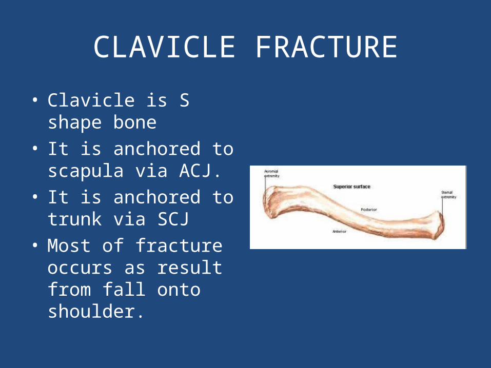
CLAVICLE FRACTURE
• Clavicle is S shape bone• It is anchored to scapula
via ACJ.• It is anchored to trunk
via SCJ• Most of fracture occurs
as result from fall onto shoulder.
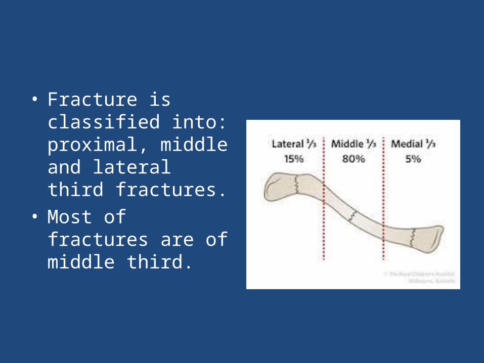
• Fracture is classified into: proximal, middle and lateral third fractures.
• Most of fractures are of middle third.
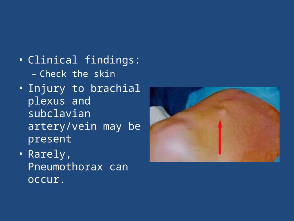
• Clinical findings:– Check the skin
• Injury to brachial plexus and subclavian artery/vein may be present
• Rarely, Pneumothorax can occur.
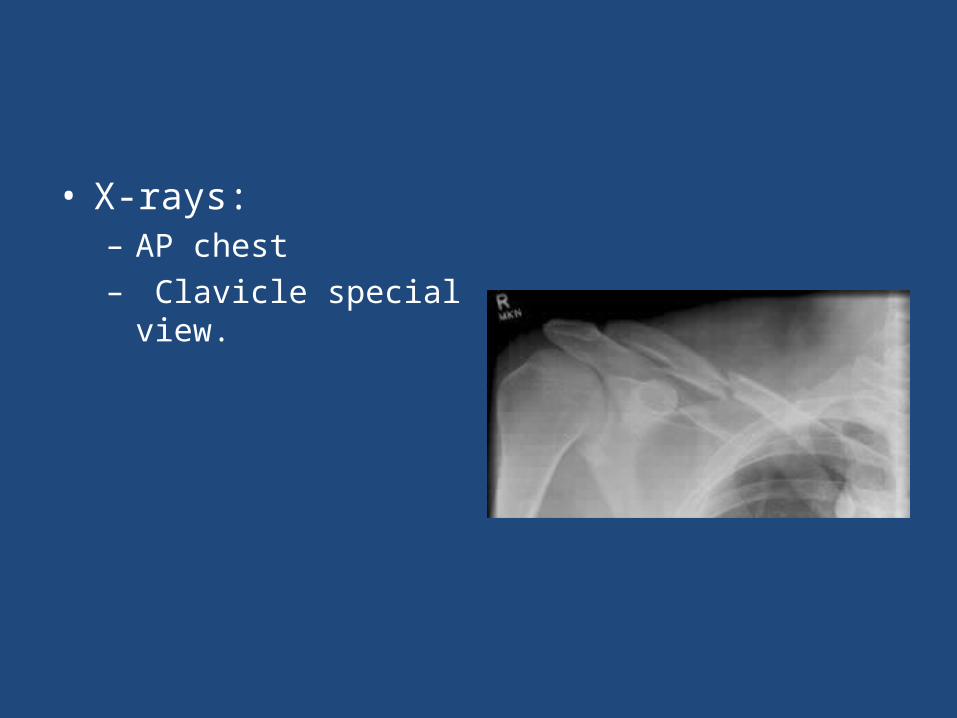
• X-rays:– AP chest– Clavicle special view.
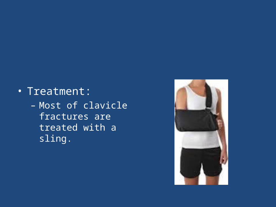
• Treatment: – Most of clavicle
fractures are treated with a sling.
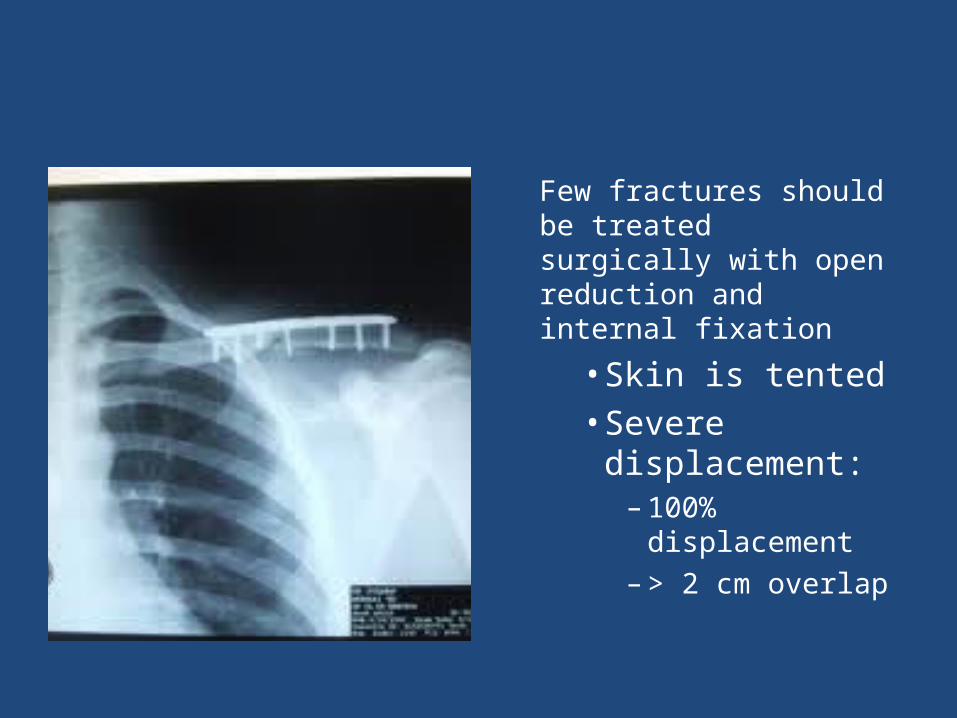
Few fractures should be treated surgically with open reduction and internal fixation
• Skin is tented • Severe
displacement:– 100%
displacement– > 2 cm overlap
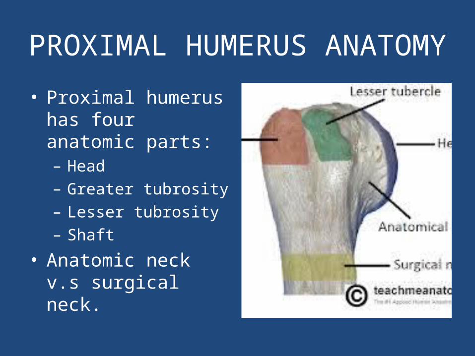
PROXIMAL HUMERUS ANATOMY
• Proximal humerus has four anatomic parts:– Head– Greater tubrosity– Lesser tubrosity – Shaft
• Anatomic neck v.s surgical neck.
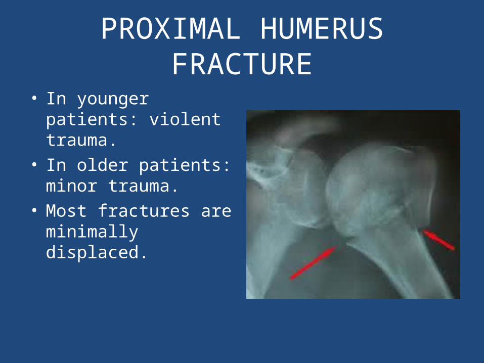
PROXIMAL HUMERUS FRACTURE
• In younger patients: violent trauma.
• In older patients: minor trauma.
• Most fractures are minimally displaced.
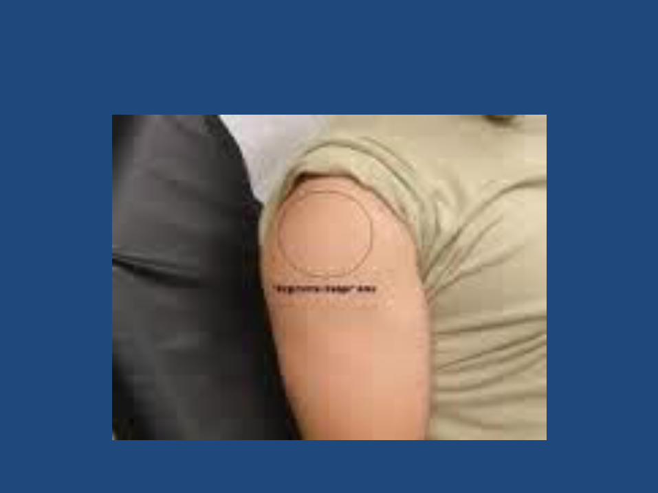
PHYSICAL EXAM
• Expose the shoulder very well.• Look for fracture signs• Check the skin.• Peripheral N/V exam.• Axillary nerve: lateral skin patch.• Examine cervical spine.
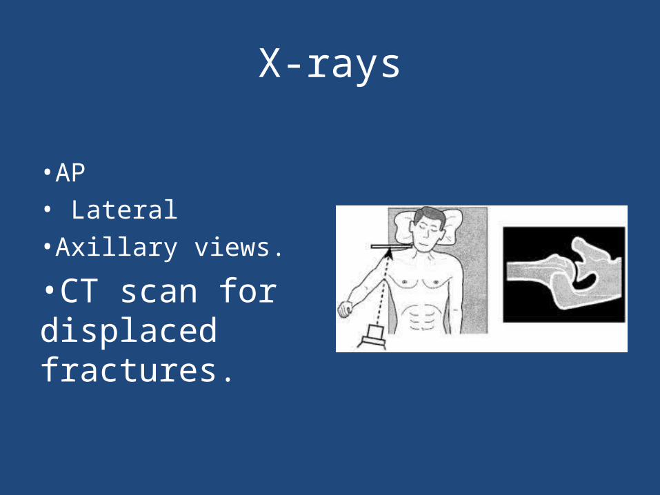
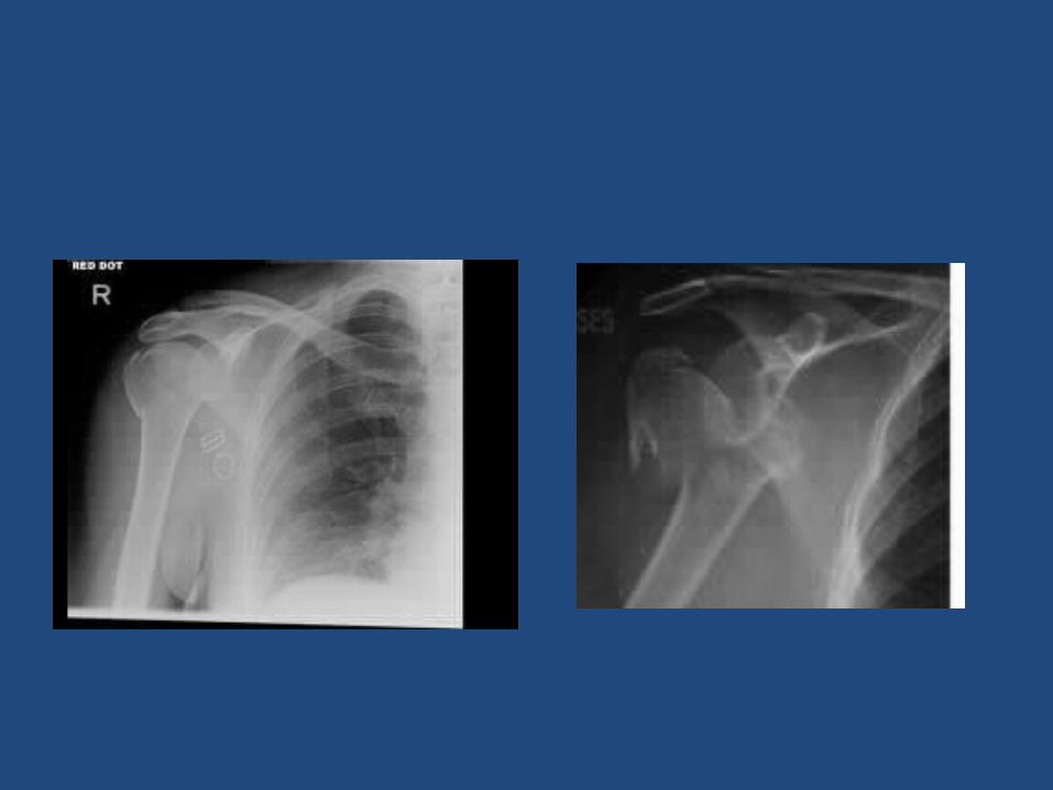
X-rays
•AP• Lateral•Axillary views.
•CT scan for displaced fractures.
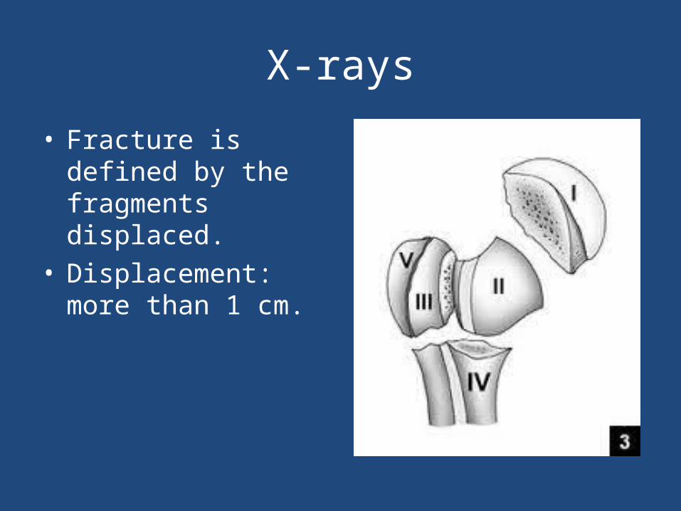
X-rays
• Fracture is defined by the fragments displaced.
• Displacement: more than 1 cm.
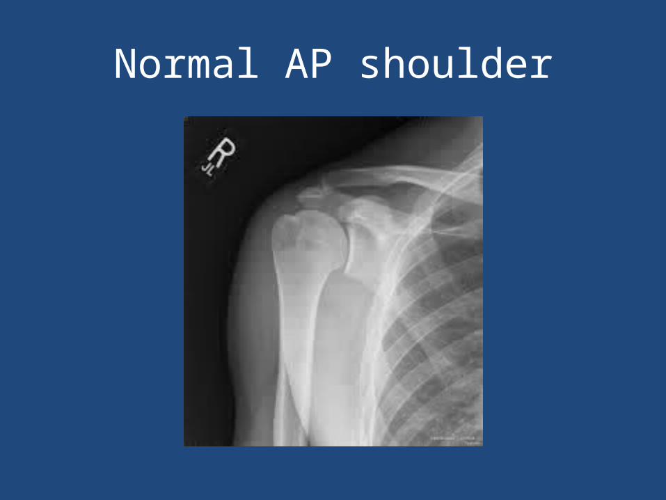
Normal AP shoulder
• If fracture is not displaced: – Treatment with sling and NWB of UE for 6-8
weeks.– Early ROM exercises after 2-4 weeks.– Normal function can be resumed after 3-4
months.
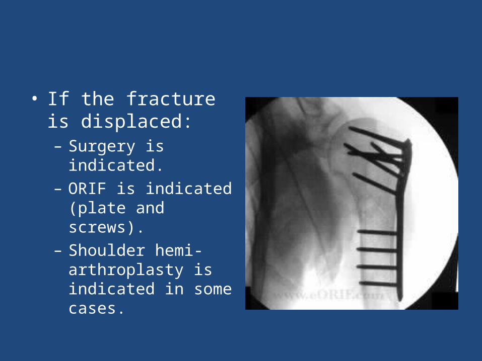
• If the fracture is displaced:– Surgery is indicated.– ORIF is indicated (plate
and screws).– Shoulder hemi-
arthroplasty is indicated in some cases.
HUMERUS SHAFT FRACTURE
• It can be classified based on location of fracture. (proximal, middle and distal)
• Fracture symptoms.• On exam:– Skin– N/V– Compartment
• Watch for radial nerve palsy.
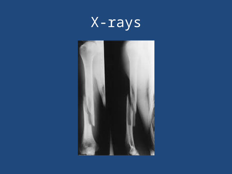
X-rays
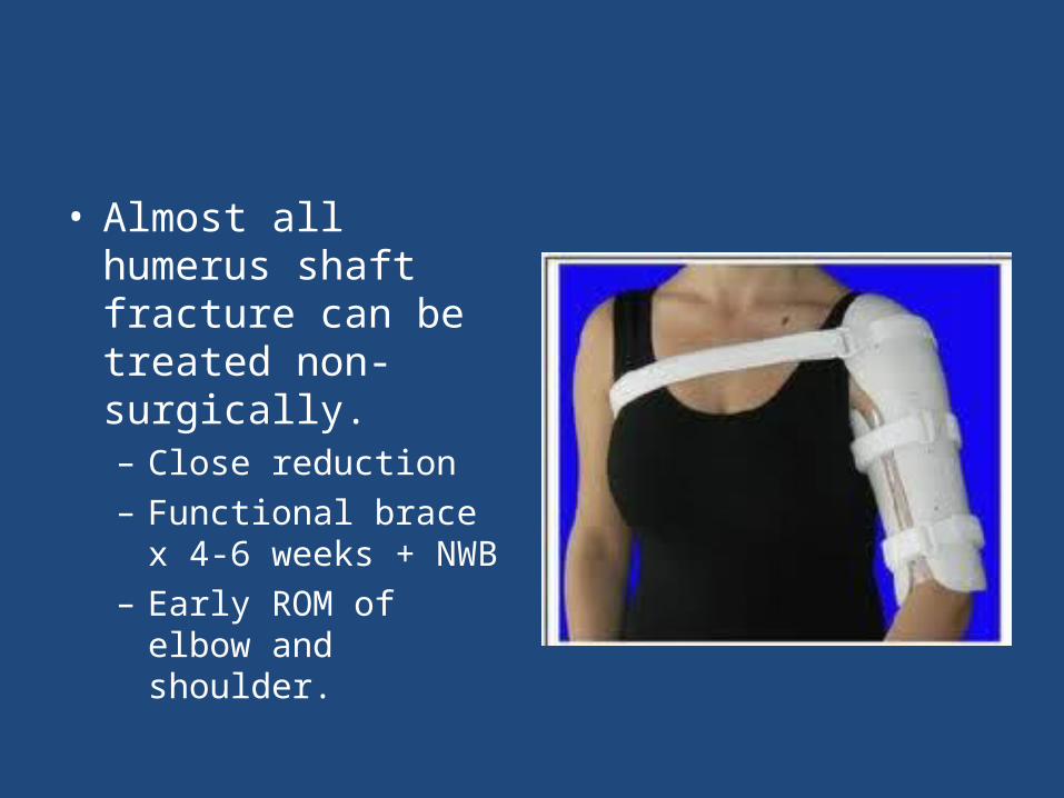
• Almost all humerus shaft fracture can be treated non-surgically.– Close reduction– Functional brace x 4-6
weeks + NWB– Early ROM of elbow and
shoulder.
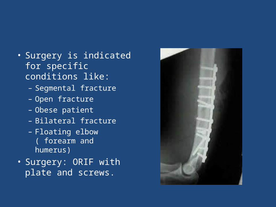
• Surgery is indicated for specific conditions like:– Segmental fracture– Open fracture– Obese patient– Bilateral fracture– Floating elbow ( forearm
and humerus)
• Surgery: ORIF with plate and screws.
BOTH BONES FOREARM FRACTURE
• Forearm is complex with two mobile parallel bones.
• Radius and ulna articulate proximally and distally.
• It very unlikely to fracture only one bone without disruption of their articulation:– Both bone fracture– Monteggia fracture – Galeazzi fracture.
• Fractures are often from fall or direct blow.• Both bones fracture:– Means radius and ulna are broken.
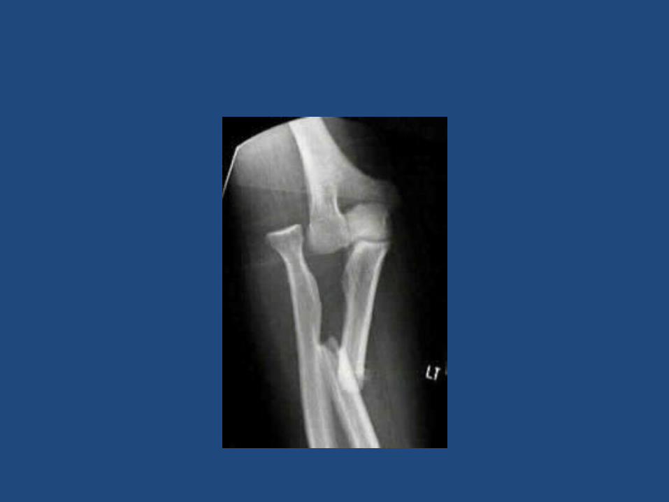
• Monteggia fracture: – Means proximal or middle third ulna shaft fracture
with dislocation of radius proximally (at elbow)
• Galeazzi fracture: – Means distal third shaft radius fracture with
disruption of DRUJ.
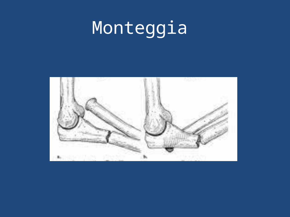
Monteggia
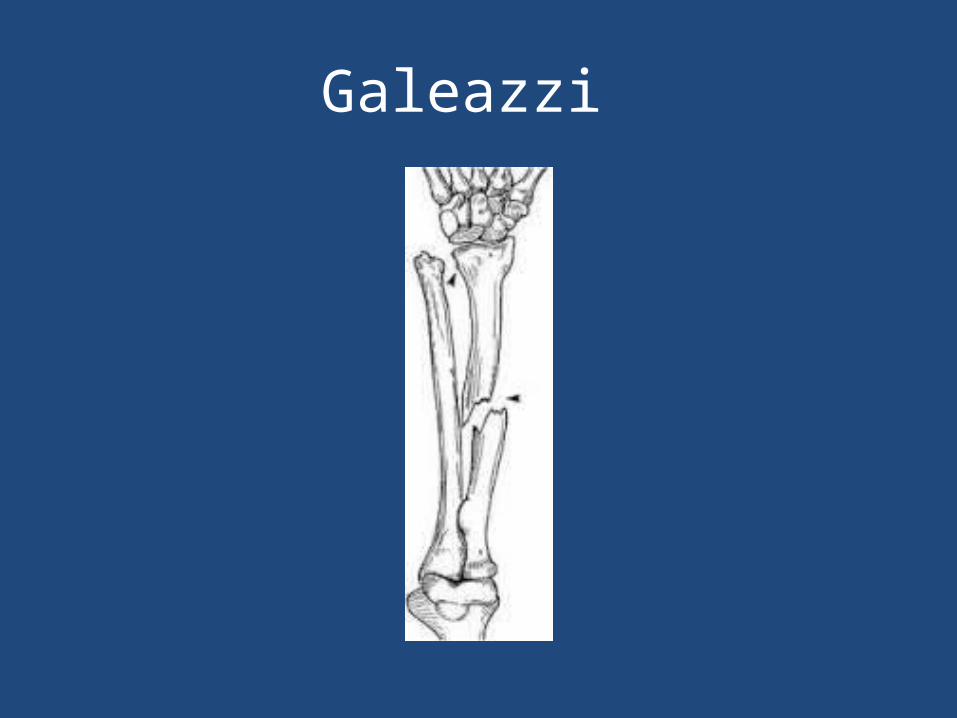
Galeazzi

Galeazzi
CLINICAL
• Symptoms and signs of fracture• Check the skin• Check the compartments of forearm• Check Ulnar, median and radial nerve
(PIN,AIN)• Check vascularity: color, temperature,
capillary refill and pulse.

Investigations
• 2 orthogonal views• CT scan if fracture
extends into joint.
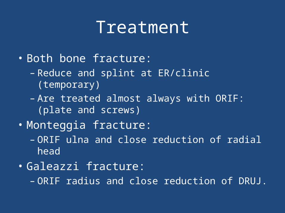
Treatment
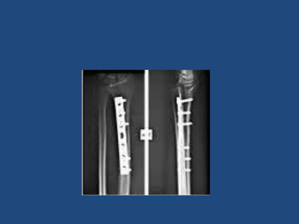
• Both bone fracture:– Reduce and splint at ER/clinic (temporary)– Are treated almost always with ORIF: (plate and
screws)
• Monteggia fracture:– ORIF ulna and close reduction of radial head
• Galeazzi fracture:– ORIF radius and close reduction of DRUJ.

DISTAL RADIUS FRACTURE
• Most common fracture of upper extremity.• Most frequently are seen in older women.• Young adults fractures are most commonly
secondary to high energy trauma.
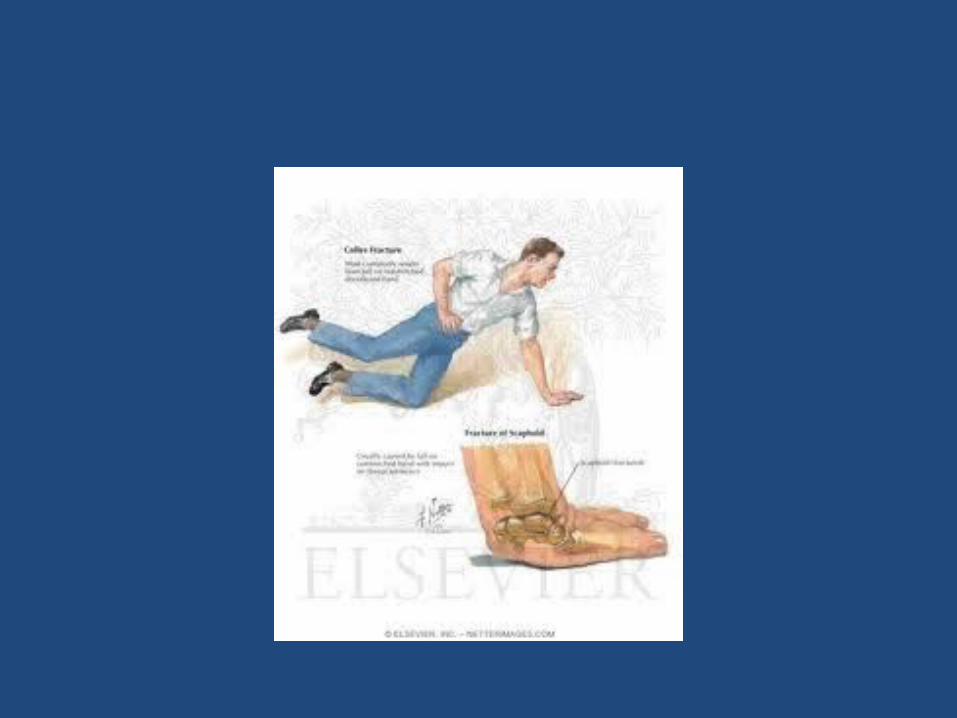
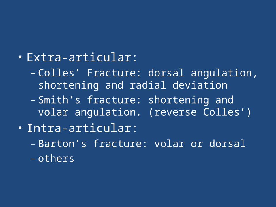
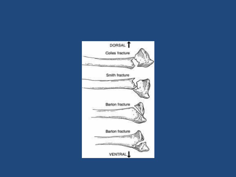
• Extra-articular:– Colles’ Fracture: dorsal angulation, shortening and
radial deviation – Smith’s fracture: shortening and volar angulation.
(reverse Colles’)
• Intra-articular:– Barton’s fracture: volar or dorsal– others
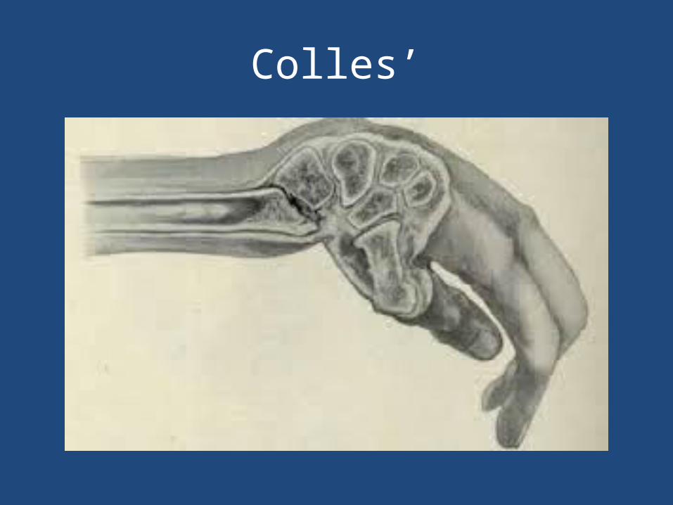
Colles’
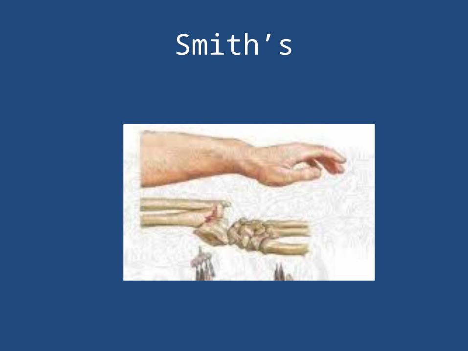
Smith’s

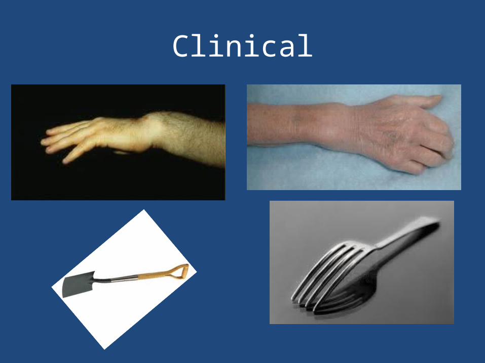
Clinical
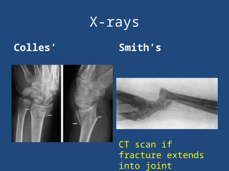
X-rays
Colles’ Smith’s
CT scan if fracture extends into joint
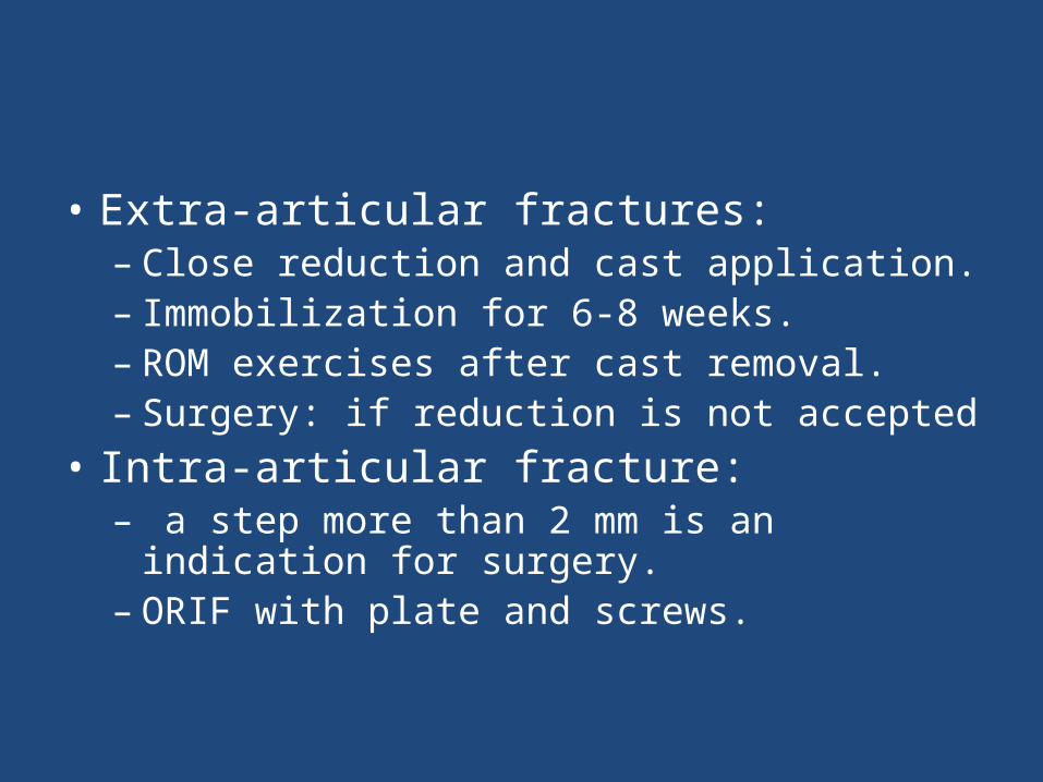
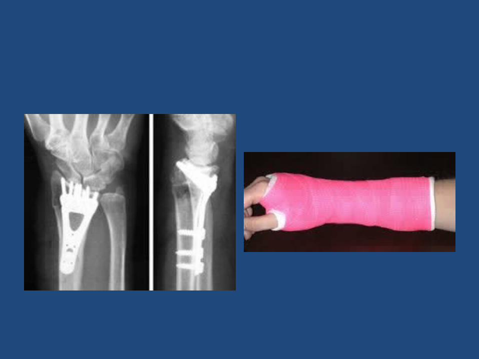
• Extra-articular fractures:– Close reduction and cast application.– Immobilization for 6-8 weeks.– ROM exercises after cast removal.– Surgery: if reduction is not accepted
• Intra-articular fracture:– a step more than 2 mm is an indication for
surgery.– ORIF with plate and screws.

Lower extremity
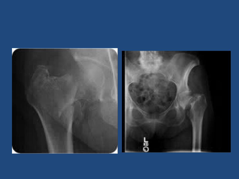
HIP FRACTURE (Old Patients: > 60 yrs)
• It is the most common fracture of LL.• It is associated with osteoporosis. • Most common mechanism is a fall from
standing height.• Other causes of fall (stroke, MI) should be
rolled out during clinical evaluation.• It is a life changing event.
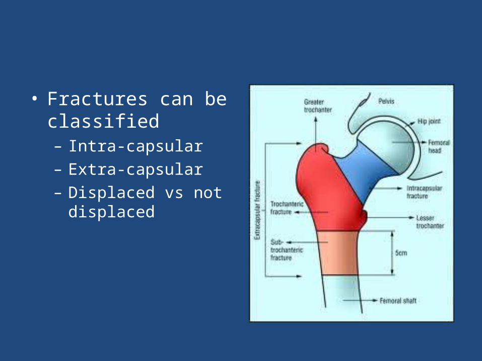
• Fractures can be classified– Intra-capsular– Extra-capsular – Displaced vs not
displaced
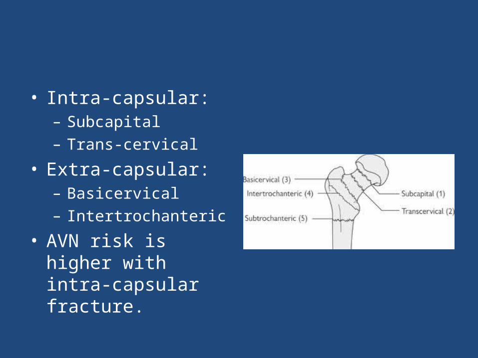
• Intra-capsular:– Subcapital – Trans-cervical
• Extra-capsular:– Basicervical– Intertrochanteric
• AVN risk is higher with intra-capsular fracture.
Clinical
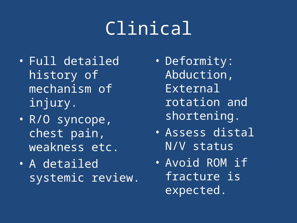
• Full detailed history of mechanism of injury.
• R/O syncope, chest pain, weakness etc.
• A detailed systemic review.
• Deformity: Abduction, External rotation and shortening.
• Assess distal N/V status• Avoid ROM if fracture is
expected.

• Common associated injuries:1. Distal radius fracture 2. Proximal humerus
fracture3. Subdural hematoma
• R/O:– ACS– Stroke

• 3 views are needed: – AP pelvis– AP hip– Lateral hip
• MRI is sensitive for occult fracture.

Treatment
• No close reduction is needed.• No traction is needed.• Patient needs surgery ideally within 48 hrs.• The goal is to ambulate patient as soon as
possible.• Be sure that DVT prophylaxis is started.• Be sure that patient will be evaluated for
osteoporosis after discharge.
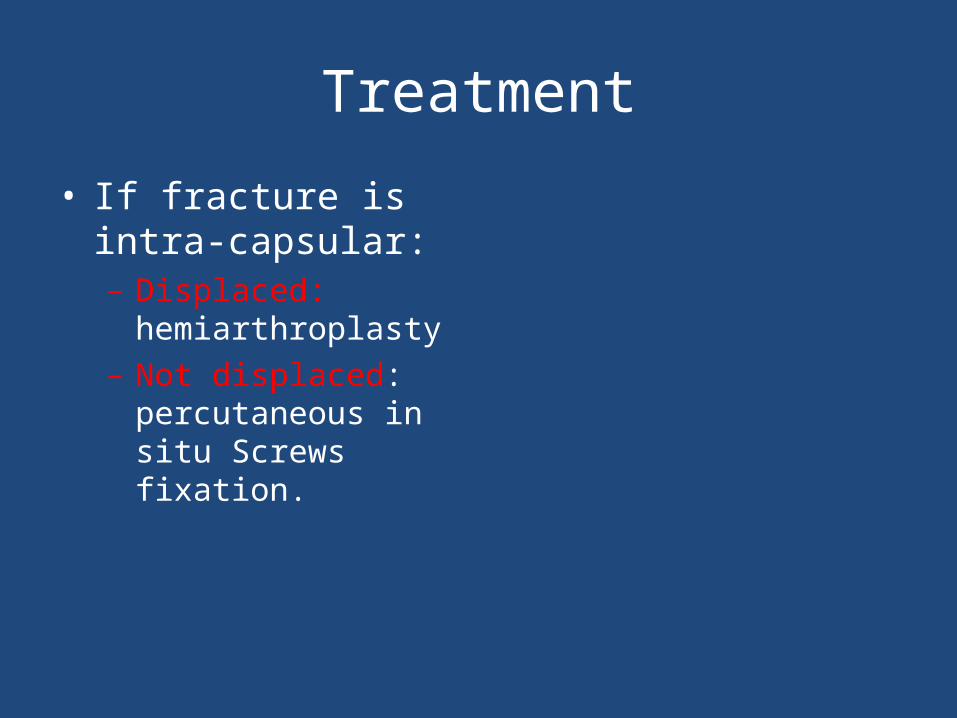
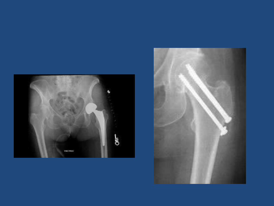
Treatment
• If fracture is intra-capsular:– Displaced:
hemiarthroplasty– Not displaced:
percutaneous in situ Screws fixation.
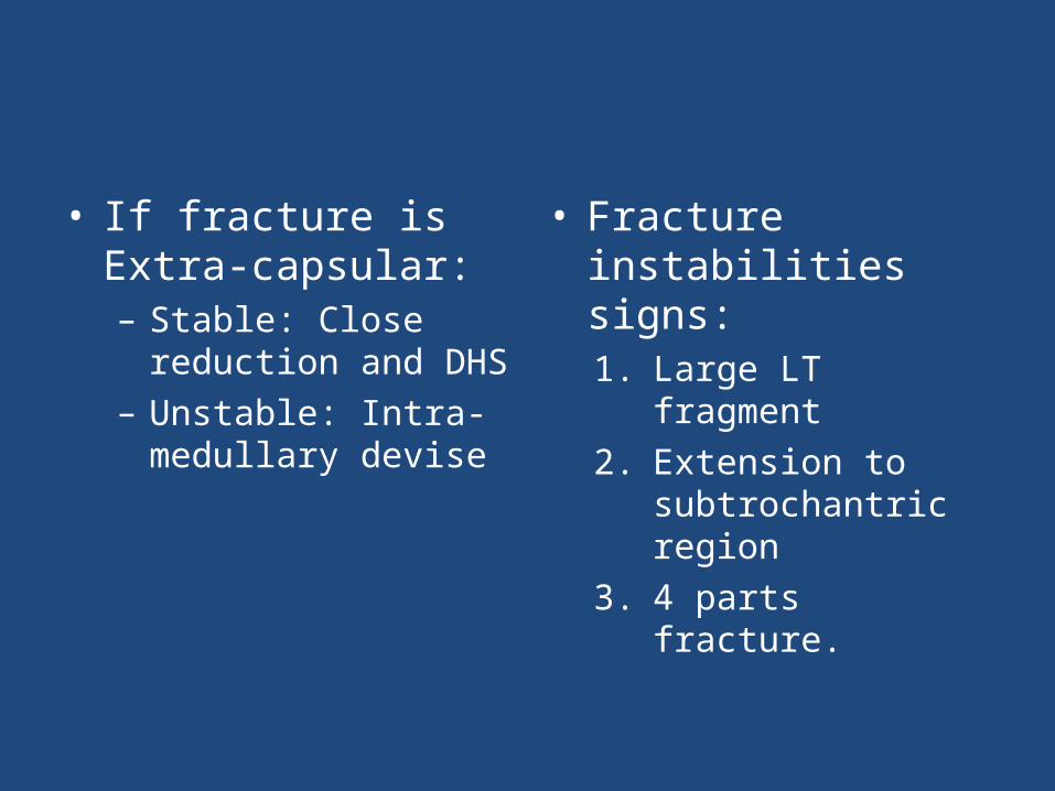
• If fracture is Extra-capsular: – Stable: Close reduction
and DHS– Unstable: Intra-
medullary devise
• Fracture instabilities signs:1. Large LT fragment2. Extension to
subtrochantric region 3. 4 parts fracture.
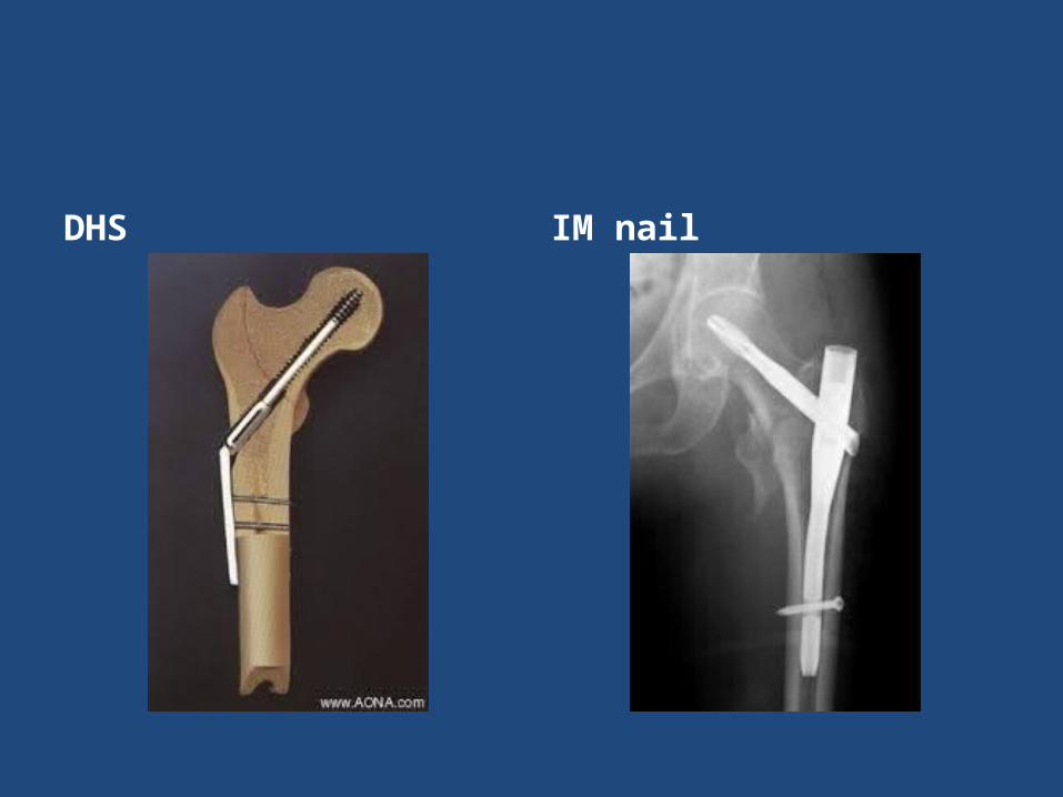
DHS IM nail
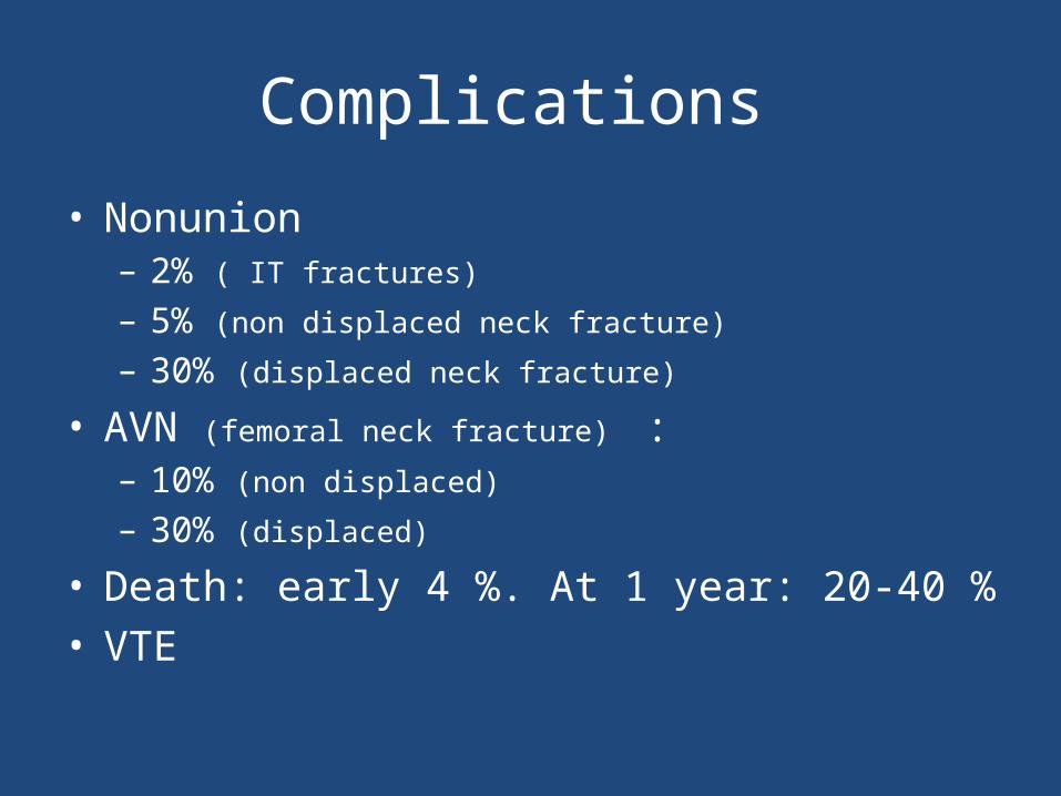
Complications
• Nonunion – 2% ( IT fractures)
– 5% (non displaced neck fracture)
– 30% (displaced neck fracture)
• AVN (femoral neck fracture) : – 10% (non displaced)
– 30% (displaced)
• Death: early 4 %. At 1 year: 20-40 %• VTE
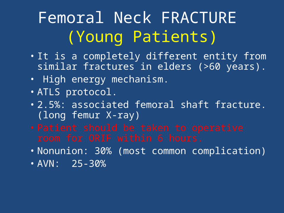
Femoral Neck FRACTURE (Young Patients)
• It is a completely different entity from similar fractures in elders (>60 years).
• High energy mechanism.• ATLS protocol.• 2.5%: associated femoral shaft fracture. (long
femur X-ray) • Patient should be taken to operative room for
ORIF within 6 hours.• Nonunion: 30% (most common complication)• AVN: 25-30%
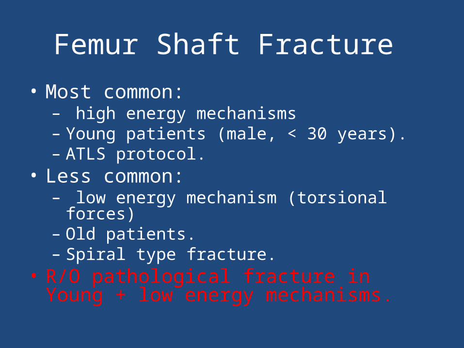
Femur Shaft Fracture • Most common:– high energy mechanisms– Young patients (male, < 30 years).– ATLS protocol.
• Less common:– low energy mechanism (torsional forces)– Old patients.– Spiral type fracture.
• R/O pathological fracture in Young + low energy mechanisms.
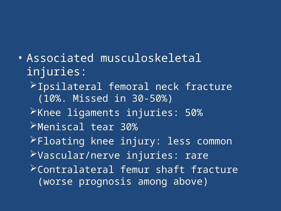
• Associated musculoskeletal injuries: Ipsilateral femoral neck fracture (10%. Missed in
30-50%)Knee ligaments injuries: 50%Meniscal tear 30%Floating knee injury: less commonVascular/nerve injuries: rare Contralateral femur shaft fracture (worse
prognosis among above)

• Associated non-MS injuries:Fat embolism ARDSHead injuries. Abdominal injuries
Clinical
• ATLS• Fracture symptoms and signs• Skin integrity • N/V exam.• Compartment assessment• Knee swelling or ecchymosis.
Investigations
• AP and lateral views femur• 15° Internal rotation AP view ipsilateral hip.• Lateral view ipsilateral view • If femoral neck fracture is suspected: CT scan
hip. • Knee AP and lateral views
Management
• ATLS: ABC resuscitation.• Skeletal traction (proximal tibial pin)• Early surgical fixation:
Proven to reduce Pulmonary complications.Must be within 24 hrs (ideally < 6 hrs)If patient is unstable: External fixation.If Patient is stable IM nailing

FEMUR SHAFT FRACTURE

Complications
• Malunion: most common.More common with proximal fracture
(subtrochantric fracture)Rotational, angulation and shortening
• Nonunion: rare • Infection. • VTE.

TIBIA SHAFT FRACTURE
• It is a subcutaneous bone ( high suspicion for skin injury.
• Most common large long bone fracture.• It can be secondary to low or high energy
mechanism.• It carries the highest risk of compartment
syndrome.• 20 % of tibial fracture can be associated with
ankle intra-articular fracture.
• It can be classified based on location and morphology:– Proximal third– Middle third– Distal third
• Displaced vs. Non-displaced:
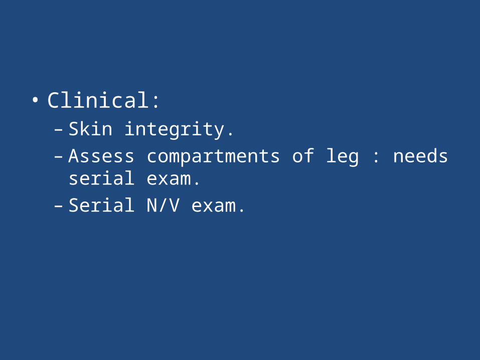
• Clinical:– Skin integrity. – Assess compartments of leg : needs serial exam.– Serial N/V exam.
INVESTIGATIONS
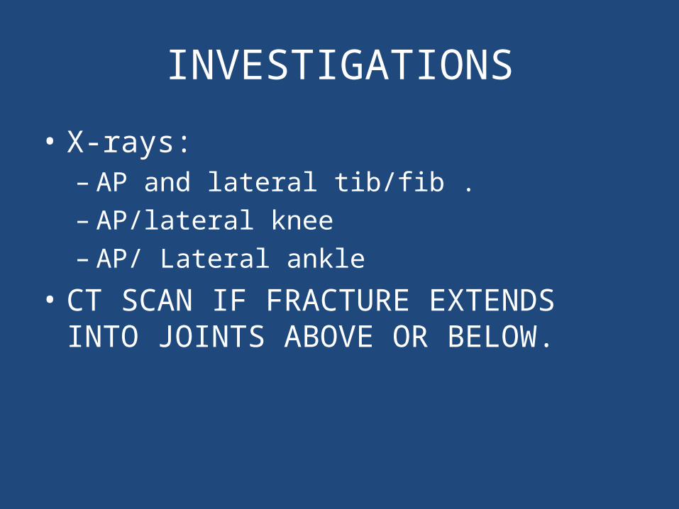
• X-rays:– AP and lateral tib/fib .– AP/lateral knee – AP/ Lateral ankle
• CT SCAN IF FRACTURE EXTENDS INTO JOINTS ABOVE OR BELOW.
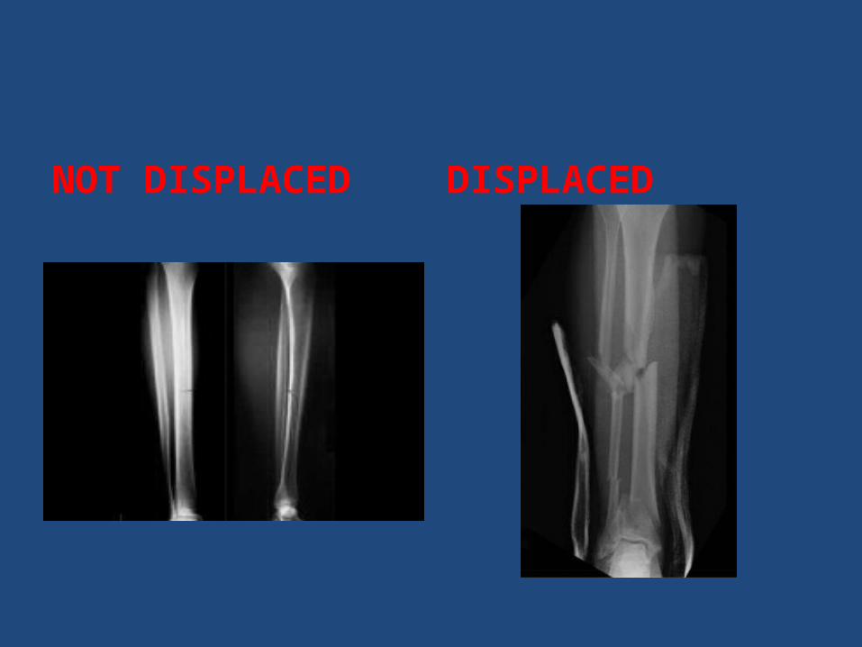
NOT DISPLACED DISPLACED
MANAGEMENT
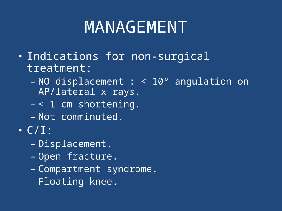
• Indications for non-surgical treatment:– NO displacement : < 10° angulation on AP/lateral x
rays.– < 1 cm shortening.– Not comminuted.
• C/I:– Displacement.– Open fracture.– Compartment syndrome.– Floating knee.
MANAGEMENT
• Close reduction and cast immobilization:– Above knee back slab and U slab if surgical
treatment is chosen. – Above knee full cast if non-surgical treatment is
chosen: it must be bivalved to minimized compartment syndrome.
– Always provide patient with Compartment Syndrome checklist if patient is discharged home with cast.
– NWB for 8 weeks with cast immobilization.
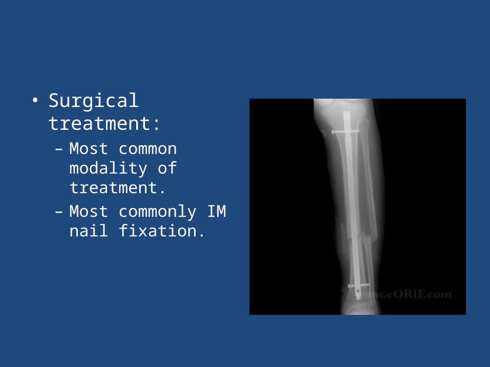
• Surgical treatment:– Most common modality
of treatment.– Most commonly IM nail
fixation.
COMPLICATIONS
• Non-union: most common complication.
• Delayed union • Infection: open fracture• DVT/PE
ANKLE FRACTURE
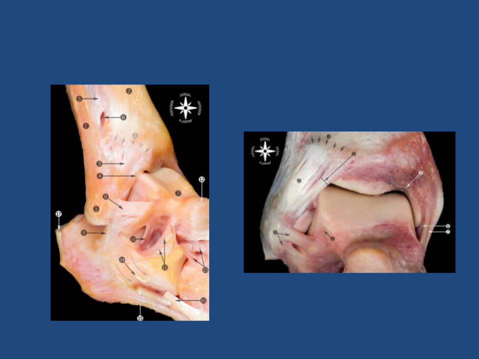
• Ankle anatomy:– Medial and lateral malleoli, distal tibia and talus.– Highly congruent joint– Fibula is held to distal tibia by syndosmotic
ligament.– Medial malleolus is held to talus by deltoid
ligament. – Lateral malleolus is held to talus by LCL.

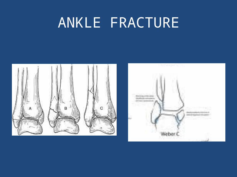
• Low energy (torsional): malleoli fracture.• Classification:
Stable v.s Unstable fracture: lateral displacement of talus
Medial, lateral or bimalleolar fractureLateral malleolus: Weber A, B, C
ANKLE FRACTURE
CLINICAL
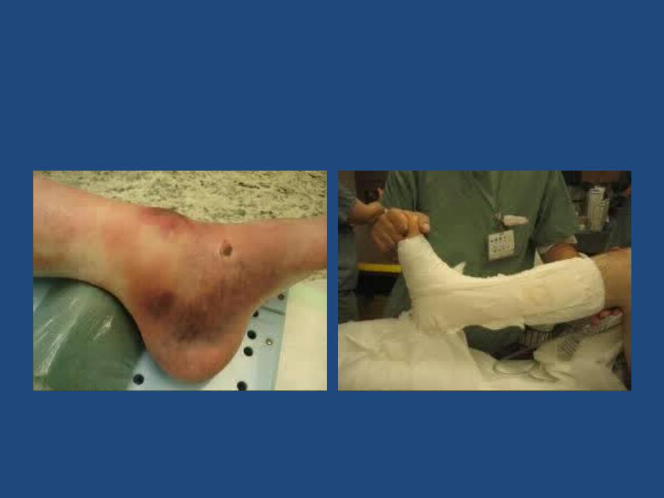
• Look for Fracture symptoms and signs.• Assess medial joint ecchymosis or tenderness
to assess medial malleolus and deltoid ligament integrity.
• Assess N/V status (before and after reduction).

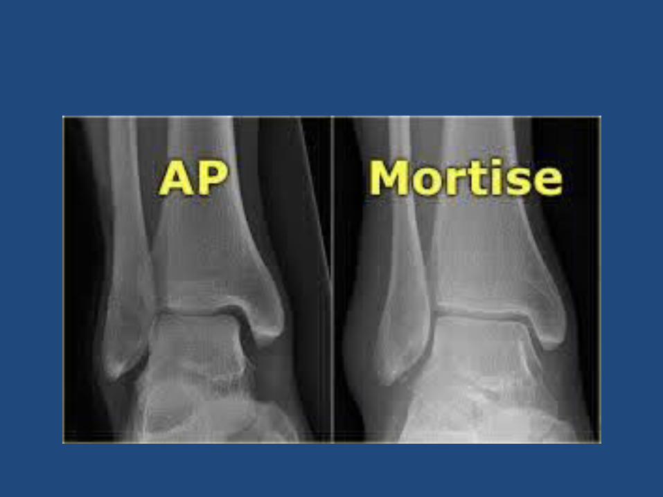
• X-rays:– AP– Lateral – Mortise view– Long leg x-rays: if only
medial malleolus is broken.
• CT SCAN IF FRACTURE EXTENDS TO ARTICULAR DISTAL TIBIA SURFACE.
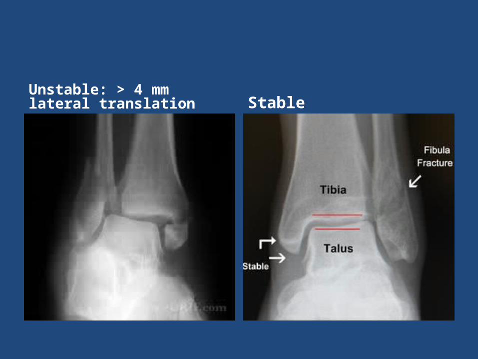
Unstable: > 4 mm lateral translation Stable
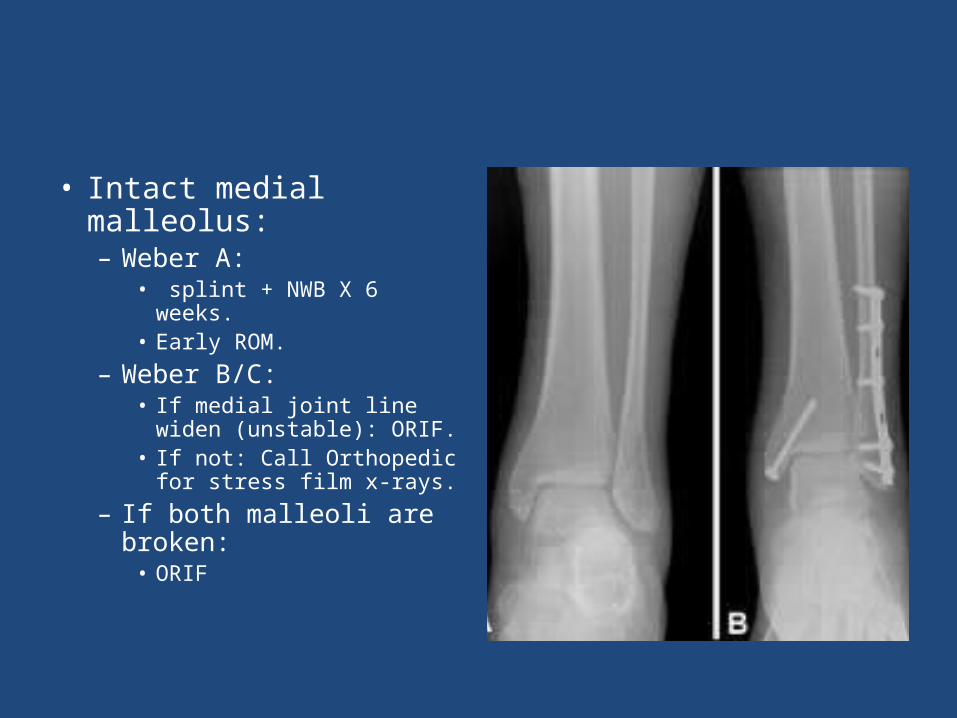
• Intact medial malleolus:– Weber A:
• splint + NWB X 6 weeks. • Early ROM.
– Weber B/C: • If medial joint line widen
(unstable): ORIF.• If not: Call Orthopedic for
stress film x-rays.– If both malleoli are
broken:• ORIF

Thanks


![SURGICAL MANAGEMENT FRACTURE … · surgical management fracture shaft tibia by intramedullary interlocking nail a study done at bhanu orthopaedic home karimnagar [a.p.] 505001 india](https://img.dokumen.tips/doc/110x75/5af9b85e7f8b9a5f588e6f99/surgical-management-fracture-management-fracture-shaft-tibia-by-intramedullary.jpg)
![WELCOME [] · •ICD-9-CM 821.01 Closed fracture of shaft of femur • ICD-10-CM S72.344 Nondisplaced spiral fracture of shaft of right femur ICD-10-PCS (Inpatient procedure)](https://img.dokumen.tips/doc/110x75/5ecd8d93ff7ebd45234ce855/welcome-aicd-9-cm-82101-closed-fracture-of-shaft-of-femur-a-icd-10-cm-s72344.jpg)