Embed Size (px)
Citation preview
Objective 10
• TSWBAT identify the structure, composition
and function of cell organelles.

• The nucleus contains most of the genes in a eukaryotic cell.• Some genes are located in mitochondria and chloroplasts.
• The nucleus averages about 5 microns in diameter.
• The nucleus is separated from the cytoplasm by a double membrane.• These are separated by 20-40 nm.
• Where the double membranes are fused, a pore allows large macromolecules and particles to pass through.
The nucleus contains a eukaryotic cell’s genetic library
Copyright © 2002 Pearson Education, Inc., publishing as Benjamin Cummings
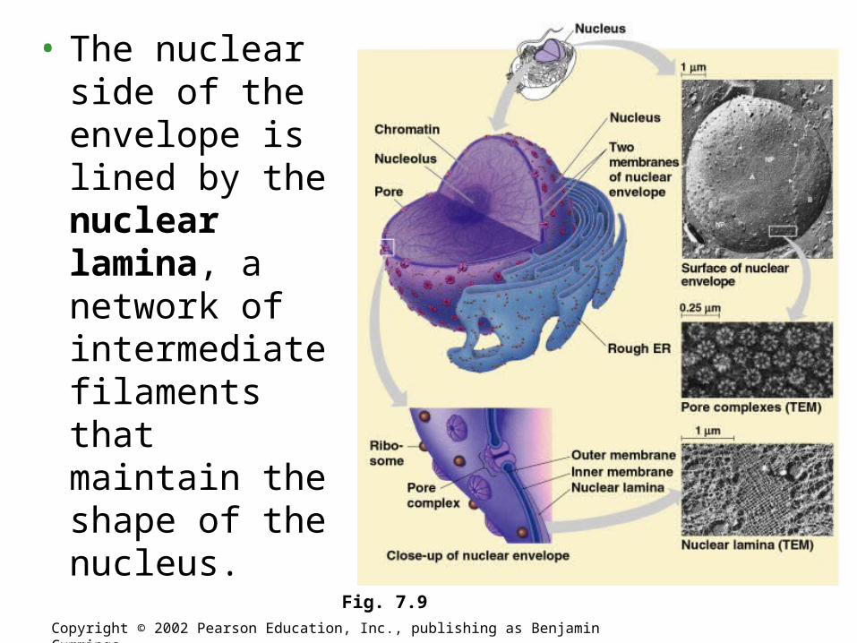
• The nuclear side of the envelope is lined by the nuclear lamina, a network of intermediate filaments that maintain the shape of the nucleus.
Copyright © 2002 Pearson Education, Inc., publishing as Benjamin Cummings
Fig. 7.9
• Within the nucleus, the DNA and associated proteins are organized into fibrous material, chromatin.
• In a normal cell they appear as diffuse mass.
• However when the cell prepares to divide, the chromatin fibers coil up to be seen as separate structures, chromosomes.
• Each eukaryotic species has a characteristic number of chromosomes.
• A typical human cell has 46 chromosomes, but sex cells (eggs and sperm) have only 23 chromosomes.
Copyright © 2002 Pearson Education, Inc., publishing as Benjamin Cummings
• In the nucleus is a region of densely stained fibers and granules adjoining chromatin, the nucleolus.
• In the nucleolus, ribosomal RNA (rRNA) is synthesized and assembled with proteins from the cytoplasm to form ribosomal subunits.
• The subunits pass from the nuclear pores to the cytoplasm where they combine to form ribosomes.
• The nucleus directs protein synthesis by synthesizing messenger RNA (mRNA).
• The mRNA travels to the cytoplasm and combines with ribosomes to translate its genetic message into the primary structure of a specific polypeptide.
Copyright © 2002 Pearson Education, Inc., publishing as Benjamin Cummings
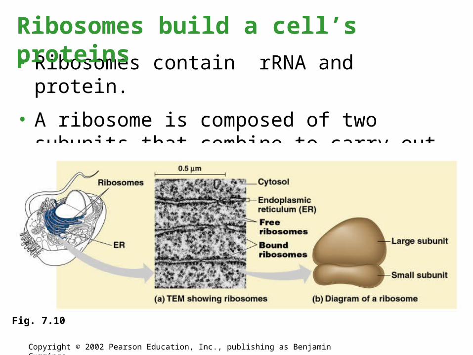
• Ribosomes contain rRNA and protein.
• A ribosome is composed of two subunits that combine to carry out protein synthesis.
Ribosomes build a cell’s proteins
Copyright © 2002 Pearson Education, Inc., publishing as Benjamin Cummings
Fig. 7.10
• Cell types that synthesize large quantities of proteins (e.g., pancreas) have large numbers of ribosomes and prominent nuclei.
• Some ribosomes, free ribosomes, are suspended in the cytosol and synthesize proteins that function within the cytosol.
• Other ribosomes, bound ribosomes, are attached to the outside of the endoplasmic reticulum.
• These synthesize proteins that are either included into membranes or for export from the cell.
• Ribosomes can shift between roles depending on the polypeptides they are synthesizing.
Copyright © 2002 Pearson Education, Inc., publishing as Benjamin Cummings
• Many of the internal membranes in a eukaryotic cell are part of the endomembrane system.
• These membranes are either in direct contact or connected via transfer of vesicles, sacs of membrane.
• In spite of these links, these membranes have diverse functions and structures.
• In fact, the membranes are even modified during life.
• The endomembrane system includes the nuclear envelope, endoplasmic reticulum, Golgi apparatus, lysosomes, vacuoles, and the plasma membrane.
Copyright © 2002 Pearson Education, Inc., publishing as Benjamin Cummings
Endomembrane System
• The endoplasmic reticulum (ER) accounts for half the membranes in a eukaryotic cell.
• The ER includes membranous tubules and internal, fluid-filled spaces, the cisternae.
• The ER membrane is continuous with the nuclear envelope and the cisternal space of the ER is continuous with the space between the two membranes of the nuclear envelope.
The endoplasmic reticulum manufacturers membranes and performs many other biosynthetic functions
Copyright © 2002 Pearson Education, Inc., publishing as Benjamin Cummings
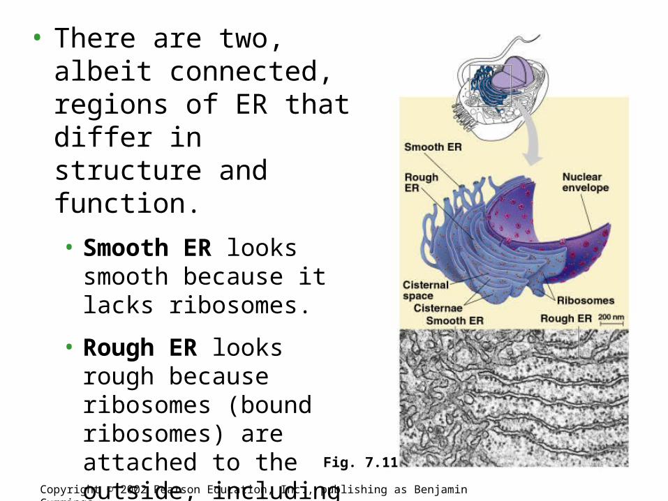
• There are two, albeit connected, regions of ER that differ in structure and function.
• Smooth ER looks smooth because it lacks ribosomes.
• Rough ER looks rough because ribosomes (bound ribosomes) are attached to the outside, including the outside of the nuclear envelope.
Copyright © 2002 Pearson Education, Inc., publishing as Benjamin Cummings
Fig. 7.11
• The smooth ER is rich in enzymes and plays a role in a variety of metabolic processes.
• Enzymes of smooth ER synthesize lipids, including oils, phospholipids, and steroids.
• These includes the sex hormones of vertebrates and adrenal steroids.
• The smooth ER also catalyzes a key step in the mobilization of glucose from stored glycogen in the liver.
• An enzyme removes the phosphate group from glucose phosphate, a product of glycogen hydrolysis, permitting glucose to exit the cell.
Copyright © 2002 Pearson Education, Inc., publishing as Benjamin Cummings
• Other enzymes in the smooth ER of the liver help detoxify drugs and poisons.
• These include alcohol and barbiturates.
• Frequent exposure leads to proliferation of smooth ER, increasing tolerance to the target and other drugs.
• Muscle cells are rich in enzymes that pump calcium ions from the cytosol to the cisternae.
• When nerve impulse stimulates a muscle cell, calcium rushes from the ER into the cytosol, triggering contraction.
• These enzymes then pump the calcium back, readying the cell for the next stimulation.
Copyright © 2002 Pearson Education, Inc., publishing as Benjamin Cummings
• Rough ER is especially abundant in those cells that secrete proteins.
• As a polypeptide is synthesized by the ribosome, it is threaded into the cisternal space through a pore formed by a protein in the ER membrane.
• Many of these polypeptides are glycoproteins, a polypeptide to which an oligosaccharide is attached.
• These secretory proteins are packaged in transport vesicles that carry them to their next stage.
Copyright © 2002 Pearson Education, Inc., publishing as Benjamin Cummings
• Rough ER is also a membrane factory.
• Membrane bound proteins are synthesized directly into the membrane.
• Enzymes in the rough ER also synthesize phospholipids from precursors in the cytosol.
• As the ER membrane expands, parts can be transferred as transport vesicles to other components of the endomembrane system.
Copyright © 2002 Pearson Education, Inc., publishing as Benjamin Cummings
• Many transport vesicles from the ER travel to the Golgi apparatus for modification of their contents.
• The Golgi is a center of manufacturing, warehousing, sorting, and shipping.
• The Golgi apparatus is especially extensive in cells specialized for secretion.
The Golgi apparatus finishes, sorts, and ships cell products
Copyright © 2002 Pearson Education, Inc., publishing as Benjamin Cummings
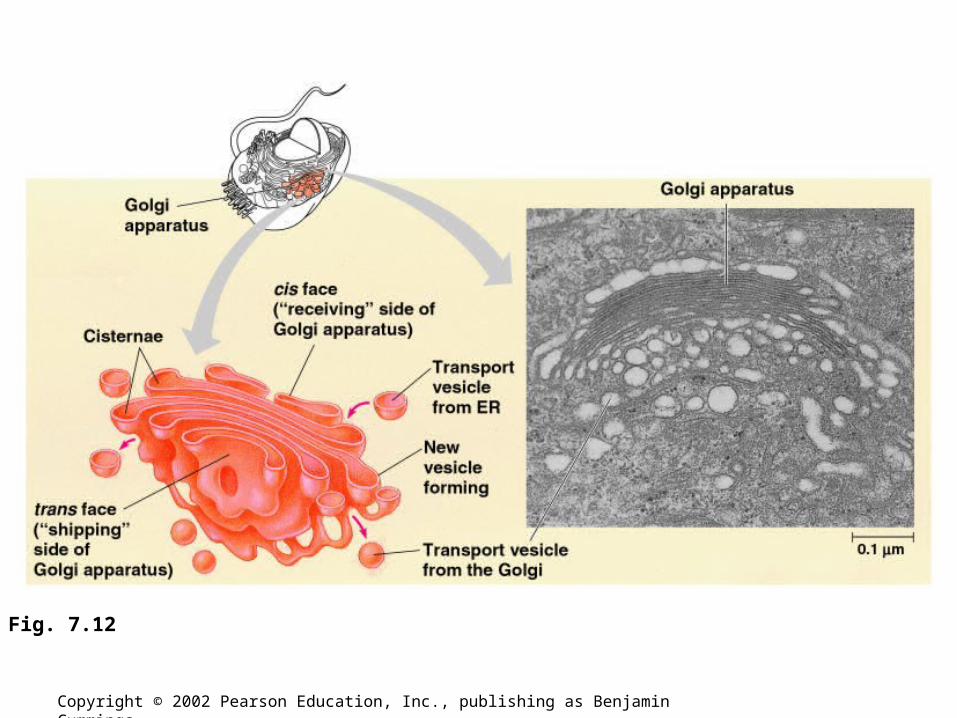
• The Golgi apparatus consists of flattened membranous sacs - cisternae - looking like a sac of pita bread.
• The membrane of each cisterna separates its internal space from the cytosol
• One side of the Golgi, the cis side, receives material by fusing with vesicles, while the other side, the trans side, buds off vesicles that travel to other sites.
Copyright © 2002 Pearson Education, Inc., publishing as Benjamin Cummings
Copyright © 2002 Pearson Education, Inc., publishing as Benjamin Cummings
Fig. 7.12
• During their transit from the cis to trans pole, products from the ER are modified to reach their final state.
• This includes modifications of the oligosaccharide portion of glycoproteins.
• The Golgi can also manufacture its own macromolecules, including pectin and other noncellulose polysaccharides.
• During processing material is moved from cisterna to cisterna, each with its own set of enzymes.
• Finally, the Golgi tags, sorts, and packages materials into transport vesicles.
Copyright © 2002 Pearson Education, Inc., publishing as Benjamin Cummings
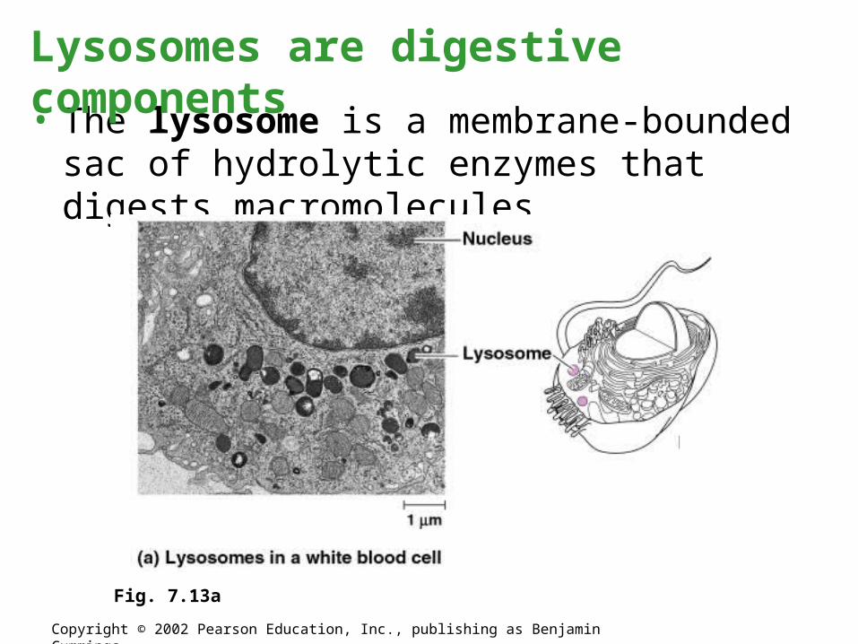
• The lysosome is a membrane-bounded sac of hydrolytic enzymes that digests macromolecules.
Lysosomes are digestive components
Copyright © 2002 Pearson Education, Inc., publishing as Benjamin Cummings
Fig. 7.13a
• Lysosomal enzymes can hydrolyze proteins, fats, polysaccharides, and nucleic acids.
• These enzymes work best at pH 5.
• Proteins in the lysosomal membrane pump hydrogen ions from the cytosol to the lumen of the lysosomes.
• While rupturing one or a few lysosomes has little impact on a cell, but massive leakage from lysosomes can destroy an cell by autodigestion.
• The lysosomes creates a space where the cell can digest macromolecules safely.
Copyright © 2002 Pearson Education, Inc., publishing as Benjamin Cummings
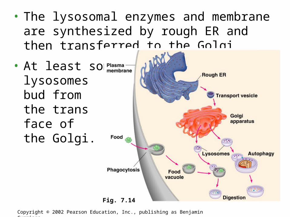
• The lysosomal enzymes and membrane are synthesized by rough ER and then transferred to the Golgi.
• At least some lysosomes bud from the trans face of the Golgi.
Copyright © 2002 Pearson Education, Inc., publishing as Benjamin Cummings
Fig. 7.14
Copyright © 2002 Pearson Education, Inc., publishing as Benjamin Cummings
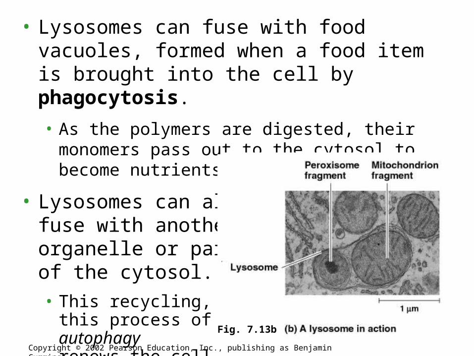
• Lysosomes can fuse with food vacuoles, formed when a food item is brought into the cell by phagocytosis.
• As the polymers are digested, their monomers pass out to the cytosol to become nutrients of the cell.
• Lysosomes can also fuse with another organelle or part of the cytosol.
• This recycling,this process of autophagyrenews the cell.
Fig. 7.13b
• The lysosomes play a critical role in the programmed destruction of cells in multicellular organisms.
• This process allows reconstruction during the developmental process.
• Several inherited diseases affect lysosomal metabolism.
• These individuals lack a functioning version of a normal hydrolytic enzyme.
• Lysosomes are engorged with indigestable substrates.
• These diseases include Pompe’s disease in the liver and Tay-Sachs disease in the brain.
Copyright © 2002 Pearson Education, Inc., publishing as Benjamin Cummings
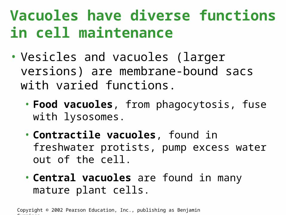
• Vesicles and vacuoles (larger versions) are membrane-bound sacs with varied functions.
• Food vacuoles, from phagocytosis, fuse with lysosomes.
• Contractile vacuoles, found in freshwater protists, pump excess water out of the cell.
• Central vacuoles are found in many mature plant cells.
Vacuoles have diverse functions in cell maintenance
Copyright © 2002 Pearson Education, Inc., publishing as Benjamin Cummings
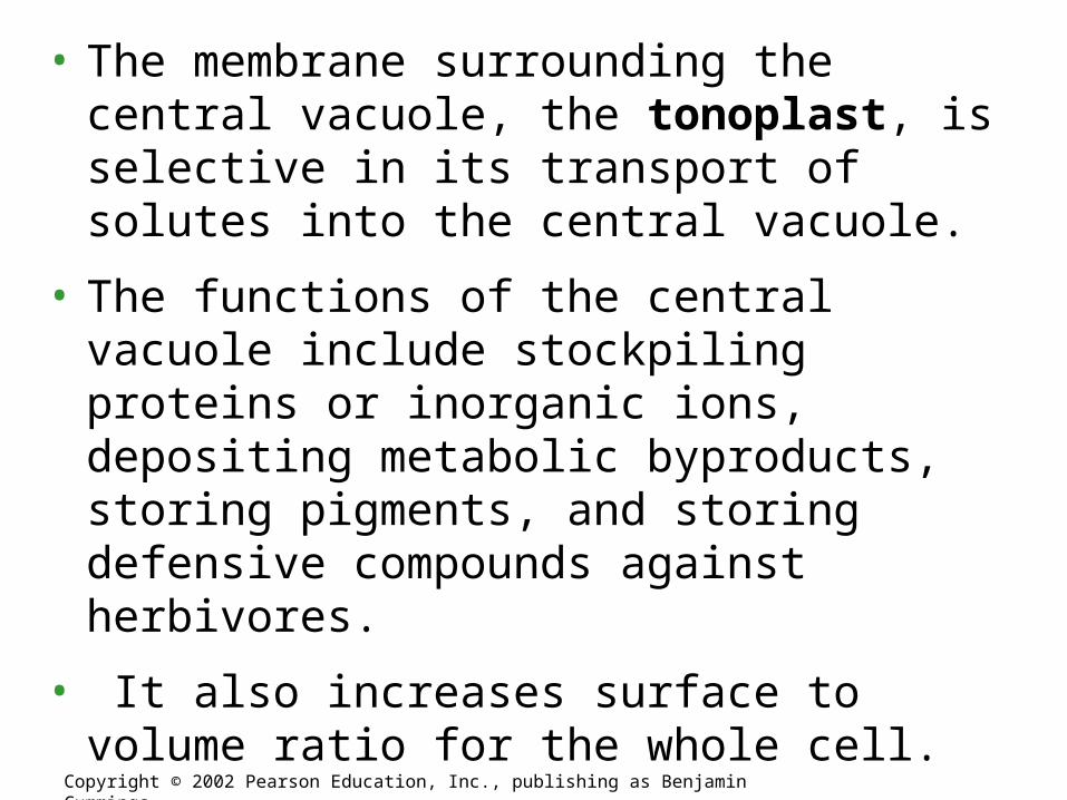
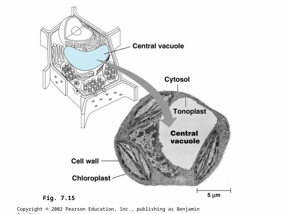
• The membrane surrounding the central vacuole, the tonoplast, is selective in its transport of solutes into the central vacuole.
• The functions of the central vacuole include stockpiling proteins or inorganic ions, depositing metabolic byproducts, storing pigments, and storing defensive compounds against herbivores.
• It also increases surface to volume ratio for the whole cell.
Copyright © 2002 Pearson Education, Inc., publishing as Benjamin Cummings
Copyright © 2002 Pearson Education, Inc., publishing as Benjamin Cummings
Fig. 7.15
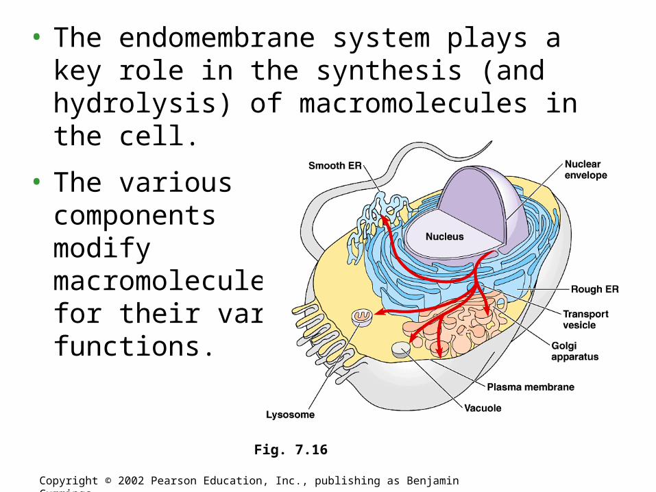
• The endomembrane system plays a key role in the synthesis (and hydrolysis) of macromolecules in the cell.
• The various components modify macromolecules for their various functions.
Copyright © 2002 Pearson Education, Inc., publishing as Benjamin Cummings
Fig. 7.16
• Mitochondria and chloroplasts are the organelles that convert energy to forms that cells can use for work.
• Mitochondria are the sites of cellular respiration, generating ATP from the catabolism of sugars, fats, and other fuels in the presence of oxygen.
• Chloroplasts, found in plants and eukaryotic algae, are the site of photosynthesis.
• They convert solar energy to chemical energy and synthesize new organic compounds from CO2 and H2O.
Mitochondria and chloroplasts are the main energy transformers of cells
Copyright © 2002 Pearson Education, Inc., publishing as Benjamin Cummings
• Mitochondria and chloroplasts are not part of the endomembrane system.
• Their proteins come primarily from free ribosomes in the cytosol and a few from their own ribosomes.
• Both organelles have small quantities of DNA that direct the synthesis of the polypeptides produced by these internal ribosomes.
• Mitochondria and chloroplasts grow and reproduce as semiautonomous organelles.
Copyright © 2002 Pearson Education, Inc., publishing as Benjamin Cummings
• Almost all eukaryotic cells have mitochondria.
• There may be one very large mitochondrion or hundreds to thousands in individual mitochondria.
• The number of mitochondria is correlated with aerobic metabolic activity.
• A typical mitochondrion is 1-10 microns long.
• Mitochondria are quite dynamic: moving, changing shape, and dividing.
Copyright © 2002 Pearson Education, Inc., publishing as Benjamin Cummings
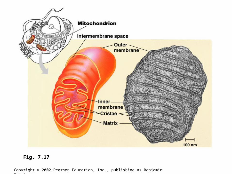
• Mitochondria have a smooth outer membrane and a highly folded inner membrane, the cristae.
• This creates a fluid-filled space between them.
• The cristae present ample surface area for the enzymes that synthesize ATP.
• The inner membrane encloses the mitochondrial matrix, a fluid-filled space with DNA, ribosomes, and enzymes.
Copyright © 2002 Pearson Education, Inc., publishing as Benjamin Cummings
Copyright © 2002 Pearson Education, Inc., publishing as Benjamin Cummings
Fig. 7.17
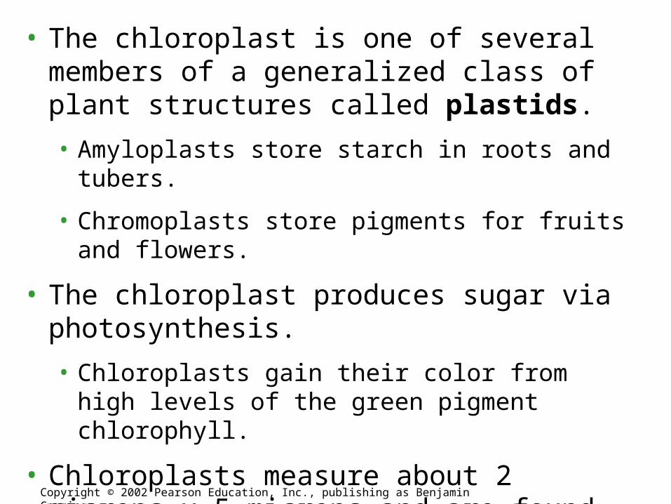
• The chloroplast is one of several members of a generalized class of plant structures called plastids.
• Amyloplasts store starch in roots and tubers.
• Chromoplasts store pigments for fruits and flowers.
• The chloroplast produces sugar via photosynthesis.
• Chloroplasts gain their color from high levels of the green pigment chlorophyll.
• Chloroplasts measure about 2 microns x 5 microns and are found in leaves and other green structures of plants and in eukaryotic algae.
Copyright © 2002 Pearson Education, Inc., publishing as Benjamin Cummings
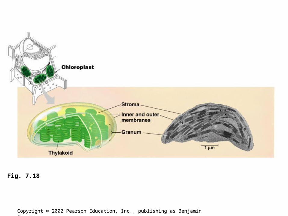
• The processes in the chloroplast are separated from the cytosol by two membranes.
• Inside the innermost membrane is a fluid-filled space, the stroma, in which float membranous sacs, the thylakoids.
• The stroma contains DNA, ribosomes, and enzymes for part of photosynthesis.
• The thylakoids, flattened sacs, are stacked into grana and are critical for converting light to chemical energy.
Copyright © 2002 Pearson Education, Inc., publishing as Benjamin Cummings
Copyright © 2002 Pearson Education, Inc., publishing as Benjamin Cummings
Fig. 7.18
• Like mitochondria, chloroplasts are dynamic structures.
• Their shape is plastic and they can reproduce themselves by pinching in two.
• Mitochondria and chloroplasts are mobile and move around the cell along tracks in the cytoskeleton.
Copyright © 2002 Pearson Education, Inc., publishing as Benjamin Cummings
• Peroxisomes contain enzymes that transfer hydrogen from various substrates to oxygen
• An intermediate product of this process is hydrogen peroxide (H2O2), a poison, but the peroxisome has another enzyme that converts H2O2 to water.
• Some peroxisomes break fatty acids down to smaller molecules that are transported to mitochondria for fuel.
• Others detoxify alcohol and other harmful compounds.
• Specialized peroxisomes, glyoxysomes, convert the fatty acids in seeds to sugars, an easier energy and carbon source to transport.
Peroxisomes generate and degrade H2O2 in performing various metabolic functions
Copyright © 2002 Pearson Education, Inc., publishing as Benjamin Cummings
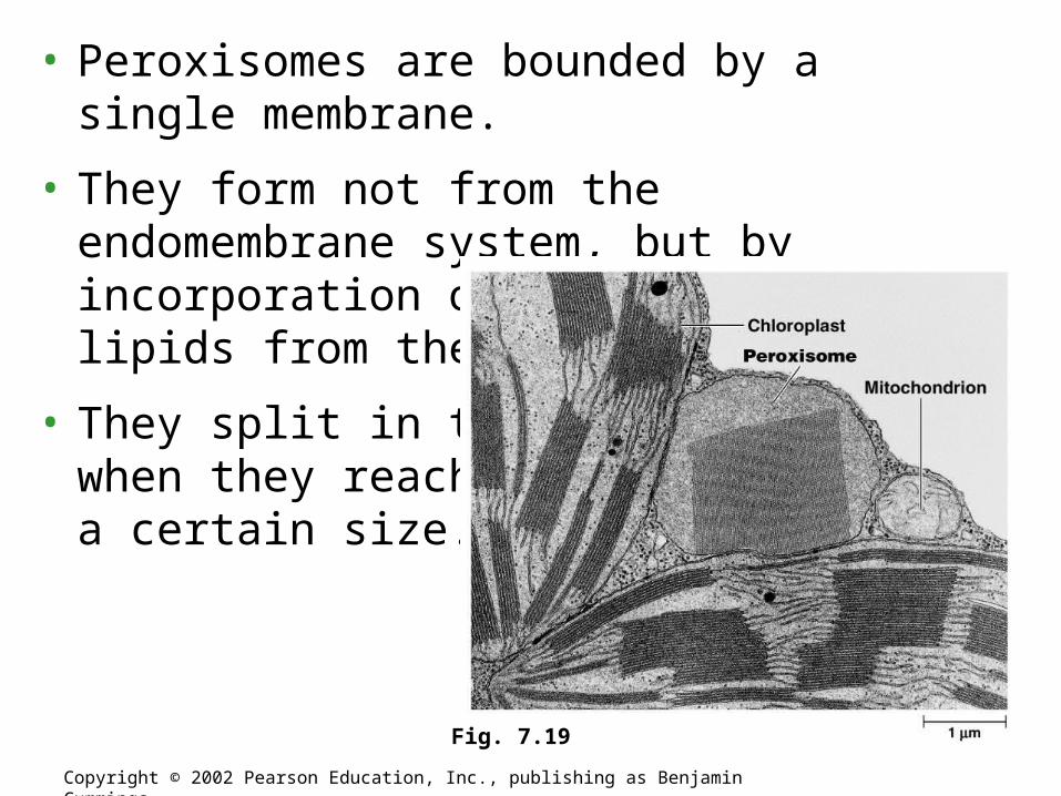
• Peroxisomes are bounded by a single membrane.
• They form not from the endomembrane system, but by incorporation of proteins and lipids from the cytosol.
• They split in two when they reach a certain size.
Copyright © 2002 Pearson Education, Inc., publishing as Benjamin Cummings
Fig. 7.19