Embed Size (px)
Citation preview

BRAINA JOURNAL OF NEUROLOGY
Obesity induces functional astrocytic leptinreceptors in hypothalamusHung Hsuchou, Yi He, Abba J. Kastin, Hong Tu, Emily N. Markadakis, Richard C. Rogers,Paul B. Fossier and Weihong Pan
Pennington Biomedical Research Center, Baton Rouge, LA 70808, USA
Correspondence to: Weihong Pan, MD, PhD,
Blood-Brain Barrier Group,
Pennington Biomedical Research Center,
6400 Perkins Road,
Baton Rouge, 70808 LA, USA
E-mail: [email protected]
The possible role of astrocytes in the regulation of feeding has been overlooked. It is well-established that the endothelial cells
constituting the blood–brain barrier transport leptin from blood to brain and that hypothalamic neurons respond to leptin to
induce anorexic signaling. However, few studies have addressed the role of astrocytes in either leptin transport or cellular
activation. We recently showed that the obese agouti viable yellow mouse has prominent astrocytic expression of the leptin
receptor. In this study, we test the hypothesis that diet-induced obesity increases astrocytic leptin receptor expression and
function in the hypothalamus. Double-labelling immunohistochemistry and confocal microscopic analysis showed that all
astrocytes in the hypothalamus express leptin receptors. In adult obese mice, 2 months after being placed on a high-fat diet,
there was a striking increase of leptin receptor (+) astrocytes, most prominent in the dorsomedial hypothalamus and arcuate
nucleus. Agouti viable yellow mice with their adult-onset obesity showed similar changes, but the increase of leptin receptor (+)
astrocytes was barely seen in ob/ob or db/db mice with their early-onset obesity and defective leptin systems. The marked
leptin receptor protein expression in the astrocytes, shown with several antibodies against different receptor epitopes, was
supported by RT–PCR detection of leptin receptor-a and -b mRNAs in primary hypothalamic astrocytes. Unexpectedly, the
protein expression of GFAP, a marker of astrocytes, was also increased in adult-onset obesity. Real-time confocal imaging
showed that leptin caused a robust increase of calcium signalling in primary astrocytes from the hypothalamus, confirming their
functionality. The results indicate that metabolic changes in obese mice can rapidly alter leptin receptor expression and astro-
cytic activity, and that leptin receptor is responsible for leptin-induced calcium signalling in astrocytes. This novel and clinically
relevant finding opens new avenues in astrocyte biology.
Keywords: leptin; obesity; astrocyte; ObR; calcium signalling
IntroductionIt is increasingly recognized that astrocytes play an important
role in gliotransmission and neurovascular coupling (Haydon and
Carmignoto, 2006). However, the role of astrocytes in the
regulation of obesity is not yet clear. Obese agouti viable yellow
(Avy) mice express leptin receptors (ObR) in both astrocytes and
neurons in the hypothalamus. While in lean adult B6 mice 7% of
the ObR(+) cells are astrocytes (the remaining 93% being neu-
rons), in adult Avy mice 25% of ObR is expressed in astrocytes,
doi:10.1093/brain/awp029 Brain 2009: 132; 889–902 | 889
Received November 11, 2008. Revised December 31, 2008. Accepted January 28, 2009. Advance Access publication March 17, 2009
� The Author (2009). Published by Oxford University Press on behalf of the Guarantors of Brain. All rights reserved.
For Permissions, please email: [email protected]
Dow
nloaded from https://academ
ic.oup.com/brain/article/132/4/889/287812 by guest on 05 February 2022

with a corresponding decrease of ObR (+) neurons to 75% (Pan
et al., 2008a). Obese rats also have higher levels of expression of
agouti-related protein (Dunbar et al., 2005). This leads to the next
question: do astrocytes participate in the CNS control of obesity by
upregulation of their leptin receptors?
Leptin is a 16 kD protein mainly produced by adipocytes, and its
blood concentration correlates with fat mass. As a neuroendocrine
hormone, one of the major targets of leptin action is the brain.
Obesity is related to impaired transport of leptin across the
blood–brain barrier (Kastin et al., 1999; Banks and Farrell, 2003;
Banks, 2003; Banks et al., 2001, 2004), reduced neuroplasticity
(Stranahan and Mattson, 2008), and decreased neurogenesis in
the hippocampus (Lindqvist et al., 2006). The model of diet-
induced obesity provides an additional tool to determine whether
astrocytic upregulation of ObR expression is related to an intrinsic
genetic predisposition in the Avy mice or is caused by epigenetic
influences occurring after the onset of obesity.
The structural and biochemical changes in brain parenchyma
induced by obesity appear to be rather trivial. There is no clear
evidence of reactive gliosis or inflammatory cell infiltration, and
there is no change in whole brain protein carbonyls (index of
protein oxidation) or 4-hydroxynonenal (index of membrane
lipid oxidation) in mice with a higher percent of fat (10%) in
the diet as compared with the controls (5%). Yet, the triglyc-
eride lowering agent gemfibrozil reduces protein carbonyls and
4-hydroxynonenal in these obese mice on a diet containing
10% fat for 10 months (Farr et al., 2008). But would leptin
provide a metabolic switch for astrocytes?
The CNS effects of leptin are rather diverse. It is trophic to
neural progenitor cells and promotes glial as well as neuronal
development (Udagawa et al., 2006). It is anti-epileptic and can
directly affect calcium and potassium channels (Shanley et al.,
2002; Harvey, 2007; Xu et al., 2008). In the feeding circuitry,
leptin facilitates the production of melanocortin in proopiomelano-
cortin neurons and suppresses the production of neuropeptide Y
and agouti-related protein in neuropeptide Y neurons of the arcu-
ate nucleus of the hypothalamus (Schwartz, 2001). Both pro-
inflammatory and anti-inflammatory effects of leptin have been
shown (Matarese et al., 2001; Lord, 2006; Ahmed et al., 2007;
Pinteaux et al., 2007; Lin et al., 2007). The central leptin system,
therefore, might be involved in neuroinflammation after the onset
of the metabolic syndrome and hyperleptinaemia. Astrocytes could
constitute an ideal cellular substrate for such actions. Thus, in
this study we examine the important issue of astrocytic ObR
regulation.
Materials and methods
Induction of diet-induced obesity andthe specifics of the genetic models ofobesity used in this studyC57BL/6J male mice (B6, Jackson Laboratories, Bar Harbor, ME, USA)
were used to induce diet-induced obesity following a protocol
approved by the Institutional Animal Care and Use Committee.
Four-week-old mice were group-housed and randomly assigned to
either a high fat diet (HFD, 45% kcal fat) (D12451, Research Diets,
New Brunswick, NJ, USA) or regular rodent chow for 2 months or
longer, as specified in the Results section. Body weight was meas-
ured weekly after induction of obesity for both the diet-induced
obesity and control groups. The percentage of body fat was deter-
mined with a nuclear magnetic resonance (NMR) Bruker minispec Live
Mice Analyser (model mq7.5, LF50; Bruker Optics, Inc., Billerica MA,
USA), as described previously (Pan and Kastin, 2007).
The obesity phenotype of Avy mice (Jackson Laboratories) has
been characterized previously (Pan and Kastin, 2007; Pan et al.,
2008a). Small numbers of male ob/ob and db/db mice (Jackson
Laboratories) were used in the validation of immunohistochemistry.
The mutant mice were studied in parallel with age-matched male
B6 controls. The ages and sample sizes are specified in the Results
section.
Fluorescent immunohistochemistry andconfocal microscopic analysesThe mice were anaesthetized by intraperitoneal injection of 40% ure-
thane, and perfused intracardially with 4% paraformaldehyde (PFA).
The brain was post-fixed in 4% paraformaldehyde for 3 days at 4�C.
Afterwards, it was transferred to phosphate-buffered saline (PBS) with
15% sucrose for 8 h at 4�C, and then to 30% sucrose until it sank.
HistoPrep frozen tissue embedding media (Fisher Scientific, Pittsburg,
PA, USA) was used to embed the brain. The embedded tissue block
was stored at �80�C for future use. Free-floating coronal sections of
20 mm thickness were obtained by a cryostat and stored in cryoprotec-
tant (30% sucrose and 30% ethylene glycol in 0.1 M phosphate buffer)
at �20�C until staining.
After being washed with PBS, the sections were blocked with
10% normal donkey serum in PBS with 0.3% Triton X-100 at room
temperature for 1 h. Several ObR primary antibodies were used for
immunolabelling, as shown in Table 1. Anti-glial fibrillary acidic protein
(anti-GFAP, AB5804, 1:100 to 1:500; Chemicon, Temecula, CA, USA)
or anti-neuronal nuclei (anti-NeuN, MAB377B, 1:100; Chemicon)
primary antibodies were also used in the double-labelling process.
Table 1 Features of leptin receptor (ObR) antibodies
Epitope Name Source Concentration (kg/ml)
membrane juxtapositional cytoplasmicdomain (aa 877–894; greater affinityto short cytoplasmic forms ofmembrane-bound ObR)
M18 SC-1834, Santa Cruz Biotechnology 2
N-terminus of ObR (all isoforms) K20 SC-1834, Santa Cruz Biotechnology 2
ObRb CH14014 CH14014, Neuromics 10
890 | Brain 2009: 132; 889–902 H. Hsuchou et al.
Dow
nloaded from https://academ
ic.oup.com/brain/article/132/4/889/287812 by guest on 05 February 2022

After overnight incubation at 4�C, the sections were incubated with
Alexa488 or Alexa594-labeled secondary antibodies (Invitrogen,
Eugene, OR, USA), or FITC-conjugated anti-chicken secondary anti-
body (Jackson ImmunoResearch Laboratories, West Grove, PA, USA)
at room temperature for 1 h. The sections were thoroughly washed
with PBS between the steps. After staining, the sections were mounted
with Vectashield mounting media (Vector Laboratories, Burlingame,
CA, USA).
The specificity of the staining was shown by the lack of signal in
negative controls, including pre-adsorption of the primary antibody
with a specific blocking peptide, and omission of primary antibody.
For double-labelling immunofluorescence studies with a second pri-
mary antibody against GFAP, single staining experiments with each
primary antibody were performed and compared to rule out the
cross-reactivity of the irrelevant antibodies. Confocal images were
acquired on an Olympus FV1000 microscope in our laboratory, with
Argon laser excitation/filter for emission at 488/505–525 and
543/560–660 (nm) for Alexa488 and Alexa594, respectively.
Statistical analysis of the number of ObR(+), GFAP(+) or double-
labelled cells in the diet-induced obesity, Avy or control groups was
performed on matching sections from three different mice. The signif-
icant differences were determined by analysis of variance (ANOVA)
followed by Tukey’s post hoc test.
Primary culture of mouse hypothalamicastrocytesFollowing approved animal protocols, the hypothalami of 7-day-old
FVB/NJ (FVB) mouse pups were dissected, pooled (3 pups/ml),
mechanically dissociated and cultured in Dulbecco’s modified eagle
medium (DMEM) containing 10% fetal bovine serum (FBS) and
antibiotics/antimycotics in a 5% CO2 incubator at 37�C. At about
90% confluency (about 1 week in culture), the cells were agitated
in an orbital shaker at 250 r.p.m. for 2 h at 37�C to detach microglial
cells and remove them by change of medium. The remaining cells
showed astrocytic morphology. Immunocytochemistry confirmed that
495% of the cells were GFAP (+). Two days later, the cells were used
for RT–PCR analysis of ObR mRNA, or passaged to poly-D-lysine
coated glass coverslips for calcium imaging.
RT–PCR analysis of ObR mRNAsubtypes in hypothalamic astrocytesThe cells were lysed in RNA lysis buffer containing b-mercaptoethanol.
In addition to primary mouse hypothalamic astrocytes, rat C6 astro-
cytoma cells were also studied. C6 cells have been shown to express
ObR mRNA and protein (Morash et al., 2000) and thus were used as
a positive control. Total RNA was extracted by use of an Absolutely
RNA Miniprep kit (Stratagene, La Jolla, CA, USA) and reversely tran-
scribed with a High Capacity cDNA Reverse Transcription Kit (Applied
Biosystems, Foster City, CA, USA). The primers used for PCR amplifi-
cation are listed in Table 2. Mouse primers were used for tissue stud-
ies, whereas rat primers were used for C6 astrocytoma cells. The PCR
conditions were: denaturing at 95�C for 5 min, followed by 35 cycles
of denaturing at 95�C for 30 s, annealing at 60�C for 30 s, extension at
72�C for 30 s, with a final extension at 72�C for 10 min. A negative
control without template (water only) was included. The PCR products
were electrophoresed on 1.5% agarose gel containing ethidium bro-
mide, and imaged under a UV lamp with a Kodak EDAS 290 imaging
system.
Real-time imaging of leptin-inducedcalcium responsePrimary hypothalamic astrocytes on glass coverslips were loaded with
calcium green-1AM (4 mM, Invitrogen) for 30 min at 37�C. The cover-
slip was placed on a temperature-controlled perfusion chamber on
the stage of an Olympus FV1000 inverted microscope, and perfused
with oxygenated Kreb’s solution at 1.0 ml/min. Real-time imaging was
performed with a 40� objective at a frequency of 2 frames/s, and
600 frames were captured. Calcium green was excited by argon laser,
and the resulting emission at 488 nm was captured. After application
of leptin (6.2 nM; R&D Systems, Minneapolis, MN) or Kreb’s solution
for 2 min, the cells were allowed to recover for 5 min, at which time
the baseline had returned to normal. The cells were then challenged
with ATP (50 mM; Sigma, St Louis, MO, USA) for 30 s. This served as
a positive control with calcium oscillation being elicited consistently,
as has been described (Rogers and Hermann, 2008). Only one field
of each coverslip was recorded, since desensitization of the leptin
response had been observed that was not explained by bleaching.
The percentage of responsive cells and the amplitude of increase
from baseline fluorescent intensity were calculated for each coverslip.
The cells treated with Kreb’s solution had no change in calcium signal
and thus served as negative controls. All results were replicated at least
four times, and representative images are shown.
Results
Immunohistochemistryof ObR andcellular phenotypic characterizationWe have observed co-staining of ObR and GFAP in mouse
hypothalamus with the M-18 antibody (sc-1834; Santa Cruz
Biotechnology, Santa Cruz, CA, USA) that recognizes the
Table 2 PCR primers for ObR subtypes
ObR subtype Forward primer Reverse primer Species Product size (bp)
ObRa GAAGTCTCTCATGACCACTACAGATGA TTGTTTCCCTCCATCAAAATGTAA Mouse/rat 98
ObRb GCATGCAGAATCAGTGATATTTGG CAAGCTGTATCGACACTGATTTCTTC Mouse/rat 81
ObRc AGGGCTTTATGTTGTTGTGTTC TTTCTCTGATCAAATCCCAAAC Mouse 88
ObRc0 TTA ATT TCC AAA AGG TCA CT TTT TTG CCT TTT AAA GAT GT Rat 105
ObRd CACACCAGAGAATGAAAA TCTGAAAATAAAAACTTCATGT Mouse 119
ObRe TAATGAAGATGATGGAATGAAG ATTGCCAGTCTACAGTGTCA Mouse 117
ObRe0 GTA AAT TGG CAA TCC TGA TAA TAA AG ATT CAG TTG GAA GGT TGA CAT C Rat 72
Obesity and astrocytic ObR Brain 2009: 132; 889–902 | 891
Dow
nloaded from https://academ
ic.oup.com/brain/article/132/4/889/287812 by guest on 05 February 2022
membrane juxtapositional cytoplasmic domain (aa 877–894) of
ObR in B6 and Avy mice in the original study (Pan et al.,
2008a). To determine whether this is a general phenomenon
and further identify ObR subtypes, we tested additional rodent
models and ObR antibodies against different epitopes of the
receptor protein. For all three antibodies used, sections incubated
with secondary antibody only or with primary antibody after pre-
adsorption with the respective blocking peptide overnight did not
show specific signals. The negative controls thus assured the spec-
ificity of the staining.
In hypothalamic sections of obese ob/ob mice (Fig. 1A) that do
not produce leptin, and in obese db/db mice (Fig. 1B) that have
no ObRb receptor because of an insertional mutation although
other ObR isoforms are present (Chua et al., 1996), the ObR
staining pattern with the M18 antibody was mainly neuronal
rather than astrocytic. Figure 1C shows higher magnification
images of GFAP and ObR co-localization in these strains.
In hypothalamic sections from B6 and Avy mice, the K-20
polyclonal antibody against the N-terminus of the mouse ObR
(sc-1835) also showed immunoreactivity (Fig. 2). The staining
patterns were similar to those seen with the M-18 antibody. In
contrast to the weak immunoreactivy in neurons, the astrocytes
in the Avy mice showed strong reactivity to the K-20 antibody
in both the arcuate nucleus (Fig. 2A) and dorsomedial hypothala-
mus (Fig. 2B). Figure 2C shows higher magnification images of the
co-localization of GFAP and ObR.
A third antibody used was targeted to the unique sequence of
ObRb at its cytoplasmic tail (CH14104, Neuromics, Edina, MN,
USA). This antibody was raised against rat ObRb but cross-reacted
with mouse tissue. Figure 3A shows that the ObRb staining had a
rather diffuse background with both neuronal and astrocytic dis-
tribution. The cells double-labelled with both the M-18 antibody
(against the membrane juxtapositional cytoplasmic domain) and
the CH14104 antibody (against the membrane distal cytoplasmic
domain of ObRb) were mainly astrocytes. The partial overlap of
the staining in cells showing typical astrocyte morphology, proba-
bly representing the co-expression of ObRb with short transmem-
brane forms of ObR, was seen not only in the B6 mice but also in
the diet-induced obesityand Avy mice (Fig. 3B).
Time course of the development ofdiet-induced obesityMale B6 mice were fed a 45% HFD (the diet-induced obesity group,
n = 9) or rodent chow (lean control group, n = 7) between 1 and
5 months of age. Repeated measures ANOVA showed that the diet-
induced obesity group had significantly higher body weight than the
lean controls (P50.005). The post hoc test showed that the differ-
ence was apparent by 13 weeks of age (P50.05 on week 13–17,
and P50.01 afterwards). Concurrent with increased body weight,
there was also a significant (P50.005) increase of fat composition
(% fat/body weight) shown by serial measurement with NMR, at
8.5 week after being placed on HFD, or at 12.5 weeks of age
(Fig. 4). Based on the results, we used 3-month-old mice for immu-
nohistological studies, immediately before the full development of
the obesity phenotype.
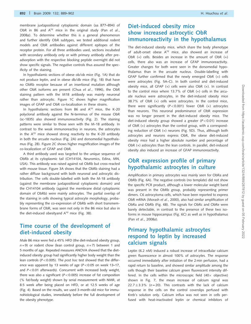
Diet-induced obesity miceshow increased astrocytic ObRimmunoreactivity in the hypothalamusThe diet-induced obesity mice, which share the body phenotype
of adult-onset obese Avy mice, also showed an increase of
ObR (+) cells. Besides an increase in the amount of ObR (+)
cells, there also was an increase of GFAP immunoreactivity.
Greater changes for both were seen in the dorsomedial hypo-
thalamus than in the arcuate nucleus. Double-labelling with
GFAP further confirmed that the newly emerged ObR (+) cells
were astrocytes (Fig. 5A–C). In both control and diet-induced
obesity mice, all GFAP (+) cells were also ObR (+). In contrast
to the control mice where 13.7% of ObR (+) cells in the arcu-
ate nucleus were astrocytes, in the diet-induced obesity mice
38.7% of ObR (+) cells were astrocytes. In the control mice,
there were significantly (P50.001) fewer ObR (+) astrocytes
than neurons. This neuronal predominance of ObR expression
was no longer present in the diet-induced obesity mice. The
diet-induced obesity group showed a greater (P50.01) increase
of ObR (+) astrocytes than the control group, with a correspond-
ing reduction of ObR (+) neurons (Fig. 5D). Thus, although both
astrocytes and neurons express ObR, the obese diet-induced
obesity mice had a higher absolute number and percentage of
ObR (+) astrocytes than the lean controls. In parallel, diet-induced
obesity also induced an increase of GFAP immunoreactivity.
ObR expression profile of primaryhypothalamic astrocytes in cultureAmplification in primary astrocytes was mainly seen for ObRa and
ObRb (Fig. 6A). The negative controls (no template) did not show
the specific PCR product, although a lower molecular weight band
was present in the ObRb group, probably representing primer
dimers. C6 astrocytoma cells, which have been reported to express
ObR mRNA (Morash et al., 2000), also had similar amplification of
ObRa and ObRb (Fig. 6B). The signals for ObRc and ObRe were
barely detectable, in contrast to the presence of these two iso-
forms in mouse hippocampus (Fig. 6C) as well as in hypothalamus
(Pan et al., 2008a).
Primary hypothalamic astrocytesrespond to leptin by increasedcalcium signalsLeptin (6.2 nM) induced a robust increase of intracellular calcium
green fluorescence in almost 100% of astrocytes. The response
occurred immediately after initiation of the 2 min perfusion, had a
rapid return to baseline, and showed similar amplitude among the
cells though their baseline calcium green fluorescent intensity dif-
fered. In the cells within the microscopic field (40� objective)
shown in Fig. 7, the mean increase of calcium signal was
22.7� 3.3% (n = 20). This contrasts with the lack of calcium
response in the cells on the control coverslips perfused with
Kreb’s solution only. Calcium influx was not seen in cells per-
fused with heat-inactivated leptin or chemical inhibitors of
892 | Brain 2009: 132; 889–902 H. Hsuchou et al.
Dow
nloaded from https://academ
ic.oup.com/brain/article/132/4/889/287812 by guest on 05 February 2022

Figure 1 Expression of ObR in arcuate nucleus of different mouse models shown by immunostaining with M18 antibody. Astrocytes
were immunostained with anti-GFAP (red, arrows), whereas neurons were immunostained with anti-NeuN (red, arrow heads). Yellow
colouration indicates co-localization. (A) ob/ob mouse; (B) db/db mouse (confocal image, scale bar = 50 mm for GFAP; scale
bar = 100 mm for NeuN). (C) Higher magnification of the co-localization of ObR and GFAP from the demarcated areas in A and B.
Scale bar: 25mm.
Obesity and astrocytic ObR Brain 2009: 132; 889–902 | 893
Dow
nloaded from https://academ
ic.oup.com/brain/article/132/4/889/287812 by guest on 05 February 2022

Figure 2 Expression of ObR in the arcuate nucleus (A) and dorsomedial hypothalamus (B) shown by immunostaining with the K20
antibody. In both regions, there were more ObR (+) astrocytes (arrows) in Avy than in age-matched B6 mice (confocal image, scale
bar = 50 mm). (C) Higher magnification of the co-localization of ObR and GFAP from the demarcated areas in (A and B).
894 | Brain 2009: 132; 889–902 H. Hsuchou et al.
Dow
nloaded from https://academ
ic.oup.com/brain/article/132/4/889/287812 by guest on 05 February 2022

mitogen-activated protein kinases (MAPK), indicating specificity.
Repetitive application of leptin on the same coverslip 5 min later
elicited a much smaller amplitude of response (4.5� 1.1%), sug-
gesting desensitization (tachyphylaxis). Nonetheless, ATP (50 mM
for 30 s) treatment of the same cells induced a robust and persis-
tent increase of calcium response at the end of the study in each
coverslip (32.6� 2.4%) (Fig. 7).
DiscussionThe notion of astrocytic expression of ObR has encountered much
resistance among some obesity researchers focusing on endothelial
and neuronal ObR in the CNS. Nonetheless, Avy mice, which
manifest a genetic predisposition to adult-onset obesity, recently
have been shown to express increased astrocytic ObR in the
hypothalamus (Pan et al., 2008a). This indicates regulatory
changes of astrocytic ObR. Here we further determined whether
this upregulation is a general mechanism in obesity or is specific
for genetic models of obesity.
The distribution of ObR mRNA in rat brain has been mapped
by in situ hybridization with radioactive probes for ObRb and
the entire ObR (Elmquist et al., 1998). In the rat hypothalamus,
we also found co-localization of ObRb mRNA and GFAP pro-
tein, by fluorescent in situ hybridization and double labelling
immunohistochemistry (Hsuchou et al., unpublished observations).
ObR is a single-transmembrane receptor that interacts with Janus
kinase (JAK)-2, Signal Transducer and Activator of Transcription
Figure 3 Expression of ObR in the arcuate nucleus of B6 (top), diet-induced obesity (DIO) (middle) and Avy (bottom) mice shown
by double immunostaining with M-18 (red) and CH14104 (green) antibodies. (A) M-18 immunoreactivity was mainly seen in
astrocytes (arrows). CH14104 antibody stained mostly neurons (arrow heads). In comparison with the lean B6 control, the diet-induced
obesityB6 mice and the Avy mice on a B6 background showed a greater number of ObR(+) astrocytes seen with the M18 antibody
(epifluorescence image, scale bar = 50 mm). (B) Higher magnification images showing that astrocytes can be stained with both M-18
and CH14104 antibodies, the former providing a stronger signal with a crisper background (confocal image, scale bar = 10 mm).
Obesity and astrocytic ObR Brain 2009: 132; 889–902 | 895
Dow
nloaded from https://academ
ic.oup.com/brain/article/132/4/889/287812 by guest on 05 February 2022

(STAT)-3, phosphoinositide-3 kinase, and MAPK. There are at
least four major forms of ObR splice variants in mice, including
the soluble ObRe, the short cytoplasmic tail ObRa and ObRc,
and the long cytoplasmic tail ObRb that activates STAT-3. ObRd
is not detected in the mouse hypothalamus or cerebral vasculature
(Pan et al., 2008b). By use of three different polyclonal antibodies
against different epitopes of ObR and co-immunostaining of
cellular phenotypic markers, we differentiated the relative abun-
dance of the ObR isoforms in astrocytes and neurons.
The K20 antibody recognizes all ObR isoforms, the CH14104
antibody is specific for ObRb, whereas the relative selectivity of
the M18 antibody is highest for ObRa, less for ObRc and ObRd
(if present), and the least for ObRb. The M18 antibody revealed
both neuronal and astrocytic patterns of ObR immunoreactivity,
Figure 3 Continued.
896 | Brain 2009: 132; 889–902 H. Hsuchou et al.
Dow
nloaded from https://academ
ic.oup.com/brain/article/132/4/889/287812 by guest on 05 February 2022

similar in rats and mice. The specificity of the staining was illus-
trated not only by the lack of staining in the presence of blocking
peptide or secondary antibody only, but also by different patterns
in the ob/ob mice, in which leptin is not produced, and in
the db/db mice, in which an insertional mutation abolishes the
production of ObRb although other ObR isoforms are present
(Chua et al., 1996). In the ob/ob and db/db mice, there were
few ObR (+) astrocytes, and the neuronal ObR staining was also
less intense than the age-matched B6 lean controls.
Confocal analyses of the images showed that astrocytes (GFAP-
positive) had strong immunoreactivity to the M18 antibody, K20
antibody, and to a lesser extent the CH14104 antibody. Neurons
(NeuN-positive), in contrast, revealed a greater immunreactivity to
CH14104 and weaker signals with M18 and K20. Although the
fluorescent intensity is influenced by the affinity of the antibodies,
comparison from the same antibody staining between the two
major populations of ObR (+) cells in CNS parenchyma suggests
that astrocytes showed short ObR predominance whereas the
neurons had more ObRb.
It should be noted that the lack of an endothelial or microvas-
cular pattern of staining does not detract from the sensitivity of
the detection method nor indicate the absence of ObR at the
blood–brain barrier. Microvessels composing the blood-brain bar-
rier have a specific array of ObR subtypes and mediate the trans-
port of leptin from blood to brain (Bjørbæk et al., 1998; Boado
et al., 1998; Kastin et al., 1999; Maness et al., 2000; Hileman
et al., 2002; Pan et al., 2008b). The blood-brain barrier is a three-
dimensional structure. In human brain averaging 1200 g, capillaries
span 650 km with a volume of 1 ml. These thin structures provide
tremendous surface area for exchange of information between the
circulation and CNS parenchyma, with no neuron48–10 mm away
from a capillary (Yoshida and Ikuta, 1984; Abbott et al., 2006;
Abbott, 2005, 2008). Thus, enriched microvessels show a many-
fold greater extent of expression of cytokine receptors than tissue
homogenates from the same region (Pan et al., 2008a, 2008b),
yet the immunostaining in individual endothelial cells may not
be significant.
In general, astrocytes have a close structural association with
the endothelia and play important roles in regulating blood-brain
barrier functions (Nedergaard et al., 2003). Specialized astrocytic
endfeet cover up to 90% of vessel walls, as shown by electron
microscopy and aquaporin-4 staining (Abbott, 2008). Other inves-
tigators even estimate that499% of the cerebrovascular surface is
ensheathed by astrocyte processes (Kacem et al., 1998; Agulhon
et al., 2008). In primary astrocytes from mouse hypothalamus,
both ObRa and ObRb mRNAs were detected. This is consistent
with findings in C6 cells shown both here and in the literature
(Morash et al., 2000), and supports the immunohistochemical
findings described above. The composition of ObR subtypes in
astrocytes is similar to that shown in cerebral microvessels
(Hileman et al., 2002; Pan et al., 2008b). Although it is yet to
be determined how the endothelial ObR, astrocytic ObR, and
neuronal ObR act in concert or differentially in response to
blood-borne leptin, astrocytes apparently play an intricate role in
the blood–brain barrier transport of leptin into the CNS.
A high fat diet increases body weight and blood leptin levels,
and induces leptin resistance shown both by defective STAT3
signalling in neurons that express ObRb, and by saturation of
blood–brain barrier transport (Coculescu, 1999; El Haschimi
et al., 2000; Hileman et al., 2002; Banks et al., 2003; Pan and
Kastin, 2003; Kastin and Pan, 2003; Levin et al., 2004; Pan et al.,
2004). We also showed that different ObR isoforms all participate
in leptin transport (Tu et al., 2007, 2008), that interactions of
leptin with other ingestive peptides may transform feeding behav-
iour (Kastin et al., 2002; Pan et al., 2004), and that the ObR
isoforms are differentially regulated by neonatal development as
well as obesity (Pan et al., 2008a, 2008b). In addition, this study
indicates that astrocytic ObR is implicated in the mechanism of
leptin resistance. The elevation of astrocytic ObR in the diet-
induced obesity mice is similar to that seen in obese Avy mice
(Pan et al., 2008a). The percent of ObR (+) astrocytes in the
control group was higher than in the previous study, probably
related to differences in staining intensity, individual mice, and
cell counting resulting from different demarcation of the borders
of the hypothalamic nuclei. Regardless, the results indicate that
metabolic signals in obesity can rapidly alter astrocytic phenotype,
representing a unique aspect of CNS plasticity.
Besides our observation of regulatory changes of ObR in
astrocytes, several groups have shown functional leptin recep-
tors in astrocytes. Systemically delivered hydroxystilbamidine
(FluoroGold equivalent) can be taken up by astrocytes in selective
regions in the brain 2–6 h later, mainly in the median eminence
and the adjacent ventral part of the arcuate nucleus. Many of
these GFAP (+) cells also express leptin receptors and neuropep-
tide Y Y1 receptors, whereas the surrounding neurons (NeuN-
positive) or adjacent tanycytes (vimentin-positive) do not take
up the dye (Cheunsuang and Morris, 2005). In the subcommis-
sural organ of rabbits, ObR staining was seen in astrocytes, and
fasting decreased ObR distribution in the cytoplasm but increased
it in plasma membranes of these GFAP (+) cells (Cecilia et al.,
2006). Leptin induces differentiation of astrocytes from neuro-
spheres, and helps to maintain BrdU (+) cells in the embryonic
5 10 15 2010
20
30
40
*
***
**
******
**
0
10
20
30
40***
Bo
dy
wei
gh
t (g
)
Age (wk)
DIOcontrol
% F
at (
/BW
)control DIO
Figure 4 Obesity phenotype of the diet-induced obesity mice
(n = 9), shown by more rapid increase of body weight over
time and higher percentage of body fat at 8.5 weeks in
comparison with their former littermates fed with regular
rodent chow (n = 7). �P50.05; ��P50.01; ���P50.005.
Obesity and astrocytic ObR Brain 2009: 132; 889–902 | 897
Dow
nloaded from https://academ
ic.oup.com/brain/article/132/4/889/287812 by guest on 05 February 2022

neuroepithelium (Udagawa et al., 2006). C6 astrocytoma cells
express the short form of ObR mRNA and cytoplasmic staining
of ObR; since the leptin transcript is also found in these cells, it is
suggested that leptin may be an autocrine factor acting on ObR
(Morash et al., 2000). In primary rat astrocytes from the nucleus
tractus solitarius, all membrane-bound ObR isoforms are present,
and are differentially regulated by fasting and estradiol treatment
(Dallaporta et al., 2009). Consistent with observations in Avy mice
(Pan et al., 2008a), the increased astrocytic expression in diet-
induced obesity mice in this study suggests its role in feeding
regulation.
In the obese Avy and diet-induced obesity mice, there also was
an increase of GFAP immunoreactivity. This indicates that obesity
increases the level of expression of GFAP, an intermediate filament
Figure 5 Diet-induced obesity increased ObR (+) astrocytes in the arcuate nucleus (A) and to an even greater extent in the
dorsomedial hypothalamic nucleus (B) (confocal image, scale bar = 100mm). (C) Higher magnification of the co-localization of ObR
and GFAP from the demarcated areas above in diet-induced obesity. Scale bar: 25 mm. (D) Percent of ObR (+) cells that were also
GFAP or NeuN (+); the diet-induced obesity group had significantly more ObR (+) astrocytes in the arcuate nucleus than the controls,
with corresponding reduction of ObR (+) neurons. ��P50.01.
898 | Brain 2009: 132; 889–902 H. Hsuchou et al.
Dow
nloaded from https://academ
ic.oup.com/brain/article/132/4/889/287812 by guest on 05 February 2022

Figure 5 Continued.
Figure 6 RT–PCR analysis shows that astrocytes expressed mainly ObRa (98 bp) and ObRb (81 bp) mRNA. (A) Primary astrocytes from
1-week-old mouse pups. Lane 1: no template control; lane 2: primary astrocytes from mouse hypothalamus; lane 3: primary astrocytes
from mouse striatum. (B) C6 astrocytoma cells with rat-specific primers. (C) In contrast, mouse hippocampus had not only ObRa and
ObRb, but also ObRc and ObRe. Lane 1: negative control; lanes 2–5: hippocampal tissue from different mice.
Obesity and astrocytic ObR Brain 2009: 132; 889–902 | 899
Dow
nloaded from https://academ
ic.oup.com/brain/article/132/4/889/287812 by guest on 05 February 2022

protein that plays essential roles in astrocyte function. In the Avy
mice, Western blotting analysis showed that the level of GFAP
protein was indeed increased in the hypothalamus (He et al.,
unpublished observations). Although there is astrogenesis in the
adult hypothalamus, and our preliminary data showed an increase
of BrdU-labeled cells, some of which co-localized with GFAP and
S100b (Hsuchou et al., unpublished observations), the newly
emerged astrocytes from de novo mitosis would not be sufficient
Figure 7 Leptin-induced calcium signalling in primary astrocytes from mouse hypothalamus. (A) A field of astrocytes on glass
coverslips after calcium green-1AM loading. (B) Time-lapse image showing that all cells responded to leptin and ATP. (C) Time-lapse
image showing average amplitude of the calcium response (n = 20). Green area surrounding the line shows SD of the response from
individual cells.
900 | Brain 2009: 132; 889–902 H. Hsuchou et al.
Dow
nloaded from https://academ
ic.oup.com/brain/article/132/4/889/287812 by guest on 05 February 2022

to explain the abundant increase of GFAP (+) astrocytes in the
obese mice. Rather, it is more likely that the expression level of
both GFAP and ObR in the existing astrocytes was enhanced.
Further, there seems to be an interaction between leptin and
GFAP, either directly or modified by signals induced by the
changed feeding status,
Calcium signaling is an important means of astrocytic activity.
Our study is the first to show that leptin activates calcium sig-
naling in astrocytes. The brief time course of the increase of
calcium signal was related to the short application time by per-
fusion and showed desensitization to repetitive application of
leptin. Desensitization to the second leptin stimulation, 5 min
after recovery to the baseline, was reflected by 80% reduction
in the amplitude of the response and probably was explained by
leptin-mediated endocytosis of the receptors, as we have shown
in cellular trafficking studies (Tu et al., 2008). Nonetheless, the
cells retained their response to ATP by calcium oscillation similar
to that seen in naı̈ve cells. ATP is a P2X3 purinergic receptor
agonist that increases cation channel permeability and spon-
taneous glutamate mediated excitatory postsynaptic currents
(Jin et al., 2004). In non-astrocytes, both stimulatory and inhibi-
tory effects of leptin on calcium influx have been reported. Leptin
increases calcium influx in somatotropes (Glavaski-Joksimovic
et al., 2004) and NMDA-receptor mediated calcium influx in hip-
pocampal neurons (Shanley et al., 2001). In cerebellar granular
cells, leptin acts through ObR and MAPK to facilitate N-methyl-
D-aspartate (NMDA) receptor-mediated Ca2+ influx (Irving et al.,
2006). Leptin directly inhibits voltage-gated calcium channels in
lateral hypothalamic neurons (Jo et al., 2005), and reduces angio-
tensin II-induced calcium influx in muscle cells (Fortuno et al.,
2002). These different results may depend on the type of prepara-
tion, CNS region, ObR subtype, and intracellular pathways.
It has been shown that activation of calcium signaling in
hypothalamic astrocytes indicates the specific role of astrocytes
in the regulation of synaptic transmission and neurovascular cou-
pling (Haydon and Carmignoto, 2006). A subpopulation of astro-
cytes that are immunopositive for brain fatty acid binding protein
is adjacent to neurons that respond to leptin by STAT3 activation
(Young, 2002). Much needs to be done to further determine the
mechanisms of leptin-induced calcium activation in astrocytes;
however, the alteration of astrocytic calcium signaling by leptin
suggests a novel role of these ObR(+) astrocytes in neuroendo-
crine regulation. With upregulation of astrocytic ObR by obesity,
it is possible that the astrocytic calcium signaling in response
to leptin is also enhanced. Thus, the ObRs in astrocytes may be
critically involved in nutritional sensing and regulation of feeding
circuitry.
In summary, we showed the presence and obesity-induced
regulation of astrocytic ObR in the hypothalamus by immunohis-
tochemistry with three different antibodies and blocking peptides,
and by RT–PCR demonstrating ObRa and ObRb mRNA in purified
astrocytes from mouse brain. The increased astrocyte ObR in
obese diet-induced obesity and Avy mice was not seen in ob/ob
or db/db obese mice, further supporting the specific regulatory
changes in adult-onset obesity. Hypothalamic astrocytes showed
increased GFAP expression, and responded to leptin by calcium
influx. They probably play an important role in communication
with blood-borne factors across the blood–brain barrier and with
neurons. Overall, the upregulation of astrocytic ObR by a high-fat
diet shows the plasticity of the brain in response to obesity, and
opens a new direction for determination of the role of these astro-
cytes in neurovascular coupling.
AcknowledgementsWe thank Ms. Montina J. Van Meter for helpful discussions
on ObRb staining, and the Comparative Biology and Animal
Metabolic and Behavior Core Services of PBRC for providing
facilities.
FundingNational Institutes of Health (DK54880, NS45751, NS46528,
NS62291).
ReferencesAbbott NJ. Dynamics of CNS barriers: evolution, differentiation, and
modulation. Cell Mol Neurobiol 2005; 25: 5–23.
Abbott NJ. Cellular compositions of the blood-brain barrier (BBB). In:
Kastin AJ, Pan W, editors. The Henry Stewart Talk series: the blood-
brain barrier. London: Henry Stewart Talks; 2008.
Abbott NJ, Ronnback L, Hansson E. Astrocyte-endothelial interactions at
the blood-brain barrier. Nat Rev Neurosci 2006; 7: 41–53.
Agulhon C, Petravicz J, McMullen AB, Sweger EJ, Minton SK, Taves SR,
et al. What is the role of astrocyte calcium in neurophysiology?
Neuron 2008; 59: 932–46.
Ahmed M, Shaban Z, Yamaji D, Okamatsu-Ogura Y, Soliman M,
Abd EM, et al. Induction of proinflammatory cytokines and caspase-
1 by leptin in monocyte/macrophages from holstein cows. J Vet Med
Sci 2007; 69: 509–14.
Banks WA. Is obesity a disease of the blood-brain barrier? Physiological,
pathological, and evolutionary considerations. Curr Pharm Des 2003;
9: 801–9.
Banks WA, Altmann J, Sapolsky RM, Phillips-Conroy JE, Morley JE.
Serum leptin levels as a marker for a Syndrome X-like condition in
wild baboons. J Clin Endocrinol Metab 2003; 88: 1234–40.
Banks WA, Coon AB, Robinson SM, Moinuddin A, Shultz JM,
Nakaoke R, et al. Triglycerides induce leptin resistance at the blood-
brain barrier. Diabetes 2004; 53: 1253–60.
Banks WA, Farrell CL. Impaired transport of leptin across the blood-brain
barrier in obesity is acquired and reversible. Am J Physiol 2003; 285:
E10–15.
Banks WA, King BM, Rossiter KN, Olson RD, Olson GA, Kastin AJ.
Obesity-inducing lesion of the central rervous system alter leptin
uptake by the blood-brain barrier. Life Sci 2001; 69: 2765–73.
Bjørbæk C, Elmquist JK, Michl P, Ahima RS, van Bueren A, McCall AL,
et al. Expression of leptin receptor isoforms in rat brain microvessels.
Endocrinology 1998; 139: 3485–91.
Boado RJ, Golden PL, Levin N, Pardridge WM. Up-regulation of blood-
brain barrier short-form leptin receptor gene products in rats fed a
high fat diet. J Neurochem 1998; 71: 1761–4.
Cecilia D, Ceccarelli P, Pascucci L, Brecchia G, Boiti C. Receptors for
leptin and estrogen in the subcommissural organ of rabbits are differ-
entially modulated by fasting. Brain Res 2006; 1124: 62–9.Cheunsuang O, Morris R. Astrocytes in the arcuate nucleus and median
eminence that take up a fluorescent dye from the circulation express
Obesity and astrocytic ObR Brain 2009: 132; 889–902 | 901
Dow
nloaded from https://academ
ic.oup.com/brain/article/132/4/889/287812 by guest on 05 February 2022

leptin receptors and neuropeptide Y Y1 receptors. Glia 2005; 52:228–33.
Chua SC Jr, Chung WK, Wu-Peng XS, Zhang Y, Liu SM, Tartaglia L,
et al. Phenotypes of mouse diabetes and rat fatty due to mutations in
the OB (leptin) receptor. Science 1996; 271: 994–6.Coculescu M. Blood-brain barrier for human growth hormone and
insulin-like growth factor-I. J Pediatr Endocrinol Metab 1999; 12:
113–24.
Dallaporta M, Pecchi E, Pio J, Jean A, Horner KC, Troadec J. Expressionof leptin receptors by glial cells of the nucleus tractus solitarius: pos-
sible involvement in energy homeostasis. J Neuroendocrinol 2009; 21:
57–67.Dunbar J, Lapanowski K, Barnes M, Rafols J. Hypothalamic agouti-
related protein immunoreactivity in food-restricted, obese, and
insulin-treated animals: evidence for glia cell localization. Exp Neurol
2005; 191: 184–92.El Haschimi K, Pierroz DD, Hileman SM, Bjorbaek C, Flier JS. Two defects
contribute to hypothalamic leptin resistance in mice with diet-induced
obesity. J Clin Invest 2000; 105: 1827–32.
Elmquist JK, Bjorbaek C, Ahima RS, Flier JS, Saper CB. Distributions ofleptin receptor mRNA isoforms in the rat brain. J Comp Neurol 1998;
395: 535–47.
Farr SA, Yamada KA, Butterfield DA, Abdul HM, Xu L, Miller NE, et al.
Obesity and hypertriglyceridemia produce cognitive impairment.Endocrinology 2008; 149: 2628–36.
Fortuno A, Rodriguez A, Gomez-Ambrosi J, Muniz P, Salvador J, Diez J,
et al. Leptin inhibits angiotensin II-induced intracellular calciumincrease and vasoconstriction in the rat aorta. Endocrinology 2002;
143: 3555–60.
Glavaski-Joksimovic A, Rowe EW, Jeftinija K, Scanes CG, Anderson LL,
Jeftinija S. Effects of leptin on intracellular calcium concentrations inisolated porcine somatotropes. Neuroendocrinology 2004; 80: 73–82.
Harvey J. Leptin regulation of neuronal excitability and cognitive func-
tion. Curr Opin Pharmacol 2007; 7: 643–7.
Haydon PG, Carmignoto G. Astrocyte control of synaptic transmissionand neurovascular coupling. Physiol Rev 2006; 86: 1009–31.
Hileman SM, Pierroz DD, Masuzaki H, Bjorbaek C, El Haschimi K,
Banks WA, et al. Characterization of short isoforms of the leptin recep-tor in rat cerebral microvessels and of brain uptake of leptin in mouse
models of obesity. Endocrinology 2002; 143: 775–83.
Irving AJ, Wallace L, Durakoglugil D, Harvey J. Leptin enhances NR2B-
mediated N-methyl-D-aspartate responses via a mitogen-activated pro-tein kinase-dependent process in cerebellar granule cells. Neuroscience
2006; 138: 1137–48.
Jin YH, Bailey TW, Li BY, Schild JH, Andresen MC. Purinergic and vanil-
loid receptor activation releases glutamate from separate cranial affer-ent terminals in nucleus tractus solitarius. J Neurosci 2004; 24:
4709–17.
Jo YH, Chen YJ, Chua SC Jr, Talmage DA, Role LW. Integration of endo-cannabinoid and leptin signaling in an appetite-related neural circuit.
Neuron 2005; 48: 1055–66.
Kacem K, Lacombe P, Seylaz J, Bonvento G. Structural organization of
the perivascular astrocyte endfeet and their relationship with theendothelial glucose transporter: a confocal microscopy study. Glia
1998; 23: 1–10.
Kastin AJ, Pan W. Feeding peptides interact in several ways with the
blood-brain barrier. Curr Pharm Des 2003; 9: 789–94.Kastin AJ, Pan W, Akerstrom V, Hackler L, Wang CF, Kotz CM. Novel
peptide-peptide cooperation may transform feeding behavior. Peptides
2002; 23: 2189–96.
Kastin AJ, Pan W, Maness LM, Koletsky RJ, Ernsberger P. Decreasedtransport of leptin across the blood-brain barrier in rats lacking the
short form of the leptin receptor. Peptides 1999; 20: 1449–53.
Levin BE, Dunn-Meynell AA, Banks WA. Obesity-prone rats have normalblood-brain barrier transport but defective central leptin signaling
before obesity onset. Am J Physiol 2004; 286: R143–50.
Lin J, Yan GT, Xue H, Hao XH, Zhang K, Wang LH. Leptin protects vital
organ functions after sepsis through recovering tissue myeloperoxidase
activity: an anti-inflammatory role resonating with indomethacin.
Peptides 2007; 28: 1553–60.Lindqvist A, Mohapel P, Bouter B, Frielingsdorf H, Pizzo D, Brundin P,
et al. High-fat diet impairs hippocampal neurogenesis in male rats. Eur
J Neurol 2006; 13: 1385–8.
Lord GM. Leptin as a proinflammatory cytokine. Contrib Nephrol 2006;
151: 151–64.
Maness LM, Banks WA, Kastin AJ. Persistence of blood-to-brain trans-
port of leptin in obese leptin-deficient and leptin receptor-deficient
mice. Brain Res 2000; 873: 165–7.
Matarese G, Sanna V, Di Giacomo A, Lord GM, Howard JK, Blood SR,
et al. Leptin potentiates experimental autoimmune encephalomyelitis
in SJL female mice and confers susceptibility to males. Eur J Immunol
2001; 31: 1324–32.
Morash B, Johnstone J, Leopold C, Li A, Murphy P, Ur E, et al. The
regulation of leptin gene expression in the C6 glioblastoma cell line.
Mol Cell Endocrinol 2000; 165: 97–105.
Nedergaard M, Ransom B, Goldman SA. New roles for astrocytes: rede-
fining the functional architecture of the brain. Trends Neurosci 2003;
26: 523–30.Pan W, Akerstrom V, Zhang J, Pejovic V, Kastin AJ. Modulation of
feeding-related peptide/protein signals by the blood-brain barrier.
J Neurochem 2004; 90: 455–61.
Pan W, Hsuchou H, He Y, Sakharkar A, Cain C, Yu C, et al. Astrocyte
leptin receptor (ObR) and leptin transport in adult-onset obese mice.
Endocrinology 2008a; 149: 2798–806.
Pan W, Hsuchou H, Tu H, Kastin AJ. Developmental changes of leptin
receptors in cerebral microvessels: unexpected relation to leptin trans-
port. Endocrinology 2008b; 149: 877–85.
Pan W, Kastin AJ. Interactions of cytokines with the blood-brain barrier:
implications for feeding. Curr Pharm Des 2003; 9: 827–31.
Pan W, Kastin AJ. Mahogany, blood-brain barrier, and fat mass surge in
A(VY) mice. Int J Obesity 2007; 31: 1030–2.
Pinteaux E, Inoue W, Schmidt L, Molina-Holgado F, Rothwell NJ,
Luheshi GN. Leptin induces interleukin-1beta release from rat micro-
glial cells through a caspase 1 independent mechanism. J Neurochem
2007; 102: 826–33.
Rogers RC, Hermann GE. Mechanisms of action of CCK to activate
central vagal afferent terminals. Peptides 2008; 29: 1716–25.Schwartz MW. Brain pathways controlling food intake and body weight.
Exp Biol Med 2001; 226: 978–81.
Shanley LJ, Irving AJ, Harvey J. Leptin enhances NMDA receptor func-
tion and modulates hippocampal synaptic plasticity. J Neurosci 2001;
21: RC186.
Shanley LJ, O’Malley D, Irving AJ, Ashford ML, Harvey J. Leptin inhibits
epileptiform-like activity in rat hippocampal neurones via PI 3-kinase-
driven activation of BK channels. J Physiol 2002; 545: 933–44.
Stranahan AM, Mattson MP. Impact of energy intake and expenditure
on neuronal plasticity. Neuromolecular Med 2008; 10: 209–18.
Tu H, Kastin AJ, Hsuchou H, Pan W. Soluble receptor inhibits leptin
transport. J Cell Physiol 2008; 214: 301–5.
Tu H, Pan W, Feucht L, Kastin AJ. Convergent trafficking pattern of
leptin after endocytosis mediated by ObRa - ObRd. J Cell Physiol
2007; 212: 215–22.
Udagawa J, Hashimoto R, Suzuki H, Hatta T, Sotomaru Y, Hioki K, et al.
The role of leptin in the development of the cerebral cortex in mouse
embryos. Endocrinology 2006; 147: 647–58.Xu L, Rensing N, Yang XF, Zhang HX, Thio LL, Rothman SM, et al.
Leptin inhibits 4-aminopyridine- and pentylenetetrazole-induced sei-
zures and AMPAR-mediated synaptic transmission in rodents. J Clin
Invest 2008; 118: 272–80.
Yoshida Y, Ikuta F. Three-dimensional architecture of cerebral micro-
vessels with a scanning electron microscope: a cerebrovascular casting
method for fetal and adult rats. J Cereb Blood Flow Metab 1984; 4:
290–6.
Young JK. Anatomical relationship between specialized astrocytes and
leptin-sensitive neurones. J Anat 2002; 201: 85–90.
902 | Brain 2009: 132; 889–902 H. Hsuchou et al.
Dow
nloaded from https://academ
ic.oup.com/brain/article/132/4/889/287812 by guest on 05 February 2022