Embed Size (px)
Citation preview

9/20/2011
1
Chapter 7 - Membranes Outline
I. Biological membranesII. Plasma membrane
A. Structure and functionB. ProteinsC. Transport
III. Cell to cell contact
Fig. 4.6
Biological membranes
Membranes all have a similar structure
Plasma membrane surrounds the cell – this membrane controls what enters and exits the cell - exhibits selective permeability
There are many membrane bound organelles within the cell. The membranes that surround these organelles have the same basic structure
Functions of the Plasma Membrane
1. Regulates passage of materials
2. Participates in biochemical reactions
3. Receives information about environment – signal transduction
4. Communicates with other cells
membranes are fluid mosaics of lipids and proteins
Phospholipids are the most abundant lipid in the plasma membrane
Phospholipids are amphipathic molecules, containing hydrophobic and hydrophilic regions
The fluid mosaic model states that a membrane is a fluid structure with a “mosaic” of various proteins embedded in it
© 2011 Pearson Education, Inc.

9/20/2011
2
© 2011 Pearson Education, Inc.
• In 1935, Hugh Davson and James Danielli proposed a sandwich model in which the phospholipid bilayer lies between two layers of globular proteins
Later studies found problems with this model, particularly the placement of membrane proteins, which have hydrophilic and hydrophobic regions
In 1972, S. J. Singer and G. Nicolson proposed that the membrane is a mosaic of proteins dispersed within the bilayer, with only the hydrophilic regions exposed to water
Plasma Membrane
Bilayer
Mixture of phospholipids, sterols (cholesterol), and proteins
Phospholipids arrange to have the polar head on the outside and the non-polar, lipid portion on the inside
Proteins arranged on surfaces, some form channels
Figure 7.2
Hydrophilichead
Hydrophobictail
WATER
WATER
Figure 7.3
Phospholipidbilayer
Hydrophobic regionsof protein
Hydrophilicregions of protein
The Fluidity of Membranes
Phospholipids in the plasma membrane can move within the bilayer
Most of the lipids, and some proteins, drift laterally
Rarely does a molecule flip-flop transversely across the membrane
© 2011 Pearson Education, Inc.
Figure 7.7
Membrane proteins
Mouse cellHuman cell Hybrid cell
Mixed proteinsafter 1 hour
RESULTS
9/20/2011
3
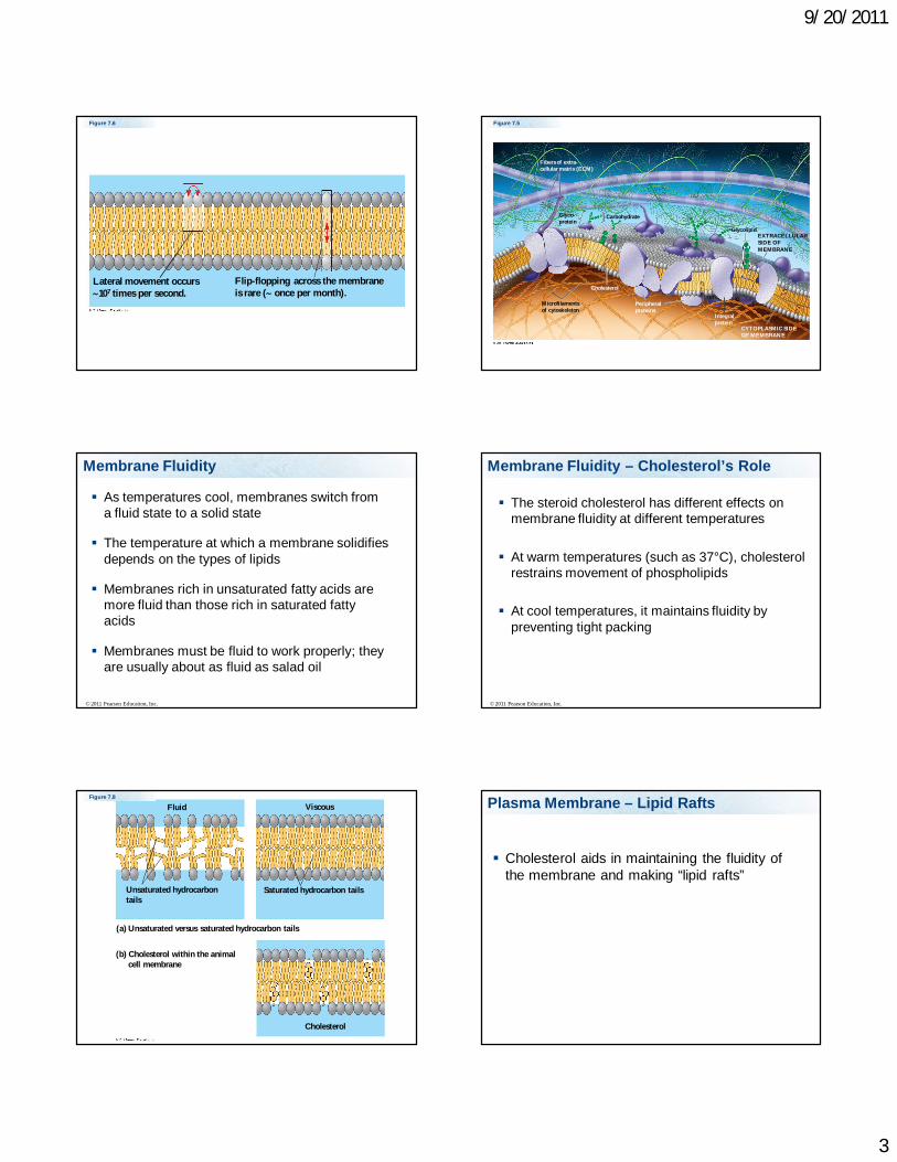
Figure 7.6
Lateral movement occurs107 times per second.
Flip-flopping across the membraneis rare ( once per month).
Figure 7.5
Glyco-protein
Carbohydrate
Glycolipid
Microfilamentsof cytoskeleton
EXTRACELLULARSIDE OFMEMBRANE
CYTOPLASMIC SIDEOF MEMBRANE
Integralprotein
Peripheralproteins
Cholesterol
Fibers of extra-cellular matrix (ECM)
Membrane Fluidity
As temperatures cool, membranes switch from a fluid state to a solid state
The temperature at which a membrane solidifies depends on the types of lipids
Membranes rich in unsaturated fatty acids are more fluid than those rich in saturated fatty acids
Membranes must be fluid to work properly; they are usually about as fluid as salad oil
© 2011 Pearson Education, Inc.
The steroid cholesterol has different effects on membrane fluidity at different temperatures
At warm temperatures (such as 37°C), cholesterol restrains movement of phospholipids
At cool temperatures, it maintains fluidity by preventing tight packing
© 2011 Pearson Education, Inc.
Membrane Fluidity – Cholesterol’s Role
Figure 7.8
Fluid
Unsaturated hydrocarbontails
Viscous
Saturated hydrocarbon tails
(a) Unsaturated versus saturated hydrocarbon tails
(b) Cholesterol within the animalcell membrane
Cholesterol
Plasma Membrane – Lipid Rafts
Cholesterol aids in maintaining the fluidity of the membrane and making “lipid rafts”

9/20/2011
4
Figure 7.5
Glyco-protein
Carbohydrate
Glycolipid
Microfilamentsof cytoskeleton
EXTRACELLULARSIDE OFMEMBRANE
CYTOPLASMIC SIDEOF MEMBRANE
Integralprotein
Peripheralproteins
Cholesterol
Fibers of extra-cellular matrix (ECM)
Five main components in membrane
1. Phospholipid bilayer – controls what passes through membrane
2. Cholesterol – maintains proper fluidity of membrane
3. Proteins – transport, support, communication, recognition, enzymatic, junctions
4. Glycoproteins - form binding sites and function in cell recognition
5. Glycolipids - form binding sites and function in cell recognition
Phospholipids
Amphipathic - have both hydrophilic and hydrophobic regions
Phosphate head (hydrophilic) Fatty acid tail (hydrophobic)
Glycoproteins and Glycolipids
Short chains of carbohydrates (usually less than 15 sugars) attached to proteins or lipids
Glycoproteins Glycolipids
Cell to Cell Interactions
Indentity Markers:
Glycolipids – Identify tissue types
Glycoproteins – Identify cells as “our cells”. MHC proteins – major histocompatibility complex proteins

9/20/2011
5
Six major functions of membrane proteins Transport Enzymatic activity Signal transduction Cell-cell recognition Intercellular joining Attachment to the cytoskeleton and extracellular
matrix (ECM)
© 2011 Pearson Education, Inc.
Membrane Proteins - Functions Figure 7.10
Enzymes
Signaling molecule
Receptor
Signal transduction
Glyco-protein
ATP
(a) Transport (b) Enzymatic activity (c) Signal transduction
(d) Cell-cell recognition (e) Intercellular joining (f) Attachment tothe cytoskeletonand extracellularmatrix (ECM)
Figure 7.10a
Enzymes
Signaling molecule
Receptor
Signal transductionATP
(a) Transport (b) Enzymatic activity (c) Signal transduction
Figure 7.10b
Glyco-protein
(d) Cell-cell recognition (e) Intercellular joining (f) Attachment tothe cytoskeletonand extracellularmatrix (ECM)
Synthesis and Sidedness of Membranes
Membranes have distinct inside and outside faces
The asymmetrical distribution of proteins, lipids, and associated carbohydrates in the plasma membrane is determined when the membrane is built by the ER and Golgi apparatus
© 2011 Pearson Education, Inc.
Membrane Proteins - Production
Proteins that will be associated with the inner surface of the membrane are manufactured by free floating ribosomes
Proteins that will be associated with the outer surface of the membrane are manufactured by ribosomes on the rough endoplasmic reticulum.

9/20/2011
6
The proteins destined to be on the outer surface of the plasma membrane have a sugar chain is added to these proteins transmembrane integral outer surface peripherial proteins
These proteins are glycoproteins.
The sugar chain is added in the lumen of the RER
Figure 7.12
Transmembraneglycoproteins
ERER lumen
Glycolipid
Plasma membrane:Cytoplasmic faceExtracellular face
Secretoryprotein
Golgiapparatus
Vesicle
Transmembraneglycoprotein Secreted
protein
Membraneglycolipid
Membrane Proteins
Peripheral membrane proteins Not embedded in the cell membrane, either
found inner or the outer surface of the membrane
Integral membrane proteins Embedded in the cell membrane. Some completely cross the membrane =
transmembrane proteins
34
Membrane Proteins - Peripheral
Peripheral membrane proteins
anchored to a phospholipid in one layer of the membrane
possess nonpolar regions that are inserted in the lipid bilayer
are free to move throughout one layer of the bilayer
35 36
Membrane Proteins - Intergral
Integral membrane proteins
span the lipid bilayer (transmembrane proteins)
nonpolar regions of the protein are embedded in the interior of the bilayer
polar regions of the protein protrude from both sides of the bilayer

9/20/2011
7
Integral Proteins
Integral proteins penetrate the hydrophobic core
Integral proteins that span the membrane are called transmembrane proteins
The hydrophobic regions of an integral protein consist of one or more stretches of nonpolaramino acids, often coiled into alpha helices
© 2011 Pearson Education, Inc.
Figure 7.9
N-terminus
helix
C-terminus
EXTRACELLULARSIDE
CYTOPLASMICSIDE
39 40
Membrane Proteins - integral
Extensive polar regions within a transmembrane protein can create a pore through the membrane.
–pleated sheets in the protein secondary structure form a cylinder called a -barrel
-barrel interior is polar and allows water and small polar molecules to pass through the membrane
41
Types of membrane proteins
1. Spectrins – give shape to the cell, form a scaffold.
2. Clatherins – line coated pits to facilitate receptor mediated endocytosis
3. Adhesion proteins – adhere cells to each other or to surface Integrins and cadherins – proteins that anchor
the cell to a substrate or cell to cell, they also attach to microfilaments inside the cell, also can play a role in signal transduction

9/20/2011
8
Types of membrane proteins
4. Cell Surface Receptors – transmit messages from outside the cell to inside the cell = signal transduction
5. Cell Surface identity markers – proteins and glycoproteins characteristic for that cell type
6. Junction proteins – form channels between cells (Gap junctions)
7. Transport proteins – Carrier and transport proteins. Transport of items across the cell membrane Aquaporins – Transport water, important in
kidneys and in certain bacteria Ion channels
8. Enzymes – catalyze reactions
45
Proteins that anchor the cell to a substrate, they also attach to microfilaments inside the cell
1. Spectrins2. Receptors3. Integrins4. Enzymes
Junction
Rec
eptor
s
Integrin
s
33% 33%33%
Passage Through the Cell Membrane
The cell membrane is selectively permeable or semi-permeable.
This means some molecules can pass but not all molecules.
The Permeability of the Lipid Bilayer
Hydrophobic (nonpolar) molecules, such as hydrocarbons, can dissolve in the lipid bilayer and pass through the membrane rapidly
Polar molecules, such as sugars, do not cross the membrane easily
© 2011 Pearson Education, Inc.
9/20/2011
9
What can freely pass through the membrane
1. Hydrophobic compounds (non-polar)2. Gases – oxygen, carbon dioxide3. Very small uncharged polar compounds
(Water, glycerol) – but they will pass slowly
What can not freely pass through the membrane
1. Ions2. Most hydrophillic compounds 3. Charged compounds
Transport Proteins
Transport proteins allow passage of hydrophilic substances across the membrane
Some transport proteins, called channel proteins, have a hydrophilic channel that certain molecules or ions can use as a tunnel
Channel proteins called aquaporins facilitate the passage of water
© 2011 Pearson Education, Inc.
Other transport proteins, called carrier proteins, bind to molecules and change shape to shuttle them across the membrane
A transport protein is specific for the substance it moves
© 2011 Pearson Education, Inc.
Passive transport is diffusion of a substance across a membrane with no energy investment
Diffusion is the tendency for molecules to spread out evenly into the available space
Although each molecule moves randomly, diffusion of a population of molecules may be directional
At dynamic equilibrium, as many molecules cross the membrane in one direction as in the other
© 2011 Pearson Education, Inc.

9/20/2011
10
© 2011 Pearson Education, Inc.
Animation: Membrane SelectivityRight-click slide / select “Play”
© 2011 Pearson Education, Inc.
Animation: DiffusionRight-click slide / select “Play”
Figure 7.13a
Molecules of dyeMembrane (cross section)
WATER
(a) Diffusion of one solute
Net diffusion Net diffusion Equilibrium
Figure 7.13b
(b) Diffusion of two solutes
Net diffusion Net diffusion
Net diffusion Net diffusion
Equilibrium
Equilibrium
Movement through the membrane
If you add molecules to water they will disperse until it is equally distributed in the water = diffusion
Equilibrium is reached when the molecules are equally distributed
Substances diffuse down their concentration gradient, the region along which the density of a chemical substance increases or decreases
No work must be done to move substances down the concentration gradient
The diffusion of a substance across a biological membrane is passive transport because no energy is expended by the cell to make it happen
© 2011 Pearson Education, Inc.

9/20/2011
11
Movement through the membrane
Concentration gradient
Molecules will go from higher concentration to lower concentration.
62
Movement through the membraneOsmosis
The cell membrane is semipermeable
The cytoplasm is mainly water (solvent) and molecules and ions are dissolved in it (solutes)
Water will travel across the membrane to try to restore the balance of solutes = Osmosis
Effects of Osmosis on Water Balance
Osmosis is the diffusion of water across a selectively permeable membrane
Water diffuses across a membrane from the region of lower solute concentration to the region of higher solute concentration until the solute concentration is equal on both sides
© 2011 Pearson Education, Inc.
Figure 7.14 Lowerconcentrationof solute (sugar)
Higher concentrationof solute
Sugarmolecule
H2O
Same concentrationof solute
Selectivelypermeablemembrane
Osmosis
Water Balance of Cells Without Walls
Tonicity is the ability of a surrounding solution to cause a cell to gain or lose water
Isotonic solution: Solute concentration is the same as that inside the cell; no net water movement across the plasma membrane
Hypertonic solution: Solute concentration is greater than that inside the cell; cell loses water
Hypotonic solution: Solute concentration is less than that inside the cell; cell gains water
© 2011 Pearson Education, Inc.

9/20/2011
12
Figure 7.15
Hypotonicsolution
Osmosis
Isotonicsolution
Hypertonicsolution
(a) Animal cell
(b) Plant cell
H2O H2O H2O H2O
H2O H2O H2O H2OCell wall
Lysed Normal Shriveled
Turgid (normal) Flaccid Plasmolyzed
Hypertonic or hypotonic environments create osmotic problems for organisms
Osmoregulation, the control of solute concentrations and water balance, is a necessary adaptation for life in such environments
The protist Paramecium, which is hypertonic to its pond water environment, has a contractile vacuole that acts as a pump
© 2011 Pearson Education, Inc.
Figure 7.16
Contractile vacuole50 m
Water Balance of Cells with Walls
Cell walls help maintain water balance
A plant cell in a hypotonic solution swells until the wall opposes uptake; the cell is now turgid (firm)
If a plant cell and its surroundings are isotonic, there is no net movement of water into the cell; the cell becomes flaccid (limp), and the plant may wilt
© 2011 Pearson Education, Inc.
In a hypertonic environment, plant cells lose water; eventually, the membrane pulls away from the wall, a usually lethal effect called plasmolysis
© 2011 Pearson Education, Inc. © 2011 Pearson Education, Inc.
Video: Plasmolysis

9/20/2011
13
73
If you put a cell in very salty water, would water enter or leave the cell?
1. Enter2. Leave
Enter
Leave
50%50%
Can Calcium (Ca2+) pass freely through the membrane?
1. Yes2. No
Yes No
50%50%
Can glucose pass freely through the membrane?
1. Yes2. No
Yes No
50%50%
Transporting molecules across the membrane
Passive transport – molecules travel across using a concentration gradient
1. Simple diffusion2. Facilitated diffusion
Active Transport – requires energy, usually in the form of ATP, to move molecules against a concentration gradient
Passive Transport: 1. Simple diffusion
Simple diffusion: molecules that can freely passthrough the membrane are controlled by concentration gradient
They will travel from a high concentration to a low concentration
Gases like oxygen and CO2
Very small molecules that are not charged (H2O, glycerol) hydrophobic (non-polar) molecules (steroids)

9/20/2011
14
Passive Transport: Facilitated diffusion Passive Transport: 2. Facilitated diffusion
Facilitated diffusion: Aided by a transport protein, still controlled by concentration gradient, does not require energy
Molecules use this type of transport if they can’t pass freely through the membrane, but travels from high concentration to low concentration
Hydrophilic molecules like glucose and polar amino acids Ions or charged compounds
Passive Transport: Facilitated diffusion Figure 7.17EXTRACELLULARFLUID
CYTOPLASM
Channel protein Solute
SoluteCarrier protein
(a) A channelprotein
(b) A carrier protein
Passive Transport Active Transport
Sometimes our cells want to move a molecule across the membrane but against the concentration gradient. The cell wants more of the solute on one side of the membrane.
The cell will use energy to maintain the higher concentration. The energy is usually in the form of ATP
Example: Used to transport ions

9/20/2011
15
Types of Active Transport Proteins
1. Uniporters – transport a single type of molecule
2. Symporter – transport two types of molecules in the same direction
3. Antiporter – transport two molecules in the opposite direction
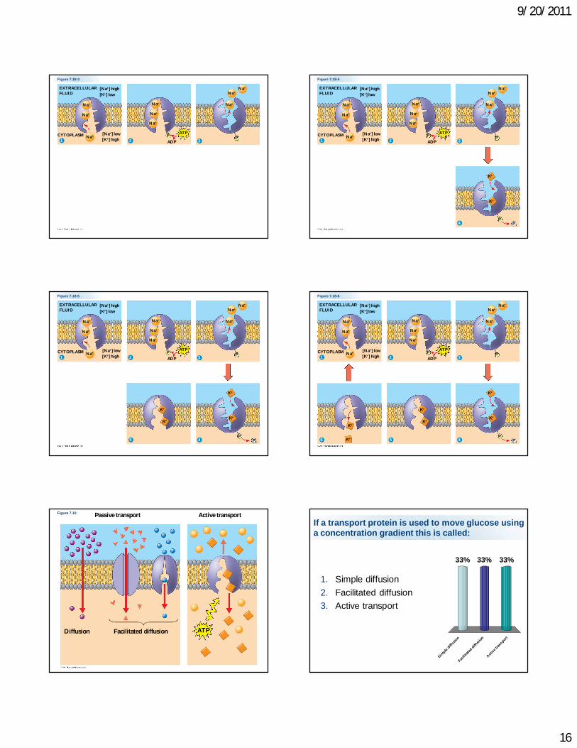
Active Transport - Example
Sodium-potassium pump Energy is used to transport sodium ions to the
outside of a cell in exchange for potassium ion which enter the cell. Transport proteins are used Two potassium ions enter in exchange for three
sodium ions that leave the cell Used in nerve cells = neurons
How Ion Pumps Maintain Membrane Potential
Membrane potential is the voltage difference across a membrane
Voltage is created by differences in the distribution of positive and negative ions across a membrane
© 2011 Pearson Education, Inc. © 2011 Pearson Education, Inc.
Animation: Active TransportRight-click slide / select “Play”
Figure 7.18-1
EXTRACELLULARFLUID
[Na] high[K] low
[Na] low[K] high
CYTOPLASM
Na
Na
Na1
Figure 7.18-2
EXTRACELLULARFLUID
[Na] high[K] low
[Na] low[K] high
CYTOPLASM
Na
Na
Na1 2
Na
Na
Na
P ATP
ADP
9/20/2011
16
Figure 7.18-3
EXTRACELLULARFLUID
[Na] high[K] low
[Na] low[K] high
CYTOPLASM
Na
Na
Na1 2 3
Na
Na
Na
NaNa
Na
P PATP
ADP
Figure 7.18-4
EXTRACELLULARFLUID
[Na] high[K] low
[Na] low[K] high
CYTOPLASM
Na
Na
Na1 2 3
4
Na
Na
Na
NaNa
Na
K
K
P P
PP i
ATP
ADP
Figure 7.18-5
EXTRACELLULARFLUID
[Na] high[K] low
[Na] low[K] high
CYTOPLASM
Na
Na
Na1 2 3
45
Na
Na
Na
NaNa
Na
K
K
K
K
P P
PP i
ATP
ADP
Figure 7.18-6
EXTRACELLULARFLUID
[Na] high[K] low
[Na] low[K] high
CYTOPLASM
Na
Na
Na1 2 3
456
Na
Na
Na
NaNa
Na
K
K
K
K
K
K
P P
PP i
ATP
ADP
Figure 7.19 Passive transport Active transport
Diffusion Facilitated diffusion ATP
If a transport protein is used to move glucose using a concentration gradient this is called:
1. Simple diffusion2. Facilitated diffusion3. Active transport
Simple
diffusio
n Fac
ilitate
d diffu
sion A
ctive t
ransport
33% 33%33%

9/20/2011
17
Bulk Transport
When the cell needs to transport larger things they can use vesicles to transport things in and out of the cell.
Exocytosis: moving things out of the cell Endocytosis: moving things into the cell
Used to transport macromolecules including: whole cells (bacteria), cholesterol, and proteins
© 2011 Pearson Education, Inc.
Animation: ExocytosisRight-click slide / select “Play”
Endocytosis
In endocytosis, the cell takes in macromolecules by forming vesicles from the plasma membrane
Endocytosis is a reversal of exocytosis, involving different proteins
There are three types of endocytosis Phagocytosis (“cellular eating”) Pinocytosis (“cellular drinking”) Receptor-mediated endocytosis
© 2011 Pearson Education, Inc.
Endocytosis
Phagocytosis – when cells transport large particles and cells (bacteria) into the cell using vesicles
Pinocytosis – when cells transport fluid into the cell using vesicles
Receptor-mediated endocytosis – molecules attach to a receptor, and are then transported into the cell. The molecules that specifically bind to a receptor are called ligands
© 2011 Pearson Education, Inc.
Animation: Exocytosis and Endocytosis IntroductionRight-click slide / select “Play”
In phagocytosis a cell engulfs a particle in a vacuole
The vacuole fuses with a lysosome to digest the particle
© 2011 Pearson Education, Inc.
9/20/2011
18
© 2011 Pearson Education, Inc.
Animation: PhagocytosisRight-click slide / select “Play”
In pinocytosis, molecules are taken up when extracellular fluid is “gulped” into tiny vesicles
© 2011 Pearson Education, Inc.
© 2011 Pearson Education, Inc.
Animation: PinocytosisRight-click slide / select “Play”
receptor-mediated endocytosis
In receptor-mediated endocytosis, binding of ligands to receptors triggers vesicle formation
A ligand is any molecule that binds specifically to a receptor site of another molecule
© 2011 Pearson Education, Inc.
© 2011 Pearson Education, Inc.
Animation: Receptor-Mediated EndocytosisRight-click slide / select “Play”
Figure 7.22
Solutes
Pseudopodium
“Food” orother particle
Foodvacuole
CYTOPLASM
Plasmamembrane
Vesicle
Receptor
Ligand
Coat proteins
Coatedpit
Coatedvesicle
EXTRACELLULARFLUID
Phagocytosis Pinocytosis Receptor-Mediated Endocytosis

9/20/2011
19
Figure 7.22a
Pseudopodium
Solutes
“Food”or otherparticle
Foodvacuole
CYTOPLASM
EXTRACELLULARFLUID
Pseudopodiumof amoeba
BacteriumFood vacuole
An amoeba engulfing a bacteriumvia phagocytosis (TEM).
Phagocytosis
1 m
Figure 7.22b
Pinocytosis vesicles formingin a cell lining a small bloodvessel (TEM).
Plasma membrane
Vesicle
0.5
m
Pinocytosis
Figure 7.22c
Top: A coated pit. Bottom: Acoated vesicle forming duringreceptor-mediated endocytosis(TEMs).
Receptor
0.25
m
Receptor-Mediated Endocytosis
Ligand
Coat proteins
Coatedpit
Coatedvesicle
Coatproteins
Plasma membrane
112
Receptor-mediated endocytosis
HIV (Human Immunodeficiency Virus) entering a cell. HIV infects a number of cells in the body, though the main target is the lymphocyte cell, which is a type of T-cell. Once the HIV virus has infected the host cell it then replicates itself with thousands of copies. Image 1 of 3 in series. TEM X120,000.
Credit: © Dr. Hans Gelderblom/Visuals Unlimited
229674 HIV (Human Immunodeficiency Virus) being surrounded by a cell. HIV infects a number of cells in the body, though the main target is the lymphocyte cell, which is a type of T-cell. Once the HIV virus has infected the host cell it then replicates itself with thousands of copies. Image 2 of 3 in series. TEM X120,000.
Credit: © Dr. Hans Gelderblom/Visuals Unlimited
229675

9/20/2011
20
HIV (Human Immunodeficiency Virus) in a cell. HIV infects a number of cells in the body, though the main target is the lymphocyte cell, which is a type of T-cell. Once the HIV virus has infected the host cell it then replicates itself with thousands of copies. Image 3 of 3 in series. TEM X120,000.
Credit: © Dr. Hans Gelderblom/Visuals Unlimited
229676
when cells transport fluid into the cell using vesicles this is called:
1. Phagocytosis2. Pinocytosis3. Exocytosis
Phagoc
ytosis
Pinocy
tosis
Exocy
tosis
33% 33%33%
Important Concepts
Reading Chapter 6 Know the vocabulary from this lecture
What are the functions of the plasma membrane
Identify the main components in membrane and their functions. Be able to draw a membrane.
Be able to identify what can pass freely through a membrane and what can’t pass freely
What happens to cells in isotonic, hypertonic, and hypertonic situations
Important Concepts
Be able to describe how things are transported across membranes – know all the mechanisms
What are the types of membrane proteins, what are the secondary structures of intergral membrane proteins
What are the different cell to cell junctions, their functions and locations where there are found in high concentration
Important Concepts
Understand how things are transported across membranes – know the different mechanisms
Know all the types of membrane proteins and their functions
Know the different cell to cell junctions, their functions and locations where there are found in high concentration