Embed Size (px)
Citation preview
MOH NURSING CLINICAL PRACTICE GUIDELINES 1/2010
Nursing Management of Nasogastric Tube Feeding
in Adult Patients
July 2010
i
STATEMENT OF INTENT
This set of guidelines serves as a guide for caregivers of adults with nasogastric tube feeding. The recommendations are based on the available research findings and existing evidence-based guidelines. However there are some aspects in which there are insufficient published researches and therefore, consensus of expert in the field has been utilized to provide guidelines specific to conventional practice. As practitioners, each individual must exercise clinical judgement in the nursing management of patients with nasogastric tube feeding. Therefore, these guidelines should be implemented according to individual patient’s condition, overall treatment goal, resource availability, institutional policies and treatment options available.
Copyright © 2010 by Ministry of Health, Singapore.
ii
FOREWORD
“Every careful observer of the sick will agree in this that thousands of patients are annually starved in the midst of plenty, from want of attention to the ways which alone make it possible for them to take food.”
Florence Nightingale, 1859
Nurses have recognised the importance of meeting patients’ nutritional needs since the beginning of our profession. Enteral nutrition is the preferred feeding method for those patients with normal functioning gastrointestinal tracts but with inability to ingest adequate nutrients solely by mouth. Significant advances and changes have since evolved in nutritional care of hospitalised patients. Enteral nutrition as compared to parenteral feeding, is considered to be safer and less expensive. Nurses have always assumed the role of providing nutritional therapy to patients. They would find the publication of this clinical practice guidelines timely and useful to guide them in enteral nutrition therapy. Adhering closely to these guidelines would significantly reduce and, in many instances, prevent complications commonly associated with enteral feeding.
PAULINE TAN CJ
CHIEF NURSING OFFICER
iii
CONTENTS
1 INTRODUCTION 1
1.1 Background 1
1.2 Definition of Nasogastric Tube Feeding 1
1.3 Highlights of Patient Management 2
1.4 Scope of the Guidelines 2
2 DEVELOPMENT OF GUIDELINES 3
2.1 Training and Guidelines 3
2.2 Strategy and Literature Review 3
2.3 Evaluation of Evidence and Grading of Recommendations 3
2.3.1 Individual Study Validity Rating 4
2.3.2 Levels of Evidence 5
2.3.3 Grade of Recommendation 6
2.3.4 Interpretation of the D/4 Grading 6
2.4 Guideline Review and Revision 7
2.5 Limitations 7
3 ALGORITHM FOR MANAGEMENT OF NASOGASTRIC TUBE FEEDING 8
4 SELECTION OF NASOGASTRIC TUBES 9
4.1 Feeding Tube Selection 9
5 NASOGASTRIC TUBES PLACEMENT AND CARE 10
5.1 Tube Insertion and Stabilization 10
5.2 pH Testing 10
iv
5.3 Radiological Determination of Feeding Tube Placement 12
5.4 Auscultatory Method 13
5.5 Frequency in Checking Placement 14
5.6 Tube Clogging 14
5.6.1 Maintaining Tube Patency 14
6 ADMINISTRATION OF NASOGASTRIC TUBE FEEDING 16
6.1 Preparation of Formula Feeds and Delivery System 16
6.2 Feeding Position 17
6.3 Bolus Feeding 17
6.4 Continuous Feeding 18
6.5 Prevention of Diarrhoea 19
7 ADMINISTRATION OF MEDICATIONS 21
7.1 Administration of Medications 21
8 MONITORING AND MANAGEMENT 23
8.1 Monitoring and Management of Gastro-intestinal Tolerance 23
9 COMPLICATIONS 25
9.1 Management of Feeding Intolerance 25
9.2 Declogging 25
9.3 Diarrhoea 26
9.3.1 Assessment / Monitoring 26
9.3.2 Treatment of Diarrhoea 26
9.4 Aspiration Pneumonia 27
9.5 Feeding Devices 28
v
10 QUALITY ASSURANCE 29
10.1 Parameters for Evaluation 29
10.1.1 Incidence of misplaced tubes 29
10.1.2 Incidence of aspiration as a result of nasogastric tube feeding 29
10.2 Sentinel Events 29
10.3 Management Role 30
11 IMPLEMENTATION OF GUIDELINES 31
12 REFERENCES 32
13 GLOSSARY 36
14 WORKGROUP MEMBERS 39
APPENDIX 1 NASOGASTRIC TUBE ANCHORING TECHNIQUE 41
APPENDIX 2 ALGORITHM FOR ASSESSING PLACEMENT OF THE NASOGASTRIC TUBE 42
APPENDIX 3 SELF ASSESSMENT 43
1
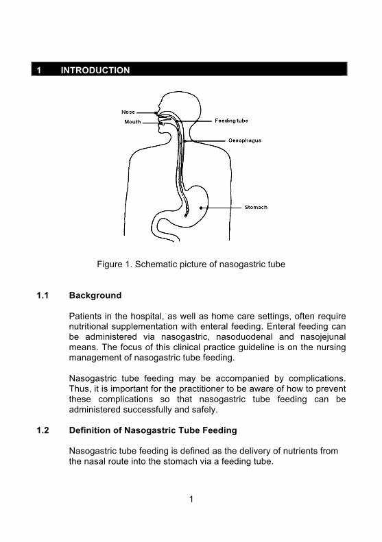
1 INTRODUCTION
Figure 1. Schematic picture of nasogastric tube
1.1 Background
Patients in the hospital, as well as home care settings, often require nutritional supplementation with enteral feeding. Enteral feeding can be administered via nasogastric, nasoduodenal and nasojejunal means. The focus of this clinical practice guideline is on the nursing management of nasogastric tube feeding. Nasogastric tube feeding may be accompanied by complications. Thus, it is important for the practitioner to be aware of how to prevent these complications so that nasogastric tube feeding can be administered successfully and safely.
1.2 Definition of Nasogastric Tube Feeding
Nasogastric tube feeding is defined as the delivery of nutrients from the nasal route into the stomach via a feeding tube.
2
1.3 Highlights of Patient Management
Patients and their caregivers are important team players in the effective management of nasogastric tube feeding. The practitioner should: • encourage patients and their caregivers to be active participants
in their care • develop an effective plan of care that is consistent with the
patient’s goals
The recommendations focus on: • implementation of nasogastric tube feeding • monitoring and management of nasogastric tube feeding • management of complications
1.4 Scope of the Guidelines
This set of guidelines is intended as a simple and readable reference for caregivers of adults with nasogastric tube feeding. These clinical practice guidelines are tools for assisting clinical decision making with regard to the nursing care given to a patient with a nasogastric tube. They are the result of systematic identification and synthesis of published research findings and expert opinions. However, they should be adapted locally to suit a particular situation and patient. It is the intention of the workgroup not to be encyclopaedic in coverage but to produce a concise, readable and practical format that addresses key topics which can be used as a reference and training tool.
3
2 DEVELOPMENT OF GUIDELINES
2.1 Training and Guidelines
Members of the workgroup attended a two-day interactive training workshop to learn about and discuss the theory and practical issues of developing evidence-based guidelines under the guidance of Dr Edwin Chan and Dr Miny Samuel of the then Clinical Trials and Epidemiology Research Unit. The practical training revolved around topic selection and the development of “mock” evidence-based guidelines which developed into this present set of guidelines.
2.2 Strategy and Literature Review
Three highly regarded evidence-based guidelines were reviewed: • The A.S.P.E.N. Nutrition Support Practice Manual (2
nd Edition),
The American Society for Parenteral and Enteral Nutrition. 2005 [ASPEN, 2005]
• Insertion and management of Nasogastric Tubes for Adults. The Joanna Briggs Institute: Systematic Reviews, 2006 [JBI, 2006]
• A.S.P.E.N. Enteral Nutrition Practice Recommendations. 2009 [ASPEN, 2009]
The members felt that an updated literature search on the specific topics addressed would suffice. The electronic databases (MEDLINE, EMBASE, Cochrane Library, SPRINGNET and CINAHL) and hard copies of relevant journals (Journal of Parenteral and Enteral Nutrition, Hospital Pharmacy, Critical Care Nurse, American Journal of Critical Care, Medical Surgical Nursing, Journal of Clinical Nursing, Consultant Pharmacist, Gastroenterology Nursing, Nursing Research, Heart and Lung) were searched.
2.3 Evaluation of Evidence and Grading of Recommendations
The revised Scottish Intercollegiate Guidelines Network (SIGN) system was adopted as a guide to evaluate the design of individual studies and assign a grade to each study’s level of evidence. The recommendation was given a grade after taking into account the external validity, result consistency, local constraints and expert
4
opinion. The extensive reliance on the ASPEN and JBI guidelines are acknowledged and treated as our main published expert opinion. For areas where available evidence is inconsistent or inconclusive, recommendations were made based on the clinical experience and judgement of the workgroup or expert committee reports.
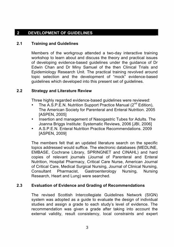
2.3.1 Individual Study Validity Rating
All primary studies and reviews addressing a particular topic were appraised using a SIGN checklist appropriate to the study's design. These were individually rated for internal validity using the system below:
Rating Description
++ All or most of the criteria have been fulfilled. Where they have not been fulfilled the conclusions of the study or review are thought very unlikely to alter.
+ Some of the criteria have been fulfilled. Those criteria that have not been fulfilled or not adequately described are thought unlikely to alter the conclusions.
–
Few or no criteria fulfilled. The conclusions of the study are thought likely or very likely to alter.
5
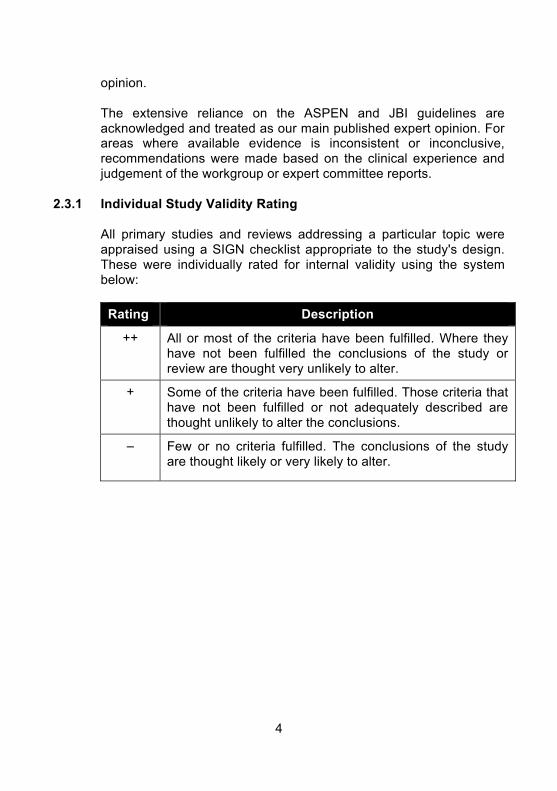
2.3.2 Levels of Evidence
Each study is assigned a level of evidence by combining the design designation and its validity rating using the system below:
Level Type of Evidence
1++ High quality meta-analyses, systematic reviews of RCTs, or RCTs with a very low risk of bias.
1+ Well-conducted meta-analyses, systematic reviews, or RCTs with a low risk of bias.
1- Meta-analyses, systematic reviews, or RCTs with a high risk of bias.
2++ High quality systematic reviews of case-control or cohort or studies.
High quality case-control or cohort studies with a very low risk of confounding or bias and a high probability that the relationship is causal.
2+ Well-conducted case-control or cohort studies with a low risk of confounding or bias and a moderate probability that the relationship is causal.
2-
Case-control or cohort studies with a high risk of confounding or bias and a significant risk that the relationship is not causal.
3 Non-analytic studies e.g. case reports, case series.
4 Expert opinion.
6
2.3.3 Grade of Recommendation The detailed results of each study and mitigating local circumstances were considered in formulation of each recommendation which was then graded using the system below:
Grade Recommendation
A At least one meta-analysis, systematic review, or RCT rated as 1
++,, and directly applicable to the target
population; or A body of evidence, consisting principally of studies rated as 1
+, directly applicable to the target population,
and demonstrating overall consistency of results.
B A body of evidence, including studies rated as 2++
, directly applicable to the target population, and demonstrating overall consistency of results; or Extrapolated evidence from studies rated as 1
++ or 1
+.
C A body of evidence including studies rated as 2+,
directly applicable to the target population and demonstrating overall consistency or results; or Extrapolated evidence from studies rated as 2
++.
D Evidence level 3 or 4 ; or Extrapolated evidence from studies rated as 2
+.
2.3.4 Interpretation of the D/4 Grading
The grading system emphasises the quality of the experimental support underpinning each recommendation. The grading D/4 was assigned in cases where • it would be unreasonable to conduct a RCT because the correct
practice is logically obvious;
• recommendations derived from existing high quality evidence-based guidelines. We alert the user to this special status by appending the initials of their source e.g. D/4 - ASPEN, 2005.
7
2.4 Guideline Review and Revision Drafts of the guidelines were circulated to healthcare institutions for peer review on validity, reliability and practicality of the recommendations. These guidelines will be reviewed and revised periodically to incorporate the latest relevant evidence and expert clinical opinion.
2.5 Limitations
These guidelines offer recommendations that are based on current scientific evidence and professional judgement. They are not intended as the legal standard of care. Users of these guidelines should determine the appropriate and safe patient care practices, based on assessment of the circumstances of the particular patient, their own clinical experiences and the knowledge of the most recent research findings.
8
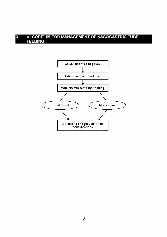
3 ALGORITHM FOR MANAGEMENT OF NASOGASTRIC TUBE FEEDING

9
4 SELECTION OF NASOGASTRIC TUBES
4.1 Feeding Tube Selection
Select the feeding tubes based on the tube’s composition, intended use, estimated length of time required, cost-effectiveness and tube features. • Soft, flexible, small diameter tube (8 Fr to 12 Fr) is recommended
for nasogastric feeding. (D/4 - ASPEN, 2005 & JBI, 2006)
• Use Polyurethane or silicone tubes for anticipated long term
feeding rather than polyvinylchloride tubes. (D/4 - ASPEN, 2005)
• Polyvinylchloride (PVC) tubes should be used for a short period
of time usually for gastric drainage, decompression, lavage or diagnostic procedures.
(D/4 - ASPEN, 2005)
Rationale: Smaller size feeding tube improves patient comfort. Common complications associated with the use of larger and stiffer tubes include nasopharyngeal erosions / necrosis, sinusitis and otitis media.
(Kirby and Opilla, 2005 as cited in ASPEN, 2005) Polyurethane or silicone tubes are better for long-term (> 4-8 weeks) use because they are more flexible and less irritating to tissues.
(Kirby and Opilla, 2005 as cited in ASPEN, 2005) PVC feeding tubes are used for short term duration (< 3 weeks) and they tend to harden and become brittle with time and may cause tissue irritation or necrosis.
(Kirby and Opilla, 2005 as cited in ASPEN, 2005)
For short-term usage, PVC feeding tubes have adequate efficacy and are more cost effective.
(Kirby and Opilla, 2005 as cited in ASPEN, 2005)
10
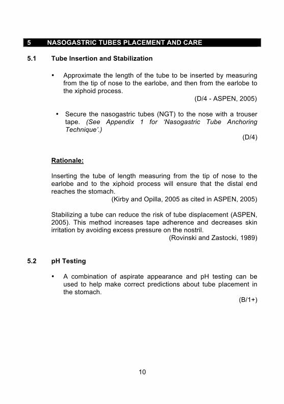
5 NASOGASTRIC TUBES PLACEMENT AND CARE
5.1 Tube Insertion and Stabilization
• Approximate the length of the tube to be inserted by measuring
from the tip of nose to the earlobe, and then from the earlobe to the xiphoid process.
(D/4 - ASPEN, 2005)
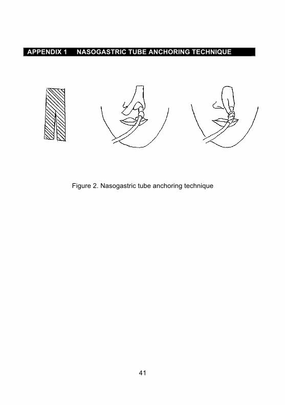
• Secure the nasogastric tubes (NGT) to the nose with a trouser tape. (See Appendix 1 for ‘Nasogastric Tube Anchoring Technique’.)
(D/4)
Rationale: Inserting the tube of length measuring from the tip of nose to the earlobe and to the xiphoid process will ensure that the distal end reaches the stomach.
(Kirby and Opilla, 2005 as cited in ASPEN, 2005) Stabilizing a tube can reduce the risk of tube displacement (ASPEN, 2005). This method increases tape adherence and decreases skin irritation by avoiding excess pressure on the nostril.
(Rovinski and Zastocki, 1989)
5.2 pH Testing
• A combination of aspirate appearance and pH testing can be
used to help make correct predictions about tube placement in the stomach.
(B/1+)
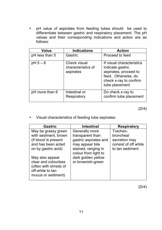
11
• pH value of aspirates from feeding tubes should be used to differentiate between gastric and respiratory placement. The pH values and their corresponding indications and action are as follows:
Value Indications Action
pH less than 5 Gastric Proceed to feed
pH 5 – 6 Check visual characteristics of aspirates
If visual characteristics indicate gastric aspirates, proceed to feed. Otherwise, do check x-ray to confirm tube placement
pH more than 6 Intestinal or Respiratory
Do check x-ray to confirm tube placement
(D/4)
• Visual characteristics of feeding tube aspirates:
Gastric Intestinal Respiratory
May be grassy green with sediment, brown (if blood is present and has been acted on by gastric acid) May also appear clear and colourless (often with shreds of off-white to tan mucus or sediment)
Generally more transparent than gastric aspirates and may appear bile stained, ranging in colour from light to dark golden yellow or brownish-green
Tracheo-broncheal secretion may consist of off white to tan sediment
(D/4)
12
Rationale:
Combination of pH and visual characteristics can be helpful in distinguishing between respiratory and gastrointestinal tube position.
(Metheny, Reed, Berglund and Wehrle 1994; Metheny and Titler, 2001; Metheny and Stewart, 2002)
The pH value can offer correct predication of gastric versus respiratory placement.
(Metheny, Wehrle and Wiersema, 1998)
Gastric pH values are lower than that of intestinal or respiratory pH. (Metheny and Titler, 2001; Metheny and Stewart, 2002)
pH less than 5 indicates gastric placement, whereas a pH more than 5 indicates intestinal or respiratory placement.
(Metheny, 1993; Metheny and Stewart, 2002)
Patient receiving H2 receptor antagonist or with recent alkaline reflux from the intestine may have elevated gastric pH. Proton Pump Inhibitors and Histamine receptor blocking agents, such as Omeprazole and Famotidine, fail to decrease gastric pH to below 6.5 but tends to elevate gastric pH.
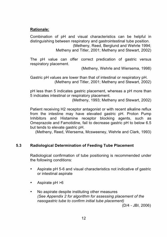
(Metheny, Reed, Wiersema, Mcsweeney, Wehrle and Clark, 1993) 5.3 Radiological Determination of Feeding Tube Placement
Radiological confirmation of tube positioning is recommended under the following conditions: • Aspirate pH 5-6 and visual characteristics not indicative of gastric
or intestinal aspirate
• Aspirate pH >6
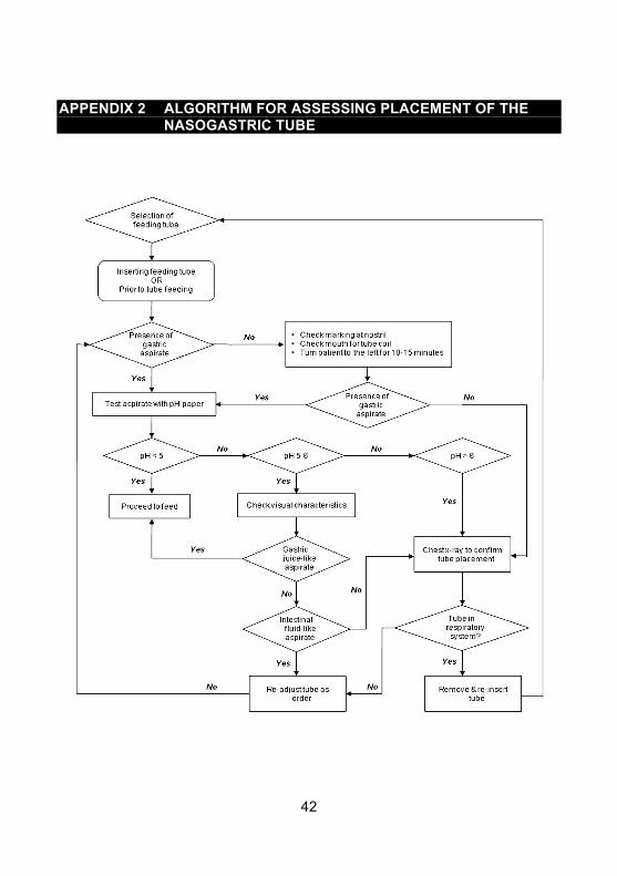
• No aspirate despite instituting other measures (See Appendix 2 for algorithm for assessing placement of the nasogastric tube to confirm initial tube placement)
(D/4 - JBI, 2006)
13
Rationale: Radiography determination of tube location is the most accurate method of checking tube placement.
(Grifffiths et al, 2006 as cited in JBI, 2006)
X-ray can visualise the entire course of the tube. (Metheny and Titler, 2001)
5.4 Auscultatory Method
• Auscultatory method (also known as air insufflations test) should not be relied on as the sole method to determine the location of the feeding tube.
(D/4 - ASPEN, 2005 & JBI, 2006)
Rationale: The auscultatory method is not effective in distinguishing between respiratory and gastrointestinal placement of feeding tube. “Pseudoconfirmatory gurgling” sound can be heard when feeding tubes were positioned in the tracheobronchial tree or pleural spaces. A tube that is inadvertently placed in the respiratory tract or oesophagus can transmit a sound similar to that of air entry in the stomach.
(Metheny, 1993; Metheny and Titler, 2001; Griffiths, Thompson, Chau and Fernandez, 2006 as cited in JBI, 2006)
A positive air insufflations test combined with aspiration of gastric contents is a fairly reliable predictor of successful placement.
(Kirby and Opilla, 2005 as cited in ASPEN, 2005; Griffiths et al, 2006 as cited in JBI, 2006)
14
5.5 Frequency in Checking Placement
Mark the intersection where the nasogastric tube enters the nostril, use this marking to check the tube placement:
• after initial insertion, before each intermittent feeding, and at
every 8-hourly during continuous feedings. (D/4)
• if patients complain of discomfort, coughing, retching or
vomiting and show sudden signs of respiratory difficulties. (D/4)
• if the visible part of the tube changes in the length.
(D/4)
Rationale: Regular assessment of feeding tube placement is important because a tube can be partially pulled out during movement or when tugged at by a confused patient. Tube malposition may be caused by faulty initial placement or upward dislocation after bouts of coughing or vomiting.
(Metheny and Stewart, 2002)
5.6 Tube Clogging
5.6.1 Maintaining Tube Patency
• Flush feeding tubes with 30 ml of water before and after
intermittent feeding, every 4-hourly during continuous feeding and after checking for gastric residuals. More frequent flushing might be ordered according to patient’s condition.
(B/1+) • Flush feeding tube before and after administration of each
medicine and after checking for residual. (C/2+)
15
Rationale:
Flushing enteral tubes with water have been found to prevent clogging and restore the patency of partially obstructed tubes. Water was found to be the most effective flushing solution in maintaining tube patency. However, the patient’s medical condition needs to be considered before deciding on the volume of water and frequency for irrigation.
(ASPEN, 2009; Mateo, 1994) Compatibility of enteral formula and medications affect drug concentration and causes clog formation. Irrigating tubes before and after medication administration resulted in fewer clogged tubes.
(Mateo, 1994) Residual, containing gastric aspirate with an average pH of 4.6 or less interacts with protein formula to form clots. Formula residue that adheres to the tube lumen may cause tube clogging. Thus flushing the tube with water will maintain the patency by eliminating acid precipitation of formula in the feeding tube.
(Mateo, 1994)
16
6 ADMINISTRATION OF NASOGASTRIC TUBE FEEDING
6.1 Preparation of Formula Feeds and Delivery System
• Wash hands before preparing and handling delivery sets. (D/4)
• Use mask if handler has a cold, sore throat or upper respiratory
tract infection. (D/4)
• Use clean technique when handling the feeding system. Limit the
hang time of the formula to: • 4 hours for powdered, reconstituted formula and enteral
nutrition formula with additives • 8 hours for sterile, decanted formula in open system • 24–48 hours per manufacturer’s guidelines for closed-system
enteral nutrition formula (D/4 – ASPEN, 2009)
• Use pre-prepared feeds for enteral feeding.
(D/4)
• Initiate all formulas at full strength. (A/1)
Rationale: Handwashing reduces the risk of contamination and cross infection.
(Kennedy, 1997) The use of mask has shown to reduce the risk of cross-infection.
(Kennedy, 1997) Bacterial contamination is most often due to contamination of a product. It has been found that colonization of distal administration set by enteric bacteria present in the enteral tube hub leads to an easily accessible reservoir of gastrointestinal organisms.
(Eisenberg, 2002)
17
Pre-prepared feeds reduce the risk of cross -cross infection.
(Kennedy, 1997) As new formulas and technology develop, it is recognized that the osmolality of formula does not cause diarrhoea and the dilution of the formula to or strength is not necessary.
(Eisenberg, 2002)
6.2 Feeding Position
• The patient should be placed at a semi-recumbent position or elevated to an angle of at least 30
o during and after feeding for at
least one hour. (D/4 - ASPEN, 2005)
Rationale:
Elevation of the head of bed at least 30
0 during tube feeding
decreases the risk of aspiration of gastric content. (Lord and Harrington, 2005 as cited in ASPEN, 2005)
Gravity reduces the likelihood of regurgitation of gastric contents from the distended stomach. There is evidence that a sustained supine position (with the head of the bed flat) increases gastroesophageal reflux (GER) and the probability for aspiration.
(Metheny and Stewart, 2002)
6.3 Bolus Feeding
• The total volume of feeds administered should not exceed 400ml during each bolus feed.
(D/4)
contamination and
18
Rationale:
Both the speed and volume with which formula is delivered has an impact on intragastric pressure and the probability of gastro-oesophageal reflux (GER).
(Metheny, 2002) Bolus feeding of more than 400 ml and rapid infusion may result in abdominal distension and discomfort.
(Marian and Allen, 1998)
6.4 Continuous Feeding
• Use feeding pump for administration of continuous feeds. Start with an initial slow rate of 10 to 40 ml/hour. Advance to the goal rate by increasing the rate by 10 to 20 ml/hour every 8-12 hours as tolerated.
• (D/4 - ASPEN, 2005)
• Continuous feeding is recommended for patients who are critically ill, receiving jejunal feedings; or patients who are unable to tolerate intermittent feedings. Pump-assisted delivery of enteral feeds is highly recommended for small-bowel feeding.
(D/4 - ASPEN, 2005)
Rationale:
Continuous feeding minimizes the amount of formula in the stomach at any given time. Less abdominal discomfort occurred during continuous feedings compared with intermittent feedings.
(Lord and Harrington, 2005 as cited in ASPEN, 2005) There is evidence that aspiration is less likely with continuous feedings because this method minimizes the amount of formula in the stomach at any given time.
(Metheny, 2002)
19
Pump-assisted continuous feeding helps to prevent gastrointestinal complications associated with rapid infusion. The pump usually delivers the feed much more accurately than the gravity method and is likely to reduce gastric intolerance
(Lord and Harrington, 2005 as cited in ASPEN,2005; Metheny, 2002)
6.5 Prevention of Diarrhoea
• Administration sets and containers should be discarded every 24 hours.
(D/4) • Feeds should not be decanted before use.
(D/4) • Limit the hang time of the formula to:
• 4 hours for powdered, reconstituted formula and enteral nutrition formula with additives
• 8 hours for sterile, decanted formula in open system • 24–48 hours per manufacturer’s guidelines for closed-system
enteral nutrition formula (D/4 – ASPEN, 2009)
Rationale:
Continuous infusion raises gastric pH and promotes bacteria overgrowth. Bacteria can spread up the giving set from gastric or enteral sources and thus increase the risk of contamination.
(Stroud, Duncan and Nightingale, 2003) Enteral feed is an ideal culture medium for bacteria growth and once contaminated, bacteria will multiply rapidly.
(Stroud et al, 2003)
20
Limiting the hang-time of formula helps to reduce the potential for bacterial contamination of formula feeds.
(Bankhead, Boullata, Brantley, Corkins, Guenter, Krenitsky, Lyman, Methany, Mueller, Robbins, Wessel, and the
A.S.P.E.N. Board of Directors, 2009 in ASPEN 2009)

21
7 ADMINISTRATION OF MEDICATIONS
7.1 Administration of Medications
• Consult pharmacist to evaluate each patient’s medication profile before medications are given via enteral tube.
(D/4)
• Enteric-coated and sustained release medications should not be crushed.
(D/4 - ASPEN, 2005)
• Medications administered through nasogastric tube should preferably be in liquid form.
(D/3 - ASPEN, 2005)
• Flush feeding tube with water prior to medication administration.
(B/2+)
• Do not administer sublingual and buccal medications via feeding tube.
(D/4)
• Medications should not be added directly to the enteral formula or into the enteral feeding bag.
(D/4)
Rationale: Every dosage form (tablet, capsules, immediate release, extended release, enteric coated, elixirs, syrups, granules) has the potential to cause incompatibilities, complications, or intolerance.
(Lord and Harrington, 2005 as cited in ASPEN, 2005)
22
Pharmaceutical incompatibilities occur when there is an alteration in the drug form which interferes with drug efficacy, potency, or tolerance. Crushing of enteric–coated medications can induce pharmaceutical incompatibilities.
(Guenter, 1999) Medications should be evaluated by the pharmacist to determine if the form of medication needs to be changed. Liquid medications decrease the risk of tube clogging and increase absorption of the medication.
(Pancorbo-Hidalgo, Garcia-Fernandez and Ramirez-Perez, 2001; Guenter, 1999)
Hyperosmolar liquid medications have the potential to cause diarrhoea, cramping, and increased gastric residuals.
(Guenter, 1999) Flushing the feeding tube prior to drug administration removes any enteral feeds that remain in the tube to prevent drug nutrient interaction.
(Maka and Murphy, 2000)
Sublingual and buckle medications are designed to be absorbed into the systemic circulation by placement under the patient’s tongue or in the cheek pouch. Thus, these should not be administered via the feeding tube.
(Lord and Harrington, 2005 as cited in ASPEN, 2005)
Adding medications into formula feeds may result in tube clogging and cause undesirable effects or incompatibilities.
(Lord and Harrington, 2005 as cited in ASPEN, 2005)
23
8 MONITORING AND MANAGEMENT
8.1 Monitoring and Management of Gastro-intestinal Tolerance
• Gastric residual volume (GRV) should be checked 4-8 hourly in
continuously fed patients and before each intermittent feeding. (B/1)
• GRV greater than 200 ml should prompt careful bedside
evaluation and initiation of appropriate feeding method and feeding volume. GRV reading should be evaluated in conjunction with physical examination for abdominal distension, absence of bowel sounds, and presence of nausea and vomiting.
(D/4)
• A trend in GRV may be more important than an isolated high level of GRV.
(D/3)
• There is no significant difference between returning and not returning gastric residuals.
(D/3)
Rationale: Gastric residuals are more likely to be high during the early phase of tube feedings; therefore it is necessary to monitor residuals more closely during that period.
(Metheny, 1993) GRV greater than 200 ml was associated with physical findings of gastrointestinal dysmotility. Bedside evaluation and careful initiation of feeding method may reduce aspiration risk.
(Metheny, Schallom and Edwards, 2004)
24
GRV volume during intra-gastric feedings is determined by the balance between the amount of infused formula feeds plus the endogenous secretions of saliva and gastric juice, less the amount of fluid emptied from the stomach. This amount varies between individuals. Thus, it is important to note for changes in the trend.
(Metheny et al, 2004) Discarding gastric residual may contribute to loss of gastric juices and subsequently may lead to electrolyte imbalance, however returning the gastric residual may cause potential contamination and tube clogging.
(Booker, Niedringhaus, Eden and Arnold, 2000)
25
9 COMPLICATIONS
9.1 Management of Feeding Intolerance
When patient shows signs of feeding intolerance such as nausea, vomiting, abdominal distension and pain:
• Perform a physical examination of the abdomen including
assessment for presence of abdominal pain and bowel sounds. (D/3)
• Feeding should only be stopped abruptly for those patients who
demonstrate overt regurgitation or aspiration. (D/3)
Rationale: Presence of nausea, vomiting, abdominal pain, distension, flatus, stool and bowel sounds, and abnormal abdominal X-ray may indicate tube-feeding intolerance.
(McClave, Snider, Lowen, McLaughlin, Greene, McCombs, Rodgers, Wright, Roy, Schumer and Pfeifer, 1992 and McClave &
Snider, 2003) Inappropriate cessation of feeding may contribute to inadequate caloric intake and may not be physiologically sound.
(McClave et al, 1992)
9.2 Declogging
• Use warm water to declog obstructed feeding tubes. If unsuccessful, pancreatic enzyme with sodium bicarbonate may be used.
(D4 – ASPEN, 2005)
26
Rationale:
Warm water is the most effective irrigant in declogging blocked feeding tubes. However, a solution of digestive enzymes (pancrealipase) mixed with sodium bicarbonate (to activate the enzyme ) has been shown to be fairly effective in dissolving formula occlusions.
(Lord and Harrington, 2005 as cited in ASPEN, 2005; Reising and Neal, 2005; Wilson and Haynes-Johnson, 1987)
9.3 Diarrhoea
9.3.1 Assessment / Monitoring
• Patients on antibiotics should be monitored for symptoms of diarrhoea.
(A/1+)
Rationale: Characteristics of the formula composition, method of administration and contamination of formulas can cause diarrhoea in tube fed patients. The antibiotics reduce bacteria within the colon that is necessary for the digestion of fibre and subsequent release of short-chain fatty acids (SCFA).
(Eisenberg, 2002; Guenter, Settle, Perlmutter, Marino, DeSimone and Rolandelli, 1991)
9.3.2 Treatment of Diarrhoea
• Do not dilute enteral feeds.
(D/4)
• For patients with diarrhoea, use enteral feeds with soluble fibre but do not dilute standard feeds.
(B/1)
27
Half strength dilution of enteral feeds does not decrease the patient’s diarrhoea episode. It is not recommended to dilute formulas as osmolarity of formula does not cause diarrhoea.
(Eisenberg, 1993) Supplementation of an enteral formula with soluble fibre significantly reduces the incidence of diarrhoea in patients on enteral feeds.
(Homann, Kemen, Fuessenich, Senkal and Zumtobel, 1993)
9.4 Aspiration Pneumonia
• Monitor for the signs and symptoms of aspiration pneumonia, including unexplained fever spikes; changes in sputum colour or consistency; changes in breath sounds; worsening oxygenation and setbacks in ventilator weaning.
(D/4) • Be more alert when feeding elderly patients via nasogastric tube.
(D/3) Rationale:
The presence of an orogastric or nasogastric tube can predispose a patient to aspiration due to increased oropharyngeal secretions, impairment of laryngeal elevation, and disruption of the upper and lower oesophageal sphincters. It also increases risk of nosocomial pneumonia from multiple factors such as long-term hospital isolation and length of stay in the intensive care unit.
(DeLegge, 2002; Pannunzio, 1996)
Rationale:
28
The incidence of aspiration pneumonia increases with age, with the risk being almost six times higher in those above 75 years old, compared to those below 60 years of age. Feeding should be stopped immediately when patient shows signs of regurgitation or aspiration
(Pancorbo-Hildago et al, 2001; Marik and Kaplan, 2003)
9.5 Feeding Devices
• Use a 50 ml syringe to aspirate gastric content from
nasogastric tubes that are 12F or less. (D/4 - ASPEN, 2005)
Rationale: Larger syringes reduce collapse of the feeding tube upon aspiration. Smaller syringes exert a higher pressure per square inch and may cause the tube to collapse.
(Lord and Harrington, 2005 as cited in ASPEN, 2005)
29
10 QUALITY ASSURANCE
Nasogastric tube feeding is a high-risk problem-prone treatment and shall be addressed in the nutrition support health professional’s quality improvement and outcome measurement activities.
10.1 Parameters for Evaluation
In the nursing management for nasogastric tube feeding in adult patients, the quality of care may be evaluated using indicators such as:
10.1.1 Incidence of misplaced tubes
Number of misplaced tubes
Number of patients on nasogastric tube feeding
10.1.2 Incidence of aspiration as a result of nasogastric tube feeding
Number of aspiration incidence as a result of nasogastric feeding
Number of patients on nasogastric tube feeding
10.2 Sentinel Events Sentinel events (rare but serious adverse outcomes) related to nasogastric tube feeding shall be appropriately addressed and reported to regulatory agencies. Data to be collected shall include but not limited to: • mortality • hospital readmission • complications
30
10.3 Management Role
Hospital and institution administrators, together with quality assurance teams, should ensure that outcome indicators are met. They may benchmark against hospitals or institutions that perform well.
31
11 IMPLEMENTATION OF GUIDELINES
It is expected that these guidelines be adopted after discussion with the hospital, institution management and clinical staff. They may review how these guidelines may complement or be incorporated into their existing institution protocols.
Feedback may be directed to the Ministry of Health for consideration for future review.
32
12 REFERENCES
Bankhead, R., Boullata, J., Brantley, S., Corkins, M., Guenter, P., Krenitsky, J., Lyman, B., Methany, N. A., Mueller, C., Robbins, S., Wessel, J., and the A.S.P.E.N. Board of Directors. (2009). A.S.P.E.N. Enteral Nutrition Practice Recommendations. Journal of Parenteral and Enteral Nutrition Online. First published on January 27, 2009 as doi 10.1177/0148607108330314. [ASPEN, 2009] Booker, K.J., Niedringhaus, L., Eden, B., & Arnold, J.S. (2000). Comparsion of 2 methods of managing gastric residual volumes from the feeding tubes. American Journal of Critical Care, 9(5), 318-324. DeLegge, M.H. (2002). Aspiration Pneumonia: Incidence, Mortality, and At-Risk Populations, Journal of Parenteral and Enteral Nutrition, 26(6), S19-25. Eisenberg, P.G. (1993). Clinical observation: Causes of diarrhea in tube-fed patients. A comprehensive approach to diagnosis and management. Nutrition in Clinical Practise, 8(3), 1-5. Eisenberg, P.G. (2002). An overview of diarrhea in the patient receiving enteral nutrition. Gastroenterology Nursing, 25(3), 95-104. Griffiths, R.D., Thompson, D.R., Chau, J.P.C, & Fernandez, R.S. (2006). Insertion and Management of Nasogastric Tubes for Adults. The Joanna Briggs Institute: Systematic Reviews, from http://www.joannabriggs.edu.au/protocols/protnasotube.php (last assessed on 11 March 2010) [JBI, 2006] Guenter, P.A., Settle, R.G., Perlmutter, S., Marino, P.L., DeSimone, G.A., & Rolandelli, R.H. (1991). Tube feeding-related diarrhoea in acutely ill patients. Journal of Parenteral and Enteral Nutrition, 15(3), 227-280. Guenter, P.A. (1999). Administering medications via feeding tubes : What consultant pharmacists need to know. American Society of Consultant Pharmacist, from http://www.ascp.com/public/pubs/tcp/1999/jan/tubes.shtml Homann, H.H., Kemen, M., Fuessenich, C., Senkal, M., & Zumtobel, V. (1993). Reduction in diarrhea incidence by soluble fiber in patients receiving
33
total or supplemental enteral nutrition. Journal of Parenteral and Enteral Nutrition, 18(6), 486-490. Kennedy, J.F. (1997). Enteral feeding for the critically ill patients. Nursing Standard, 11(33), 39-43. Kirby, D.F., & Opilla, M. (2005). Enteral Access and Infusion Equipment. in Merritt, R, ed. The A.S.P.E.N. Nutrition Support Practice Manual. 2
nd Edition.
American Society of Parenteral and Enteral Nutrition, 3.1 to 3.10. [ASPEN, 2005] Lord, L. & Harrington, M. (2005) Enteral Nutrition Implementation and Management. in Merritt, R, ed. The A.S.P.E.N. Nutrition Support Practice Manual. 2
nd Edition. American Society of Parenteral and Enteral Nutrition, 5.1
to 5.5. [ASPEN, 2005] Maka, D.A., & Murphy, L.K. (2000). Drug-nutrient Interactions: A Review. American Association of Critical Care Nurses, Clinical Issues, 11(4), 580-589 Marian, M.J., & Allen, P. (1998). Nutrition support for patients in long-term acute care and subacute care facilities. Advanced Practice in Acute Critical Care, 9(3), 427-447. Marik, P.E., & Kaplan, D. (2003). Aspiration pneumonia and dsysphagia in the elderly. The Cardiopulmonary and Critical Care Journal, 124(1), 328- 336. Mateo, M.A. (1994). Maintaining the patency of enteral feeding tubes. The Online Journal of Knowledge Synthesis for Nursing, Vol 1, Document Number 9. McClave, S.A., & Snider, H.L. (2002). Clinical use of gastric residual volumes as a monitor for patients on enteral tube feeding. Journal of Parenteral and Enteral Nutrition, 26(6), S43-50. McClave, S.A., Snider, H.L., Lowen, C.C., McLaughlin, A.J., Greene, L.M., McCombs, R.J., Rodgers, L., Wright, R.A., Roy, T.M., Schumer, M.P., & Pfeifer, M.A. (1992). Use of residual volume as a marker for enteral feeding intolerance: Prospective blinded comparison with physical examination and radiographic findings. Journal of Parenteral and Enteral Nutrition, 16(2), 99-105.
34
Metheny, N.A., Reed, L., Berglund, B., & Wehrle, M.A. (1994). Visual characteristics of aspirates from feeding tubes as a method for predicting tube location. Nursing Research, 43(5), 282 -287. Metheny, N.A. (1993). Minimizing respiratory complications of nasoenteric tube feedings: State of the science. Heart & Lung, 22(3), 213-223. Metheny, N.A. (2002). Risk Factors for Aspiration. Journal of Parenteral and Enteral Nutrition, 26(6), S26-29. Metheny, N.A., & Stewart, B.J. (2002). Testing feeding tube placement during continuous tube feedings. Applied Nursing Research, 15(4), 254-258. Metheny, N.A., & Titler, M.G. (2001). Assessing placement of feeding tubes. American Journal of Nursing, 101(5), 36-45. Metheny, N.A., Reed, L., Wiersema, L., Mcsweeney, M., Wehrle, M.A., & Clark, J. (1993). Effectiveness of pH measurement in predicting feeding tubes placement: An update. Nursing Research, 42(6), 324-331. Metheny, N.A., Schallom, M., & Edwards, S. (2004). Effect of Gastrointestinal motility and feeding tube site on aspiration risk in critically ill patients: A review. Heart & Lung: The Journal of Acute & Critical Care, 33(3), 131-145. Metheny, N.A., Wehrle, M.A., & Wiersema, L. (1998). Testing feeding tube placement: Auscultation vs. pH method. American Journal of Nursing, 98(5), 37-42. Pancorbo-Hidalgo, P.L., Garcia-Fernandez, F.P., & Ramirez-Perez, C. (2001). Complications associated with enteral nutrition by nasogastric tube in an internal medicine unit. Journal of Clinical Nursing, 10(4), 482-490. Pannunzio, T.G. (1996). Aspiration of oral feeding in patients with tracheostomies. American Association of Critical Care Nurses, 7(4), 560-569. Reising, D. L. and Neal, R. S. (2005) Enteral Tube Flushing: What you think are the best practices may not be. American Journal of Nursing. 105(3):58-63.
35
Rovinski, C.A., & Zastocki, D.K. (1989). Gastric tube feeding procedure. Home care: A technical manual for the professional nurse. W.B Saunders Co. Philadelphia. Stroud, M., Duncan, H., & Nightingale, J. (2003). Guidelines for enteral feeding in adult hospital patients. Gut, 53(Suppl VII), vii1-vii12. Wilson M.F., & Haynes-Johnson, V. (1987). Cranberry Juice or Water? A comparison of feeding-tube irrigants. Nutrition Support Services, Volume 7 (7), July.
36
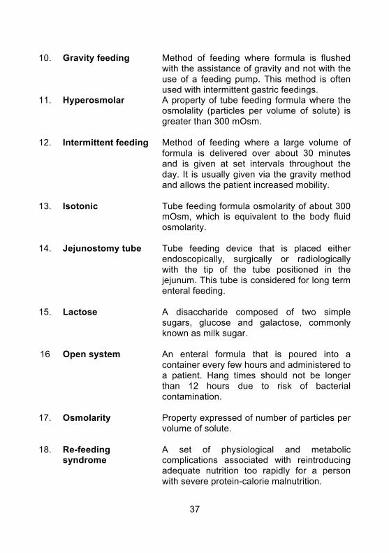
13 GLOSSARY
1. Aspiration The inhalation of food or liquid into the lungs.
2 Bolus feeding Method of feeding where a large volume of
formula is given in a short period of time, usually through a large syringe.
3 Continuous feeding Method of feeding where formula is delivered in low volumes over 8 to 24 hours, usually using a feeding pump.
4 Diarrhoea Stool output that is considered in excess of normal amounts. Usually consists of loose or watery frequent stools.
5. Drug Nutrient Interaction
Drug nutrient interaction applies to any situation in which incompatibility involves changes in medication bioavailability, absorption, distribution, metabolism or excretion.
6. Dysphagia Physiological swallowing difficulty that can lead to severe nutritional compromise without treatment.
7. Enteral feeding The delivery of formulas and medications through a tube placed into the stomach or intestine.
8. Flushing Process of pushing water through the feeding tube in order to clear it of formula or medications and/or restore patency.
9. Gastric residual volume (GRV)
The amount of tube feeding remaining upon aspiration of gastric contents. It is used to determine how quickly a patient is tolerating tube feeding formula or if there is a problem with peristalsis.
37
10. Gravity feeding Method of feeding where formula is flushed with the assistance of gravity and not with the use of a feeding pump. This method is often used with intermittent gastric feedings.
11. Hyperosmolar A property of tube feeding formula where the osmolality (particles per volume of solute) is greater than 300 mOsm.
12. Intermittent feeding Method of feeding where a large volume of formula is delivered over about 30 minutes and is given at set intervals throughout the day. It is usually given via the gravity method and allows the patient increased mobility.
13. Isotonic Tube feeding formula osmolarity of about 300 mOsm, which is equivalent to the body fluid osmolarity.
14. Jejunostomy tube Tube feeding device that is placed either endoscopically, surgically or radiologically with the tip of the tube positioned in the jejunum. This tube is considered for long term enteral feeding.
15. Lactose A disaccharide composed of two simple sugars, glucose and galactose, commonly known as milk sugar.
16 Open system
An enteral formula that is poured into a container every few hours and administered to a patient. Hang times should not be longer than 12 hours due to risk of bacterial contamination.
17. Osmolarity Property expressed of number of particles per volume of solute.
18. Re-feeding syndrome
A set of physiological and metabolic complications associated with reintroducing adequate nutrition too rapidly for a person with severe protein-calorie malnutrition.
38
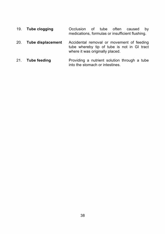
19. Tube clogging Occlusion of tube often caused by medications, formulas or insufficient flushing.
20. Tube displacement Accidental removal or movement of feeding tube whereby tip of tube is not in GI tract where it was originally placed.
21. Tube feeding Providing a nutrient solution through a tube into the stomach or intestines.
39
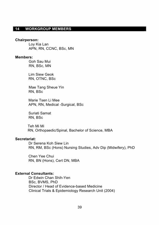
14 WORKGROUP MEMBERS
Chairperson:
Loy Kia Lan APN, RN, CCNC, BSc, MN
Members: Goh Sau Mui RN, BSc, MN Lim Siew Geok RN, OTNC, BSc
Mae Tang Sheue Yin RN, BSc Marie Tsen Li Mee APN, RN, Medical -Surgical, BSc Suriati Samat RN, BSc Teh Mi Mi RN, Orthopaedic/Spinal, Bachelor of Science, MBA Secretariat: Dr Serena Koh Siew Lin RN, RM, BSc (Hons) Nursing Studies, Adv Dip (Midwifery), PhD Chen Yee Chui RN, BN (Hons), Cert DN, MBA External Consultants: Dr Edwin Chan Shih-Yen
BSc, BVMS, PhD Director / Head of Evidence-based Medicine Clinical Trials & Epidemiology Research Unit (2004)
40
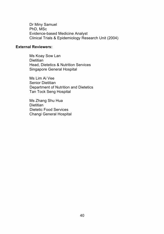
Dr Miny Samuel PhD, MSc Evidence-based Medicine Analyst Clinical Trials & Epidemiology Research Unit (2004) External Reviewers: Ms Koay Sow Lan Dietitian Head, Dietetics & Nutrition Services Singapore General Hospital Ms Lim Ai Vee Senior Dietitian
Department of Nutrition and Dietetics Tan Tock Seng Hospital Ms Zhang Shu Hua Dietitian
Dietetic Food Services Changi General Hospital
41
APPENDIX 1 NASOGASTRIC TUBE ANCHORING TECHNIQUE
Figure 2. Nasogastric tube anchoring technique
42
APPENDIX 2 ALGORITHM FOR ASSESSING PLACEMENT OF THE NASOGASTRIC TUBE
A FOSSESSI OF TH
43
[[E ASO APPENDIX 3 SELF ASSESSMENT
1. Selection of feeding tube should be based on the tube’s composition,
intended use, estimated length of time required, cost-effectiveness and tube features.
True/False 2. Polyvinylchloride feeding tube tends to harden with time and may
produce tissue irritation or necrosis. True/False
3. All feeding tubes are for long term use. True/False
4. Polyurethane or silicone tubes are better for long-term use because they
are more flexible and less irritating to tissues. True/False
5. Stabilizing a tube can reduce the risk of tube displacement, increased
tape adherence and decrease skin irritation by avoiding excess pressure on the nostril.
True/False 6. If the aspiration obtained from naso-gastric tube is whitish with tinge of
blood. It has a pH reading of 7, X-ray must be carried out to determine tube placement.
True/False 7. The value of gastric pH is less than 5.
True/False
8. Proceed with tube feeding if aspirate has a pH of 8 with off white to tan sediment.
True/False
44
9. Patient receiving H2 receptor antagonist or with recent alkaline reflux
from the intestine may have lower gastric pH. True/False
10. In the absence of gastric aspirate, the alternative method of checking
tube placement is to reposition patient and attempt to aspirate again. True/False
11. Auscultatory method should not be relied on as the sole method to
determine the feeding tube’s location. True/False
12. Patients who are on Omeprazole and Famotidine tend to have elevated
gastric pH. True/False
13. Duration for hanging of the formula feeds should be limited to 4 hours for
powdered, reconstituted formula and enteral nutrition formula with additives.
True/False
14. The patient should be placed at a semi-recumbent position or elevated to an angle of at least 30
o during and after feeding for at least one hour.
True/False
15. Administration of bolus feeds should not exceed 400ml during each bolus feed.
True/False 16. Pump feeding is recommended for patients who are ill and are unable to
tolerate intermittent bolus feedings. True/False
17. A trend in gastric residual volume may be more important than an
isolated high level of GRV. True/False
45
18. Flush feeding tube before and after administration of formula feeds, medication and checking of gastric residue.
True/False 19. There is no need to flush the feeding tube after checking gastric residual.
True/False 20. Enteral feed is an ideal culture medium and once contaminated, bacteria
will multiply rapidly. True/False
21. Administration sets and containers should be discarded every 24 hours.
True/False
22. Carbonated soda is the most effective method for declogging of blocked tube.
True/False
23. Supplementation of an enteral formula with soluble fibre significantly reduces the incidence of diarrhoea in patients on enteral feeds.
True/False 24. Patients with signs and symptoms of aspiration pneumonia, including
unexplained fever spikes, change in sputum colour or consistency, changes in breath sounds and worsening oxygenation should be monitored closely.
True/False 25. Whenever possible, medications administered through nasogastric tube
should be in liquid form. True/False
26. Enteric-coated and sustained release medications can be crushed.
True/False 27. Flushing the feeding tube prior to drug administration removes any
enteral feeds that remain in the tube to prevent drug nutrient interaction. True/False
46
28. Pump-assisted continuous feeding helps to prevent gastrointestinal complications associated with rapid infusion.
True/False 29. Handwashing reduces the risk of contamination and cross infection.
True/False 30. Adding medications into formula feeds may result in tube clogging and
cause undesirable effects or incompatibilities. True/False
47
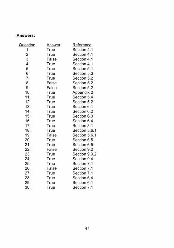
Answers:
Question Answer Reference 1. True Section 4.1 2. True Section 4.1 3. False Section 4.1 4. True Section 4.1 5. True Section 5.1 6. True Section 5.3 7. True Section 5.2 8. False Section 5.2 9. False Section 5.2
10. True Appendix 2 11. True Section 5.4 12. True Section 5.2 13. True Section 6.1 14. True Section 6.2 15. True Section 6.3 16. True Section 6.4 17. True Section 8.1 18. True Section 5.6.1 19. False Section 5.6.1 20. True Section 6.5 21. True Section 6.5 22. False Section 9.2 23. True Section 9.3.2 24. True Section 9.4 25. True Section 7.1 26. False Section 7.1 27. True Section 7.1 28. True Section 6.4 29. True Section 6.1 30. True Section 7.1