Embed Size (px)
Citation preview

1
Document Review History
Review Date Reviewed By Signature
2018
Document Change History
Change to Document Reason for Change
Part 2of 2
Nursing Guidelines on Peritoneal Dialysis.
Part 2 of 2
Version Number 1
Date of Issue 13th July 2015
Reference Number PD-07-2015-2of2-GDFMcHRUTSCUH-V1
Review Interval 3 yearly
Approved By
Name: Fionnuala O’Neill
Title: Nurse Practice Development Coordinator
Signature: Date
June 2015
Authorised By
Name: Rachel Kenna
Title: Director of Nursing
Signature:
June 2015
Author/s
Name: Grainne Doran (Renal C.N.S) Fiona McHugh (Renal C.N.F.) With thanks to all who have contributed feedback/suggestions and advice from the renal units in OLCHC and TSCUH.
Location of Copies On Hospital Intranet and locally in department

2
Table of contents
PG
1. Peritoneal Dialysis
2. Manual Dialysis
2
3, 4
Appendix 1. PD Catheter choice
Appendix 2. 3 Minute Hand wash.
Appendix 3. Dialysis fluid choice
Appendix 4. Peritoneal Dialysis prescription and sample guide
Appendix 5. Patient Assessment and troubleshooting
5
6
7, 8,
9, 10, 11
12, 13, 14
1.0 PERITONEAL DIALYSIS:

3
Introduction: Manual dialysis is a form of peritoneal dialysis used for a low weight child unsuitable for the
home choice machine It can also be used in emergency situations where peritoneal dialysis is required. Indications for manual peritoneal Dialysis:
A child’s weight determines the need for manual dialysis. Manual dialysis is required when
a fill volume of less than 60mls is required.( Home Choice Machine Minimum Fill is 60 mls or more)
Manual Dialysis may also be indicated in the acute intensive care setting when GFR falls between 15-10ml/lminute/1.73m ².
Definition of Peritoneal dialysis:
Dialysate Fluid is instilled into the peritoneal cavity via a Tenckhoff catheter, left to dwell
for a prescribed time and drained out. (Total Aseptic Technique). Peritoneal dialysis uses the peritoneum, a semi-permeable membrane, to transport
molecules and fluid across this membrane using the following three principles: Osmosis: is the movement of water through a semi-permeable membrane, from a solution of low solute concentration (high water potential) to a solution of high solute concentration (low water potential), up a solute concentration gradient. Diffusion: is solute movement from high solute concentration to low solute concentration across a semi-permeable membrane until equilibrium occurs. Convection: (Solvent Drag) Small solutes are dragged along with fluid.. Ultra filtration (UF): Solutes move down a concentration gradient by diffusion and water by osmosis. Ultrafiltration (UF) causes movement of solutes by convection, so that solutes may be carried across the membrane (solvent Drag) even in the absence of a concentration gradient. Water removal (UF) and dissolved solutes flow down a pressure gradient. This occurs as a result of Hydrostatic pressure (B/P) and osmotic force (glucose pull). UF is recorded and used throughout treatment to assess patient’s fluid balance. Total UF is documented at the end of a dialysis session. This can be a positive figure (more fluid off the patient .than fluid retained by the patient) or a negative figure (fluid retained in the patient i.e. more stays in than comes out) Cumulative fluid retention from different cycles in the programme = patient becomes more positive. Cumulative fluid removal from different cycles in a programme=patient becomes more negative.
THE PERITONEAL MEMBRANE: Visceral and parietal layer, rich in vascular supply (mesenteric artery, portal veins) Fluid is instilled between these two layers i.e. the cavity, and left to dwell for a prescribed length of time, and then drained and replaced with a fresh supply. The access to the cavity is via a catheter called a Tenckhoff catheter. This is tunnelled and anchored (usually) with internal cuffs. A permanent catheter usually takes six weeks to embed. This is the patient’s life line, and must be treated with respect i.e. to prevent peritonitis. An episode of peritonitis causes scarring and decreased peritoneal function. PERITONITIS: Signs and symptoms:
Pyrexia
Painful Abdomen,

4
Fibrin(cloudy bit’s of white protein) or Turbid fluid
Nausea/vomiting/Diarrhoea
“guarded abdomen” EXIT SITE: Refer to exit site care protocol for new or established catheter.

5
2.0 MANUAL DIALYSIS:
Dialysis sets should be changed every 24 hours using ASEPTIC TECHNIQUE (OLCHC, 2013) Physioneal 2.5 litre bags are used only When using Physioneal 2.5litre bags add additives to top chamber before breaking seal using aseptic technique. See protocol(s) / guidelines for addition of medicines COMPLICATIONS ASSOCIATED WITH PERITONEAL DIALYSIS:
1. Dehydration 2. Fluid overload 3. Infections -PERITONITIS (see protocol)Take Sample from port on drain
Buretrol(Aseptic Technique) 4. Contamination 5. Disconnection 6. Hypertension 7. Hypotension 8. Diaphragmatic Splinting (ventilated patients-see fill volumes in appendix) 9. Pleural Effusions 10. Pulmonary oedema 11. Catheter malfunction/blockage 12. Leaks 13. Bowel/Bladder perforation-ensure that bladder and bowel is emptied prior to catheter
insertion 14. Haemorrhage 15. Hernias(inguinal /scrotal/umbilical) 16. Air embolism 17. Nausea/vomiting 18. Disequilibrium (rapid removal of blood urea → fluid shifts → cerebral oedema
GUIDELINES FOR SETTING UP MANUAL DIALYSIS Equipment:
1. Manual Dialysis Baxter set (acute set for children CAPD) Ref JMC3437. 2. Dialysis fluid bag(s) as prescribed. 3. 3 connection shields 4. 5 prong Baxter manifold if using a mix of PD fluid 5. Fluid Heater 6. 1 trolley 7. 2 sterile drapes 8. Sterile gloves. 9. 1 Baxter drainage bag ( 3L empty bag system) (reference)XMC4284 10. Disinfection wipes 11. 1 minicap cap (for end of procedure) 12. 1 Blue clamp in patient’s room at all times
3 GUIDELINE FOR SETTING UP MANUAL DIALYSIS
ACTION
1.Explain the procedure to the child/family 2. Close all windows and doors. 3. Decontaminate hands/Gather all equipment. 4. Check the Dialysis prescription is correct. 5. Heat Dialysis Fluid on heater (optional).

6
6.Decontaminate hands for 1 minute and apply Alcohol rub/gel. 7. Open the Physioneal bags break seals between two fluid chambers and hang on a drip stand. 8. Open out sterile drape on the trolley. 9. Empty all sterile equipment onto the trolley. 10.Decontaminate hands and apply sterile gloves. 11. Close all clamps on the manual set. 12. Attach connection shield at junction of upper Buretrol and manifold. 13. Attach drainage bag and connection shield to the lower ‘drain’ Buretrol. 14.Attach dialysis fluid bag(s) once completely mixed 15.Break the green seal on the dialysis fluid bag at the Buretrol connection 16. Fill the Buretrol to 80mls of dialysis fluid and squeeze the bubble chamber to fill. 17. Slowly release the clamp to prime the line. 18. Ensure the line is primed. Clamp. Do not remove the cap at end of line. 19. Prime drain side. Clamp. 20. Fill Buretrol to required fill volume and clamp. 21. Clamp all lines. Cover the trolley with a sterile drape when transferring to the patient’s room. 22. N.B If doing this procedure with two nurses, sterility can be maintained. Second nurse will assist with checks 24-29. 23. Hang heated dialysis fluid on the stand. 24. Ensure all windows and doors are closed in the patient’s room. 25. Check patient’s identification band with a second nurse. 26. Clean the Transfer set with Disinfection wipes and wrap. 27. Decontaminate hands. 29..Change sterile gloves.(if single nurse doing procedure) 30. Apply connection shield to patient line. 31. Connect the patient line to transfer set ensuring the line is primed. 32.Check that correct fill volume is in the Buretrol 33. Fill patient (see below).
Fill: 34.Open blue twist clamp on Transfer set, roller clamp, patient line clamp and allow the fluid to infuse into the patient (3-5 minutes) 35. Clamp blue twist clamp on transfer set, patient line and roller clamp when required volume has infused. 36. Observe the Buretrol closely to prevent air from entering the chamber. 37. Observe the exit site and inform the Nephro-Urology team if a leak is noted. 38.Secure drain bag and Buretrol at lower level to patient
Dwell:
39. Set the clock (if used) for the prescribed dwell time. Record commencement time of dwell.
Drain:
40. Ensure roller clamp and drain bag clamp are clamped. 41. Open drain line clamp (leading to drain Buretrol), patient line clamp and blue twist clamp on transfer set. 42. Make sure the large drain bag clamp is shut until drain is complete. 43. Observe the effluent draining. 44. Drain time may be15-20minutes approx. 1. Observe the colour of the effluent for colour clarity, fibrin, and blood. 2. Reposition the patient if required to obtain maximum drainage. 3. When drain complete, close blue twist clamp on transfer set and drain line clamp 4. Record Volume. 5. Open clamp to large bag until the Buretrol is empty. Clamp.

7
6. Repeat for designated number of cycles.
7. N.B Please look at appendix 5 for information on the following Fill and Drainage problems. Resolutions of the above. Trouble shooting. Patient prescription of dialysis regime.
Nursing care related issues.
Appendix 1
PERITONEAL DIALYSIS CATHETER CHOICE:
The Choice of Single or Double Cuff is at the discretion of the surgeon. Quinton is Preferred for older children above 8 kgs, Double Cuff.
FLEX NECK CATHETERS
Size Catheter Code weight
Neonate Medigroup Infant’Flex-neck’Coiled Catheter 1 Cuff
(CF-4235) Under 3Kg
Neonate The Medigroup Infant ‘Flex-neck’ Coiled Catheter 2 Cuff FIRST CHOICE
(CF-4230)
Under 3 Kg
All of the above are supplied by L.I.N.C Medical Systems LTD PH.0044 1572 7175 15.
QUINTON TENCKHOFF CATHTERS
Size Catheter Code Weight guide
31cm Neonatal PD catheter double cuff straight
8814-4227 Under 4kg
38.9cm Infant PD catheter swan neck curl catheter double cuff
8888-413100 8kg or less
42cm Paediatric PD catheter swan neck curl catheter double cuff
8888-414813 Greater than
8 kg
59cm Paediatric swan neck curl catheter double cuff
8888 413102 30-40kg
62.2cm
Adult PD catheter swan neck curl catheter double cuff
8888-413807 Greater than 45kg
All of the above are supplied by Health Care 21, Jackie Knox phone number: (087)6976800
Swan neck catheters decrease stress on exit site and the curl catheter helps keep the catheter positioned low in the peritoneum where drainage is optimum.

8
Appendix 2 3 MINUTE HAND WASH.
Action
1. Ensure all windows and doors are closed.
2. Remove hand and wrist jewellery. (Plain wedding band allowed).
3. Ensure finger nails are short and clean.
4. Individually place both hands and arms under running water from fingers to elbow.
5. Apply Hydrex Surgical Scrub (Chlorhexidine Gluconate 4% skin cleanser to the
elbows.
6. Start with the fingertips of the hand, then rub the length of the fingers using rotating
movements, repeat each finger 10 times paying attention to the thumbs, on each hand.
7. Rub the back of your hand with the palm of the other hand 10 times to each hand.
8. Rub Palm to Palm 10 times over entire hands and fingers.
9 Rub palm to palm 10 times with the fingers interlocked.
10. Using the tips of the fingers rub the palm of each hand using a circular motion-10 times
to each hand.
11. Place right palm over back of left hand. Rub in a side to side motion
12. Rub the area between tips of index finger and thumb on each hand 10 times.
13. Rub the area from tip of thumb to the wrist (on lateral aspect) 10 times and then from
tip of little finger (on lateral aspect) to the wrist the area 10 times to each hand.
14. Next rub the wrist area on each hand 10 times.
15. Finally using a downward motion rub the arm from the wrist to the elbow, repeat on
the other arm.
16. Rinse off the soap from the fingers to the elbow on both hands/arms.
17. Ensure this hand wash takes 3 minutes, if not repeat the above.
18. Dry with paper towel working from the fingers to the elbow in a downwards motion.
19. Apply Alcohol gel (Purell) to both hands
9
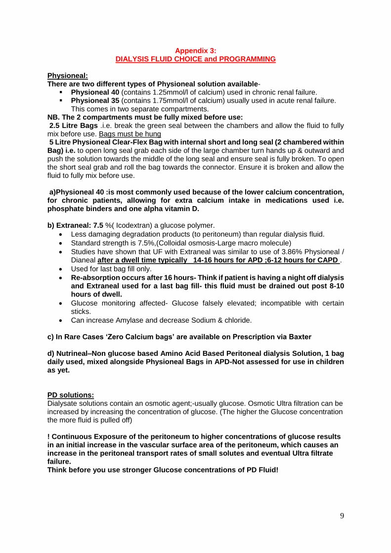
Appendix 3: DIALYSIS FLUID CHOICE and PROGRAMMING
Physioneal: There are two different types of Physioneal solution available-
Physioneal 40 (contains 1.25mmol/l of calcium) used in chronic renal failure. Physioneal 35 (contains 1.75mmol/l of calcium) usually used in acute renal failure.
This comes in two separate compartments. NB. The 2 compartments must be fully mixed before use: 2.5 Litre Bags .i.e. break the green seal between the chambers and allow the fluid to fully mix before use. Bags must be hung 5 Litre Physioneal Clear-Flex Bag with internal short and long seal (2 chambered within Bag) i.e. to open long seal grab each side of the large chamber turn hands up & outward and push the solution towards the middle of the long seal and ensure seal is fully broken. To open the short seal grab and roll the bag towards the connector. Ensure it is broken and allow the fluid to fully mix before use. a)Physioneal 40 :is most commonly used because of the lower calcium concentration, for chronic patients, allowing for extra calcium intake in medications used i.e. phosphate binders and one alpha vitamin D. b) Extraneal: 7.5 %( Icodextran) a glucose polymer.
Less damaging degradation products (to peritoneum) than regular dialysis fluid.
Standard strength is 7.5%,(Colloidal osmosis-Large macro molecule)
Studies have shown that UF with Extraneal was similar to use of 3.86% Physioneal / Dianeal after a dwell time typically 14-16 hours for APD ;6-12 hours for CAPD .
Used for last bag fill only.
Re-absorption occurs after 16 hours- Think if patient is having a night off dialysis and Extraneal used for a last bag fill- this fluid must be drained out post 8-10 hours of dwell.
Glucose monitoring affected- Glucose falsely elevated; incompatible with certain sticks.
Can increase Amylase and decrease Sodium & chloride. c) In Rare Cases ‘Zero Calcium bags’ are available on Prescription via Baxter d) Nutrineal–Non glucose based Amino Acid Based Peritoneal dialysis Solution, 1 bag daily used, mixed alongside Physioneal Bags in APD-Not assessed for use in children as yet. PD solutions: Dialysate solutions contain an osmotic agent;-usually glucose. Osmotic Ultra filtration can be increased by increasing the concentration of glucose. (The higher the Glucose concentration the more fluid is pulled off) ! Continuous Exposure of the peritoneum to higher concentrations of glucose results in an initial increase in the vascular surface area of the peritoneum, which causes an increase in the peritoneal transport rates of small solutes and eventual Ultra filtrate failure. Think before you use stronger Glucose concentrations of PD Fluid!
10
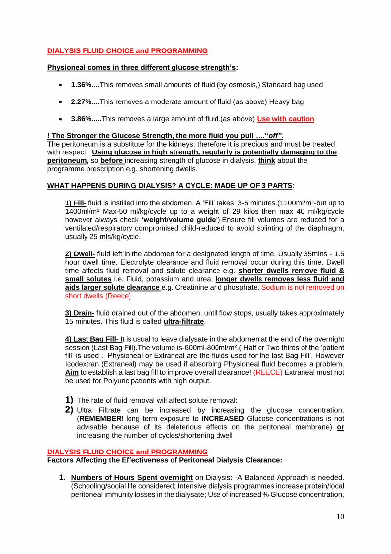
DIALYSIS FLUID CHOICE and PROGRAMMING Physioneal comes in three different glucose strength’s:
1.36%....This removes small amounts of fluid (by osmosis,) Standard bag used
2.27%....This removes a moderate amount of fluid (as above) Heavy bag
3.86%.....This removes a large amount of fluid.(as above) Use with caution ! The Stronger the Glucose Strength, the more fluid you pull ….“off”. The peritoneum is a substitute for the kidneys; therefore it is precious and must be treated with respect. Using glucose in high strength, regularly is potentially damaging to the peritoneum, so before increasing strength of glucose in dialysis, think about the programme prescription e.g. shortening dwells. WHAT HAPPENS DURING DIALYSIS? A CYCLE: MADE UP OF 3 PARTS:
1) Fill- fluid is instilled into the abdomen. A ‘Fill’ takes 3-5 minutes.(1100ml/m²-but up to 1400ml/m² Max-50 ml/kg/cycle up to a weight of 29 kilos then max 40 ml/kg/cycle however always check ‘weight/volume guide’).Ensure fill volumes are reduced for a ventilated/respiratory compromised child-reduced to avoid splinting of the diaphragm, usually 25 mls/kg/cycle.
2) Dwell- fluid left in the abdomen for a designated length of time. Usually 35mins - 1.5 hour dwell time. Electrolyte clearance and fluid removal occur during this time. Dwell time affects fluid removal and solute clearance e.g. shorter dwells remove fluid & small solutes i.e. Fluid, potassium and urea; longer dwells removes less fluid and aids larger solute clearance e.g. Creatinine and phosphate. Sodium is not removed on short dwells (Reece)
3) Drain- fluid drained out of the abdomen, until flow stops, usually takes approximately 15 minutes. This fluid is called ultra-filtrate.
4) Last Bag Fill- It is usual to leave dialysate in the abdomen at the end of the overnight session (Last Bag Fill).The volume is-600ml-800ml/m²,( Half or Two thirds of the ‘patient fill’ is used . Physioneal or Extraneal are the fluids used for the last Bag Fill’. However Icodextran (Extraneal) may be used if absorbing Physioneal fluid becomes a problem. Aim to establish a last bag fill to improve overall clearance! (REECE) Extraneal must not be used for Polyuric patients with high output.
1) The rate of fluid removal will affect solute removal:
2) Ultra Filtrate can be increased by increasing the glucose concentration, (REMEMBER! long term exposure to INCREASED Glucose concentrations is not advisable because of its deleterious effects on the peritoneal membrane) or increasing the number of cycles/shortening dwell
DIALYSIS FLUID CHOICE and PROGRAMMING Factors Affecting the Effectiveness of Peritoneal Dialysis Clearance:
1. Numbers of Hours Spent overnight on Dialysis: -A Balanced Approach is needed. (Schooling/social life considered; Intensive dialysis programmes increase protein/local peritoneal immunity losses in the dialysate; Use of increased % Glucose concentration,

11
long term, is a contributing factor for development of peritoneal sclerosis & decreased peritoneal membrane function
2. Number of cycles AND Number of nights on Dialysis.
3. The length of dwell.
4. The Fill Volume: 1100ml/m² (-1400ml/m²) when child is in lying/supine position (intra-abdominal pressure is less in the prone position, if intra-abdominal pressure starts to rise this will decrease Ultra filtrate) (REECE).
5. Last Bag Fill: (Dialysate left in the abdomen at the end of the overnight session). Volume used is 600-800ml/m²,this adds considerably to the overall clearance of the dialysis programme, however if there is poor return on this during the initial drain i.e. less than two thirds of the last bag fill, (when commencing overnight dialysis),Icodextran(Extraneal) can be used.(Baxter)
6. UF Rate Affects Solute Removal: Ultra filtrate rate can be increased by:
Increasing Glucose concentration.
Increasing Numbers of cycles in the total therapy time. (Shorter dwell time).
7. Peritoneal Blood Flow will be affected by cardiac output.
8. Peritoneal Membrane Characteristics (surface area, vasculature, permeability, peritoneal sclerosis and/or adhesions)
A Day time Dwell: clearance of small and large molecules significantly increased (as much as one third) Paediatric NEPHROLOGY, LESLEY REES, OXFORD

12
Appendix 4 Dialysis Prescription Guide and Sample Prescription: Prescribing cycle/Fill Volume:
Initially start with flushes of 10 mls/ kg/cycle until fluid clear or mildly rosé.
Gradually work up fill volume over a period of days/ weeks to avoid increased intra-abdominal pressure, and leakage at exit site.
When Tenckhoff catheter is embedded (6 weeks) start with small fills working upwards, when established aim for volumes of 30-40 mls /kg/cycle. Maximum 50 mls/kg/cycle.1.1L/m²-1.4L/m²
Established patients: are usually on fill volumes of 1.1L/ m². (Usually 30 - 40 ms/kg/cycle, Max-50 ml/kg/cycle up to a weight of 29 kilos then max 40 ml/kg/cycle however always check ‘weight/volume guide’).
Ventilated patients: maximum fill volume- 25ml/kg/cycle. (Avoidance Diaphragmatic Splinting)
For the Established End Stage Renal Failure Patient:
Infant-Usually on dialysis for 12-14 hours with a ratio of 1:1 (I cycle to 1 hour of dialysis time) (REECE) All dialysis regimes for infants (under 2yrs) will have an extra cycle added to prescription to allow for a ‘flushing 1st cycle’ to ensure clearance of residual iodine in lines, unless initial drain volume is greater than 35mls. If bypassing the 1st cycle it is necessary to take into consideration that the average dwell will be lengthened.
Polyuric child/Older Child- Up to 10-12 hours overnight dialysis with a ratio of 2:3 (2 cycles to 3 hours)
Oliguric/anuric child - Up to 12 hours with a ratio of 1:1 (1 cycle to 1 Hour).
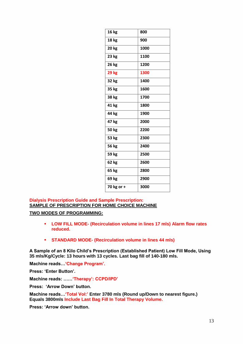
Dialysis Prescription Guide and Sample Prescription: Always Check Weight/Volume-Guide;
Fill Volume Limit for weight: As per Baxter
Weight (kg)
Fill Vol. Limit.
Millilitres(Mls)
2 kg 100
3 kg 150
4 kg 200
6 kg 300
8 kg 400
10 kg 500
12 kg 600
14 kg 700
13
16 kg 800
18 kg 900
20 kg 1000
23 kg 1100
26 kg 1200
29 kg 1300
32 kg 1400
35 kg 1600
38 kg 1700
41 kg 1800
44 kg 1900
47 kg 2000
50 kg 2200
53 kg 2300
56 kg 2400
59 kg 2500
62 kg 2600
65 kg 2800
69 kg 2900
70 kg or + 3000
Dialysis Prescription Guide and Sample Prescription: SAMPLE OF PRESCRIPTION FOR HOME CHOICE MACHINE
TWO MODES OF PROGRAMMING:
LOW FILL MODE- (Recirculation volume in lines 17 mls) Alarm flow rates reduced.
STANDARD MODE- (Recirculation volume in lines 44 mls)
A Sample of an 8 Kilo Child’s Prescription (Established Patient) Low Fill Mode, Using 35 mls/Kg/Cycle: 13 hours with 13 cycles. Last bag fill of 140-180 mls.
Machine reads…‘Change Program’.
Press: ‘Enter Button’.
Machine reads: ……‘Therapy’: CCPD/IPD’
Press: ‘Arrow Down’ button.
Machine reads…‘Total Vol:’ Enter 3780 mls (Round up/Down to nearest figure.) Equals 3800mls Include Last Bag Fill In Total Therapy Volume.
Press: ‘Arrow down’ button.

14
Machine reads:….. ‘Therapy time’: Enter 13 Hours.
Press: ‘Arrow down’ button.
Machine reads:….. ‘Fill Vol’: Enter 280 mls.
Press: ‘Arrow down’ button.
Machine reads:…. ‘Last fill Vol’: Enter 140 mls. (Half – Two thirds of patient’s fill volume)
Machine reads:…. ‘Dextrose: ‘Same’ or ‘Different’ is entered depending if same fluid for the dialysis session is used or not.
Machine reads…… ‘Weight units’ ….. Kg
Machine reads…… ‘Patient Weight’…kg
Press the ‘Stop’ button. The number of cycles and the dwell time will be displayed.
Dwell: 43 mins
Cycles 13
Dialysis Prescription Guide and Sample Prescription:
SAMPLE OF PRESCRIPTION
A Big Child Approx. 25 kgs-On 40 mls /kg / cycle:
Total Therapy time: - 10 Hours
Number of cycles: 7.
Fill Volume: - 1000 mls
Total Therapy Volume: - 7700 mls (Round Up/Down to nearest figure)
Last Bag Fill – Extraneal: - 650 mls (Always include last bag fill in total therapy volume.)
Dwell:
Cycles: 7
! Always ensure that enough fluid is used to prime and flush lines during set up, (Allow 100mls per bag hung) e.g. 3 bags→300ml. Checking patency of patient line during therapy, (15ml ‘pushback’) this volume is not included in the total therapy volume. !

15
Appendix 5:
PATIENT ASSESSMENT AND TROUBLE SHOOTING ON PERITONEAL DIALYSIS:
1. Over Load- Signs & Symptoms.
Weight Gain
Large Positive Balance
Hypertension
Generalised oedema,(genital, legs ,feet, ascites)
Periorbital oedema
Facial Oedema
Enlarged Liver
Tachypnoea…→Nasal flaring→Dyspnoea→Desaturation→Pulmonary oedema
Tachycardia/Gallop/full, bounding pulses
Visible, full external jugular’s OVERLOADED WHY?
High Fluid Intake also remember ‘hidden fluids’
Low UF-Low volume Drains (i.e. low volume of fluid “off”)
Oliguric or anuric
Retaining Fluid Observe for fluid OVERLOAD?
Observe for Weight↑, B/P↑, tachycardia, ↑pulse volume. (Full bounding pulses present).
Engorged or prominent neck veins.
Generalised Oedema, skin marks easily,
Tachypnoea/dyspnoea/nasal flaring/↓ O2 Saturations,
Have they Poor output & Large Positive Balance?
Usually 85% of the fill volume or more comes out, It is not safe to refill, if NO Fluid, or less than 50% of the fill volume has come out.
The patient will become increasingly more positive/and may display respiratory compromise).
IF THE NEXT CYCLE YIELDS LESS THAN 50% CALL NEPHROLOGY TEAM FOR ADVICE. If no fluid has come out you must always try repositioning the patient and checking the
line for obstruction, kinks, and clamps are open and the position of the machine at the level of the patient or max 12 inches above (should not be too low as omentum can be pulled by gravity up against catheter pores, obstructing flow). If no result, Contact renal team!
2. DEHYDRATED- Signs and symptoms
Decreased Weight
Tachycardia
Sunken eyes
Cool peripheries
> 2ºGap between core and peripheral temperature
Abdominal pain
nausea and vomiting

16
DEHYDRATED WHY?
Have they Diarrhoea and vomiting
Is the weather or the environment hot?-Insensible losses may be high (What are the child’s insensible losses for the 24 hour period? Insensible losses are usually 400ml/m²/24 hours
High UF
Poor fluid intake
Child may need to retain, for example if he/she is dehydrated due to excess fluid loss example vomiting or diarrhoea, increased activity during hot summers ,If the child is retaining you must ask yourself if it is O.K.?-It must be within safe boundaries.
3. CATHETER BLOCKED?
If no fluid has come out, check all clamps, check for kinks in the line, or any mechanical obstruction.
Is there fibrin, visible in the lines or dialysate fluid? (Fluid should always be crystal clear)
If the fluid is cloudy or fibrin present peritonitis must be out ruled and a sample sent to the lab for cell count and C&S. Discuss with the nephrologists.
Heparin will need to be added to the dialysis fluid (aseptic technique) Refer to Renal guideline folder for protocol.
The catheter may need to be flushed, with Saline or Heparin and saline. Refer to renal guidelines folder, for protocol for catheter flush Refer to renal guidelines for Urokinase protocol. 4. MACHINE LEVEL /HEIGHT?
Check the position of the machine. Is it too high or too low? The recommended height is at the level of the patient it usually is level with the height of the bed or 12 inches above, (if it is too low the gravity “pull” might pull the omentum up against the internal Tenckhoff pores, and stop drainage.
5. DWELL TIME TOO LONG?
The longer the dwell time the more likely the patient will retain, usually small babies need fast cycles with short dwell times, i.e. around 30- 45 min dwells. This will be decided by the Nurse Specialist (or Nephrologists).
6. SERUM ALBUMIN LOW?
Large amounts of Albumin (nephrotic range) are washed away from the peritoneum during dialysis.
Losses also occur secondary to poor diet or other underlying disease. If serum albumin is low the oncotic pressure is low. The hydrostatic pressure is greater than the “pullback” force from albumin/protein, so fluid shifts from the intravascular space into the interstitial space, compounding the oedema of the tissues and making fluid inaccessible for removal.
8. POSITION OF CATHETER.
Is there Shoulder pain or pain on filling /draining? (Causes Migration of internal catheter tip- should be low down in pelvis)
Is there poor draining?
Is there Slow filling/draining or resistance on flushing the line with saline (Aseptic Technique) see protocol.
X-ray catheter (plain abdominal film).
U/S of the catheter- checking position.
9. INADEQUATE FILL VOLUME?

17
Retention is likely to occur using a small fill volume. (E.g. with a new catheter) This may need to be increased if blood/electrolyte clearance is poor or fluid removal inadequate?
↑Volumes gradually (dictated by new exit site and catheter) Maximum fill volume for ICU patients is 25mls/kg.
10. CONSTIPATION. This can be a very important reason why the patient might retain (also plays a part in catheter migration) so keep a close eye on bowel habit, stool softeners are often indicated.
THINGS TO THINK ABOUT: Sometimes fluid does not come out if inguinal hernias are present, or if there are adhesions in the abdomen (pocketing of fluid). Check for scrotal oedema. Occasionally diaphragmatic leakage can occur, all peritoneal fluid into the thoracic cavity restricting breathing- (confirmed by X-ray and clinical history& examination)
Do you need to flush the catheter (see protocol)?
Does the Dialysis programme need to be changed?
Does the PD fluid Prescription need to be changed? i.e. shorter dwells-more fluid off, longer dwells – more fluid retained.
Maybe a mix of 1.36% and 2.27% glucose is needed Or 2.27% only
The higher the glucose concentration the more fluid is pulled off. PATIENT ASSESSMENT AND TROUBLE SHOOTING ON PERITONEAL DIALYSIS:
Assessment Recap: 1. Weight ↑or↓, i.e. overloaded, (wet) or dehydrated (dry). 2. Balance, positive (retaining) negative (ultra filtrating too much, or
other body losses- Gastric/diarrhoea, insensible losses, urine, blood/serum, chest drain losses)
3. The catheter- slow filling or slow draining? 4. What % volume of the cycle has drained out? LOOK AT YOUR PATIENT! A child with a persistently high urine output when on dialysis is more likely to retain fluid, causing increased ‘Low Drain volume Alarms’ during cycles. Try decreasing ‘Minimum Drain Volume’ to 75% and keep ‘Drain time’ at 15 minutes. Too short a dwell time may take off excess fluid, leading to poor Creatinine/phosphate clearance and is more likely to cause low potassium levels. Clearance is decreased on very low fill volumes. eg.10mls /kg/cycle. It is important to keep a regular fluid balance and record how positive or negative the child is. (We tend to include colloid as part of our intake when looking at balance). Dialysis prescription regularly changes throughout the regime, depending on the patients balance so it is important to inform the team if too much or too little is ultra-filtrated. Therefore, it is the responsibility of the nurse to continuously assess the appropriateness of the dialysis fluid type/programme in relation to patient balance. Aim to stay near your ideal weight (Dry weight)!

18
UF Failure may be due to excessive salt and water ingestion, excessive fluid absorption during long dwell exchanges, over exposure to stronger glucose solution, non-compliance with dialysis prescription, or post recurrent peritonitis, or peritoneal sclerosis and /or adhesions. References National Institute of Clinical Excellence (NICE) (2003) Infection control: Prevention of healthcare-associated infection in primary and community care, Thames Valley University, London. National Hospitals Office (NHO)(2009) Code of Practice for Healthcare Records Management, Version 2 (2010), Health Service Executive, Dublin Ireland. Our Lady’s Children’s Hospital Crumlin (OLCHC) (2009) Guidelines on the Care of the Child Requiring Clinical Holding, OLCHC, Dublin. Our Lady’s Children’s Hospital Crumlin (OLCHC) (2013) Guideline for Hand Hygiene, OLCHC, Dublin. Our Lady’s Children’s Hospital Crumlin (OLCHC) (2014b) Waste Management Policy. OLCHC, Dublin. Our Lady’s Children’s Hospital Crumlin (OLCHC) (2015) The Hospital Formulary and Prescribing Guide, OLCHC, Dublin. Our Lady’s Children’s Hospital Crumlin (2011a) Standard Universal Precautions, OLCHC, Dublin. Our Lady’s Children’s Hospital Crumlin (OLCHC) (2013) Aseptic Non Touch Technique. OLCHC, Dublin. Our Lady’s Children’s Hospital for Sick Children (OLCHC) (2009) Guidelines on Performing a Wound Swab, OLCHC, Dublin.