Embed Size (px)
Citation preview

Nursing Care of Patients experiencing Critical Illness
C. Cummings EdD,RN
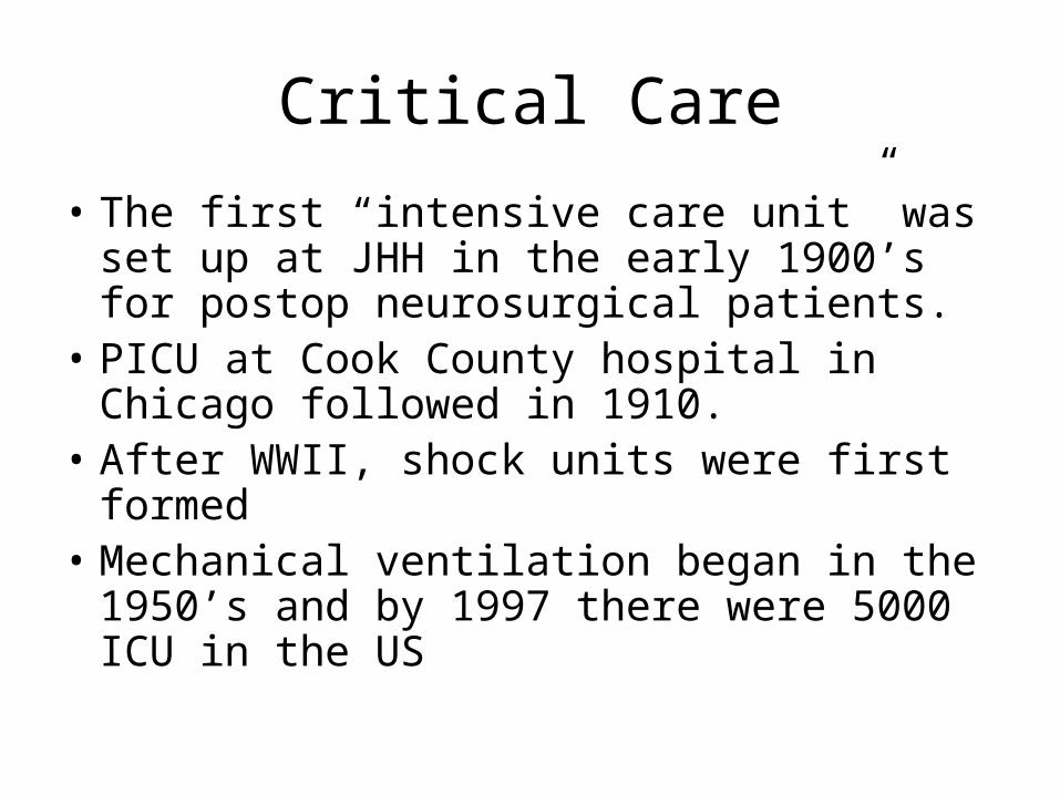
Critical Care
• The first “intensive care unit” was set up at JHH in the early 1900’s for postop neurosurgical patients.
• PICU at Cook County hospital in Chicago followed in 1910.
• After WWII, shock units were first formed• Mechanical ventilation began in the 1950’s
and by 1997 there were 5000 ICU in the US
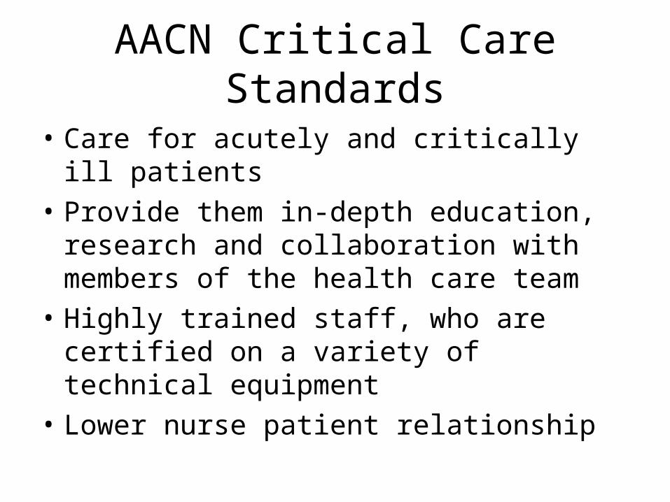
AACN Critical Care Standards
• Care for acutely and critically ill patients• Provide them in-depth education, research
and collaboration with members of the health care team
• Highly trained staff, who are certified on a variety of technical equipment
• Lower nurse patient relationship

ICU

Golden Hour• R. Adams Cowley, shock trauma center was the first in
the country in 1969• Golden Hour
– Treat the patient within 1 hour of injury– Mortality rate prior to 1970 was 70%, at MIEMS, it
is now 3%– Full body helial CT within 20 minutes of arrival,
many new technological advances• Pulse ox, capnography, inline blood gases, etc.
– Developed teams to perform all processes and were the first with computerized medical records
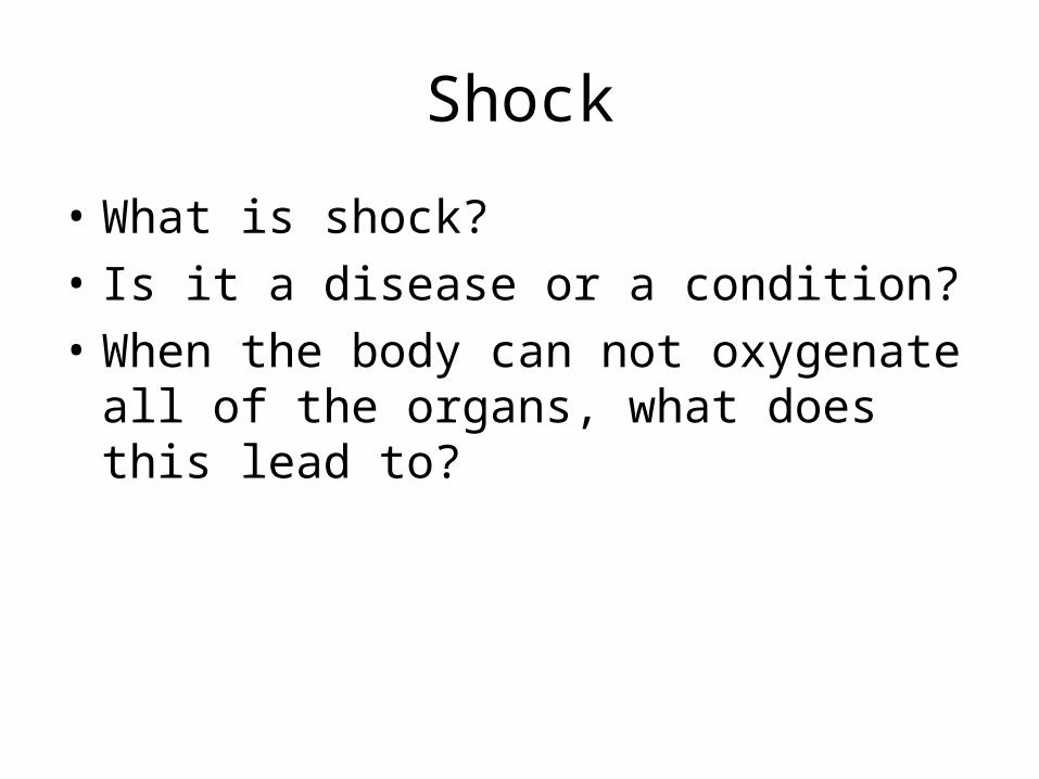
Shock
• What is shock? • Is it a disease or a condition?• When the body can not oxygenate all of the
organs, what does this lead to?

Shock Classifications
• Four functional impairment systems• Key features of shock are similar• Organ perfusion r/t MAP (mean arterial
pressure) What organs poorly tolerate hypoxia??
• Factors that influence the MAP are:– Total blood volume– Cardiac output– Size of the vascular bed

Shock Classifications
• Hypovolemic– Total body fluid decreased– Hemorrhage & dehydration
• Cardiogenic– Pump failure, fluid volume ok– MI, valvular problems, dyrhythmias, cardiac
arrest, myopathies
Shock Classifications
• Distributive– Fluid shifted from central vascular space, body
fluid remains normal or increased– Neural loss of vascular tone, why??– Chemical loss of vascular tone, why??
• Obstructive– Cardiac function decreased by non-cardiac factors,
fluid not affected– Pulmonary hypertension, pneumothorax,
pericarditis, tamponade (what is that?)

What happens with shock?
• Anaerobic cell metabolism• Main trigger is a decrease in MAP, results from
decreased C.O., decreased blood volume or expansion of the vascular bed (think of what can cause this?)
• C.O.= HR x what??• Drop of just 5-10mm Hg is detected by
baroreceptors in the aortic arch and carotid sinus

Process of Shock
• Baroreceptors send info to the brain, which stimulates systems to shunt blood into vital areas: heart, brain, lungs (what systems are stimulated?)
• Leads to an increase in lactic acid, protein destroying enzymes and oxygen radicals
• If corrected in 1-2 hours, it may be reversible, if not, can lead to MODS
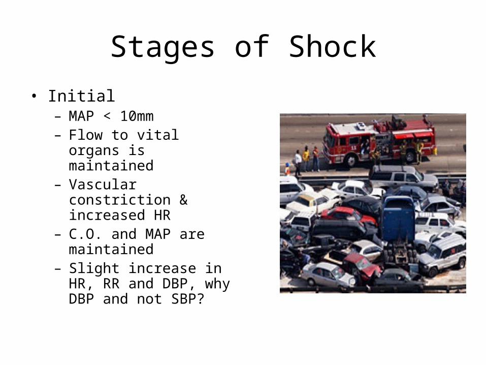
Stages of Shock• Initial
– MAP < 10mm– Flow to vital organs is
maintained– Vascular constriction &
increased HR– C.O. and MAP are
maintained– Slight increase in HR, RR and
DBP, why DBP and not SBP?
Stages of Shock
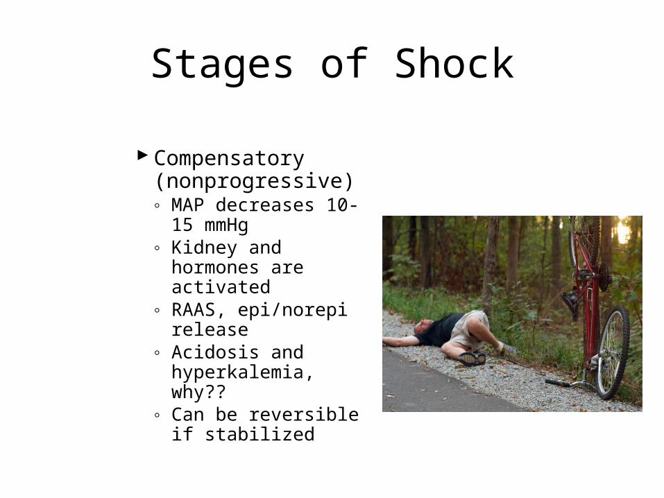
Compensatory (nonprogressive)◦ MAP decreases 10-15
mmHg◦ Kidney and hormones
are activated◦ RAAS, epi/norepi
release◦ Acidosis and
hyperkalemia, why??◦ Can be reversible if
stabilized

Stages of Shock
• Intermediate (progressive)– Sustained decrease in
MAP >20mm Hg– Less vital organs
become anoxic, what are they?
– Ischemia occurs, can only tolerate for a short time
– Life-threatening– Must correct in 1 hour
Stages of Shock
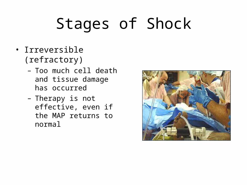
• Irreversible (refractory)– Too much cell death and
tissue damage has occurred– Therapy is not effective,
even if the MAP returns to normal
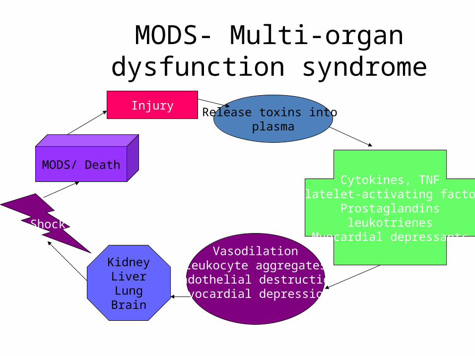
MODS- Multi-organ dysfunction syndrome
InjuryRelease toxins into
plasma
Cytokines, TNFPlatelet-activating factor
Prostaglandinsleukotrienes
Myocardial depressantsVasodilation
Leukocyte aggregatesEndothelial destructionMyocardial depression
KidneyLiverLungBrain
Shock
MODS/ Death
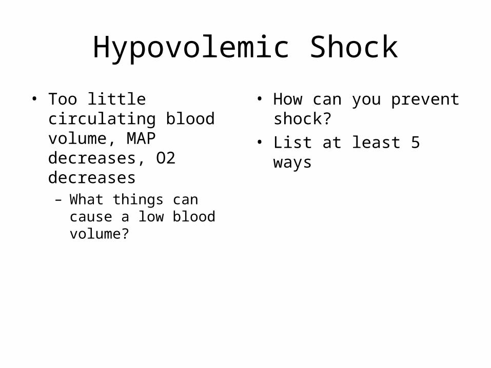
Hypovolemic Shock
• Too little circulating blood volume, MAP decreases, O2 decreases– What things can cause a low
blood volume?
• How can you prevent shock?
• List at least 5 ways

Case Study
• A 36-year-old motorcyclist, J.M., is admitted to the ICU after being hit by a car. His initial injuries are diffuse cerebral contusions, a left-sided flail chest, a ruptured spleen, a lacerated liver and a fractured left femur. He has undergone a laparotomy at which his spleen was removed, his liver repaired and a corpak feeding tube inserted. His femur has been internally fixed. During initial resuscitation and surgery he received 24 units of blood, as well as other fluids and blood products

JM- 36 years old, married with one child, ETOH present in system
Case Study
• What type of shock is this patient in? Why?• What stage of shock is he in?• What do you think his vital signs may be? How is his
cardiac output?• What is a flail chest?• Can there be a complication from receiving a large
volume of blood products?• Why the corpak tube??
Case Study
• He has decreased C.O.
• Inadequate tissue perfusion
• Inadequate fluid volume• Risk for injury
• What interventions can be done for this?
• Name an intervention for this
• List 2 things to be done for restoration
• How can you protect him?

Case Study
• Note what changes may be seen next to each item.
• MAP• Stroke Volume• Pulse Pressure• pH• pCO2• Lactic Acid• Hematocrit• K

Case Study
• It is 4 days postop and J.M.’s leg has begun to swell around the femur and there is a decreased pulse and sensation in that leg.
• What could this be?• What interventions would be done to assess
this?

Case Study
• J.M. returns to surgery for a revision of the IF and clot removal, it is 8 hours postop and while doing your assessment, he seems confused. He can’t remember his name and his skin is very cool and clammy. His capillary refill is >10sec and his breathing is rapid and shallow. He is quickly becoming more lethargic and his BP is now 76/45.

Case Study
• What do you do???• List 5 emergent interventions• Should his head be up or down?• He has a 20 ga IV in his R hand, is this enough?• What type of fluids may be ordered?• What labs should be done?

Case Study
• The MD orders the following medications to be started, you know that each does what?
• Dopamine• Epinephrine• Norepinephrine• Phenylephrine• Milrinone• Atropine• Dobutamine• Nitroprusside

Case Study
• It is now 1 week later and JM is improving, what value would you look at to assess for good organ function?
• What types of therapy may JM need at home?

Cardiogenic Shock
• Heart muscle is unhealthy and the pump is impaired, when would this occur?
• Besides and MI, what else can cause this?• Think back to the MODS example, what could
cause damage to cardiac muscle?

Cardiogenic Case Study
• Ed is a 54 year old teacher, who has been having chest pain for a the past 6 hours. He came to the ED and was immediately taken to the cath lab for a PTCA. They found an 80% occlusion in the RCA and a 90% occlusion in the LAD. The following is his story

OHS patient
• http://www.youtube.com/watch?v=XfZGNZEdRuk&feature=related
Coronary Artery Bypass Surgery (CABG)
• One of the most common surgeries in the US for older adults, half are over 65
• Blockage is removed and the patient’s own venous or arterial blood vessel is used.
• Internal Mammary (IMA), Saphenous Vein (SV) and radial artery are used
• Vessels are typically occluded >70%• Ejection Fraction, EF>40%• 70% remain pain free 5 years post surgery

Open Heart Surgery (OHS)
• Performed not only for CABG, but also for valve replacement, tumors, cardiogenic shock requiring revascularization, VAD placement
• What types of things should be discussed with the patient preoperatively?

Operative Procedure
• Cardiopulmonary Bypass (CPB) machine
• Cannulate the IVC and SVC (inferior and superior vena cava)
• Blood is totally diverted, cooled, oxygenated and then returned

CABG procedure
• Heart is stopped with a Potassium solution and then the grafting begins
• Heart is rewarmed and observed for patency
• Epicardial wires are placed and attached to an external pacemaker
• Mediastinal Chest Tubes are also placed, usually 4

Epicardial Pacemaker & Chest tubes

Postoperative Care
• Patient is on mechanical ventilation until he is awake, can take breaths sufficient to meet a set tidal volume and can lift his head off the bed
• He has epicardial wires, mediastinal tubes, foley, Central line, may or may not have a Swan Ganz Catheter, IV fluids and maybe vasopressor drips
• What kind may these be?

Post operative care
• How would you monitor fluid balance?• What electrolytes may need to be replaced?• What IV medications may be given to
maintain his BP?• What do you think can be done to warm the
patient?• What if the BP becomes too high?? Why
would this happen?

Rewarming- Bear Hugger
• What do we want the temp to be?

Swan Ganz Catheter
Hemodynamic monitoring

Use of Invasive Catheters
• Remember:– Zero line to atmospheric air– Level it at the phlebostatic
axis: midaxillary, 4th ICS– Use a pressure bag

Swan Ganz Monitoring
• Why would a Swan be used?• What kind of things can be monitored off of it?• What is a PAWP? What does this monitor?• Why shouldn’t you leave the wedge balloon inflated?• Cardiac Output and SVO2 (systemic venous oxygenation) can
even be monitored by a swan• http://www.edwards.com/products/pacatheters/thermodilut
ioncatheter.htm

Complications Post CABG
• Fluid & Electrolyte Imbalance• Dysrhythmias- PVC’s common• Hypo/Hypertension- < C.O.• Hypothermia• Bleeding
– Bleeding from chest tubes, if >150ml/hr call MD, may need to return to surgery

Complications
Cardiac Tamponade- narrow pulse pressure, drop in BP and C.O., decrease in CT drainage (around the heart), JVD, pulsus paradoxis (BP > 10 mmHg on expiration than inspiration)
EMERGENCY

Complications
• Heart failure- failure of the LV to recover• Cardiogenic Shock- pump failure• MI- clot can dislodge or reocclude• Fatal dysrhythmias- ventricular or heart block• Pneumonia- VAP( ventilator associated)• Atelectasis- chest tube complications or pneumonia• CVA• ARF• Infection- medianstinitis- fever 4 days post, redness at incision
line, may reopen• Stress Ulcer• Pericardiotomy syndrome- pain in pericardial area, fluid may
remain and may feel like tightness, pain on inspiration, may need antibiotics and steroids

Cardiogenic Shock and the Intraaortic Balloon Pump (IABP)

Balloon Pump
• Acts to divert blood back into the coronary artery on diastole, so that they maintain a better perfusion and so a better LV filling and push
• Should increase the BP and C.O.• Be careful that the balloon does not move or
cause dysrhythmias, has to be timed to the patient’s rhythm

Open Heart Surgery- Home Care
• What type of teaching should be done for the patient on discharge?
• Can they return to work? To exercise?• What types of medication may they be on?• What should they do if they have chest pain?

Distributive Shock
• Loss of sympathetic tone, blood vessel dilation, pooling of blood in capillary beds and increased vessel permeability
• There are two types of Shock– Neural and Chemical– What may be a cause of each type?

Anaphylaxis and Sepsis
• Anaphylaxis is the result of type 1 allergic reactions, antigen-antibody reaction, IgE, basophils and mast cells– Can lead to decreased cardiac contraction, dysrhythmias, bronchial
edema and hypoxia death
• Sepsis is caused by toxins released by organisms invade the body and cause a whole-body inflammatory process or SIRS (systemic inflammatory response syndrome)

Sepsis
• SIRS leads to DIC or (disseminated intravascular coagulation)– Small clots form as the result of toxins and
inflammation in order to stop the infectious process, that causes platelets to be used up and the patient can have massive bleeding
– As the inflammation continues, capillaries leak to release toxins, fluid shifts from the vessels to interstitial space and the BP drops SHOCK

Septic Shock
Sepsis
Inflammation
Tissue DamageBlood vessels/lungs
ARDS
MODS
Death
DIC
Phase 1Warm shock
Vasodilation
Phase 2Cold Shock/ low output
Ischemia
Septic Shock
• First Phase– May last hours or days– Manage there before it gets worse– Hyperdynamic, HR up, BP up, bounding pulses,
warm, RR up– Respiratory alkalosis– Shift to the left (immature neutrophils)– Decreased platelets and C reactive protein
(enzyme released in septis and DIC)– Increased D dimers, why??

Septic Shock
• Second Phase– Hypodynamic– Fluid in the lungs may become ARDS (adult
respiratory distress syndrome)- high mortality– Cold, C.O., BP drop, pulse pressure decreases,
diastolic low, dilated out– DIC develops, patient is confused or lethargic– Hypoxia to organs MODS

Treatment of Septic Shock
• Mechanical Ventilation if hypoxic
• Drugs to increase BP, dopamine, epinephrine, vasopressin
• Heparin for DIC if early, if late stage, give Platelets, Plasma and Clotting Factors (Factor 7 & Cyroprecipitate)
Patient with DIC

Treatment of Septic Shock
• Antibiotics– Usually multiple– Drotrecogin alpha or Xigris
may be given as an activated C reactive protein
– Interleukins, Interferons
• Watch for ARF• ARDS
• How do you know that the patient is recovering?
• Would the patient be acidotic or alkalotic?
• How can you reduce sepsis?

ARDS (Adult Respiratory Distress Syndrome)
• Acute respiratory failure with:– Hypoxemia even with 100% O2– Decreased pulmonary compliance– Dyspnea– Bilateral pulmonary edema, noncardiac– Dense pulmonary infiltrates (ground glass)
• Mortality rate is 50-60%• Systemic inflammatory response
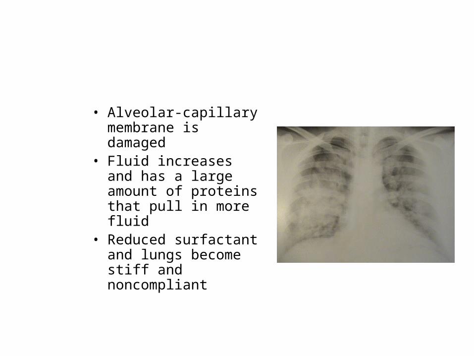
• Alveolar-capillary membrane is damaged
• Fluid increases and has a large amount of proteins that pull in more fluid
• Reduced surfactant and lungs become stiff and noncompliant

ARDS
• What other processes besides sepsis can cause ARDS?
• What will the patient look like?
• Note if the following is up or down.
• RR• BP• O2 level• CO2

ARDS
• What is the treatment of choice for a patient with ARDS?
• What should the paO2 be before intubation?• What medications may be given for
pulmonary infiltrates?• What is the main diagnoses related to this
problem?

Emergent Intubation• Oxygenate patient with
100% • Laryngoscope is used,
lighted, MD inserts it into the mouth and looks for the vocal cords, the tube slides past over the scope and into the trachea, above the carina
Intubation
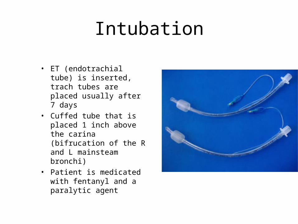
• ET (endotrachial tube) is inserted, trach tubes are placed usually after 7 days
• Cuffed tube that is placed 1 inch above the carina (bifrucation of the R and L mainsteam bronchi)
• Patient is medicated with fentanyl and a paralytic agent

Intubation Procedure
• Once inserted, you have to check for placement by:– CO2 device, turns from
yellow to purple– Listen to breath sounds
(bilateral)– Chest X ray
• Tape the tube in place, now use velcro
• Inflate the cuffed tube and continue to bag until placed on ventilator

Mechanical Ventilation
• Two types– Negative Pressure, non
invasive, body wrap that puts pressure on the patient’s body, they must breathe on their own
– Positive Pressure, what is used in the hospital

Mechanical Ventilation• During inspiration, pressure is generated that pushes air into
the lungs and expands the chest– Pressure cycled- preset airway pressure, push air in until
pressure reached, short periods of time, not used much– Time-cycled- push air into for a period of time– Volume-cycled-push air in to a preset volume, set to tidal
volume and pressure limits- type commonly used, with different modes

Volume-Cycled Ventilation
• Set the mode you want to deliver– Assist-Control- breathes for the patient, tidal
volume and rate are set, if the patient doesn’t breathe, the vent comes in at the preset rate and volume, because it is set to deliver a preset rate, once the patient starts to breathe on their own, the rate may be too fast. Used mostly when they come from surgery or if they are nonresponsive

Volume-Cycled Modes
• SIMV (synchronized intermittent Mandatory ventilation)– TV and rate are preset, but they don’t come in if the patient is
meeting the preset rate and volume– Positive airway pressure and PEEP may also be set
• PAP refers to a preset airway pressure to keep the bronchioles open (5-15cm)
• PEEP- positive end expiratory pressure- used to keep the alveoli open for gas exchange (5 cm)
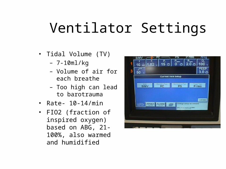
Ventilator Settings
• Tidal Volume (TV)– 7-10ml/kg– Volume of air for each
breathe– Too high can lead to
barotrauma• Rate- 10-14/min• FIO2 (fraction of inspired
oxygen) based on ABG, 21-100%, also warmed and humidified

Ventilator Settings
• Sighs- 1.5-2 x’s the TV delivered 6-10x’s/hr to mimic the patient’s natural sigh, not always used
• CPAP (continuous positive airway pressure), keeps alveoli open and prevents collapse on expiration -5-15cm
Ventilator Settings• Peak Airway Pressure (PIP)
– Pressure needed by the ventilator to deliver a set tidal volume
– Highest pressure reached during inspiration
– Increased PIP may be poor compliance, patient resistance, increasing fluid or kinked tubing
• Flow– How fast the ventilator delivers
each breathe– Set at 40L/min– If agitated may be that the flow
is too low• I/E ratio
– Inspiratory and expiratory ratio– Can set to have a longer
expiration to clear CO2 or longer inspiration to take in more O2

ARDS settings
• Typical early ARDS settings:– FIO2- 100%– TV- 500– Rate-14– AC mode– PAP of 10cm– PEEP of 5 cm
• Patient is sedated• Antibiotics and
Vasopressors are given

Nursing Care for the Ventilator patient
• Monitor breath sounds, bilateral, clear, etc
• Monitor Vital signs, BP and C.O. can drop if the PEEP is too high, creates too great a positive pressure and decreases return to the heart
• Monitor ABG’s, O2 sats• Assess the ETT, taped in
place, cuff inflated whenever on a vent, no leaks
• Keep patient comfortable, sedate as needed
Nursing Care for the Ventilated patient
• Monitor the vent settings, RT will monitor too and change along with MD
• Know your vent alarms- always an emergency
• Ventilator alarms• High- PIP is too high• What can cause this?
• Low alarm- can’t meet the presets
• What causes this?

Ventilator complications
• Cardiac complications– Hypotension- positive pressures, dehydration and high PIP– Fluid retention- decreased C.O., ARF, humidified air contributes
• Pulmonary complications– Barotrauma- pressure damage to alveoli, high PEEP, stiff lungs, can
lead to pneumothorax– VAP- ventilator associated pneumonia– Aspiration- often an orogastric tube is placed to suction out
secretions

Ventilator complications• VAP is a major nosocomial
infection• 48 hours after placement• Good aseptic technique• Inline suction- closed
system• HOB elevated 30 degrees• Mouth care

Ventilator complications• GI and nutritional
complications– Stress ulcers- give protonix
to all– Malnutrition- burn a lot of
calories on the vent– Need electrolyte and calorie
replacement, often corpak placed for feedings
• Muscle deconditioning– Become very weak and have
muscle atrophy• Ventilator dependence
– Inability to wean– Muscles fatigue and have to
relearn– Physical and psychological
Weaning
• Process of decreasing vent dependence• Can take a long time, months• Procedure is explained to the patient• Given an antianxiety medication, not a sedative, which would
decrease respirations• Go from SIMV mode down to PSV (pressure support mode) by
decreasing vent rate and volume. • On a trach by this time and they may be placed on a T-piece
of vent attachment only at night

Extubation
• Patient must be awake and alert• Be able to breath on their own, take a
sufficient TV• Lift their head off of the bed• Have adequate ABG’s• Stable hemodynamics (BP and C.O. stable, so
they won’t decompensate)• Suction before, oxygenate,deflate the balloon
and remove, then suction again