Embed Size (px)
Citation preview

NURS 2410 Unit 8 and 9 plus cardiac
Nancy Pares, RN, MSNMetro Community College
Pediatric Respiratory System Anatomy and Physiology Variances from the Adult
• Anatomy of airway• Comparison of airway structures

Figure 25-1 It is easy to see that a child’s airway is smaller and less developed than an adult’s airway, but why is this important? The infant and child are more vulnerable to the consequences of an upper respiratory tract infection, enlarged tonsils and adenoids, an allergic reaction, positioning of the head and neck during sleep, and small objects that can be aspirated. All can cause an airway obstruction that results in respiratory distress.

Pediatric Respiratory System Anatomy and Physiology Variances from the Adult
• Upper airway differences– Airway diameter

Figure 25-3 The diameter of an infant’s airway is approximately 4 mm, in contrast to an adult’s airway diameter of 20 mm. An inflammatory process in the airway causes swelling that narrows the airway, and airway resistance increases. Note that swelling of 1 mm reduces the infant’s airway diameter to 2 mm, but the adult’s airway diameter is only narrowed to 18 mm. Air must move more quickly in the infant’s narrowed airway to get the same amount of air to the lungs. The friction of the quickly moving air against the side of the airway increases airway resistance. The infant must use more effort to breathe and breathe faster to get adequate oxygen.
Pediatric Respiratory System Anatomy and Physiology Variances from the Adult
• Upper airway differences– Position of trachea

Figure 25-2 In children, the trachea is shorter and the angle of the right bronchus at bifurcation is more acute than in the adult. Where is an aspirated foreign body likely to land? When you are resuscitating or suctioning, you must allow for the differences in the length of the trachea because it is easier to slip into the right bronchus with an endotracheal tube or suction catheter.

Pediatric Respiratory System Anatomy and Physiology Variances from the Adult
• Upper airway differences– Position of right mainstem bronchus– Airway resistance

Pediatric Respiratory System Anatomy and Physiology Variances from the Adult
• Lower airway differences– Growth of alveoli
• Diaphragm use for respirations– Use of accessory muscles
• Immaturity of respiratory system

Respiratory Conditions and Injuries That Can Cause Respiratory Distress in Infants and Children
• Airway obstruction• Blockage of airway passages by different
causes– Foreign-body aspiration

Figure 25-5 An aspirated foreign body (coin) is clearly visible in the child’s trachea on this chest radiograph.Source: Courtesy of Rockwood Clinic, Spokane, WA.

Respiratory Conditions and Injuries That Can Cause Respiratory Distress in Infants and Children
• Acute respiratory distress syndrome (ARDS)

Figure 25-7 A ventilation-perfusion mismatch can occur when an infant or child has an abnormal distribution of ventilation or perfusion. A, Children with normal lung function and circulation have a ventilation-perfusion ratio of 0.8 to 0.9 because perfusion is greater than ventilation (air exchange) in the lung bases. B, When ventilation is inadequate to well-perfused areas of the lungs, the ventilation-perfusion ratio is low or mismatched, resulting in shunting. Blood passing through the pulmonary capillaries gets less oxygen exchange than normal, and hypoxemia occurs. This is the case in asthma due to bronchoconstriction and in pneumonia because alveoli are filled with fluid. C, In the case of neonatal hyaline membrane disease the alveoli are collapsed, so blood passes through the alveolar capillaries and no oxygenation occurs. The ventilation-perfusion ratio is very low with significant shunting that does not respond to oxygen therapy because the capillary bed never gets exposed to the supplemental oxygen.

Respiratory Conditions and Injuries That Can Cause Respiratory Distress in Infants and Children
• Multiple factors may cause ARDS– Sepsis– Pneumonia– Meconium aspiration– Gastric content aspiration– Smoke inhalation– Near drowing


Clinical Manifestations of Respiratory Distress
• Dyspnea• Tachypnea• Grunting• Nasal flaring• Retractions

Figure 25-4 The chest wall is flexible in infants and young children because the chest muscles are immature and the ribs are cartilaginous. With respiratory distress, the negative pressure created by the downward movement of the diaphragm to draw in air is increased, and the chest wall is pulled inward causing retractions. Intercostal retractions are seen in mild respiratory distress. As the severity of respiratory distress increases, retractions can be seen in the substernal and subcostal areas. In cases of severe distress, accessory muscles (sternocleidomastoid and trapezius muscles) are used, and retractions are seen in the supraclavicular and suprasternal areas.

Assessment of Respiratory Status
• Quality of pulse• Quality of respirations• Color• Cough• Behavior changes• Signs of dehydration
Nursing Care
• ABC—airway, breathing, circulation• Determine if cause can be alleviated
– Foreign body
• Supportive care– Supplemental oxygen

Diagnostic Tests to Determine Oxygen Saturation
• Pulse oximetry• Arterial blood gases


Figure 25-10 The phrase “thumb sign” has been used to describe this enlargement of the epiglottis. Recall the trachea’s usual “little finger” size. Do you see the stiff, enlarged “thumb” above it in this lateral neck radiograph?

Pulmonary Function for Chronic Conditions
• Force vital capacity (FVC)• Peak expiratory flow rate (PEFR)


Pulmonary Function for Chronic Conditions
• Forced expiratory volume in 1 second (FEVI)

Apnea in Infants and Children
• Cessation of respirations for longer than 20 seconds
• Obstructive apnea• Central apnea• Mixed apnea• Apnea of prematurity• Apparent life-threatening events

Apnea Monitors
• Polysomnography

Respiratory Assessment
• Determine baseline status of child• Provide pulmonary therapies as needed• Maintain oxygenation

Increased Metabolic Activity
• Increased need for calories/nutrition• Increased need for fluid
Anxiety and Fear Common
• Psychosocial support for parent• Psychosocial support for child• Discharge Planning
– Education about duration of illness– Need for follow up– When to seek emergency care
• Home care planning– Education to parents
Nursing Considerations for Chronic Respiratory Conditions
• Oxygenation• Activity intolerance• Nutrition• Growth and development• Treatment management• Social interactions

Oxygenation
• Most important consideration– Assess and reassess– Hypoxia leads to chronic changes– Permanent changes in body systems

Figure 25-18 Digital clubbing.

Oxygenation
• Activity intolerance

Growth and Development
• Nutritional concerns– Need increased calories to meet body
requirements
• Developmental– Appropriate activities and interactions

Social Interactions
• Lack of peers for some• Decreased activity tolerance• Decreased age activities

Treatment Management
• Family collaboration required– Plan around family, if possible

Cystic Fibrosis
• Inherited autosomal recessive
• S/S: salty taste to skin; thick, sticky mucous, stool abnormalities; huge appetite, wt maintenance
• Dx: lab value of IRT

• Treatment:– Focus on airway maintenance, infection
prevention; GI tract therapy, nutrition
– Meds: pg 898– Story pg 901

Broncho pulmonary dysplasia
• Persistence of premature lungs; usually in neonates on oxygen-esp ventilators
• S/S: increased resp effort, grunting, retractions, intermittent bronchospasms
• Dx: x ray; barrel shaped chest• Tx: focused on prevention by close monitoring
in ICU; meds pg 876; health promotion pg 878

Anatomy of Heart
• Atria• Ventricles• Vena cava• Pulmonary artery and vein

Hemodynamics of Heart (Circulatory System)
• Heart pumps blood– Pulmonary system
• Receives oxygen
– Return to heart– To systemic system
• Provides oxygen to organs and tissues• Depletes oxygen stores
– Return to heart

Transition from Fetal to Pulmonary Circulation
• Occurs within few hours after birth• Completes at approximately days 10 to 21
with permanent closure of ductus arteriosus


Transition from Fetal to Pulmonary Circulation
• Hemodynamics change– Increased pulmonary blood flow– Decreased pulmonary vascular resistance– Left atrium increased blood flow
• From lungs through pulmonary veins

Figure 26-4 The arrows indicate the flow of blood through the heart while the color indicates the level of oxygen saturation in the blood. A, Fetal (prenatal) circulation. B, Pulmonary (postnatal) circulation. LA, left atrium; LV, left ventricle; RA, right atrium; RV, right ventricle.

Transition from Fetal to Pulmonary Circulation
• Hemodynamics change– Right atrial pressure falls– Increased pressure in left atrium
• Stimulates closure of foramen ovale
– Higher oxygen saturation, then fetal circulation• Stimulates closure of ductus arteriosus

Normal Hemodynamics of Heart
• Cardiac function• Pressure gradients

Figure 26-1 Normal pressure gradients and oxygen saturation levels in the heart chambers and great arteries. The ventricle on the right side of the heart has a lower pressure during systole than the left ventricle because less pressure is needed to pump blood to the lungs through the rest of the body.

Heart Size
• Proportionately larger in children
Cardiovascular System Growth
• Continues until puberty

Increased Pulmonary Blood Flow
• Defects that cause

Patent Ductus Arterious
• Incidence and etiology
• Patho: Left to right shunting
• Clinical manifestations:– Asymptomatic– CHF
• Dx– Continuous murmur below left clavicle– X ray
• Treatment– Indomethocin for preterm only– Surgery– Non surgical closure


ASD
• Etiology
• Patho:• Dx:• Treatment:
– Diuretics– Surgical repair

Table 26-7 (continued) Pathophysiology, Clinical Manifestations, and Clinical Therapy for Heart DefectsThat Increase Pulmonary Blood Flow


VSD
• Patho:– Left to right shunting– Heart enlargement– Pulmonary vessel congestion
• Dx: loud holosytolic murmur• Tx: may close by 2 years of age; surgery

Table 26-7 (continued) Pathophysiology, Clinical Manifestations, and Clinical Therapy for Heart DefectsThat Increase Pulmonary Blood Flow

Increased Pulmonary Blood Flow
• Common manifestations– Tachypnea– Tachycardia– Congestive heart failure

Decreased Pulmonary Blood Flow
• Defects that cause

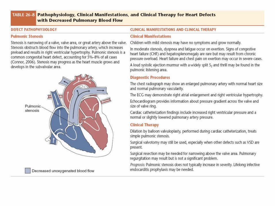
Pulmonary stenosis
• Path: – Obstruction of flow from RV to PA; increase RV
pressure
• S/S: dyspnea on exertion
• Tx: surgical; balloon valvuloplasty

Tetrology of Fallot
• Ventricular septal defect; pulmonary stenosis; right ventricular hypertrophy; overriding aorta;
• S/S: cyanotic vs. non cyanotic• Tx: surgical correction: pre op management;
modified Blalock-Taussig shunt

Table 26-8 (continued) Pathophysiology, Clinical Manifestations, and Clinical Therapy for Heart Defectswith Decreased Pulmonary Blood Flow


Decreased Pulmonary Blood Flow
• Common manifestations– Cyanosis– Hypercyanotic spells– Poor weight gain– Polycythemia– Tricuspid atresia
Obstructed Systemic Blood Flow
• Defects that cause

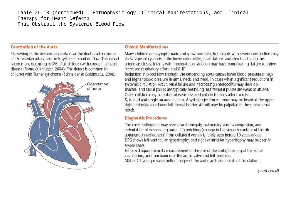
Table 26-10 (continued) Pathophysiology, Clinical Manifestations, and Clinical Therapy for Heart DefectsThat Obstruct the Systemic Blood Flow

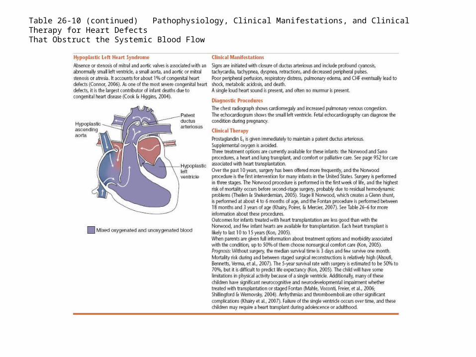
Table 26-10 (continued) Pathophysiology, Clinical Manifestations, and Clinical Therapy for Heart DefectsThat Obstruct the Systemic Blood Flow

Obstructed Systemic Blood Flow
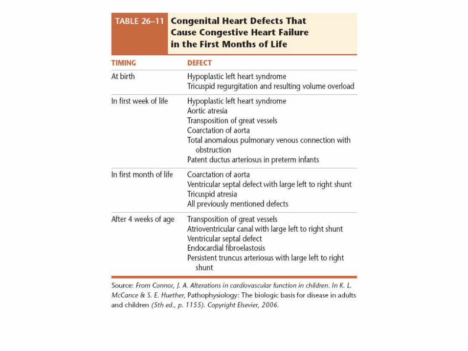
• Common manifestations– Diminished pulses– Pale color– Delayed capillary refill– Decreased urinary output– Signs of congestive heart failure

Education of Family
• Family-centered plan• Home care and planning• Assessment for complications• Assessment for worsening condition• Oxygenation requirements• Metabolic and nutritional needs• Fluid-volume balance

Education of Family
• Skin integrity• Management of illness• Medications• Other therapeutic interventions• Prevention of complications• Family interactions• Family adjustment and issues

Postoperative Care of Heart Surgery
• Immediate care– Intensive care unit until stable
• One or more days

Postoperative Care of Heart Surgery
• Hospital management focus– Pain
• Medications• Nonmedicated management of pain
– Rest– Respiratory functions– Fluid balance

Postoperative Care of Heart Surgery
• Hospital management focus– Nutrition status– Discharge planning– Home care teaching– Home care follow-up– Long-term care and follow-up

Congestive Heart Failure (CHF)
• Etiology

Assessment of CHF
• Respiratory• Pulse• Blood pressure• Color• Heart

Assessment of CHF
• Fluid status• Activity• Behavior• General

Clinical Manifestations
• Subtle signs– Early stage CHF
• Advanced signs– Late stage CHF

Nursing Management of CHF
• Assessment of child and family• Promote oxygenation• Cardiovascular function• Administration of medications• Growth and development• Family planning• Family education for home care

Congenital Heart Disease
• Definition—born with defect
Acquired Heart Disease
• Definition—defect related to illness– Infective endocarditis– Rheumatic fever– Kawasaki syndrome

Figure 26-15 This child has returned for one of her frequent follow-up visits to assess her cardiac status after treatment for Kawasaki syndrome. Notice the lips that show the inflammation and cracking.

Hypovolemic Shock
• Definition—acute complex state of circulatory dysfunction
• Results in failure to deliver sufficient oxygen to meet demands

Figure 26-17 If hemorrhage reduces the circulating blood volume sufficiently then vasoconstriction occurs, shifting blood to larger blood vessels that maintain the perfusion of vital organs. When the blood loss exceeds 20% to 25%, the child’s body can no longer compensate and hypovolemic shock ensues.
Etiology of Shock
• Hemorrhage• Dehydration• Sepsis• Obstruction of blood flow• Cardiac pump failure

Nursing Management
• Early intervention to treat etiology• Interventions aimed to prevent falling blood
pressure

Gastrointestinal System
• Digestion takes place in duodenum• Enzymes aid in the digestion process

Figure 30-1 The internal anatomic structures of the stomach, including the pancreatic, cystic, and hepatic ducts; the pancreas; and the gallbladder.

Child vs. Adult Gastrointestinal System
• Liver function immature at birth• Enzymes deficient until 4 to 6 months old• Abdominal distention from gas common with
infants• Stomach capacity smaller

Disorders of Gastrointestinal System
• Define congenital defects• Define acquired defects• Define infectious defects

Types of Pathophysiology Associated with Structural Defects of the Gastrointestinal System
• Cleft lip and cleft palate– Definition– Failure of the maxillary processes to fuse between
5 and 12 weeks’ gestation– Failure of the tongue to move down at the correct
time prevents the palatine processes from fusing– Multifactorial causes

Figure 30.3 A, Unilateral cleft lip.

Figure 30.3 (continued) B, Bilateral cleft lip. Source: Courtesy of Dr. Elizabeth Peterson, Spokane, WA.
Types of Pathophysiology Associated with Structural Defects of the Gastrointestinal System
• Esophageal atresia and tracheoesophageal fistula– Definition– Foregut fails to lengthen, separate, and fuse into
two parallel tubes (esophagus and trachea) at 4 to 5 weeks’ gestation
• Associated with maternal polyhydramnios

Figure 30-7 In the most common type of esophageal atresia and tracheoesophageal fistula, the upper segment of the esophagus ends in a blind pouch connected to the trachea; the fistula connects the lower segment to the trachea.
Types of Pathophysiology Associated with Structural Defects of the Gastrointestinal System
• Pyloric Stenosis– Definition– Etiology unknown– Hypertrophy of the circular pylorus muscle– Stenosis occurs between stomach and duodenum

Figure 30-9 In pyloric stenosis, the hypertrophied pyloric muscle causes symptoms of projectile vomiting and visible peristalsis.

Types of Pathophysiology Associated with Structural Defects of the Gastrointestinal System
• Gastroesophageal reflux– Definition– Three mechanisms allow reflux to occur
• Lower esophageal relaxations• Incompetent lower esophageal sphincter• Anatomic disruption of esophagogastric junction
– Reflux acidity damages the esophageal mucosa– Causes

Types of Pathophysiology Associated with Structural Defects of the Gastrointestinal System
• Gastroschisis and omphalocele– Definition– Gastroschisis usually occurs to the right of the
umbilicus and omphalocele occurs through the umbilical cord
– Occurs in week 11 of gestation when abdominal contents fail to return to the abdomen
– Multifactorial causes
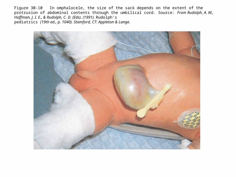
Figure 30-10 In omphalocele, the size of the sack depends on the extent of the protrusion of abdominal contents through the umbilical cord. Source: From Rudolph, A. M., Hoffman, J. I. E., & Rudolph, C. D. (Eds). (1991). Rudolph’spediatrics (19th ed., p. 1040). Stamford, CT: Appleton & Lange.
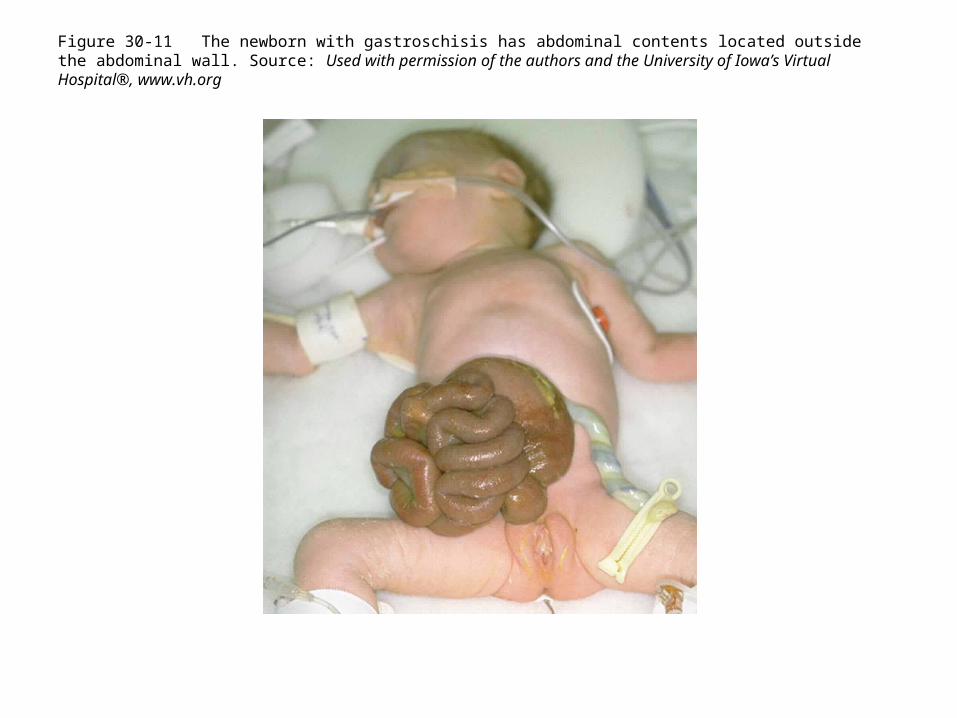
Figure 30-11 The newborn with gastroschisis has abdominal contents located outside the abdominal wall. Source: Used with permission of the authors and the University of Iowa’s Virtual Hospital®, www.vh.org
Types of Pathophysiology Associated with Structural Defects of the Gastrointestinal System
• Intussusception– Intestine invaginates into another– Mesentery becomes inflamed and obstruction can
occur– Multifactorial causes

Figure 30-12 In infants, intussusception is commonly associated with viral illnesses and gastroenteritis.
Types of Pathophysiology Associated with Structural Defects of the Gastrointestinal System
• Volvulus– Occurs in 7th to 12th week of gestation– 1 in 6,000 live births– Malrotation of bowel interrupts blood flow and
causes bowel necrosis– Surgical emergency

Types of Pathophysiology Associated with Structural Defects of the Gastrointestinal System
• Hirschsprung disease– Definition– Congenital absence of ganglion cells in the rectum
and colon– Genetically acquired and occurs when there is
failure of the migration of neural crest cells in utero
– Colon becomes a “megacolon”

Types of Pathophysiology Associated with Structural Defects of the Gastrointestinal System
• Anorectal malformations– Anal stenosis and anal atresia– Failure of growth of urorectal septum, lateral
mesoderm structures, and ectodermal structures– Associated anomalies up to 70% of the time

Types of Pathophysiology Associated with Structural Defects of the Gastrointestinal System
• Congenital diaphragmatic hernia– Protrusion of abdominal contents into thoracic
cavity– Occurs in 4th week of gestation– Failure of pleuroperitoneal musculature to close

Types of Pathophysiology Associated with Structural Defects of the Gastrointestinal System
• Umbilical hernia– Definition– Etiology unknown– Around week 11 of gestation, the obliterated
umbilical vessels occupy the space in the umbilical ring

Figure 30-13 The umbilical hernia of the newborn usually closes as the muscles strengthen in later infancy and childhood. Source: From Zitelli, B., & Davis, H. (Eds.). (2007). Atlas of pediatric physical diagnosis. (5th ed., p. 48). St. Louis, MO: Mosby.

Pathophysiology Associated with Inflammatory Disease
• Necrotizing enterocolitis– Inflammatory disease producing vascular
compromise of bowel mucosa– More common in premature infants– Caused by intestinal ischemia, bacterial or viral
infection, and immature gastrointestinal mucosa

Pathophysiology Associated with Inflammatory Disease
• Meckel’s diverticulum– Omphalomesenteric duct fails to atrophy– Outpouching of the ileum remains and contains
gastric contents, causing ulceration– Bowel obstruction, perforation, or peritonitis can
occur

Pathophysiology Associated with Inflammatory Disease
• Inflammatory bowel disease (Crohn’s disease and ulcerative colitis)– Faulty regulation of the immune response of the
intestinal mucosa– Usually genetically triggered– Crohn’s disease can cause inflammation and
ulcers anywhere throughout the GI tract– Ulcerative colitis affects large intestine and rectal
mucosa

Pathophysiology Associated with Inflammatory Disease
• Pathophysiology of motility disorders• Gastroenteritis
– Definition– Acute vs. chronic diarrhea caused by viruses,
bacteria, or parasites– Causes of diarrhea in children


Pathophysiology of Disorders of Malabsorption
• Celiac disease– Immunologic disorder; characterized by
intolerance for gluten– Impairs absorptive process in the small intestine– Affects fat absorption

Pathophysiology of Disorders of Malabsorption
• Lactose intolerance– Inability to digest lactose– Lactose enzyme deficiency– Usually acquired, but can be congenital

Pathophysiology of Disorders of Malabsorption
• Short bowel syndrome– Shortened intestine resulting from bowel
resection– Extent of bowel loss determines severity of
disorder– Location of bowel resection determines type of
malabsorption
Pathophysiology of Hepatic Disease
• Hepatic disorders– Biliary atresia– Viral hepatitis– Cirrhosis
Pathophysiology of Injuries to the Gastrointestinal System
• Abdominal trauma– Blunt or penetrating trauma to the abdomen– Common causes
• Falls• Motor vehicle accidents• Automobile vs. pedestrian accidents• Child abuse• Gunshot wounds

Pathophysiology of Injuries to the Gastrointestinal System
• Abdominal trauma– Organs commonly involved
• Liver• Spleen

Signs of Hepatic Disorders
• Jaundice• Easy bruising, intense itching• White or clay-colored stools• Tea-colored urine

Clues to Gastrointestinal Disorders in Children
• Vomiting or abdominal pain• Failure to thrive• Stool changes
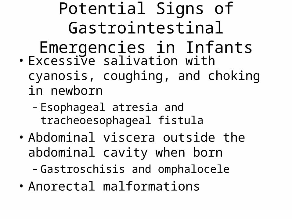
Potential Signs of Gastrointestinal Emergencies in Infants
• Excessive salivation with cyanosis, coughing, and choking in newborn– Esophageal atresia and tracheoesophageal fistula
• Abdominal viscera outside the abdominal cavity when born– Gastroschisis and omphalocele
• Anorectal malformations

Potential Signs of Gastrointestinal Emergencies in Children
• Abdominal pain• Changes in appearance of stool• Vomiting and/or anorexia• Changes in activity• Changes in level of consciousness

Cleft Lip and Cleft Palate
• Nursing care• Pre- and postoperative care

Esophageal Atresia and Tracheoesophageal Fistula
• Nursing care– Identifying signs and symptoms of these infants
• Pre- and postoperative care– Suction is important preoperatively– Care of the gastrostomy tube postoperatively

Pyloric Stenosis
• Nursing care• Pre- and postoperative care

Gastroesophageal Reflux
• Nursing care• Important education

Infant (Birth to 1 Year Old)
• Congenital defects• Gastroesophageal reflux in infant vs. older
child• Gastrointestinal disorders specific to this age
group

Toddlers (1 Year Old to 3 Years Old)
• Meckel’s diverticulum• Offer age-appropriate toys• Childproof the room• Use pictures for education of older toddler

Children (3 Years Old to 12 Years Old)
• Body image starts becoming important after 5 years old
• Offer age-appropriate toys• Use pictures for education of younger child• Umbilical hernia repaired

Adolescents (13 Years Old to 17 Years Old)
• Appendicitis (10 to 19 years old)• Body image extremely important• Allow use of phone to satisfy peer needs• Give them handouts about peers with
conditions and experiences

LEARNING OUTCOME 6
• Discuss nursing management of the child with an injury to the abdomen.

Abdominal Trauma
• Provide emotional support• Follow care orders• Prevention teaching once stabilized