Embed Size (px)
Citation preview
7/23/2019 Novel Therapeutic Agents From Plants
http://slidepdf.com/reader/full/novel-therapeutic-agents-from-plants 1/464

7/23/2019 Novel Therapeutic Agents From Plants
http://slidepdf.com/reader/full/novel-therapeutic-agents-from-plants 2/464
Novel Therapeutic Agents from Plants
© 2009 by Taylor & Francis Group, LLC

7/23/2019 Novel Therapeutic Agents From Plants
http://slidepdf.com/reader/full/novel-therapeutic-agents-from-plants 3/464
Editors:
María Cecilia CarpinellaProfessor and Researcher
School of ChemistryCatholic University of Córdoba
Córdoba, Argentina
Mahendra RaiProfessor and Head
Department of BiotechnologySGB Amravati University
Maharashtra, India
Novel Therapeutic
Agents from Plants
Science PublishersEnfield (NH) Jersey Plymouth
© 2009 by Taylor & Francis Group, LLC

7/23/2019 Novel Therapeutic Agents From Plants
http://slidepdf.com/reader/full/novel-therapeutic-agents-from-plants 4/464
Science Publishers www.scipub.net234 May StreetPost Office Box 699Enfield, New Hampshire 03748
United States of America
General enquiries : [email protected]
Editorial enquiries : [email protected]
Sales enquiries : [email protected]
Published by Science Publishers, Enfield, NH, USAAn imprint of Edenbridge Ltd., British Channel IslandsPrinted in India
© 2009 reserved
ISBN 978-1-57808-546-0
Library of Congress Cataloging-in-Publication Data
Novel therapeutic agents from plants/editors: Maria CeciliaCarpinella, Mahendra Rai. — 1st ed.
p. cm.
Includes bibliographical references and index.
ISBN 978-1-57808-546-0 (hardcover)
1. Materia medica, Vegetable. 2. Medicinal plants. I. Carpinella,
María Cecilia. II. Rai, Mahendra.
RS164.N68 2009
615’.321—dc22
2008041306
All rights reserved. No part of this publication may be reproduced,stored in a retrieval system, or transmitted in any form or by anymeans, electronic, mechanical, photocopying or otherwise, withoutthe prior permission of the publisher, in writing. The exception tothis is when a reasonable part of the text is quoted for purpose of book review, abstracting etc.
This book is sold subject to the condition that it shall not, by wayof trade or otherwise be lent, re-sold, hired out, or otherwise circulatedwithout the publisher’s prior consent in any form of binding or coverother than that in which it is published and without a similar conditionincluding this condition being imposed on the subsequent purchaser.
© 2009 by Taylor & Francis Group, LLC

7/23/2019 Novel Therapeutic Agents From Plants
http://slidepdf.com/reader/full/novel-therapeutic-agents-from-plants 5/464

7/23/2019 Novel Therapeutic Agents From Plants
http://slidepdf.com/reader/full/novel-therapeutic-agents-from-plants 6/464
Preface ix
1. Anticancer Compounds of Plant Origin 1 Modhumita Ghosh, Manisha Thapliyal and Krish Gurumurthi
2. Antimicrobial and Antiviral Metabolites from 36
Polypore Fungi Jordan K. Zjawiony
3. Naturally Occurring Anti-Salmonella Agents and 60Their Modes of ActionIsao Kubo, Ken-ichi Fujita and Aya Kubo
4. Natural Chemotherapeutic Agents in the 84
Control of MalariaEdith O. Ajaiyeoba
5. Anti-Candida Activity of Extracts and Essential Oils 123from Native and Exotic Medicinal Plants in Brazil
Marta Cristina Teixeira Duarte and Glyn Mara Figueira
6. Bridging the Gap: Using Natural Products in 167Drug Discovery Research in Academia and Industry
Alan L. Harvey
7. Fungal Endophytes: A Potential Source of 176Anticancer CompoundsSunil Deshmukh and Shilpa A. Verekar
8. A Review of Antifungal and Antiviral Proteins 208Tzi Bun Ng and Jack H. Wong
9. Compounds with Antioxidant Activity from Herbs 239Tzi Bun Ng and Jack H. Wong
Contents
© 2009 by Taylor & Francis Group, LLC

7/23/2019 Novel Therapeutic Agents From Plants
http://slidepdf.com/reader/full/novel-therapeutic-agents-from-plants 7/464
10. Bioactivity of Medicinal Plants: Progress and 266Perspectives José-Luis Ríos and Rosa A. Sendra
11. Cannabimimetics: The Pharmacological Potential of 301Cannabinoid Receptor Ligands Jürg Gertsch
12. Anti-inflammatory Herbal Medicines for the 339Control of Pain Julia Elodie Vlachojannis, Rujee K. Duke, Van H. Tran,Colin C. Duke and Sigrun Chrubasik
13. Metabolic Engineering for the Fabrications of 368Pharmaceutically Central Metabolites fromMicroorganisms and Higher Plants Mahmud Tareq Hassan Khan and Arjumand Ather
14. Pharmacogenomics of Biotic and Abiotic Natural 387Products Derived from Traditional Chinese
Medicine for Cancer TherapyThomas Efferth and Bernd Kaina
15. Plant Derived Antimycobacterial Metabolites: 405An Overview Adewole L. Okunade and Memory P.F. Elvin-Lewis
Index 453
vi
Color Plate Section 479
© 2009 by Taylor & Francis Group, LLC

7/23/2019 Novel Therapeutic Agents From Plants
http://slidepdf.com/reader/full/novel-therapeutic-agents-from-plants 8/464
Adewole L. Okunade
Center for Human Nutrition, Division of Geriatrics and Nutritional Science,Department of Internal Medicine, Washington University School of Medicine,St. Louis, MO 63110, U.S.A. Tel.: 314 747 4052; Fax: 314 362 8032; E-mail:[email protected]
Alan L. HarveyStrathclyde Institute for Drug Research, University of Strathclyde, 27 TaylorStreet, Glasgow G4 0NR, UK. E-mail: [email protected] [email protected]
Edith O. Ajaiyeoba
Department of Pharmacognosy, Faculty of Pharmacy, University of Ibadan,Ibadan, Nigeria. E-mail: [email protected]
Isao Kubo
Department of Environmental Science, Policy and Management, Universityof California, Berkeley, California 94720-3114. E-mail: [email protected]
Jordan K. Zjawiony
Department of Pharmacognosy, School of Pharmacy, Post Office Box 1848,The University of Mississippi, University MS 38677, USA. Tel.: (662) 915-7290;
Fax: (662) 915-6975; E-mail: [email protected]
José-Luis Ríos
Departament de Farmacologia, Facultat de Farmacia, Universitat de Valencia,Av. Vicent Andres Estelles s/n, 46100 Burjassot (Valencia, Spain). Tel./Fax: +34 963 544 498; E-mail: [email protected]
Julia Elodie Vlachojannis
Orthodontic Department, Columbia University, 630 West 168th, VC 9, Rm219B, NYC 10032, NY. E-mail: [email protected]
List of Contributors
© 2009 by Taylor & Francis Group, LLC

7/23/2019 Novel Therapeutic Agents From Plants
http://slidepdf.com/reader/full/novel-therapeutic-agents-from-plants 9/464
Jürg Gertsch
Institute of Pharmaceutical Sciences, ETH Zurich, Wolfgang-Pauli-Str. 10, CH-8093 Zürich, Switzerland. E-mail: [email protected]
Mahendra RaiDepartment of Biotechnology, SGB Amravati University, Amravati-444 602,Maharashtra, India. Tel.: 91 721-2667380 (Home); 91 721-2662207; Extn. 267;Cell: 9422857196; Fax: 91 721-2660949; E-mail: [email protected] [email protected]
Mahmud Tareq Hassan Khan
School of Molecular and Structural Biology, and Department of Pharma-
cology, Institute of Medical Biology, University of Tromsø, Tromsø 9037,Norway. E-mail: [email protected]
María Cecilia Carpinella
Fine Chemicals and Natural Products Laboratory, School of Chemistry,Catholic University of Córdoba, Camino a Alta Gracia Km 10, (5000)Córdoba, Argentina. E-mail: [email protected]
Marta Cristina Teixeira Duarte
CPQBA/UNICAMP—Centro Pluridisciplinar de Pesquisas Químicas,Biológicas e Agrícolas—Universidade Estadual de Campinas, Campinas—São Paulo, Brasil, P.O. Box 6171, CEP: 13081-970. E-mail: [email protected]
Modhumita Ghosh
Division of Plant Biotechnology, Institute of Forest Genetics & Tree Breeding,Forest Campus, R.S. Puram, Coimbatore-641 002. India. E-mail: ghoshm@
ifgtb.res.in
Sunil Deshmukh
Department of Natural Products, Piramal Life Sciences Limited, 1, NirlonComplex, Off Western Express Highway, Goregaon (East), Mumbai 400 063,India. E-mail: [email protected]
Thomas Efferth
Pharmaceutical Biology (C015), German Cancer Research Center, ImNeuenheimer Feld 280, 69120 Heidelberg, Germany. Tel.: 49-6221-423426;Fax: 49-6221-423433; E-mail: [email protected]
Tzi Bun Ng
Department of Biochemistry, The Chinese University of Hong Kong, Shatin,New Territories, Hong Kong, China. E-mail: [email protected]
viii
© 2009 by Taylor & Francis Group, LLC

7/23/2019 Novel Therapeutic Agents From Plants
http://slidepdf.com/reader/full/novel-therapeutic-agents-from-plants 10/464
Since the advent of synthetic drugs, the use of natural products hasdiminished. However, the diversity of natural molecules still surpassesthose from synthetic compounds, and this ensures that natural productswill continue to be important for drug discovery. Besides, many of thecurrently used synthetic drugs are responsible for side-effects and are
sometimes extremely costly.Plants and microorganisms are capable of producing secondary
metabolites with a wide range of biological activities which are involvedin defensive and protective processes in the hosts. This property of determined organisms should be exploited by man for obtaining newtherapeutic preparations or to serve as leading molecules for the synthesisof analogues.
The use of plants in medicine has evolved because, from the biochemical point of view, they produce different chemical compoundswith particular modes of action for controlling pathogenic organisms orinhibiting processes involved in many diseases. There are a large numberof chemical structures and only a few are well known. The differentchemical structures present in a plant -derived extract ensure that targetorganisms do not develop resistance, and have high effectiveness due todifferent modes of actions and/or synergism between compounds. Mostof these secondary metabolites are harmless, thus ensuring safe clinicaluse.
A scientific approach to understand the relationship between activechemicals and cure of diseases is recent. More properties and characteristicsof the wide range of natural compounds come to light everyday. Suchvoluminous data has to be compiled and described on the basis of scientificcriteria.
While there are several books on natural drugs, the present book coversmultiple curative aspects of natural chemicals. This book is a complete
Preface
© 2009 by Taylor & Francis Group, LLC

7/23/2019 Novel Therapeutic Agents From Plants
http://slidepdf.com/reader/full/novel-therapeutic-agents-from-plants 11/464
review of medicinally active metabolites produced by nature and lookedat from different approaches.
The book describes the effects of natural extracts and/or their isolated
compounds and also gives an update of their in vitro and in vivo effective-ness, active doses, modes of action, production and commercialisation.
María Cecilia CarpinellaMahendra Rai
x
© 2009 by Taylor & Francis Group, LLC

7/23/2019 Novel Therapeutic Agents From Plants
http://slidepdf.com/reader/full/novel-therapeutic-agents-from-plants 12/464
Introduction
Cancer is a malignant neoplastic disease characterized by uncontrolledgrowth of cells with the ability of the cells to migrate and spread to distantsites (American Cancer Society, 2006). There are different types of cancers.Carcinomas are cancers that arise in the epithelium (layers of cells coveringthe body’s surface and lining the internal organs and various glands).Ninety percent of human cancers fall into this category. Melanomas arecancers that originate in the skin, usually in the pigment cells (melanocytes)while sarcomas are cancers of the supporting tissues of the body such as
bone, muscle and blood vessels. Cancers of the blood and lymph glandsare called leukemias and lymphomas respectively, while gliomas are
cancers of the nerve tissue.Cancer is a leading cause of death worldwide. In a total of 58 million
Anticancer Compounds ofPlant Origin1Modhumita Ghosh
1, *
, Manisha Thapliyal2
andKrish Gurumurthi3
1Division of Plant Biotechnology, Institute of Forest Genetics& Tree Breeding, Forest Campus, R.S. Puram, Coimbatore641 002, India. E-mail: [email protected],
2Forest Tree Seed Laboratory, Silviculture Division, ForestResearch Institute, Dehradun, India. E-mail: [email protected].
3Director (Retd.), Institute of Forest Genetics and TreeBreeding, Forest Campus, R.S. Puram, Coimbatore-641 002, India. E-mail: [email protected]
*Corresponding author: Modhumita Ghosh , Division of PlantBiotechnology, Institute of Forest Genetics and TreeBreeding, Forest Campus, R.S. Puram, Coimbatore-641 002, India. Tel.: 91-422-2431540, 2435541, 2450302, Fax:91-422-2430549, E-mail: [email protected]
© 2009 by Taylor & Francis Group, LLC

7/23/2019 Novel Therapeutic Agents From Plants
http://slidepdf.com/reader/full/novel-therapeutic-agents-from-plants 13/464
2
deaths worldwide in 2005, cancer accounted for 7.6 million (or 13 percent)of deaths. The main types of cancer leading to overall cancer mortalityinclude lung (1.3 million deaths/year); stomach (almost 1 million deaths/year); liver (662,000 deaths/year); colon (655,000 deaths/year) and breast(502,000 deaths/year). Deaths from cancer in the world are projected tocontinue rising, with an estimated 9 million people dying from cancer in2015 and 11.4 million dying in 2030 (WHO, 2006).
Cancer treatment
The usual treatments for different groups of cancer are surgery, radiation
therapy and chemotherapy (treatment with anticancer drugs) or combi-nation of these methods.
Surgery is the main treatment for many types of solid tumors, especiallywhen the cancer has not spread to other parts of the body. This involvessurgical removal of all or part of the cancerous tissue. Sometimes it is usedin conjunction with chemotherapy and/or radiation therapy.
Radiation therapy involves the use of high-level ionizing radiation to
destroy cancer cells. Both tumor cells and healthy cells may be affected bythis radiation. Radiation injures the cancer cells and they can no longercontinue to divide or multiply. They can be generated either by X-rays or
by gamma rays. Other recent radiotherapy research has focused on the useof radio labeled antibodies to deliver doses of radiation directly to thecancer site (radioimmunotherapy).
Chemotherapy is the treatment of cancer with ‘anticancer’ drugs to
destroy cancer cells throughout the body. Chemotherapy may be used to:• Cure the cancer
• Prevent the cancer from spreading
• Destroy cells that have spread beyond the tumor
• Decrease the size of the tumor
• Relieve symptoms, such as pain
Drugs used in chemotherapy have been classified according to themechanism of action. It includes alkylating agents like cisplastin andmitomycin; antimetabolites like methorexate, trimetrexate, hydroxyurea andfluorouracil; DNA cutters like bleomycin and DNA binders like dactino-mycin. The plant derived or semi-synthetically plant derived drugs thatare used in chemotherapy include, taxol, taxotere, vinbalstine, vincristine,vindesine, vinorelbine, teniposide, etaposide, topotecan and irinotecan
© 2009 by Taylor & Francis Group, LLC

7/23/2019 Novel Therapeutic Agents From Plants
http://slidepdf.com/reader/full/novel-therapeutic-agents-from-plants 14/464
3
PLANT DERIVED ANTICANCER DRUGS
Anticancer drugs derived from plants are believed to have tremendouspotential for treatment of various types of cancer. Seven plant based
anticancer drugs have received Food and Drug Administration approvalfor commercial production in the USA (Taylor, 2000). Some of the moststudied and marketed anticancer drugs are described below.
Paclitaxel (Taxol®)
Paclitaxel, a diterpene, is derived from Taxus spp. belonging to the family
Taxaceae, a genus of evergreen trees and shrubs. Seven species of Taxushave been recognized and the two major species are Taxus baccata Linn.and Taxus brevifolia Nutt. T. baccata, commonly known as English yew, isnative to England, occurring locally in South Scotland, Ireland and Wales.It is also indigenous to central and southern Europe, Algeria and northernSpain and the distribution is primarily based on edaphic conditions. T.brevifolia also known as pacific yew, grows in moist soils along the Pacificcoast of south eastern Alaska, southward through western British
Columbia to central California (Elbert, 1979). It grows as an understory of conifers and has high shade tolerance (Klinka et al., 2000). The species isgenerally propagated through seeds (Mitiska, 1954) or by vegetativepropagation as in T. baccata (Mitter and Sharma, 1999) and T. brevifolia(Mitchell, 1997). The active components of the tree include taxol and
baccatin, isolated from the root bark and needles.
Taxol was first isolated from the bark of T. brevifolia by Wall and Wani
at the Research Triangle Institute (RTI) in 1967 (Wani et al., 1971) (Figure1). The broad range antitumor activity of the compound was reportedagainst rodent tumors (Wani et al., 1971). Taxol is the first drug of choicein several tumorous cancers including breast, ovarian, lung cancer,squamosa cell carcinoma, head and neck cancer and AIDS-related Kaposi’ssarcoma. In 1992, FDA approved the use of paclitaxel for ovarian cancerthat was refractory and in 1994 it was approved for treating breast cancer.In 1997, FDA approved it for treating AIDS-related Kaposi’s sarcoma whilein 1998 it received approval for use against ovarian and lung cancer. Therevenue earned from 1997-1998 by marketing the drug was US$ 45 millionwhile the gross revenue from all the patents in 1997-1998 was around US$57.3 million (Eisenstein and Resnick, 2001).
Originally, the only source of Taxol was the Pacific yew but presentlythe drug is made by a semi-synthetic process. Taxol has been identified inlesser quantities in other Taxus sp. like T. canadensis Marshall (American
yew) (Hezari et al., 1997; Phisalaphong and Linden, 1999), T. cuspidate Sieb.(Japanese yew) (Zhang and Su, 2000; Zhang and Su, 2002) and T. mairei
© 2009 by Taylor & Francis Group, LLC

7/23/2019 Novel Therapeutic Agents From Plants
http://slidepdf.com/reader/full/novel-therapeutic-agents-from-plants 15/464
4
Fig. 1. Structure of Paclitaxel.
Lemée & H. Lév. (Cui and Ge, 2004). Among different Taxus species anddifferent tissues of the tree, there is a variable taxol production rangingfrom zero to 0.069 percent (Castor and Theodore, 1993; Guy et al., 2002). Asemi-synthetic compound, Docetaxel (Taxotere®) from T. baccata was
developed by Rhone-Poulenc Rorer which was similar to paclitaxel. It wasmore water-soluble than taxol and was produced semi synthetically from10-deacetylbaccatin III, an inactive precursor, extracted from needles of T.baccata (Lavelle et al., 1995). It has been approved as a second-line agentfor advanced breast cancer and is found to be more effective than taxol.Other than taxol, a diterpenolignan was also isolated from bark of T.brevifolia called brevitaxin, showing cytotoxicity against tumor cell lines(Arslanian et al., 1995).
Studies have shown that endophytic fungi like Sporormia minimaAuersw. and Trichothecium sp. growing in T. wallichiana Zucc. producedpaclitaxel in culture medium (Shreshtha et al., 2001). Similarly Tuberculariasp. growing in T. mairei produced taxol in potato dextrose liquid medium.
Three total synthesis of taxol has been reported by Holton Route,Nilcolaou and Danishefsky route and Morihira route (Holton et al., 1994;Nicolaou et al., 1994; Danishefsky et al., 1996; Morihira et al., 1998).
However, the complex structure of the compound requires many chemicalreaction steps that make commercial-scale production of synthetic taxolunfeasible (Guo et al., 2006).
Mode of action
Taxol binds to β-tubulin subunit of microtubules and promotes assemblyof microtubules, stabilizes microtubules by preventing depolymerization
and causes formation of stabilized abnormal microtubule bundles (Schiff et al., 1979). It disrupts the equilibrium between free tubulin and
© 2009 by Taylor & Francis Group, LLC

7/23/2019 Novel Therapeutic Agents From Plants
http://slidepdf.com/reader/full/novel-therapeutic-agents-from-plants 16/464
5
microtubule by shifting direction of assembly and inhibits normal dynamicreorganization of microtubule network that is required for mitotic functions(Ganesh et al., 2004). This novel mode for action made taxol a prototypefor a new class of anticancer drugs. The fungal taxol was found to enhancemicrotuble stability and bundling in culture cells and induced tubulinpolymerization in vitro similar to the authentic plant taxol (Wang et al.,2000a).
Vinca alkaloids
Vinca alkaloids are derived from a small genus of perennial, evergreen
herbs commonly known as periwinkles (divided into 2 distinct groups,treated as separate genera—Vinca and Catharanthus) which belongs to thefamily Apocynaceae. The main area within which the species of Vinca arenative, extends eastwards from Morocco, Algeria, Portugal, Spain andFrance and over central and southern Europe and southwestern Russia.The genus Catharanthus is distributed in Madagascar and India. It ispropagated generally through seeds and occasionally by cuttings.
Work on the periwinkle plant, Catharanthus roseus Linn. G.Donn. wasindependently taken up in two different laboratories for its allegedhypoglycaemic activity. The Canadian group of Nobel Beer and Cuttssucceeded in isolating vinblastine while Eli Lilly group isolated vinblastineand vincristine along with two other active dimeric alkaloids (Duffin,2000). These alkaloids were present in extremely low concentrations in acomplex mixture of 50 different alkaloids. Vinblastine was introduced
(Velban
®
, Eli Lilly) in 1961 and vincristine (Oncovin
®
, Eli Lilly) in 1963 asanticancer drugs Figure 2). In India, CIPLA has improved upon the processof isolating vinblastine and vincristine from C. roseus as developed byNational Chemical Laboratory, Pune (Kaul, 2004).
The vinca alkaloids are considered to be cell phase-specific. It is usedfrequently in acute lymphatic leukemia, Ewing’s sarcoma, Hodgkins’sdisease, small cell lung cancer, non-Hodgkins’s lymphoma, rhabdo-myosarcoma, Wilm’s tumor, brain tumor, breast cancer, cervical cancer,chronic lymphocytic leukemia, chronic myelogenous leukemia, colorectalcancer, hepatoblastoma in children, Kaposi’s sarcoma, malignantmelanoma, multiple myeloma, neuroblastoma, osteogenic sarcoma, softtissue sarcoma and testicular cancer. A very important feature of vincaalkaloids is their relatively low toxicity.
Vinblastine is an antineoplastic drug and is the first drug of choice inmany forms of leukemias and since 1950s it has increased the survival
rate of childhood leukemias by 80 percent. It is used for treating cancer of breast, testicles, bladder, kidney, lungs, prostrate and malignant melanoma.
© 2009 by Taylor & Francis Group, LLC

7/23/2019 Novel Therapeutic Agents From Plants
http://slidepdf.com/reader/full/novel-therapeutic-agents-from-plants 17/464
6
It has a half-life of 24 hours in the bloodstream and is also known tointerfere with the glutamic acid metabolism. Vincristine is anotherantileukemic, antineoplastic drug marketed by Eli Lilly with a half-life of 85 hours in the serum.
Other than vinblastine and vincristine, a new semi-syntheticcompound with structural modification from catharanthine unit wassynthesized and named vinorelbine (Figure 3). Vinorelbine is effectiveagainst non-small cell lung cancer and breast cancer. It is used as a singleagent or in combination with cisplatin for the first-line treatment of advanced, unresectable non-small cell lung cancer (NSLC), and is the onlysingle-agent therapeutic to treat NSLC. Another semi synthetic compound
derived from vinblastine is Vindesine (Figure 4), which is mainly used totreat melanoma and lung cancer (Huang, 1999).
Fig. 2. Structure of Vinblastine and Vincristine.
Fig. 3. Structure of Vinorelbine.
HO
C H2 5
N
N
N
R
HO
NH
H C O C3 2
H CO3
CO CH2 3
OCOCH3
C H2 5
H
R =Ch
Vinblastine3 R =CHO
Vincristine
© 2009 by Taylor & Francis Group, LLC

7/23/2019 Novel Therapeutic Agents From Plants
http://slidepdf.com/reader/full/novel-therapeutic-agents-from-plants 18/464
7
Fig. 4. Structure of Vindesine.
Mode of action
The vinca alkaloids and their derivatives inhibit mitosis in metaphase.They bind to tubulin, thus preventing spindle formation and arresting celldivision. They also interfere with the cells’ ability to synthesize DNA andRNA.
Podophyllotoxins
Podophyllotoxin and Epipodophyllotoxin are active cytostatic glucosidesderived from the rhizomes and roots of May apple plant, Podophyllum peltatum Linn. Podophyllum belonging to the family of Berberidaceae, is asmall genus of herbs distributed in the north temperate regions in Canadaand eastern US. Three species have been reported from the Himalayanregion of India of which P. hexandrum Royle. (syn. P. emodi Wallich ex Hook.f. & Thomson) is the source of podophyllotoxin (Figure 5) and a resin,
podophylline (Foster 1993). Recent findings concluded that the leaf bladesof P. peltatum could serve as an alternative and renewable source of podophyllotoxin (Canel et al., 2001; Moraes et al., 2001). Other than P.hexandrum and P. peltatum, other sources of podophyllotoxin and its analogswere reported from Linum, Juniperus, Hyptis, Teucrium, Nepeta, Dysosma, Jeffersonia, Thymus and Thuja (Kupchan et al., 1965; San Feliciano et al.,1989a,b; Broomhead and Dewick, 1990a,b; Yu et al., 1991; Kuhnt et al., 1994;Konuklugil et al., 1996a,b). Other anticancer principles of Podophyllum are
contained as resins called podophylline. American Podophyllum yieldsabout 2-8 percent, while Indian Podophyllum yields about 6-12 percent of
© 2009 by Taylor & Francis Group, LLC

7/23/2019 Novel Therapeutic Agents From Plants
http://slidepdf.com/reader/full/novel-therapeutic-agents-from-plants 19/464
8
the resin. The Indian Podophyllum produces higher resin while theAmerican Podophyllum produces more peltatins. The podophyllotoxincontent in Himalayan mayapple is high (4.3 percent) compared with otherspecies of Podophyllum, notably P. peltatum (0.25 percent) (Jackson andDewick, 1984).
Podophyllotoxin is a precursor of semi synthetic compounds likeetoposide, teniposide and etophos (Figure 6 & 7). These compounds have
been used for the treatment of lung and testicular cancer as well as certainleukemias (Stahelin and Wartburg, 1991; Imbert, 1998). Synthetic studiesof podophyllotoxin derivatives are divided into four general approaches—the oxo-ester route, the dihydroxy acid route, the tandem conjugate addition
route and the Diels-Alder route (Botta et al., 2001).
Fig. 5. Structure of Podophyllotoxin.
Fig. 6. Strcture of Etoposide.
© 2009 by Taylor & Francis Group, LLC

7/23/2019 Novel Therapeutic Agents From Plants
http://slidepdf.com/reader/full/novel-therapeutic-agents-from-plants 20/464
9
Fig. 7. Structure of Teniposide.
Etoposide is used in the combination therapy for testicular cancer, lungand breast cancer, some lymphomas, acute nonlymphocytic leukemias andKaposi’s sarcoma. It has been tested in 167 clinical trials for the use as a
new investigative cancer treatment or as positive control (Ekstrom et al.,1998; Holm et al., 1998; Ajani et al., 1999). Teniposide is antineoplasticand is used to treat acute lymphoblastic leukemia. It is also used for acutelymphocytic leukemia and monocytic leukemia in children, brain tumorsin adults, neuroblastoma in children and non-Hodgkin’s lymphoma.
Mode of action
Both etoposide and teniposide block the cell cycle in two specific places, between the last division and the start of DNA replication (G1 phase) andalso the replication of DNA (S phase), thus preventing cells from enteringmitosis. It is known to inhibit topoisomerase II activity and forms stabilizedcleavable complex (DNA-topo II drug) leading to DNA double strand breakswhich blocks cells at S-G2 interphase.
Camptothecin alkaloids
Camptothecin, a monoterpenoid indole alkaloid (Figure 8), is derived fromthe bark of Camptotheca acuminate Decne. and Nothapodytes foetida (Wight)Sleumer. C. acuminata belonging to the family Nyssaceae, is a rapidly-growing, deciduous tree native to China and Tibet, where it is known asXi Shu (‘happy tree’). It occurs at elevations from 150m to 2,400m in South-eastern China, and it also grows in Myanmar and northern Thailand. Thetree forms part of the Chinese mixed mesophytic forest in warm, moist,temperate regions. N. foetida ( Mappia foetida) renamed as N. nimmoniana
© 2009 by Taylor & Francis Group, LLC

7/23/2019 Novel Therapeutic Agents From Plants
http://slidepdf.com/reader/full/novel-therapeutic-agents-from-plants 21/464
10
Graham. belonging to the Icacinaceae family is a small tree native to warm, broad-leaved forests in India. It has been recorded at an altitude of 1,830metres in the Himalayan foothills, northern India, at locations includingLopchu and Rungbi, near Darjeeling (Grierson and Long, 1984) andWestern Ghats (Padmanabha et al., 2006). It is a far richer source of camptothecin than C. acuminata (where camptothecin content isapproximately 0.001 percent) with an average camptothecin content of approximately 0.1 percent. Camptothecin was also isolated from a varietyof plant species including Merriliodendron megacarpum (Hemsl.) Sleumer(Arisawa et al., 1981), Ophiorrhiza mungos Linn. (Tafur et al., 1976), O.
pumila Champ. (Aimi et al., 1990), Ervatamia heyneana (Wall.) T. Cooke and
Mostuea brunonis Didr. (Gunasekera et al., 1979). Traditionally, camptothecinwas extracted from root, root bark and fruits but later its presence wasdetected in young leaves (Lopez-Meyer et al., 1994; Zhang and Yang, 1997).The camptothecin content in leaves was found to be linearly correlated tothe leaf area, climatic factors where high temperature, high evaporationcapacity and low precipitation were found to increase its content (Yan etal., 2003).
Fig. 8. Structure of Camptothecin.
Its potential as a source of anticancer agent was first noted by MonroeE. Wall and Jonathan Hartwell of National Cancer Institute (Wall et al.,1966). The intact lactone E ring of the camptothecin is known to be essentialfor its cytotoxic activity. The compound undergoes a reversible pH-dependent hydrolysis of the active lactone form to an inactive ring-openedhydroxy acid anion form. Camptothecins are lauded as one of the mostpromising anticancer drugs of the 21st century. Topotecan (TPT)(Lilenbaum et al., 1995; Romanelli et al., 1998; Clements et al., 1999) andirinotecan (CPT II) (Masuda et al., 1992; Abigerges et al., 1995; Bleiberg,1999) are two water soluble derivatives of camptothecins and have gainedapproval by FDA for treating colorectal and ovarian cancer. Topotecan isan analog of the first generation compound discovered by RTI called 10-hydroxy camptothecin (Figure 9). It was approved by FDA for treatment of ovarian and small cell lung cancers. It is currently in clinical trials, either
alone or in combination with other anticancer drugs for several other typesof cancer. Irinotecan, another analog of 10-hydroxy camptothecin (Figure
© 2009 by Taylor & Francis Group, LLC

7/23/2019 Novel Therapeutic Agents From Plants
http://slidepdf.com/reader/full/novel-therapeutic-agents-from-plants 22/464
11
10) was discovered by Daiichi Pharmaceutical Co. Ltd. and was approved by FDA for treatment of metastatic colorectal cancer. It is used for treatingrefractory ovarian cancer in Japan, non-small-cell lung cancer in Europeand metastatic colorectal cancer in the USA.
Fig. 9. Structure of Topotecan.
Fig. 10. Structure of Irinotecan.
Other than topotecan and irinotecan, C. acuminata is a source of otherpromising anticancer drugs like 9-aminocamptothecin (9AC), a second-generation analog, presently in clinical trials against ovarian and stomachcancers and T-cell lymphoma, 9-nitro camptothecin (9NC) and 7-(4-methylpiperazino-methylene)-10,11-ethylenedioxy camptothecin (GG211) (Wall
and Wani, 1996; Giovanella, 1997; Jeha et al., 1998; Stevenson et al., 1999).
© 2009 by Taylor & Francis Group, LLC

7/23/2019 Novel Therapeutic Agents From Plants
http://slidepdf.com/reader/full/novel-therapeutic-agents-from-plants 23/464
12
Mode of action
It is a topoisomerase I inhibitor and forms cleavable complex with DNAand topo I causing single-strand breaks (Kohn and Pommier, 2000; Bruschi
et al., 2001). Studies have shown that the down regulation of topoisomeraseI is via an ubiquitin/26S proteasome pathway (Desai et al., 2001).
A list of potential plant derived anticancer drugs is given in Table 1.
Other than natural or semi synthetic drugs, bioactive compounds inplants are used in Complementary/Alternative medicine (CAM) forprevention and treatment of cancer. CAM can be defined as “diagnosis,treatment and/or prevention which complements mainstream medicine by
contributing to a common whole, by satisfying a demand not met byorthodoxy or by diversifying the conceptual frameworks of medicine”(Ernst et al., 1995). It is immensely popular with about 40 percentprevalence in the USA (Astin, 1998); 50 percent in Australia (MacLennanet al., 1996) and 65 percent in Germany and the overall prevalence of thistreatment in 13 countries is about 31 percent (Ernst and Cassileth, 1998).The most popular therapies include dietary treatments, herbalism,
homeopathy, hypnotherapy, imagery/visualization, meditation, mega-vitamins, relaxation and spiritual healing (Ernst and Cassileth, 1999).Glycoproteins like sphyrnastatin 1 and 2 from hammerhead shark(Folkman, 1992); mistletoe lectin, viscumin (Kleijnen and Knipschild, 1994;Kaegi, 1998a) thymus extract (Gieldanowski et al., 1987); hydrazinesulphate (Gold, 1968); herbal mix of Arctium lappa L, Rheum palmatum L.,Rumex acetosella L. and Ulmus fulva Michx. (Kaegi, 1998b); diallyl sulphatefound in Allium sp. (Schaffer et al., 1996), Panax ginseng C.A. Mey (Yun,1996) and green tea (Wang et al., 1992) have all found their use in CAM.
Even though a high percentage of cancer patients depend on CAM,oncologists have their reservations in the efficacy of these medicines sincemost of them are not clinically tested. Hence, more reliable treatmentsinclude chemotherapy, radiation therapy and surgical oncology.
Other potential sources of anticancer compounds
Several bioactive compounds with cytotoxic and antineoplastic activityhave been identified from plants, which could later become a marketablesource of anticancer drugs. Some of them have been discussed in thepresent chapter.
Cephalotaxus harringtonia (Siebold & Zucc.) Koidz belonging to thefamily Cephalotaxaceae is an endangered species found in Northwest
China, Korea and Japan (Tripp, 1995). It is a source of two antitumor esters,harringtonine and homoharringtonine (HHT) and is under clinical trials
© 2009 by Taylor & Francis Group, LLC

7/23/2019 Novel Therapeutic Agents From Plants
http://slidepdf.com/reader/full/novel-therapeutic-agents-from-plants 24/464
13
T a b l e
1 . P l a n t d e r i v e d a n t i c a n c e r d r u g s
S p e c i e s
P l a n t p a r
t
R e l e v a n t
E f f e c t i v i t y a g a i n s t d i f f e r e n t c a n c e r
T r a d e n a m e
R e f e r e n c e
u s e d
a n t i c a n c e r
c o m p o u n d
T a x u s
b r e v i f o l i a
B a r k
P a c l i t a x e l
O v a r i a n , b r e a s t , l u n g c a n c e r ,
T a x
o l ®
C r a g g e t a l . , 1 9 9 3 ;
N u t t .
A I D S - r e l a t e d K a p o s i ’ s s a r c o m a
T a y l o r , 2 0
0 0
T a x u s
b a c c a t a L .
N e e d l e s
D o c e t a x e l
A d v a n c e d b r e a s t c a n c e r
T a x
o t e r e ®
T a y l o r , 2 0
0 0
C a t h a r a n t h u s
R o o t s /
V i n b l a s t i n e
A c u t e l y m p h a t i c l e u k e m i a , E d w i g ’ s
V e l b a n ®
H u a n g , 1 9 9 9 ;
r o s e u
s ( L . )
L e a v e s
V i n c r i s t i n e
s a r c o m a , s m a l l c e l l l u n g c a n c e r ,
O n
c o v i n ®
T a y l o r , 2 0
0 0
G . D
o n
V i n d e s i n e
b r a i n t u m o r , b r e a s t ,
c e r v i c a l ,
E l d
i s i n e ® ,
V i n o r e l b i n e
p r o s t r a t e b l a d d e r , k i d n e y c o l o r e c t a l
F i l d e s i n ®
c a n c e r , h e p a t o b a l s t o
m a , m a l i g n a n t
N a
v e l b i n e ®
m e l a n o m a , n e u r o b l a s t o m a , s o f t
t i s s u e s a r c o m a , o s t e
o g e n i c s a r c o m a
P o d o p h y l l u m
R h i z o m e s /
P o d o p h y l l o t
T e s i c u l a r c a n c e r , l u n
g a n d b r e a s t
V u m o n
E k s t r o m
e t a l . , 1 9 9 8 ;
p e l t a t u m L .
R o o t s
o x i n
c a n c e r , s o m e l y m p h
o m a s , a c u t e
V e p e s i d ,
H o l m e t a
l . , 1 9 9 8 ;
T e n i p o s i d e
n o n l y m p h a t i c l e u k e m i a s , K a p o s i ’ s
E t a
p o p h o s
A j a n i e t a l . , 1 9 9 9 ;
E t a p o c i d e
s a r c o m a
T a y l o r , 2 0
0 0
C a m p t o t h e c a
R o o t , r o
o t
C a m p t o t h e c i n
O v a r i a n , c o l o n , l u n g
, g a s t r i c , n o n -
H y
c a m t i n ®
M a s u d a e
t a l . , 1 9 9 2 ;
a c u m
i n a t e
b a r k , f r u
i t s
T o p o t e c a n
s m a l l c e l l l u n g , c o l o r e c t a l , s t o m a c h ,
C a m p t o s a r ®
R o m a n e l l i e t a l . ,
D e c n e ;
a n d y o u
n g
I r i n o t e c a n
c a n c e r , l e u k e m i a s
1 9 9 8 ; C l e m
e n t s e t a l . ,
N . n
i m m o n i a n a
l e a v e s
9 - a m i n o c a m p t
1 9 9 9 ; B l e i b e r g , 1 9 9 9
G r a h
a m .
o t h e c i n
© 2009 by Taylor & Francis Group, LLC
7/23/2019 Novel Therapeutic Agents From Plants
http://slidepdf.com/reader/full/novel-therapeutic-agents-from-plants 25/464
14
by National Cancer Institute, USA against refractory acute non-lymphocyticleukemias, acute promyelocytic leukemia and chronic phase chronicmyelogenous leukemia (Kantarjian et al., 2001; O’Brien et al., 2002). Theprincipal mechanism of action of HHT is the inhibition of proteinsynthesis in a dose-and time-dependent manner by acting on the ribosomesof cancer cells. It blocks the progression of cells from G1 phase into S phaseand from G2 phase into M phase (Zhou et al., 1995).
Bruceantin, a triterpene of the quassinoid type was isolated from the bark of the Ethiopian tree Brucea antidysenterica Mill belonging to the familySimaroubaceae, was found to be a potential chemotherapeutic agent(Sneden, 1979). Nine additional quassinoids, bruceantarin, bruceantinol,
bruceine B, bruceolide, dehydro bruceantin, dehydrobruceantarin,dehydrobruceine B, dehydrobruceantol, and isobruceine B were isolatedfrom the same plant. Bruceantin, bruceantarin, bruceantinol, bruceine B,and dehydrobruceantin were also isolated from the Ghanaian tree Brucea guineensis Don. Bruceantin demonstrated significant activity in vivo againstseveral tumor systems including lymphocytic leukemia, lymphoidleukemia, adriamycin resistant P388 leukemia, cytoxan resistant P388
leukemia, melanocarcinoma, and the Lewis Lung carcinoma. The mode of action of the compound was irreversible inhibition of protein synthesisand partial inhibition of DNA synthesis (Huang, 1999).
Maytansine was first isolated from the African plant Maytenus ovatus(Wall. ex Wight & Arn.) Loes. ( Maytenus serrata (Hochst. ex A. Rich.) R.Wilczek) belonging to the family Celastraceae. Later, M. buchananii wasidentified as a better source of maytansine yielding about 1.5 mg/kg. It isalso a source of several other metabolites termed as maytansinoides (Larsonet al., 1999). The compound showed antileukemic activity and cytotoxicityagainst the KB cell culture derived from a human epidermoid carcinomaof the mouth. Similar cytotoxic aromatic triterpenes were isolated from Maytenus ilicifolia Mart. ex Reissek (Shiroto, 1994). The cytotoxicity of maytansine was attributed to the inhibition in tubulin polymerizationresulting in mitotic block and cell death. Its mode of action is similar tothe vinca alkaloids but the cytotoxicity of maytansine was 200-1000 fold
more than vinblastine and vincristine (Huang, 1999).Betulinic acid, a pentacyclic triterpene is a novel cytotoxic compound
derived from the bark of Betula alba L. and Zizyphus mauritiana Lam. andis cytotoxic against medulloblastoma and glioblastoma cells. Cancer cellstreated with betulinic acid showed enhanced mitochondrial membranedamage leading to apoptosis (Fulda et al., 1999).
Colchicine derived from Colchicum autumnale L. exhibit antimitotic
properties by interfering with microtubule—dependent cell function and bind to tubulin irreversibly. Demecolcine is less toxic than colchicines and
© 2009 by Taylor & Francis Group, LLC

7/23/2019 Novel Therapeutic Agents From Plants
http://slidepdf.com/reader/full/novel-therapeutic-agents-from-plants 26/464
15
have been shown as anti leukemia agent (Yamamoto et al., 2001).Colchicine is abundantly present in Gloriosa superba L., and Colchicumautumnale (Wildman and Pursey, 1968), and is effective against breast,thyroid and esophagus cancer (Huang, 1999).
Combretastin A was isolated from the South African tree Combretumcaffrum (Eckl. & Zeyh.) Kuntze and is presently in phase I clinical trial. Itshowed concentration-dependent cytotoxicity against human tumors (El-Zayat et al., 1993). Its prodrug, combretastatin A-4 phosphate was foundto be both antitumor and antivascular causing complete shutdown in tumorcells leading to necrosis (Holwell et al., 2002).
The benzophenanthridine alkaloid sanguinarine and the proto- berberine alkaloid berberine occur in several genera of the familiesPapaveraceae ( Agremone, Bocconia, Chelidonium, Dicranostigma, Escholtzia,Glaucium, Hunnemannia, Hylomecon, Macleaya, Meconopsis, Papaver, Platy-stemon, Romneya, Sanguinaria, Stylomecon and Stylophorum), Berberidaceae(Berberis and Mahonia), Fumariceae (Corydalis), Hypococaceae (Hypecoum)and less abundantly in Menispermaceae ( Jateorhiza), Ranunculaceae(Thalictrum), Rutaceae (Zanthoxylum) and Sapindaceae (Pteridophyllum).
These alkaloids bind to microtubules, inhibit several enzymes, includingNa+, K+-ATPase, uncouple oxidative phosphorylation and intercalate inGC-rich regions of DNA (Krey and Hahn, 1969; Smekal and Kubova, 1984).Sanguinarine is a potent inhibitor of the nuclear factor NF-kappa Bactivation (Chaturvedi et al., 1997). Berberine has been reported to reducethe in vitro growth of brain tumor cells, teratocarcinoma cells and HepG2cells.
Hadi et al. (2000) have shown that several plant derived polyphenoliccompounds can possess anticancer and apoptosis-inducing activity incancer cells. Evidence shows that polyphenols such as gallotannins,curcumins and resveratrol can have cytotoxic activity against cancer cellsthat are involved in mobilization of endogenous copper and the consequentprooxidation. Curcumin (deferlolylmethane) is a polyphenol derived fromCurcuma zedoaria (Christm.) Roscoe, C. aromatica salisb., C. kwangsiensis S.G.Lee et C.F. Liang and C. longa L. (turmeric). It suppresses tumor inhibition,promotion and metastasis. Pharmacologically it was found safe even athigher concentrations of 10g/day (Aggarwal et al., 2003). The antiproli-ferative effect of this compound was tested against several breast tumorcell lines caused due to arrest of G2/S phase of cell cycle (Mehta et al.,1997). It is also effective against ovarian carcinoma, skin cancer, genitalcarcinoma, malignant lymphoma, primary hepatoma thyroid cancer, gastriccarcinoma, and lung carcinoma. Its chemopreventive activity against
induction of tumors in various target organs was also documented (Iqbalet al., 2003). Resveratrol is a cancer preventive agent found in grapes and
© 2009 by Taylor & Francis Group, LLC

7/23/2019 Novel Therapeutic Agents From Plants
http://slidepdf.com/reader/full/novel-therapeutic-agents-from-plants 27/464
16
red wine (Jang et al., 1997). The antileukemic activity of the compound wasattributed to its conversion into piceatannol, a phytoestrogen that triggeredcell death in cancer cells (Potter et al., 2002).
The dried aerial parts of Rabdosia rubescens (Hemsl.) Hara (Labiatae)are widely used in the province of Hunan, China, to treat esophagealcancer. Several terpines, including rubescensine B, oridonin andponicidine, are probably responsible for the antitumor activity of the herb(Huang, 1999).
The stigmas of the flowers from Crocus sativa L. (Iridaceae) containseveral carotene derivatives and glycosides including crocin and
picrocrocin. Crocin and dimethyl-crocetin show a potent cytotoxic activityagainst human cancer cells by disrupting DNA-protein interaction.(Huang, 1999).
Ipomeanol, a pneumotoxic furan derivative produced by sweet potatoesIpomoea batatas (L.) Lam., infected with the fungus, Fusarium solani, has beenin clinical trials for treatment of lung cancer (Kinghorn and Balandrin,1993).
Limonene (1-cyclohexene-1-methyl-4-isopropenyl) is one of the mostabundant naturally occurring monocyclic monoterpenes found in oils of citrus fruit peel. A number of mechanisms of limonene action have beensuggested including induction of carcinogen metabolizing enzymes(Maltzman et al., 1991), growth factor/growth factor receptor expression(Jiritle et al., 1993), inhibition of 3-hydroxy-3-methylglutaryl CoA reductaseand inhibition of Ras protein farnesylation (Crowell et al., 1991) suggestingits cytotoxic nature.
Falcarinol, also named panaxynol, is a common constituent of manyplants especially Panax ginseng belonging to the family Araliaceae.Bioassays have shown that falcarinol has selective in vitro cytotoxicityagainst L1210, MK-1, B-16 and other cancer cell lines (Zheng et al., 1999).
Ukrain is a semisynthetic compound consisting of alkaloids fromChelidonium majus L. (Papaveraceae) conjugated to thiophosphoric acidshowed antineoplastic and immunomodulatory properties (Nowicky et al.,
1987; Liepins and Nowicky, 1996; Jagieto-Wojtowicz et al., 1999). The druginterferes directly with the metabolism of cancer cells.
Criptolepine hydrochloride is an indoloquinoline alkaloid isolatedfrom the roots of Cryptolepis sanguinolenta (Lindl.) Schltr. (Periplocaceae).The alkaloid binds tightly to DNA and behaves as a typical intercalatingagent. The drug interacts preferentially with GC-rich sequences.Investigations also reveal that cryptolepine is a potent inhibitor of
topoisomerase II and a promising antitumor agent (Nowicky et al., 1996; Jagieto-Wojtowicz et al., 1999).
© 2009 by Taylor & Francis Group, LLC
7/23/2019 Novel Therapeutic Agents From Plants
http://slidepdf.com/reader/full/novel-therapeutic-agents-from-plants 28/464
17
Recently, it has been demonstrated that some indole alkaloids isolatedfrom the root bark of Alstonia macrophylla Wallich ex G. Don possessed apronounced cytotoxic activity against different human tumor cell lines—MOR-P (lung adenocarcinoma), COR-L23 (large cell carcinoma of the lung),StMII Ia (melanoma), Caki-2 (renal cell carcinoma), MCF7 (breastadenocarcinoma), and LS174T (colon adenocarcinoma) (Keawpradub etal., 1999).
Cui et al. (1999) identified 10 cytotoxic compounds composed of 5 novelxanthanolide derivatives, a novel nerolidol derivative, 3 sesquiterpenelactones and hispidulin, a flavonoid from Ratibida columnifera (Nutt.) Woot.and Standl. The cytotoxicity of one of the sesquiterpene lactone was studied
in detail and was found to induce G1 arrest during cell division.A diphyllin glycoside called cleistanthin was isolated from the tropical
plant Cleistanthus collinus (Roxb.) Hook. f. The compound showedpreferential cytotoxicity against several tumor cell lines and was foundmost effective for oral carcinoma cell lines and cervical carcinoma cell lines.It is less toxic compared to other drugs. This compound inhibited DNAsynthesis and cell division and caused vigorous membrane blebbing
resulting in apoptosis (Kumar and Shanmugam, 1999).An antitumor/antimetastatic compound called Cassiagrol A was
purified from the heartwood of Cassia garrettiana Craib. It inhibited theplasmin activity and caused formation of tubes (angiogenesis) in tumorcell (Kimura et al., 2000).
A powerful antineoplastic agent was isolated from bulbs and roots of Hymenocallis littoralis (Jacq.) Salisb. (Pancratium littorale Jacq.) termed
pancratistatin (Pettit et al., 2000), which was found to be toxic to leukemia,ovary sarcoma, melanoma, brain, colon, lung and renal cancers.
Gossypol derived from cotton seeds has antiproliferative andantineoplastic effects on tumor cell lines derived from testis, lung, breast,cervix, melanoma, colorectal carcinoma, and others (Blackstaffe et al., 1997;Wang et al., 2000b). Its cytotoxicity to the tumor cells has been attributedto its telomarase inhibitor activity (Mego, 2002).
Ginkgetin and isoginkgetin are biflavonoids identified from Ginkgobiloba L. with anti-proliferative activity (Lee et al., 1995) against humanovarian cancer, breast cancer and hepatocellular cancer (Chao and Chu,2004).
Lapachol derived from Tabebuia barbata (E. Mey) Sandw. belonging toBignoniaceae, is known to have antitumor activity against laryngealepidermoid carcinoma. It activates DNA unwinding activity of topoiso-merase I b and also enhances the cytotoxic effects of DNA damaging agentsand induces DNA strand incisions (Colman de Saizarbitoria et al., 1997;Pinto et al., 2000).
© 2009 by Taylor & Francis Group, LLC

7/23/2019 Novel Therapeutic Agents From Plants
http://slidepdf.com/reader/full/novel-therapeutic-agents-from-plants 29/464
18
Glycine max (L.) Merr. is a source of an isoflavone called Genisteinderived from phenylalanine through the Phenyl propanoid pathway. Itshows cytotoxicity against breast and prostrate cancer. It inhibitsangiogenesis and interacts with topoisomerase regulating cellularproliferation (Barnes, 1995).
Several other plant compounds have also been found to haveanticarcinogenic/antiproliferative effect like alkaloids from Sedumsarmentosum Bunge (Kang et al., 2000) and Peganum harmala L. seeds(Lamchouri et al., 1999); Brassica olearaceae L. var capitata extract (Isbir etal., 2000; Van Poppel et al., 2000); prenylflavanones from Sophora tomentosaL. and S. moorcroftiana Benth. (Shirataki et al., 2001); sesquiterpene helenalin
from Helenium autumnale L. (Dirsch et al., 2001); Ursolic acid from Hyptiscapitata Jacq. (Andersson et al., 2003); diterpene acids pseudolaric acid Aand B from Pseudolarix kaempferi (Lamb.) Gordon. (Pan et al., 1990);
bufadienolide called Bryophillin B from Bryophyllum pinnatum (Lam.) Oken(Yamagishi et al., 1989); Rottlerin-like phloroglucinol derivatives of Mallotus japonicus Thunb. ex L.f. Müll (Arisawa, 2003); cantharidin from Mylabrissp. (Wang, 1989); dicine N-oxide (a pyrrolizidine alkaloid) from
Heliotropium indicum L., ellipticine (a monomeric indole alkaloid) fromseveral Ochrosia species (Appendino, 1993), baccharin from Baccharismegapotamica Spreng. (Kupchan et al., 1976) and herbal formulation like‘muthu marunthu’ made from eight plants (Palani et al., 1999). Otherpromising agents include Ostodes paniculata BI, Peddiea fischeri Engl,Soulamea soulameoides (A. Gray) Noot. (Handa et al., 1983a,b,c), Dircaoccidentalis A. Gray. (Badawi et al., 1983); Passerina vulgaris Thoday (Guoet al., 1984), indirubin from Baphiacanthus cusia (Nees) Brem., Indigofera
tinctoria Linn., Polygonum tinctorium Lour., Isatis tinctoria Linn. (Dharma-nanda, 1991) and rhein from Rheum sp. (Losi et al., 1993).
A traditional remedy consisting of an aqueous extract of mixed partsof the tree Abies alba Mill. and its mistletoe Viscum album L. was tested on
benzo ∝ pyrene (BaP) induced tumors in Wistar rats and L-1210 malignantcell lines. The results proved that the extract showed anticarcinogenic/antiproliferative effects due to lectins and thionins contained in Viscum
album and the monoterpenes contained in Abies alba (Karkabournas et al.,2000).
A list of anticancer compounds with potential anticancer propertiesare listed in Table 2.
© 2009 by Taylor & Francis Group, LLC

7/23/2019 Novel Therapeutic Agents From Plants
http://slidepdf.com/reader/full/novel-therapeutic-agents-from-plants 30/464
19
Table 2. Anticancer compounds of plant origin
Species Plant part Relevant Effectivity against cancerused anticancer
compound
Tabebuia barbata Heartwood Beta lapachone, Cancer of lung, breast,(E. Mey) Sandw., lapachol colon, prostrate,T. impetiginosa malignant melanomas(Mart. ex DC.) laryngeal epidermoidStandl., T. carcinomaavellanedaeLor. ex Griseb.
Betula alba L. Bark Betulinic acid Melanomas, malignant brain tumor
Colchicum Rhizome Colchicine Antileukemicautumnale L. Demecolcine
Cephalotaxus Root and Homoharring- Colon tumors,harringtonia rhizome tonine, harring- melanoma, leukemia,(Knight ex tonine, isohar- adenocarcinomaForbes) K. Koch ringtonine
Cassia garrettiana Heartwood Cassigarol A Lung cancerCraib
Gossypium sp. Seeds Gossypol 1a Testicular cancer
Cleistanthus Cleistanthin Oral, cervical carcinomacollinus (Roxb.)Hook. f.
Brucea antidysen- Bark Bruceantin Leukemia,
terica Mill. melanocarcinoma, LewisBrucea guineensis lung carcinomaG. Don
Maytenus ovatus Root bark Maytansine Epidermoid carcinoma(Wall. Ex Wt &Arn.) Loes, Maytenusbuchananii (Loes)
R. Wilezck Hymenocallis Bulb and Pancratistatin Leukemia, ovary
littoralis (Jacq.) roots sarcoma, melanoma,Salisb. brain, colon, lung and
renal cancers
Citrus sinensis L. Fruit peel Limonene Mammary carcinoma breast, colon, liver, skinand pancreatic tumors
Contd.
© 2009 by Taylor & Francis Group, LLC

7/23/2019 Novel Therapeutic Agents From Plants
http://slidepdf.com/reader/full/novel-therapeutic-agents-from-plants 31/464
20
Table 2 continued
Species Plant part Relevant Effectivity against cancerused anticancer
compound
Ipomoea batatas (L.) Roots infected Ipomeanol Lung cancer,Lam. with Fusarium hepatocellular carcinoma
solani
Crocus sativus L. Stigma, corm Crocin and Ovarian and breast cancer,dimethyl- fibrosarcoma,crocetin osteosarcoma
Rabdosia rubescens Dried aerial Rubescensine Esophageal cancer(Hemsl.) Hara parts B, oridonin,ponicidine
Chelidonium Roots and Ukrain Leukemia, bladder cancer,majus L. rhizomes melanoma, prostrate and
breast cancer
Glycine max (L.) Beans Genistein, Breast and prostrateMerr., Trifolium daidzein cancer
pretense L.Ginkgo biloba L. Leaves Ginkgetin and Ovarian cancer, breast
isoginkgetin cancer and hepatocellularcancer
FUTURE PERSPECTIVES
Presently the rate of propagation of pharmacologically relevant plantspecies in nature is far less than the rate of its exploitation. Researchtowards either genetically improving the species for higher production of the active component or increasing the production of the compounds in invitro culture conditions is essential. The genetic improvement program isdirected on identifying and propagating high yielding genotypes for theactive compound. Such studies have been reported in Podophyllum peltatumwhere 28 wild populations were sampled for the podophylotoxin content,
which ranged from 0 to 23.6 mg/g (Moraes et al., 2001). Genetic diversitystudies aimed at ex situ conservation of the species also has been attempted(Bhadula et al., 1996; Singh et al., 2001). However, ex situ conservation wasfound to cause decline in podophyllotoxin content in rhizomes and rootsof P. hexandrum (Sharma et al., 2000).
Further, studies in genotype-environment interactions on increasingthe content of the active principle in vegetatively propagated species are
essential. A classical example of this was demonstrated by Idso et al. (2000)in Hymenocallis littorale, where increase in atmospheric CO2
by 75 percent
© 2009 by Taylor & Francis Group, LLC

7/23/2019 Novel Therapeutic Agents From Plants
http://slidepdf.com/reader/full/novel-therapeutic-agents-from-plants 32/464
21
increased the bioactive component pancratistatin in the bulbs.
Another alternative is to enhance the production of the active principlesunder in vitro culture conditions followed by their effective extraction. The
concentrations of the active components in plants are generally low (Table3) because of slow growth rate and high susceptibility to geographical andenvironmental conditions. In addition, the in vitro culture method affordsversatile capabilities for tailoring the chemical structures to maximize their
beneficial effects. Most of the research in this field has been directedtowards tissue culture of Taxus spp., Catharanthus roseus, Podophyllum peltatum, Cephalotaxus harringtonia and Camptotheca acuminata.
Table 3. Concentration of antitumor compounds in plants
Antitumor compound Dry weight (%) in plants
Baccharin 2.0 x 10⁻²
Bruceantin 1.0 x 10⁻²
Camptothecin 5.0 x 10⁻³
Ellipticine 3.2 x 10⁻⁵
Homoharringtonine 1.8 x 10⁻⁵
Maytansine 2.0 x 10⁻⁵
Podophyllotoxin 6.4 x 10⁻¹
Taxol 5.0 x 10⁻¹
Tripdiolide 1.0 x 10⁻³
Vinblastine, vincristine 5.0 x 10⁻³
Source: Plant tissue culture: an alternative for production of useful metabolite,Misawa, 1994.
Alternative ways of production of taxol, other than extraction from bark, include plant tissue and cell cultures as well as chemical trans-formation processes from baccatin extracted from needles of the T. baccata(Gueritte-Voegelein et al., 1986). Shuler et al. (1994) reported that a cell lineof T. brevifolia in suspension produced 3.9 mg/L taxol in medium after 26
days of culture and the total taxol produced was secreted into the medium.Similar studies in other Taxus species for overproduction of taxol is widelydocumented (Flores et al., 1993; Wickremesinhe and Arteca, 1993, 1994;Frett-Neto et al., 1994, 1995; Srinivasan et al., 1996; Cheng et al., 1996;Hezari et al., 1997; Goleniowski, 2000; Guy et al., 2002; Hu et al., 2003; Hoet al., 2005). Several factors are known to enhance the taxol productionunder culture conditions like selection of suitable cell lines (Chang et al.,
1996), higher concentration of sugar and fructose (Kim et al., 1995) andinduction by using methyl jasmonate (Yukimune et al., 1996) and fungal
© 2009 by Taylor & Francis Group, LLC

7/23/2019 Novel Therapeutic Agents From Plants
http://slidepdf.com/reader/full/novel-therapeutic-agents-from-plants 33/464
22
elicitors (Ciddi et al., 1995), arachidonic acid (Srinivasan et al., 1996), silverion (Zhang et al., 2000), chitosan (Zhang et al., 2000; Liden andPhisalaphong, 2000), La3+ ion (Wu et al., 2001) and ethylene inhibitors(Zhang and Wu, 2003). It was also reported that haploid and haploid-derived cultures of cells from any species of Taxus produced taxanes. Mostsignificantly, female gametophyte tissues taken from immature seedsproduced significant quantity of taxanes under culture conditions (Durzanand Ventimiglia, 1994).
Vinbalstin and vincristin, produced commercially by extraction fromCatharanthus roseus is reported to be present in very low concentration(0.0005 percent as dry weight basis) in plants. Misawa (1994) studied the
production of vinblastine by alternative way and established the productionof catharanthine by plant cell fermentation and simple chemical orenzymatic coupling. Similar studies on catharanthine production from theroot cultures of C. roseus was also reported (Jung et al., 1992; Vazquez-Flotaet al., 1994; Moreno et al., 1995; Rijhwani and Shanks, 1998).
Reports are also available on tissue culture of Podophyllum peltatumand Juniperus chinensis for the production of podophyllotoxin (Moraes-
Cerdeira et al., 1998; Muranaka et al., 1998; Petersen and Alferman, 2001),Cephalotaxus harringtonia for the production of homoharringtonine,harringtonine and isoharringtonine (Westgate et al., 1991; Wickremesinheand Arteca, 1993; Janick et al., 1994), Camptotheca acuminata andNothapodytes foetida for camptothecin (Jain and Nessler, 1996; Ciddi andShuler, 2000; Fulzele et al., 2002) and pancratistatin from Hymenocallislittoralis (Backhaus et al., 1992). Although tissue culture of these plants is
a lucrative source of active component for the pharmaceutical industry,very limited success has been achieved in increasing the content of theactive principle in culture conditions (Misawa, 1994).
Genetic improvement of species like Taxus spp. through conventional breeding strategy is mainly limited by the slow growth rate, long generationtime of the species and high degree of heterozygosity in the genome.Genetic engineering promises to augment the in vitro production of theactive principle by direct manipulation of the genes encoding the keyenzymes in the biosynthetic pathway. This will lead to a much higher yieldof active compounds either in in vitro cultivation of tissues or inregenerated plants expressing the genes in question at higher levels.Genetic transformation in T. brevifolia, T. baccata and T. mairei was reported
by Han and coworkers (Han et al., 1994, 1999; Ho et al., 2005). Research isalso in progress to identify key enzymes and their corresponding genesinvolved in the metabolic pathway as achieved by identification and
cloning of the gene encoding taxadiene synthase, the cyclization enzymein the taxol pathway (Hezari et al., 1995; Wildung and Croteau, 1996; Wu
© 2009 by Taylor & Francis Group, LLC

7/23/2019 Novel Therapeutic Agents From Plants
http://slidepdf.com/reader/full/novel-therapeutic-agents-from-plants 34/464
23
et al., 1999). Identification of such target genes will lead to a generation of transgenics with increased yield of the secondary metabolite.
Thus, genetic improvement of pharmacologically important plants,
their propagation through seeds and vegetative means, in vitro cultureprotocols to enhance the production of secondary metabolites, biochemicalpathway engineering for increasing the production of active principles,need to be researched and developed for formulation of drugs for combatingvarious kinds of carcinomas
CONCLUSION
Tropical forests have been the source of 60 percent of the anticancer drugsdiscovered in the last 10 years and offers a US$ 200 billion market for plantderived drugs and a potentially huge economic reason for preserving theforests. Analysis of the FDA approved drugs in United States in a span of 12 years (1983-1994) showed that 157 drugs out of 520 drugs approved(30 percent), were natural products and their derivatives (Cragg et al.,1997a) and about 61 percent of the anticancer agents approved were
natural products and their derivatives. Nature has provided many of theeffective anticancer agents in current use, such as the microbially deriveddrugs, dactinomycin, bleomycin, and doxorubicin, and plant-derived drugslike vinblastine, irinotecan, topotecan, etoposide, and paclitaxel (Cragg etal., 1997b). While the chemo synthesized drugs are often more potent thanneutraceuticals, they are expensive, toxic and less eco friendly. Henceenhanced research on promising plant products towards sustainabledevelopment of anticancer drugs can help in continuous availability of these products to the society.
References
Abigerges, D., G.G. Chabot, J.P. Amand, P. Herait, A. Gouyette and D. Gandia.1995. Phase I and pharmacologic studies of the camptothecin analog
irinotecan administered every 3 weeks in cancer patients. J. Clin. Oncol. 13:210-221.
Aggarwal, B.B., A. Kumar and A.C. Bharti. 2003. Anticancer potential of curcumin:preclinical and clinical studies. Anticancer Res. 23: 363-398.
Aimi, N., H. Hoshino, M. Nishimura, S. Sakai and J. Haginiwa. 1990. Chaboside,first natural glycocamptothecin found from Ophirrohiza pumila. TetrahedronLett. 31: 5169-5172.
Ajani, J.A., P.F. Mansfield and P. Dumas. 1999. Oral etoposide for patients with
metastatic gastric adenocarcinoma. Cancer J. Sci. Am. 5: 112-114.American Cancer Scociety. 2006. Cancer facts and figures 2006. Internet address
© 2009 by Taylor & Francis Group, LLC

7/23/2019 Novel Therapeutic Agents From Plants
http://slidepdf.com/reader/full/novel-therapeutic-agents-from-plants 35/464
24
http://www.cancer.org/downloads/STT/CAFF2006PWSecured.pdf lastaccessed on 29-5-2006.
Andersson, D., J.J. Liu, A. Nilsson and R.D. Duan. 2003. Ursolic acid inhibitsproliferation and stimulates apoptosis in HT29 cells following activation of
alkaline sphingomyelinase. Anticancer Res. 23: 3317-3322.Appendino, G. 1993. Taxol: History and ecological aspects. Fitoterapia. 64: 5-24.Arisawa, M. 2003. Constituents of the pericarps of Mallotus japonicus
(Euphorbiaceae). Yakugaku Zasshi. 123: 217-224.Arisawa, M., S.P. Gunasekera, G.A. Cordell and N.R. Farnsworth. 1981. Plant
anticancer agents XXI. Constituents of Merriliodendron megacarpum. PlantaMed. 43: 404-407.
Arslanian, R.L., D.T. Bailey, M.C. Kent, S.L. Richheimer, K.R. Thornburg, D.W.
Timmons and Q.Y. Zheng. 1995. Brevitaxin, a new diterpenolignan from bark of Taxus brevifolia. J. Nat. Prod. 58: 583-585.Astin, J.A. 1998. Why patients use alternative medicine. Results of a national study.
J. Am. Med. Assoc. 279: 1548-1553.Backhaus, R.A., G.R. Pettit III, D.S. Huang, G.R. Pettit, G. Groszek, J.C. Odgers,
J. Ho and A. Meerow. 1992. Biosynthesis of the antineoplastic pancratistatinfollowing tissue culture of Hymenocallis littoralis (Amaryllidaceae). Acta Hort.320: 364-366.
Badawi, M.M., S.S. Handa, A.B. Kinghorn, G.A. Cordell and N.R. Farnsworth.1983. Plant anticancer agents XIVII. Antileukemia and cytotoxic constituentsof Dirca occidentalis (Thymeleaceae). J. Pharm. Sci. 72: 1285-1287.
Barnes, S. 1995. Effect of genistein on in vitro and in vivo models of cancer. J.Nutr. 125: 77-783.
Bhadula, S.K., A. Singh, H. Lata, C.P. Kuniyal and A.N. Purohit. 1996. Geneticresources of Podphyllum hexandrum Royle, an endangered medicinal speciesfrom Garhwal Himalaya, India. Plant Genet. Resour. Newsl. 106: 26-29.
Blackstaffe, L., M.D. Shelley and R.G. Fish. 1997. Cytotoxicity of gossypol enanti
omers and its quinone metabolite gossypolone in melanoma cell lines.Melanoma Res. 7: 364-372.
Bleiberg, H. 1999. CPT-II in gastrointestinal cancer. Eur. J. Cancer 35: 371-379.Botta, B., G.D. Monache, D. Misiti, A. Vitali and G. Zappia. 2001. Aryltetralin
lignans: Chemistry, Pharmacology and Biotransformation. CurrentMedicinal Chemistry 8: 1363-1381.
Broomhead, A.J. and P.M. Dewick. 1990a. Tumor-inhibitory aryltetralin lignansin Podophyllum versipelle, Diphylleia cymosa and Diphylleia grayi. Phyto-
chemistry 29: 3831-3837.Broomhead A.J. and P.M. Dewick. 1990b. Aryltetralin lignans from Linum flavumand Linum capitatum. Phytochemistry 29: 3839-3844.
Bruschi, G.C., C.C. de Souza, M.R. Fagundes, M.A. Dani, M.H. Goldman, N.R.Morris, L. Li and G.H. Goldman. 2001. Sensitivity of camptothecin in Aspergillus nidulens a novel gene SCAT; related to the cellular DNA damageresponse. Mol. Genet. Genomic 265: 264-275.
Canel, C., F.E. Dayan, M. Ganzera, A. Rimando, C. Burandt, I. Khan and R.M.Moraes. 2001. Increased yield of podophyllotoxin from leaves of Podophyllum peltatum L. by in situ conversion of podophyllotoxin 4-O-b-D gluco-pyranoside. Planta Med. 67: 1-3.© 2009 by Taylor & Francis Group, LLC

7/23/2019 Novel Therapeutic Agents From Plants
http://slidepdf.com/reader/full/novel-therapeutic-agents-from-plants 36/464
25
Castor, T.P. and A.T. Theodore. 1993. Determination of taxol in Taxus media needlesin the presence of interfering components. J. Liquid Chromatogr. 16: 723-731.
Chang, S.H., C.K. Ho and J.Y. Tsay. 1996. Callus initiation, growth and taxol
production of Taxus mairei. Taiwan J. For. Sci. 11: 445-453.Chao, J.C.J. and C.C. Chu. 2004. Effects of Ginkgo biloba extract on cell
proliferation and cytotoxicity in human hepatocellular carcinoma cells. World J. Gasteroenterol. 10: 37-41.
Chaturvedi, M., A. Kumar, B. Darnay, G. Chainy, S. Agarwal and B. Aggarwal.1997. Sanguinarine (pseudochelerythrine) is a potent inhibitor of NF-kappaBactivation, IkappaBalpha phosphorylation, and degradation. J. Biol. Chem.272: 30129-30134.
Cheng, K., W. Fang, Y. Yang, H. Xu, C. Meng, M. Kong, W. He and Q. Fang.1996. C-14 oxygenated taxanes from Taxus yunnanensis cell cultures.Phytochemistry 42: 73-75.
Ciddi, V. and M.L. Shuler. 2000. Camptothecin from callus cultures of Nothapodytes foetida. Biotechnology Letters 22: 129-132.
Ciddi, V., V. Srinivasan and M.L. Shuller. 1995. Elicitation of Taxus sp. Cell culturesfor production of taxol. Biotechnol. Lett. 17: 1343-1346.
Clements, M.K., C.B. Jones, M. Cumming and S.S. Daoud. 1999. Antiangiogenicpotential of camptothecin and topotecan. Cancer Chemother. Pharmacol.44: 411-416.
Colman de Saizarbitoria, T., J.E. Anderson, D. Alfonso and J.L. Mc.Laughin. 1997.Bioactive furonaphtoquinones from Tabebuia barbata (Bignoniaceae). Acta.Cient. Venez. 48: 42-46.
Cragg, G.M., M.R. Boyd, J.H. Cardellina, M.R. Grever, S. Schepartz, K.M. Snaderand M. Suffness. 1993. The search for new pharmaceutical crops: Drugdiscovery and development at the National Cancer Institute, pp. 161-167.In: J. Janick and J.E. Simon [eds.]. New crops. Wiley, New York, USA.
Cragg, G.M., D.J. Newman and R.B. Weiss. 1997a. Coral reefs, forests, andthermal vents: the worldwide exploration of nature for novel antitumoragents. Semin. Oncol. 24: 156-63.
Cragg, G.M., D.J. Newman and K.M. Snader. 1997b. Natural products in drugdiscovery. J. Nat. Prod. 60: 52-60.
Crowell, P.L., R.C. Rebekab, Z. Ren. et al., 1991. Selective inhibition of isoprenylation of 21-26 Kda proteins by the anticarcinogen d-limonene andits metabolites. J. Biol. Chem. 266: 17676-17685.
Cui, B., Y.H. Lee, H. Chai, J.C. Tucker, C.R. Fairchild, C. Raventos-Suarez, B.Long, K.E. Lane, A.T. Menendez, C.W. Beecher, G.A. Cordell, J.M. Pezzotoand A.D. Kinghorn. 1999. Cytotoxic sesquiterpenoids from Ratibidacolumnifera. J. Nat. Prod. 62: 1545-1550.
Cui, H. and F. Ge. 2004. Studies on constituents from Taxus mairei Bark. ZhongYao Cai. 27: 566-568.
Danishefsky, S.J., J.J. Masters, W.B. Young, J.T. Link, L.B. Snyder, T.V. Magee,D.K. Jung, R.C.A. Isaacs, W.G. Bornmann, C.A. Alaimo, C.A. Coburn andM.J. Di Grandi. 1996. Total synthesis of baccatin III and taxol. J. Am. Chem.Soc. 118: 2843-2859.
Desai, S.D., T.K. Li, A.R. Bauman, E.H. Rubin and L.F. Liu. 2001. Ubiquitin/26S© 2009 by Taylor & Francis Group, LLC

7/23/2019 Novel Therapeutic Agents From Plants
http://slidepdf.com/reader/full/novel-therapeutic-agents-from-plants 37/464
26
proteasome-mediated degradation of topoisomerase I as a resistancemechanism to camptothecin in tumor cell. Cancer research. 61: 5926-5932.
Dharmananda, S. 1991. Chinese herbal therapies for immune disorders. Institutefor Traditional Medicine, Hawthorne, Portland, USA.
Dirsch, V.M., H. Stuppner and A.M. Vollmar. 2001. Helenalin triggers a CD95death receptor-independent apoptosis that is not affected by over expressionof Bcl-X
Lor Bcl-2. Cancer Res. 61: 5817-5823.
Duffin, J. 2000. Poisoning the Spindle: Serendipity and Discovery of the Anti-Tumor Properties of the Vinca Alkaloids. CBMH/BCHM 17: 155-192.
Durzan, D.J. and F. Ventimiglia. 1994. Free taxanes and the release of boundcompounds having taxane antibody reactivity by xylanase in female,haploid-derived cell suspension cultures of Taxus brevifolia. In Vitro Plant
Cellular and Developmental Biology 30: 219-227.Eisenstein, R.I. and D.S. Resnick. 2001. Going for the big one. Nature
Biotechnology 19: 881-882.Ekstrom, K., K. Hoffman, T. Linne, B. Eriksoon and B. Glimelius. 1998. Single-
dose etoposide in advanced pancreatic and biliary cancer; aphase II study.Oncology Report 5: 931-934.
Elbert, L. L. Jr. 1979. Checklist of United States trees (native and naturalized).Agric. Handbook 541, Department of Agriculture, Forest Service,
Washington, DC, USA.El-Zayat, A.A., D. Degen, S. Drabek, G.M. Clark, G.R. Pettit and D.D. Von Hoff.1993. In vitro evaluation of the antineoplastic activity of combretastatin A-4,a natural product from Combretum caffrum (acid shrub). Anticancer Drugs 4:19-25.
Ernst, E. and B.R. Cassileth. 1998. The prevalence of complementary/alternativemedicine in cancer. Cancer 83: 777-782.
Ernst, E. and B.R. Cassileth. 1999. How useful are unconventional cancer
treatments? European Journal of Cancer 35: 1608-1613.Ernst, E, K.L. Resch, S. Mills, R. Hill, A. Mitchell, M. Willoughby and A. White.1995. Complementary medicine—a definition. Br. J. Gen. Pract. 45: 506.
Flores, T., L.J. Wagner and H.E. Flores. 1993. Embryo culture and taxaneproduction in Taxus spp. In vitro Cell Dev. Biol. Plant 29: 160-165.
Folkman, J. 1992. The role of angiogenesis in tumor growth. Semin. Cancer. Biol.3: 65-71.
Foster, S. 1993. Medicinal plant conservation and genetic resources: Examplesfrom temperate Northern hemisphere. Act. Hort. 330: 67-73.
Frett-Neto, A.G., S.J. Melanson, S.A. Nocholson, J.J. Pennington and F. Dicosmo.1994. Improved taxol yield by aromatic carboxylic acid and amino acidfeeding to cell cultures of Taxus cuspidate. Biotechnol. Bioeng. 44: 966-971.
Frett-Neto, A.G., J.J. Pennington and F. Dicosmo. 1995. Effects of white light ontaxol and baccatin III accumulation in cell cultures of Taxus cuspidata Sieband Zucc. J. Plant Physiol. 146: 584-590.
Fulda, S., I. Jeremias, H.H. Steiner, T. Pietsch and K.M. Debatin. 1999. Betulinicacid: a new cytotoxic agent against malignant brain-tumor cells. Int. J. Cancer
82: 435-441.Fulzele, D.P., R.K. Satdivel and B.B. Poll. 2002. Untransformed root cultures of
© 2009 by Taylor & Francis Group, LLC

7/23/2019 Novel Therapeutic Agents From Plants
http://slidepdf.com/reader/full/novel-therapeutic-agents-from-plants 38/464
27
Nothapodytes foetida and production of camptothecin. Plant Cell, Tissue andOrgan Culture 69: 285-288.
Ganesh, T., R.C. Guza, S. Bane, R. Ravindra, N. Shanker, A.M. Lakdawala, J.P.Snyder and D.G.I. Kingston. 2004. The bioactive Taxol conformation on
tubulin: Experimental evidence from highly active constrained analogs. PNAS101: 10006-10011.
Gieldanowski, J., E.M. Lemmel and B. Blaszczyk. 1987. The influence of thymushormones on the NK cells activity. Arch. Immunol. Ther. Exp. Warsz. 35:57-61.
Goleniowski, M.E. 2000. Cell lines of Taxus species as a source of the anticancerdrug taxol. Bio. Cell. 24: 139-144.
Giovanella, B.C. 1997. Topoisomerase I inhibitors. pp. 137-152. In: B.A. Teicher
[ed.] Cancer Therapeutics: experimental and clinical agents. Humana Press,Totowa, USA.Gold, J. 1968. Proposal treatment of cancer by inhibition of gluconeogenesis.
Oncology 22: 185-207.Grierson, A.J.C. and D.G. Long. 1984. Flora of Bhutan including a record of plants
from Sikkim. Volume 1, Part 2, Royal Botanical Garden, Edinburgh and TheRoyal Government of Bhutan.
Gueritte-Voegelein, F., V. Senilh, B. David, D. Guenard and P. Potier. 1986.Chemical Studies of 10-Deacetyl Baccatin III. Hemisynthesis of TaxolDerivatives. Tetrahedron 42: 4451-4460.
Gunasekera, S.P., M.M. Badawi, G.A. Cordell, N.R. Farnsworth and M. Chitnis.1979. Plant anticancer agents X. Isolation of camptothecin and 9-methoxycamptothecin from Ervatamia heyneana. J. Nat. Prod. 42: 475-477.
Guo, B.H., G.Y. Kai, H.B. Jin and K.X. Tang. 2006. Taxol synthesis. African Journalof Biotechnology 5: 15-20.
Guo J.X., S.S. Handa, J.M. Pezzuto, A.D. Kinghorn and N.R. Farnsworth. 1984.1: Jun; Plant anticancer agents XXXIII. Constituents of Passerina vulgaris.
Planta Med. 50: 264-265.Guy, P., C. Aurelie, L. Pierre, H. Reynald, C. Dominique and M. Meyer. 2002.
Production of taxoids with biological activity by plants and callus culturefrom selected Taxus genotypes. Phytochemistry 59: 725-730.
Hadi, S.M., S.F. Asad, S. Singh and A. Ahmad. 2000. Putative mechanism foranticancer and apoptosis-inducing properties of plant-derived polyphenoliccompounds. IUBMB Life 50: 167-171.
Han, K.H., P. Fleming, K. Walker, M. Loper, W.S. Chilton, U. Mocek, M.P. Gordon
and H.G. Floss. 1994. Genetic transformation of mature Taxus: an approachto genetically control the in vitro production of the anticancer drug, taxol.Plant Science 95: 187-196.
Han, K.H., M.P. Gordon and H.G. Floss. 1999. Genetic transformation of Taxus(Yew) to improve production of taxol. pp. 291-306. In: Y.P.S. Bajaj [ed.]Biotechnology in agriculture and forestry; Transgenic Trees. Springer-Verlag,Berlin, Heidelberg, Germany.
Handa, S.S., A.D. Kinahorn, G.A. Cordell and N.R. Farnsworth. 1983a. Plantanticancer agents XXII. Isolation of a Phorbol diester and its delta -5,6-7 beta-hydroperoxide derivatives from Ostodes paniculata. J. Nat. Prod. 46: 123-126.
Handa, S.S., A.D. Kinahorn, G.A. Cordell and N.R. Farnsworth. 1983b. Plant© 2009 by Taylor & Francis Group, LLC

7/23/2019 Novel Therapeutic Agents From Plants
http://slidepdf.com/reader/full/novel-therapeutic-agents-from-plants 39/464
28
anticancer agents XXVI. Constituents of Peddiea fischeri. J. Nat. Prod. 46: 248-250.
Handa, S.S., A.D. Kinahorn, G.A. Cordell and N.R. Farnsworth. 1983c. Plantanticancer agents XXI. Constituents of Soulamea soulameoides (Simaroubaceae).
J. Nat. Prod. 46: 359-364.Hezari, M., N.G. Lewis and R. Croteau. 1995. Purification and characterization
of Taxa-4 (5), 11(12)-diene synthase from pacific yew (Taxus brevifolia) thatcatalyses the first committed step of taxol biosynthesis. Arch. Biochem.Biophys. 322: 437-444.
Hezari, M., R.E.B. Ketchum, D.M. Gibson and R. Croteau. 1997. Taxol productionand taxadiene synthase activity in Taxus canadensis cell suspension cultures.Arch. Biochem. Biophys. 337: 185-190.
Ho, C.K., S.H. Chang, J. Lung, C.J. Tsai and K.P. Chen. 2005. The strategies toincrease taxol production by using Taxus mairei cells transformed with TSand DBAT genes. International Journal of Applied Science and Engineering3: 179-185.
Holm, B., M. Sehested and P.B. Jesen. 1998. Improved targeting of brain tumorsusing dexrazoxane rescue of topoisomerase II combined with supralethaldoses of etoposide and teniposide. Clinical Cancer Res. 4: 1367-1373.
Holton, R.A., H.B. Kim, C. Somoza, F. Liang, R.J. Biediger, P.D. Boatman, M.Shindo, C.C. Smith, S. Kim, H. Nadizadeh, Y. Suzuki, C. Tao, P. Vu, S. Tang,P. Zhang, K.K. Murthi, L.N. Gentile and J.H. Liu. 1994. First total synthesisof taxol. 2. Completion of the C and D rings. J. Am. Chem. Soc. 116: 1599-1600.
Holwell, S.E., P.A. Cooper, M.J. Thompson, G.R. Pettit, L.W. Lippert, S.W. Matinand M.C. Bibby. 2002. Anti-tumor and anti-vascular effects of the noveltubulin-binding agent combretastatin A-1 phosphate. Anticancer Res. 22:3933-3940.
Hu, Y.M., F.Y. Gan, C.H. Lu, H.S. Ding and Y.M. Shen. 2003. Production of Taxol
and related taxanes by cell suspension cultures of Taxus yunnanensis. Acta.Bot. Sin. 45: 373-378.
Huang, K.C. 1999. The Pharmacology of Chinese Herbs. CRC Press, Boca Raton,Florida, USA.
Idso, S.B., B.A. Kimball, G.R. Pattit III, L.C. Garner, G.R. Pettit and R.A. Backhaus.2000. Effects of atmospheric CO
2enrichment on the growth and
development of Hymenocallis littoralis (Amarylidaceae) and the concentrationsof several antineoplastic and antiviral constituents of its bulbs. Am. J. Bot.
87: 769-773.Imbert, F. 1998. Discovery of podophyllotoxins. Biochimie. 80: 207-222.Iqbal, M., S.D. Sharma, Y. Okazaki, M. Fujisawa and S. Okada. 2003. Dietary
supplementation of curcumin enhances antioxidant and phase II metaboli-zing enzymes in ddY male mice: possible role in protection against chemicalcarcinogenesis and toxicity. Pharmacol. Toxicol. 92: 33-38.
Isbir, T., I. Yaylim, M. Aydin, O. Ozturk, H. Koyuncu, U. Zeybek, B. Agachanand H. Yilmaz. 2000. The effects of Brassica oleraceae var capitata on epidermalglutathione and lipid peroxides in DMBA-initiated-TPA-promoted mice.Anticancer Res. 20: 219-224.
Jackson, D.E. and P.M. Dewick. 1984. Biosynthesis of Podophyllum lignans-II.© 2009 by Taylor & Francis Group, LLC

7/23/2019 Novel Therapeutic Agents From Plants
http://slidepdf.com/reader/full/novel-therapeutic-agents-from-plants 40/464
29
Interconversion of aryltralin lignan in Podophyllum hexandrum. Phyto-chemistry 23: 1037-1042.
Jagieto-Wojtowicz, E., Z. Kleinrok and E.M. Urbanska. 1999. Ukrain (NSC-631570)in experimental and clinical studies: a review. Drugs Exp. Clin. Res. 24: 213-
219. Jain, K. and C.L. Nessler. 1996. Clonal propagation of Camptotheca acuminata
through shoot bud culture. Plant Cell Tiss. Org. Cult. 44: 229-233. Jang, M., L. Cai, G.O. Udeani, K.V. Slowing, C.F. Thomas, C.W.W. Beecher, H.H.S.
Fong, N.R. Farnsworth, A.D. Kinghorn, R.G. Mehta, R.C. Moon and J.M.Pezzuto. 1997. Cancer chemopreventive activity of resveratrol, a naturalproduct derived from grapes. Science 10: 218-221.
Janick, J. , A. Whipkey, S.L. Kitto and J. Frett. 1994. Micropropagation of
Cephalotaxus harringtonia. Hort. Science 29: 120-122. Jeha, S., H. Kantarjian, S. O’Brien, L. Vitek and M. Beran. 1998. Activity of oral
and intravenous 9-aminocamptothecin in SCID mice grafted with humanleukemia, Leuk. Lymphoma 32: 159-164.
Jiritle, R., J. Haag, E. Ariazi and M.N. Gould. 1993. Increased mannose 6phosphate/insulin like growth factor II receptor and transforming growthfactor (1 levels during monoterpeneinduced regression of mammarytumors. Cancer Res. 53: 3849-3852.
Jung, K.H., S.S. Kwak, S.W. Kim, C.W. Choi and J.R. Liu. 1992. Improvement inthe catharanthine productivity in hairy root cultures of Catharanthus roseus by using monosaccharides as a carbon source. Biotechnol. Lett. 14: 695-700.
Kaegi, E. 1998a. Unconventional therapies for cancer: 1 Essiac. Can. Med. Ass. J.158: 897-902.
Kaegi, E. 1998b. Unconventional therapies for cancer: Iscador. Can. Med. Ass. J.158: 1157-1159.
Kang, T.H., H.O. Pae, J.C. Yoo, N.Y. Kim, Y.C. Kim, G.I. Ko and H.T. Chung.
2000. Antiproliferative effects of alkaloids from Sedum sarmentosum onmurine and human hepatoma cell lines. J. Ethnopharmacol. 70: 177-182.Kantarjian, H.M., M. Talpaz, V. Santini, A. Murgo, B. Cheson and S.M. O’Brien.
2001. Homoharringtonine: history, current research, and future direction.Cancer 15: 1591-1605.
Karkabournas, S., D. Assimakopoulos, M. Malamas, A.L. Skaltsounis, S. Leonce, J. Zelovitis, D. Stefanou and A. Evangelou. 2000. Antiproliferative andanticarcenogenic effects of aqueous preparation of Abies alba and Viscumalbum se albies on a L-1210 malignant cell lines and tumor-bearing Winstarrats. Anticancer Res. 20: 4391-4395.
Kaul, V.K. 2004. Strategic approach to strengthening the internationalcompetitiveness in knowledge bound industries: Indian chemical industry.RIS Discussion papers, Research and Information system for non-alignedand other developing countries, New Delhi, India. Internet address http://www.newasiaforum.org/dp96_pap.pdf, last accessed on 29-5-2006.
Keawpradub, N., E. Eno-Amooquaye, P.J. Burke, and P.J. Houghton. 1999.Cytotoxic activity of indole alkaloids from Alstonia macrophylla. Planta Med.
65: 311-315.Kim, J.H., J.H. Yun, Y.S. Hwang, S.Y. Byun and D.I. Kim. 1995. Production of
© 2009 by Taylor & Francis Group, LLC

7/23/2019 Novel Therapeutic Agents From Plants
http://slidepdf.com/reader/full/novel-therapeutic-agents-from-plants 41/464
30
taxol and related taxanes in Taxus brevifolia cell cultures: effect of sugar.Biotechnol. Lett. 17: 101-106.
Kimura, Y., K. Baba and H. Okuda. 2000. Inhibitory effects of active substancesisolated from Cassia garrettiana heartwood on the tumor growth and lung
metastasis in Lewis lung carcinoma-bearing mice (Part 1). Anticancer Res.20: 2899-2906.
Kinghorn, A.D. and M.F. Balandrin. 1993. Human medical agents from plants.Am. Chem. Soc. Symposium Series 534, Washington, DC, pp. 80-95.
Kleijnen, J. and P. Knipschild. 1994. Mistletoe treatment for cancer-review of controlled trials in humans. Phytomedicine 1: 255-260.
Klinka, K., J. Worrall, L. Skoda, P. Varga and V.J. Krajina. 2000. The distributionand synopsis of ecological and silvical characteristics of tree species of British
Columbia’s forest. Canadian Cartographics Ltd., Coquitlam B.C., Canada.Kohn, K.W. and Y. Pommier. 2000. Molecular and biological determinants of the
cytotoxic actions of camptothecins. Perspective for the development of newtopoisomerase I inhibitor. Ann. New York Acad. Sci. 922: 11-26.
Konuklugil, B. 1996a. Aryltetralin lignans from genus Linum. Fitoterapia. 67: 379-381.
Konuklugil, B. 1996b. Investigation of podophyllotoxin in some plants inLamiaceae using HPLC. J. Fac. Pharm., Ankara 25: 23-27.
Krey, A.K. and F.E. Hahn. 1969. Berberine. Complex with DNA. Science 166:755-757.Kuhnt, M., H. Rimpler and M. Henrich. 1994. Lignans and other compounds
from the mixed Indian medicinal plant Hyptis verticillata. Phytochemistry36: 485-489.
Kumar, C.P.P. and G. Shanmugam. 1999. Anticancer potential of Cleistanthin Aisolated from the tropical plant Cleistanthus collinus. Oncol. Res. 11: 225-232.
Kupchan, S.M., J.C. Hemingway and J.R. Knox. 1965. Tumor inhibitors VII.
Podophyllotoxin, the active principle of Juniperus virginiana. J. Pharma. Sci.54: 659-660.Kupchan, S.M., B.B. Jarvis, R.J. Dailey Jr, W. Bright, R.F. Bryan and Y. Shizuri.
1976. Baccharin, a novel potent antileukemic trichothecene triepoxide fromBaccharis megapotamic. J. Am. Chem. Soc. 98: 7092-7093.
Lamchouri, F., A. Settaf, Y. Cherrah, M. Zemzami, B. Lyoussi, A. Zaid, N. Atif and M. Hassar. 1999. Antitumor principles from Peganum harmala seeds.Theraoie 54: 753-758.
Larson, G.M., B.T. Schaneberg and A.T. Sneder. 1999. Two new maytansinoidesfrom Maytenus buchnanii. J. Nat. Prod. 62: 361.
Lavelle, F., C. Combeau and A. Commercon. 1995. Taxoids: Structural andexperimental properties. Bull. Cancer 82: 249-264.
Lee, S.J., J.H. Choi, K.H. Son, H.W. Chang, S.S. Kang and H.P. Kim. 1995.Suppression of mouse lymphocyte proliferation in vitro by naturallyoccurring biflavonoids. Life Sci. 57: 551-558.
Liden, J.C. and M. Phisalaphong. 2000. Oilgosaccharides potentiate methyl jasmonate-induced production of paclitaxel in Taxus chinensis. Plant Sci. 158:
41-51.Liepins, A. and J.W. Nowicky. 1996. Modulation of immune effector cell cytolytic
© 2009 by Taylor & Francis Group, LLC

7/23/2019 Novel Therapeutic Agents From Plants
http://slidepdf.com/reader/full/novel-therapeutic-agents-from-plants 42/464
31
activity and tumor growth inhibition in vitro by Ukrain (NSC 631570). DrugsExp. Clin. Res. 22: 103-113.
Lilenbaum, R.C., M.J. Ratain, A.A. Miller, J.B. Hargis, D.R. Hollis, G.L. Rosner,S.M. O’Brien, L. Brewster, M.R. Green and R.L. Schilsky. 1995. Phase I study
of paclitaxel and topotecan in patients with advanced tumors: A cancer andleukemic group B study. J. Clin. Oncol. 13: 2230-2237.
Lopez-Meyer, M., C.L. Nessler and T.D. Mcknight. 1994. Sites of accumulation of the antitumor alkaloid camptothecin in Camptotheca acuminate. Planta medica60: 558-560.
Losi, F., M.T. Santini and W. Marloni. 1993. Membrane and cytoskeleton areintracellular target of rhein in A431 cells. Anticanc. Res. 13: 545-554.
MacLennan, A.H., D.H. Wilson and A.W. Taylor. 1996. Prevalence and cost of
alternative medicine in Australia. Lancet 347: 569-573.Maltzman, T.H., M. Cristou, M.N. Gould and C.R. Jefcoate. 1991. Effects of monoterpenoids on in vitro DMBA-DNA adduct formation and on phase Ihepatic metabolizing enzymes. Carcinogenesis 12: 2081-2087.
Masuda, N., M. Fukuoka, Y. Kusunoki, K. Matsui, N. Takifuji, S. Kudoh, S.Negoro, M. Nishioka, K. Nakagawa and M. Takada. 1992. CPT-II a newderivative of camptothecin for the treatment of refractory or relapsed small-cell lung cancer. J. Clin. Oncol. 10: 1225-1229.
Mego, M. 2002. Telomerase inhibitors in anticancer therapy: Gossypol as apotential telomerase inhibitor. Bratisl. Lek. Listy. 103: 378-381.
Mehta, K., P. Pantazis, T. McQueen and B.B. Aggarwal. 1997. Antiproliferativeeffect of curcumin (diferulolylmethane) against human breast tumor celllines. Anticancer Drugs 8: 470-481.
Misawa, M. 1994. Products of interest to industry. In: Plant tissue culture: analternative for production of useful metabolite. FAO Agricultural services bulletin, No. 108, Viale delle Terme di Caracalla, Rome. Internet address:http://www.fao.org/docrep/t0831e/t0831e00.htm last accessed on 24-5-
2006.Mitchell, A.K. 1997. Propagation and growth of Pacific yew (Taxus brevifolia Nutt.)
cuttings. Northwest Science 71: 56-63.Mitiska, L.J. 1954. The Propagation of Taxus by Seeds. Plant Propagators Society
Proceedings 4: 69-75.Mitter, H. and A. Sharma. 1999. Propagation of Taxus baccata Linn. by stem
cuttings. Indian Forester 125: 159-162.Moraes-Cerdeira, R.M., C.L. Burandt Jr, J.K. Bastos, N.P.D. Nanayakkara and
J.D. McChesney. 1998. In vitro propagation of Podophyllum peltatum. PlantaMedica 64: 42-46.Moraes, R.M., C.L. Burandt Jr, M. Ganzera, X. Li, I. Khan and C. Canel. 2001.
The American mayapple revisited-Podophyllum peltatum-still a potential cashcrop? Econ. Bot. 54: 471-476.
Moreno, P.R.H., R. van der Heiden and R. Verpoorte. 1995. Cell and tissue culturesof Catharanthus roseus: A literature survey II. Plant Cell Tissue Organ Culture42: 1-25.
Morihira, K., R. Hara, S. Kawahara, T. Nishimori, N. Nakamura, H. Kusamaand I. Kuwajima. 1998. Enantio-selective total synthesis of Taxol. J. Am.Chem. Soc. 120: 12980-12981.© 2009 by Taylor & Francis Group, LLC

7/23/2019 Novel Therapeutic Agents From Plants
http://slidepdf.com/reader/full/novel-therapeutic-agents-from-plants 43/464
32
Muranaka, T., M. Miyata, K. Ito and S. Tachibana. 1998. Production of podo-phyllotoxin in Juniperus chinensis callus cultures treated with oligosaccharidesand a biogenetic precursor. Phytochemistry 49: 491-496.
Nicolaou K.C., Z. Yang, J.J. Liu, H. Ueno, P.G. Nantermet, R.K. Guy, C.F.
Claiborne, J. Renaud and E.A. Couladouros. 1994. Total sysnthesis of taxol.Nature 367: 630-634.
Nowicky, W., M. Grief, F. Hamler, W. Hismayr and W. Staub. 1987. Biologicalactivity of ukrain in vitro and in vivo. Chemotherapia 6: 683-685.
Nowicky, J.W., W. Hiesmayer, W. Nowicky and A. Liepins. 1996. Influence of Ukrain on human xenografts in vitro. Drugs Exp. Clin. Res. 22: 93-97.
O’Brien S., M. Talpaz, J. Cortes, J. Shan, F.J. Giles, S. Faderl, D. Thomas, G. Garcia-Manero, S. Mallard, M. Beth, C. Koller, S. Kornblau, M. Andreeff, A. Murgo,
M. Keating and H.M. Kantarjian. 2002. Simultaneous homoharringtonineand interferon-alpha in the treatment of patients with chronic-phase chronicmyelogenous leukemia. Cancer 94: 2024-2032.
Padmanabha, B.V., M. Chandrashekar, B.T. Ramesha, H.C.H. Gowda, R.P.Gunaga, S. Suhas, R. Vasudeva, K.N. Ganeshaiah and R. Uma Shaanker. 2006.Patterns of accumulation of camptothecin, an anti-cancer alkaloid inNothapodytes nimmoniana Graham., in the Western Ghats, India: Implicationsfor identifying high-yielding sources of the alkaloid. Current Science 90: 95-100.
Palani, V., R.K. Senthilkumaran and S. Govindasamy. 1999. Biochemical evaluationof antitumor effect of muthu marunthu (a herbal formulation) onexpertimental fibrosarcoma in rats. J. Ethnopharmacol. 65: 257-265.
Pan, D.J., Z.L. Li, G.Q. Hu, K. Chen, J.J. Chang and K.H. Lee. 1990. The cytotoxicprinciples of Pseudolarix kaempferi pseudolaric acid-A and B and relatedderivatives. Planta Med. 56: 383-385.
Petersen, M. and A.W. Alferman. 2001. The production of cytotoxic lignans byplant cell cultures. Appl. Microbiol. Biotechnol. 55: 135-142.
Pettit, G.R., B. Orr and S. Ducki. 2000. Antineoplastic agents 453. Synthesis andpancratistatin prodrugs. Anticancer Drug Res. 15: 389-395.
Phisalaphong, M. and J.C. Linden. 1999. Kinetic studies of paclitaxel production by Taxus canadensis cultures in batch and semicontinuous with total cellrecycle. Biotechnol Prog. 15: 1072-1077.
Pinto, C.N., A.P. Dantas, K.C. Demoura, F.C. Emery, P.F. Polequevitch, M.C. Pinto,S.L. De Castro and A.V. Pinto. 2000. Chemical reactivity studies withnapthoquinones from tebebuia with anti-trypanosomal efficacy. Arzeimit-
relforschung 50: 1120-1128.Potter, G.A., L.H. Patterson, E. Wanogho, P.J. Perry, P.C. Butler, T. Ijaz, K.C.Ruparelia, J.H. Lamb, P.B. Farmer, L.A. Stanley and M.D. Burke. 2002. Thecancer preventive agent resveratrol is converted to the anticancer agentpiceatannol by the cytochrome P450 CYP1B1. British Journal of Cancer 86:774-778.
Rijhwani, S.K. and J.V. Shanks. 1998. Effect of elicitor dosage and exposure timeon biosynthesis of Indole alkaloids by Catharanthus roseus hairy root cultures.Biotechnol. Prog. 14: 442-449.
Romanelli, S., P. Perego, G. Pratesi, N. Carenini, M. Tortoreto and F. Zunino.1998. In vitro and in vivo interactions between cisplatin and topotecan in© 2009 by Taylor & Francis Group, LLC

7/23/2019 Novel Therapeutic Agents From Plants
http://slidepdf.com/reader/full/novel-therapeutic-agents-from-plants 44/464
33
ovarian carcinoma systems. Cancer Chemother. Pharmacol. 41: 385-390.San Feliciano, A., J.M.M. Del Corral, M. Gordaliza and M.A. Castro. 1989a.
Acetylated lignans from Juniperus sabinai. Phytochemistry 28: 659-660.San Feliciano, A., M. Medarde, J.L. Lopez, P. Pueble, J.M.M. del Corral and A.F.
Barrero. 1989b. Lignans from Juniperus thurifera. Phytochemistry 28: 2863-2866.
Schaffer, E.M., J.Z. Liu, J. Green, A. Ch Dangler and J.A. Milner. 1996. Garlic andassociated allylsulfur components inhibit N-methyl-N-nitrosourea inducedmammary carcinogenesis. Cancer Lett. 102. 199-204.
Schiff, P.B., J. Fant and S.B. Horwitz. 1979. Promotion of microtubule assemblyin vitro by Taxol. Nature 277: 665-667.
Sharma, T.R., B.M. Singh, N.R. Sharma and R.S. Chauhan. 2000. Identification of
high podophyllotoxin producing biotypes of Podophyllum hexandrum fromnorthwestern Himalaya. J. Plant Biochem. Biotech. 9: 49-51.Shirataki, Y., N. Motohashi, S. Tani, H. Sakagami, K. Satoh, H. Nakashima, S.K.
Mahapatra, K. Ganguly, S.G. Dastidar and A.N. Chakrabarty. 2001. In vitro biological activity of prenylflavanones. Anticancer Res. 21: 275-280.
Shiroto, O., H. Morita, K. Takeya and H. Itokawa. 1994. Cytotoxic aromatictriterpenes from Maytenus ilicifolia and Maytenus chunchuhuasca. J. Nat. Prod.57: 1675-1681.
Shreshtha, K., G.A. Strobel, S.P. Shrivastava and M.B. Gewali. 2001. Evidence forpaclitaxel from three new endophytic fungi of Himalayan yew of Nepal.Planta Med. 67: 374-376.
Shuler, M.L. 1994. Bioreactor Engineering as an Enabling Technology to TapBiodiversity: The Case of Taxol. Ann. N.Y. Acad. Sci. 745: 455-461.
Singh, B.M., K.D. Sharma, M. Katoch, S. Guleri and T.R. Sharma. 2001. Molecularanalysis of variability in Podophyllum hexandrum Royle-an endangeredmedicinal herb of northwestern Himalaya. Plant Genet. Resour. Newsl. 124:57-61.
Smekal, E. and N. Kubova. 1984. Alkaloids of protoberberine and benzo-phenanthridine groups as intercalating drugs with DNA. Stud. Biophys. 101:121-123.
Sneden, A.T. 1979. The Development of Bruceantin as a Potential Chemo-therapeutic Agent. pp. 75-82. In: B.W. Fox. [ed] Advances in MedicalOncology, Research and Education. Pergamon Press, New York, USA.
Srinivasan, V., V. Ciddi, V. Bringi and M.L. Shuler. 1996. Metabolic inhibitors,elicitors and precursors as tools for probing yield limitation in taxane
production by Taxus chinensis cell cultures. Biotechnol Progr. 12: 457-465.Stahelin, H.F. and A.V. Wartburg. 1991. The chemical and biological route frompodophyllotoxin glucoside to etoposide. Cancer Res. 51: 5-15.
Stevenson, J.P., D. DeMaria, J. Sludden, S.B. Kaye, L. Paz-Ares, L.B. Grochow, A.McDonald, K. Selinger, P. Wissel, P.J. O’Dwyer and C. Twelves. 1999. PhaseI/pharmacokinetic study of the topoisomerase I inhibitor GG211administered as a 21 day continous infusion. Ann. Oncol. 10: 339-344.
Tafur, S., J.D. Nelson and D.C. DeLong. 1976. Antiviral components of Ophiorrhizamungos. Isolation of camptothecin and 10-methoxycamptothecin. Lloydia 39:261-262.
Taylor, L. 2000. Plant based drugs and medicines. Raintree Nutrition Inc. Internet© 2009 by Taylor & Francis Group, LLC

7/23/2019 Novel Therapeutic Agents From Plants
http://slidepdf.com/reader/full/novel-therapeutic-agents-from-plants 45/464
34
address http://www.rain-tree.com/plantdrugs.htm. last downloaded on 31-05-2006.
Tripp, K.E. 1995. Cephalotaxus: the plum yews. Arnoldia. 55: 24-39.Van Poppel, G., D.T.H. Verhoeven, H. Verhagen and R.A. Goldbohm. 2000.
Brassica vegetables and cancer prevention: Epidemiology and mechanisms.Advances in Experimental Medicine and Biology 472: 159-168.
Vazquez-Flota, F., O. Moreno-Valenzuela, J.A. Miranda-Ham, J. Voello-Coelloand V.M. Loyola-Vargas. 1994. Catharanthine and ajmalicine synthesis inCatharanthus roseus hairy root cultures. Medium optimization and elicitation.Plant Cell Tissue Organ Culture 38: 273-279.
Wall, M.E. and M.C. Wani. 1996. Camptothecin: discovery to clinic. pp. 1-12. In:P. Pantazis, B.C. Giovanella and M.L. Rothenberg [eds.] The Camptothecins
from discovery to patient, The New York Academy of Sciences, New York,USA.
Wall, M.E., M.C. Wani, C.E. Cook and K.H. Palmer. 1966. Plant Antitumor Agents.I. The isolation and structure of camptothecin, a novel alkaloidal leukemiaand tumor inhibitor from Camptotheca acuminate. J. Amer. Chem. Soc. 88:3888-3890.
Wani, M.C., H.L. Taylor, M.E. Wall, P. Coggon and A.T. McPhail. 1971. Plantantitumor agents. VI. The isolation and structure of taxol, a novel
antileukemic and antitumor agent from Taxus brevifolia. Journal of theAmerican Chemical Society 93: 2325.Wang, G.S. 1989. Medicinal uses of Mylabris in ancient China and recent studies.
J. Ethnopharmacol. 26: 147-162.Wang, J., G. Li, H. Lu, Z. Zheng, Y. Huang and W. Su. 2000a. Taxol from
Tubercularia sp. strain TF5, an endophytic fungus of Taxus mairei. FEMSMicrobiol. Lett. 193: 249-253.
Wang, X., J. Wang, S.C. Wong, L.S. Chow, J.M. Nicholls, Y.C. Wong, Y. Liu, D.L.
Kwong, J.S., Sham and S.W. Tsa. 2000b. Cytotoxic effect of gossypol on coloncarcinoma cells. Life Sci. 67: 2663-2671.Wang, Z.Y., M.T. Huang, C.T. Ho, R. Chang, W. Ma, T. Ferraro, et al., 1992.
Inhibitory effect of green tea on the growth of established skin papillomasin mice. Cancer Research 52: 6657-6665.
Westgate, P.J., W.R. Curtis, A.H. Emery, P.M. Hasegawa and P.H. Heinstein. 1991.Approximation of continuous growth of Cephalotaxus harringtonia plant cellcultures using fed-batch operation. Biotechnol. Bioengineering 38: 241-246.
[WHO] World Health Organization. 2006. Cancer fact sheet. Internet address:http://www.who.int/mediacentre/factsheets/fs297/en/index.html. lastaccessed on 28-5-2006.
Wickremesinhe, E.R. and R.N. Arteca. 1993. Taxus callus cultures: Initiation,growth optimization, characterization and taxol production. Plant Cell Tissueand Organ Culture 35: 181-193.
Wickremesinhe, E.R. and R.N. Arteca. 1994. Taxus cell suspension cultures:Optimizing growth and production of taxol. J. Plant Physiol. 144: 183-188.
Wildman, W.C. and B.A. Pursey. 1968. Colchicine and related compounds.
Alkaloids 11: 407-457.Wildung, M.R. and R. Croteau R. 1996. A cDNA clone for taxadiene synthase,
© 2009 by Taylor & Francis Group, LLC

7/23/2019 Novel Therapeutic Agents From Plants
http://slidepdf.com/reader/full/novel-therapeutic-agents-from-plants 46/464
35
the diterpene cyclase that catalyzes the committed step of taxol biosynthesis. J. Biol. Chem. 71: 9201-9204.
Wu, F.S., Y.H. Hsu and H.H. Chang. 1999. Partial sequence comparison betweentaxadiene synthase cDNA clones of Taxus brevifolia and T. mairei. In: Plant
and Animal Genome VII Conference. January 17th-21st, San Diego , CA, USA.Wu, J.Y., C.G. Wang and X.G. Mei. 2001. Stimulation of taxol production and
excretion in Taxus spp. cell cultures by rare earth chemical lanthanum. J.Biotechnol. 85: 67-73.
Yamagishi, T., M. Haruna, X.Z. Yan, J.J. Chang and K.H. Lee. 1989. Antitumoragents, 110. Byophillin B, a novel potent cytotoxic bufadienolide fromByophyllum pinnatum. J. Nat. Prod. 52: 1071-1079.
Yamamoto, F.K., S. Wang, H. Yamagishi, C. Ohdoi, H. Murano and T. Ikeda.
2001. Establishment of tetraploid Meth-A cell line through polyploidization by democolcine but not by staurosporine, K-252A and Paclitaxel. Cell Prolif 34: 211-222.
Yan, X.F., Y. Wang, T. Yu, Y.H. Zhang and S. Jun Dai. 2003. Variation incamptothecin content in Camptotheca acuminata leaves. Bot. Bull. Acad. Sin.44: 99-105.
Yu, P., L. Wang and Z. Chen. 1991. A new podophyllotoxin-type lignan fromDysosma versipellis var. tomentosa. J. Nat. Prod. 54: 1422-1424.
Yukimune, Y., H. Tabata, Y. Higashi and Y. Hara. 1996. Methyl jasmonate-inducedoverproduction of paclitaxel and baccatin III in Taxus cell suspension cultures.Nature Biotech. 14: 1129-1132.
Yun, T.K. 1996. Experimental and epidemiological evidence of the cancer-preventive effects of Panax ginseng. C.A. Meyer Nutr. Rev. 54: 71-81.
Zhang, C.H. and J.Y. Wu. 2003. Ethylene inhibitors enhance elicitor-inducedpaclitaxel production in suspension cultures of Taxus spp. Cells. Enzyme andMicrobial technology 32: 71-77.
Zhang, C.H., X.G. Mei, L. Liu and L.J. Yu. 2000. Enhanced paclitaxel production
induced by the combination of elicitors in cell suspension cultures of Taxuschinensis. Biotechnol. Lett. 22: 1561-1564.
Zhang, L.Y. and Y.Q. Yang. 1997. Determination of camptothecin in the fruits of Camptotheca acuminata Decne by R P HPLC. J. Chin. Materia Medica 22: 234-235.
Zhang, Z. and Z. Su. 2000. Recovery of taxol from the extract of Taxus cuspidatecallus cultures with Al [2] O [3] chromatography. Journal of LiquidChromatography & Related Technologies 23: 2683-2693.
Zhang, Z. and Z. Su. 2002. Catalysis mechanism to increase taxol from the extractof Taxus cuspidate callus cultures with alumina chromatography. SeparationScience and Technology 37: 733-743.
Zheng, G., W. Lu, H.A. Aisa and J. Cai. 1999. Absolute configuration of falcarinol,a potent antitumor agent commonly occurring in plants. Tetrandron Letters40: 2181-2182.
Zhou, D.C., R. Zittoun and J.P. Marie. 1995. Homoharringtonine: an effectivenew natural product in cancer chemotherapy. Bull. Cancer 82: 987-995.
© 2009 by Taylor & Francis Group, LLC

7/23/2019 Novel Therapeutic Agents From Plants
http://slidepdf.com/reader/full/novel-therapeutic-agents-from-plants 47/464
Introduction
Polypores and corticioid fungi are members of the Aphyllophorales, agroup of morphologically complex, terrestrial basidiomycetes. Aphylogenetic classification for these fungi is under development, but the
groups are probably not monophyletic (Hibbet and Thorn, 2001). As aresult of updates in nomenclature and systematics information over thelast 30 years, escalating even more from DNA sequence analyses, there arenumerous changes in the names of these fungi to reflect the phylogeneticsituation. This causes a problem in evaluating the older literature and incomparing studies of the same fungus that has been known by one or moredifferent names. The fungal names used in this chapter generally followthose of Gilbertson and Ryvarden or other recent monographic materials(Gilbertson and Ryvarden, 1986, 1987; Kirk et al., 2001; Index Fungorumdatabase, 2004).
Many polypores are saprobic wood decayers, and as such these fungiare most often found on logs, stumps, or other dead wood. They aretypically tough and woody, and produce basidiospores on walls of tubesof the undersurface hymenophore (the tissue that bears the fertile layer).Common names for the fruiting bodies or basidiocarps of polypores
include conks, shelf, and bracket fungi. Some basidiocarps are perennialand others often do not rot readily; they may remain undecayed to the
Antimicrobial and AntiviralMetabolites from Polypore Fungi2
Jordan K. ZjawionyDepartment of Pharmacognosy and National Center forNatural Product Research, Research Institute of Pharma-ceutical Sciences, School of Pharmacy, The University of Mississippi, University, Mississippi 38677-1848, U.S.A. Tel.:+1-662-915-7290, Fax: +1-662-915-6975, E-mail: [email protected]
© 2009 by Taylor & Francis Group, LLC

7/23/2019 Novel Therapeutic Agents From Plants
http://slidepdf.com/reader/full/novel-therapeutic-agents-from-plants 48/464
37
point that algae or mosses begin to grow on their surfaces. As mentionedabove, the majorities of polypores absorb nutrients from the dead woodyplant parts and as such are saprobic. These may include polypores thatgrow on living trees and cause decay of the nonfunctional heart wood. Afew of these fungi invade conducting plant tissues and as such areparasites; a few others are mycorrhizal and exchange nutrients and carbonwith the roots of plants.
Several excellent review articles have been published on the subject of biologically active metabolites from basidiomycetes (Ayer and Browne,1981; Anke, 1989; Vidari and Vita-Finzi, 1995; Lorenzen and Anke, 1998;Wasser and Weis, 1999a,b,c; Brandt and Piraino, 2000; Abraham, 2001;
Reshetnikov et al., 2001; Stamets, 2001; Smith et al., 2002a,b; Stamets, 2002;Wasser, 2002a,b; Chang and Buswell, 2003; Smith et al., 2003; Lindequistet al., 2005; Lull et al., 2005; Petrova et al., 2005; Zaidman et al., 2005; Fanet al., 2006; Quang et al., 2006; Piraino, 2006; Sullivan et al., 2006; Moradaliet al., 2007; Xiao et al., 2007; Zjawiony, 2007). This chapter focusesexclusively on polypores, which are considered by many authors as amajor source of pharmacolo-gically active natural products. The primaryand secondary metabolites of polypores exhibit a wide range of biologicalactivities such as antimicrobial, antiviral, anticancer, cardiovascular, anti-inflammatory, antioxidant, immunostimulating, nematocidal, and otheractivities (Stamets, 2002; Zjawiony, 2004). The antimicrobial and antiviralactivities of metabolites from polypores are the most commonly observedin this group of fungi.
Antibacterial and antifungal secondary metabolites
According to biological evaluation of over 200 mushroom species, morethan 75% of screened polypores showed strong antimicrobial activity (Suayet al., 2000) In another qualitative screening an even higher ratio of antimicrobial activity for tested polypores has been shown (Keller et al.,2002). These activities are associated not only with small moleculesecondary metabolites but also with high molecular weight cell-wallpolysaccharides.
The major philosophy of the search for antimicrobial compounds from basidiomycetes is that humans and animals share common microbialpathogens with fungi, such as Escherichia coli, Staphylococcus aureus, andPseudomonas aeruginosa. Thus, humans may benefit from defensive strategiesused by fungi against microorganisms, also explaining the fungal originsof many antibiotics.
Basidiomycetes, especially polypores, have a long history of medicinal
use. For instance, the tinder polypore, Fomes fomentarius, was used in the18th and 19th centuries as haemostatic dressings and bandages (Roussel
© 2009 by Taylor & Francis Group, LLC

7/23/2019 Novel Therapeutic Agents From Plants
http://slidepdf.com/reader/full/novel-therapeutic-agents-from-plants 49/464
38
et al., 2002). The same polypore together with the birch polypore (Piptoporusbetulinus), that has had a variety of medicinal and other uses, was foundwith the body of the famous 5,300 years old ‘Ice Man’ in a glacier of theItalian Alps in 1991. It is not known, however, how he used either of thesefungi (Peintner et al., 1998; Stamets, 2002).
There are numerous publications describing antimicrobial propertiesof secondary metabolites isolated from various polypores (Robbins et al.,1947; Locquin et al., 1948; Birkinshaw et al., 1952; Marcus, 1952; Takeuchiet al., 1969; Takeuchi et al., 1971; Mellows et al., 1973; Mantle and Mellows,1973; Nair and Anchel, 1975; Nakajima et al., 1976; Nair and Anchel, 1977;Deol et al., 1978; Giannetti et al., 1978; Quack et al., 1978; Kupka et al.,
1981; Anke et al., 1982; Lauer et al., 1989; Clough, 1993; Abate and Abraham,1994; Dagne et al., 1994; DeJong et al., 1994; Anke et al., 1995; Keller et al.,1996; Smania et al., 1997; Smania et al., 1998; Smania Jr. et al., 1999; Gerberet al., 2000; Mothana et al., 2000; Aqueveque et al., 2002; Kawagishi et al.,2002; Morrison et al., 2002; Angawi et al., 2003; Kamo et al., 2003; Levy etal., 2003; Wang et al., 2004; Chu et al., 2005; Gao et al., 2005; Hashimoto etal., 2005; Luo et al., 2005; Jin and Zjawiony, 2006; Wang and Ng, 2006).
Screening of crude extracts of Ganoderma basidiocarps, such asGanoderma lucidum (reishi mushrooms), G. pfeifferi, and G. resinaceum,revealed selective activity against Bacillus subtilis.
For example, two secondary metabolites, ganomycins A (1) and B (2),isolated from G. pfeifferi showed moderate growth inhibition of several
bacterial strains, particularly Gram-positive strains such as Bacillus subtilis,
Staphylococcus aureus, and Micrococcus flavus (Mothana et al., 2000).The antifungal isocoumarin, oospolactone (3), was identified as a
secondary metabolite of Gleophyllum sepiarium (Nakajima et al., 1976). Thiscompound was most active against strains of the asexual ascomycete Alternaria, showing MIC values of 12.5-25 µg/mL (66-132 µM).1
HO
OH
R
CO2H
ganomycin A (1) R=OHganomycin B (2) R=H
1MIC or IC50
values wherever possible were recalculated by the author of
this article to µM concentrations to facilitate comparison of biological activity of metabolites.
© 2009 by Taylor & Francis Group, LLC

7/23/2019 Novel Therapeutic Agents From Plants
http://slidepdf.com/reader/full/novel-therapeutic-agents-from-plants 50/464
39
An antifungal polyketide-derived secondary metabolite, lowdenic acid(4), was obtained from non-sporulating cultures of Verticillium speciesisolated from basidiomata of a polypore Poria sp. (Coriolaceae) (Angawi
et al., 2003). Lowdenic acid (4) is active against Aspergillus flavus with anMIC value of 6 µg/mL (13 µM), and also inhibits the growth of Candidaalbicans, Staphylococcus aureus, and Bacillus subtilis in the standard disk zoneinhibition assay (Angawi et al., 2003).
Another group of antimicrobial metabolites of polyketide origin aremerulinic acids A, B, and C (5-7) isolated from the fruiting bodies of polypores Merulius tremellosus and Phlebia radiate (Giannetti et al., 1978).
The merulinic acids showed antimicrobial activity with MIC values of 0.4-10 µg/mL (1-24 µM), particularly against Arthrobacter citreus, Bacillussubtilis, Corynobacterium insidiosum, Micrococcus roseus, and Sarcina lutea. Mycobacterium phlei was selectively inhibited by 6 and 7, while 5 wasinactive. Similarly, Staphylococcus aureus and Proteus vulgaris were inhibited
only by merulinic acid B (6). It is interesting that mycelial cultures of Merulius tremellosus do not produce merulinic acids, but instead a highly
O
OOH
oospolactone (3)
CO2H
OH
merulinic acid A (5) R1=OH, R2=H
merulinic acid B (6) R1=H, R2=OHmerulinic acid C (7) R1=H, R2=H
R1
R2
HOO
O
O
O
HO
lowdenic acid (4)
© 2009 by Taylor & Francis Group, LLC

7/23/2019 Novel Therapeutic Agents From Plants
http://slidepdf.com/reader/full/novel-therapeutic-agents-from-plants 51/464
40
antifungal sesquiterpenoid, merulidial (8) (Giannetti et al., 1978). Thisoccurrence serves as an example of the influence of different life cyclesstages on the production of fungal secondary metabolites.
Biological activities of merulidial (8) are associated with the presenceof two aldehyde functions. The triol obtained by the reduction of merulidialis inactive (Quack et al., 1978).
Structurally similar to merulinic acids, the antifungal grifolin (9),grifolic acid (10), grifolic acid methyl ether (11), and grifolinol (12), wereisolated from Albatrellus dispansus (Hashimoto et al., 2005).
One-step reaction of grifolic acid (10) with DDQ produced a racemicmixture of daurichromenic acids (13 and 14). This was resolved separatedinto the individual enantiomers by chiral HPLC.
Compounds 9, 10, 11 and two semi-synthetic enantiomeric daurichro-menic acids (13 and 14) have shown significant growth inhibition against
OH
OR2
R1
grifolin (9) R1=H, R2=Hgrifolic acid (10) R1=COOH, R2=Hgrifolic acid methyl ether (11) R1=COOH, R2=Me
OH
OH
HOOC
OH
grifolinol (12)
OHH
CHOOHC
merulidial (8)
OH
OH
HOOC
1. DDQ
2. chiral HPLC
O
OH
HOOC
O
OH
HOOC
+
grifolic acid (10)
(+)-daurichromenic acid (13)
(-)-daurichromenic acid (14)
© 2009 by Taylor & Francis Group, LLC

7/23/2019 Novel Therapeutic Agents From Plants
http://slidepdf.com/reader/full/novel-therapeutic-agents-from-plants 52/464
41
five bacteria (Escherichia coli, Klebsiella pneumoniae, Pseudomonas aeruginosa,Staphylococcus aureus, Salmonella enteriditis and two fungal species( Aspergillus niger and Candida albicans). In the standard disk zone inhibitionassay all of these compounds showed up to three-fold larger zones of growth inhibition than corresponding controls (gentamycin for bacteriaand nystatin for fungi). Grifolin (9) and grifolic acid (10) were also activeagainst the Gram-positive bacteria Bacillus subtilis and B. brevis (Zechlin etal., 1981).
A crude, ethanolic extract of the fruiting bodies of Albatrellus dispansusfrom Yunnan province in China and its major active metabolite grifolin(9) have shown antifungal activity against plant pathogenic fungi (Luo et
al., 2005). Antifungal activities of grifolin were evaluated in vitro againstnine plant pathogenic fungi and in vivo against the wheat powdery mildewErysiphe graminis (Luo et al., 2005). In vitro, Sclerotina sclerotium and Fusarium graminearum were the most sensitive fungi to grifolin and their mycelialgrowth inhibition were 86.4 and 80.9% respectively, at a concentration of 304.9 µM of grifolin. Spore germination of F. graminearum, Gloeosporum
fructigenum and Pyricularia oryzae was almost completely inhibited by a
38.1 µM solution of grifolin. In vivo, the curative effect of grifolin againstErysiphe graminis was 65.5% at 304.9 µM concentration after an eight daytreatment.
A series of methacilin-resistant Staphylococcus aureus (MRSA) inhibiting5-alkylresorcinols (15-20) with C-15 (15-16) and C-17 (17-20) unsaturatedside chain was isolated from Merulius incarnatus (Corticiaceae) (Jin andZjawiony, 2006). Compound 19 was the most active, with an IC
50value of
2.5 µg/mL. Compounds 15-18 and 20 were moderately active, with IC50values of 9.5, 5.0, 8.0, 6.5, and 15 µg/mL (30, 16, 23, 19, and 44 µM),
respectively, as compared to ciproflaxin (IC50
0.1 µg/mL, 0.2 µM).Compound 17 also showed moderate activity against Mycobacteriumintracellulare, with an IC
50value of 10 µg/mL (29 µM) (ciproflaxin, IC
500.4
µg/mL, 1 µM), and compound 16 showed weak activity againstCryptococcus neoformans with an IC
50value of 20 µg/mL (63 µM)
(amphotericin B, IC50
0.75 µg/mL, 0.8 µM).
The results indicate that an increase in the number of double bonds inthe side chain increases anti-MRSA activity and that the change of isolatedcis-cis double bonds system to conjugated trans-cis moieties decreases theantimicrobial activity.
In the screening of basidiomycete cultures from Chile for the productionof antimicrobial metabolites, the resupinate polypore Serpula himantoides(Coniophoraceae) was identified as species inhibiting growth of bacteria
and fungi (Aqueveque et al., 2002). Mycelial cultures of an active strainwere derived from the spore print of a fruiting body. Fermentation was
© 2009 by Taylor & Francis Group, LLC

7/23/2019 Novel Therapeutic Agents From Plants
http://slidepdf.com/reader/full/novel-therapeutic-agents-from-plants 53/464
42
carried out in YMG medium at 24ºC for 240 hours when the antibacterialactivity against Bacillus brevis and B. subtilis of the extract had reached itsmaximum. Bioactivity-guided isolation provided four active metabolites,himanimides A-D (21-24). Himanimide C (23) exhibited the strongest effectsof all four compounds. It showed fungicidal effects against Alternaria porri, Aspergillus ochraceus and Phytium irregulare with MIC values of 25 µg/mL(69 µM) and fungistatic effects against Absidia glauca, Cladosporium
cladosporiodes, Curvluaria lunata, Zygorhynchus moelleri, Nadsonia fulvescensand Saccharomyces cerevisiae with the MIC values of 10 µg/mL (28 µM).
NR
O
O
O
NH
O
O
O
NHO
O
O
O
OH
OH
himanimide A (21) R=Hhimanimide C (23) R=OH
himanimide B (22) himanimide D (24)
OH
HO 5
17'
5-heptadec-8'Z -enylresorcinol (17)
OH
HO
5
17'
5-heptadec-8'Z -enylresorcinol (17)
OH
HO 5 17'
5-heptadeca-8'Z ,11'Z -dienylresorcinol (18)
OH
HO 5 17'
5-heptadeca-8'Z ,11'Z -dienylresorcinol (18)
OH
HO 5 17'
5-heptadeca-8'Z ,11'Z ,16-trienylresorcinol (19)
OH
HO
5 17'
5-heptadeca-8'Z ,11'Z ,16-trienylresorcinol (19)
OH
OH 5 17'
5-heptadeca-9'E ,11'Z ,16-trienylresorcinol (20)
OH
OH 5 17'
5-heptadeca-9'E ,11'Z ,16-trienylresorcinol (20)
OH
HO 5
15'
5-pentadec-8'Z -enylresorcinol (15)
OH
HO
5
15'
5-pentadec-8'Z -enylresorcinol (15)
OH
HO 5 15'
5-pentadeca-8'Z ,11'Z -dienylresorcinol (16)
OH
HO 5 15'
5-pentadeca-8'Z ,11'Z -dienylresorcinol (16)
© 2009 by Taylor & Francis Group, LLC

7/23/2019 Novel Therapeutic Agents From Plants
http://slidepdf.com/reader/full/novel-therapeutic-agents-from-plants 54/464
43
It also showed weak to moderate antibacterial activity against Gram-positive bacteria and yeast. No activity was observed against Gram-negative bacteria. It is interesting to note the difference in activity betweenhimanimide C (23) and the other three metabolites (21,22 and 24), whichis possibly due to the presence of the double bond and the hydroxylatedimine functional group in the maleinimide part of the molecule.
Antimicrobial sesquiterpenes, desoxyhypnophilin (25), hypnophilin(26), 6,7-epoxy-4(15)-hirsutene-5-ol (27), and 6,7-epoxy-4(15)-hirsutene-1,5-diol (28) with a hirsutane skeleton were isolated from the wood-decayingpolypore, Lentinus crinitus (Abate and Abraham, 1994).
Desoxyhypnophilin (25) and hypnophilin (26) are active against theGram-positive bacterium Bacillus cereus, and spores of Aspergillus niger, A. flavus, and Mucor rouxii with MIC values of 1-5 µg/mL (4-20 µM). The α,β-unsaturated exomethylene ketone system, present in compounds 25 and26, is responsible for antimicrobial activity. Reduction of the carbonyl groupin 25 leads to a significant drop in antimicrobial activity of 27 (Abate andAbraham, 1994). Hypnophilin (26) together with pleurotellol (29) and
pleurotellic acid (30) were also isolated from fermentation of the gilledmushroom, Pleurotellus hypnophilus (Agaricaceae) (Kupka et al., 1981).Hypnophilin (26) and pleurotellol (29) act in addition as plant growthinhibitors (Lorenzen and Anke, 1998).
Two other hirsutane derivatives, hirsutic acid (31) and complicatic acid(32), were isolated from the wood-decaying polypore, Stereum complicatum(Mellows et al., 1973). Similar to other hirsutanes with an α,β-unsaturatedexomethylene ketone system, complicatic acid (32) showed moderate anti-microbial activity against Staphylococcus aureus (Mantle and Mellows, 1973).
O H
O
RH
O H
HO
RH
6,7-epoxy-4(15)-hirsutene-5-ol (27) R=H6,7-epoxy-4(15)-hirsutene-1,5-diol (28) R=OH
desoxyhypnophilin (25) R=Hhypnophilin (26) R=OH
O H
OR
H
pleurotellol (29) R=CH2OHpleurotellic acid (30) R=CO2H
O H
OR
H
pleurotellol (29) R=CH2OHpleurotellic acid (30) R=CO2H
© 2009 by Taylor & Francis Group, LLC

7/23/2019 Novel Therapeutic Agents From Plants
http://slidepdf.com/reader/full/novel-therapeutic-agents-from-plants 55/464
44
Another antimicrobial hirsutane sesquiterpene, coriolin (33), wasisolated from the white-rot basidiomycete, Coriolus consors (Takeuchi et al.,1969). Coriolin is active against S. aureus, Micrococcus flavus, Bacillus subtilis,and B. anthracis with the same MIC values of 12.5 µg/mL (45 µM). A closely
related compound isolated from the same fungus, coriolin B (34), did notshow any antimicrobial activity, but its synthetic derivative, diketocoriolinB (35) obtained by oxidation of coriolin B (34) showed antimicrobial activitysimilar to that of coriolin (33) (Takeuchi et al., 1971).
A potent antifungal sesterterpene β-D-xyloside, aleurodiscal (36), wasisolated from another wood-rotting polypore Aleurodiscus mirabilis (Laueret al., 1989). Aleurodiscal (36) is selectively active against zygomycetes,especially against Mucor miehei.
O H
O
OHH
O H
HO
OCO(CH2)6CH3
HO
OH
O
OH
O H
O
OCO(CH2)6CH3
HO
Ocoriolin (33) coriolin B (34)
diketocoriolin B (35)
O H
CO2HO
H
O H
CO2HHOH
hirsutic acid (31) complicatic acid (32)
H
H
H
OHC
H
O OHOOH
OHaleurodiscal (36)
© 2009 by Taylor & Francis Group, LLC

7/23/2019 Novel Therapeutic Agents From Plants
http://slidepdf.com/reader/full/novel-therapeutic-agents-from-plants 56/464
45
Ganoderma australe, and G. applanatum, known commonly as the artist’sconk, provide sterols, 5α-ergost-7-en-3β-ol (37), 5α-ergost-7,22-dien-3β-ol(38), 5,8-epidioxy-5α,8α-ergost-6,22-dien-3β-ol (39), and a novellanostanoid (40) that are active predominantly against Gram-positive
bacteria (Smania Jr. et al., 1999; Gerber et al., 2000).
Several lanostanoid derivatives, polyporenic acid C (41 ), 3α-acetyloxylanosta-8,24-dien-21-oic acid (42 ), pinicolic acid A (43),trametenolic acid B (44), and fomitopsic acid (45) isolated from the polypore,Fomitopsis pinicola have shown antimicrobial activity against Bacillus subtilisin a TLC-bioautography assay in quantities from 0.01 to 1 µg, but did not
O
HO2C
H
OH
AcO
HO2C
H
O
HO2C
H
HO
HO2C
H
O
HO2C
HH3CO2C
OOH
polyporenic acid (41)
3α-acetyloxylanosta-8,24-dien-21-oic acid (42)
pinicolic acid (43)
trametenolic acid B (44) fomitopsic acid (45)
HO
H H
HO
H H
5α-ergost-7-en-3β-ol (37) 5α-ergost-7,22-dien-3β-ol (38)
HO
H H
O
OHO O
O
O
O
HO
CO2H
(40)
OH
5,8-epidioxy-5α,8α-ergost-6,22-dien-3β-ol (39)
© 2009 by Taylor & Francis Group, LLC

7/23/2019 Novel Therapeutic Agents From Plants
http://slidepdf.com/reader/full/novel-therapeutic-agents-from-plants 57/464
46
inhibit B. subtilis in a classic agar dilution assay at concentrations up to50 µg/mL (Keller et al., 1996).
Polyporenic acid C (41) was also isolated from Polyporus benzoinus and
Piptoporus betulinus (as Polyporus betulinus or Ungulina betulina) togetherwith a weaker antibacterial metabolite of undefined structure, ungulinicacid (Locquin et al., 1948; Birkinshaw et al., 1952; Marcus, 1952; Kawagishiet al., 2002; Kamo et al., 2003). Ungulinic acid has 1/6 of the activity of penicillin and tyrothricin but the same as streptomycin, but is inactiveagainst fungi and aerobic bacteria (Locquin et al., 1948; Birkinshaw et al.,1952; Marcus, 1952).
A culture of an Ethiopian Favolaschia species produced favolon (46),an unusual ergosterone with a B/C-cis ring junction. This compounddisplayed strong antifungal activity against numerous fungal pathogens,with the strongest inhibitions in the agar diffusion assay for Mucor miehei,Paecilomyces varioti, and Penicillium islandicum (Anke et al., 1995).
Basidiocarps of the genera Agaricus, Favolaschia, and Filoboletus
produced strobilurins A (47), E (48), F1 (49), 9-methoxystrobilurin A (50)and oudemansin A (51), aromatic antifungal compounds derived by wayof the shikimic acid pathway. These compounds exhibited potentantifungal activity (MIC values of 0.1-1 µg/mL, 0.2-3 µM), but they hadno discernible antibacterial properties.
The strobilurins and oudemansins also inhibited the growth of a
number of fungal plant pathogens at very low concentrations (Lorenzenand Anke, 1998). They have a unique mode of action, selectively inhibiting
O
OHO
OO O
H HO
OH
favolon (46)
OH
R1
H3CO2COCH3
R2
H3CO2COCH3
O
O
OO
strobilurin A (47) R1=H, R2=Hstrobilurin F1 (49) R1=OH, R2=H9-methoxystrobilurin A (50) R1=H, R2=OCH3
strobilurin E (48)
H3CO2COCH3
OCH3
oudemansin A (51)
© 2009 by Taylor & Francis Group, LLC

7/23/2019 Novel Therapeutic Agents From Plants
http://slidepdf.com/reader/full/novel-therapeutic-agents-from-plants 58/464
47
the respiration of fungi by interfering with the ubiquinol oxidation centerof the mitochondrial bc1 complex (Lorenzen and Anke, 1998). Thesecompounds have served as natural product prototypes for the design anddevelopment of synthetic analogs. Their lack of mammalian toxicity hasmade them good lead compounds for the development of commercialagricultural fungicides (Clough, 1993).
Very simple aromatic compounds, such as anisaldehyde (52) and (4-methoxyphenyl)-1,2-propanediol (53), showing weak antifungal activity,were isolated from Pleurotus pulmonarius and Bjerkandera adusta (DeJong etal., 1994).
One of the first antimicrobial compounds ever isolated from a polyporewas biformin (54), a polyacetylenic carbinol. Biformin (54) is produced byTrichaptum biforme (as Polyporus biformis), and is active against a widevariety of bacteria and fungi (Robbins et al., 1947).
A methyl homolog of biformin (54), trans-2,3-epoxydeca-4,6,8-triyn-1-ol (55), was isolated from the liquid culture of the Ethiopian polypore,Trametes pubescens (Dagne et al., 1994). In contrast to biformin, compound55 did not show antibacterial activity but exhibited antifungal activityagainst Aspergillus niger, A. ochraceus, Rhodotorula glutinis, Candida tropicalis,and C. albicans with MIC values of 1, 1, 10, 50, and 50 µg/mL (6, 6, 62,312, 312 µM), respectively. The aromatic acetylene derivatives, frustulosin
(56) and frustulosinol (57), isolated from the liquid cultures of Stereum frustulosum were active against several bacteria such as Staphylococcus
CHO
OCH3 OCH3
HOOH
anisaldehyde (52) (4-methoxyphenyl)-1,2-propanediol (53)
OH
OH
R
frustulosin (56) R=CHOfrustulosinol (57) R=CH2OH
HO
O
biformin (54) R=Htrans -2,3-epoxydeca-4,6,8-triyn-1-ol (55) R=CH3
R
© 2009 by Taylor & Francis Group, LLC

7/23/2019 Novel Therapeutic Agents From Plants
http://slidepdf.com/reader/full/novel-therapeutic-agents-from-plants 59/464
48
aureus, Bacillus mycoides, B. subtilis, and also moderately active against Vibriocholera and V. cholera phage (Nair and Anchel, 1975; Nair and Anchel, 1977).
Another example of an acetylenic compound exhibiting antifungal
activity is the 1-hydroxy-2-nonyn-4-one (58) isolated from the fermentationof polypore Ischnoderma benzoinum (Anke et al., 1982).
A liquid culture of the strain of the polypore mushroom Aporpiumcaryae (Schweinitz) Teixeira & D.P. Rogers provided a series of furanoneswith the side chain containing an acetylenic unit (aporpinones).Aporpinone B (59) and acetylaporpinone B (60) showed weak to moderateantibacterial activity against Bacillus subtilis, Staphylococcus aureus andEscherichia coli (Levy et al., 2003).
The red polypore, Pycnoporus sanguineus, produces cinnabarin (61), aphenoxazinone with antimicrobial activity. Bacillus cereus and Leuconostoc plantarum were the most sensitive to cinnabarin, each being inhibited with
an MIC value of 62.5 µg/mL (218 µM) (Smania et al., 1997; Smania et al.,1998).
O
OH
1-hydroxy-2-nonyn-4-one (58)
OO
OR
O
aporpinone B (59) R=H
acetylaporpinone B (60) R=COCH3
O
N
O
H2N
CO2H CH2OH
cinnabarin (61)
© 2009 by Taylor & Francis Group, LLC

7/23/2019 Novel Therapeutic Agents From Plants
http://slidepdf.com/reader/full/novel-therapeutic-agents-from-plants 60/464
49
A large group of antimicrobial secondary metabolites isolated frompolypores also includes a cyclodepsipeptide, beauvericin (62). Beauvericin(62) is produced by the bright yellow polypore Laetiporus sulphureus (asPolyporus sulphureus), commonly known as ‘Chicken-of-the-Woods’ (Deolet al., 1978). Beauvericin is also considered as a mycotoxin produced byhypocrealean ascomycetes in grain (Morrison et al., 2002).
An antifungal polypeptide, alveolarin with molecular weight of 28kDawas isolated from fresh fruiting bodies of Polyporus alveolaris (Wang et al.,2004). Alveolarin inhibited mycelial growth of Botrytis cinerea, Fusariumoxysporum, Mycosphaerella arachidicola and Physalospora piricola. Anotherantifungal polypeptide, pleurostrin (MW = 7 kDa) with antifungal activityagainst the same species was isolated from fresh fruiting bodies of oyster
mushroom (Pleurotus ostreatus) (Chu et al., 2005). Recently, a 15kDaantifungal protein, ganodermin, was isolated from the very well knownpolypore Ganoderma lucidum (Wang and Ng, 2006). Ganodermin inhibitedthe mycelial growth of Botrytis cinerea, Fusarium oxysporum, and Physalos- pora piricola with an IC
50value of 15.2, 12.4, and 18.1 µM, respectively.
A number of polypores exhibit immunoprotective activity and provideprotection against a variety of infectious diseases. This kind of activity isassociated mainly with the presence of polysaccharides. PSK, a protein-
bound polysaccharide isolated from Trametes versicolor (as Coriolusversicolor), was found to increase resistance in mice against infection withListeria monocytogenes by enhancing oxygen metabolism of the hostmacrophages (Saito et al., 1988).
Antiviral metabolites
In their excellent review article on mushroom antivirals, Brandt and Pirainodivided the antiviral compounds from fungi into two major classes: (i) those
O
N
O
N N
O
O
O
O
O
O
O
beauvericin (62)
© 2009 by Taylor & Francis Group, LLC

7/23/2019 Novel Therapeutic Agents From Plants
http://slidepdf.com/reader/full/novel-therapeutic-agents-from-plants 61/464
50
that act indirectly as biological response modifiers (usually frompolysaccharide fractions); and (ii), those that act directly as viral inhibitors(Brandt and Piraino, 2000; Piraino, 2006). In polypores, however, severalpolysaccharide fractions display direct inhibitory effects on various viruses.The polysaccharide preparation, PSK from Trametes versicolor, commonlyknown as turkey tail, was found to have an antiviral effect on humanimmunodeficiency virus (HIV) in vitro (Tochikura et al., 1987; Tochikura etal., 1989). One of the mechanisms of this effect was due to inhibition of the
binding of HIV with lymphocytes. PSK inhibited reverse transcriptase of avian myeloblastosis virus in vitro (Hirose et al., 1987). PSK was also shownto provide protection against exogenous and endogenous infections by the
murine cytomegalovirus (MCMV) (Okada and Minamishima, 1987).In contrast to PSK, another protein-bound polysaccharide, PSP, isolated
from Trametes versicolor is not a ‘true’ antiviral and acts indirectly byimmunostimulation (Brandt and Piraino, 2000). PSP was reported to inhibitthe binding of HIV-1 gp120 to immobilized CD4 receptor with an IC
50value
of 150 µg/mL, and recombinant HIV-1 reverse transcriptase with an IC50
value of 6.25 µg/mL (Colins and Ng, 1997). Both the polysaccharides PSK
and PSP are heteroglucans with α-(1→4) and β-(1→3)glucosidic linkages,with a protein or polypeptide component. The presence of fucose in PSK,and rhamnose and arabinose in PSP, distinguishes the two protein-boundpolysaccharides, which are otherwise chemically similar (Ng, 1998).
An extract of the culture medium of mycelia of very well knownpolypore mushroom Shiitake (Lentinus edodes) showed activity against HIVin vitro (Tochikura et al., 1988). The spore extract of the same mushroominhibited the A/SW-15 influenza virus infection in mice after the singlei.p. injection by inducing an interferon (Suzuki et al., 1976).
Aqueous extracts from four polypores, Fomitella supina, Phellinusrhabarbarinus, Trichaptum perrottotti, and Trametes cubensis showed stronganti-HIV-1 activity without toxicity toward lymphocytic cells. It wasdemonstrated that the active compounds of these extracts act by amechanism of direct virion inactivation and inhibition of syncytiumformation in an in vitro culture system (Walder et al., 1995).
Water-soluble preparations from carpophores of Ganoderma applanatum(as Elfvingia applanata) exhibited potent antiviral activity against vesicularstomatitis virus Indiana serotype VSV (IND) (Eo et al., 2001).
Water-soluble lignins isolated from the sclerotia of the polyporeInonotus obliquus, commonly known as ‘chaga’, inhibited HIV protease withan IC
50value of 2.5 µg/mL (Ichimura et al., 1998).
Two phenolic compounds, hispolon (63) and hispidin (64), isolated
from the basidiocarps of Inonotus hispidus showed considerable antiviralactivity against influenza viruses type A and B (Awadh Ali et al., 2003).
© 2009 by Taylor & Francis Group, LLC

7/23/2019 Novel Therapeutic Agents From Plants
http://slidepdf.com/reader/full/novel-therapeutic-agents-from-plants 62/464
51
The filtrate from the culture of the polypore Fomes fomentarius, ‘tinderconk’, is highly active against the mechanical transmission of tobaccomosaic virus (TMV) with an IC
50value of 10 µg/mL, and it has similar
effects against TMV infection on bell pepper and tomato plants (Aoki etal., 1993).
The fruiting bodies of Ganoderma lucidum are the source of antiviraltriterpenoids. Ganoderic acid β (65) isolated from the spores of G. lucidumshowed significant anti-HIV-1 protease activity with an IC
50value of 20
µM (Min et al., 1998). The same species also produced ganoderiol F (66)and ganodermanontriol (67) that have anti-HIV-1 activity (El-Mekkawy etal., 1998).
Antiviral triterpenes were also isolated from the European polyporeGanoderma pfeiferri. Ganodermadiol (68), lucidadiol (69), and applanoxidicacid (70) showed antiviral activity against influenza virus type A andHSV-1 (Mothana et al., 2003).
Conclusions and future perspectives
Polypore fungi are major sources of biologically active natural productsamong species of the diverse fungal phylum Basidomycota. They provide
CO2H
ganoderic acid β (65)
O
OHO OH
CH2OH
CH2OH
ganoderiol F (66)
O
CH2OH
OH
ganodermanontriol (67)
O
OH
HO
HO
OH O
HO
HO
O
OH
O
hispolon (63) hispidin (64)
HO
CH2OH
HO
CH2OH
O
CO2H
O
O
OHO
OH
O
ganodermadiol (68) lucidadiol (69) applanoxidic acid (70)
© 2009 by Taylor & Francis Group, LLC

7/23/2019 Novel Therapeutic Agents From Plants
http://slidepdf.com/reader/full/novel-therapeutic-agents-from-plants 63/464
52
a rich variety of active secondary metabolites and polysaccharides. Certainpolypores have repeatedly been found to contain active compounds. Theseinclude Trametes versicolor, Laetiporus sulphureus, and several species of Ganoderma, with long-lived fruiting bodies that ideally resist decay duringtheir relatively long periods (weeks to months) of active basidiosporeproduction. This is evident from the large number of compounds isolatedfrom polypores that have proved to have significant antimicrobial activities,making them good candidates for critically needed new antibiotics.Sclerotia, the long-lived underground resistant mycelial structures of polypores such as Grifola umbellata and Wolfiporia cocos, also are goodsources of secondary metabolites. Polysaccharide fractions of many
polypores have shown remarkable anticancer effects in vivo throughpotentiation and stimulation of the entire immune system.
While a functional role for antibiotics can be assumed, a role for thecompounds in the fungi that synthesize them, however, is unknown.Another neglected area of research in relation to the secondary metabolitesof basidiomycetes is the difference in production of different compoundsin different life history states, the mycelium (somatic assimilative state) and
basidiocarp (reproductive state). They apparently are distinctive not onlyin function, but also, in production of metabolites.
The emergence of H5N1 avian influenza, SARS and the threat of otherviruses in bioterrorism has created an urgent need for identifying newantiviral agents. Polypore mushrooms provide extracts and compoundsthat are active against a large spectrum of viruses including avianinfluenza, SARS, West Nile virus, Dengue, yellow fever and other viruses(Stamets, 2006).
Among the biologically active compounds from Basidiomycetes, anumber from Aphyllophorales (polypores) have found their way to themarket. In Japan, the polysaccharide anticancer drug PSK (Krestin®)isolated from the polypore Trametes versicolor (as Coriolus versicolor) is onthe market, together with two other drugs from non-polyporous wood-decaying fungi: Lentinan (EnzolenTM) from Lentinus edodes (Shiitake), andSchizophyllan (Sonifilan®) from Schizophyllum commune. Several
polysaccharide preparations from basidiomycetes, including polyporessuch as Grifola frondosa, Ganoderma lucidum, and Trametes versicolor, are inclinical trials in the Peoples Republic of China. Extracts from numerousAphyllophorales are also available all over the world as nutritionalsupplements or herbal remedies. There is an intense interest in these so-called ‘mushroom nutriceuticals’ by consumers. The worldwide marketvalue of mushroom dietary supplement products from Ganoderma lucidumspecies is estimated to be US $ 5-6 billion per year, with US $ 1.6 billion
for the United States (Chang and Buswell, 1999; Wasser et al., 2000; Changand Buswell, 2003).
© 2009 by Taylor & Francis Group, LLC

7/23/2019 Novel Therapeutic Agents From Plants
http://slidepdf.com/reader/full/novel-therapeutic-agents-from-plants 64/464
53
Major research on isolation of pharmacologically active compoundsfrom polypores, as well as other Basidiomycetes, comes from Germany,
Japan, Korea, and the People’s Republic of China, the countries with thehistorically best established tradition of the use of medicinal mushrooms.Unfortunately, the United States has been poorly represented in thisresearch field. Considering, however, the leading role of the U.S in the studyof natural products worldwide, this gap could soon be filled. The largeand well preserved natural resources of North America, with a richdiversity of higher fungi, including polypores, provide a good base forextensive research in isolation and biological evaluation of naturalproducts from mushrooms.
Acknowledgements
The author would like to thank his wife Krystyna for her patience, Dr.Daneel Ferreira and Dr. William Day for their editorial assitance.
References
Abate, D. and W.-R. Abraham. 1994. Antimicrobial metabolites from Lentinuscrinitus. J. Antibiot. 47: 1348-1350.
Abraham, W.-R. 2001. Bioactive sesquiterpenes produced by fungi: are they usefulfor humans as well? Curr. Med. Chem. 8: 583-606.
Angawi, R.F., D.C. Swenson, J.B. Gloer and D.T. Wicklow. 2003. Lowdenic acid:A new antifungal polyketide-derived metabolite from a new fungicolousVerticillium sp. J. Nat. Prod. 66: 1259-1262.
Anke, T., B.M. Giannetti and W. Steglich. 1982. Antibiotics from basidiomycetes.XV. 1-Hydroxy-2-nonyn-4-one, an antifungal and cytotoxic metabolite fromIschnoderma benzoinum (Wahl.) Karst. Z. Naturforsch. 37C: 1-4.
Anke, T. 1989. Basidiomycetes: A source for new bioactive secondary metabolites.pp. 51-66. In: M.E. Bushell and U. Grafe. [eds.]. Progress in IndustrialMicrobiology; Vol. 27. Bioactive Metabolites from Microorganisms. Elsevier,Amsterdam, The Netherlands.
Anke, T., A. Werle, S. Zapf, R. Velten and W. Steglich. 1995. Favolon, a new
antifungal triterpenoid from a Favolaschia species. J. Antibiot. 48: 725-726.Aoki, M., M. Tan, A. Fukushima, T. Hieda, S. Kubo, M. Takabayashi, K. Ono and
Y. Mikami. 1993. Antiviral substances with systemic effects produced byBasidiomycetes such as Fomes fomentarius. Biosci. Biotech. Biochem. 57: 278-282.
Aqueveque, R., T. Anke and O. Sterner. 2002. The himanimides, new bioactivecompounds from Serpula himantioides (Fr) Karst. Z. Naturforsch. C. J. Biosci.57: 257-262.
Awadh Ali, N.A., R.A.A. Mothana, A. Lesnau, H. Pilgrim and U. Lindequist. 2003.Antiviral activity of Inonotus hispidus. Fitoterapia 74: 483-485.
© 2009 by Taylor & Francis Group, LLC

7/23/2019 Novel Therapeutic Agents From Plants
http://slidepdf.com/reader/full/novel-therapeutic-agents-from-plants 65/464
54
Ayer, W.A. and L.M. Browne. 1981. Terpenoid metabolites of mushrooms andrelated Basidiomycetes. Tetrahedron 37: 2199-2248.
Birkinshaw, J.H., E.N. Morgan and W.P.K. Findlay. 1952. Biochemistry of thewood-rotting fungi. VII. Metabolic products of Polyporus benzoinus. Biochem.
J. 50: 509-516.Brandt, C.R. and F. Piraino. 2000. Mushroom antivirals. Rec. Res. Dev.
Antimicrob. Agents Chemother. 4: 11-26.Chang, S.T. and J.A. Buswell. 1999. Ganoderma lucidum (Curt.: Fr.) P. Karst.
(Aphyllophoromycetodeae)—a mushrooming medicinal mushroom. Int. J.Med. Mush. 1: 139-146.
Chang, S.T. and J.A. Buswell. 2003. Medicinal mushrooms—a prominent sourceof nutraceuticals for the 21st century. Curr. Topics Nutraceut. Res. 1: 257-
279.Chu, K.T., L. Xia and T.B. Ng. 2005. Pleurostrin, an antifungal peptide from theoyster mushroom. Peptides (New York, NY) 26: 2098-2103.
Clough, J.M. 1993. The strobilurins, oudemansins, and myxothiazols, fungicidalderivatives of β-methoxyacrylic acid. Nat. Prod. Rep. 10: 565-574.
Colins, R.A. and T.B. Ng. 1997. Polysaccharopeptide from Coriolus versicolor haspotential for use against human immunodeficiency virus type 1 infection.Life Sci. 60: PL383-PL387.
Dagne, E., S. Asmellash and D. Abate. 1994. An antifungal polyacetyleniccompound from the culture filtrate of the fungus Tramtes pubescens. J. Nat.Prod. 57: 390-392.
DeJong, E., J. Field, H.E. Spinnler, J.B.P.A. Wijnberg and J.A.M. DeBont. 1994.Significant biogenesis of chlorinated aromatics by fungi in naturalenvironments. Appl. Environ. Microbiol. 60: 264-270.
Deol, B.S., D.D. Ridley and P. Singh. 1978. Isolation of cyclodepsipeptides fromplant pathogenic fungi. Aust. J. Chem. 31: 1397-1399.
El-Mekkawy, S., M.R. Meselhy, N. Nakamura, Y. Tezuka, M. Hattori, N. Kakiuchi,
K. Shimotohno, T. Kawahata and T. Otake. 1998. Anti HIV-1 and anti HIV-1protease substances from Ganoderma lucidum. Phytochemistry 49: 1651-1657.
Eo, S.K., Y.S. Kim, K.W. Oh, C.K. Lee, Y.N. Lee and S.S. Han. 2001. Mode of antiviral activity of water soluble components isolated from Elfvingiaapplanata on vesicular stomatitis virus. Arch. Pharm. Res. 24: 74-78.
Fan, L., H. Pan, A.T. Soccol, A. Pandey and C.R. Soccol. 2006. Advances inmushroom research in the last decade. Food Tech. Biotech. 44: 303-311.
Gao, Y., W. Tang, H. Gao, E. Chan, J. Lan, L. Jin and Z.S. Zhou. 2005. Antimicrobial
activity of the medicinal mushroom Ganoderma. Food Rev. Int. 21: 211-229.Gerber, A.L., A. Smania Jr., F. Delle Monache, N. Biachci Jr. and E.F.A. Smania.2000. Triterpenes and sterols from Ganoderma australe (Fr.) pat.(Aphyllophoromycetideae). Int. J. Med. Mush. 2: 303-311.
Giannetti, B.M., W. Steglich, W. Quack, T. Anke and F. Oberwinkler F. 1978.Antibiotics from basidiomycetes. VI. Merulinic acids A, B and C, newantibiotics from Merulius tremellosus and Phlebia radiata. Z. Naturforsch. C., J. Biosci. 33C: 807-816.
Gilbertson, R.L. and L. Ryvarden. 1986. North American Polypores. Vol. 1. Abortiporus-Lindtneria, Fungiflora, Oslo, Norway.
Gilbertson, R.L. and L. Ryvarden. 1987. North American Polypores. Vol. 2© 2009 by Taylor & Francis Group, LLC

7/23/2019 Novel Therapeutic Agents From Plants
http://slidepdf.com/reader/full/novel-therapeutic-agents-from-plants 66/464
55
Megasporoporia-Wrightoporia, Fungiflora, Oslo, Norway.Hashimoto, T., D.N. Quang, M. Nukada and Y. Asakawa. 2005. Isolation,
synthesis and biological activity of grifolic acid derivatives from the inediblemushroom Albatrellus dispansus. Heterocycles 65: 2431-2439.
Hibbett, D.S. and R.G. Thorn. 2001. Basidiomycota: Homobasidiomycetes. pp.121-168. In: D.J. McLaughlin, E.G. McLaughlin and P.E. Lempke, [eds.] TheMycota, Vol. VII, Part B Systematics and Evolution. Springer-Verlag, NewYork, USA.
Hirose, K., M. Hakozaki, J. Kakuchi, K. Matsunaga, C. Yoshikumi, M. Takahashi,T.S. Tochikura and N. Yamamoto. 1987. A biological response modifier, PSK,inhibits reverse transcriptase in vitro. Biochem. Biophys. Res. Commun. 149:562-567.
Ichimura, T., O. Watanabe and S. Maruyama. 1998. Inhibition of HIV-1 protease by water-soluble lignin-like substance from an edible mushroom, Fuscoporiaobliqua. Biosci. Biotechnol. Biochem. 62: 575-577.
Index Fungorum. 2004. The CABI Biosciences Database of Fungal Names. http://www.indexfungorum.org
Jin, W. and J.K. Zjawiony. 2006. 5-Alkylresorcinols from Merulius incarnatus. J.Nat. Prod. 69: 704-706.
Kamo, T., M. Asanoma, H. Shibata and M. Hirota. 2003. Anti-inflammatorylanostane-type triterpene acids from Piptoporus betulinus. J. Nat. Prod. 66:1104-1106.
Kawagishi, H., K. Hamajima and Y. Inoue. 2002. Novel hydroquinone as a matrixmetallo-proteinase inhibitor from the mushroom, Piptoporus betulinus. Biosci.,Biotech., and Biochem. 66: 2748-2750.
Keller, A.C., M.P. Maillard and K. Hostettmann. 1996. Antimicrobial steroids fromthe fungus Fomitopsis pinicola. Phytochemistry 41: 1041-1046.
Keller, C., M. Maillard, J. Keller and K. Hostettmann. 2002. Screening of Europeanfungi for antibacterial, antifungal, larvicidal, molluscicidal, antioxidant, and
free-radical scavenging activities, and subsequent isolation of bioactivecompounds. Pharm. Biol. 40: 518-525.
Kirk, P.M., P.F. Cannon, J.C. David and J.A. Stalpers. [eds.] 2001. Ainsworth andBisby’s Dictionary of Fungi, 9th Edition, CAB International Publishing,Wallingford, Oxon OX10 8DE, UK.
Kupka, J., T. Anke, B.M. Giannetti and W. Steglich. 1981. Antibiotics from basidiomycetes. XIV. Isolation and biological characterization of hypnophilin,pleurotellol, and pleurotellic acid from Pleurotellus hypnophilus (Berk.) Sacc.
Arch. Microbiol. 130: 223-227.Lauer, U., T. Anke, W.S. Sheldrick, A. Scherer and W. Steglich. 1989. Antibioticsfrom Basidiomycetes. XXXI. Aleurodiscal: An antifungal sesterterpenoid from Aleurodiscus mirabilis (Berk. & Curt.) Hohn. J. Antibiot. 42: 875-882.
Locquin, M., J. Locquin and A.-R. Prevot. 1948. Research on the antibioticungulinic acid produced by Ungulina betulina. Revue de Mycologie 13: 1-9.
Levy, L.M., G.M. Cabrera, J.E. Wright and A.M. Seldes. 2003. 5H-Furan-2-onesfrom fungal cultures of Aporpium caryae. Phytochemistry 62: 239-243.
Lindquist, U., T.H.J. Niedermeyer and W.-D. Julich. 2005. The pharmacologicalpotential of mushrooms. eCAM 2: 285-299.
Lorenzen, K. and T. Anke. 1998. Basidiomycetes as a source for new bioactive© 2009 by Taylor & Francis Group, LLC

7/23/2019 Novel Therapeutic Agents From Plants
http://slidepdf.com/reader/full/novel-therapeutic-agents-from-plants 67/464
56
natural products. Curr. Org. Chem. 2: 329-364.Lull, C.H.J. Wichers and H.F.J. Savelkoul. 2005. Antiinflammatory and
immunomodulating properties of fungal metabolites. Mediators of inflammation. 2005: 63-80.
Luo, D.-Q., H.-J. Shao, H.-J. Zhu and J.-K. Liu. 2005. Activity in vitro and in vivoagainst plant pathogenic fungi of grifolin isolated from the basidiomycete Albatrellus dispansus. Z. Naturforsch. C.J. Biosci. 60: 50-56.
Mantle, P.G. and G. Mellows. 1973. Production of hirsutanes by Stereumcomplicatum. Trans. Brit. Mycol. Soc. 61: 513-519.
Marcus, S. 1952. Appendix. Antibacterial activity of the triterpenoid acid(polyporenic acid C) and of ungulinic acid, metabolic products of Polyporusbenzoinus. Biochem. J. 50: 516-517.
Mellows, G., P.G. Mantle, T.C. Feline and D.J. Williams. 1973. Sesquiterpenoidmetabolites from Stereum complicatum. Phytochemistry 12: 2717-2720.Min, B.-S., N. Nakamura, H. Miyashiro, K.-W. Bae and M. Hattori. 1998.
Triterpenes from the spores of Ganoderma lucidum and their inhibitory activityagainst HIV-1 protease. Chem. Pharm. Bull. 46: 1607-1612.
Moradali, M.-F., H. Mostafavi, S. Ghods and G.-A. Hedjaroude. 2007.Immunomodulating and anticancer agents in the realm of macromycetesfungi (macrofungi). Int. Immunopharm. 7: 701-724.
Morrison, E., B. Kosiak, A. Ritieni, A.H. Aastveit, S. Uhlig and A. Bernhoft. 2002.Mycotoxin production by Fusarium avenaceum strains isolated fromNorwegian grain and the cytotoxicity of rice culture extracts to porcinekidney epithelial cells. J. Agric. Food Chem. 50: 3070-3075.
Mothana, R.A.A., R. Jansen, W.-D. Juelich and U. Lindequist. 2000. GanomycinsA and B, new antimicrobial farnesyl hydroquinones from the basidiomyceteGanoderma pfeifferi. J. Nat. Prod. 63: 416-418.
Mothana, R.A.A., N.A. Awadh Ali, R. Jansen, U. Wegner, R. Mentel and U.Lindequist. 2003. Antiviral lanostanoid triterpenes from the fungus
Ganoderma pfeifferi. Fitoterapia 74: 177-180.Nair, M.S.R. and M. Anchel. 1975. Frustulosin, an antibiotic metabolite of Stereum
frustulosum. Tetrahedron Lett. 2641-2642.Nair, M.S.R. and M. Anchel. 1977. Frustulosinol, an antibiotic metabolite of
Stereum frustulosum: revised structure of frustulosin. Phytochemistry 16: 390-392.
Nakajima, S., K. Kawai, S. Yamada and Y. Sawai. 1976. Studies on fungal products.Part IV. Isolation of oospolactone as the antifungal principle of Gloeophyllum
sepiarium. Agric. Biol. Chem. 40: 811-812.Ng T.B. 1998. A review of research on the protein-bound polysaccharide(polysaccharopeptide, PSP) from the mushroom Coriolus versicolor(Basidiomycetes: Polyporaceae). Gen. Pharmacol. 30: 1-4.
Okada M. and Y. Minamishima. 1987. The efficacy of biological response modifiersagainst murine cytomegalovirus infection in normal and immunodeficientmice. Microb. Immun. 31: 45-57.
Peintner, U., R. Poder and T. Pumpel. 1998. The Ice Man’s fungi. Mycol. Res. 102:1153-1162.
Petrova, R.D., S.P. Wasser, J.A. Mahajna, C.M. Denchev and E. Nevo. 2005.Potential role of medicinal mushrooms in breast cancer treatment: current© 2009 by Taylor & Francis Group, LLC

7/23/2019 Novel Therapeutic Agents From Plants
http://slidepdf.com/reader/full/novel-therapeutic-agents-from-plants 68/464
57
knowledge and future perspectives. Int. J. Med. Mush. 7: 141-155.Piraino, F.F. 2006. Emerging antiviral drugs from medicinal mushrooms. Int. J.
Med. Mush. 8: 101-114.Quack, W., T. Anke, F. Oberwinkler, B.M. Gianetti and W. Steglich. 1978.
Antibiotics from basidiomycetes. V. Merulidial, a new antibiotic from the basidiomycete Merulius tremellosus Fr. J. Antibiot. 31: 737-741.
Quang, D.N., T. Hashimoto and Y. Asakawa. 2006. Inedible mushrooms: a goodsource of biologically active substances. Chem. Rec. 5: 79-99.
Reshetnikow, S.V., S.P. Wasser and K.K. Tan. 2001. Higher Basidiomycota as asource of antitumor and immunostimulating polysaccharides (review). Int. J. Med. Mush. 3: 361-394.
Robbins, W.J., F. Kavanagh and A. Hervey. 1947. Antibiotic substances from
Basidiomycetes. II. Polyporus biformis. Proc. Natl. Acad. Sci. USA 33: 176-182.Roussel, B., S. Rapior, C. Charlot, C.-I. Masson and P. Boutie. 2002. History of the therapeutic uses of the tinder polypore, Fomes fomentarius (L. Fr.) Rev.Hist. Pharm. (Paris) 50: 599-614.
Saito, H., H. Tomioka and K. Sato. 1988. PSK, a polysaccharide from Coroliusversicolor, enhances oxygen metabolism of murine peritoneal macrophagesand the host resistance to listerial infection. J. Gen. Microbiol. 134: 1029-1035.
Smania, E.F.A., A. Smania Jr., C. Loguercio-Leite and M.L. Gil. 1997. Optimalparameters for cinnabarin synthesis by Pycnoporus sanguineus. J. Chem. Tech.Biotech. 70: 57-59.
Smania, E.F.A., A. Smania Jr. and C. Loguercio-Leite. 1998. Cinnabarin synthesis by Pycnoporus sanguineus strains and antimicrobial activity against bacteriafrom food products. Rev Microbiol. 29: 317-320.
Smania, Jr. A., F. Delle Monache, E.F.A. Smania and S.R. Cuneo. 1999.Antibacterial activity of steroidal compounds isolated from Ganodermaapplanatum (Pers) pat. (Aphyllophoromycetideae) fruit body. Int. J. Med.Mush. 1: 325-330.
Smith, J.E., N.J. Rowan and R. Sullivan. 2002a. Medicinal mushrooms: a rapidlydeveloping area of biotechnology for cancer therapy and other bioactivities.Biotech. Lett. 24: 1839-1845.
Smith, J.E., N.J. Rowan and R. Sullivan. 2002b. Medicinal mushrooms: theirtherapeutic properties and current medical usage with special emphasis oncancer treatments. http://sci.cancerresearchuk.org/labs/med_mush/med_mush.html, University of Strathclyde, Glasgow, UK.
Smith, J.E., R. Sullivan and N.J. Rowan. 2003.The role of polysaccharides derived
from medicinal mushrooms in cancer treatment programs: currentperspectives (review). Int. J. Med. Mush. 5: 217-234.Stamets, P. 2001. New antiviral compounds from mushrooms. HerbalGram, 51:
24, 27.Stamets, P. 2002. Novel antimicrobials from mushrooms. HerbalGram, 54: 28-
33.Stamets, P. 2006. Antiviral activity from medicinal mushrooms. U.S. Pat. Appl.
US 2006171958 A1 20060803. Chem. Abstr. 145: 195587 (2006).Suay, I., F. Arenal, F.J. Asenio, A. Basilio, M.A. Cabello, M.T. Diez, J.B. Garcia, A.
Gonzalez del Val, J. Gorrochategui, P. Hernandez, F. Pelaez and M.F. Vicente,M. 2000. Antonie van Leeuwenhoek. 78: 129-139.© 2009 by Taylor & Francis Group, LLC

7/23/2019 Novel Therapeutic Agents From Plants
http://slidepdf.com/reader/full/novel-therapeutic-agents-from-plants 69/464
58
Sullivan, R., J.E. Smith and N.J. Rowan. 2006. Medcinal mushrooms and cancertherapy: translating a traditional practice into Western medicine. Persp. Biol.Med. 49: 159-170.
Suzuki, F., T. Koide, A. Tsunoda and N. Ishida. 1976. Mushroom extract as an
interferon inducer. I. Biological and physicochemical properties of sporeextract of Lentinus edodes. Mushroom Sci. 9: 509-520.
Takeuchi, T., H. Iinuma, J. Iwanaga, S. Takahashi, T. Takita and H. Umezawa.1969. Coriolin, a new Basidiomycetes antibiotic. J. Antibiot. 22: 215-217.
Takeuchi, T., S. Takahashi, H. Iinuma and H. Umezawa. 1971. Diketocoriolin B.an active derivative of coriolin B produced by Coriolus consors. J. Antibiot.24: 631-635.
Tochikura, T.S., H. Nakashima, K. Hirose and N. Yamamoto. 1987. A biological
response modifier, PSK, inhibits human immunodeficiency virus infectionin vitro. Biochem. Biophys. Res. Commun. 148: 726-733.Tochikura, T.S., H. Nakashima, Y. Ohashi and N. Yamamoto. 1988. Inhibition (in
vitro) of replication and of the cytopathic effect of human immunodeficiencyvirus by an extract of the culture medium of Lentinus edodes mycelia. Med.Microbiol. Immun. 177: 235-244.
Tochikura, T.S., H. Nakashima and N. Yamamoto. 1989. Antiviral agents withactivity against human retroviruses. J. Acq. Immun, Def. Synd. 2: 441-447.
Vidari, G. and P. Vita-Finzi. 1995. Sesquiterpenes and other secondary metabolitesof genus Lactarius (Basidiomycetes): chemistry and biological activity. Nat.Prod. Chem. 17: 153-206.
Walder, R., Z. Kalvatchev, D. Garzaro D and M. Barrios M. 1995. Natural productsfrom the tropical rain forest of Venezuela as inhibitors of HIV-1 replication.Acta Cient. Venezolana. 46: 110-114.
Wang, H., T.B. Ng and Q. Liu. 2004. Alveolarin, a novel antifungal polypeptidefrom the wild mushroom Polyporus alveolaris. Peptides (New York, NY) 25:693-696.
Wang, H. and T.B. Ng. 2006. Ganodermin, an antifungal protein from fruiting bodies of the medicinal mushroom Ganoderma lucidum. Peptides (New York,NY) 27: 27-30.
Wasser, S.P. and A.L. Weis. 1999a. Therapeutic effects of substances occurring inhigher Basidiomycetes mushrooms: a modern perspective. Crit. Rev.Immunol. 19: 65-96.
Wasser, S.P. and A.L. Weis. 1999b. Medicinal properties of substances occurringin higher Basidiomycetes mushrooms: current perspectives (review). Int. J.
Med. Mush. 1: 31-62.Wasser, S.P. and A.L. Weis. 1999c. General description of the most importantmedicinal higher Basidiomycetes mushrooms. 1. Int. J. Med. Mush. 1: 351-370.
Wasser, S.P., E. Nevo, D. Sokolov, S. Reshetnikov and M. Timor-Tismenetsky.2000. Dietary supplements from medicinal mushrooms: diversity of typesand variety of regulations. Int. J. Med. Mush. 2: 1-19.
Wasser, S.P. 2002a. Review of medicinal mushrooms advances. Good news fromold allies. HerbalGram. 56: 29-33.
Wasser, S.P. 2002b. Medicinal mushrooms as a source of anticancer and immuno-modulating polysaccharides. Appl. Microbiol. Biotechnol. 60: 258-274.© 2009 by Taylor & Francis Group, LLC

7/23/2019 Novel Therapeutic Agents From Plants
http://slidepdf.com/reader/full/novel-therapeutic-agents-from-plants 70/464
59
Xiao, J.-H. and J.J. Zhong. 2007. Secondary metabolites from Cordyceps speciesand their antitumor activity studies. Rec. Pat. Biotech. 1: 123-137.
Zaidman, B.-Z., M. Yassin, J. Mahajna and S.P. Wasser. 2005. Medicinal mushroommodulators of molecular targets as cancer therapeutics. Appl. Microbiol.
Biotechnol. 67: 453-468.Zechlin, L., M. Wolf, W. Steglich and T. Anke. 1981. Cristatsaure, ein modifiziertes
farnesylphenol aus fruchtkorpern von Albatrellus cristatus. Liebigs Ann.Chem. 2099-2105.
Zjawiony, J.K. 2004. Biologically active compounds from Aphyllophorales(Polypore) fungi. J. Nat. Prod. 67: 300-310.
Zjawiony, J.K. 2007. Antitubercular activity of mushrooms (Basidiomycetes) andtheir metabolites. Nat. Prod. Comm. 2: 315-318.
© 2009 by Taylor & Francis Group, LLC

7/23/2019 Novel Therapeutic Agents From Plants
http://slidepdf.com/reader/full/novel-therapeutic-agents-from-plants 71/464
1. Introduction
Salmonellosis is one of the most frequently occurring bacterial food-borneillness. The salmonellae are Gram-negative non-spore-forming rods. Thereare over 2500 serovars of Salmonella, all of which are pathogenic forhumans. The salmonellae ferment glucose but not lactose or sucrose. Thestudy to search for anti-Salmonella agents was initiated by the request tosolve the problem of contamination in the fruit of Piper nigrum (Piperaceae),commonly known as pepper, in the Amazon basin. On the basis of apreliminary survey, this contamination has been likely caused by increasedlarge-scale poultry farms around the area, since important sources of Salmonella contamination for foods are poultry and rodents. For example,chickens may be infected with any number of types of Salmonella, whichare then found in the fecal matter. In addition, infected rodents, rats, miceand bats may contaminate unprotected pepper with their feces and thusspread Salmonella bacteria (Frazier and Westhoff, 1988; Tyrrel and Quinton,
2003). In order to search for anti-Salmonella agents, Salmonella choleraesuissubsp. choleraesuis ATCC 35640 was selected as an example since this bacterium most frequently causes septicemia, although septicemia can becaused by any Salmonella. There is a great need for effective antibacterialagents but no appropriate anti-Salmonella agent for food is currentlyavailable; hence, safe and effective anti-Salmonella agents are urgentlyneeded. Phytochemicals characterized from edible plants have the potentialof filling this need because their structures are different from those of thewell-studied microbial sources; therefore, their modes of action may verylikely differ.
Naturally Occurring Anti-SalmonellaAgents and Their Modes of Action3Isao Kubo*, Ken-ichi Fujita and Aya KuboDepartment of Environmental Science, Policy and Manage-
ment, University of California, Berkeley, California 94720-3114, USA. E-mail: [email protected]
© 2009 by Taylor & Francis Group, LLC

7/23/2019 Novel Therapeutic Agents From Plants
http://slidepdf.com/reader/full/novel-therapeutic-agents-from-plants 72/464
61
In our continuing search for antimicrobial agents from food spices,several phytochemicals previously characterized as antibacterial agentsagainst Gram-negative bacteria were found to exhibit antibacterial activityagainst S. choleraesuis. Among them, volatile compounds such as (2E)-alkenals and alkanals were characterized from various edible plants suchas the coriander Coriander sativum L. (Umbelliferae) (Kubo et al., 2004a),the olive Olea europaea L. (Oleaceae) (Kubo et al., 1995a), and the cashew Anacardium occidentale (Anacardiaceae) (Muroi et al., 1993) were found toexhibit antibacterial activity against S. choleraesuis (Kubo et al., 2004b). Inaddition, a bicyclic sesquiterpene dialdehyde, polygodial (1) (Figure 1)isolated from various food spices and medicinal plants such as Polygonum
hydropiper and P. punctatum (Polygonaceae); Warburgia ugandensis and W.stuhlmannii (Canaraceae); and Drymis winteri and Tasmania lanceolata(Winteraceae) (Ban et al., 2000) was described to show moderateantibacterial activity against S. choleraesuis (Kubo et al., 2001). Themaximum antimicrobial activity of (2E)-alkenals is dependent on the balance of the hydrophobic alkyl (tail) chain length from the hydrophilicaldehyde group (head) (Kubo et al., 1995b and 2003a). It is well knownthat the hydrophobicity of molecules is often associated with biologicalaction (Hansch and Dunn, 1972). However, the rationale for thisobservation, especially the role of the hydrophobic portion, is still poorlyunderstood and widely debated. To clarify this, (2E)-alkenals are a superiormodel for structure and anti-Salmonella activity relationship (SAR) study
because these molecules possess the same hydrophilic portion, the enalgroup, which explain the role of the hydrophobic alkyl portion. In addition,a series of (2E)-alkenals and their related analogues are common in many
plants (Kim et al., 1995; Kubo and Kubo, 1995; Kubo et al., 1995, 1996 and1999; Kubo and Fujita, 2001) and are readily available. Therefore, a seriesof aliphatic (2E)-alkenals, as well as the corresponding alkanals andalkanols, from C
5to C
13were tested for their antibacterial activity against
S. choleraesuis to gain new insights into their antibacterial action on amolecular basis (Kubo et al., 2004b). This work has been reported as a result
Fig. 1. Structures of polygodial (1), warburganal (2) and anethole (3).
H
CHOCHOR
1: R = H2: R = OH
OCH3
3
© 2009 by Taylor & Francis Group, LLC

7/23/2019 Novel Therapeutic Agents From Plants
http://slidepdf.com/reader/full/novel-therapeutic-agents-from-plants 73/464
62
of sporadic research (Kubo et al., 1993a, 1993b, 1995a, 1995b, 1996, 1999,2003a, 2003b, 2004a and 2004b; Muroi et al., 1993; Kubo and Fujita, 2001),so it is an opportune time to review and include all that is currently knowninto one chapter.
2. (2E)-Alkenals
A homologous series of (2E)-alkenals and alkanals were assayed for theirantibacterial activity against S. choleraesuis using a 2-fold serial brothdilution method. The results are listed in Table 1. As expected, theirantibacterial activity against this food-borne bacterium is correlated with
the hydrophobic alkyl (tail) chain length from the hydrophilic aldehydegroup (head). The activity grew with increasing alkyl chain length up to(2E)-dodecenal (C
12). Thus, (2E)-dodecenal is the most effective bactericide
against S. choleraesuis, followed by (2E)-undecenal (C11
). It appears that S.choleraesuis showed different susceptibilities to aldehydes possessingdifferent chain lengths. This result is broadly similar to those of thecorresponding alkanols against many microorganisms (Kubo et al. 1995band 2003b), indicating, at least in part, the similarity of their mode of action. The range of the antibacterial activity of the (2E)-alkenals testedagainst S. choleraesuis is between 6.25 and 200 µg/ml, and the MICs andMBCs are markedly the same. Both the MIC and MBC of the most potent(2E)-dodecenal are 6.25 µg/ml (34 µM). Notably, this MBC value is slightlymore potent than that of gentamicin (Table 2).
Table 1. Antibacterial activity (µg/ml) of aldehydes against
S. choleraesuis subsp. choleraesuis ATCC 35640
MIC (MBC) Aldehydes tested
(2E)-Alkenal Alkanal
C5
200 (200) >800 (–)
C6
100 (100) 400 (800)
C7
100 (100) 400 (400)
C8
100 (100) 200 (400)
C9
25 (25) 100 (200)
C10
50 (50) 100 (100)
C11
12.5 (12.5) 100 (100)
C12
6.25 (6.25) 100 (100)
C13
25 (200) >800 (–)
Numbers in italic type in parenthesis are MBC.(–) Not tested.
© 2009 by Taylor & Francis Group, LLC

7/23/2019 Novel Therapeutic Agents From Plants
http://slidepdf.com/reader/full/novel-therapeutic-agents-from-plants 74/464
63
Fig. 2. Effect of (2E)-dodecenal on the growth of S. choleraesuis subsp. choleraesuis.Exponentially growing cells were inoculated into NYG broth and then culturedat 37°C with 0 ( ); 1.56 ( ); 3.13 ( ); and 6.25 ( ) µg/ml of (2E)-dodecenal.
The drug was added after 1-h incubation. Viability was established by the numberof colonies formed on NYG plate after incubation at 30°C in for 24 h.
Table 2. Antibacterial activity (µg/ml) of the C6
compounds andgentamycin against S. choleraesuis ATCC 35640
Compounds tested MIC (MBC)
(2E)-Hexenal 100 (100)
Hexanal 400 (800)
Hexanol >1600 (>1600)
Hexanoic acid 400 (400)
Gentamicin 12.5 (12.5)
Numbers in italic type in parenthesis are MBC.
The bactericidal effect of (2E)-dodecenal was confirmed by the time killcurve method as shown in Figure 2. Cultures of S. choleraesuis, with a celldensity of 1 × 10⁵ CFU/ml, were exposed to three different concentrationsof (2E)-dodecenal. The number of viable cells was determined followingdifferent periods of incubation with (2E)-dodecenal. The result verifies thatthe MIC and MBC are the same. It shows that ½MIC slowed growth, butthe final cell count was not significantly different from the control. Notably,lethality occurred quickly, within the first 1 h after the addition of (2E)-dodecenal. This rapid lethality very likely indicates that antibacterialactivity of (2E)-dodecenal against S. choleraesuis is associated with the
Time (h)
0 4 8 12 16 20 24
L o g C
F U / mL
0
1
2
3
4
56
7
8
© 2009 by Taylor & Francis Group, LLC

7/23/2019 Novel Therapeutic Agents From Plants
http://slidepdf.com/reader/full/novel-therapeutic-agents-from-plants 75/464
64
disruption of the membrane (Trombetta et al., 2002), similar to its effectsfound against Saccharomyces cerevisiae.
The bactericidal effect of (2E)-hexenal was also confirmed by the time
kill curve experiment as shown in Figure 3. Cultures of S. choleraesuis, witha cell density of 1 × 10⁵ CFU/ml, were exposed to two differentconcentrations of (2E)-hexenal. The number of viable cells was determinedfollowing different periods of incubation with (2E)-hexenal. The resultverifies that MIC and MBC are the same. Lethality occurred slower thanthat of (2E)-dodecenal. The result obtained indicates that the mode of antibacterial activity of (2E)-hexenal and (2E)-dodecenal against S.choleraesuis differs to some extent.
Fig. 3. Effect of (2E)-hexenal on the growth of S. choleraesuis subsp. choleraesuis.Exponentially growing cells were inoculated in NYG broth and then cultured at37°C with 0 ( ); 50 ( ), or 100 ( ) µg/ml of (2E)-hexenal. The drug was addedafter 1-h incubation. Viability was established by the number of colonies formed
on NYG plate after incubation at 30°C in for 24 h.
The bactericidal effect of (2E)-hexenal (C6
) needed 7 h. Such a slowcell death is thought to proceed independent of the membrane disruptiveaction. The effects of (2E)-dodecenal and (2E)-hexenal against S. choleraesuiswere further tested by holding the viable cell number in the presence of chloramphenicol. This antibiotic is known to restrict cell division byinhibiting protein synthesis. Figure 4 shows that the effect of chloram-phenicol against S. choleraesuis cells is bacteriostatic for the first 3 h afterthe addition of the drug. It should be noted that chloramphenicol is known
to be bacteriostatic for a wide range of Gram-positive and Gram-negative bacteria, but this antibiotic expressed a bactericidal effect against S.
Time (h)
0 4 8 12 16 20 24
L o g C F U / mL
0
1
2
3
4
5
6
7
8
© 2009 by Taylor & Francis Group, LLC

7/23/2019 Novel Therapeutic Agents From Plants
http://slidepdf.com/reader/full/novel-therapeutic-agents-from-plants 76/464
65
choleraesuis after 8-h incubation. In the presence of chloramphenicol, (2E)-hexenal decreased viable cell numbers slightly more quickly than in itsabsence. (2E)-dodecenal induced rapid decrease in viability regardless of the presence of chloramphenicol. The inhibition of cell division bychloramphenicol did not influence the bactericidal effects of (2E)-hexenaland (2E)-dodecenal. The reduced viability might not be due to interactionwith the biosynthesis of cell wall or plasma membrane components. The
Fig. 4. Effect of (a) (2E)-hexenal and (b) (2E)-dodecenal in the presence of chloramphenicol against S. choleraesuis subsp. choleraesuis. Exponentially growingcells were inoculated into NYG broth and then cultured at 37°C. Chloramphenicol0 ( ); and 6.25 ( , , ) µg/ml, was added to the culture after 1-h cultivation.
(a) (2E)-Hexenal (100 µg/ml) or (b) (2E)-Dodecenal (6.25 µg/ml) were added at1 ( ), 2 ( ), and 3 ( ) h.
Time (h)
0 2 4 6 8 10 12
L o g C F U / mL
0
1
2
3
4
5
6
7
8
9b
Time (h)
0 2 4 6 8 10 12
L o g C F U / mL
0
1
2
3
4
5
6
7
8
9a
© 2009 by Taylor & Francis Group, LLC

7/23/2019 Novel Therapeutic Agents From Plants
http://slidepdf.com/reader/full/novel-therapeutic-agents-from-plants 77/464
66
synthesis of macromolecules such as DNA, RNA and proteins was notrelated to the reduction. The observation that the rapid bactericidal effectof (2E)-dodecenal very likely indicates that the primary target of (2E)-dodecenal is on the cell membrane.
Subsequently, hexanal (C6) was also found to exhibit the antibacterial
activity against S. choleraesuis with MIC and MBC of 400 and 800 µg/ml,respectively. It appears that the antibacterial activity against S. choleraesuisshould not be specific to (2E)-alkenals because the conjugated double bondis not essential in eliciting activity, but is involved with increasing theactivity. This prompted us to assay the corresponding alkanals for theirantibacterial activity against S. choleraesuis for comparison. The results are
listed in Table 1. The activity of alkanals is weaker than those of thecorresponding (2E)-alkenals. Similar to (2E)-alkenals, their MIC and MBCvalues are approximately the same and the activity also increased in generalwith increasing carbon chain length up to dodecanal (C
12). It should be
noted, however, that there is a slight difference between (2E)-alkenals andalkanals. For example, decanal (C
10), undecanal (C
11), and dodecanal are
the most effective but their MIC and MBC values against S. choleraesuis are
all the same. The increase in the activity as carbon-chain length increasesis not distinct in the case of alkanals as compared to those of (2E)-alkenals.The bactericidal effect of hexanal and dodecanal were also confirmed bythe time kill curve method (data not shown).
The activity often disappears after the chain length reached themaximum activity and this phenomenon is known as the cutoff. Asexpected, dodecanal (C
12) was the most effective against S. choleraesuis with
both MIC and MBC of 100 µg/ml, while tridecanal (C13) did not show anyactivity up to 800 µg/ml. Noticeably, this cutoff was not observed withthe (2E)-alkenal series against S. choleraesuis. That is, (2E)-tridecenalexhibits some activity, though to a lesser extent than (2E)-dodecenal. Thisdifference in susceptibility of S. choleraesuis to (2E)-alkenals possessingdifferent chain lengths still remains largely unclear. Since the hydrophobicforces are more favorable than hydrogen bonding forces, this may help toexplain the cutoff in that the compound is pulled further into membrane
and loses the orientation required for bilayer disruption. The hydrophilicaldehyde group first binds with an intermolecular hydrogen bond like a‘hook’ by attaching itself to the hydrophilic portion of the membranesurface, at which point the hydrophobic alkyl portion of the molecule isable to enter into the membrane lipid bilayers (Kubo et al., 1995a and 2003a).
In the current study, (2E)-hexenal was found to be effective againstPseudomonas aeruginosa with an MBC of 800 µg/ml. This troublesome
bacterium is the most resistant organism to phytochemicals, followed byEscherichia coli and Enterobacter aerogenes (Kubo et al., 1996). The activity of
© 2009 by Taylor & Francis Group, LLC

7/23/2019 Novel Therapeutic Agents From Plants
http://slidepdf.com/reader/full/novel-therapeutic-agents-from-plants 78/464
67
(2E)-alkenals against P. aeruginosa decreased with an increasing carbonchain length. (2E)-Decenal did not exhibit any activity against P. aeruginosaup to 1600 µg/ml. The different susceptibilities between S. choleraesuis andP. aeruginosa may be caused by their different permeabilities of the outermembrane layer since this plays a major role in the general resistance of Gram-negative bacteria especially to lipophilic antibiotics. The outermembrane is known to surround most Gram-negative bacteria and thisfunction as an effective but less specific barrier (Nikaido, 1994). It is logicalto assume that most of the lipophilic (2E)-alkenal molecules beingdissolved in the medium are incorporated into the outer membrane andhence hardly reach the plasma membrane of P. aeruginosa. Notably, (2E)-
hexenal was the only active compound against all of the Gram-negative bacteria tested. Thus, the other (2E)-alkenals as well as alkanals tested didnot show any activity against E. coli, P. aeruginosa, E. aerogenes, and Proteusvulgaris up to 800 µg/ml. In our continuing search for antimicrobial agentsfrom plants, a number of active principles have been characterized.However, only a few of them showed activity against Gram-negative bacteria, especially the Pseudomonas species. (2E)-hexenal is one of the rarephytochemicals found as antibacterial agents against P. aeruginosa. The bactericidal effect of (2E)-dodecenal against S. choleraesuis occurred fasterthan that of (2E)-hexenal (Kubo and Fujita, 2001). The phenomenonobserved very likely indicates that primary action of (2E)-dodecenal is onthe cell membrane.
We first characterized (2E)-hexenal as the principal antimicrobial agentfrom the cashew apple (Muroi et al. 1993) and subsequently olive oil (Kuboet al., 1995a). Soon after, we became aware that this common α,β-
unsaturated aldehyde known as ‘leaf aldehyde’ is widely distributed inmany plants (Hatanaka, 1993). (2E)-hexenal may be a key defense chemical(postinhibitin) in plants against microbial attacks. Safety is a primaryconsideration for anti-Salmonella agents, especially those in food products,which may be utilized in unregulated quantities on a regular basis. Thephytochemicals characterized as anti-Salmonella agents isolated from plants
being used as food spices and/or characterized, as flavor substances in
many edible plants should be superior as compared to a nonnatural one.The antibacterial activity of (2E)-alkenals is nonspecific, and the
potency of the activity against S. choleraesuis was distinctly increased witheach additional CH
2group up to (2E)-dodecenal. In the time kill experiment
of (2E)-dodecenal against S. choleraesuis, (i) lethality occurred notablyquickly, within the first 1 h after the addition of (2E)-dodecenal, (ii) the
bactericidal activity was found at any growth stage, and (iii) (2E)-dodecenalrapidly killed S. choleraesuis cells in which cell division was inhibited bychloramphenicol. Taking together this study and our previous report (Kuboet al., 1995a), the antibacterial activity of (2E)-dodecenal against S.
© 2009 by Taylor & Francis Group, LLC

7/23/2019 Novel Therapeutic Agents From Plants
http://slidepdf.com/reader/full/novel-therapeutic-agents-from-plants 79/464
68
choleraesuis is mediated primarily due to its nonionic surfactant property,although it cannot be inferred that membrane damage is the only cause of the lethal effect.
The greater bactericidal activity of (2E)-dodecenal than that of (2E)-hexenal against S. choleraesuis is due primarily to a balance between thehydrophilicity of the unsaturated aldehyde subunit and the hydrophobicityof the alkyl portion of the molecule similar to their action against S.cerevisiae (Kubo et al., 2003a). The amount of (2E)-alkenals entering intothe cytosol or lipid bilayer is dependent on the length of the alkyl chain.The short chain (2E)-alkenals enter the cell by passive diffusion across theplasma membrane and/or through porin channels. The more lipophilic
long-chain (2E)-alkenals molecules being dissolved in the medium areincorporated into the lipid bilayers, similar to those found for alkanols(Kubo et al., 1995b and 2003b). In contrast to alkanols, α,β-unsaturatedaldehydes are chemically highly reactive substances and they readily reactwith biologically important nucleophilic groups, such as sulfhydryl, amino,or hydroxyl. The main reaction appears to be 1,4-addition underphysiological conditions, although the formation of Schiff bases is also
possible (Schauenstein et al., 1977). Once inside the cells, (2E)-alkenals mayreact with various intercellular components. For instance, bacteria areknown to protect themselves against reactive oxygen species in variousways, and some of the most ubiquitous systems include glutathione (Bruland Coote, 1999). It appears that (2E)-dodecenal mainly acts as a surfactantand then inhibits various cellular functions in an ordered sequentialmechanism, while (2E)-hexenal behaves reversely. In our previousexperiment that (2E)-undecenal rapidly adsorbed onto the surface of S.cerevisiae cells but (2E)-hexenal did slightly (Kubo et al., 2003a). It appearsthat S. cerevisiae showed different affinities to (2E)-alkenal having differentalkyl chain lengths (Kubo et al., 2003b), and this may support theaforementioned postulate.
The same series of (2E)-alkenals has been reported to inhibit thesuccinate-supported respiration of intact mitochondria isolated from ratliver, and the potency grew with increasing chain length up to (2E)-
undecenal, similar to those found for alkanols (Hammond and Kubo, 2000).On the other hand, (2E)-alkenals did not inhibit the bacterial membranerespiratory system (Haraguchi et al., 1992). For example, neither (2E)-hexenal nor (2E)-undecenal inhibited NADH oxidase prepared from the P.aeruginosa IFO 3080 cells up to 100 µM.
In our previous articles on structure-antifungal activity relationship(SAR) studies with the same series of acyclic (2E)-alkenals, we reported
that the antifungal activity of amphipathic medium chain (C9-C12) (2E)-alkenals against S. cerevisiae was mediated. This was largely due to their
© 2009 by Taylor & Francis Group, LLC

7/23/2019 Novel Therapeutic Agents From Plants
http://slidepdf.com/reader/full/novel-therapeutic-agents-from-plants 80/464
69
nonionic surface-active properties and hence, the maximum activity can be obtained when balance between hydrophilic and hydrophobic portions becomes the most appropriate, similar to being described for acyclicalkanols (Kubo et al., 1995b and 2003b). In other words, the antifungalactivity of (2E)-alkenals against S. cerevisiae is due mainly to biophysicalprocess. This concept can be extended to the antibacterial activity of thesame medium chain (2E)-alkenals against S. choleraesuis. Moreover, theantimicrobial activity of (2E)-alkenals is non-specific and the potency of the activity against S. choleraesuis was distinctly increased with eachadditional CH
2group, up to (2E)-dodecenal. The results observed support
medium chain (2E)-alkenals’ ability to function at least in part as nonionic
surfactants.The common nature among these aldehydes should be considered in
that the electron negativity on the aldehyde oxygen atom forms anintermolecular hydrogen bond with a nucleophilic group in the membrane,thereby creating disorder in the fluid bilayer of the membrane. The fluidityof the cell membrane can be disturbed maximally by hydrophobiccompounds of particular hydrophilic aldehyde group. They could enter
the molecular structure of the membrane with the polar aldehyde grouporiented into the aqueous phase by hydrogen bonding and nonpolar carbonchain aligned into the lipid phase by dispersion forces. Eventually, whenthe dispersion force becomes greater than the hydrogen bonding force, the balance is destroyed and the activity disappears. In connection with this,the hydrophobic bonding energy between an average fatty acid ester anda completely hydrophobic peptide is approximately 12 kcal/mol. Addition
of a hydrogen bond between a peptide and a fatty ester’s carbonyl addsanother 3-6 kcal/mol. Furthermore, aldehydes first approach the bindingsite with the electron negativity of the aldehyde oxygen atom. Thishydrogen bond acceptor will affect the hydrogen bonds that regulate thepermeability of the lipid bilayer.
Given the surfactant-like properties of medium-chain (2E)-alkenals, itis possible to suggest that (2E)-alkenals also act at the lipid-protein interfaceof integral proteins, such as ion channels and/or transport proteins,denaturing their functional conformation in a similar manner found foralkanols (Kubo et al., 1995b and 2003b). Thus, the amphipathic medium-chain (2E)-alkenals disrupt the hydrogen bonding in the lipid-proteininterface in S. choleraesuis. The data obtained are consistent with an effecton the bulk membrane rather than a direct interaction of the specific targetproteins, and (2E)-alkenals’ non-specificity of antimicrobial activitysupports this assumption. The possibility of the anti-Salmonella activity of
the medium-chain (2E)-alkenals is due to their nonionic surfactantproperty, but this may not be the case for short-chain (< C
9) (2E)-alkenals.
© 2009 by Taylor & Francis Group, LLC

7/23/2019 Novel Therapeutic Agents From Plants
http://slidepdf.com/reader/full/novel-therapeutic-agents-from-plants 81/464
70
The short chain (2E)-alkenals enter into the cell by passive diffusion acrossthe plasma membrane and/or through porin channels (Schulz, 1996). Onceinside the cell, their α,β-unsaturated aldehyde (enal) moiety is chemicallyhighly reactive and hence, they may readily react with biologicallyimportant nucleophilic groups such as sulfhydryl, amino, or hydroxyl(Schauenstein et al., 1977). Sulfhydryl groups in proteins and lowermolecular weight compounds such as glutathione are known to play animportant role in the living cell. Bacteria protect themselves againsthydrogen peroxide in various ways (Brul and Coote, 1999), and some of the most ubiquitous systems include glutathione. (2E)-alkenals causesdepletion of cytoplasmic and mitochondrial glutathione which functions
in eliminating reactive oxygen species, similar to found for polygodial(Machida et al., 1999). This (2E)-alkenal mediated depletion of intercellularglutathione can be explained by a direct interaction between the enalmoiety and the sulfhydryl group of glutathione by a Michael-type addition.This may reveal the reason why (2E)-alkenals exhibit in general more potentand broader antimicrobial activity than those of the correspondingalkanals and alkanols. In the case against S. choleraesuis, (2E)-hexenal
exhibited the bactericidal activity against this food borne bacterium withan MBC of 100 µg/ml, whereas hexanol did not show any activity up to1600 µg/ml.
Moreover, the leakage of carboxyfluorescein (CF) in liposomes of phosphatidylcholine (PC) following exposure to (2E)-alkenals waspreviously reported (Trombetta et al., 2002). Interestingly, (2E)-alkenalscaused rapid CF leakage from PC liposomes and the effectiveness order
correlated well with the alkyl chain length. Thus, (2E)-nonenal was moreeffective in inducing CF leakage from PC liposomes than that of (2E)-hexenal. This previous report also supports the surfactant concept.
The process by which antibacterial agents reach the action sites inliving bacteria is usually neglected in the cell-free experiment, but this must be taken into account in the current study. The inner and outer surfaces of the membrane are hydrophilic while the interior is hydrophobic, so theincreased lipophilicity of (2E)-alkenals should affect their movement furtherinto the membrane lipid bilayer portions. It should be logical to assumethat most of the lipophilic (2E)-alkenal molecules being dissolved in themedium are partially incorporated into the lipid bilayers (Franks and Lieb,1986) in which they may react with biologically important substances. Theamount of (2E)-alkenals entering into the cytosol or lipid bilayer isdependent on the length of the alkyl chain. Hence, the length of the alkylchain is associated with eliciting activity to a large extent (Kubo and Kubo,
1995; Kubo et al., 1995a).
© 2009 by Taylor & Francis Group, LLC

7/23/2019 Novel Therapeutic Agents From Plants
http://slidepdf.com/reader/full/novel-therapeutic-agents-from-plants 82/464
71
3. Polygodial
A bicyclic sesquiterpene dialdehyde, polygodial (1), was first isolated as apungent principle from the sprout of Polygonum hydropiper (Polygonaceae)
(Barnes and Loder, 1962; Ohsuka, 1963), known as ‘tade’ and used as afood spice in Japan. Its congener, warburganal (2), was isolated from twoEast African Warburgia trees together with polygodial in minute amounts(Kubo et al., 1976) and these two plants are also locally used as food spice(Watt and Breyer-Brandwijik, 1962). Their potent fungicidal activity,especially against yeasts such as C. albicans and S. cerevisiae, wassubsequently reported, although they possessed little or no activity against
bacteria (McCallion et al., 1982; Taniguchi et al., 1984). In previous reports,the antibacterial activity of polygodial was tested against only a fewselected bacteria and the highest concentration tested was 100 µg/ml, because of polygodial’s potent antifungal activity (Taniguchi et al., 1984).The antifungal activity of polydial studied using S. cerevisiae as a modelwas found to involve its multifunction. Polygodial first acts as a surface-active agent (surfactant) and is then involved in biochemical processes.The primary antifungal action of polygodial comes from its ability to act
as a surfactant, which thus disrupts the lipid-protein interface of integralproteins nonspecifically, denaturing their functioning conformation. Onthe basis of the surfactant concept found mainly against S. cerevisiae,polygodial can be expected to possess a broad spectrum due to itsnonspecific mechanism. Hence, polygodial was reexamined against theselected bacteria including S. choleraesuis and found to show moderateantibacterial activity (Kubo and Fujita, 2001). Subsequently, polygodial was
found to exhibit moderate antibacterial activity against four commonGram-negative bacteria, Escherichia coli, Pseudomonas aeruginosa, Enterobacteraerogenes and Proteus vulgaris (Kubo et al., 2005). Since in the previousexperiments, polygodial did not exhibit any antibacterial activity againstthese Gram-negative bacteria up to 100 µg/ml, the result obtained wasunexpected.
Polygodial exhibited the activity against S. choleraesuis with both MICand MBC of 50 µg/ml (170 µM), suggesting that no residual bacteriostaticactivity is involved. The bactericidal activity was confirmed by the timekill curve experiment as shown in Figure 5. Cultures of S. choleraesuis, witha cell density of 4.4 × 10⁴ CFU/ml, were exposed to three differentconcentrations of polygodial. The number of viable cells was determinedfollowing different periods of incubation with polygodial. It shows thatMIC significantly reduced the growth rate, but that the final cells countwas not different from the control. It should be noted that lethality occurred
quickly, within the first 1 h after adding polygodial. This rapid lethalityvery likely indicates that antibacterial activity of polygodial against S.
© 2009 by Taylor & Francis Group, LLC

7/23/2019 Novel Therapeutic Agents From Plants
http://slidepdf.com/reader/full/novel-therapeutic-agents-from-plants 83/464
72
choleraesuis is associated with the disruption of the membrane, similar toits effect found against S. cerevisiae (Kubo et al., 2001).
In previous reports, the antifungal activity of polygodial against S.cerevisiae was described to be significantly enhanced in combination with
anethole (3) (Kubo and Himejima, 1992). Hence, polygodial was combinedwith anethole to see if the same combination effect can also be observedagainst S. choleraesuis. Anethole itself exhibits antibacterial activity againstthis food-borne bacterium with both MIC and MBC of 200 µg/ml (1350µM). Similar to polygodial and (2E)-hexenal, no differences in MIC andMBC were noted, suggesting that no residual bacteriostatic activity wasinvolved. The bactericidal effect of anethole was confirmed by the time killcurve method as shown in Figure 6. The lethality occurred slower than
that of polygodial, 4 h after the addition of anethole. It shows that ½MICreduced the growth rate, but that the final cell count was not significantlydifferent from the control. S. choleraesuis is one of the few Gram-negativesusceptible bacteria to anethole, which thus resembles polygodial.
The combination of polygodial and anethole synergistically retardedthe growth rate of S. choleraesuis to a large extent, but this combinationshowed only marginal synergism on their bactericidal action. Thus, S.
choleraesuis cells appeared to adapt to this combination stress, eventuallyrecovering and growing normally. These results may indicate possibly
Fig. 5. Bactericidal effect of polygodial against S. choleraesuis subsp. choleraesuis.Exponentially growing cells of S. choleraesuis were inoculated into NYG brothwith 0 ( ); 12.5 ( ), 25 ( ), 50 ( ) or 100 ( ) µg/ml of polygodial. Incubation
was done at 37°C without shaking. Viability was estimated by the number of colonies formed on NYG plate after incubation at 37°C for 24 h.
© 2009 by Taylor & Francis Group, LLC

7/23/2019 Novel Therapeutic Agents From Plants
http://slidepdf.com/reader/full/novel-therapeutic-agents-from-plants 84/464
73
different antimicrobial mechanism of the combination between yeasts and bacteria, or more specifically between S. cerevisiae and S. choleraesuis.Anethole was also combined with (2E)-hexenal to see if the combinationhas any enhancing activity. This combination also exhibited strong
synergism on their bacteriostatic action, but only marginal synergism ontheir bactericidal action. The reason for the residual bacteriostatic activityagainst S. choleraesuis remains unknown.
4. Alkanols
Since α,β-unsaturated aldehydes are chemically highly reactive substances,some practical application may limit the scope of the use of (2E)-alkenalsor polygodial as anti-Salmonella agents. On the other hand, alkanols areconsidered to be chemically stable, and hence, a series of aliphatic alkanolsfrom C
5to C
14were tested for their antibacterial activity against S.
choleraesuis. The results are listed in Table 3 and are basically similar tothose observed for the corresponding (2E)-alkenals. The range of theantibacterial activity of the alkanols tested against this food-borne bacterium is between 6.25 and 800 µg/ml, and MICs and MBCs are the
same. Dodecanol (C12) was found to be the most effective against S.choleraesuis with an MBC of 6.25 µg/ml (34 µM), followed by undecanol
Fig. 6. Bactericidal effect of anethole against Salmonella choleraesuis subsp.choleraesuis. Exponentially growing cells of S. choleraesuis were inoculated intoNYG broth with 0 ( ); 100 ( ), 200 ( ) µg/ml of anethole. Incubation was doneat 37°C without shaking. Viability was estimated by the number of colonies
formed on NYG plate after incubation at 37°C for 24 h.
© 2009 by Taylor & Francis Group, LLC

7/23/2019 Novel Therapeutic Agents From Plants
http://slidepdf.com/reader/full/novel-therapeutic-agents-from-plants 85/464
74
(C11
) with an MBC of 12.5 µg/ml (67 µM). Hexanol (C6) did not exhibit any
activity against S. choleraesuis up to 1600 µg/ml. Notably, no alkanolexhibits any antibacterial activity against the other Gram-negative bacteriatested, Escherichia coli, Pseudomonas aeruginosa, Enterobacter aerogenes, Proteusvulgaris and Helicobacter pylori. They are all resistant to alkanols and thismay be caused by their different permeability of the outer membrane layersince this plays a major role in the general resistance of Gram-negative bacteria especially to lipophilic antibiotics.
Table 3. Antibacterial activity (µg/ml) of alkanols againstSalmonella choleraesuis subsp. choleraesuis ATCC 35640
Alkanols Tested MIC (MBC)
C5
>1600 (>1600)
C6
>1600 (>1600)
C7
800 (800)
C8
400 (400)
C9
200 (200)
C10 50 (50)C
1112.5 (12.5)
C12
6.25 (6.25)
C13
6.25-50* (6.25-100)*
C14
>100 (>100)
Numbers in italic type in parenthesis are MBC.(*) The values are variable.
The bactericidal effect of dodecanol was also confirmed by the timekill curve method as shown in Figure 7. Cultures of S. choleraesuis, with acell density of 5 × 10⁵ CFU/ml, were exposed to two different concentrationsof dodecanol. The number of viable cells was determined following differentperiods of incubation with dodecanol. The result verifies that MIC and MBCare the same. It shows that ½MIC slowed growth, but that the final cell
count was not significantly different from the control. In the time kill curveexperiment, lethality occurred notably quickly, within the first 1 h afteradding dodecanol. This rapid lethality very likely indicates that theantibacterial activity of dodecanol against S. choleraesuis is associated withthe disruption of the membrane, similar to its effect described against S.cerevisiae (Kubo et al., 2003b).
Subsequently, the effect of dodecanol on the growth of S. choleraesuis
treated with chloramphenicol was examined. Chloramphenicol restrictedcell division thereby inhibiting bacterial protein synthesis. In fact, cell
© 2009 by Taylor & Francis Group, LLC

7/23/2019 Novel Therapeutic Agents From Plants
http://slidepdf.com/reader/full/novel-therapeutic-agents-from-plants 86/464
75
Fig. 7. Effect of dodecanol on the growth of S. choleraesuis subsp. choleraesuis.Exponentially growing cells were inoculated into NYG broth and then culturedat 37°C. The arrow indicates the time when drug was added. Dodecanol, 0 ( ),
3.13 ( ), and 6.25 ( ) µg/ml.
Fig. 8. Effect of dodecanol on the growth of S. choleraesuis subsp. choleraesuis inthe presence of chloramphenicol. Exponentially growing cells were inoculatedinto NYG broth and then cultured at 37°C. Chloramphenicol at 0 ( , ), and
6.25 ( , ) µg/ml was added to each culture at 0 h. Dodecanol at 12.5 µg/mlwas added at 0 ( ) and 1 ( ) h.
© 2009 by Taylor & Francis Group, LLC

7/23/2019 Novel Therapeutic Agents From Plants
http://slidepdf.com/reader/full/novel-therapeutic-agents-from-plants 87/464
76
viability of S. choleraesuis in the presence of 6.25 µg/ml of chloramphenicolwas kept at the same level during incubation as shown in Figure 8.Dodecanol reduced the viability rapidly regardless of the treatment of chloramphenicol. Hence, the bactericidal effect of dodecanol is not thoughtto be the necessary function accompanying reproduction of S. choleraesuiscells, which involves macromolecule biosyntheses such as DNA, RNA andprotein, and cell wall synthesis.
The antibacterial activity of medium chain (C9-C
12) alkanols against S.
choleraesuis was mediated primarily due to their nonionic surface-activeproperties disrupting lipid-protein interface, and the maximum activity can be obtained when balance between the hydrophilic and hydrophobic
portions becomes the most appropriate. This surfactant concept can besupported by the observation that the potency of the activity against S.choleraesuis was distinctly increased with each additional CH
2group, up
to dodecanol. As abovementioned for medium chain alkanols, firstapproach the binding site with the electron negativity of the hydroxyloxygen atom. This hydrogen bond acceptor will affect the hydrogen bondsthat regulate the permeability of the lipid bilayers. For example, the hydroxyl
group of cholesterol resides near the membrane-water interface in the lipid bilayers and is likely to be bonded with the carbonyl group of phospho-lipids. Alkanols may function by disrupting and disorganizing the hydro-gen bonds. Cholesterol is a major component of the plasma membrane andowes its membrane-closing properties to its rigid longitudinal orientationin the membrane. Cholesterol has profound influences on membranestructure and function, therefore, if the hydrogen bond is broken, cellfunction will be impaired. Hence, it is possible to suggest that alkanols
also act at the lipid-protein interface of integral proteins nonspecifically,such as ion channels and/or transport proteins, denaturing their function-ing conformation, in a similar manner described against S. cerevisiae. Thecommon nature among these alkanols should be considered in that theelectron negativity on the hydroxyl oxygen atom forms an intermolecularhydrogen bond with a nucleophilic group in the membrane, therebycreating disorder in the fluid bilayer of the membrane. The fluidity of the
cell membrane can be increased maximally by hydrophobic compoundsof particular hydrophilic hydroxyl group (Fujita and Kubo, 2005a). Thus,the medium-chain alkanols disrupt the hydrogen bonding in the lipid-protein interface in S. choleraesuis. The data obtained are consistent withan effect on the bulk membrane rather than a direct interaction of thespecific target protein and alkanols’ non-specificity of antimicrobial activitysupports this assumption. The possibility of the anti-Salmonella activity of the medium-chain alkanols is due to their nonionic surfactant property,
but this may not be the case for short-chain alkanols. The short chainalkanols enter the cell by passive diffusion across the plasma membrane
© 2009 by Taylor & Francis Group, LLC

7/23/2019 Novel Therapeutic Agents From Plants
http://slidepdf.com/reader/full/novel-therapeutic-agents-from-plants 88/464
77
and/or through porin channels. On the other hand, the more lipophiliclong chain alkanol molecules are incorporated in part into the lipid bilayers.The amount of alkanols entering into the cytosol or lipid bilayer isdependent on the length of the alkyl chain. Nonetheless, alkanols arechemically stable compounds and may not react with any biologicallyimportant substances in the cytosol or lipid bilayer. Hence, the primaryantibacterial action of medium-chain alkanols comes from their ability tofunction as nonionic surfactants (physical disruption of the membrane). Itcan be concluded that the medium-chain alkanols target the extra-cytoplasmic region as surfactants. This is highly desirable since they donot need to enter the cell, thus, avoiding most cellular pump-based
resistance mechanisms. In addition, alkanols are considered to bechemically stable, colorless, inexpensive, biodegradable and essentiallynon-toxic to humans. Therefore, the medium-chain alkanols can be usedfor food as anti-Salmonella agents.
5. Discussion
The study shows that polygodial as well as medium-chain (2E)-alkenalsand alkanols are bactericidal against S. choleraesuis and kills the cellsquickly. Since polygodial possesses potent antifungal activity, its mode of action has been extensively studied using S. cerevisiae as a model. In brief,the fungicidal activity of polygodial is exerted by its multiple functions but primarily comes from its ability to act as a nonionic surfactant, therebydisrupting lipid-protein interface (Kubo et al., 2001a; Fujita and Kubo,2005b). This surfactant concept can also be applicable in part against S.
choleraesuis because the lethality against this food-borne bacterium occurredremarkably quickly in the time kill experiment, within the first 1 h afteradding polygodial. This rapid lethality observed supports its ability tofunction as a nonionic surfactant. If this is the case, polygodial very likelytargets the extracytoplasmic region as a nonionic surfactant and thus doesnot need to enter the cell, thereby avoiding most cellular pump-basedresistance mechanisms. A further study to investigate this idea was notperformed since polygodial falls short of the broad spectrum of activity asfar as Gram-negative bacteria are concerned. S. choleraesuis was the raresusceptible Gram-negative bacterium to polygodial, indicating that S.choleraesuis differs from other Gram-negative bacteria in some aspects. Thisdifference may be caused by their different permeability of the outermembrane layer since this layer in Gram-negative bacteria plays a majorrole in the general resistance against drugs. In general, antibacterial activityagainst Gram-negative bacteria decreases by increasing the lipophilicity
of molecules. However, the antibacterial agents against S. choleraesuischaracterized so far are not the case, but they are rather similar to those
© 2009 by Taylor & Francis Group, LLC
7/23/2019 Novel Therapeutic Agents From Plants
http://slidepdf.com/reader/full/novel-therapeutic-agents-from-plants 89/464
78
against Gram-positive bacteria and fungi.
In contrast to polygodial, (2E)-hexenal is noted to possess a broadantimicrobial spectrum (Muroi et al., 1993; Kubo et al., 1996). Although
the precise mode of antimicrobial action of this alkenal is not yet clear, itshould be nonspecific mechanism due to its broad spectrum. (2E)-hexenalunlikely acts as a surfactant but likely permeates by passive diffusionacross the plasma membrane. Once inside cells, its α,β-unsaturatedaldehyde moiety readily reacts with biologically important nucleophilicgroups. For example, this aldehyde moiety is known to react with sulfhydrylgroups mainly by 1,4-additions under physiological conditions(Schauenstein et al., 1977). Sulfhydryl groups in proteins and lower
molecular weight compounds such as glutathione are known to play animportant role in the living cell. Limited to this reactivity, (2E)-hexenal andpolygodial resemble. However, the precise target molecule remains unclear.
In our continuing search for antimicrobial agents from plants, a numberof active principles have been characterized. However, only a few of themshowed activity against Gram-negative bacteria, especially the Pseudomonasspecies. Among the compounds we characterized as antibacterial agents,
(2E)-hexenal is one of the two phytochemicals characterized as anti- bacterial agents against Pseudomonas aeruginosa. We first characterized (2E)-hexenal as the principal antimicrobial agent from the cashew apple andsubsequently olive oil. This common α,β-unsaturated aldehyde is knownas ‘leaf aldehyde’ (Hatanaka, 1993) and is widely distributed. It may be akey defense chemical (postinhibitin) against microbial attacks.
The antibacterial activity of (2E)-hexenal against E. coli, P. aeruginosa,
E. aerogenes, and P. vulgaris (Kubo et al., 1996) as well as H. pylori (Kubo etal., 1999) was previously reported. Due to this broad antimicrobial activityand availability in many edible plants (Schauenstein et al., 1997), thisaliphatic α,β-unsaturated aldehyde known as ‘leaf aldehyde’ (Hatanaka,1993), was studied in more detail. In our continuing search forantimicrobial agents from edible plants, (2E)-hexenal was also previouslycharacterized from the volatile fraction of the cashew apple (Muroi et al.,1993) and olive oil (Kubo et al., 1995a). In contrast to (2E)-hexenal, hexanol
did not show any activity up to 1600 µg/ml, but hexanal and hexanoicacid still exhibited some activity, though to a lesser extent than (2E)-hexenal.Thus the conjugated double bond is not essential to elicit the activityalthough it increases it.
Currently, no appropriate anti-Salmonella agent for pepper is available.Hence, the phytochemicals characterized as anti-Salmonella agents can beapplicable to disinfect and prevent the contamination. In the case to
disinfect the Salmonella contaminated pepper, volatile α,β-unsaturatedaldehydes, specifically (2E)-hexenal seems to fit nicely for the purpose. This
© 2009 by Taylor & Francis Group, LLC

7/23/2019 Novel Therapeutic Agents From Plants
http://slidepdf.com/reader/full/novel-therapeutic-agents-from-plants 90/464
79
is currently under examination and the result will be reported elsewhere.On the other hand, anti-Salmonella phytochemicals can be mixed intoartificial fodder to eliminate Salmonella in their sources.
Furthermore, it may be worthwhile to consider the anti-Salmonellaactivity of rather common phytochemicals from an ecological point of view.For example, it should be remembered that chickens peck green leaves. Thegreen leaves contain a variety of antibacterial agents against Salmonella bacteria, especially those known as green leaf aldehydes and alcohols(Hatanaka, 1993). This indicates that Salmonella are very likely controlledin nature when chickens were continuously fed green leaf based food. Inthe Amazon basin, Salmonella contamination of post-harvest pepper has
been increasingly noted with increasing large-scale poultry farms. Thismay be caused by shifting their foods from plant-based natural foods toartificial fodders. (2E)-Alkenals and alkanols may have potential as croppreservatives to inhibit or prevent the growth of Salmonella bacteria. Forexample, a minute amount of the medium-chain (2E)-alkenals such as (2E)-dodecenal and (2E)-undecenal can be added to the artificial fodder. Onthe other hand, high concentrations are needed to cause the loss of viability,
but (2E)-hexenal may be considered as a genuine anti-Salmonella agent because of its high volatility (Wilson and Winiewski, 1989; Corbo et al.,2000) and wide distribution in many edible plants such as, apples, pears,grapes, strawberries, kiwi, tomatoes, olives, etc (Schauenstein et al., 1977).
Safety is a primary consideration for anti-Salmonella agents, especiallythose in food products. The anti-Salmonella agents isolated from plants
being used as food spices and/or characterized, as flavor substances inmany edible plants should be superior as compared to non-natural ones.The knowledge obtained may provide insights into bactericidal action of aldehydes and alkanols on a molecular basis, and a more rational andscientific approach to use or design efficient and safe anti-Salmonella agents.On the basis of the data obtained, the hydrophilic aldehyde group can bereplaced by any hydrophilic groups as long as the ‘head and tail’ structureis balanced. Hence, various additional biological activities can beintroduced mainly by selecting appropriate head portions. For example, a
series of each alkyl gallates (Fujita and Kubo, 2002; Kubo et al., 2002a)and alkyl protocatechuates (Nihei et al., 2003; Nihei et al., 2004) weresynthesized as antioxidative anti-Salmonella agents.
6. Antibacterial bioassay
The test strain, Salmonella choleraesuis subsp. choleraesuis ATCC 35640 waspurchased from the American Type Culture Collection (Manassas, VA).NYG broth (0.8% nutrient broth, 0.5% yeast extract, and 0.1% glucose) wasused for the antibacterial assay. The nutrient broth was purchased from
© 2009 by Taylor & Francis Group, LLC

7/23/2019 Novel Therapeutic Agents From Plants
http://slidepdf.com/reader/full/novel-therapeutic-agents-from-plants 91/464
80
BBL Microbiology System (Cockeysville, MD). The yeast extract wasobtained from Difco Lab (Detroit, MI).
The cells of S. choleraesuis subsp. choleraesuis ATCC 35640 were
precultured in 3 ml of NYG broth without shaking at 37°C for 16 h. Thepreculture was used for the following antibacterial assay and time killstudy. The test compounds were first dissolved in DMF and the highestconcentration tested was 1600 µg/ml. It should be noted however thathigher concentrations reported might not be accurate because of theirsolubility limitation in the water-based medium. The final concentrationof DMF in each medium was 1%, which did not affect the growth of thetest strain. Broth macrodilution methods were used as previously described
(Kubo et al., 1995a) with slight modifications. Briefly, serial 2-fold dilutionsof the test compounds were prepared in DMF, and 30 µl of each dilutionwas added to 3 mL of NYG broth. These were inoculated with 30 µl of preculture of S. choleraesuis. After the cultures were incubated at 37°C for24 h, the minimum inhibitory concentration (MIC) was determined as thelowest concentration of the test compound that demonstrated no visiblegrowth. The MBC was determined as follows. After the determination of
the MIC, 100-fold dilutions with drug-free NYG broth from each tubeshowing no turbidity were incubated at 37°C for 48 h. The MBC was thelowest concentration of the test compound that showed no visible growthin the drug-free cultivation.
The bactericidal activity of the selected compounds was confirmed bythe time kill curve experiment. The cultivation with each compound wasdone the same as the above MIC assay. Samples were withdrawn at selected
time points, and serial dilutions were performed in sterile saline beforethe samples were plated onto NYG agar plates. After the plates wereincubated at 37°C for 16 h, colony forming units (CFU) were estimated.
It should be noted that the MIC and MBC values against S. choleraesuiswere noted to be variable in some degree. The maximum extent and rate of antimicrobial activity is known to vary with the experimental conditionssuch as the seed culture mediums, the physiological age of the culture,and the type of culture medium. The variation observed may be caused inpart by volatilization of the test compounds from the test medium duringthe incubation. This postulate can be supported by the observation thatthe nonvolatile compounds tested, such as polygodial, were relativelyconstant compared to that of volatile (2E)-hexenal. Nonetheless, the activityagainst S. choleraesuis seems to be more affected in general by experimentalconditions compared to other microorganisms.
© 2009 by Taylor & Francis Group, LLC

7/23/2019 Novel Therapeutic Agents From Plants
http://slidepdf.com/reader/full/novel-therapeutic-agents-from-plants 92/464
81
Acknowledgements
The authors are indebted to ABEP, Pará, Brazil for the opportunity toexplore this interesting science, Dr. S.H. Lee and Dr. H. Muroi for obtaining
the antibacterial activity data at earlier stage of the work, and Dr. H.Haraguchi and Dr. D.G. Hammond for performing the respiratoryinhibition assay.
References
Ban, T., I.P. Singh and H. Etoh. 2000. Polygodial, a potent attachment-inhibitingsubstance for the blue mussel, Mytilus edulis galloprovincialis from Tasnnialanceolata. Biosci. Biotechnol. Biochem. 64: 2699-2701.
Barnes, C.S. and J.W. Loder. 1962. The structure of polygodial, a newsesquiterpene dialdehyde from Polygonum hydropiper L. Aust. J. Chem. 15:322-327.
Brul, S. and P. Coote. 1999. Preservative agents in foods, mode of action andmicrobial resistance mechanisms. Int. J. Food Microbiol. 50: 1-17.
Corbo, M.R., R. Lanciotti, F. Gardini, M. Sinigaglia and M.E. Guerzoni. 2000.Effects of hexanal, (E)-2-hexenal, and storage temperature on shelf of freshsliced apples. J. Agric. Food Chem. 48: 2401-2408.
Franks, N.P. and W.R. Lieb. 1986 Partitioning of long-chain alcohols into lipid bilayers: Implications for mechanisms of general anesthesia. Proc. Natl. Acad.Sci. USA 83: 5116-5120.
Frazier, W. C. and D.C. Westhoff. 1988. Food Microbiology. 4th Edition., McGraw-Hill, New York, USA., pp. 410-439.
Fujita, K. and I. Kubo. 2002. Plasma membrane injury by nonyl gallate in
Saccharomyces cerevisiae. J. Appl. Microbiol. 92: 1035-1042.Fujita, K. and I. Kubo. 2005a. Naturally occurring antifungal agents against
Zygosaccharomyces bailii and their synergism. J. Agric. Food Chem. 53: 5187-5191.
Fujita, K. and I. Kubo. 2005b. Multifunctional action of antifungal polygodialagainst Saccharomyces cerevisiae: involvement of pyrrole formation on cellsurface in antifungal action. Bioorg. Med. Chem. 13: 6742-6747.
Hammond, D.G. and I. Kubo. 2000. Alkanols inhibit respiration of intact
mitochondria and display cutoff similar to that measured in vivo. J. Pharm.Exp. Ther. 293: 822-828.
Hansch, C. and W.J. Dunn III. 1972. Linear relationships between lipophiliccharacter and biological activity of drugs. J. Pharm. Sci. 61: 1-19.
Haraguchi, H., Y. Hamatani, K. Shibata and K. Hashimoto. 1992 An inhibitor of acetolactate synthase from a microbe. Biosci. Biotech. Biochem. 56: 2085-2086.
Hatanaka, A. 1993. The biogeneration of green odour by green leaves.
Phytochemistry 34: 1201-1218.Kim, J.M., M.R. Marshall, J.A. Cornell, J.F. Preston III and C.I. Wei. 1995.Antibacterial activity of carvacrol, citral, and geraniol against Salmenella© 2009 by Taylor & Francis Group, LLC

7/23/2019 Novel Therapeutic Agents From Plants
http://slidepdf.com/reader/full/novel-therapeutic-agents-from-plants 93/464
82
typhimurium in culture medium and on fish cubes. J. Food Sci. 60: 1364-1368.Kubo, I. and M. Himejima. 1992. Potentiation of antifungal activity of
sesquiterpene dialdehydes against Candida albicans and two other fungi.Experientia 48: 1162-1164.
Kubo, A. and I. Kubo. 1995. Antimicrobial agents from Tanacetum balsamita. J.Nat. Prod. 58: 1565-1569.
Kubo, I. and K. Fujita. 2001. Naturally occurring anti-Salmonella agents. J. Agric.Food Chem. 49: 5750-5754.
Kubo, I., Y.W. Lee, M. Pettei, F. Pilkiewicz and K. Nakanishi. 1976. Potent armyworm antifeedants from the East African Warburgia plants. J.C.S. Chem.Comm. 1013-1014.
Kubo, I., A. Kubo and H. Muroi. 1993a. Antibacterial activity of long-chain
alcohols against Streptococcus mutans. J. Agric. Food Chem. 41: 2447-2450.Kubo, I., H. Muroi, M. Himejima and A. Kubo. 1993b. Antibacterial activity of long-chain alcohols: the role of hydrophobic alkyl groups. Bioorg. Med.Chem. Lett. 3: 1305-1308.
Kubo, A., C.S. Lunde and I. Kubo. 1995a. Antimicrobial activity of the olive oilflavor compounds. J. Agric. Food Chem. 43: 1629-1633.
Kubo, I., H. Muroi, M. Himejima and A. Kubo. 1995b. Structural functions of antimicrobial long-chain alcohols and phenols. Bioorg. Med. Chem. 3: 873-880.
Kubo, A., C.S. Lunde and I. Kubo. 1996. Indole and (E)-2-hexenal, phytochemicalpotentiators of polymyxins against Pseudomonas aeruginosa and Escherichiacoli. Antimicrob. Agents Chemother. 40: 1438-1441.
Kubo, J., J.R. Lee and I. Kubo. 1999. Anti- Helicobacter pylori agents from thecashew apple. J. Agric. Food Chem. 47: 533-537.
Kubo, I., K. Fujita and S.H. Lee. 2001 Antifungal mechanism of polygodial. J.Agric. Food Chem. 49: 1607-1611.
Kubo, I., K. Fujita and K. Nihei. 2002a. Anti-Salmonella activity of alkyl gallates.
J. Agric. Food Chem. 50: 6692-6696.Kubo, I., P. Xiao and K. Fujita. 2002b. Anti-MRSA activity of alkyl gallates. Bioorg.
Med. Chem. Lett. 12: 113-116.Kubo, I., K. Fujita, A. Kubo, K. Nihei and C.S. Lunde. 2003a. Modes of antifungal
activity of (2E)-alkenals against Saccharomyces cerevisiae. J. Agric. Food Chem.51: 3951-3957.
Kubo, I., T. Fujita, A. Kubo and K. Fujita. 2003b. Modes of antifungal action of alkanols against Saccharomyces cerevisiae. Bioorg. Med. Chem. 11: 1117-1122.
Kubo, I., K. Fujita, K. Nihei, A. Kubo and T. Ogura. 2004a. Antibacterial activityof coriander volatile compounds against Salmonella choleraesuis. J. Agric. FoodChem. 52: 3329-3335.
Kubo, I., K. Fujita, K. Nihei and A. Kubo. 2004b. Anti-Salmonella activity of (2E)-alkenals. J. Appl. Microbiol. 96: 693-699.
Kubo, I., K. Fujita, S. Lee, S.H. and T.J. Ha. 2005. Antibacterial activity of polygodial. Phytother. Res. 19: 1013-1017.
Machida, K., T. Tanaka and M. Taniguchi. 1999. Depletion of glutathione as acause of the promotive effects of polygodial, a sesquiterpene on theproduction of reactive oxygen species in Saccharomyces cerevisiae. J. Biosci.Biotechnol. 88: 526-530.© 2009 by Taylor & Francis Group, LLC

7/23/2019 Novel Therapeutic Agents From Plants
http://slidepdf.com/reader/full/novel-therapeutic-agents-from-plants 94/464
83
McCallion, R.F., A.L. Cole, J.R.L. Walker, J.W. Blunt and M.H.G. Munro. 1982.Antibiotic compounds from New Zealand plants II. Polygodial, an anti-Candida agent from Pseudowintera colorata. Planta Med. 44: 134-138.
Muroi, H., A. Kubo and I. Kubo. 1993. Antimicrobial activity of cashew apple
flavor compounds. J. Agric. Food Chem. 41: 1106-1109.Nihei, K., A. Nihei and I. Kubo. 2003. Rational design of antimicrobial agents:
Antifungal activity of alk(en)yl dihydroxybenzoates and dihydroxyphenylalkanoates. Bioorg. Med. Chem. Lett. 13: 3993-3996.
Nihei, K., A. Nihei and I. Kubo. 2004. Molecular design of multifunctional foodadditives: antioxidative antifungal agents. J. Agric. Food Chem. 52: 5011-5020.
Nikaido, H. 1994. Prevention of drug access to bacterial targets: permeability
barriers and active efflux. Science 264: 383-388.Ohsuka, A. 1963. The structure of tadeonal and isotadeonal components of Polygonum hydropiper L. Nippon Kagaku Zasshi 84: 748-752.
Schauenstein, E., H. Esterbauer and H. Zollner. 1977. Aldehydes in BiologicalSystems. Pion, London, U.K., pp. 172-200.
Schulz, G.E. 1996. Porins: general to specific, native to engineered passive pores.Curr. Opin. Struct. Biol. 6: 485-490.
Taniguchi, M., T. Adachi, S. Oi, A. Kimura, S. Katsumura, S. Isoe and I. Kubo.1984. Structure-activity relationship of the Warburgia sesquiterpenedialdehydes. Agric. Biol. Chem. 48: 73-78.
Trombetta, D., A. Saija, G. Bisignano, S. Arena, S. Caruso, G. Mazzanti, N. Uccellaand F. Castelli. 2002. Study on the mechanisms of the antibacterial action of some plant α,β-unsaturated aldehydes. Lett. Appl. Microbiol. 35: 285-290.
Tyrrel, S.F. and J.N. Quinton. 2003. Overland flow transport of pathogens fromagricultural land receiving faecal wastes. J. Appl. Microbiol. 94: 87S-93S.
Watt, J.M. and M.G. Breyer-Brandwijik. 1962. Medicinal and Poisonous Plants of Douthern and Eastern Africa; E. & S. Livingston Ltd., Edinburgh and
London, p. 120.Wilson, C.L. and M.E. Winiewski. 1989. Biological control of postharvest diseases
of fruits and vegetables. An emerging technology. Ann. Rev. Phytopathol.27: 425-441.
© 2009 by Taylor & Francis Group, LLC

7/23/2019 Novel Therapeutic Agents From Plants
http://slidepdf.com/reader/full/novel-therapeutic-agents-from-plants 95/464
Introduction
Huge efforts are continuously being made in finding effective remedies forthe control of malaria. Globally, malaria remains one of the most infectiousdiseases, which is also life threatening, infects about half a billion peopleresulting in the death of between 1.5 and 2.7 persons, annually with a highmortality rate among children (WHO, 1997, 2000a; Snow et al., 2001). Theobserved increase in morbidity and mortality is attributed to the loss of effectiveness of readily available and effective chemotherapeutic agents tothe malaria parasite (Ollairo and Bloland, 2001). Malaria is caused by theapicomplex parasite, Plasmodium. It has four main species affecting humansnamely: Plasmodium falciparum, P. vivax, P. malaraie and P. ovale. By far, P. falciparum is the most virulent and widespread etiological agent for humanmalaria.
The disease occurs in well over 100 countries and territories. More than
40% of the earth’s population is at risk including large areas of Africa,Central and South America, South East Asia and the Middle East. Over90% of the cases of malaria occur in Sub-Saharan Africa and this hascontributed in no small measure to increased mortality and economichardships in the developing world. In USA, about 1200 cases of malariaare diagnosed yearly (Roll back malaria, 1998; Marsh, 1998; Breman, 2001;Ohaeri, 2004).
The importance of plants as a source of new antimalarial as well asvector control agents is the highlight of this chapter. The urgent need to
Natural Chemotherapeutic Agentsin the Control of Malaria4Edith O. AjaiyeobaDepartment of Pharmacognosy, Faculty of Pharmacy,
University of Ibadan, Ibadan, Nigeria. E-mail: [email protected]
© 2009 by Taylor & Francis Group, LLC

7/23/2019 Novel Therapeutic Agents From Plants
http://slidepdf.com/reader/full/novel-therapeutic-agents-from-plants 96/464
85
continue to seek new entities for malaria control even as malaria vaccinedevelopment is being awaited is also discussed.
Various proposals are identified in the control of malaria and most
governments in Africa advocate the need for an integrated approach.Prompt diagnosis, the use of chemotherapeutic agents, vector control,improved management and search for an effective vaccine are some of theseapproaches (Okenu, 1999).
Today, malaria is still one of the diseases for which not manyappropriate drugs are available. Various reasons can be ascribed to thisdeficiency. This includes insufficient efficacy and increasing loss of effectiveness to readily available drugs, due to resistance by the parasite,P. falciparum. Consequently, therapeutic agents against malaria arecontinuously being sought. Effective drugs are urgently needed and willstill be needed in the near future (Canianto and Puricelli, 2003). In thecourse of the past few years, scientific progress in chemotherapy of malariahas focused on existing drugs, rather than development of new ones. Theneed to develop new antimalarial drugs is of high priority because of thedevelopment of resistance by the parasites to cheap first line drugs in both
uncomplicated and severe malaria. In addition, recrudescence observedin the use of some newer antimalarial drugs has led to the use of drugcombination with the hope of achieving delay or total circumvent toresistance (Sowunmi et al., 1997; WHO, 2000b). Antimalarial drugdiscovery is dependent on identification of new targets. With the adventof improved research on molecular biology, the sequencing of P. falciparumgenome though highly laborious has been successfully completed. This
will definitely facilitate genomic approaches to drug discovery (Olliaro andBloland, 2001).
Ultimately, plants have been a rich source of new chemotherapeuticagents and some classical antimalarial drugs in use today: quinine 1 andartemisinin 2 were either obtained directly from plant sources or developedfrom plant compounds (Klayman, 1985; Dhingra et al., 2000). Withincreasing resistance to antimalarial drugs like chloroquine 3 andmefloquine 4, there is certainly the dire need to find newer and effectivedrugs (Ridley, 2002). In contributing to the discovery of naturalchemotherapeutic agents that could be developed for the control of malaria,compounds found in literature (Olliaro and Yuthavong, 1999; Schwikkardand van Heerden, 2002; Canianto and Puricelli, 2003; Kumar et al., 2003)in addition to the research findings of our group, the malaria researchgroup, University of Ibadan, Nigeria, until 2004 are presented. Presently,no vaccines have been developed and there are no prospects for vaccines
being available soon enough! Until such time, chemotherapeutic agentsstill remain the major form of control of malaria, a most devastating disease.
© 2009 by Taylor & Francis Group, LLC

7/23/2019 Novel Therapeutic Agents From Plants
http://slidepdf.com/reader/full/novel-therapeutic-agents-from-plants 97/464
86
NATURAL PRODUCTS IN MALARIA CHEMOTHERAPY
Acetylenes
Phenyl acetylene 5 and phenyl heptatriyne 6, isolated from the roots of
Bidens pilosa (Asteraceae) have been reported to be active against P. falciparum. Evaluation of nine species of Bidens, for antimalarial propertiesindicated that the acetylenic compounds were probably associated withflavonoids in the roots of the plants (Brandao et al., 1997, 1998).Minquartynoic acid 7, isolated from the South American plant Minquartia guinensis (Olacaceae) is an aliphatic acid with four ethynic bonds. It wasfound to be active in the in vitro antiplasmodial bioassay-led fractionationof M. guinensis. It however failed to elicit any selectivity to the malaria
parasite, as it was found to be toxic in the cytotoxicity studies (Rasmussenet al., 2000).
© 2009 by Taylor & Francis Group, LLC

7/23/2019 Novel Therapeutic Agents From Plants
http://slidepdf.com/reader/full/novel-therapeutic-agents-from-plants 98/464
87
Amides
The root bark of a Tanzanian medicinal plant, Zanthoxylum gilletti(Zanthoxylaceae), in the work reported by Weenen et al. (1990) furnished
the antiplasmodial compounds, fagaramide (IC50 = 12.34 µg/ml) 8 and N-isobutyldeca-2,4-dieneamide 9 (IC
50= 5.37 µg/ml). Deca-2, 4-dienamide (E,
Z), 10 (IC50
of 192.8 µM), isolated from Phyllanthus fraternus (Euphor- biaceae) was also found to display moderate in vitro antiplasmodial activity(Sittie et al., 1998). The root bark of the Hazalea plant, Fagara rhetza(Rutaceae) yielded the amide, hazaleamide 11, which displayed goodactivity against chloroquine resistant P. falciparum as reported by Shibuyaet al. (1992).
© 2009 by Taylor & Francis Group, LLC

7/23/2019 Novel Therapeutic Agents From Plants
http://slidepdf.com/reader/full/novel-therapeutic-agents-from-plants 99/464
88
Anthraquinones
From Morinda lucida (Rubiaceae), a popularly used remedy for themanagement of malaria in West Africa, three compounds digitolutine 12,
rubiadin-1-methyl ether 13, and damnacanthal, 14 were isolated from thestem bark and roots of the plant. The antimalarial properties of thecompounds were investigated on the growth of P. falciparum using theschizont inhibition assay. They displayed a dose-dependent activityattaining 100% inhibition with 30-40 µg of compounds. Damnacanthal,14 showed the highest activity and this was attributed to the aldehydegroup at position C-2 (Koumaglo et al., 1992; Sittie et al., 1999).
From the Nigerian phytomedicine, emodin 15, was identified as theantimalarial principle in extracts of both Cassia nigricans and C. siamea stem bark (Fabaceae), respectively. In the antiplasmodial assay using the multi-resistant strain of P. falciparum K1, it had IC
50of 5 µg/ml in the parasite
lactate dehydrogenase assay (Obodozie et al., 2004; Ajaiyeoba et al., 2007).
Alkaloids
Several alkaloids have displayed in vitro antiplasmodial properties as wellas in animal models. Ever since the identification and development of quinine 1, a quinoline alkaloid, there has been tremendous effort to sourceplants for alkaloids with antimalarial properties. The reason in so doingis to enhance structural novelty (Hadi and Bremner, 2001). The quininemolecule inspired the synthesis of chloroquine 3, which became the drugof choice for chemotherapy of malaria until the advent of notable resistance.
Several alkaloids with diverse structures have displayed antiplasmodialproperties. This includes, aporphine, bisbenzylisoquinoline, indole and
© 2009 by Taylor & Francis Group, LLC
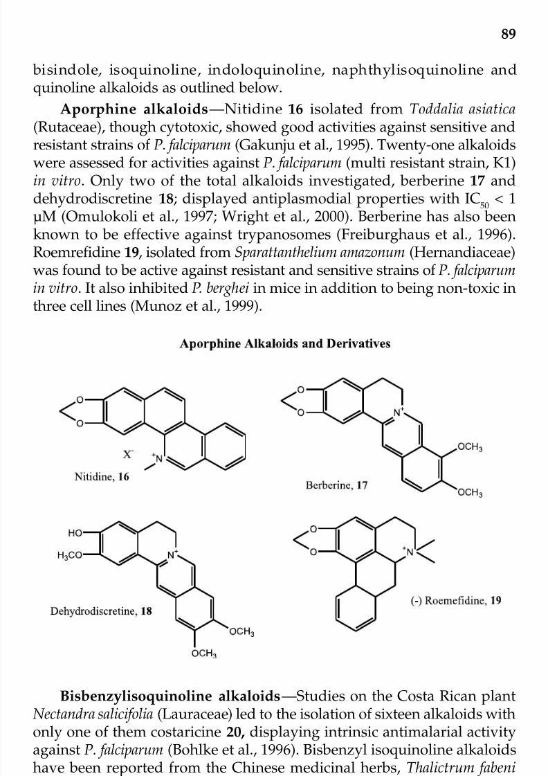
7/23/2019 Novel Therapeutic Agents From Plants
http://slidepdf.com/reader/full/novel-therapeutic-agents-from-plants 100/464
89
bisindole, isoquinoline, indoloquinoline, naphthylisoquinoline andquinoline alkaloids as outlined below.
Aporphine alkaloids—Nitidine 16 isolated from Toddalia asiatica
(Rutaceae), though cytotoxic, showed good activities against sensitive andresistant strains of P. falciparum (Gakunju et al., 1995). Twenty-one alkaloidswere assessed for activities against P. falciparum (multi resistant strain, K1)in vitro. Only two of the total alkaloids investigated, berberine 17 anddehydrodiscretine 18; displayed antiplasmodial properties with IC
50< 1
µM (Omulokoli et al., 1997; Wright et al., 2000). Berberine has also beenknown to be effective against trypanosomes (Freiburghaus et al., 1996).Roemrefidine 19, isolated from Sparattanthelium amazonum (Hernandiaceae)
was found to be active against resistant and sensitive strains of P. falciparumin vitro. It also inhibited P. berghei in mice in addition to being non-toxic inthree cell lines (Munoz et al., 1999).
Bisbenzylisoquinoline alkaloids—Studies on the Costa Rican plantNectandra salicifolia (Lauraceae) led to the isolation of sixteen alkaloids withonly one of them costaricine 20, displaying intrinsic antimalarial activityagainst P. falciparum (Bohlke et al., 1996). Bisbenzyl isoquinoline alkaloidshave been reported from the Chinese medicinal herbs, Thalictrum fabeni(Rananculaceae) and Cyclea barbata (Menispermaceae). Three of such
alkaloids were evaluated for antimalarial properties. Though thalifacine21, most found to be most active, it was also found cytotoxic against human
© 2009 by Taylor & Francis Group, LLC

7/23/2019 Novel Therapeutic Agents From Plants
http://slidepdf.com/reader/full/novel-therapeutic-agents-from-plants 101/464
90
© 2009 by Taylor & Francis Group, LLC

7/23/2019 Novel Therapeutic Agents From Plants
http://slidepdf.com/reader/full/novel-therapeutic-agents-from-plants 102/464
91
cell lines (Lin et al., 1993). Valentin et al. (1997) also isolated penduline22, from Isopynum thalictriodes (Ranunculaceae) and it was found to beactive against chloroquine-resistant strains of P. falciparum. Several bisbenzyl isoquinoline alkaloids from a variety of plant families (checkreferences Steele et al., 1999; Likhitwitayawuid et al., 1993a, 1993b) werescreened for antiplasmodial properties. Of a total of 53 alkaloids that werescreened using the chloroquine sensitive and resistant strains (D6 and W2),respectively, candicusine 23, cepharanthine 24, were most active againstthe D6 strain, while the most active against the W2 strain was cyclea-pentine 25. Cycleamine , 26 was the most selective in the study, apart from being the most potent compound, it also had the least toxicity cytoxocity
assay, it was active in the in vivo animal studies for malaria. Cycleanine isa good candidate for drug development. There have been reports of bisbenylisoquinolines acting as modulators to chloroquine resistancein P. falciparum investigations in vitro (Tian and Pan, 1997; Hakuri et al.,2000). There is need for these compounds to be evaluated in laboratoryanimals. In which case these could be worth considering for drug develop-ment.
Indole alkaloids and derivatives—Cadabamine, 27, isolated from anAsian plant, Anthocephalus chinensis (Rubiaceae) was shown by Kitagawaet al. (1996a) to have activity against a chloroquine resistant strain of P. falciparum with an IC
50value of 6.8 µM. From the Bolivian ethnomedicinal
plant, Pogonopus tubulosus (Rubiaceae) three antiplasmodial indolealkaloids: cephaline, psychotrine and tubulosine 28 were identified.Tubulosine 28, was the most active in the assays and showed an IC
50values
of 0.006 µg/ml and 0.011 µg/ml on chloroquine-sensitive and resistant
strains, respectively. It was also found to be non-toxic using the KB cellscytotoxicity assay. It elicited activity in the in vivo animal antimalarialanalysis using P. vinckei petteri and P. berghei and was active at doses thatwere not lethal. Tubulosine 28 seems to have promise as a suitablecandidate for drug development (Sauvain et al., 1996).
Strychnos (Strychnaceae) species have furnished the indole alkaloids,icajine, isoretuline and strychnobrasiline 29, which though did not display
antiplasmodial properties in vitro were able to reverse P. falciparum activityat a concentration range of 2.5-25 µg/ml (Frederich et al., 2000). Vocamine30, a bisindole alkaloid isolated initially from Voacanga species and morerecently from Peschiera fuchsiaefolia (Apocynaceae), exhibited in vitroantiplasmodial activities against chloroquine sensitive D
6(IC
50= 0.238 µg
g/ml) and chloroquine resistant W2
(IC50
= 0.290 µg/ml) strainsrespectively. These activities were more pronounced than those of thereference drugs included in the assay (Federici et al., 2000). It also displayedinteresting in vivo activities though it was less than that of the referencecompounds and there was low toxicity against mouse and human cell
© 2009 by Taylor & Francis Group, LLC

7/23/2019 Novel Therapeutic Agents From Plants
http://slidepdf.com/reader/full/novel-therapeutic-agents-from-plants 103/464
92
lines. It has been suggested that the possible mode of action of 30 is onthe parasite DNA and or protein synthesis (Ramanitrahasimbola et al.,2001). Voacamine 30 is thus a candidate worthy of drug development because of the good selectivity index. From the Alstonia plant species(Apocynaceae), commonly found in Africa, Asia, Central America andprominently used for management of malaria in these ethnomedicines,
several indole alkaloids have been isolated (Wright et al., 1993). The mostactive of the several alkaloids tested for antimalarial properties,
© 2009 by Taylor & Francis Group, LLC

7/23/2019 Novel Therapeutic Agents From Plants
http://slidepdf.com/reader/full/novel-therapeutic-agents-from-plants 104/464
93
Villalstonine 31, was identified from A. angustifolia, A. glaucescens and A.macrophylla (Keawpradub et al., 1999). Cryptolepine 32, an indoloquinolinealkaloid, isolated from the roots of Crytolepsis sanguinolenta (Asclepia-daceae) a Central and West African traditional medicinal plant that has been reported to display antiplasmodial properties. The workers employedthe chloroquine sensitive and resistant strains of P. falciparum in severalstudies (Kirby et al., 1995; Greiller et al., 1996; Cimanga et al., 1997; Pauloet al., 2000). Three other alkaloids were isolated from the plant species,namely: 11-hydroxycryptolepine 33, quindoline 34 and neocryptine 35. Thedetails of the in vitro antiplasmodial and in vivo antimalarial properties of the four compounds 32-35 are displayed in Table 1.
Table 1. In vitro and in vivo antimalarial studies of alkaloids of Crytolepsis sanguinolenta*
Compound Activity/Parasite strain
Name (Figure) In vitro (P. falciparum, In vivo (P. bergheiIC
50in µg/ml) berghei % Parasitemia
D6 W 2 K1 strain** in mice) AnkaCryptolepine (32) 27 41 33 45.4
11-Hydroxycryptolepine (33) 31 52 62 43.2
Neocryptolepine (34) 35 65 81 –
Quindoline (35) 63 108 87 48.2
*Cimanga et al. (1997)**D
6= Chloroquine sensitive; W
2= Chloroquine resistant; K1 = multi resistant.
Naphthylisoquinoline alkaloids—The African and Asian ethno-medicines have resulted in the bioactivity-led identification of theDioncophyllaceae and Anchstrocladaceae plant families as sources forantimalarial agents. The in vitro assay, (chloroquine resistant P. falciparumNF 54 strain and clone A1A9) and in vivo animal studies (Anka strain of P. berghei in mice) of Triphyophyllum peltatum (Dioncophyllaceae) furnished
dioncophylline C 36 and dioncopeltatine A, 37 as the most activecompounds (Bringmann et al., 1997; Bringmann and Feineis, 2000). Furtherevaluations revealed that the compounds also exhibited good activityagainst exoerthrocytic stages of P. berghei in human hepatoma cells thatwere cultured with 10 µg/ml of the alkaloids. From the Cameroonian plant Ancistrocladus korupensis (Ancistrocladaceae), two novel naphthyliso-quinoline alkaloids, korupensis B 38 and youndamine A 39 were evaluated(Bringmann, 2003). The alkaloids displayed exceptional activities in boththe in vitro analysis and in animal studies. For details of their anti-plasmodial activities, see Table 2.
© 2009 by Taylor & Francis Group, LLC

7/23/2019 Novel Therapeutic Agents From Plants
http://slidepdf.com/reader/full/novel-therapeutic-agents-from-plants 105/464
94
Table 2. Antimalarial properties of naphthylisoqunoline alkaloids*
Compound/Plant source Activity/Parasite clone
Name (Figure) P. falciparum, IC50
µg/ml) P. berghei bergheiNF54 clone A1A9** (% Parasitemia in mice)
Anka
Dioncophylline C (36) 0.014 0.00
Triphyophyllum peltatumDioncopeltine A (37) 0.021 0.03Triphyophyllum peltatum
Korupsinamine B (38) 0.041 Ancistocladus korupensis
Youndamine A (39) 2.200 Ancistocladus korupensis
*Bringmann, 2003.**Chloroquine sensitive clone.
Isoquinoline alkaloids—Campbell et al. (1998) identified theantimalarial principles in the Bolivian plant, Brunsvigia littoralis(Amaryllidaceae) to be lycorine 40, 1,2-di-O-acetyllycorine 41, amberllineand crinine 42 using 2 strains of P. falciparum. They were also evaluatedfor cytotoxicity using the BL
6mouse melanoma cells. From the Madagascan
antimalarial ethnomedicine, Hernandia voyronni (Hernandiaceae) has beenreported to be used in combination with chloroquine to treat malaria
© 2009 by Taylor & Francis Group, LLC

7/23/2019 Novel Therapeutic Agents From Plants
http://slidepdf.com/reader/full/novel-therapeutic-agents-from-plants 106/464
95
(Rasoanaivo et al., 1998). Isolation of active of the antimalarial principlesled to the identification of herveline A 43 (IC
503.28 µM) , herveline B ,
herveline C and herveline D 44 (IC50
2.22 µM) and laudanosine 45 (IC50
26.27 µM). These compounds displayed moderate activity against P.
falciparum. Hervelines A, C and laudanosine enhanced chloroquine activitywhile compound 44 acted as antagonist (Rasoanaivo et al., 1998).
Quinazolones—Febribugine 46 and Isofebrifugine, alkaloids found inthe Chinese medicinal plant, Dichroa febrifuga (Saxifragaceae) were theforemost alkaloids that displayed antimalarial properties. In the earlystudies, febrifugine 46 was a hundred times more active than quinine 1,in the monkey model in vivo antimalarial assay, while compound
isofebrifugine was found to be active in chicks. The toxicicologicalevaluation of febrifugine in mice revealed that it was toxic and so notrelevant for drug development (Takaya et al., 1999). Jiang et al. (2005)evaluated 10 analogs of febrifugine 46 identifed from a febrifuginestructure-based search of the Walter Reed Chemical Information System.These compounds inhibited parasite growth with IC
50ranging 0.141-290
ng/ml with both the chloquine-sensitive and resistant strains of P. falciparum. Some of these compounds displayed feasible therapeutic indices
with selectivity indices >1000. One of the analogs, Halofuginone, reducedparasitemia to non-detectable levels and had curative effects in P. berghei
© 2009 by Taylor & Francis Group, LLC

7/23/2019 Novel Therapeutic Agents From Plants
http://slidepdf.com/reader/full/novel-therapeutic-agents-from-plants 107/464
96
infected-mice. Details of the IC50
values for four of these compounds aredisplayed in Table 3 below.
Two other quinazolones; hydranthine A, 47 and sampangine 48,isolated from Duguetia hadrantha (Annonaceae), a South African anti-malarial plant were found to demonstrate antiplasmodial properties(Muhammed et al., 2000).
Table 3. Antimalarialand cytocoxicity properties of febrifugine ands analogs*
Parasite Cytotoxicity
Drug** P. falciparum, IC50
ng/ml) Neuronal cells SelectivityW2 D6 (NG108)
Febrifugine 0.53 0.34 63.50 119.81
Halofuginone 0.15 0.12 177.06 1222.12
WR092103 1.46 1.23 6864.29 2346.77
WR140085 15.07 12.94 15479.39 1027.00
*Jiang et al. 2005; **WR092103 & WR140085 compounds from Walter ReedChemical library.
Chalcones—Uvaretin, 49 from the Tanzanian species of Uvaria(Apocynaceae) was one of the foremost chalcones shown to possessantimalarial properties, though not as active as chloroquine (Pavanand et
al., 1989). Phorizidine, a bitter tasting compound, isolated from Micromelumthephrocarpum (Rutaceae) displayed antiparasitic properties againstmalaria parasites. Roots of Glycyrrhiza sp. (licorice) popularly used in theChinese ethnomedicine displayed antimalarial properties. Phytochemicalexamination of the plant, led to the identification of licochalcone A 50, asthe active principle using the chloroquine-sensitive strain (3D7) andchloroquine-resistant strain (Dd2) of P. falciparum in (3H) hypoxanthineuptake assay. The in vivo evaluation against P. yoelii in mice, at 1000 mg/kg completely eradicated the malaria parasite without exhibiting toxicity(Chen et al., 1994). This makes licochalcone A 50, a candidate for further
© 2009 by Taylor & Francis Group, LLC
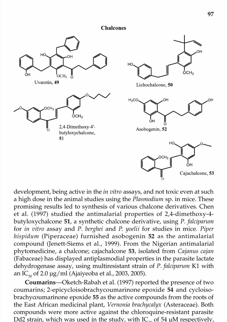
7/23/2019 Novel Therapeutic Agents From Plants
http://slidepdf.com/reader/full/novel-therapeutic-agents-from-plants 108/464
97
development, being active in the in vitro assays, and not toxic even at sucha high dose in the animal studies using the Plasmodium sp. in mice. Thesepromising results led to synthesis of various chalcone derivatives. Chenet al. (1997) studied the antimalarial properties of 2,4-dimethoxy-4-
butyloxychalcone 51, a synthetic chalcone derivative, using P. falciparumfor in vitro assay and P. berghei and P. yoelii for studies in mice. Piperhispidum (Piperaceae) furnished asobogenin 52 as the antimalarialcompound (Jenett-Siems et al., 1999). From the Nigerian antimalarialphytomedicine, a chalcone; cajachalcone 53, isolated from Cajanus cajan(Fabaceae) has displayed antiplasmodial properties in the parasite lactatedehydrogenase assay, using multiresistant strain of P. falciparum K1 withan IC
50
of 2.0 µg/ml (Ajaiyeoba et al., 2003, 2005).
Coumarins—Oketch-Rabah et al. (1997) reported the presence of twocoumarins; 2-epicycloisobrachycoumarinone epoxide 54 and cycloiso-
brachycoumarinone epoxide 55 as the active compounds from the roots of the East African medicinal plant, Vernonia brachycalyx (Asteraceae). Bothcompounds were more active against the chloroquine-resistant parasiteDd2 strain, which was used in the study, with IC
50of 54 µM respectively,
compared to 111-160 µM for the chloroquine-sensitive strain, 3D7.
Several coumarins were identified from Exostema mexicana (Rubiaceae)stem bark and were subjected to in vitro antimalarial studies. The most
© 2009 by Taylor & Francis Group, LLC

7/23/2019 Novel Therapeutic Agents From Plants
http://slidepdf.com/reader/full/novel-therapeutic-agents-from-plants 109/464
98
active of the coumarin was O-methyl exostimin 56. It had an IC50
value of 3.6 µg/ml against the chloroquine-sensitive strain of P. falciparum and anIC
50value of 1.6 µg/ml against the chloroquine resistant strain (Kohler et
al., 2001).
Flavones and Flavonoids—The detection of antiplasmodial flavonoidsin Artemisia annua (Asteraceae) led to the study of this class of compounds
with renewed interest. Two methoxylated flavones, artemetin and casticinact synergistically with artemisinin 2, against P. falciparumin in the in vitroanalysis. None of the other compounds were as active as artemisinin.Further examination of Artemisia species led to the identification of moreantiparasitic flavonoids. One of such studies in the Thai ethnomedicinefurnished exiguaflavanone A 57 and exiguaflavanone B 58 from A. indica.Both compounds exhibited activity with EC
50values of 4.6 and 7.1 µg/ml
respectively, in the in vitro analysis using P. falciparum (Chaphen et al.1998). From A. arbrotanum, isofraxidin 59 was identified as the antimalarialcomponent (Cubucku et al., 1990). The isoflavones, calycosin 60 andgenistein 61 isolated from Andira inermis (Fabaceae) have been shown tohave antimalarial properties.
© 2009 by Taylor & Francis Group, LLC

7/23/2019 Novel Therapeutic Agents From Plants
http://slidepdf.com/reader/full/novel-therapeutic-agents-from-plants 110/464
99
Limonoids—Practically all the plants belonging to the familyMeliaceae have been investigated for antimalarial properties because of the use of these plants in several ethnomedicines in Africa, Asia, and SouthAmerica for malaria therapy. Over 60 extracts from various parts of over20 plants were tested against P. falciparum. Of particular interest in thisregard is the Neem tree, Azadirachta indica (Meliaceae), which hasundergone various antiparasitic studies. The antimalarial activity of A.indica has been attributed to the presence of gedunin 62 (MacKinnon et al.,1997). In a previous study, Khalid et al. (1986) reported that 62 add effectiveconcentration, isolated from Melia azedarach had activity comparable toquinine in the in vitro assay using P. falciparum and 3H- hypoxanthine in
continuous culture. The in vivo mice model activity of this compound usingP. berghei was not promising for further development (Bray et al. 1990).From the Indian ethnomedicine, meldenin 63 was the most active of fourlimonoids isolated from in a bioassay-guided isolation of A. indica leaves.Azadirachtin 64 and three semisynthetic limonoid derivatives were foundto be active against the erythrocytic stages of P. falciparum. The neem extractswere reported to affect all the stages of maturation of the gametocytes. This
© 2009 by Taylor & Francis Group, LLC

7/23/2019 Novel Therapeutic Agents From Plants
http://slidepdf.com/reader/full/novel-therapeutic-agents-from-plants 111/464
100
was novel and unlike the reference drugs artemisinin 2 and primaquinethat inhibit only the immature stages of growth (Dhar et al., 1998). Fromother Meliaceae plants, most especially the genus Khaya had providedantiplasmodial agents. Fissinolide 65 from K. senegalensis was active againstchloroquine-resistant P. falciparum. The non-polar extract from K. grandifololia was found active in the in vivo antiplasmodial analysis usingP. berghei in mice (Khalid et al., 1998; Agbedahunsi et al., 1998).
Peroxides—Ever since the artemisinin 2, a macrocylic sesquiterpeneendoperoxide, from A. annua and its analogs have been shown to possessgreat promise as rapidly acting and potent antimalarials, a lot of attentionhas been focused on various natural peroxides and several have been
reported to display antimalarial properties (Klayman, 1985; Dhingra et al.,2000). Artemisia annua (Asteraceae) is the Chinese herb that had been usedin the Chinese ethnomedicine for over three centuries for the treatment of malaria. Presently, artemisinin and its derivatives are by far the most usefulcompounds for treatment of chloroquine-resistant malaria. Though severalsynthesis of artemisinin has been reported, the most cost-effective meansof obtaining the compound is still through the plant (van Agtamael et al.,
1999). The peroxide yingzhaosu 66, also from the Chinese ethnomedicalplant Artabotrys uncinatus (Annonaceae), used for malaria treatment wasreported to have exceptionally good activity. This initiated the synthesisof arteflene 67, with a high potential as a candidate worthy of development.Ascaridole 68 , a terpene endoperoxide isolated from Chenopodiumambrosiodes (Chenopodaceae) has been shown to possess antiplamodialproperties (Pollack et al., 1989). The bisabolene endoperoxides zingiberene3, 6-α- and β-endoperoxides 69, identified from the Brazilian plants
Eupatorium rufescens and Senecio selloi (Asteraceae) were found to display
Peroxides
O
O
OH
OH
O
O
O
F3C
CF3
Yingzhaosu, 66
Arteflene, 67
© 2009 by Taylor & Francis Group, LLC

7/23/2019 Novel Therapeutic Agents From Plants
http://slidepdf.com/reader/full/novel-therapeutic-agents-from-plants 112/464
101
antimalarial properties. The sesquiterpene endoperoxide, 10,12-peroxy-calamenene was found to be the most active compound in the in vitro bioassay-guided isolation of Cyperus rotundus, the Tanzanian and Thaiantimalarial plant (Weenen et al., 1990). Nardoperoxide 70 andisonardoperoxide 71 were isolated from the roots of Nardostachys chinensisand they had EC
50of 1.5 µM and 0.6 µM respectively in the in vitro studies
using P. falciparum (Takaya et al., 1998).
Phloroglucinol compounds—This group of compounds have showna wide range of biological activities including antimalarial and anti HIVproperties. Phloroglucinol compounds occur widely in Eucalpytus sp and
Hypericum sp. The leaves of E. robusta (Myritaceae) used in China fortreatment of malaria among other ailments, furnished robustaol A 72,robustadial A 73 and B 74 as the antimalarial compounds (Lopes et al.,1999). The relative prenylated stilbene derivatives; (75, 76 and 77) fromthe fruit and aerial parts of Artrocarpus interger (Moraceae) showedantimalarial activity (Boonlaksiri et al., 2000). Sarothralen B 78 and Japonicine A 79, from Hypericum japonicum (Clusiaceae) and the prenylated
phloroglucinol derivative 80 from H. calycinum were found to possess goodantimalarial activity (Xu et al., 1984).
O
O
Ascaridole, 68
OO
H
H
Zingiberene 3,6-ß-endoperoxide, 69
-
O
O
O
HO
O
O
O
O H
Nardoperoxide, 70Isonardoperoxide, 71
Peroxides (continued)
© 2009 by Taylor & Francis Group, LLC
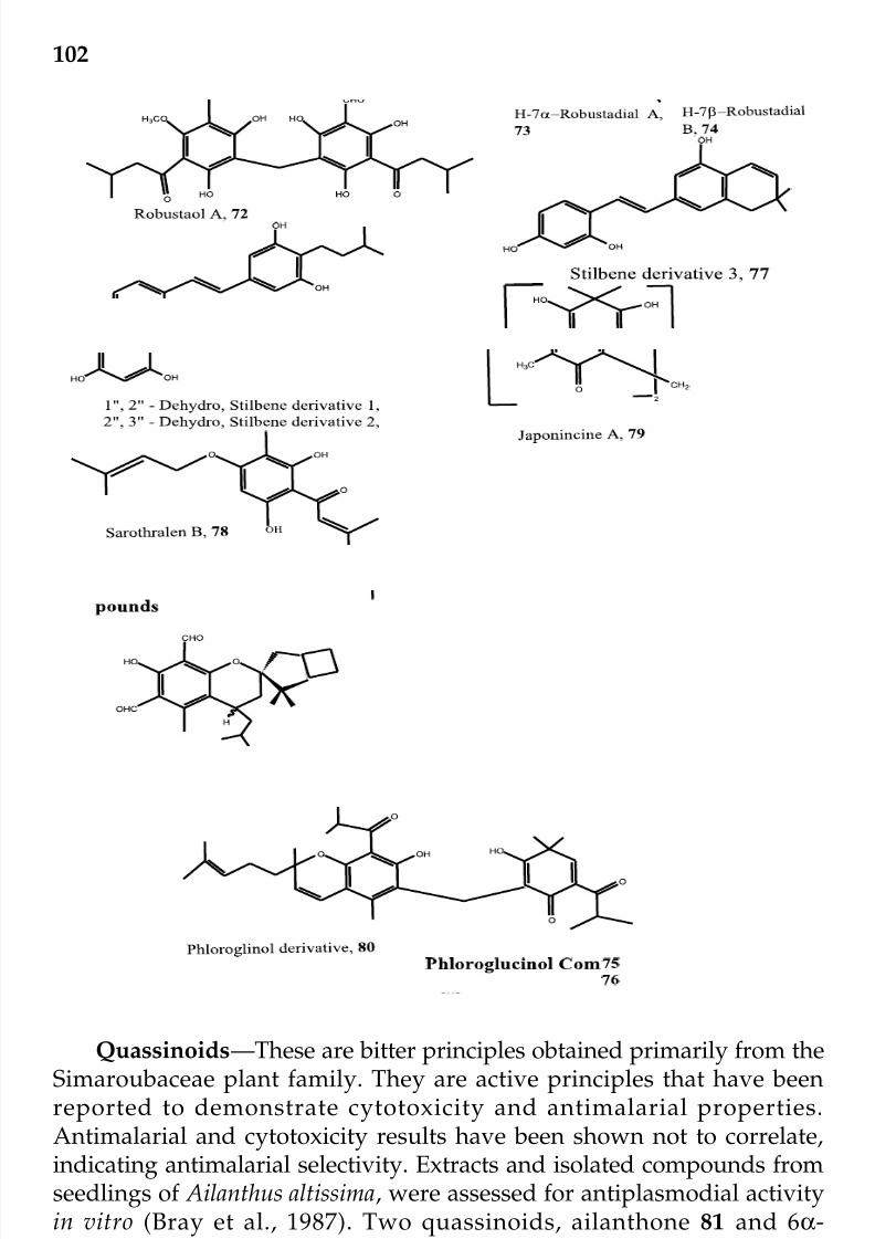
7/23/2019 Novel Therapeutic Agents From Plants
http://slidepdf.com/reader/full/novel-therapeutic-agents-from-plants 113/464
102
Quassinoids—These are bitter principles obtained primarily from theSimaroubaceae plant family. They are active principles that have beenreported to demonstrate cytotoxicity and antimalarial properties.Antimalarial and cytotoxicity results have been shown not to correlate,indicating antimalarial selectivity. Extracts and isolated compounds fromseedlings of Ailanthus altissima, were assessed for antiplasmodial activityin vitro (Bray et al., 1987). Two quassinoids, ailanthone 81 and 6α-tigloyloxychaparrinone, isolated from the active extracts showed activity
against chloroquine-resistant and chloroquine-sensitive strains of Plasmodium falciparum in the in vitro analysis. Only ailanthone 81
© 2009 by Taylor & Francis Group, LLC

7/23/2019 Novel Therapeutic Agents From Plants
http://slidepdf.com/reader/full/novel-therapeutic-agents-from-plants 114/464
103
demonstrated low toxicity against the Vero cell line (Okuande et al., 2003).Brucea javanica was one of the foremost quassinoid-containing plants fromthe Simaroubaceae plant family to be evaluated for antimalarial activity.In Thai and Chinese ethnomedicines, B. javanica is used in the treatmentof malaria usually as a tea in combination with chloroquine. Bruceine A82, bruceine B 83 and bruceine C 84 isolated from the chloroform fractionof the fruit of the plant displayed activity against multi drug-resistant strainof P. falciparum comparable to mefloquine (Pavanand et al., 1988) Addeffective concentration. Yandanziolide 85 of the 12 quassinoids isolated by O’ Neill et al. (1987) from the same plant was found most active andwas even more so than chloroquine salt used in the study. Compounds
82-84, brustanol 86 and bruceine D 87 were active in the mice modelantimalarial study using P. berghei. Though these compounds were toxicand reported to be antagonistic to chloroquine, they may contribute todeveloping chloroquine - resistant malaria chemotherapeutic agents. Bruceasumatrana also furnished bruceolide 88 and this demonstrated activityagainst in vivo using P. berghei in mice. Dou et al. (1996), identified sevenactive quassinoids from Castela texana which demonstrated in vitroantiplasmodial properties using both the chloroquine sensitive andresistant strains of P. falciparum, the most active of which was holacanthone89 with IC
50of 0.010 µg/ml. From Malaysian Eurycoma longifolia roots,
eurycomanol 2-O-glucopyranoside 90 was found to be very active onchloroquine resistant strain of P. falciparum with an IC
50of 0.39-3.50 µg/
ml (Ang et al., 1995). Of the four quassinoids isolated from the stem barkof Hannoa chlorantha and Quassia undulata (formerly, H. klaineana),Chapparinone 91, displayed a good activity in the in vitro studies using
the NF54, clone AIA9 of P. falciparum. It also exhibited antimalarialproperties in the mice model study with the Anka strain of mice (Francoiset al., 1998). Quassia undulata and Q. amara extracts also displayedantimalarial activities in mice using Anka clones of P. berghei. At aconcentration of 100 mg/kg mouse, Q. amara leaf methanol extract hadthe highest suppressive activity with a parasite density of 0.16% andchloroquine had a parasite density of 0.34% in the same assay on day 4(Ajaiyeoba et al., 1999). Quassia indica furnished antiplasmodialcompounds, the samaderine X, samaderine Z, samaderine B andsamaderine E 92 and which were active against chloroquine resistant (K1)strain of P. falciparum with an IC
50of 0.14-0.21 µM (Kitagawa et al., 1996a).
Gutolactone 93 and simalikalactone D 94 from Simaba quaianesis and Q.africana, have demonstrated interesting antimalarial and antiviral activities(Apers et al., 2002). Both compounds have demonstrated the highestactivities of all the quassinoids with IC
50of 4.0 and 1.6 ng/ml each. In the
same assay, chloroquine and mefloquine had IC50 values of 63.2 and 1.5ng/ml, respectively. Moretti et al. (1998) identified cedronin 95 from Simana
© 2009 by Taylor & Francis Group, LLC

7/23/2019 Novel Therapeutic Agents From Plants
http://slidepdf.com/reader/full/novel-therapeutic-agents-from-plants 115/464
104
cendron and it showed a much better activity against both chloroquinesensitive and resistant strains of P. falciparum in the in vitro investigationas well as in vivo study using P. vinckei petteri in mice. A quassinoidregistered under CO7D493-08, 96 in the patent registry, with a structure
similar to cendronin was reported much earlier, to increase the survivaltime in male mice, when innoculated with P. berghei NK-65 compared with
Quassinoids
O
O
O
O
O
O
O
OH
HH
OH
O
O
HO
O
Ailanthone, 81
HO O
H
O
HH
OH
R2O
O
O
O
R1
O
Bruceine A, 82 R1
= Isobutyl, R2
= H; Bruceine B, 83 R1
= CH3, R2
= H
Bruceine C, 84 R1 = CH=C(CH3)C(OH)(CH3)2, R2 = H;
Yadanziolide I, 85,R1= CH3, R
2= glucopyranoside;
Brustanol 86, R1 = CH=C(CH3)2, R2 = H; Bruceine D, 87 R1 = H, R2 = CH3
HO
H
H
O
H
O
OH
Bruceolide, 88
HO
O
H
O H OOH
OH
OH
H
HO
HO
OH
O
O
O
O
HO
OH
O
OH
Chapparinone, 91
OH
OAc
H
HOO
H
OH
O
O
O
R2O
OH
O
OR1
H
H
OR
OH O
H
O
O
OH
HO
O
H
OHHO
O
O
Holacatnhone, 89
HO
O
HO
O
OO
Samaderine A, 92
Eurycomanol glucoside, 90
Gutalactone, 93- R = Me2 C=CHCO, R1
,R2
= H
Simakilactone D, 94 = Me CH2CH(Me)CO, R1 , R2
=Ac
Cendronin, 95
Quassinoid C07D493-08, 96
O
© 2009 by Taylor & Francis Group, LLC

7/23/2019 Novel Therapeutic Agents From Plants
http://slidepdf.com/reader/full/novel-therapeutic-agents-from-plants 116/464
105
11.3 days for bruceantin (Takenuchi, 1989). The presence of an ester groupat C-15 has been reported to be vital for activity and the glycosides weregenerally less active than the aglycones (Bray et al., 1987). Table 4 belowdisplays the antiplasmodial properties of some compounds in Simarou- baceae family.
Table 4. Antiplasmodial properties of quassinoids of the simaroubaceae plant family
Compound ( Figure) Plant source P. falciparum in vitro activity/ strain (IC
50µg/ml)
D6 W 2 K1 strain* Holacanthone (90) Castela texana 0.010 0.120
Eurycomanol glucoside (91) Eurycoma longifolia – 0.420
Chapparinone (92) Quassia undulata 0.250 0.20
Samaderine E (95) Q. indica – – 0.056 a
Gutolactone (97) Q. africana/Simaba – – 0.004quaianensis
Simikalactone D (98) Q. africana/Simaba - - 0.002quaianensis
*D6
= Chloroquine sensitive; W2
= Chloroquine resistant; K1 = multi resistanta = IC
50in µM.
Quinones—Bulbine capitata (Asphodelaceae) from the Botswanaethnomedicine furnished 5 naphothoquinone furan diones with 1-
acetoxymethyl-8-hydroxynaphtho(2,3-c)furan-4,9 dione showing thehighest activity against chloroquine sensitive (3D7, IC
50of 23 µM) and
chloroquine resistant (K1, IC50
of 52 µM) strains of P. falciparum (Bezabihet al., 2001). From the roots and aerial parts of B. capitata, knipholone 97,4’-O-demethylknipholone and 6’-O-methylknipholone were identified andisolated. Demethyl knipholone also exhibited the highest activity againstchloroquine sensitive (NF54, IC
50of 1.55 µM) while knipholone 97 was
more active against the chloroquine resistant K1 strain of P. falciparum withan IC50
of 1.06 µM. Knipholone anthrone 98 was also identified as a veryactive quinone from Knipholia foliosa and was more active than the otherquinones (IC
50of 0.31 µM) with the chloroquine resistant parasite
(Bringmann et al., 1999). Plumbagin 99, identified in Nephenthes thorelli(Nepethenceae) was the most active of the five naphthoquinones againstP. falciparum with an IC
50of 0.27 µM (Likhitwitayawuid et al., 1998a). The
antimalarial properties of lapachacol 100, identified from several
Bignoniaceae plants initiated the synthesis of the antimalarial drug,atovaquine 101. Isopinnatal 102 isolated from Kigelia pinnata was also
© 2009 by Taylor & Francis Group, LLC

7/23/2019 Novel Therapeutic Agents From Plants
http://slidepdf.com/reader/full/novel-therapeutic-agents-from-plants 117/464
106
found to display antimalarial properties. Diospyros montana (Ebeneceae)has furnished diosporin 103 with a reported in vitro activity against P.
falciparum (Hazra et al., 1995).
Tannins—The biggest drawback in the development of tannins as anantimalarial drug has been lack of selectivity with malarial parasite as
compared to cancer cells, though tannins have displayed antiplasmodialproperties. Gossypol 104, isolated from species of the cotton seed plant,
Quinones
O
R O H
O
O HO H
Knipholone, 97- R = O
Knipholone anthrone, 98 - R = H2
O
O
O
OO H
O H
Diosporin, 103
O
OO H
Plumbagin, 99
O
O
O H
Lapachacol, 100
C l
O
O
O H
Atovaquine, 101
O
H
O
O
O
Isopinnatal, 102
© 2009 by Taylor & Francis Group, LLC

7/23/2019 Novel Therapeutic Agents From Plants
http://slidepdf.com/reader/full/novel-therapeutic-agents-from-plants 118/464
107
Gossypium, has demonstrated impressive antiplasmodial properties. It wasshown to inhibit chloroquine-sensitive and chloroquine-resistant P. falciparum parasites with IC
50in the magnitude of 10 µM. It has also been
shown to be cytotoxic (Coyle et al., 1994; Deck et al., 1998). The traditionalhealers in Southwest Nigeria identified Gossypium arboreum leaves as themost potent of the three species including G. hirsitum and G. barbadenseplant for treatment of febrile illness. Though the crude methanol extractselicited weak activities (IC
50= 197.9 µg/ml) with the multi-resistant strain
of P. falciparum K1 strain, in the clinical observation study, a P. falciparumparasite clearance of over 70% was recorded in febrile clients of theherbalists. In the in vitro analysis using KB cells, the crude methanol extractof the leaves was also found to be non-toxic with ED
50> 300 µg/ml
(Ajaiyeoba et al., 2004a, 2004b).
Terpenoids—Following the discovery and subsequent development of the sesquiterpene endoperoxide, Artemisinin 2, from Artemisia annua,
several sesquiterpenoids mainly from the Asteraceae plant family andindeed most terpenoids have been evaluated for antimalarial properties(Trigg, 1989). From the Central American phytomedicinal plant, Neurolaenalobata, several antiplasmodial sesquiterpenoids have been isolated. Thethree most active compounds being Neurolenin B, 105, Neurolenin C 106(each with IC
50of 0.62 µM), using the chloroquine sensitive P. falciparum
NF54 strain (Francois et al., 1996). From another Asteraceous plant, Takayaet al. (1998) isolated the sesquiterpenoids, Nardoperoxides 83 and
isonardoperoxide 84 (as previously discussed), among others from theroots of Nardostachys chinensis (Asteraceae). The Kenyan plant Vernoniabrachycalyx (Asteraceae) furnished the sesquiterpenoid 16,17 Dihydro- brachycalyxolide, 107 which was studied for antimalarial properties usingseveral P. falciparum strains. It was most sensitive to the chloroquine-resistant P. falciparum V1/S strain and in the bioassay; it had an IC
50value
of 5.9 µM (Oketch-Rabah et al., 1998).
From Surinam, Lopes et al. (1999) identified nerolidol as one of theantimalarial components in Virola surinamensis. An antimalarial eudesmane
Tannins
OHCHO
HO
HO
CHOOH
OH
OH
Gossypol, 104
© 2009 by Taylor & Francis Group, LLC

7/23/2019 Novel Therapeutic Agents From Plants
http://slidepdf.com/reader/full/novel-therapeutic-agents-from-plants 119/464
108
sesquiterpenoid has also been reported from Jasonia glutinosa (Villaescusa-Castillo et al., 2000). The Cameroonian ethnomedicine furnished the two
antiplasmodial sesquiterpenoids 108 and 109 from Reneilma cincinnata.Their IC
50values renged from 1.5-1.6 µg/ml for the chloroquine-sensitive
strain, D6 and between 1.9-31.9 µg/ml for the resistant strain W2(Tchuendem et al., 1999).
The diterpenoid, taxol 110, from the Pacific yew, Taxus brevifolia ananticancer agent was shown by Pouvelle et al. (1994) to displayantimalarial properties in vitro using P. falciparum and in vivo with P.
chabaudi adami in mice.From Vernonia brasiliana (Asteraceae), Lupeol 111 and β-amyrin 112© 2009 by Taylor & Francis Group, LLC

7/23/2019 Novel Therapeutic Agents From Plants
http://slidepdf.com/reader/full/novel-therapeutic-agents-from-plants 120/464
109
were identified as the antiplasmodial compounds (de Almeida-Alves etal., 1997). Lupeol was also identified from the Nigerian phytomedicine asthe antimalarial compound from Cassia siamea and C. nigricans (Fabaceae)respectively with an IC
50
of 5.0 µg/ml using the multi-resistant strain K1of P. falciparum (Ajaiyeoba et al., 2007; Obodozie et al., 2004). Betunilic acidisolated and identified from several plant species has shown weakantiplasmodial properties using different P. falciparum strains. Steele et al.(1999) and Traore-Keita et al. (2000a) reported that it had IC
50of 19.6 µg/
ml (multi-resistant K1 strain) and 18.10 µg/ml (resistant W2 strain).Muzanzagenin 113 was identified by Oketch-Rabah et al. (1997) as theantimalarial triterpenoid from the Kenyan medicinal plant, Asparagus
africanus (Liliaceae). The diterpenoid, taxol 114, from the Pacific yew, Taxusbrevifolia an anticancer agent was shown by Pouvelle et al. (1994) to displayantimalarial properties in vitro using P. falciparum and in vivo with P.chabaudi adami in mice. The leaf extract of Hyptis suaveolens (Lamiaceae),an abietane type diterpenoid endoperoxide 115, was identified as theantiplasmodial agent with an IC
50of 4.1 µg/ml using a chloroquine
sensitive strain of P. falciparum, D10 (Chukwujekwu et al., 2005). FromVernonia brasiliana (Asteraceae), Lupeol 116 and β-amyrin 117 wereidentified as the antiplasmodial compounds (de Almeida Alves et al., 1997).Lupeol was also identified from the Nigerian phytomedicine as theantimalarial compound from Cassia siamea and C. nigricans (Fabaceae)respectively with an IC
50of 5.0 µg/ml using the multi-resistant strain K1
of P. falciparum (Obodozie et al., 2004; Ajaiyeoba et al., 2007). Betunilic acidisolated identified from several plant species has shown weak anti-plasmodial properties using different P. falciparum strains. Steele et al.
(1999) and Traore-Keita et al. (2000) reported that it had IC50 of 19.6 µg/ml(multi-resistant K1 strain) and 18.10 µg/ml (resistant W2 strain).Muzanzagenin 118 was identified by Oketch-Rabah and Dossaji (1997)as the antimalarial triterpenoid from the Kenyan medicinal plant, Asparagusafricanus (Liliaceae).
Xanthones—Species of Garcinia have furnished several xanthones thathave been active in antimalarial assays. Of the five xanthones isolated from
G. cowa (Guttiferae), Cowaxanthone 119 and Cowanol 120 were the mostimpressive with IC50
of 1.5 µg/ml, and 1.6 µg/ml, respectively in the studywith P. falciparum in vitro (Likhitwitayawuid et al., 1998a). Five xanthoneswere also isolated from G. dulcis and the most active antimalarialcompound was found to be garciniaxanthone 121 (Likhitwitayawuid etal., 1998b; Wright et al., 2000). Dua et al. (2004) identified six xanthonesfrom the leaves of Andrographis paniculata, a popular ethnomedicinal plantin China and Southeast Asia. In vitro antimalarial studies showed that
1,2-dihydroxy-6,8-dimethoxy xanthene-9-one (TDR 130011), 122 exhibitedthe highest activity with an IC
50of 4 µg/ml with P. falciparum and gave a
© 2009 by Taylor & Francis Group, LLC

7/23/2019 Novel Therapeutic Agents From Plants
http://slidepdf.com/reader/full/novel-therapeutic-agents-from-plants 121/464
110
70% reduction using P. berghei in mice without showing toxicity in thehuman lung fibroblast (Dou et al., 1999).
Other compounds—The lignan nysol 123 from Asparagus africanus(Liliaceae) was also shown to possess antimalarial properties in vitro andin vivo mice model (Oketch-Rabah et al., 1997). Termenalia bellerica(Combretaceae) fruit rind from the Indian ethnopharmacology haspresented termilignan 124 and anonlignan 125 as antiplasmodialcompounds. The two most active lignans isolated from Rhaphidophora
decursiva (Araceae) leaves and stems were polysyphorin 126 andrhaphidecurperoxin 127. Polysyphorin, 128 had an IC
50value of 0.040 µg/
© 2009 by Taylor & Francis Group, LLC

7/23/2019 Novel Therapeutic Agents From Plants
http://slidepdf.com/reader/full/novel-therapeutic-agents-from-plants 122/464
111
ml against the chloroquine-sensitive strain and 0.037 µg/ml (3D7) withthe chloroquine resistant strain (Dd2), used in the study (Zhang et al., 2001).
Future perspectives in chemotherapeutic control of malaria
Malaria is a major health burden in developing countries and is aremaining health problem socio-economically in developed countries; it isimportant that various strategies and interventions are planned andexecuted. Malaria chemotherapy is even more complex and challenging
© 2009 by Taylor & Francis Group, LLC

7/23/2019 Novel Therapeutic Agents From Plants
http://slidepdf.com/reader/full/novel-therapeutic-agents-from-plants 123/464
112
because of multi-drug resistant strains of Plasmodium falciparum, which arereadily available and well known chemotherapeutic agents are nowineffective. There is no doubt that the new drugs against malaria are greatlyneeded (Ridley, 2002). Rosenthal (2003) in his review of old and newapproaches of antimalarial drug discovery highlighted the under-listedstrategies: discovery of natural products, development of analogs of existingchemotherapeutic agents evaluation of other compounds used previouslyfor other diseases, evaluation of drug resistant reversers and considerationof new drug targets. These are basically the five practical areas that shouldconstitute the focus in malaria drug discovery and development for thefuture.
Presently, no vaccines are available for malarial treatment and theprospect for vaccine development is not soon enough, hence drugs willremain the main stay of disease control (WHO, 2004). Since the two mosteffective drugs for control of malaria originate from plants, that is quinine1 and artemisinin 2, plants are a major resource for discovering of newermalaria drugs. The screening of herbs using local biodiversities should beenhanced (Wilcox and Bodeker, 2000). Approaches to discovery of
antimalarial agents from various biodiversities are not different from thosefor other diseases. These could be through:
1. Ethnomedicinal leads2. Random screening3. Literature4. Chemotaxonomy
These are the four basic approaches used in the selection of plants.
Thereafter, selected plants are primarily subjected to in vitro antiplasmodialscreens, to identify a ‘hit’. This refers to that plant sample/drug that hasan index of inhibitory activity greater than the reference drug utilized inthe same assay. In vivo animal screens mainly in murine animals areregarded as the secondary screens. This also helps in identifying plantsthat contain constituents that may be prodrugs. Isolation, mainly by acombination of chromatographic techniques and subsequent spectroscopicstructural elucidations of isolated bioactive components usually follow.Structure-activity relationships may be initiated to maximize observed bioactivity or reduce toxicity to optimize lead antimalarial compound(s).Various stages of toxicological assessments in animal models will benecessary for the selected compound to progress to the next stage, whichusually precede biopharmaceutical studies (pharmacokinetics, bio-availability properties of the lead compound). Other stages of drug develop-ment generally will include other tertiary bioassays and studies including
the possibility of synthesis of the compound. By so doing, in a couple of years, usually more than 10 years, a development candidate emerges.
© 2009 by Taylor & Francis Group, LLC

7/23/2019 Novel Therapeutic Agents From Plants
http://slidepdf.com/reader/full/novel-therapeutic-agents-from-plants 124/464
113
In most developing countries where malaria is endemic, it is reportedthat over 80% of the rural population with no access to modern healthcarefacilities, depend on traditional medicine for management of most diseasesincluding malaria. In the short term, the use of herbal remedies consistingof plants like the neem tree, Azadirachta indica (Meliaceae), Khayasenegalensis (Meliaceae), Morinda lucida (Rubiaceae), Cassia sp. (Fabaceae),Phyllanthus sp. (Euphorbiaceae), to mention a few, has been reported(Ekanem, 1978; Makinde et al., 1994; Agbedahunsi et al., 1998; Ajaiyeoba,2002; Ajaiyeoba et al., 2002, 2003, 2004, 2005, 2006, 2007) from Nigeria.Investigations on some of the individual plants have indicated someantiplasmodial properties, both in the primary in vitro and in vivo assays.
This has assisted the indigenous communities in the management of uncomplicated malaria, while awaiting drug development from hit plantsfrom these ethnomedicines, as in the case of the Peruvian and Chineseethnomedicines that provide the two major drugs for chemotherapeutictreatment of malaria; quinine 1 and artemisinin 2, respectively.
Conclusion
Malaria still poses the greatest threat of all parasites to human health.Malaria is a major health problem and its eradication has so far provedimpossible. The eradication of malaria seems to be ampiclox, mainly dueto resistance of the parasite to readily available drugs and so loss of efficacy. The death toll from malaria especially in Africa is likely to be onthe increase with the spread of drug resistance to chloroquine. This hasmade the chemotherapy of malaria shift to drug combination and a re-
visitation of abandoned drugs. Vaccine development is still being awaited.So wither hence? The future of chemotherapy as the mainstay for malariamay look clouded. In this article, a large number of compounds withstructurally diverse chemical structures have been mentioned, there is nodoubt that these plant secondary metabolites present a plethora of compounds that could be developed into the new antimalarial drugs.Ethnopharmacological approach has by far proved superior to otherempirical approaches in identification of new sources of antimalarial drug.Hence scientific validation of traditional medicinal properties of indigenous medicinal plants used for management of malaria should beencouraged. A concerted effort by all stakeholders in this regard, may wellprovide the answer for the immediate control of malaria in sub-SaharanAfrica and other endemic regions. Nature has continued to providesolutions to problems of people on earth and chemotherapeutic malariacontrol is one of the problems that can be overcome from natural sources.
© 2009 by Taylor & Francis Group, LLC

7/23/2019 Novel Therapeutic Agents From Plants
http://slidepdf.com/reader/full/novel-therapeutic-agents-from-plants 125/464
114
References
Addae-Kyereme, J., S.L. Croft, H. Kendrick and C.W. Wright. 2001. Anti-plasmodial activities of some Ghanaian plants traditionally used for fever/
malarial treatment and of some alkaloids isolated from Pleiocarpa mutica; invitro antimalarial activity of pleiocarpine. J. Ethnopharmacol. 76: 99-103.
Agbedahunsi, J.M., O.O. Elujoba, J.M. Makinde and A.M.J. Oduola. 1998.Antimalarial activity of Khaya grandifololia stam bark. Phamaceut. Biol. 36: 8-12.
Ajaiyeoba, E.O. 2002. Identification and chemical evaluation of potentialantimalarial components from the Nigerian phytomedicine compendium.UNDP/TDR/WHO/MIM Africa grant ID 980046, 2nd Year Report and
renewal request, pp. 47.Ajaiyeoba, E.O., U.I. Abalogu, H.C. Krebs and A.M.J. Oduola. 1999. In vivo
Antimalarial activities of Quassia undulata and Quassia amara plant extractsin Mice. J. Ethnopharmacol. 67: 312-325.
Ajaiyeoba, E.O., O.S. Osowole, O.O. Oduola, J.S. Ashidi, D.O. Akinboye, G.O.Gbotosho, C.O. Falade, O.A.T. Ogundahunsi, O.I. Fawole, O.M. Bolaji, M.O.Falade, O.O. Oladepo, O.A. Itiola and A.M.J. Oduola. 2002. Nigerianantimalarial ethnomedicine 2: Ethnobotanical surveys of Herbal Remedies
used in the treatment of febrile illnesses in the middle belt of Nigeria. J.Phytomed. & Therapeut. 7: 26-39.
Ajaiyeoba, E.O., O.I. Fawole, O.M. Bolaji, D.O. Akinboye, O.S. Osowole, O.Oladepo, O.O. Oduola, G.O. Gbotosho, O.O. Ebong, C.O. Falade, O.A.T.Ogundahunsi, O.A. Itiola, J.S. Ashidi, T.C. Happi, F.O. Falade and A.M.J.Oduola. 2003. Cultural categorization of febrile illnesses in correlation withherbal recipes for treatment in Southwestern Nigeria. J. Ethnopharmacol.85: 179-185.
Ajaiyeoba, E., J. Ashidi, O. Abiodun, L. Okpako, O. Ogbole, D. Akinboye, C.Falade, O. Bolaji, G. Gbotosho, M. Falade, O. Itiola, P. Houghton, C. Wrightand A. Oduola. 2004a. Antimalarial Ethnobotany: In vitro antiplasmodialactivity of seven Plants identified in the Nigerian middle Belt. Pharmaceut.Biol. 42(8): 588-591.
Ajaiyeoba, E.O., C.O. Falade, O.I. Fawole, D.O. Akinboye, G.O. Gbotosho, O.M.Bolaji, J.S. Ashidi, O.O. Abiodun, O.S. Osowole, O.A. Itiola, O. Oladepo, A.Sowunmi and A.M.J. Oduola. 2004b. Efficacy of herbal remedies used by
herbalists in Oyo State Nigeria for treatment of Plasmodium falciparuminfections—a survey and an observation, Afri. J. Med. & Med. Sci. 33: 115 –119.
Ajaiyeoba, E.O., J.S. Ashidi, D.O. Akinboye, O.M. Bolaji, G.O. Gbotosho, C.O.Falade, L.C. Okpako, O.O. Oduola, M.O. Falade, P.J. Houghton, C.W. Wright,O.A.T. Ogundahunsi and A.M.J. Oduola. 2005. In vitro antiplasmodial andcytotoxicity activities of six plants from the South West Nigerianethnomedicine. J. Nat. Rem. 5: 1-6.
Ajaiyeoba, E.O., O.O. Abiodun, M.O. Falade, N.O. Ogbole, J.S. Ashidi, C.T. Happiand D.O. Akinboye. 2006. In vitro Cytotoxicity studies of 20 plants used inthe Nigerian Antimalarial Ethnomedicine. Phytomedicine 13(4): 295-298.© 2009 by Taylor & Francis Group, LLC

7/23/2019 Novel Therapeutic Agents From Plants
http://slidepdf.com/reader/full/novel-therapeutic-agents-from-plants 126/464
115
Ajaiyeoba, E.O., J.S. Ashidi, L.C. Okpako, P.J. Houghon and C.W. Wright. 2007.Antiplasmodial compounds from Cassia siamea stem bark extract.Phytother. Res. 22(2): 254-255.
Almeida-Alves, T.M., T.J. Nagem, L.H. Carvalho, A.U. Krettli and C.L. Zani. 1997.
Antiplasmodial triterpene from Vernonia brasiliana. Planta Medica 63: 554-555.
Ang, H.H., K.L. Chan and J.W. Mak. 1995. In vitro antimalarial activity of quassinoids from eurycoma longifolia against Malaysian chloroquine-resistantPlasmodium falciparum isolates. Planta Medica 61: 177-178.
Apers, S., K. Cimanga, D. van Berghe, E. van Mueen, A.O. Longanga, A. Fories,A. Vlietnick and L. Pieters. 2002. Antiviral activity of Simalactone D,Aquassinoid from Ouassia africana. Plant Medica 68, 20-24.
Bezabih, M., B.M. Abegaz, K. Dufall, K. Croft, T. Skinner-Adams and T.M.E.Davies. 2001. Antiplasmodial and antioxidant isofuranonaphthaquinonesfrom the roots of Bulbine capitata. Planta Medica 67: 340-344.
Bickii, J., N. Njifutie, J.A. Foycre, L.K. Basco and P. Ringwald. 2000. In vitroantimalarial activity of limonoids from Khaya grandifoliola C.D.C. (Meliaceae) J. Ethnopharmacol. 69: 27-33.
Bohlke, M., H. Guinaudeau, C.K. Angehofer, V. Wongpanich, D.D. Soejarto, W.R.Farnsworth, N.R. Mora and L.J. Poveda. 1996. Costraricine, a newantiplasmodial bisbenzylisoquinoline alkaloid from Nectranda salicifolia trunk bark. J. Nat. Prod. 59: 576-580.
Boonlaksiri, C., W. Oonanant, P. Kongsaeree, P. Kittakoop, M. Tianticharoen andY. Tebtaranoonth. 2000. An antimalarial stilbene from Artcarpus integer.Phytochemisty 55: 415-417
Boye, G.L. and O. Ampofo. 1983. Clinical uses of Cryptolepis sanguinolenta.Proceedings of the First International Seminar on Cryptolepine. July 1983.University of Science and Tech. Kumasi, pp. 37-40.
Brandao, M.G.L., A.U. Krettli, L.S.R. Soares, C.G.C. Nevy and H.C. Marinuzzi.
1997. Antimalarial activity of extracts and fractions from Bidens pilosa andother Bidens species (Asteraceae) correlated with the presence of acetyleneand flavonoids compound. J. Ethnopharmacol. 51: 131-138.
Brandao, M.G.L., C.G.C. Nevy, M.A.S. Mamao and A.U. Krettli. 1998. Twomethoxylated flavone glycosides from Bidens pilosa. Phytochemistry 48: 397-399.
Bray, P.H., P. Boardman, M.J. O Neill, K.L. Chan, J.D. Phillipson, D.C. Wrahurst,and M. Stuffness. 1987. Phytother. Res. 1: 22-24.
Bray, D.H., J.D. Connolly, J. O’Neill, J.D. Phillipson and D.C. Warhurst. 1990.Plants as sources of antimalarial drugs. VI. Activity of some Meliaceae plantsand their constituent limonoids. Phytother. Res. 4: 29-35.
Breman, J.G. 2001. The ears of the hippopotamus: manifestations, determinantsand estimates of the malaria burden. Am. J. Trop. Med. Hyg. 64 (supp): 1-11.
Bringmann, G. 2003. From Tropical Lianas to novel antiplasmodial agents: TheNaphthylisoquinoline alkaloids, pp. 145-152. In: H. Vial, A. Fairlamb and R.Ridley (eds) Guidelines and Issues for the Discovery and Drug DevelopmentAgainst African Diseases World Health Organisation, Geneva, Switzerland.
Bringmann, G. and D. Feineis. 2000. Novel antiparasitic biaryl alkaloids from© 2009 by Taylor & Francis Group, LLC

7/23/2019 Novel Therapeutic Agents From Plants
http://slidepdf.com/reader/full/novel-therapeutic-agents-from-plants 127/464
116
West African Dioncophyllaceae plants. Acta. Chem. Therapeut. 26: 151-157.Bringmann, G., W. Saeb, L.A. Assi, A.S.S. Narayanan, K. Peters and E.M. Peters.
1997. Betulinic acid: isolated from Triphyophyllum peltatum and Ancistrocladusheyneanus, antimalarial activity, and crystal structure of Benzyl ester. Planta
Med. 63: 255-257.Bringmann, G., D. Menche, M. Bezabih, B.M. Abegaz and R. Kaminsky. 1999.
Antiplasmodial activity of knipholone and related natural phenyl-anthraquinones. Planta Med. 65: 757-758.
Campbell, W.E, J.J. Nair, D.W. Gamma, J. Bastida, C. Codina, I. Valadomat, P.G.Smith and C.F. Albrecht. 1998. Cytotoxic and antimalarial alkaloids fromBrunsvigia littoralis. Planta Medica 64(11): 91-93.
Canianto R. and L. Puricelli. 2003. Review: natural products antimalarial agents
(1995-2001). Crit. Rev. Plant Sci. 22(1): 79-105.Chaphen, R., Y. Thebtaranonth, S. Wanauppathamkul and Y. Yothovong. 1998.Antimalarial principles from Artemisia indica. J. Nat. Prod. 61: 1146-1147.
Chen, M., T.G. Theander, S.B. Christensen, Haviid, l., L. Zhai and A. Kharaz A.1994. Lichochalcone, a new antimalarial agent inhibits in vitro growth of human malaria parasite, Plasmodium falciparum and protects mice from P. yoeliiinfection. Antimicrob. Agents and Chemother. 38(77): 1470-1475.
Chen, M., S.B. Christensen, L. Zhai, M.H. Rasmussen, T.C. Theander, S. Frokjaer,B. Steffanssen, J. Davidson and A. Kharazni. 1997. The novel oxygenatedchalcone 2,4-dimethoxy-4’-butyloxychalcone exhibits potent activity againsthuman malaria parasite Plasmodium falciparum in vitro and rodent parasitesP. berghei and P. yoelii in vivo. J. infectious Diseases 176: 1327-1333.
Chukwujekwu, J.C., P. Smith, P.H. Combes, D.A. Mulholland and J.van Staden.2005. Antiplasmodial diterpenoid from the leaves of Hyptis suaveolens. J.Ethnopharmacol. 102: 295-297
Cimanga, K., T. De Bruyne, L. Pieters and A.J. Vlientinck. 1997. In vitro and invivo antiplasmodial activity of cryptolepine and related alkaloids from
Cryptolepis sanguinolenta. J. Nat. Prod. 60: 688-691.Coyle, T., S. Levante, M. Shetler and J. Winfield. 1994. In vitro and in vivo
cytotoxicity of Gossypol against central nervous system tumor cell lines. J.Neuro. Oncol. 19: 25-35.
Cubucku, B., D.H. Bray, D.C. Warhurst, A.H. Mericli, N. Ozhatay and G. Sariyar.1990. In vitro antimalarial activity of crude extracts and compounds from Artemisia abrotanum. Phythther. Res. 4: 203-204.
De Almeida Alves, T.M., T.G. Nagem, L.H. de Carvalho, A.U. Krettli and C.L.
Zani. 1997. Antiplasmodial triterpene from Vernonia brasiliana. Planta Medica63: 554-555.Deck, L.M., R.E. Royal, B.B. Chamblee, V.M. Hermandez, R.R. Malone, J.E. Torres,
L.A. Hunsaker, R.C. Piper, M.T. Markler and D.L. Vander Jagt. 1998. Selectiveinhibitors of human lactate dehydrogenases and lactate dehydrogenase frommalarial parasite Plasmodium falciparum. J. Med. Chem. 41: 3879-3887.
Dhar, R., K. Zhang, G.P. Talwar, S. Garg and N. Kumar. 1998. Inhibition of thegrowth of the human malaria parasite Plasmodium falciparum by Neem( Azadirachta indica). J. Erthnopharmacol. 61: 31-39.
Dhingra, V., R.K. Vishwesherwar and N.M. Lakshmi. 2000. Current status of artemisinin and its derivatives as antimalarial drugs. Life Sci. 66(4): 279-300.© 2009 by Taylor & Francis Group, LLC

7/23/2019 Novel Therapeutic Agents From Plants
http://slidepdf.com/reader/full/novel-therapeutic-agents-from-plants 128/464
117
Dou, J., J.D. McChesney, R.D. Sindelar, D.K. Goins and L.A. Walker. 1996. A newquassinoid from Castela texana. J. Nat. Prod. 59: 73-75.
Dua, V.K., R. Roy, B.C. Joshi, N. Valecha, C.U. Devi, M.C. Bhatnagar, V.P. Shamarand S.K. Subbarao. 2004. Antimalarial activity of some xanthones isolated
from the roots of Andrographis paniculata. J. Ethnopharmacol. 95: 247-251.Ekanem, O.J. 1978. Has Azadirachta indica any antimalarial activity? Nig. Med. J.
8: 8-10.Federici, E., G. Palazzino, M. Nicoletti and C. Galeffi C. 2000. Antiplasmodial
activity of alkaloids of Peschiera fuchsiaefolia. Planta Med. 66: 93-95.Feiburghaus, F.B., R. Kaminsky, M.H.H. Nkunya and R. Brun. 1996. Evaluation
of African medicinal plants for their in vitro trypanocidal activity. J.Ethnopharmacol. 55: 1-11.
Francois, G., C. Diakanamwa, G. Timperman, G. Bringmann, T. Steenackers, G.Attassi, M. Van Looveren, J. Holenz, J.P. Tassin, L. Ake Assi, R. Vanhaelen-Fastre and M. Vanhaelen. 1996. Antimalarial and cytotoxic potentials of fourquassinoids from Hannoa chlorantha and Hannoa klaineana and their structure–activity relationships. Int J. Parasitol. 28, 635-640.
Frederich, M., M.C. De Pauw, G. Llabres, M. Tits, M.P. Hayette, V. Brandt, J.Penelle, P. Demol and L. Angebot. 2000. New antimalarial and cytotoxicsanguicine derivatives from Strychnos icaja roots. Planta Medica 66: 262-269.
Gakunju, D.M.N., E.K. Mberu, S.F. Dossaji, A.I. Gray, R.D. Waigh, P.G. Watermanand W.M. Watkins. 1995. Potent antimalarial activity of Nitidine, isolatedfrom Kenyan herbal remedy. Antimicrob. Agents Chemother. 25: 2606-2609.
Greiller, P., L. Ramiaramanana, V. Millerioux, E. Deharo, J. Schrevel, F. Frappier,F. Trigalo, B. Bodo and J.L. Pousset. 1996. Antimalarial activity of cryptolepine and isocryptolepine alkaloids from Ancistocladus korupensis.Tetrahedron. 53: 8121-8128.
Hadi S. and J.B. Bremner. 2001. Initial studies on alkaloids from Lombokmedicinals. Plants Molecules. 6: 117-129.
Haruki, K., P.G. Bray, M. Ono and S.A. Ward. 2000. Potent enhancement of sensitivity of Plasmodium falciparum to chloroquine by bisbenzylisoquinolinealkaloids. Antimicob. Agents Chemother. 44: 2706-2708.
Hazra, B., R. Ghosh, A. Banerjee, G.C. Kirby, D.C. Warhurst and J.D. Phillipson.1995. In vitro Antiplasmodial effects of Diosporin, a plant derivedNaphthoquinoiod, and a series of derivatives. Phytother. Res. 9: 72-74.
Jenett-Siems, K., P.P. Mockenhaupt, U. Bienzle, M.P. Gupta and E. Eich. 1999. Invitro antiplasmodial activity of Central American medicinal plants. Trop. Med.
Int. Health. 4: 611-615. Jiang, S., Q. Zeng, M. Gettayacamin, A. Tungtaeng, S. Wannayin, A. Lim, P.Hansukjariya, C.O. Okunji, S. Zhu and D. Fang. 2005. Antimalaria activityand therapeutic efficacy of Febrifugine and analogs. Antimicrob. Agents &Chemother. 49: 1169-1176.
Keawpradub, N., G.C. Kirby, J.C. Steele and P.J. Houghton. 1999. Antiplasmodialactivity of extracts and alkaloids from three Alstonia species from Thailand.Planta Medica. 6(8): 690-694.
Khalid, S.A., A. Farouk, T.G. Geary and J.B. Jensen. 1986. Potential antimalarialcandidates from African plants. An in vitro approach using Plasmodium falciparum. J. Ethnopharmacol. 15: 201-209.© 2009 by Taylor & Francis Group, LLC

7/23/2019 Novel Therapeutic Agents From Plants
http://slidepdf.com/reader/full/novel-therapeutic-agents-from-plants 129/464
118
Khalid, S.A., G.M. Friedrichsen, A. Kharazami, T.G. Theander, C.E. Olsen and S.Brogger-Christensen. 1998. Limmonoids from Khaya senegalensis.Phytochemistry 49, 1769-1772.
Kirby, G.C., A. Paine, D.C. Warhurst, B.K. Noamese and J.D. Phillipson. 1995. In
vitro and in vivo antimalarial activity of cryptolepine, a plant derivedindoloquinoline. Phytother. Res. 9: 359-363.
Kitagawa, I., T. Mahmud, K. Yokota, S. Nakagawa, T. Mayumi, M. Kobayashiand Shibuya II. 1996a. Indonesian medicinal plants. XVII. Characterizationof quassinoids from the stems of Quassia indica. Chem. Pharm. Bull. 44: 2009-2014.
Klayman, D.L. 1985. Quinghaosu (Artemisinin): An antimalarial drug from China.Science 228: 1049-1055.
Kohler, I., K. Jenett-Siems, F.P. Mokenhaupt, J.C. Gondzalez, M.A. Hernandez,R.A. Ibarra, W.G. Berendsohn, U. Bienzle and E. Eich. 2001. In vitroAntiplasmodial activity of 4-phenylcoumarins from Exostema mexicanum.Planta Medica 60: 532-537.
Koumaglo, K., M. Gbeasor, O. Nikabu, C. de Souza and W. Werner. 1992. Effectsof three compounds extracted from Morinda lucida on Plamodium falciparum.Planta medica 58: 533-534.
Kumar, A., S. Katiyar, A. Agarwal and P.M.S. Chauhan. 2003. Perspective in
Antimalarial Chemotherapy. Current Med. Chem. 10: 1137-1150.Likhitwitayawuid, K., C.K. Angehofer, G.A. Cordell and J.M. Pezzuto. 1993a.Cytotoxic and antimalarial bisbenzylisoquinoline alkaloids from Stephaniaerecta. J. Nat. Prod. 56: 30-38.
Likhitwitayawuid, K., C.K. Angehofer, H. Chai, J.M. Pezzuto and G.A. Cordell.1993b. Cytotoxic and antimalarial alkaloids from the tubers of Stephania pierrei. J. Nat. Prod. 56: 1468-1475.
Likhitwitayawuid, K., R. Kaewamatawong, N. Ruangrungsi and J. Krungkrai J.
1998a. Antimalarial naphthoquinones from Nepenthes thorelii. Planta Med.64: 237-241.Likhitwitayawuid, K., T. Phadungeharoen and J. Krungkrai. 1998b. Antimalarial
xanthones from Garcinia cowa. Planta Med. 64, 70-72.Lin, L.Z., S.F. Hu, K. Zaw, C.K. Angerhorfer, H. Chai, J.M. Pezzuto and G.A.
Cordell. 1993. Cytotoxic and antimalarial bisbenzylisoquinoline alkaloidsfrom Cyclea barta J. Nat. Prod. 56: 22-29.
Lopes, N.P., M.J. Kato, E.H. de A. Andrale, J.G.S. Maia, M. Yoshida, A.R. Planchartand A.M. Katzin. 1999. Antimamlarial use of the volatile oil from the leavesof Virola surinamensis (Rol.) Warb. By Waiapi Indians. J. Ethnopharmacol.67: 311-319.
MacKinnon, S., T. Dust, J.T. Arnason, C. Angehofer, J. Pezzuto, P.E. Sanchez-Vindas, L.J. Poveda and M. Gbeasor. 1997. Antimalarial activity of tropicalMeliaceae extracts and gedunin derivatives. J. Nat. Prod. 60: 336-341.
Makinde, J.M., S.O. Awe and L.A. Salako. 1994. Seasonal variation in theantimalarial activity of Morinda lucida on Plasmodium berghei in mice.Fitoterapia 65: 124-130.
Marsh, K. 1998. Malaria disaster in Africa. Lancet 352: 924.Moretti, C., M. Sauvain, C. Lavand, G. Massiot, J.A. Bravo and V. Munoz. 1998.
© 2009 by Taylor & Francis Group, LLC

7/23/2019 Novel Therapeutic Agents From Plants
http://slidepdf.com/reader/full/novel-therapeutic-agents-from-plants 130/464

7/23/2019 Novel Therapeutic Agents From Plants
http://slidepdf.com/reader/full/novel-therapeutic-agents-from-plants 131/464
120
development of blood-stage Plasmodium falciparum in vitro and Plasmodiumchabaudi adami in malaria-infected mice. Clin. Invest. 94: 413-417.
Rasoanaivo, P., S. Ratsimamanga-Urverg, R. Milijaona, A. Rakolo-Ratsimmamanga, C. Gallefi and M. Nicolette. 1994. In vitro and in vivo
chloroquine potentiating action of Strychnos myrtoides against chloroquine-resistant strain of Plasmodium malariae. Planta Medica 60, 13-16.
Rasoanaivo, P., S. Ratsimamanga-Uverg, H. Rafatro, D. Ramanitrahasimbola, G.Palazzino, C. Galeffi and M. Nicoletti. 1998. Alkaloids of Hernandia voyronii:Chloroquine potentiating activity and structural elucidation of herveline D.Planta Medica 64: 58-62.
Ramanitrahasimbola, D., P. Rasoanaivo, S. Ratsimamanga-Urverg, E. Federici,G. Palazzino, C. Galeffi and M. Nicoletti. 2001. Biological activities of plant-
derived bisindole voacamine with reference to malaria. Phytother. Res. 15:30-33.Ramussen, H.B., S.B. Christensen, L.P. Kvist, A. Kharazmi and A.G. Huansi. 2000.
Absolute configuration and antiprotozoal activity of mintrquartynoic acid. J. Nat. Prod. 63: 1295-1295.
Reawpradub, N., G.C. Kirby, J.C. Steele and P.J. Houghton. 1999. Antiplasmodialactivity of extracts and alkaloids from three Alstonia species from Thailand.Planta Medica. 65(8): 690-694.
Ridley, R.G. 2002. Medical need, scientific opportunity and the drive forantimalarial drugs. Nature 415, 686-693.
Roll Back Malaria. 1998. Malaria in Africa. Roll back malaria information sheets,www.who.int/cmc/0/000/015/370.
Rosenthal, P.J. 2003. Antimalarial drug discovery: old and new approaches. J.Exptal. Biol. 206: 3735-3744.
Sauvain, M., C. Moretti, J.A. Bravo, J. Callapa, V. Munoz, E. Ruiz, B. Richard andL. Le-Menoliver. 1996. Antimalarial activity of alkaloids from Pogonopustubulosis. Phytother. Res. 10: 198-201.
Schwikkard S. and F.R. van Heerden. 2002. Antimalarial activity of plantmetabolites. Nat. Prod. Rep. 19: 675-692.
Shibuya, H., Y. Takeda, R.S. Zhang, R.X. Tong and I. Kitagawa. 1992. On theconstituents of the bark of Fagara rhetza (Rutaceae). 2. Lignan glycosidesand apioglucosides from Indonesian plants IV. Chem. Pharm. Bull. 40: 2325-2330.
Sittie, A.A., E. Lemmich, C.E. Olsen, L. Hviid and S.B. Christensen. 1998.Alkylamides from Phyllanthus fraternus. Planta Medica. 64: 192-193.
Sittie, A.A., E. Lemmich, C.E. Olsen, L. Hviid, A. Kharazami, F.L. Nkunnah andS.B. Christensen. 1999. Structure-activity studies in vitro antileishmanial andantimalarial activities of anthraquinones from Morinda lucida. Planta Medica.65: 259-261.
Sowunmi, A., A.M. Oduola, O.A. Ogundahunsi, C.O. Falade, G.O. Gbotosho andL.A. Salako. 1997. Enhanced efficacy of chloroquine-chlorpheniraminecombination in acute uncomplicated falciparum malaria in children. Trans.R. Soc. Trop. Med. Hyg. 91: 63-67.
Snow, R.W., J.F. Trape and K. Marsh. 2001. The past, present and future of childhood malaria mortality in Africa. Trends Parasitol. 17: 593-597.
Steele, J.C.P., M.S.J. Simmonds, N.C. Veitch and D.C. Warhurst. 1999. Evaluation© 2009 by Taylor & Francis Group, LLC

7/23/2019 Novel Therapeutic Agents From Plants
http://slidepdf.com/reader/full/novel-therapeutic-agents-from-plants 132/464
121
of the antiplasmodial activity of bisbenzyl isoquinoline alkaloids from Abuta grandifolia. Planta Medica. 65: 413-416.
Steele, J.C.P., D.C. Warhurst, G.C. Kirby and M.S.J. Simmonds. 1999. In vitro andin vivo evaluation of betulinic acid as an antimalarial. Phytother. Res. 13:
115-119.Takaya, Y., K. Kummada, Y. Takayi, H.S. Kim, Y. Shibata, N. Ikemoto, Y. Wataya
and Y. Oshima. 1998. Novel antimalarial guaiane-type sesquiterpenoids fromNardostachys chinensis roots. Tet. Lett. 39: 1361-1364.
Takaya, Y., H. Tasaka, T. Chiba, K. Uwai, M. Tanitsu, H. Kim, Y. Wataya, M. Muira,M. Takeshita and Y. Oshima. 1999. New type of febrifugine analogues, bearing a quinolizidine moiety, show potent antimalarial activity againstPlasmodium malaria parasite. J. Med. Chem. 42: 3163-3166.
Takeuchi, T. 1989. Quassinoids as antimalarials. Jpn Kokai Tokkyo Koho.Tchuendem, M.H.K., J.A. Mbah, A. Tsopmo, J.F. Ayafor, O. Sterner, C.C. Okonji,M.M. Iwu and B.M. Schuster. 1999. Natural products from Cameroonianmedicinal plants. Phytochemistry 52: 1095-1099.
Tian H. and Q. Pan. 1997. A comparative study on effect of two bisbenzyl-isoquinolines, tetrandine and berbamine on reversal of multidrug resistance.Yao Xue Xue Bao 32: 245-250.
Traore-Keita, F., R. Faure, E. Ollivier, M. Gasquet, N. Azas, L. Debrauwer, A.Keita, P. Timon-David and G. Balansard. 2000a. Structure and antiprotozoalactivity of triterpenoids saponins from Glinus oppositifolius. Planta Med. 66:368-371.
Traore-Keita, F., M. Gasquet, C. Di Giorgio, E. Ollivier, F. Delmas, A. Keita, O.Doumbo, G. Balasard and P. Timon-David. 2000b. Antimalarial activity of two plants used in traditional medicine in Mali. Phytother. Res. 14: 45-47.
Trigg, P.I. 1989. Qinghaosu (artemisinin) as an antimalarial drug. pp. 20-25. In:H. Wagner, H. Hikino and N.R. Farnsworth (eds). Economic and MedicinalPlant Research, Academic Press, London, UK.
Valentin, A., E. Benoit-Vical, C. Moulis, E. Stanislas, M. Mallie, I. Fouraste and J.M. Bastide. 1997. In vitro antimalarial activity of Penduline, a biabenzyl-isoquinoline from Isopyrum thalictroides. Antimicrob. Agents Chemother. 41,2305-2307.
van Agtamael, M.A., T.A. Eggelte and C.J. van Boxtel. 1999. Artemisinin drugsin the treatment of malaria: from medicinal herb to registered medication.Trends Pharmacol. Sci. 20: 199-205.
Villaescusa-Castillo, L., A.M. Diaz-Lanza, M. Gasquet, F. Delma, E. Olivier, R.
Faure, M. Bernabe, R. Elias and G. Balansard. 2000. Antiprotozoal activityof sesquiterpenes from Jasonia glutinosa. Pharmaceut. Biol. 38(8): 176-180.Weenen, H., M.H.H. Nkunya, D.H. Bray, L.B. Mwasumbi, L.S. Kinabo, E.B. Kilimali
and J.P. Wijniberg. 1990. Antimalarial compounds of Tanzanian medicinalplants. Planta Medica. 56: 369-370.
Wilcox, M.L. and G. Bodeker. 2000. Plant-based malaria control: Researchinitiative on traditional antimalarial methods. Parasitol. Today 16: 220-221.
World Health Organization. 1997. World malaria situation in 1994. WHO weeklyEpidemiol. Rep. 22: 161-167.
World Health Organization. 2000a. Economic costs of malaria are many timeshigher than previously estimated. Press Release WHO/28. April 25, 2000.© 2009 by Taylor & Francis Group, LLC

7/23/2019 Novel Therapeutic Agents From Plants
http://slidepdf.com/reader/full/novel-therapeutic-agents-from-plants 133/464

7/23/2019 Novel Therapeutic Agents From Plants
http://slidepdf.com/reader/full/novel-therapeutic-agents-from-plants 134/464
Introduction
Drug-resistance to human and animal microbial pathogens is one of the best-documented cases of biological evolution and a serious problem indeveloping as well as developed countries. More than one tonne of
antibiotics per day are consumed in some European countries, which hasresulted in the emergence and spread of a vast amount of antibioticresistance determinants among bacterial populations, thus creating acritical public health problem. Baquero and Blázquez (1997) stated thatthe danger of a return to a pre-antibiotic era is becoming a serious threat,particularly considering that no novel chemical class of antibiotics has been introduced in the past 20 years, despite intensive research in the
pharmaceutical industry. In view of the present scenario, the search fornew antimicrobial substances from natural sources including plants, isgaining importance for pharmaceutical companies.
The use of plants as a source of medicine is prevalent in developingcountries as an alternative solution to health problems, and is wellestablished in some cultures and traditions, especially in Asia, LatinAmerica and Africa (Shale et al., 1999). Many of these plants have not beenstudied, and can be researched for antimicrobial action, in contrast to
European native plants that have already been exhaustively studied.Phytotherapy was adopted more in the past mainly by a needy population
Anti-Candida Activity of Extracts andEssential Oils from Native and ExoticMedicinal Plants in Brazil5Marta Cristina Teixeira Duarte andGlyn Mara Figueira
CPQBA/UNICAMP—Centro Pluridisciplinar de PesquisasQuímicas, Biológicas e Agrícolas—Universidade Estadualde Campinas, Campinas—São Paulo, Brasil. P.O. Box 6171,CEP: 13081-970; Tel.: 55 19 3884 7500; Fax: 55 19 3884 7811.E-mail: [email protected]
© 2009 by Taylor & Francis Group, LLC

7/23/2019 Novel Therapeutic Agents From Plants
http://slidepdf.com/reader/full/novel-therapeutic-agents-from-plants 135/464
124
from rural or urban areas, due to easy availability and cheaper costs.However these days due to the increasing interest in natural products, theusage of herbal medicine has became more or less, general.
History has recorded several accounts on plant utilization for treatmentof diseases since 4,000 B.C. The first medical report deposited at thePennsylvania Museum dates from 2,100 B.C and includes a formulacollection of 30 different drugs arising from plants, animals and minerals(Helfand and Cowen, 1990). The Egyptian manuscript ‘Ebers Papirus’(1.500 a.C.) contains 811 prescriptions and 700 drugs, and the first Chinesetext on medicinal plants (500 a.C.) mentions names, dosage and indicationsof plants used for the treatment of diseases, which are still used such as
Ginseng (Panax spp.), Ephedra spp., Cassia spp and Rheum palmatum L.Some of them are sources for pharmaceutical industries.
Though antimicrobial properties of substances present in extracts andessential oils which plants produce as a consequence of secondarymetabolism have been recognized empirically over centuries, but werescientifically confirmed only recently (Jansen et al., 1987). Studies on theantimicrobial activities of the extracts and essential oils of native plants
have been reported in many countries such as Brazil, Cuba, India, Mexicoand Jordan, which are examples of countries that have diverse flora and arich tradition in the use of medicinal plants for both antibacterial andantifungal applications (Martínez et al., 1996; Navarro et al., 1996; Ahmadand Beg, 2001; Mahasneh et al., 1999; Rehder et al., 2004).
Since medicinal plants produce a variety of compounds withantimicrobial properties, it is expected that screening programs for some
under-represented targets, such as antifungal activity, may yield candidatecompounds for developing new antimicrobial drugs (Ahmad and Beg,2001). However, scientific investigation in order to determine thetherapeutic potential of the plants is limited and there is still a lack of experimental scientific studies confirming the possible antibiotic propertiesof a great number of these herbs. It is expected that plant compoundsshowing target sites other than those currently used by antibiotics will beactive against drug-resistant microbial pathogens.
Natural products and anti-Candida research around the world
Human infections, particularly those involving the skin and mucosalsurfaces, constitute a serious problem, especially in tropical and subtropicaldeveloping countries; dermatophytes and Candida spp. being the mostfrequent pathogens (Portillo et al., 2001). Candida albicans is an opportu-
nistic pathogen ‘yeast’ that can cause local or systemic infections inpredisposed persons, commonly affecting immunologically compromised© 2009 by Taylor & Francis Group, LLC

7/23/2019 Novel Therapeutic Agents From Plants
http://slidepdf.com/reader/full/novel-therapeutic-agents-from-plants 136/464
125
patients and those undergoing prolonged antibiotic treatment (Zhang etal., 2002a). However, information available on medicinal plants activeagainst this yeast species has, until recently, not resulted in effectiveformulations for humans or animal use, except for some patents frommaterial derived from the plant family Allium (Plummer, 1992), from Radix
gentianae (Chen, 1996) and five extracts studied by Lee et al. (2003).Recently, several research groups from different countries have studied theinhibition of C. albicans by extracts, essential oils or isolated substancesfrom plant groups. In many countries such as India and from Africa andLatin America, most of the work began from an ethnopharmacologicalsurvey, which describe the species most frequently used by the population.
Some Latin American countries have maintained research programs toscreen traditional medicines for antimicrobial activity, as is the case of Cuba(Martínez et al., 1996), Honduras (Lentz et al., 1998) and Mexico (Navarroet al., 1996; Rojas et al., 2001).
The investigation of natural products active against Candida spp.around the world increased significantly since 2003 as can be seen fromFigure 1, which shows the number of indexed papers (ISI) in the last 14
years. This increase was higher in Latin America, Europe and Asia (Figure2). Despite many studies, the present survey does not consider results onlocal divulgations such as meeting abstracts and academic degree thesesthat are generally not published.
Among the investigated species, Table 1 shows only medicinal plantsthat presented positive activity against C. albicans, as well as the plant partused and type of sample preparation. The data presented in Table 1apparently indicates that a significant number of plant families and species
Fig. 1. Number of indexed publications on anti-Candida activityaround the world for the last fourteen years.
0
20
40
60
80
100
1994-1995 1996-1997 1998-1999 2000-2001 2002-2003 2004-2005 2006-2007
Period
I n d e x e
d p
u b l i c a t i o n s
0
20
40
60
80
100
1994-1995 1996-1997 1998-1999 2000-2001 2002-2003 2004-2005 2006-2007
Period
I n d e x e
d p
u b l i c a t i o n s
© 2009 by Taylor & Francis Group, LLC

7/23/2019 Novel Therapeutic Agents From Plants
http://slidepdf.com/reader/full/novel-therapeutic-agents-from-plants 137/464
126
were already studied, but taking into account the existence of around300,000 plant species around the world much work is yet to be done. Inaddition, for most of the plants only one part such as the leaves, roots, orstems, or only one type of preparation such as oil, aqueous or ethanolicextracts were studied.
Fig. 2. Publications on anti-Candida activity around the world on thelast ten years: NA—North America; LA—Latin America; AF—Africa;
EU—Europe; AS—Asia; OC—Oceany.
0
20
40
60
NA LA except
BR
BR AF EU AS OC
World regions
N u m b e r o f i n d e x e
d p u b l i c a t i o n s
0
20
40
60
NA LA except
BR
BR AF EU AS OC
World regions
N u m b e r o f i n d e x e
d p u b l i c a t i o n s
The survey also shows that many of the works focused on the crude
extract or oil. The essential oil majority compounds are generallydetermined and the antimicrobial activity is assigned to a number of smallterpenoid and phenolic compounds, which also in pure form have beenshown to exhibit antibacterial or antifungal activity (Didry et al., 1993;Conner, 1993; Smid et al., 1996; Helander et al., 1998). The antimicrobialactivity has been attributed mainly to compounds such as, thymol, geranial,limonene, carvone, carvacrol, menthol and muurolene. However, specificmechanisms of bacterial action of these essential oil constituents remainpoorly characterized. The antibacterial properties of these compounds have been associated with their lipophilic character, leading to accumulationin membranes and to subsequent membrane-associated events such asenergy depletion (Conner, 1993; Sikkema et al., 1995). Regarding extracts,there is little investigation which exists concerning fractions or activecompounds isolated.
Another aspect to be considered are the differences with respect to the
techniques applied to investigate the action of the plants compounds, andthe great variation found in the chemical composition of the same plant
© 2009 by Taylor & Francis Group, LLC

7/23/2019 Novel Therapeutic Agents From Plants
http://slidepdf.com/reader/full/novel-therapeutic-agents-from-plants 138/464
127
Table 1
Family and Plant parta Extract, ReferencesBotanical name Essential oil,
Derivedb
ACORACEAE Acorus calamus rh et Ahmad and Beg, 2001
AGAVACEAE Agave lecheguilla lv et Verástegui et al., 1996
ALANGIACEAE Alangium salvifolium rt dc + et Kumar et al., 2006
ANACARDIACEAESchinus molle lv, fr tt, sq Quiroga et al., 2001Sclerocarya birrea rt me Hamza et al., 2006
ANNONACEAE Annona purpurea wp et Lentz et al., 1998 Annona cherimolia sd he, me Navarro-García et al.,
2003Guatteria multivenia rt et Zhang et al., 2002b
Monodora myristica sd eo Cimanga et al., 2002APIACEAE Ammoides pusilla ap eo Laouer et al., 2003 Anethum graveolens sd eo Jirovetz et al., 2003Coriandrum sativum lv or fw ae Srinivasan et al., 2001
or buDaucus carota sd he Momin et al., 2002 Hippomarathrum microcarpum ap eo Özer et al., 2007
APOCYANACEAEDecalepis hamiltonii rt eo Thangadurai et al.,
2002 Holarrhena antidysenterica bk et Ahmad and Beg, 2001Schizozygia coffaeoides lv pe, dc, Kariba et al., 2001
ea, meTabernaemontana elegans rt aq Steenkamp et al., 2007Thevetia lucida fr, lv, st et Lentz et al., 1998Thevetia nerrifolia lv dc + et Kumar et al., 2006
ARACEAE Acorus calamus rh et Ahmad and Beg, 2001Phylodendron solimoesense st me Jovel et al., 1996
ARALIACEEDendropanax arboreus fr, lv, st et Lentz et al., 1998
ARISTOLOCHIACEAE Aristolochia monticola n.s. et Murillo-Alvarez et al.,
2001Contd.
© 2009 by Taylor & Francis Group, LLC

7/23/2019 Novel Therapeutic Agents From Plants
http://slidepdf.com/reader/full/novel-therapeutic-agents-from-plants 139/464
128
Table 1 continued
Family and Plant parta Extract, ReferencesBotanical name Essential oil,
Derivedb
Aristolochia brevipes n.s. et Murillo-Alvarez et al.,2001
ASCLEPIADACEAECryptolepis sanguinolenta rt et Silva et al., 1996
Hemidesmus indicus rt et Ahmad and Beg, 2001 Hemidesmus indicus rt dc + et Kumar et al., 2006
ASTERACEAE Achillea millefolium he me, eo Candan et al., 2003 Achillea setacea ap eo Unlu et al., 2002 Achillea teretifolia ap eo Unlu et al., 2002 Artemisia asiatica ap eof Kalemba et al., 2002 Artemisia absinthium ap eo Juteau et al., 2003 Artemisia mexicana lv me Navarro et al., 1996 Artemisia herba-alba ap et Khafagi et al., 2000Baccharis notosergilia lv eo Cobos et al., 2001Echinops hussonii ap et Khafagi et al., 2000Felicia erigeroides rt et Salie et al., 1996Gnaphalium americanum n.s. ce Rojas et al., 2001Gnaphalium oxyphylum ap ce Rojas et al., 2001 Helichrysum crispum st ce Salie et al., 1996 Hymenoclea monogyra n.s. et Murillo-Alvarez et al.,
2001 Hymenoclea sp. n.s. et Murillo-Alvarez et al.,
2001Inula viscosa lv bb Maoz and Neeman,2000
Osmitopsis asteriscoides Ap eo Viljoen et al., 2003Ophryosporus peruvianus lv, st et Rojas et al., 2003Santolina chamaecyparissus ap eo Suresh et al., 1997Senecio culcitioides ap et Rojas et al., 2003Senecio graveolens lv eo Pérez et al., 1999
Soroseris hookeriana ap mt, sq Meng et al., 2000Tagetes lucida lv et Cáceres et al., 1998BERBERIDACEAE Mahonia aquifolium n.s. al Cernakova and
Kostalova, 2002BIGNONIACEAE
Crescentia alata fr ce Rojas et al., 2001Tabebuia avellanedae bk dc Portillo et al., 2001
Contd.
© 2009 by Taylor & Francis Group, LLC

7/23/2019 Novel Therapeutic Agents From Plants
http://slidepdf.com/reader/full/novel-therapeutic-agents-from-plants 140/464
129
Table 1 continued
Family and Plant parta Extract, ReferencesBotanical name Essential oil,
Derivedb
BIXACEAEBixa orellana fr dc + et Kumar et al., 2006
BORANGINACEAE Alkanna orientalis ap he, ea, et Khafagi et al., 2000 Alkanna strigosa rt et Ali-Shtayeh et al., 1998Cordia curassavica rt qn Ioset et al., 2000
BRASSICACEAEBrassica oleracea lv ae Sisti et al., 2003 Matthiola arabica ap et Khafagi et al., 2000
BURSERACEAEBoswellia serrata rs dc + et Kumar et al., 2006Commiphora mukul rs dc + et Kumar et al., 2006
CAPPARACEAECleome droserifolia ap ea, et Khafagi et al., 2000
CARYOPHYLACEAEDianthus coryophyllum lv et Erturk, 2006
CASUARINACEAECasuarina equistifolia lv, bk et Ahmad and Beg, 2001
COMBRETACEAECombretum fragrans rt me Fyhrquist et al., 2002Combretum hereroense st bk me Fyhrquist et al., 2002Combretum molle lv me Fyhrquist et al., 2002
Combretum molle rt me Steenkamp et al., 2007Combretum padoides st bk me Fyhrquist et al., 2002Combretum psidioides lv me Fyhrquist et al., 2002Combretum zeyheri rt, st, bk me Fyhrquist et al., 2002Quisqualis indica st dc + et Kumar et al., 2006Terminalia macroptera rt et Silva et al., 1997Terminalia alata bk me Taylor et al., 1996Terminalia alata rt et Silva et al., 1996
Terminalia belerica fr et Ahmad and Beg, 2001Terminalia chebula fr et Ahmad and Beg, 2001Terminalia alata rt gl Srivastava et al., 2001Terminalia kaiserana rt me Fyhrquist et al., 2002Terminalia sambesiaca rt, sd me Fyhrquist et al., 2002Terminalia sericea rt, lv ae, et, me Fyhrquist et al., 2002
COMPOSITAE Achillea millefolium L. ap dc + et Kumar et al., 2006
Eclipta alba rt dc + et Kumar et al., 2006Contd.
© 2009 by Taylor & Francis Group, LLC

7/23/2019 Novel Therapeutic Agents From Plants
http://slidepdf.com/reader/full/novel-therapeutic-agents-from-plants 141/464
130
Table 1 continued
Family and Plant parta Extract, ReferencesBotanical name Essential oil,
Derivedb
CRUCIFERAE Mathiola arabica ap et Khafagi et al., 2000Raphanus sativus sd dc + et Kumar et al., 2006
CUCURBITACEAEBryonia syriaca ap bu Mahasneh et al., 1999Citrullus colocynthis ap et Khafagi et al., 2000Luffa echinata Momordica dioica fr dc + et Kumar et al., 2006 Momordica dioica rt dc + et Kumar et al., 2006Trichosanthes anguina fr dc + et Kumar et al., 2006
CUNONIACEAE fr dc + et Kumar et al., 2006Cunonia macrophylla lv, fw me Fogliani et al., 2002
CUPRESSACEAE Juniperus oxycedrus lv me, eo Karaman et al., 2003/
Angioni et al., 2003 Juniperus oxycedrus lv et Erturk, 2006 Juniperus phoenicea lv eo Angioni et al., 2003
EBENACEAEDiospyros anisandra lv np Ankli et al., 2002Diospyros cuneata lv np Ankli et al., 2002
EUPHORBIACEAE Acalypha guatemalensis lv et Cáceres et al., 1998
Acalypha indica lv, st, rt me Solomon et al., 2005Croton ruizianus lv, st et Rojas et al., 2003Chrozophora obliqua ap et Khafagi et al., 2000Emblica officinalis fr et Ahmad and Beg, 2001Emblica officinalis fr et Ahmad and Beg, 2001 Mallotus philippinensis fr dc + et Kumar et al., 2006Spirotachys africana st me Hamza et al., 2006
FABACEAE Afzelia quanzensis bk aq Steenkamp et al., 2007Colutea arborescens lv et Erturk, 2006Desmodium molliculum ap et Rojas et al., 2003Lysiloma acapulcensis bk he, me Navarro et al., 2003Tamarindus indica lv or fw ae Srinivasan et al., 2001
or buFICOIDACEAE
Gisekia pharnaceoides wp dc + et Kumar et al., 2006
GENTIANACEAEGentiana macrophylla rt n.s. Chen, 1996Contd.
© 2009 by Taylor & Francis Group, LLC

7/23/2019 Novel Therapeutic Agents From Plants
http://slidepdf.com/reader/full/novel-therapeutic-agents-from-plants 142/464

7/23/2019 Novel Therapeutic Agents From Plants
http://slidepdf.com/reader/full/novel-therapeutic-agents-from-plants 143/464
132
Table 1 continued
Family and Plant parta Extract, ReferencesBotanical name Essential oil,
Derivedb
Albizia lebbeck lv or fw ae Srinivasan et al., 2001or bu
Caesalpinia bonducella fr dc + et Kumar et al., 2006Caesalpinia pulcherrima lv cs Ragasa et al., 2002Cassia alata wp et Somchit et al., 2003Cassia angustifolia n.s. ae Srinivasan et al., 2001Cassia podocarpa lv et Silva et al., 1996
Erythrina poeppigiana rt pd Sato et al., 2003Lupinus angustifolius sd tt, gl Woldemichael and
Wink, 2002Ononis spinosa ap bu Mahasneh et al., 1999Tamarindus indica lv or fw ae Srinivasan et al., 2001
or buLILIACEAE Allium cepa lv ae Srinivasan et al., 2001
Allium sativum lv, bu et Ahmad and Beg, 2001 Allium sativum bu ae Srinivasan et al., 2001 Allium sativum bu fs, fd Lemar et al., 2002 Allium sativum bu aq, et, ea, he Motsei et al., 2003 Allium scorodoprasum bu ce, ae Sokmen et al., 1999 Asphodeline lutea wp, ap et Ali-Shtayeh et al., 1998Dracaena cinnabari
LYCOPODIACEAE
Lycopodium cernuum ap et Zhang et al., 2002cLYRTHACEAE
Lawsonia inermis lv et Ahmad and Beg, 2001MALPIGHIACEAE
Byrsonima crassifolia bk et Cáceres et al., 1998MALVACEAE Abutilon indicum fr, rt, lv dc + et Kumar et al., 2006
MELIACEAE Azadirachta indica bk eo SaiRam et al., 2000 Melia azedarach fr et Carpinella et al., 1999 Melia dubia lv eo Nagalakshmi et al.,
2003Turrea holstii lv me Hamza et al., 2006
MENISPERMACEAECissampelos pariera wp dc + et Kumar et al., 2006
MIRISTICACEAEIryanthera lancifolia lv, st, bk et Rojas et al., 2003Contd.
© 2009 by Taylor & Francis Group, LLC

7/23/2019 Novel Therapeutic Agents From Plants
http://slidepdf.com/reader/full/novel-therapeutic-agents-from-plants 144/464
133
Table 1 continued
Family and Plant parta Extract, ReferencesBotanical name Essential oil,
Derivedb
MORACEAECudrania cochinchinensis rt et Fukai et al., 2003Dorstenia drakena lv, st et Lentz et al., 1998Ficus religiosa lv et Ahmad and Beg, 2001
MORINGACEAE Moringa pterygosperma lv or fw or bu ae Srinivasan et al., 2001
MYROTHAMNACEAE Myrothamnus flabellifolius n.s. eo Viljoen et al., 2002
MYRTACEAEBackhousia citriodora lv eo Hayes and Markovic,
2002Eucalyptus globosus lv me Navarro et al., 1996Eucalyptus globosus lv eo Cimanga et al., 2002Eucalyptus sp. lv et Ahmad and Beg, 2001Eucalyptus tereticornis lv eo Cimanga et al., 2002Eucalyptus alba lv eo Cimanga et al., 2002Eucalyptus camaldulensis lv eo Cimanga et al., 2002Eucalyptus citriodora lv eo Cimanga et al., 2002 Melaleuca alternifolia lv eo Carson and Riley, 1995;
Nenoff et al., 1996;Hammer et al., 1998,1999, 2000; Concha etal., 1998; Cox et al.,
2000; D´Auria et al.,2001; Mondello et al.,2003
Mirciria floribundia bk me Jovel et al., 1996Syzygium aromaticum bd et, eo Ahmad and Beg, 2001
NYMPHACEAENelumbo nucifera fw et Ahmad and Beg, 2001
OLEACEAE
Olea europaea lv eo Markin et al., 2003ONAGRACEAE
Epilobium angustifolium rt et Jones et al., 2000Oenothera multicaulis ap et Rojas et al., 2003
PAPAVERACEAEBocconia arborea lv me Navarro et al., 1996Chelidonium majus rt et Kokoska et al., 2002
PAPILIONACEAE
Retama raetam lv, st and yb et Ali-Shtayeh et al., 1998Contd.
© 2009 by Taylor & Francis Group, LLC

7/23/2019 Novel Therapeutic Agents From Plants
http://slidepdf.com/reader/full/novel-therapeutic-agents-from-plants 145/464
134
Table 1 continued
Family and Plant parta Extract, ReferencesBotanical name Essential oil,
Derivedb
PHYTOLACCACEAEPetiveria alliacea lv, st et Lentz et al., 1998
PINACEAE Abies lasiocarpa n.s. fs, pp Ritch-Krc et al., 1996 Abies webbiana lv dc + et Kumar et al., 2006Picea engelmannii x Picea glauca n.s. fs, pt Ritch-Krc et al., 1996Pinus contorta fs Ritch-Krc et al., 1996Pinus brutia rt, st me, he, ce, Kizil et al., 2002
pe, ea buPinus gerardiana nt dc + et Kumar et al., 2006
PIPERACEAEPiper aduncum fr, Lv, st et Lentz et al., 1998Piper lanceaefolium lv me Lopez et al., 2001Piper capense bk me Steenkamp et al., 2007
PLANTAGINACEAEPlantago lancelata sd dc + et Kumar et al., 2006Plantago major lv me Navarro et al., 1996
PLUMBAGINACEAEPlumbago zeylanica rt et Ahmad and Beg, 2001Plumbago zeylanica rt dc + et Kumar et al., 2006
POACEAECymbopogon citratus lv eo Cimanga et al., 2002
POLYGALACEAEPolygala myrtifolia lv aq, et, ea, he Motsei et al., 2003
POLYGONACEAERheum emodi rhs me Agarwal et al., 2000
PRIMULACEAE Anagallis arvensis wp et Ali-Shtayeh et al., 1998Cyclamen persicum ap bu Mahasneh et al., 1999
PTERIDACEAE
Pityrogramma calomelanos lv et Lentz et al., 1998PUNICACEAE
Punica granatum bk et Ahmad and Beg, 2001PYROLACEAE
Chimaphila umbellata wp et Jones et al., 2000RANUNCULACEAE
Clematis hirsuta lv fs Cos et al., 2002Coptis trifolia wp et Jones et al., 2000
Contd.
© 2009 by Taylor & Francis Group, LLC

7/23/2019 Novel Therapeutic Agents From Plants
http://slidepdf.com/reader/full/novel-therapeutic-agents-from-plants 146/464
135
Table 1 continued
Family and Plant parta Extract, ReferencesBotanical name Essential oil,
Derivedb
Nigella sativa sd ce Sokmen et al., 1999;Khan et al., 2003
RHAMNACEAEZizyphus jujuba lv, bk et Ahmad and Beg, 2001
ROSACEAERubus apelatus st, lv, fr me Hamill et al., 2003
Sarcopoterium spinosum rt, fr, sds et Ali-Shtayeh et al., 1998RUTACEAE Aegle marmelos fr dc + et Kumar et al., 2006Citrus sinensis bk et Ahmad and Beg, 2001Clausena anisata bk me Hamza et al., 2006 Murraya exotica lv dc + et Kumar et al., 2006Ruta chalepensis lv, rt, wp et Ali-Shtayeh et al., 1998Zanthoxylum lepieurii lv, rt, st, bks aq, et Ngane et al., 2000Zanthoxylum xanthoxyloides st, bks aq, et Ngane et al., 2000
SAPINDACEAESapindus sp. frs
SAXIFRAGACEAEBergenia crassifolia ap, rh et Kokoska et al., 2002
SCROPHULARIACEAEVerbascum sinaiticum ap he, ea, et Khafagi et al., 2000Verbascum spp n.s. fs Dulger et al., 2002
SMILACACEAESmilax lundellii rh et Cáceres et al., 1998
SOLANACEAECapsicum spp ts ae Cichewicz et al., 1996Cestrum auriculatum lv et Rojas et al., 2003Lycianthes synanthera ap me Nino et al., 2006Lycium barbarum br dc + et Kumar et al., 2006Pentagonia gigantifolia rt et Li et al., 2003
Solanum aculeastrum fr me Steenkamp et al., 2007Solanum indicum br dc + et Kumar et al., 2006Solanum mammosum fw me Jovel et al., 1996
STERCULIACEAE Helectres isora fr dc + et Kumar et al., 2006Guazuma ulmifolia bk, lv et Lentz et al., 1998
SYMPLOCACEAESymplocos racemosa st, bk dc + et Kumar et al., 2006
Contd.
© 2009 by Taylor & Francis Group, LLC

7/23/2019 Novel Therapeutic Agents From Plants
http://slidepdf.com/reader/full/novel-therapeutic-agents-from-plants 147/464
136
Table 1 continued
Family and Plant parta Extract, ReferencesBotanical name Essential oil,
Derivedb
TAXACEAETaxus baccata lv dc + et Kumar et al., 2006
URTICACEAEParietaria alsinifolia ap et Khafagi et al., 2000Parietaria diffusa ap et Ali-Shtayeh et al., 1998
VERBENACEAE
Lantana xenica ap eo Juliani et al., 2002Vitex nigundo lv dc + et Kumar et al., 2006VIOLACEAE
Leonia glycicarpa bk me Jovel et al., 1996VITACEAE
Cyphostemma flaviflorum rt me Lin et al., 1999Cyphostemma lanigerum rt me Lin et al., 1999Cyphostemma sp. lv, rt me Lin et al., 1999
Cyphostemma natalitium, lv, st, rt me Lin et al., 1999Rhoicissus digitata rt me Lin et al., 1999Rhoicissus rhomboidea st, rt me Lin et al., 1999Rhoicissus tomentosa lv, st me Lin et al., 1999Rhoicissus tridentata rt me Lin et al., 1999
ZINGIBERACEAE Aframomum danielli fr eo Martins et al., 2001Zingiber officinale rhs eo Martins et al., 2001
Zingiber officinale lv or fw ae Srinivasan et al., 2001or buZYGOPHYLACEAE
Fagonia arabicaFagonia mollis ap he, ea Khafagi et al., 2000Zygophyllum coccineum ap et Khafagi et al., 2000Perganum harmala sd ce, ae Sokmen et al., 1999/
Khan et al., 2003
alv, leaves; wp, whole plant; sd, seed; fr, fruit; st, stem; bk, bark; br, berry; rh,rhizome; rs, resin; rt, root; ap, aerial parts; he, herbal parts; fw, flower; bu, bulb; bd, bud; yb, young branches; ts, tissues; nt, nut.
bExtracts: fs, fresh; aq, aqueous; et, ethanolic; me, methanolic; ce, chloroform;ac, acetone; bu, buthanol; he, hexane; fd, freeze dried; ea, ethyl acetate; dc,dichloromethane; pe, petroleum ether; np, non polar; pl, polar. Oil: eo, essentialoil; eof, essential oil fraction; pt, pitch preparation. Derivatives: mt, monoterpene;sq, sesquiterpene; tt, triterpenes; pd, phytochemical; cs, cassave; qn, quinines;
gl, glycosides. Others: bb, borate buffer; al, alkaloid.n.s., not specified.
© 2009 by Taylor & Francis Group, LLC

7/23/2019 Novel Therapeutic Agents From Plants
http://slidepdf.com/reader/full/novel-therapeutic-agents-from-plants 148/464
137
preparations, which can yield results that are difficult to reconcile(Sivropoulou et al., 1995). One of the most common assays employed isthe disk diffusion test, and although the data obtained from this methodare qualitative and poor indicators of the relative antimicrobial activity,since the size of the cleared zone is dependent on the solubility and rateof diffusion of the sample in the aqueous medium, it is a practical way of screening large numbers of materials (Morris et al., 1979). MinimalInhibitory Concentration (MIC) quantitative test is also used frequently, but are not necessarily comparable with the first method.
Finally, the controls utilized to evaluate the efficiency of plantscompounds are standard antibiotics as indicated for each microorganism.
However, there is no an agreement on what level is acceptable for plantcompounds when compared with standards, so some authors consideronly results similar to antibiotics, while others consider even higher levels.
Anti-Candida activity from traditional medicinal plants in Brazil
Many plants from Brazilian biomes, such as Cerrado (savannah), the
Atlantic and Amazon rain forests, have been used as natural medicines by local populations in the treatment of several tropical diseases, includingschistosomasis, leishmaniasis, malaria, fungal and bacterial infections(Alves et al., 2000). Moreover, many exotics plants were introduced in Brazilsince its colonization, and have been incorporated into folk medicine.
The investigation of natural products active against Candida spp. inBrazil also increased significantly during recent years as can be seen fromFigure. 2, which shows the number of indexed and non indexed papers(ISI) in the last 10 years. As in other world regions, scientific investigationsare guided from a ethnopharmacological survey. However, despite the richflora, only data from about 44 plant species from 20 families with positiveactivity are available, including both native and exotic species, partly dueto the restricted dissemination of research results, disclosed in local/regional scientific meetings and publications.
Regarding publications of anti-Candida activity, the number of works
carried out in Brazil (indexed and non) are similar to those produced inthe rest of Latin America and Asia (Figure 2).
In Brazil, most of the research focuses on isolated tests with one or afew species generally based on ethnopharmacological information, differentfrom research which focuses on the flora from a defined region, whereseveral botanic families are studied. A wide study should be more effectiveif the investigation ranges the pharmacological potential of several species
from one determinate genus guided by folk medicinal use, such as the studycarried out by Silva and Cechinel (2002) on plants of the genus Bauhinia
© 2009 by Taylor & Francis Group, LLC

7/23/2019 Novel Therapeutic Agents From Plants
http://slidepdf.com/reader/full/novel-therapeutic-agents-from-plants 149/464
138
spp. The authors studied the chemical composition and pharmacologicalpotential of 16 extracts against Gram-positive and Gram-negativemicroorganisms. Therefore, no activity was founded against C. albicans.
Due to the great biodiversity present in the different biomes from Brazil,there is a growing demand for natural products by national andinternational pharmaceutical industries that impulses the scientificinvestigation and the search for natural drugs. This sequence of eventsresulted in a ‘sui generis’ legislation regarding biodiversity and traditionalknowledge now put into effect.
Methods—A model study in Brazil
In a recent study carried out at the Pluridisciplinar Research Center forChemistry, Biology and Agriculture—CPQBA/State University fromCampinas—São Paulo—Brazil, 45 native and exotic medicinal plantspopularly used in Brazil were selected to be screened against C. albicansATCC 10231, based on their traditional uses (Duarte et al., 2005).
A list of the plants studied, including the botanical name, voucher
specimen and data related to traditional use and plant parts used, are listedin Table 3. The plants were grown in the experimental field of the CPQBA/UNICAMP and were collected from November/2001 to November/2002.Voucher specimens were deposited at the State University of CampinasHerbarium (UEC) or at the herbarium at the CPQBA. The specimensreferred to as Mentha sp. are currently under identification at the KewGardens (London, UK).
The essential oils were obtained from 40 g of fresh plant parts by water-distillation using a Clevenger-type system for 3 h. The aqueous phase wasextracted three times with 50 mL dichloromethane. The pooled organicphases were dried with sodium sulfate, filtered and the solvent evaporateduntil dry. Dried samples were stored at -25ºC in sealed glass vials.
Extracts were obtained from dried plant material (10 g) macerated with100 mL of 70% ethanol and submitted to shaking at 200 rpm at roomtemperature for 3 h. Subsequently, the extracts were filtered and the plantsresidues were re-extracted with fresh 70% ethanol. The pooled filtrates wereconcentrated under vacuum and stored at 4ºC until further use.
Candida albicans CBMAI 0475 (ATCC 10231) was obtained from theCBMAI (Brazilian Collection of Environmental and Industrial Micro-organisms, CPQBA/UNICAMP, Brazil). The yeast was grown overnightat 36ºC in Sabouraud Dextrose Agar (Merck) plates, and inoculum for theassays was prepared by diluting scraped cell mass in 0.85% NaCl solution,
adjusted to McFarland scale 0.5 and confirmed by spectrophotometricalreading at 580 nm. Cell suspensions were finally diluted to 104 UFC/mL© 2009 by Taylor & Francis Group, LLC

7/23/2019 Novel Therapeutic Agents From Plants
http://slidepdf.com/reader/full/novel-therapeutic-agents-from-plants 150/464
139
for being used in the activity assays.MIC tests were carried out according to Eloff (1998), using a tissue
culture testplate (96 wells). The stock solutions of the extracts and oils were
diluted and transferred into the first well, and serial dilutions wereperformed so that concentrations in the range of 2-0.03 mg/mL wereobtained. Nistatin (Merck) was used as the reference antimycotic controlin the range of 0.005-0.060 mg/mL. The yeast inoculum was added to allwells and the plates were incubated at 36ºC for 48 h. Antimicrobial activitywas detected by adding 20 µL of 0.5% TTC (triphenyl tetrazolium chloride,Merck) aqueous solution. MIC was defined as the lowest concentration of oil and extract that inhibited visible growth, as indicated by the TCC
staining (dead C. albicans cells are not stained by TTC).The identification of volatile constituents was performed using a
Hewlett-Packard 5890 Series II gas chromatograph, equipped with a HP-5971 mass selective detector and capillary column HP-5 (25 m × 0.2 mm ×0.33 µm diam.). GC and GC-MS were done using split/splitless injection,with injector set at 220ºC, column set at 60ºC, with heating ramp of 3ºC.min-1 and final temperature 240ºC for 7 min, and the FID detector set
at 250ºC. Helium was used as carrier gas at 1 mL.min-1
. The GC-MSelectron ionization system was set at 70 eV. A sample of the essential oilwas solubilized in ethyl acetate for the analyses. Retention indices (RI)were determined by co-injection of hydrocarbon standards. The oilcomponents were identified by comparison with data from literature(Adams, 2001), the profiles from the Wiley 138 and Nist 98 libraries, and by co-injection of authentic standards, when available.
Oil and extract yields
Oil and extracts yields of the plant parts indicated in Table 4, expressedin relation to dry weight plant material, are presented in Table 5. Mostplants had oil yield below 1% (w/w), though larger quantities wereobtained from Mentha sp. (3.5% w/w).
Anti-Candida activity
MIC results of the oils and extracts obtained from the plants tested areshown in Table 5. In general, the controls utilized to evaluate the efficiencyof plants compounds are standard antibiotics as indicated for eachmicroorganism. However, there is not an agreement on what level isacceptable for a plant when compared with standards, so some authorsconsider only results similar to antibiotics, while others consider evenhigher values. Aligianis et al. (2001) proposed a classification for plantmaterials, based at MIC results as: strong inhibitors—MIC until 0.5 mg/
© 2009 by Taylor & Francis Group, LLC

7/23/2019 Novel Therapeutic Agents From Plants
http://slidepdf.com/reader/full/novel-therapeutic-agents-from-plants 151/464

7/23/2019 Novel Therapeutic Agents From Plants
http://slidepdf.com/reader/full/novel-therapeutic-agents-from-plants 152/464

7/23/2019 Novel Therapeutic Agents From Plants
http://slidepdf.com/reader/full/novel-therapeutic-agents-from-plants 153/464
142
Table 2 continued
Family and Plant parta Extract, ReferencesBotanical name Essential oil,
Derivedb
RUTACEAEPilocarpus trachyllophus n.s. eo Melo et al., 1994
SOLANACEAESolanum grandiflorum fr et Ferreira et al., 1998;
Silva et al., 2001Solanum lycocarpum fr et Ferreira et al., 1998;
Silva et al., 2001VERBENACEAE Aloysia tryphila ap eo Sartoratto et al., 2002Lippia sidoides ap eo, me, et, ae Nunes et al., 1998
alv, leaves; fr, fruit; rt, root; ap, aerial parts. bExtracts: aq, aqueous; ha, hydroalcoholic; me, methanolic; ch, ciclohexano;
ea, ethyl acetate; pl, polar.Oil: eo, essential oil. Derivatives: lsq, lactones sesquiterpenes; gl, glycosides;
xa, xantoxilin. Others: re, resin; al, alkaloids.n.s., not specified.
Chemical characterization of oil constituents
Oils that presented anti-Candida activity at levels below 2.0 mg/mL weresubjected to GC and GC-MS analyses (Table 3). The majority of the oilconstituents were identified using the data sources available, except in the
case of Anthemis nobilis, where only 40.30% of the oil components could beidentified. Among the identified compounds, some were previouslyreported to have antimicrobial activity, including 1,8-cineole, limonene andlinalool (Mazzanti et al., 1998), geranial (Araujo et al., 2003), germacrene-D (Ngassapa et al., 2003), and menthol (Iscan et al., 2002). The oilcomponents with molecular masses ranging from 202 to 222 g/mol, whichconsisted of sesquiterpenes, could be not identified.
In conclusion, the results of the present study indicate that essentialoils obtained from 13 of the 41 plants commonly used in Brazilian folkmedicine had anti-Candida activity. Essential oil of three plants, namely A.millefolium, M. glomerata and Stachys bizantina presenting strong activity,at levels 0.25 mg/mL will be further characterized. This study corroboratesthe importance of ethnopharmacology survey data in the selection of plantsfor bioactivity screening. The results obtained represent an expressivecontribution to the characterization of the anti-Candida activity of essentialoils and plant extracts for traditional medicinal plants of the Brazilianflora, as previous studies have not focused into this type of assays.
© 2009 by Taylor & Francis Group, LLC

7/23/2019 Novel Therapeutic Agents From Plants
http://slidepdf.com/reader/full/novel-therapeutic-agents-from-plants 154/464
143
Table 3
Botanical name Family Voucher Origina Plant Traditional partb usec,d
Achillea millefolium Asteraceae UEC 127.114 E lv antinflamma-L. tory, healing
Achyrocline Asteraceae UEC 127.116 N lv antinflamma-satureoides (DC.) tory, analgenicLam.
Allium schoeno- Liliaceae UEC 121.397 E lv, digestive, prasum L. rt antibiotic,
analgenic Aloysia gratissima Verbenaceae UEC 121.393 N lv spice, digestive,
(Gill and Hook) sedative Aloysia triphylla Verbenaceae UEC 121.412 E lv spice, digestive,
(L’hér.) Britten. sedative Anthemis nobilis L. Asteraceae UEC 121.411 E lv antispasmodic,
aromatic,digestive
Artemisia annua L. Asteraceae CPQBA 1246 E lv disinfectant,antimalaric
Baccharis dracun- Asteraceae CPQBA 622 N lv antibioticculifolia Dc.
Baccharis. trimera Asteraceae CPQBA 1 N lv digestive,(Less.) Dc. antihelminthic
Cordia curassavica Boraginaceae UEC112744 N lv antinflamma-
(Jacq.) Roem. toryCymbopogon Poaceae UEC 127.115 E lv antiseptic,
martinii Motia. repellentCymbopogon. Poaceae UEC 121.414 E lv antiseptic,
winterianus L. repellentCyperus articulatus Cyperaceae UEC 121.396 N rt antibiotic, antin-
L. flammatory
Cyperus rotundus Cyperaceae CPQBA 1252 N rt antibiotic, antin-L. flammatory Hydrocotyle asiatica Apiaceae UEC 127.111 E lv antinflamma-
L. tory, healingLippia alba (Mill) Verbenaceae UEC121413 N lv soothing,
N.E. Br. analgesic Mentha arvensis Lamiaceae CPQBA 8 E lv antidispeptic,
var. piperita L. antivomitive
Contd.
© 2009 by Taylor & Francis Group, LLC

7/23/2019 Novel Therapeutic Agents From Plants
http://slidepdf.com/reader/full/novel-therapeutic-agents-from-plants 155/464
144
Table 3 continued
Botanical name Family Voucher Origina Plant Traditional partb usec,d
Mentha piperita L. Lamiaceae UEC 127.110 E lv antiseptic,vermifug
Mentha pulegium Lamiaceae UEC 121.402 E lv antiseptic,L. anticough
Mentha sp. Lamiaceae CPQBA 1253 E lv antiseptic,vermifug
Mentha spicata L. Lamiaceae CPQBA 9 E lv antispasmodic,
diuretic Mikania glomerata Asteraceae UEC 102047 N lv expectorant,
Sprengel anticold Mikania laevigata Asteraceae UEC 102044 N lv expectorant,
Sch. Bip. ex Baker anticoldOcimum basilicum Lamiaceae UEC 121.408 E lv digestive,
L. vermifug
Ocimum gratissi- Lamiaceae UEC 121.407 N lv anticold, diureticmum L.Ocimum selloii Lamiaceae UEC 121.406 N lv gastritis,
Benth. expectorantOriganum applii Lamiaceae UEC 121.410 E lv analgesic,
(Domin) Boros expectorantOriganum vulgare Lamiaceae UEC 121.409 E lv analgesic,
subsp. virens L. expectorantPiper aduncum L. Piperaceae UEC 127.118 N lv tonic,
antispasmodicPiper marginatum Piperaceae UEC 121.395 N lv tonic, Jacq. antispasmodic
Piper regnellii Piperaceae UEC 127.120 N lv tonic,(Miq.) C.DC. antispasmodic
Plectranthus Lamiaceae UEC 121.403 N lv gastritis,
barbatus Benth. dispepsyPotomorphe Piperaceae UEC 127.123 N lv diuretic,
umbellta (L.) antiepleptic,Miquel antipyrectic
Solidago chilensis Asteraceae UEC 121.391 N lv adstringentMeyen
Spilanthes acmella Asteraceae UEC 127.272 N lv analgesic,L. dispepsy
Contd.
© 2009 by Taylor & Francis Group, LLC

7/23/2019 Novel Therapeutic Agents From Plants
http://slidepdf.com/reader/full/novel-therapeutic-agents-from-plants 156/464
145
Table 3 continued
Botanical name Family Voucher Origina Plant Traditional partb usec,d
Stachys byzantina Lamiaceae UEC 121.404 E lv antinflamma-C. Koch. tory
Stachytarphetta Verbenaceae UEC 121.394 N lv tonic, diuretic,cayenensis (L.C.) stimulant
Stevia rebaudiana Compositeae CPQBA 72 E lv swetener, tonic(Bert)
Thymus vulgaris L. Lamiaceae UEC 121.405 E lv antiseptic,
antispasmodicTropaeolum majus Tropeolaceae UEC 121.416 E lv antiseptic,
L. expectorantVernonia conden- Asteraceae UEC 121.399 N lv gastritis,
sata Baker dispepsyVetiveria zizanoides Poaceae UEC 121.415 E lv, aromatic
Stapf. rtViola odorata L. Violaceae UEC127108 E lv emetic,
expectorant
aN = native to Brazil; E = exotic; blv, leaves; rt, roots. cData from Lorenzi andMatos (2002) and dLust (1983).
Table 4
Plant speciesa Yield (% w/w)b MIC (mg.mL-1)b
Essential Ethanolic Essential Ethanolicoil extract oil extract
Achillea millefolium 0.19 61.60 0.25 * Achyrocline satureoides 0.08 32.70 * * Allium schoenoprazum— rt 0.07 26.30 * * Allium schoenoprazum—lv 0.09 45.90 * * Aloysia gratissina 0.50 20.80 * * Aloysia triphyla 0.22 27.00 0.8 * Anthemis nobilis 0.15 42.90 1.0 * Artemisia annua 0.35 34.50 * *Baccharis dracunculifolia 0.35 46.40 2.0 *Baccharis trimera 0.07 51.71 * *Cordia curassavica 0.10 15.80 * *Cymbopogon martinii 0.33 20.90 0.6 *
Contd.
© 2009 by Taylor & Francis Group, LLC

7/23/2019 Novel Therapeutic Agents From Plants
http://slidepdf.com/reader/full/novel-therapeutic-agents-from-plants 157/464
146
Table 4 continued
Plant speciesa Yield (% w/w)b MIC (mg.mL-1)b
Essential Ethanolic Essential Ethanolicoil extract oil extract
Cymbopogon winterianus 0.70 48.50 1.6 *Cyperus articulatus 0.15 17.33 0.6 *Cyperus rotundus 0.10 32.30 0.6 * Hidrocotyle asiatica 0.03 36.30 - *Lipia alba 0.26 29.60 1.1 *
Mentha arvensis 0.80 26.80 0.6 * Mentha piperita 0.42 32,00 0.74 * Mentha pullegium 0.21 43.60 * * Mentha sp. 3.50 n.d. 1.0 - Mentha spicata 0.32 43.82 * * Mikania glomerata 0.05 17.97 0.25 * Mikania laevigata 0.02 30.00 * *
Ocimum basilicum 0.10 33.00 * *Ocimum gratissimum 0.74 24.00 * *Ocimum selloii 0.18 17.33 * *Origanum applii 0.20 42.10 * *Origanum vulgare 0.13 35.02 2.0 *Piper aduncum 0.19 32.63 * *Piper marginatum 0.30 27.30 * *
Piper sp. 0.12 27.45 2.0 *Plectranthus barbatus 0.06 28.60 * *Potomorphe umbellata 0.13 30.20 * *Solidago chilensis 0.15 35.70 1.5 *Spilanthes acmella n.d. 47.70 * *Stachys bizantina 0.03 30.02 0.25 -Stevia rebaudiana n.d. 47.70 n.d. *
Thymus vulgaris 0.56 27.00 2.0 *Trapaeolum majus 0.08 46.80 * *Vernonia condensata 0.24 44.22 * *Vetiveria zizanoides— lv 0.67 5.77 * *Vetiveria zizanoides—rt 0.15 7.54 * *Viola odorata 0.06 32.55 * *
alv, leaves; rt, root;
bn.d. = not done; * = MIC > 2.0.
© 2009 by Taylor & Francis Group, LLC

7/23/2019 Novel Therapeutic Agents From Plants
http://slidepdf.com/reader/full/novel-therapeutic-agents-from-plants 158/464
147
T a b l e
5
C o m p
o u n d s a
R I b
A T c
A N
C M
C W
C A
C R
L A
M A
M
G
M P
M s p
S
B
S C
α - p i n
e n e
9 3 1
0 . 1 7
0 . 8 6
c a m p
h e n e
9 4 5
0 . 3 2
s a b i n
e n e
9 7 1
1 . 1 4
β - p i n
e n e
9 7 4
0 . 3 9
0 . 3 8
0 . 6 7
o c t e n
- 4 - o l
9 7 6
0 . 4 6
0 . 8 0
6 - m e t h y l - 5 - h e p t e n - 2 - o n e
9 8 4
3 . 6 3
β - m y
r c e n e
9 9 0
0 . 7 4
0 . 1 2
0 . 3 2
0 . 4 2
1 . 1 8
3 - o c t a n o l
9 9 8
1 0 . 0 9
d l - l i m
o n e n e
1 0 2 6
1 8 . 7 5
1 . 6 1
1 . 5 9
0 . 4 7
0 . 1 3
0 . 4 8
1 . 8 - c i n e o l
1 0 2 8
1 4 . 0 4
0 . 6 0
2 . 3 4
0 . 1 3
c i s - b - o c i m e n e
1 0 3 4
0 . 2 3
0 . 6 1 1 . 3 9
T r a n s - b - o c i m e n e
1 0 4 4
0 . 8 2
0 . 9 8
0 . 3 9
0 . 9 2
i s o p e n t i l b u t y r a t e
1 0 6 8
0 . 6 7
l i n a l o
o l
1 1 0 1
7 6 . 3 0
5 1 . 0 3
1 3 . 5 9
d e h y d r o s a b i n e k e t o n e
1 1 1 7
0 . 6 1
a - c a m
p h o n e l a l
1 1 2 3
0 . 8 3
C o n t d .
© 2009 by Taylor & Francis Group, LLC

7/23/2019 Novel Therapeutic Agents From Plants
http://slidepdf.com/reader/full/novel-therapeutic-agents-from-plants 159/464
148
T a b l e 5
c o n t i n u e d
C o m p
o u n d s a
R I b
A T c
A N
C M
C W
C A
C R
L A
M A
M
G
M P
M s p
S
B
S C
n o p i n
o n e
1 1 3 3
2 . 8 4
t r a n s - p i n o c a r v e o l
1 1 3 5
1 7 . 4 4
0 . 6 3
t r a n s - v e r b e n o l
1 1 4 1
2 . 1 0
i s o p u
l e g o l
1 1 4 3
0 . 9 9
0 . 6 1
s a b i n
e k e t o n e
1 1 5 3
0 . 9 5
p - m e
n t o n e
1 1 5 3
1 2 . 0 0
c i t r o n
e l a l
1 1 5 4
3 6 . 2 4
p i n o c
a r v o n e
1 1 6 0
3 . 6 8
M W 1 5 4
1 1 6 2
4 . 7 2
p - m e n t h a - 1 . 5 - d i e n - 8 - o l
1 1 6 4
7 . 0 6
t e r p i n
- 4 - o l
1 1 7 4
1 . 3 8
2 . 1 3
8 . 0 0
0 . 9 0
m e n t h o l
1 1 7 9
6 9 . 7 7
p - c i m
e n - 8 - o l
1 1 8 5
4 . 4 4
a - t e r p i n e o l
1 1 8 7
3 . 3 7
0 . 4 9
1 . 3 1
4 . 1 4
m y r t e n a l
1 1 9 3
8 . 1 6
m y r t e n o l
1 1 9 4
4 . 6 1
0 . 6 9
C o n t d .
© 2009 by Taylor & Francis Group, LLC

7/23/2019 Novel Therapeutic Agents From Plants
http://slidepdf.com/reader/full/novel-therapeutic-agents-from-plants 160/464

7/23/2019 Novel Therapeutic Agents From Plants
http://slidepdf.com/reader/full/novel-therapeutic-agents-from-plants 161/464
150
T a b l e 5
c o n t i n u e d
C o m p
o u n d s a
R I b
A T c
A N
C M
C W
C A
C R
L A
M A
M
G
M P
M s p
S
B
S C
n e r i l a c e t a t e
1 3 6 4
1 . 1 5
c i c l o s
a t i v e n e
1 3 6 6
1 . 7 9
a - c o p a e n e
1 3 7 0
0 . 8 8
g e r a n
i l a c e t a t e
1 3 8 2
3 . 1 5
1 . 5 2
2 8 . 8 3
1 . 0 5
0 . 1 9
2 . 4 6
b - b o u r b o n e n e
1 3 8 5
0 . 1 8
0 . 5 6
b - e l e m e n e
1 3 8 7
0 . 9 1
0 . 3 6
0 . 7 9
6
. 8 3
c i p e r e n e
1 3 9 3
3 . 0 5
t r a n s - c a r i o p h y l e n e
1 4 1 4
4 . 9 4
2 . 1 5
1 . 9 1
0 . 4 2 1 4 . 5 3
2 . 3 1
1 . 7 8
M W 2 0 2
1 4 1 5
3 . 9 1
a - h u m u l e n e
1 4 4 7
0 . 2 6
1 . 8 7
0 . 8 6
a - p a t c h o u l e n e
1 4 5 2
1 . 3 4
t r a ñ s - b - f a r n e s e n e
1 4 5 5
2 . 8 4
0 . 2 9
g - m u
u r o l e n e
1 4 7 7
8 . 2 6
1 . 8 0
0 . 4 7
0 . 8 0 6 5
. 0 0
1 0 . 0 5
M W 2 0 4
1 4 7 7
0 . 5 2
2 . 9 7
g e r m a c r e n e D
1 4 7 9
4 1 . 4 5
0 . 4 4
< A R >
c u r c u m e n e
1 4 8 1
1 . 4 3
C o n t d .
© 2009 by Taylor & Francis Group, LLC

7/23/2019 Novel Therapeutic Agents From Plants
http://slidepdf.com/reader/full/novel-therapeutic-agents-from-plants 162/464
151
T a b l e 5
c o n t i n u e d
C o m p
o u n d s a
R I b
A T c
A N
C M
C W
C A
C R
L A
M A
M
G
M P
M s p
S
B
S C
M W 2 0 4
1 4 8 7
0 . 8 0
b i c i c l
o g e r m a c r e n e
1 4 9 2
2 . 6 3
0 . 3 1
9 . 8 1
b - b i s a b o l e n e
1 5 0 9
4 . 1 8
7 - E P I
- a - s e l i n e n e
1 5 1 5
0 . 6 4
m e t i l - a - i o n o n e
1 5 1 8
1 . 8 2
c i s - c a
l a m e n e n e
1 5 2 0
0 . 8 4
d - c a d
i n e n e
1 5 2 0
1 . 6 6
1 . 4 2
M W 2 2 0
1 5 2 1
2 . 0 7
e l e m o l
1 5 4 6
7 . 1 5
1 . 9 7
0 . 6 9
0 . 8 1
M W 2 2 0
1 5 5 0
1 . 7 2
g e r m a c r e n e B
1 5 5 2
1 . 9 1
4 . 1 4
M W 2 2 0
1 5 5 2
0 . 5 6
M W 2 0 4
1 5 6 2
0 . 5 3
t r a n s - n e r o l i d o l
1 5 6 3
0 . 7 9
g e r m a c r e n e D - 4 - o l
1 5 6 9
1 . 3 0
0 . 1 9
s p a t u
l e n o l
1 5 7 2
2 . 6 1
2 . 9 7
0 . 7 3
C o n t d .
© 2009 by Taylor & Francis Group, LLC

7/23/2019 Novel Therapeutic Agents From Plants
http://slidepdf.com/reader/full/novel-therapeutic-agents-from-plants 163/464
152
T a b l e 5
c o n t i n u e d
C o m p
o u n d s a
R I b
A T c
A N
C M
C W
C A
C R
L A
M A
M
G
M P
M s p
S
B
S C
M W 2 2 2
1 5 7 5
2
. 1 2
c a r i o f i l e n e o x i d e
1 5 7 6
3 . 7 0
3 . 4 8
v e r i d i f l o r o l
1 5 8 9
2 . 4 3
1 - h e x
a d e c e n e
1 5 9 2
1 . 9 7
M W 2 0 4
1 6 2 6
2
. 8 8
g - e u d
e s m o l
1 6 2 6
1 . 1 2
M W 2 2 0
1 6 2 7
3 . 2 6
3 - i s o - t h u j o p s a n o n e
1 6 3 6
2 . 8 1
E P I - a - m u u r o l o l
1 6 4 0
2 . 5 8
2 . 0 6
b - e u d
e s m o l
1 6 4 4
0 . 7 7
a - e u d e s m o l
1 6 4 9
0 . 2 3
a - c a d
i n o l
1 6 5 1
4 . 7 7
3 . 0 3
1 . 9 9
M W = 2 2 0
1 6 6 6
3 . 0 9
v a l e r a n o n e
1 6 6 8
1 5
. 4 2
M W 2 2 0
1 6 7 5
3 . 5 7
M W 2 1 8
1 6 7 6
1 3 . 2 5
C o n t d .
© 2009 by Taylor & Francis Group, LLC

7/23/2019 Novel Therapeutic Agents From Plants
http://slidepdf.com/reader/full/novel-therapeutic-agents-from-plants 164/464
153
T a b l e 5
c o n t i n u e d
C o m p
o u n d s a
R I b
A T c
A N
C M
C W
C A
C R
L A
M A
M
G
M P
M s p
S
B
S C
M W 2 1 8
1 6 8 7
1 5 . 0 0
M W 2 1 8
1 6 9 2
2 1 . 2 6
M W 2 2 0
1 7 0 5
1 . 9 9
M W 2 2 0
1 7 1 5
2 . 6 9
M W 2 2 0
1 7 1 8
1 . 3 2
( E . E )
f a r n e s o l
1 7 2 0
1 . 5 7
4 - n o n
i l - p h e n o l
1 7 2 4
2 . 3 8
M W 2 2 0
1 7 4 0
3 . 0 6
M W 2 2 0
1 7 5 3
2 . 5 0
M W 2 2 0
1 7 6 1
1 . 6 5
M W 2 6 2
1 7 7 6
5 . 3 5
1 - o c t a d e c e n e
1 7 9 2
2 . 4 6
T o t a l
7 5 . 2 8
4 0 . 3 0
9 7 . 3 4
9 5 . 7 7
8 2 . 0 0
7 7 . 4 6
9 0 . 5 1
9 9 . 0 8 9 1 . 1 2
9 7 . 4 5
9 7 . 2 3 9 3
. 6 4
7 5 . 0 1
a M
W =
m o l e c u l a r w e i g h t . b R I = r e t e n t i o n i n d e x .
c R e s u l t s e x p r e s s e d a s % o f a r e a .
© 2009 by Taylor & Francis Group, LLC

7/23/2019 Novel Therapeutic Agents From Plants
http://slidepdf.com/reader/full/novel-therapeutic-agents-from-plants 165/464

7/23/2019 Novel Therapeutic Agents From Plants
http://slidepdf.com/reader/full/novel-therapeutic-agents-from-plants 166/464
155
Palestinian area, J. of Ethnopharmacol. 60: 265-271.Alves, T.M.A., A.F. Silva, M. Brandão, T.S.M. Grandi, E.F. Smânia, A. Smânia Jr.
and C.L. Zani. 2000. Biological screening of Brazilian medicinal plants. Mem.Inst. Oswaldo Cruz 95: 367-373.
Angioni, A., A. Barra, M.T. Russo, V. Coroneo, S. Dessi and P. Cabras. 2003.Chemical composition of the essential oils of Juniperus from ripe and unripe berries and leaves and their antimicrobial activity, J. Agric. Food Chem. 51:3073-3078.
Ankli, A., M. Heinrich, P. Bork, L. Wolfram, P. Bauerfeind, R. Brun, C. Schmid, C.Weiss, R. Bruggisser, J. Gertsch, M. Wasescha and O. Sticher. 2002. YucatecMayan medicinal plants: evaluation based on indigenous uses, J. of Etnopharmacol. 79: 43-52.
Araujo, C., M.J. Sousa, M.F. Ferreira and C. Leão. 2003. Activity of essential oilsfrom Mediterranean Lamiaceae species against food spoilage yeasts. Journalof Food Protection 66, 625-632.
Baquero F. and J. Blázquez. 1997. Evolution of antibiotic resistance, Tree 12: 482-487.
Cáceres, A., B. López, S. González, I. Berger, I. Tada and J. Maki. 1998. Plantsused in Guatemala for the treatment of protozoal infections. I. Screening of activity to bacteria, fungi and American Trypanosomes of 13 native plants,62: 195-202.
Candan, F., M. Unlu, B. Tepe, D. Daferera, M. Polissiou, A. Sökmen and H.A.Akpulat. 2003. Antioxidant and antimicrobial activity of the essential oil andmethanol extracts of Achillea millefolium subsp. Millefolium Afan. (Asteraceae), J. of Ethnopharmacol. 87: 215-220.
Carpinella, M.C., G.W. Herrero, R.A. Alonso and S.M. Palacios. 1999. Antifungalactivity of Melia azedarach fruit extracts. Fitoterapia 70: 296-298.
Carson, C.F. and T.V. Riley. 1995. Antimicrobial activity of the major componentsof the essential oil of Melaleuca alternifolia, J. Appl. Bacteriol. 78: 264-269.
Castilho, R.O., C.S. Alvino and M.A.C. Kaplan. 2002. Atividade antifungica einvestigação química de Licania tomentosa Benth, FT 053. In: D.T.O. Martins,and J.C.S. Lima [eds.]. The Proceedings of the XVII Simpósio de PlantasMedicinais do Brasil, Cuiabá, MT, Brazil.
Cechinel Filho, V., R.J. Nunes, V.M.F. Morais, S.T.A. Gomes, E. Queiroz, E.O. Limaand R.A. Yunes. 1994. Atividade antif úngica de análogos da filantimida, 123.In: The Proceedings of the XIII Simpósio de Plantas Medicinais do Brasil,Fortaleza, CE, Brazil.
Cernakova, M. and D. Kostalova. 2002. Antimicrobial activity of berberine—Aconstituent of Mahonia aquifolium, Folia Microbiol. 47: 375-378.Chen, Y. 1996. Method of treating Candida and Cryptococcus fungal infections by
administering gentian, U.S. Patent US 5837254 A.Cichewicz, R.H. and P.A. Thorpe. 1996. The antimicrobial properties of chile
peppers (Capsicum species) and their uses in Mayan medicine, J. of Etnopharmacol. 52: 61-70.
Cimanga, K., S. Apers, T. de Bruyne, S. van Miert, N. Hermans, J. Totte, L. Pietersand A.J. Vlietinck. 2002. Chemical composition and antifungal activity of essential oils of some aromatic medicinal plants growing in the DemocraticRepublic of Congo. J. Essential Oil Res. 14: 382-387.© 2009 by Taylor & Francis Group, LLC

7/23/2019 Novel Therapeutic Agents From Plants
http://slidepdf.com/reader/full/novel-therapeutic-agents-from-plants 167/464

7/23/2019 Novel Therapeutic Agents From Plants
http://slidepdf.com/reader/full/novel-therapeutic-agents-from-plants 168/464

7/23/2019 Novel Therapeutic Agents From Plants
http://slidepdf.com/reader/full/novel-therapeutic-agents-from-plants 169/464
158
in particular Melaleuca alternifolia (tea tree) oil and tea tree oil products,against Candida spp, J. Antimicrob. Chem. 42: 591-595.
Hammer, K.A., C.F. Carson and T.V. Riley. 1999. Influence of organic matter,cations and surfactants on the antimicrobial activity of Melaleuca alternifolia
(tea tree) oil in vitro, J. Appl. Microbiol. 86: 446-452.Hammer, K.A., C.F. Carson and T.V. Riley. 2000. Melaleuca alternifolia (tea tree)
oil inhibits germ tube formation by Candida albicans, Med. Mycology 38:355-362.
Hamill, F.A., S. Apio, N.K. Mubiru, M. Mosango, R. Bukenya-Ziraba, O.W.Maganyi and D.D. Soejarto. 2003. Traditional herbal drugs of southernUganda Part III: Isolation and methods for physical characterization of bioactive alkanols from Rubus apelatus, J. of Ethnopharmacol. 87: 15-19.
Hamza, O.J.M., C.J.B. van den Beukel, M.I.N. Matee, M.J. Moshi, H.M. Mikx,H.O. Selemani, Z.H. Mbwambo, A.J.A.M. Van der Ven and P.E. Verweij. 2006.Antifungal activity of some Tanzanian plants used traditionally for thetreatment of fungal infections 108: 124-132.
Hayes, A.J. and B. Markovic. 2002. Toxicity of Australian essential oil Backhousiacitriodora (Lemon myrtle). Part 1. Antimicrobial activity and in vitrocytotoxicity, Food Chem. Toxicology 40: 535-543.
Helander, I.M., H.L. Alakomi, K. Latva-Kala, T. Mattila-Sandholm, I. Pol, E.J. Smid,L.G.M. Gorris and A. Von Wright. 1998. Characterization of the Action of Selected Essencial Oil Components on Gram-Negative Bacteria, J. Agric. FoodChem. 46: 3590-3595.
Helfand W.H. and D.L. Cowen. 1990. Pharmacy—an illustrated history. Harry N.Abrams, New York, USA.
Ioset, J.R., A. Marston, M.P. Gupta and K. Hostettmann. 2000. Antifungal andlarvicidal cordiaquinones from the roots of Cordia curassavica, Phytochem.53: 613-617.
Iscan, G., N. Kirimer, M. Kurkcuoglu, K.H.C. Baser and F. Demirci. 2002.
Antimicrobial screening of Mentha piperita essential oils. Journal of Agricultural and Food Chemistry 50, 3943-3946.
Jansen, A.M., J.J.C. Scheffer and S.A. Baerheim. 1987. Antimicrobial activity of essential oils from Greek Sideritis species, Pharmazie 45: 70.
Jirovetz, L., G. Buchbauer, A.S. Stoyanova, E.V. Georgiev and S.T. Damianova.2003. Composition, quality control, and antimicrobial activity of the essentialoil of long-time stored dill ( Anethum graveolens L.) seeds from Bulgaria, J.Agric. Food Chem. 51: 3854-3857.
Jones, N.P., J.T. Arnason, M. Abou-Zaid, K. Akpagana, P. Sanchez-Vindas andM.L. Smith. 2000. Antifungal activity of extracts from medicinal plants used by First Nation Peoples of eastern Canada. J. of Ethnopharmacol. 73: 191-198.
Jovel, E.M., J. Cabanillas and G.H.N. Towers. 1996. An ethnobotanical study of the traditional medicine of the Mestizo people of Suni Miraño, Loreto, Peru, J. of Ethnopharnacology 53: 149-156.
Juliani, H.R., F. Biurrun, A.R. Koroch, M.M. Oliva, M.S. Demo, V.S. Trippi and J.A. Zygadlo. 2002. Chemical constituents and antimicrobial activity of theessential oil of Lantana xenica, Planta Med. 68: 762-764.
Juteau, F., I. Jerkovic, V. Masotti, M. Milos, J. Mastelic, J.M. Bessiere and J. Viano.© 2009 by Taylor & Francis Group, LLC

7/23/2019 Novel Therapeutic Agents From Plants
http://slidepdf.com/reader/full/novel-therapeutic-agents-from-plants 170/464
159
2003. Composition and antimicrobial activity of the essential oil of Artemisiaabsinthium from Croatia and France, Planta Med. 69: 158-161.
Kalemba, D., D. Kusewicz and K. Swiader. 2002. Antimicrobial properties of theessential oil of Artemisia asiatica Nakai. Phytother. Res. 16: 288-291.
Karaman, I., F. Sahin, M. Güllüce, H. Ögütçü, M. Sengül and A. Adlgüzel. 2003.Antimicrobial activity of aqueous and methanol extracts of Juniperusoxycedrus L, J. of Ethnopharmacol. 85: 231-235.
Kariba, R.M., G.M. Siboe and S.F. Dossaji. 2001. In vitro antifungal activity of Schizozygia coffaeoides Bail. (Apocynaceae) extracts, J. of Ethnopharmacol. 74:41-44.
Khafagi, I.K. and A. Dewedar. 2000. The efficiency of random versus ethno-directed research in the evaluation of Sinai medicinal plants for bioactive
compounds, J. of Ethnopharmacol. 71: 365-376.Khan, M.A.U., M.K. Ashfaq, H.S. Zuberi, M.S. Mahmood and A.H. Gilani. 2003.The in vitro antifungal activity of the aqueous extract from Nigella sativaseeds, Phytoterapy Res. 17: 183-186.
Kizil, M., G. Kizil, M. Yavuz and C. Aytekin. 2002. Antimicrobial activity of thetar obtained from the roots and stems of Pinus brutia. Pharmac. Biology 40:135-138.
Kokoska, L., Z. Polesny, V. Rada, A. Nepovimj and T. Vanek. 2002. Screening of some Siberian medicinal plants for antimicrobial activity, J. of Ethno-pharmacol. 82: 51-53.
Kumar, V.P., N.S. Chauhan, H. Padh and M. Rajan. 2006. Search for antibacterialand antifungal agents from selected Indian medicinal plants. J. of Ethnopharmacol. 107: 182-188.
Laouer, H., M.M. Zerroug, F. Sahli, A.N. Chaker, G. Valentini, G. Ferretti, M.Grande and J. Anaya. 2003. Composition and antimicrobial activity of Ammoides pusilla (Brot.) Breistr. essential oil, J. Essential Oil Res. 15: 135-138.
Lee, H., S. Kim, B. Park and Y. Ahn. 2003. Composition useful for the treatmentof bacterial infections e.g. athlete’s foot comprises plant extracts or theircompounds. U.S. Patent WO2003035093-A1.
Lemar, K.M., M.P. Turner and D. Lloyd. 2002. Garlic ( Allium sativum) as an anti-Candida agent: a comparison of the efficacy of fresh garlic and freeze-driedextracts. J. Appl. Microbiol. 93: 398-405.
Lemos, T.L.G., P.N. Bandeira, O.D.L. Pessoa, M.U.D. Sousa and N.A.P. Nogueira.2002. Atividade antimicrobiana versus composição química dos óleos
essenciais da resina e frutos de Protium heptaphyllum e Protium tenuifolium,FT. 027. In: D.T.O. Martins and J.C.S. Lima [eds.]. The Proceedings of theXVII Simpósio de Plantas Medicinais do Brasil, Cuiabá, MT, Brazil.
Lentz, D.L., A.M. Clark, C.D. Hufford, B. Meurer-Grimes, C.M. Passreiter, J.Cordero, O. Ibrahini and A.L. Okunade. 1998. Antimicrobial properties of Honduran medicinal plants, J. of Ethnopharmacol. 63: 253-263.
Li, X.-C., M.R. Jacob, H.N. ElSohly, D.G. Nagle, T.J. Smillie, T.J. Walker and A.M.Clark. 2003. Acetylenic acids inhibiting azole-resistant Candida albicans fromPentagonia gigantifolia. J. Nat. Prod. 66: 1132-1135.
Lima, E.O., S.T.A. Gomes, N.M.C. Oliveira, V.M.F. Morais, M.Q. Paulo, N.L.B.Carvalho and V.C. Araú jo. 1994a. Atividade antif úngica de óleos essenciais© 2009 by Taylor & Francis Group, LLC
7/23/2019 Novel Therapeutic Agents From Plants
http://slidepdf.com/reader/full/novel-therapeutic-agents-from-plants 171/464
160
sobre leveduras do gênero Candida, 034. In The Proceedings of the XIIISimpósio de Plantas Medicinais do Brasil, Fortaleza, CE, Brazil.
Lima, E.O., V.M.F. Morais, S.T.A. Gomes, V. Cechinel Filho, O.G. Miguel and R.A.Yunes. 1994b. Efeito antif úngico da xantoxilina, 122. In: The Proceedings of
the XIII Simpósio de Plantas Medicinais do Brasil, Fortaleza, CE, Brazil.Lima, E.O., V.M.F. Morais, A.O.S. Savi, A.B. Cruz, V. Cechinel Filho and J.F. Cicció.
1996. Constituintes químicos e potencial antimicrobiano do óleo essencialde Piper marginatum Jacq. (Piperaceae), M-015. In: J.B. Calixto, R. Yunes andT.C.M.L. Nogueira [eds.]. The Proceedings of the XIV Simpósio de PlantasMedicinais do Brasil, Florianópolis, SC, Brazil.
Lima, E.O., K.R.L. Freire and N.M.P. Farias. 2002. Avaliação da atividadeantimicrobiana do extrato aquoso de Punica granatum L. (Punicaceae),
Infarma 14: 46-49.Lin, J., A.R. Opoku, M. Geheeb-Keller, A.D. Hutchings, S.E. Terblanche, A.K. Jägerand J. van Staden. 1999. Preliminary screening of some traditional zulumedicinal plants for anti-inflammatory and anti-microbial activities, J. of Ethnopharmacol. 68: 267-274.
Lopez, A., J.B. Hudson and G.H.N. Towers. 2001. Antiviral and antimicrobialactivities of Colombian medicinal plants, J. of Ethnopharnacology 77: 189-196.
Mahasneh, A.M.A., M.A. Adel and A.A.B. El-Oqlah. 1999. Antimicrobial activityof extracts of herbal plants used in the traditional medicine of Jordan, J. of Ethnopharmacol. 64(3): 271-276.
Maoz, M. and I. Neeman. 2000. Effect of Inula viscosa extract on chitin synthesisin dermatophytes and Candida albicans, J. of Ethnopharmacol. 71: 479-482.
Markin, D., L. Duek and I. Berdicevsky. 2003. In vitro antimicrobial activity of olive leaves, Mycoses 46: 132-136.
Martins, A.P., L. Salgueiro, M.J. Gonçalves, A. Proença da Cunha, R. Vila, S.Cañigueral, V. Mazzoni, F. Tomi and J. Casanova. 2001. Essential oil
composition and antimicrobial activity of three Zingiveraceae from S. Tomée Príncipe, Planta Med. 67: 580-584.
Martínez, M.J., J. Betancourt, N. Alonso-González and A. Jauregui. 1996.Screening of some Cuban medicinal plants for antimicrobial activity, J. of Ethnopharmacol. 52(3): 171-174.
Mauro, C., C.M.Z. Cardoso, C. Schultze, E. Yamamichi, P.S. Lopes, E.M.C.Marcondes, J.P. Miranda, D.A.O. Arruda, M. Frota and A.L. Pacheco 2000.Estudo botânico, fitoquímico e avaliação da atividade antimicrobiana de
Rubus rosaefolius Sm.—Rosaceae, FG. 090. In: M.B.S. Maia, C.C. Chaves,C.S.A. Lima, E.L.C. Amorim, E.C.G. Pereira, I.A. Ssouza, T.S. Medeiros andU.P Albuquerque [eds.]. The Proceedings of the XVI Simpósio de PlantasMedicinais do Brasil, Recife-PE, Brazil.
Mazzanti, G., L. Battinelli and G. Salvatore. 1998. Antimicrobial properties of thelinalool-rich essential oil of Hyssopus officinalis L. var decumbens (Lamiaceae).Flavour and Fragrance Journal 13, 289-294.
Melo, V.M.M., R.C. Souza, M. Andrade Neto and E.R. Silveira. 1994. Atividadeantimicrobiana de 2-tridecanona extraída de Pilocarpus trachyllophus, 034. In:The Proceedings of the XIII Simpósio de Plantas Medicinais do Brasil,Fortaleza, CE, Brazil.© 2009 by Taylor & Francis Group, LLC

7/23/2019 Novel Therapeutic Agents From Plants
http://slidepdf.com/reader/full/novel-therapeutic-agents-from-plants 172/464
161
Meng, J.C., Q.X. Zhu and R.X. Tan. 2000. New antimicrobial mono- andsesquiterpenes from Soroseris hookeriana Subsp. Erysimoides, Planta Med. 66:541-544.
Mimica-Dukic, N., B. Bozin, M. Sokovic, B. Mihajlovic and M. Matavulj. 2003.
Antimicrobial and antioxidant activities of three Mentha species essential oils,Planta Med. 69: 413-419.
Moerman, D.E. 1996. An analysis of the food plants and drug plants of nativeNorth America, J. of Ethnopharmacology 52: 1-22.
Momin, R.A. and M.G. Nair. 2002. Pest-managing efficacy of trans-asaroneisolated from Daucus carota L. seeds. J. Agric. Food Chem. 31: 4475-4478.
Mondello, F., F. De Bernardis, A. Girolamo, G. Salvatore and A. Cassone. 2003.In vitro and in vivo activity of tea tree oil against azole-suscetible and
resistant human pathogenic yeasts. J. Antim. Chemot. 51: 1223-1229.Morais, J.P.S., C.J.M. Braga, A. Carneiro-Loureiro, A.A.C. Albuquerque, A.N.Coelho-de-Souza, J.H. Leal-Cardoso and N.A.P. Nogueira. 2002. Efeitoantimicrobiano do óleo essencial de Croton nepetaefolius Benth e seusprincipais constituintes terpineol e cineol, FT 345. In: D.T.O. Martins and J.C.S.Lima [eds.]. The Proceedings of the XVII Simpósio de Plantas Medicinais doBrasil, Cuiabá, MT, Brazil.
Morris, J.A., A. Khettry and E.W. Seitz. 1979. Antimicrobial activity of aromachemicals and essential oils, J. Am. Oil Chem. Soc. 56: 595-603.
Motsei, M.L., K.L. Lindsey, J. van Staden and A.K. Jäger. 2003. Screening of traditionally used South African plants for antifungal activity against Candidaalbicans, J. of Ethnopharmacol. 86: 235-241.
Murillo-Alvarez, J.I., D.R. Encarnacion and S.G. Franzblau. 2001. Antimicrobialand cytotoxic activity of some medicinal plants from Baja California Sur(Mexico). Pharmac. Biology 39: 445-449.
Nagalakshmi, M.A.H., D. Thangadurai and T. Pullaiah. 2003. In vitro antimicrobialefficacy of leaf essential oils of Chukrasia tabularis Adr. Juss. and Mellia dubia
Cav. (Meliaceae), Phytoterapy Res. 17: 414-416.Ngassapa, O., D.K.B. Runyoro, E. Harvala and I.B. Chinou. 2003. Composition
and antimicrobial activity of essential oils of two populations of TanzanianLippia javanica (Burm.f.) Spreng. (Verbenaceae). Flavour and Fragrance Journal 18, 221-224.
Nakamura, C.V., E. Bando, T.U. Nakamura and D.A.G. Cortez. 1996. Atividadeantif úngica do óleo essencial extraído do Ocimum gratissimum L. In: J.B.Calixto, R. Yunes and T.C.M.L. Nogueira [eds.]. The Proceedings of the XIV
Simpósio de Plantas Medicinais do Brasil, Florianópolis, SC, Brazil.Navarro, V., M.L. Villarreal, G. Rojas and L. Xavierb. 1996. Antimicrobialevaluation of some plants used in Mexican traditional medicine for thetreatment of infectious diseases, J. of Ethnopharmacol 53(3): 143-147.
Navarro-García, V.M., A. Gonzalez, M. Fuentes, M. Aviles, M.Y. Rios, G. Zepedaand M.G. Rojas. 2003. Antifungal activities of nine traditional Mexicanmedicinal plants, J. of Ethnopharmacol. 87: 85-88.
Nenoff, P., U.F. Haustein and W. Brandt. 1996. Antifungal activity of the essentialoil of Melaleuca alternifolia (tea tree oil) against pathogenic fungi in vitro,Skin Pharmacol. 9: 388-394.
Ngane, A.N., L. Biyiti, P.H. Amvam Zollo and P.H. Bouchet. 2000. Evaluation of © 2009 by Taylor & Francis Group, LLC

7/23/2019 Novel Therapeutic Agents From Plants
http://slidepdf.com/reader/full/novel-therapeutic-agents-from-plants 173/464
162
antifungal activity of extracts of two Cameroonian Rutaceae: Zanthoxylumleprieurii et Perr. and Zanthoxylum Xanthoxyloides Waterm., J. of Ethno-pharmacol. 70: 335-342.
Nino, J., Y.M. Correa and O.M. Mosquera. 2006. Antibacterial, antifungal, and
cytotoxic activities of 11 Solanaceae plants from Colombian biodiversity.Pharmac. Biology 44: 14-18.
Nostro, A., M.A. Cannatelli, I. Morelli, P.L. Cioni, A. Bader, A. Marino and V.Alonso. 2002. Preservative properties of Calamintha officinalis essential oil withand without EDTA, Lett. Appl. Microbiol. 35: 385-389.
Nunes, R.S., R.Q. Ferreira, P.J. Rolim Neto, D.P. Santana, H.S. Xavier and E.A.Ximenes. 1998. Atividade antiúngica da Lippia sidoides (CHAM), 01.046. In:A.J. Lapa, C. Souccar, M.T.R. Lima-Landman and R.O. Godinho [eds.]. The
Proceedings of the XV Simpósio de Plantas Medicinais do Brasil, 14-17october, Águas de Lindóia, SP, Brazil.Oliveira, R.A.G., E.O. Lima, E.L. Souza, W.L. Vieira, K.R. Freire, V.N. Trajano,
I.O. Lima and R.N. Silva-Filho. 2007. Interference of Plectranthus amboinicus(Lour.) Spreng essential oil on the anti-Candida activity of some clinicallyused antifungals. Braz. J. Pharmacognosy 17: 186-190.
Omar, S., B. Lemonnier, N. Jones, C. Ficker, M.L. Smith, C. Neema, G.H.N.Towers, K. Goel and J.T. Arnason. 2000. Antimicrobial activity of extracts of eastern North American hardwood trees and relation to traditional medicine. J. of Ethnopharmacol. 73: 161-170.
Özer, H., M. Sökmen, M. Güllüce, A. Adigüzel, F. Sahin, A. Sökmen, H. Kiliç andÖ. Baris. 2007. Chemical composition and antimicrobial and antioxidantactivities of the essential oil and methanol extract of Hippomarathrummicrocarpum (bieb.) from Turkey. J. Agric. Food Chem. 55: 937-942.
Paiva, J.C., R.M. Monteiro, C.C.C. Branco, S.G.C. Fonseca and M.M.F. Fonteles.2002. Avaliação antisséptica da Myracrodruon urundeuva Fr. All, FT290. In:D.T.O. Martins and J.C.S. Lima [eds.]. The Proceedings of the XVII Simpósio
de Plantas Medicinais do Brasil, Cuiabá, MT, Brazil.Perez, C., A.M. Agnese and J.L. Cabrera. 1999. The essential oil of Senecio
graveolens (Compositae): chemical composition and antimicrobial activitytests, J. of Ethnopharmacol. 66: 91-96.
Plummer, N. 1992. New antimicrobial compositions which colonise the smallintestine—contain non-pathogenic microorganisms and Allium plant material;for treating gastrointestinal and urogenital disease. U.S. Patent EP554319-A.
Portillo, R. Vila, B. Freixa, T. Adzet and S. Cañigueral. 2001. Antifungal activity
of Paraguayan plants used in traditional medicine, J. of Ethnopharnacology76: 93-98.Queiroz, M.V.F., E.O. Lima, R.C. Santos and V.B.A. Mendonça. 2000. Estudo das
propriedades antif úngicas de plantas medicinais sobre leveduras isoladasde pacientes com candidose bucal, FM 263. In: M.B.S. Maia, C.C. Chaves,C.S.A. Lima, E.L.C. Amorim, E.C.G. Pereira, I.A. Souza, T.S. Medeiros andU.P. Albuquerque [eds.]. The Proceedings of the XVI Simpósio de PlantasMedicinais do Brasil, Recife-PE, Brazil.
Quiroga, E.N., A.R. Sampietro and M.A. Vattuone. 2001. Screening antifungalactivities of selected medicinal plants, J. of Ethnopharnacology 74: 89-96.
Ragasa, C.Y., J.G. Hofilena and J.A. Rideout. 2002. New furanoid diterpenes from© 2009 by Taylor & Francis Group, LLC

7/23/2019 Novel Therapeutic Agents From Plants
http://slidepdf.com/reader/full/novel-therapeutic-agents-from-plants 174/464
163
Caesalpinia pulcherrima. J. Nat. Prod. 65: 1107-1110.Rehder, V.L.G., A.N.M. Machado, C. Delarmelina, A. Sartoratto, M.C.T. Duarte
and G.M. Figueira. 2004. Composição química e atividade antimicrobianado óleo essencial de Origanum applii and Origanum vulgare. Revista Brasileira
de Plantas Medicinais 6: 67-71.Rijo, P., C. Gaspar-Marques, M.F. Simões, A. Duarte, A. Del Carmen, M. Rojas,
F.H. Cano and B. Rodriguez. 2002. Neoclerodane and labdane diterpenoidsfrom Plectranthus ornatus, J. Nat. Prod. 65: 1387-1390.
Ritch-Krc, E.M., N.J. Turner and G.H.N. Towers. 1996. Carrier herbal medicine:an evaluation of the antimicrobial and anticancer activity in some frequentlyused remedies. J. of Ethnopharmacol. 52: 151-156.
Rojas, G., J. Lévaro, J. Tortoriello and V. Navarro. 2001. Antimicrobial evaluation
of certain plants used in Mexican traditional medicine for the treatment of respiratory diseases, J. of Ethnopharmacol. 74(1): 97-101.Rojas, R., B. Bustamante, J. Bauer, I. Fernández, J. Albán and O. Lock. 2003.
Antimicrobial activity of selectec Peruvian medicinal plants, J. of Ethnopharmacol. 88: 199-204.
Sahin, F., I. Karaman, M. Gulluce, H. Ogutcu, M. Sengul, A. Adiguzel, S. Ozturkand R. Kotan. 2003. Evaluation of antimicrobial activities of Satureja hortensisL., J. of Ethnopharmacol. 87: 61-65.
Sai-Ram, M., G. Ilavazhagan, S.K. Sharma, S.A. Dhanraj, B. Suresh, M.M. Parida,A.M. Jana, K. Devendra and W. Selvamurthy. 2000. Anti-microbial activityof a new vaginal contraceptive NIM-76 from neem oil ( Azadirachta indica), J.of Ethnopharmacol. 71: 377-382.
Salie, F., P.F.K. Eagles and H.M.J. Leng. 1996. Preliminary antimicrobial screeningof four South African Asteraceae species. J. of Ethnopharnacol. 52: 27-33.
Salvador, M.J., E.O. Ferreira, E.M.F. Pral, S.C. Alfieri, S. Albuquerque, I.Y. Itoand D.A. Dias. 2002a. Bioactivity of crude extracts and some constituents of Blutaparon portulacoides (Amaranthaceae), Phytomedicine 9: 566-571.
Salvador, M.J., C.R.F. Souza, R.C. Candido, D.A. Dias and W.P. Oliveira. 2002b.Extratos aquosos e hidroalcoólicos de Alternanthera marítima (Mart.) St.Hil. (Amaranthaceae): avaliação de parâmetros para o preparo e atividadeantif úngica, CQ.007. In: D.T.O. Martins and J.C.S. Lima. [eds.]. TheProceedings of the XVII Simpósio de Plantas Medicinais do Brasil, Cuiabá,MT, Brazil.
Santos, W.O., G.S. Mafra, A.C. Cabral, D.M. Santos, C.V.F. Santos, M.I.L. Machado,and O. Costa. 2000a. Atividade antimicrobiana do óleo essencial de poejo
( Mentha sp), FM 148. In: M.B.S. Maia, C.C. Chaves, C.S.A. Lima, E.L.C.Amorim, E.C.G. Pereira, I.A. Souza, T.S. Medeiros and U.P. Albuquerque[eds.]. The Proceedings of the XVI Simpósio de Plantas Medicinais do Brasil,Recife-PE, Brazil.
Santos, R.C., E.O. Lima, F.F.P. Carvalho, M.Q. Paulo and V.F. Queiroz. 2000b.Candidíase relacionado ao uso de prótese dentária e ação de Cymbopogoncitratus D.C. Staf (Capim Santo), FM 245. In: M.B.S. Maia, C.C. Chaves, C.S.A.Lima, E.L.C. Amorim, E.C.G. Pereira, I.A. Souza, T.S. Medeiros and U.P.Albuquerque. [eds.]. The Proceedings of the XVI Simpósio de PlantasMedicinais do Brasil, Recife-PE, Brazil.
Sartoratto, A., A.L.M. Machado, C. Delarmelina, G.M. Figueira, M.C.T. Duarte© 2009 by Taylor & Francis Group, LLC

7/23/2019 Novel Therapeutic Agents From Plants
http://slidepdf.com/reader/full/novel-therapeutic-agents-from-plants 175/464
164
and V.L.G. Rehder. 2002. Anticandidal activity of essential oils from somemedicinal plants used in Brazil. In: The Proceedings of the XVI CongressoLatinoamericano de Microbiologia (ALAM), 11 to 15 november Havana,Cuba.
Sartoratto, A., AL.M. Machado, C. Delarmelina, G.M. Figueira, M.C.T Duarteand V.L.G. Rehder. 2002. Atividade antif úngica de óleos essenciais de plantasaromáticas contra Candida albicans, FT.067. In: D.T.O. Martins and J.C.S. Lima[eds.]. The Proceedings of the XVII Simpósio de Plantas Medicinais do Brasil,Cuiabá, MT, Brazil.
Sato, M., H. Tanaka, R. Yamaguchi, T. Oh-Uchi and H. Etoh. 2003. Etythrina poeppigiana-derived phytochemical exhibition antimicrobial activity againstCandida albicans and methicillin-resistant Staphylococcus aureus, Lett. Appl.
Microbiol. 37: 81-85.Sato, M.E.O., J.L.S. Martins and M.M.S. Luz. 1996. Avaliação da atividade anti-microbiana do extrato fluido de Casearia sylvestris Swartz (“Guaçatonga”).The Proceedings of the XIV Simpósio de Plantas Medicinais do Brasil, FT-170, Florianópolis, SC, Brazil.
Savi, A.O.S., A.B. Cruz, J.F. Cicció, E.O. Lima, V.M.F. Morais and V. Cechinel Filho.1996. Composição química e atividade antimicrobiana do óleo essencial deBursera simaruba (Linneo) Sargent (Burseraceae), M-014. In: J.B. Calixto, R.Yunes and T.C.M.L. Nogueira [eds.]. The Proceedings of the XIV Simpósiode Plantas Medicinais do Brasil, Florianópolis, SC, Brazil.
Sena, K.X.F.R., A.A. Chiappeta, J.F.C. Albuquerque, R.C.M. Miranda and R.A.S.Oliveira. 2000. Atividade antimicrobiana de frutos comestíveis, FM 195. In:M.B.S. Maia, C.C. Chaves, C.S.A. Lima, E.L.C. Amorim, E.C.G. Pereira, I.A.Souza, T.S. Medeiros and U.P. Albuquerque [eds.]. The Proceedings of theXVI Simpósio de Plantas Medicinais do Brasil, Recife-PE, Brazil.
Shale, T.L., W.A. Stirk and J. van Staden. 1999. Screening of medicinal plants usedin Lesotho for anti-bacterial and anti-inflammatory activity, J. of
Ethnopharmacol. 67: 347-354.Sikkema, J., J.A.M. de Bont and B. Poolman. 1995. Mechanisms of membrane
toxicity of hydrocarbons, Microbiology Reviews 59: 201-222.Silva, O., A. Duarte, J. Cabrita, M. Pimentel, A. Diniz and E. Gomes. 1996.
Antimicrobial activity of Guinea-Bissau traditional remedies, J. of Ethnopharmacol. 50: 55-59.
Silva, O., A. Duarte, M. Pimentel, S. Viegas, H. Barroso, J. Machado, I. Pires, J.Cabrita and E. Gomes. 1997. Antimicrobial activity of Terminalia macroptera
root, J. of Ethnopharmacol. 57: 203-207.Silva, K.L. and V. Cechinel Filho. 2002. Plantas do gênero Bauhinia: composiçãoquímica e potencial farmacológico. Química Nova 25: 449-454.
Silva, M.V., T.R. Costa, E.C. Ferreira, O.F.L. Fernandes, S.C. Santos, L.M. Liao,P.H. Ferri, J.R. Paula, H.D. Ferreira and M.D.R. Silva. 2001. Growth inhibitioneffect of Brazilian cerrado plant extracts on Candida species, Pharm. Biology39: 138-141.
Sisti, M., G. Amagliani and G. Brandi. 2003. Antifungal activity of Brassica oleraceavar. botrytis fresh aqueous juice, Fitoterapia 74: 453-458.
Sivropoulou, A., S. Kokkini, T. Lanaras and M. Arsenakis. 1995. Antimicrobialactivity of mint essential oils. J. of Agric. Food Chem. 43: 2384-2388.© 2009 by Taylor & Francis Group, LLC

7/23/2019 Novel Therapeutic Agents From Plants
http://slidepdf.com/reader/full/novel-therapeutic-agents-from-plants 176/464
165
Smid, E.J., J.P.G. Koeken and L.G.M. Gorris. 1996. Fungicidal and fungistatic actionof the secondary plant metabolites cinnamaldehyde and carvone, pp. 173-180. In: H. Lyr, P.E. Russell and H.D. Sisler (eds) Modern Fungicides andAntimicrobial Compounds. Intercept: Andover, U.K.
Sokmen, A., B.M. Jones and M. Erturk. 1999. The in vitro antibacterial activity of Turkish medicinal plants, J. of Ethnopharmacol. 67: 79-86.
Solomon, R.D.J., S. Kallidass and J. Vimalan. 2005. Isolation, identification andstudy of antimicrobial property of a bioactive compound in an Indianmedicinal plant Acalypha indica (Indian-nettle). W.J. of Microbiol. and Technol.21: 1231-1236.
Somchit, M.N., I. Reezal, I.E. Nur and A.R. Mutalib. 2003. In vitro antimicrobialactivity of ethanol and water extracts of Cassia alata. J. of Ethnopharmacol.
84:1-4.Srinivasan, D., S. Nathan, T. Suresh and P.L. Perumalsamy. 2001. Antimicrobialactivity of certain Indian medicinal plants used in folkloric medicine, J. of Ethnopharmacol. 74: 217-220.
Srivastava, S.K., S.D. Srivastava and B.K. Chouksey. 2001. Fitoterapia 72: 106-112.
Steenkamp, V., A.C. Fernandes and C.E.J. Van Rensburg. 2007. Screening of Vendamedicinal plants for antifungal activity against Candida albicans. South African J. of Botany 73: 256-258.
Suresh, B., S. Sriram, S.A. Dhanaraj, K. Elango and K. Chinnaswamy. 1997.Anticandidal activity of Santolina chamaecyparissus volatile oil, 55: 151-159.
Taylor, R.S.L., F. Edel, N.P. Manandhar and G.H.N. Towers. 1996. Antimicrobialactivities of southern Nepalese medicinal plants, J. of Ethnopharnacology50: 97-102.
Thangadurai, D., S. Anitha, T. Pullaiah, P.N. Reddy and O.S. Ramachandraiah.2002. Essential oil constituents and in vitro antimicrobial activity, J. Agric.Food Chem. 50: 3147-3149.
Unlu, M., D. Daferera, E. Donmez, M. Polissiou, B. Tepe and A. Sokmen. 2002.Compositions and the in vitro antimicrobial activities of the essential oils of Achillea setacea and Achillea teretifolia (Compositae), J. of Ethnopharmacol.83: 117-121.
Verástegui, M.A., C.A. Sánchez, N.L. Heredia and J.S. García-Alvarado. 1996.Antimicrobial activity of extracts of three major plants from the Chihuahuandesert, J. of Ethnopharmacol. 52: 175-177.
Viljoen, A.M., M.E. Klepser, E.J. Ernst, D. Keele, E. Roling, S. vanVuuren, B.
Demirci, K.H.C. Baser and B.E. van Wyk. 2002. The composition andantimicrobial activity of the essential oil of the resurrection plant Myrothamnus flabellifolius, South African J. Botany 68: 100-105.
Viljoen, A., S. vanVuuren, E.J. Ernst, M.E. Klepser, B. Demirci, H. Baser and B.-E.van Wyk. 2003. Osmitopsis asteriscoides (Asteraceae)-the antimicrobial activityand essential oil composition of a Cape-Dutch remedy, J. of Ethnopharmacol.88: 137-143.
Woldemichael, G.M. and M. Wink. 2002. Triterpene glycosides of Lupinusangustifolius. Phytochemistry 60: 332-337.
Zhang, Z., H.N. Elsohly, M.R. Jacob, D.S. Pasco, L.A. Walker and A.M. Clark.2002a. Natural products inhibiting Candida albicans secreted aspartic© 2009 by Taylor & Francis Group, LLC
7/23/2019 Novel Therapeutic Agents From Plants
http://slidepdf.com/reader/full/novel-therapeutic-agents-from-plants 177/464
166
proteases from Tovomita krukovii, Planta Med. 68: 49-54.Zhang, Z., H.N. ElSohly, M.R. Jacob, D.S. Pasco, L.A. Walker and A.M. Clark.
2002b. New sesquiterpenoids from the root of Guatteria multivenia, J. Nat.Products 65(6): 856-859.
Zhang, Z., H.N. ElSohly, M.R. Jacob, D.S. Pasco, L.A. Walker and A.M. Clark.2002c. Natural products inhibiting Candida albicans secreted asparticproteases from Lycopodium cernuum, J. Nat. Prod. 65: 979-985.
© 2009 by Taylor & Francis Group, LLC

7/23/2019 Novel Therapeutic Agents From Plants
http://slidepdf.com/reader/full/novel-therapeutic-agents-from-plants 178/464
INTRODUCTION
Where do drugs come from?
Historically, natural products were the origin of all medicines. Indeed, ananalysis of the chemical ‘ancestry’ “of medicinal compounds indicated that80% originated from natural materials (Sneader, 1996; Figure 1). However,
Bridging the Gap: Using NaturalProducts in Drug Discovery Researchin Academia and Industry6Alan L. HarveyStrathclyde Institute for Drug Research, University of
Strathclyde, 27 Taylor Street, Glasgow G4 0NR, UK. Tel.:44 141 553 4155, Fax: 44 141 552 8376, E-mail: [email protected]
Fig. 1. Origin of the 244 drug prototypes used up to 1995.Source: Sneader (1996).
minerals
plants
animals
microbes
synthesis
© 2009 by Taylor & Francis Group, LLC

7/23/2019 Novel Therapeutic Agents From Plants
http://slidepdf.com/reader/full/novel-therapeutic-agents-from-plants 179/464
168
in the 20th century, synthetic chemistry and biotechnology offeredalternatives to natural products. For technological convenience, the effortsof drug discovery scientists have tended to be directed at synthetic ratherthan natural products; however, there is a shortage of successful newchemical entities being introduced to medical practice (Drews, 2000; Bakerand Gill, 2005; Cuatrecasas, 2005). This chapter will attempt to show hownatural products can still be a useful way to bridge the gap in drugdiscovery. Studies on natural products can also be a means to link researchin academia with that in pharmaceutical companies.
Ethnopharmacology
Broadly speaking, natural products can be used in drug discovery in twomain ways: in focused studies directed by information from traditional usesof natural products, and in random screening programmes.
It is often claimed that using ethnopharmacological information willgreatly increase the chances of discovering new drugs. However, it is notclear from published information whether this assertion is valid. Two large-
scale studies provide some pointers to success rates with natural productscreening. The NCI anti-cancer screening programme published thatscreening of random samples gave a hit rate of 10.4%, while plantscollected on the basis of some ethnopharmacological information gave ahit rate of 19.9%. However, plants that were known to be poisonous had amuch higher initial hit rate - 50%. The second example comes from theCentral Drug Research Institute in Lucknow, India. On a wide range of assays, randomly collected plants had a hit rate of 18.9%, whereas plants
with associated ethnopharmacological uses had a hit rate of 18.3%.More recently, the natural product group at the Strathclyde Institute
for Drug Research (SIDR) in Glasgow has worked with collaborators inLatin America to commercialise anti-anxiety leads that were found bystudies on plants used locally as sedative teas. A series of flavonoids wasfound that acted as partial agonists on some subtypes of benzodiazepinesites in the brain. The most interesting compounds could act like
benzodiazepines to reduce behaviour associated with anxiety, but withoutcausing sedation or memory impairment, which are common side effectsof benzodiazepines (Medina et al., 1990, 1997; Marder and Paladini, 2002).
In another example, a traditionally used topical preparation from theflowers of Calendula officinalis was subjected to bioassay-guidedfractionation to try to isolate compounds that might be responsible for theeffectiveness of the natural product extract in treating patients with
psoriasis. Several related compounds were defined as being cytostatic,rather than cytotoxic, to skin cells in culture (US Patent, 2001).
© 2009 by Taylor & Francis Group, LLC

7/23/2019 Novel Therapeutic Agents From Plants
http://slidepdf.com/reader/full/novel-therapeutic-agents-from-plants 180/464

7/23/2019 Novel Therapeutic Agents From Plants
http://slidepdf.com/reader/full/novel-therapeutic-agents-from-plants 181/464
170
many companies have turned to synthetic chemistry based on automatedparallel syntheses, using so-called combinatorial chemistry. These methodscan provide very high numbers of compounds, but they are generallylacking in three-dimensional diversity. Combinatorial chemistry alsosuffers from a number of practical issues relating, for example, to the purityof the screening samples, to their stability, and to the reproducibility of thesyntheses on scale-up. Few, if any, drugs have reached the market basedon initial hits from combinatorial libraries (Newman et al., 2003).
In contrast, natural products offer much greater structural diversity.In a comparison of published chemical databases, 40% of chemical scaffoldsfrom natural products were found to be absent from synthetic libraries
(Henkel et al., 1999). An analysis of the properties of a combinatorialchemistry collection, natural products and drug molecules indicated thatthe synthetic compounds provided relatively little chemical diversitycompared to that of drugs and natural products (Feher and Schmidt, 2003).Additionally, very few of the world’s natural products have been tested inHTS programmes (Harvey and Waterman, 1998, Harvey, 2000).
What holds back the use of natural products inhigh throughput screening?
Despite their chemical diversity, natural products are not favoured in HTS because of several real and perceived difficulties. These include theirchemical complexity; the difficulty of screening mixtures of compounds innatural product extracts; the time-consuming nature of natural productschemistry; the belief that screening of natural products gives rise to largenumbers of artefacts; the supposedly common occurrence of synergisticactions between different components in an extract; the fear of poorreproducibility between different batches of extracts, possibly from seasonaleffects on plant secondary metabolism; the uncertainty of being able toobtain resupplies of an interesting extract in large quantities; and thegeneral political problems of access to biodiversity and the implicationsof the United Nations Convention on Biological Diversity (CBD) (Harvey,
2001).Given the continued successes deriving from natural product lead
compounds (several of the world’s current top 20 most valuable drugs arenatural products or derived from a natural product lead; Newman et al.,2003; Butler, 2005), it seems worthwhile for the screening departments of pharmaceutical companies to consider natural products as a source of compounds for screening. The structural diversity is acknowledged to be
high, so can the other technical barriers be reduced?Some of the comparisons of the analysis of databases of structures of
© 2009 by Taylor & Francis Group, LLC

7/23/2019 Novel Therapeutic Agents From Plants
http://slidepdf.com/reader/full/novel-therapeutic-agents-from-plants 182/464
171
natural products and synthetic compounds indicate that natural productsare not necessarily much more complex chemicals than the synthetic ones(Henkel et al., 1999). Also, there have been many advances in separationchemistry and in techniques for analysis and structural elucidation of natural products (Bindseil, 2001; Wolfender, 2001; Cremin and Zeng, 2002;Eldridge et al., 2002; Hu et al., 2005). Therefore, following up initial hitsmade with extracts is not necessarily any longer or more difficult thanscaling up and reconfirming hits made from a combinatorial library.Additionally, the higher resolution of current techniques means thatstructures can be obtained from much smaller quantities of natural productsthan before (Hu et al., 2005), opening the way to early production of
synthetic material and of analogues for optimisation studies.Another perceived hindrance to the wider use of natural products in
HTS systems is the difficulty of access to biodiversity. Most companies withinterests in screening natural products are aware of the CBD, but fewcompanies are very clear on its impact. The CBD has been ratified by mostcountries in the world in recognition of a need to encourage thepreservation of the world’s biodiversity. In addition to its conservation
purposes, the CBD provides a framework for the sustainable exploitationof the genetic resources contained in biodiversity. Under the CBD, countriesare recognised as having sovereign rights over the biodiversity within theirnational boundaries. Countries have to develop appropriate regulationsto facilitate access to biodiversity for research and development purposes.Access has to be under principles of prior informed consent and to involvefair and equitable benefit-sharing. Involvement of source countries inresearch on their biodiversity and technology transfer to the bioresource-
rich countries are expected under the CBD.This raises many practical problems for companies wishing to access
samples of natural products for screening. Although many countries haveratified the CBD several years ago, very few have introduced the necessarylaws and regulations governing access. Also, it is daunting to companiesseeking the broadest range of biodiversity because they would need toestablish links with several different countries throughout the world.
Conversely, it is also daunting for research groups working ontraditional medicinal leads to know how to gain additional value fromnatural products that are no longer actively being pursued in the traditionalmedicines validation programme. Natural product researchers may notfind it easy to make appropriate contacts with counterparts in HTScompanies. Also, the numbers of samples that they can contribute may betoo small to interest a company.
For these reasons, it would be useful for groups of natural productsresearchers in different countries to operate in networks that pool their
© 2009 by Taylor & Francis Group, LLC

7/23/2019 Novel Therapeutic Agents From Plants
http://slidepdf.com/reader/full/novel-therapeutic-agents-from-plants 183/464

7/23/2019 Novel Therapeutic Agents From Plants
http://slidepdf.com/reader/full/novel-therapeutic-agents-from-plants 184/464
173
active molecules have been found against various targets, and these haveformed the basis of synthetic programmes aimed at optimising adevelopment candidate. Several routine assays are conducted within SIDRin attempts to find potential lead compounds for future exploitation. Theassays are summarised in Table 1.
Table 1. Drug discovery assays in routine use at SIDR
Assay Therapeutic area
Serotonin receptor antagonists Anti-migraine
Serotonin uptake inhibitors Anti-depressants
Potassium channel blockers Immunosuppressants
Tumor necrosis factor blockers Anti-inflammatory
Cell adhesion inhibitors Anti-inflammatory/anti-cancer
Tumor cell cytotoxics Anti-cancer
Tumor enzyme inhibitors Anti-cancer
Skin cell inhibitors Anti-psoriasis
Bacterial replication AntibioticsBacterial biofilm inhibitors Anti-infectives
Glucose uptake promoters Anti-diabetics
Parasite growth assays Anti-parasitics
Metabolic enzyme inhibitors Anti-diabetics
Proteinase inhibitors Anti-inflammatory and anti-Alzheimer’s
Muscarinic receptor agonists Analgesics
Adenosine receptor antagonists Neuroprotectives and cognitive enhancersAntioxidants Neuroprotectives
Cell growth and nuclear mutations Genotoxicity and mutagenicity
Cardiac ion channels Cardiotoxicity
CONCLUSIONS AND FUTURE PERSPECTIVES
From the average numbers of compounds that drop out of industrial drugdevelopment during toxicological, pharmacokinetic and clinical testing, itis evident that not all leads from traditionally used natural medicines willsucceed as pharmaceutical products. However, natural products can beapplied to HTS where their exceptional chemical diversity can provide newlead structures against molecular targets. Despite this, the pharmaceuticalindustry is not generally enthusiastic about using natural products in HTS
systems. The advances in the processing of natural products need to bemore widely appreciated, and there is a need for networks of natural
© 2009 by Taylor & Francis Group, LLC

7/23/2019 Novel Therapeutic Agents From Plants
http://slidepdf.com/reader/full/novel-therapeutic-agents-from-plants 185/464
174
products researchers to provide convenient and politically appropriateaccess to the world’s biodiversity and to engage in modern approaches todrug discovery. This is an opportunity for researchers based in academiato contribute to the discovery of leads for new medicines.
References
Baker, A. and J. Gill. 2005. Rethinking innovation in pharmaceutical R&D. J.Commercial Biotech. 12: 45-49.
Bindseil, K.U. 2001. Pure compound libraries: a new perspective for natural
product based drug discovery. Drug Discovery Today 6: 840-847.Butler, M.S. 2005. Natural products to drugs: natural product derived compoundsin clinical trials. Nat. Prod. Report 22: 162-195.
Cremin, P.A. and L. Zeng. 2002. High-throughput analysis of natural productcompound libraries by parallel LC-MS evaporative light scattering detection.Analytical Chem. 74: 5492-5500
Cuatrecasas, P. 2005. Drug discovery in jeopardy. J. Clin. Invest. 116: 2837-2842.Drews, J. 2000. Drug discovery: a historical perspective. Science 287: 1960-1964.Eldridge, G.R., H.C. Vervoort, C.M. Lee, P.A. Cremin, C.T. Williams, S.M. Hart,
M.G. Goering, M. O’Neil-Johnson and L. Zeng. 2002. High-throughputmethod for the production and analysis of large natural product librariesfor drug discovery. Analytical Chem. 74: 3963-3971.
Feher, M. and J.M. Schmidt. 2003. Property distributions: differences betweendrugs, natural products, and molecules from combinatorial chemistry. J.Chem. Inf. Comput. Sci. 43: 218-227.
Harvey, A.L. 1998. Advances in Drug Discovery Techniques, Wiley, Chichester,UK.
Harvey, A.L. 2000. Strategies for discovering drugs from previously unexplorednatural products. Drug Discovery Today 5: 294-300.
Harvey, A.L. 2001. Natural Product Pharmaceuticals: A Diverse Approach to DrugDiscovery, PJB Publications, Richmond, Surrey, UK.
Harvey, A.L. and P.G. Waterman. 1998. The continuing contribution of biodiversity to drug discovery. Current Opinion in Drug Discovery andDevelopment 1: 71-76.
Henkel, T., R.M. Brunne, H. Muller and F. Reichel. 1999. Statistical investigation
into the structural complementarity of natural products and syntheticcompounds. Angewandte Chemie International Edition 38: 643-647.
Hu, J.F., E. Garo, H.D. Yoo, P.A. Cremin, L. Zeng, M.G. Goering, M. O’Neil- Johnson and G.R. Eldridge. 2005. Application of capillary-scale NMR for thestructure determination of phytochemicals. Phytochem. Anal. 16: 127-133.
Marder, M. and A.C. Paladini. 2002. GABAA-Receptor ligands of flavonoid
structure. Curr. Top. Med. Chem. 2: 853-867.Medina, J.H., A.C. Paladini, C. Wolfman, M. Levi de Stein, D. Calvo, L. Diaz and
C. Peña. 1990. Chrysin (5-7 di OH flavone), a naturally-occurring ligand for benzodiazepine receptors with anticonvulsant properties. Biochem.Pharmacol. 40: 2227-2232.© 2009 by Taylor & Francis Group, LLC

7/23/2019 Novel Therapeutic Agents From Plants
http://slidepdf.com/reader/full/novel-therapeutic-agents-from-plants 186/464
175
Medina, J.H., H. Viola, C. Wolfman, M. Marder, C. Wasowski, D. Calvo and A.C.Paladini. 1997. Overview. Flavonoids: A new family of benzodiazepinereceptor ligands. Neurochem. Res. 22: 419-425.
Newman, D.J., G.M. Cragg and K.M. Snader. 2003. Natural products as sources
of new drugs over the period 1981-2002. J. Nat. Prod. 66: 1022-1037.Sneader, W. 1996. Drug Prototypes and their Exploitation, Wiley, Chichester, UK.US Patent 6,225,342; May 1, 2001: Use of Calendula glycosides for the treatment
of psoriasis.Wolfender, J.L. 2001. The potential of LC-NMR in phytochemical analysis .
Phytochem. Analysis 12: 2-22.
© 2009 by Taylor & Francis Group, LLC

7/23/2019 Novel Therapeutic Agents From Plants
http://slidepdf.com/reader/full/novel-therapeutic-agents-from-plants 187/464
Introduction
For many years the study of natural products from endophytes has beenof low priority for research as it was not known that plants serve as areservoir for untold number of microbes. The whole scenario of naturalproduct drug discovery programme changed with the discovery of Taxomyces andreanea, the producer microorganism of taxol from Taxus
brevifolia the world’s first billion dollar anticancer drug. This change notonly helped in conserving the world’s diminishing biodiversity but alsothe price of the drug was reduced as it could be produced via fermentation(Suffness, 1995).
First described by de Bary (1866), endophytes are defined asmicroorganisms that colonize internal plant tissues. Petrini (1991) definedthem as microorganisms that inhabit, at least for one period of their life
cycle, plant inner tissues without causing any apparent harm to the host.Hallmann et al. (1997) described them as those that may be isolated fromsurface sterilized plant parts or extracted from inner tissues and cause nodamage to the host plant. It has now been shown by molecular techniquesthat microorganisms that are not culturable on usual media and conditionscan be found inside plants.
The most frequently isolated endophytes are the fungi. Dreyfuss and
Chapela (1994) estimated that there may be at least 1 million species of endophytic fungi alone. Fungal endophytes have been reported from mostly
Fungal Endophytes: A PotentialSource of Anticancer Compounds7Sunil Deshmukh and Shilpa A. VerekarDepartment of Natural Products, Piramal Life Sciences
Limited, 1, Nirlon Complex, Off Western Express Highway,Goregaon (East), Mumbai 400 063. India.E-mail: [email protected]
© 2009 by Taylor & Francis Group, LLC
7/23/2019 Novel Therapeutic Agents From Plants
http://slidepdf.com/reader/full/novel-therapeutic-agents-from-plants 188/464

7/23/2019 Novel Therapeutic Agents From Plants
http://slidepdf.com/reader/full/novel-therapeutic-agents-from-plants 189/464
178
Anticancer compounds produced by endophytic fungi
Plants have a long history of use in the treatment of various diseases, thisis well documented in Ayurveda (Indian medicine system) e.g. in the
traditional medicine system the leaves of Taxus baccata are used in thetreatment of cancer (Hartwell, 1982; Kapoor, 1990). Another example isMadagascar’s periwinkle Catharanthus roseus, which is used for thetreatment of diabetes, and vinca alkaloids, vincristin and visblastin wereisolated from this plant (Brossi and Suffness, 1990). Similarly, campto-thecine, an anticancer compound was isolated from the Chineseornamental tree Campothecea acuminata (Potmeisel and Pinedo, 1995).
The endophytes, which harbours in the plants, are known to producevarious useful bioactive metabolites. Endophytic species such asTrichoderma, Phoma, Alternaria, Acremonium, Stachybotrytis, have yieldedseveral 100 bioactive metabolites out of ~ 8600 reported from fungi (Berdy,2005).
Endophytes produce certain phytochemicals originally characteristicof the host. These substances are synthesized via various metabolic
pathways e.g. polyketide, isoprenoid, or amino acid derivation, and belongto diverse structural groups, i.e. steroids, xanthones, phenols, isocoumarins,perylene derivatives, quinines, furandiones, terpenoids, depsipeptides, andcytochalasins (Tan and Zou 2001, Zhang et al. 2006). In the year 1993,Stierle et al. showed that paclitaxel (Taxol) (1) ( Fig. 1) a complex diterpeneused as an antitumor agent was isolated from endophytic fungus Taxomycesandreanea, recovered from Taxus brevifolia. Another taxol producer wasPestalotiopsis microspora isolated from T. wallachiana (Strobel et al., 1996).
Another example is vincristine (2) (Fig. 1), which was isolated fromCatharanthus roseus along with vinblastine (3) (Fig. 1), and they are veryuseful in the treatment of childhood leukemia (Chabner et al., 1996).Various endophytic fungi viz. Fusarium oxysporum 97CG3 and Myceliasterilia 97CY3 isolated from Catharanthus roseus produce vincristine (Zhanget al., 2000; Yang et al., 2004). Similarly camptothecin (4) (Fig. 1) can beisolated from various endophytic fungi (Puri et al., 2005). These compounds
are normally obtained from the host plant, but these findings showed thatmicroorganisms could also produce these drugs.
After the discovery of taxol from endophytic fungi extensive work has been done to get novel secondary metabolites from higher plants. But lessattention was paid to the sources like endophytic fungi from mangroves,ferns, lichens and algae for getting novel secondary metabolite.
Here the metabolites obtained from endophytic fungi of plants,
mangroves, and marine algae and their potential as anticancer agent arecovered. Many of these compounds are shown in Table 1.
© 2009 by Taylor & Francis Group, LLC

7/23/2019 Novel Therapeutic Agents From Plants
http://slidepdf.com/reader/full/novel-therapeutic-agents-from-plants 190/464
179
T a b l e 1 . C y t o t o x i c c o m p o u n d s r e p o r t e d
f r o m e n d o p h y t i c f u n g i
F u n g u s
P l a n t s o u r c e
C o m p o u n d
R e f e r e n c e
F r o m
p l a n t s
E n t r o p h o s p o r a i n f r e q u e n s
N o t h a p o d y t e s f o
e t i d a
C a m p t o t h
e c i n
A m n a e t a l . , 2 0 0 6
E n d o p h y t i c f u n g u s
N o t h a p o d y t e s f o
e t i d a
C a m p t o t h
e c i n
P u r i e t a l . , 2 0 0
5
( P h y
c o m y c e t o u s )
E n d o p h y t i c f u n g u s
C a m p t o t h e c a a c u m i n a t a
C a m p t o t h
e c i n
L i u a n d Y u , 2 0
0 4
C h a e t o m i u m g l o b o s u m I F B - E 0
1 9
I m p e r a t o r c y l i n d r i c a
C h a e t o g l o
b o s i n U , C , E , F a n d
D i n g e t a l . , 2 0 0 6
P e n o c h a l a
s i n A
E m e r i c e l l a v a r i e c o l o r
C r o t o n o b l o n g i f o l i u s
S h a m i x a n
t h o n e , 1 4 - m e t h o -
P o r n p a k a k u l e
t a l . , 2 0 0 6
x y t a j i x a n t h o n e - 2 5 - a c e t a t e ,
T a j i x a n t h o n e m e t h a n o a t e , a n d
T a j i x a n t h o n e h y d r a t e
A s p e r g i l l u s n i g e r I F B - E 0 0 3
C y n o d o n d a c t y l o n
A s p e r n i g e r i n
S h e n e t a l . , 2 0 0 6 a
M y r o t h e c i u m r o r i d u m I F B - E 0 0 9
T r a c h e l o s p e r m u m
M y r o t h e c i n e s A - C
S h e n e t a l . , 2 0 0 6 b
a n d
I F B - E 0 1 2
j a s m i n o i d e s a n d
A r t e m i s i a a n n u a
N e o p l a c o n e m a n a p e l l u m I F B - E
0 1 6
H o p e a h a i n a n e n
s i s
N e o p l a e t h e r
W a n g e t a l . , 2 0 0 6 a
C h a e t o m i u m g l o b o s u m
E p h e d r a f a s c i c u l a t a
G l o b o s u m
o n e s A - C
B a s h y a l e t a l . ,
2 0 0 5
P l e o s p
o r a s p . I F B - 0 0 6
I m p e r a t a c y l i n d r i c a
7 - m e t h o x y - 2 - m e t h y l - 3 , 4 , 5 -
G e e t a l . , 2 0 0 5
t r i h y d r o x y a n t h r a q u i n o n e , P h y s c i o n ,
M a c r o s p o
r i n , D e o x y b o s t r y c i n ,
A l t e r s o l a n
o l B , D a c t y l a r i o l
M i c r o s p h a e r o p s i s o l i v a c e a
P i l g e r o d e n d r o n
u v i f e r u m
G r a p h i s l a
c t o n e A , B o t r a l l i n
H o r m a z a b a l e t a l . , 2 0 0 5
C o n t d .
© 2009 by Taylor & Francis Group, LLC

7/23/2019 Novel Therapeutic Agents From Plants
http://slidepdf.com/reader/full/novel-therapeutic-agents-from-plants 191/464
180
T a b l e 1 c o n t i n u e d
F u n g u s
P l a n t s o u r c e
C o m p o u n d
R e f e r e n c e
C e p h a l o s p o r i u m a c r e m o n i u m
T r a c h e l o s p e r m u m
G r a p h i s l a
c t o n e G , G r a p h i s l a c t o n
e H ,
Z a n g e t a l . , 2 0
0 5
I F B - E 0 0 7
j a s m i n o i d e s
G r a p h i s l a
c t o n e A , A l t e r n a r i o l
m o n o e t h y
l e t h e r
P h o m o p s i s c a s s i a e
C a s s i a s p e c t a b i l i s
E t h y l 2 , 4 -
d i h y d r o x y - 5 , 6 - d i m e t h y l -
S i l v a e t a l . , 2 0 0 5
b e n z o a t e ,
P h o m o p s i l a c t o n e
U n i d e n t i f i e d E n d o p h y t i c f u n g u s
C a s t a n i o p s i s f i s s a
E r g o s t a - 8 ( 9 ) , 2 2 - d i e n e - 3 , 5 , 6 , 7 - t e t r a o l
L i e t a l . , 2 0 0 4
( 3 b , 5 a , 6 b , 7 a , 2 2 E ) ( A )
A s p e r g i l l u s n i g e r I F B - E 0 0 3
C y n o d o n d a c t y l o n
R u b r o f u s a r i n B , A u r a s p e r o n e A
S o n g e t a l . , 2 0 0 4
M y c e
l i a s t e r i l i a 9 7 C Y 3
C a t h a r a n t h u s r o s e u s
V i n c r i s t i n e
Y a n g e t a l . , 2 0 0 4
F u s a r i u m o x y s p o r u m
C a t h a r a n t h u s r o s e u s
V i n c r i s t i n e
Z h a n g e t a l . , 2
0 0 0
H o r m o n e m a d e m a t i o i d e s
P i n u s s p e c i e s
H o r m o n e
m a t e
F i l i p e t a l . , 2 0 0
3
A s p e r g i l l u s p a r a s i t i c u s
S e q u o i a s e m p e r
v i r e n s
S e q u o i a t o
n e s C - F ,
S t i e r l e e t a l . , 2 0 0 3
S e q u o i a m
o n a s c i n s A - D
C h a e t o m i u m g l o b o s u m
M a y t e n u s h o o k e r i i
C h a e t o g l o
b o s i n A
Z h a n g e t a l . , 2
0 0 2
P h o m o p s i s s p . B C C 1 3 2 3 .
T e c t o n a g r a n d i s
P h o m o x a n t h o n e s A a n d B
I s a k a e t a l . , 2 0
0 1
U n i d e n t i f i e d E n d o p h y t i c f u n g u s
F i c u s m i c r o c a r p
a
N o m o f u n
g i n
R a t n a y a k e e t a l . , 2 0 0 1 a
U n i d e n t i f i e d E n d o p h y t i c f u n g u s
F i c u s m i c r o c a r p
a
M i c r o c a r p
a l i d e
R a t n a y a k e e t a l . , 2 0 0 1 b
P h o m o p s i s l o n g i c o l l a
D i c e r a n d r a f r u t e s c e n s ,
D i c e r a n d r
o l s A , B a n d C
W a g e n a a r e t a
l . , 2 0 0 1
C y t o s p o r a s p .
C o n o c a r p u s e r e c t a
C y t o s k y r i
n s A a n d B
B r a d y e t a l . , 2 0 0 0
R h i n o c l a d i e l l a s p .
T r i p t e r y g i u m w i l f o r d i i
C y t o c h a l a
s i n E , 2 2 - o x a - ( 1 2 ) -
W a g e n a a r e t a
l . , 2 0 0 0
c y t o c h a l a s i n s 1 , 2 , 3
A c r e m
o n i u m s p .
T a x u s b a c c a t a
L e u c i n o s t a t i n A
S t r o b e l e t a l . , 1 9 9 7 b C
o n t d .
© 2009 by Taylor & Francis Group, LLC

7/23/2019 Novel Therapeutic Agents From Plants
http://slidepdf.com/reader/full/novel-therapeutic-agents-from-plants 192/464

7/23/2019 Novel Therapeutic Agents From Plants
http://slidepdf.com/reader/full/novel-therapeutic-agents-from-plants 193/464

7/23/2019 Novel Therapeutic Agents From Plants
http://slidepdf.com/reader/full/novel-therapeutic-agents-from-plants 194/464

7/23/2019 Novel Therapeutic Agents From Plants
http://slidepdf.com/reader/full/novel-therapeutic-agents-from-plants 195/464
184
Fig. 1. Structures of cytotoxic metabolites isolated from endophytic fungi.
© 2009 by Taylor & Francis Group, LLC

7/23/2019 Novel Therapeutic Agents From Plants
http://slidepdf.com/reader/full/novel-therapeutic-agents-from-plants 196/464

7/23/2019 Novel Therapeutic Agents From Plants
http://slidepdf.com/reader/full/novel-therapeutic-agents-from-plants 197/464
186
isolated from mycelia of Emericella variecolor, an endophytic fungus isolatedfrom the leaves of Croton oblongifolius (Pornpakakul et al., 2006). Allcompounds were tested for cytotoxic activity against various human tumorcell lines including gastric carcinoma, colon carcinoma, breast carci-noma, human hepatocarcinoma, and lung carcinoma. Compoundsshamixanthone, 14-methoxytajixanthone-25-acetate and tajixanthonemethanoate showed moderate activity with IC
50of 15.0, 11.5 and 20.0 mM
(KATO3—gastric carcinoma); 21.4, 14.4 and 19.2 mM (SW 620—coloncarcinoma); 12.5, 12.1 and 14.1 mM (BT 474—human breast carcinoma)respectively. Only tajixanthone hydrate exhibited moderate activity againstall cell lines tested with IC
50of 10.9 mM (KATO3—gastric carcinoma) 13.6
mM (SW 620—colon carcinoma), 12.3 mM (BT 474—human breastcarcinoma), 16.4 (HEP-G2—human liver hepatoblastoma) and 11.6 mM(CHAGO—human lung carcinoma) respectively.
Shen et al. (2006a) isolated aspernigerin (18) (Fig. 1) a novel cytotoxicalkaloid from the extract of a culture of Aspergillus niger IFB-E003, anendophyte in Cynodon dactylon. Aspernigerin was cytotoxic to the tumorcell lines like nasopharynyeal epidermoid KB, cervical carcinoma HeLa
and colorectal carcinoma SW1116 with corresponding IC50 values of 22,46, and 35 µM, respectively.
Shen et al. (2006b) isolated three novel cytotoxic 10,13-cyclotri-chothecane-derived macrolides, Myrothecines A-C (19-21) (Fig. 2) from
Myrothecium roridum IFB-E009 and IFB-E012, isolated from endophyticfungi found on the two traditional Chinese medicinal plants Trachelo-spermum jasminoides and Artemisia annua, respectively.
Wang et al. (2006a) isolated a new diphenyl ether, neoplaether (22)(Fig. 2), together with five known compounds viz. monomethylsulochrin,physcion, helvolic acid, ergosterol and ergosterol peroxide, from the cultureof Neoplaconema napellum IFB-E016, an endophytic fungus residing in thehealthy leaves of Hopea hainanensis. Neoplaether exhibited significantcytotoxic activity against the human nasopharyngeal epidermoid tumorKB cell line, with an IC
50value of 13.0 µg/ml, comparable to that of 5-
fluorouracil (2.5 µg/mL) co-assayed as a positive reference.
Three new esters of orsellinic acid, globosumones A-C (23-25) (Fig. 2),and three known compounds orsellinic acid, orcinol, and trichodion,were isolated from Chaetomium globosum endophytic on Ephedra fasciculata.All compounds were evaluated for inhibition of cell proliferation in apanel of four cancer cell lines namely NCI-H460 (non-small cell lungcancer), MCF-7 (breast cancer), SF-268 (CNS glioma), and MIA Pa Ca-2(pancreatic carcinoma), and WI-38 (normal human fibroblast cells). Only
globosumones A and B were found to be moderately active. GlobosumonesA showed the activity with IC50
of 6.50, 21.30, 8.80, 10.60 and 13.00 µM
© 2009 by Taylor & Francis Group, LLC

7/23/2019 Novel Therapeutic Agents From Plants
http://slidepdf.com/reader/full/novel-therapeutic-agents-from-plants 198/464
187
Fig. 2. Structures of cytotoxic metabolites isolated from endophytic fungi.
O
O
OH
NH2
O
MeO
OMe
MeO
Neoplaether (22)
Globosumone B (24)
O
OH
OH
O
O OH
O
OOH
H
H
HO
O
O
H
O
OH
OH
O
Myrothecine A (19)
O
OH
OH
O
O
Globosumone A (23)
Myrothecine C (21)
O
O
O
O
OH
O
O
OH
OH
OH
H
H
H
Myrothecine B (20)
O
OOH
H
H
HO
O
O
OH
O
OH
OH
O
© 2009 by Taylor & Francis Group, LLC

7/23/2019 Novel Therapeutic Agents From Plants
http://slidepdf.com/reader/full/novel-therapeutic-agents-from-plants 199/464
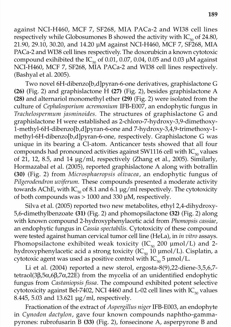
7/23/2019 Novel Therapeutic Agents From Plants
http://slidepdf.com/reader/full/novel-therapeutic-agents-from-plants 200/464
189
against NCI-H460, MCF 7, SF268, MIA PACa-2 and WI38 cell linesrespectively while Globosumones B showed the activity with IC
50of 24.80,
21.90, 29.10, 30.20, and 14.20 µM against NCI-H460, MCF 7, SF268, MIAPACa-2 and WI38 cell lines respectively. The doxorubicin a known cytotoxiccompound exihibited the IC
50of 0.01, 0.07, 0.04, 0.05 and 0.03 µM against
NCI-H460, MCF 7, SF268, MIA PACa-2 and WI38 cell lines respectively.(Bashyal et al. 2005).
Two novel 6H-dibenzo[b,d]pyran-6-one derivatives, graphislactone G(26) (Fig. 2) and graphislactone H (27) (Fig. 2), besides graphislactone A(28) and alternariol monomethyl ether (29) (Fig. 2) were isolated from theculture of Cephalosporium acremonium IFB-E007, an endophytic fungus in
Trachelospermum jasminoides. The structures of graphislactone G andgraphislactone H were established as 2-chloro-7-hydroxy-3,9-dimethoxy-1-methyl-6H-dibenzo[b,d]pyran-6-one and 7-hydroxy-3,4,9-trimethoxy-1-methyl-6H-dibenzo[b,d]pyran-6-one, respectively. Graphislactone G wasunique in its bearing a Cl-atom. Anticancer tests showed that all fourcompounds had pronounced activities against SW1116 cell with IC
50values
of 21, 12, 8.5, and 14 µg/ml, respectively (Zhang et al., 2005). Similarly,
Hormazabal et al. (2005), reported graphislactone A along with botrallin(30) (Fig. 2) from Microsphaeropsis olivacea, an endophytic fungus of Pilgerodendron uviferum. These compounds presented a moderate activitytowards AChE, with IC
50of 8.1 and 6.1 µg/ml respectively. The cytotoxicity
of both compounds was > 1000 and 330 µM, respectively.
Silva et al. (2005) reported two new metabolites, ethyl 2,4-dihydroxy-5,6-dimethylbenzoate (31) (Fig. 2) and phomopsilactone (32) (Fig. 2) alongwith known compound 2-hydroxyphenylacetic acid from Phomopsis cassiae,an endophytic fungus in Cassia spectabilis. Cytotoxicity of these compoundwere tested against human cervical tumor cell line (HeLa), in in vitro assays.Phomopsilactone exhibited weak toxicity (IC
50200 µmol/L) and 2-
hydroxyphenylacetic acid a strong toxicity (IC50
10 µmol/L). Cisplatin, acytotoxic agent was used as positive control with IC
505 µmol/L.
Li et al. (2004) reported a new sterol, ergosta-8(9),22-diene-3,5,6,7-tetraol(3β,5α,6β,7α,22E) from the mycelia of an unidentified endophytic
fungus from Castaniopsis fissa. The compound exhibited potent selectivecytotoxicity against Bel-7402, NCI 4460 and L-02 cell lines with IC
50values
8.445, 5.03 and 13.621 µg/ml, respectively.
Fractionation of the extract of Aspergillus niger IFB-E003, an endophytein Cynodon dactylon, gave four known compounds naphtho-gamma-pyrones: rubrofusarin B (33) (Fig. 2), fonsecinone A, asperpyrone B andaurasperone A (34) (Fig 3). Rubrofusarin B was cytotoxic to the colon cancer
cell line SW1116 (IC50 4.5 µg/ml), and aurasperone A inhibitory on XO(xanthine oxidase) (IC50
10.9 µM/l) (Song et al., 2004).
© 2009 by Taylor & Francis Group, LLC

7/23/2019 Novel Therapeutic Agents From Plants
http://slidepdf.com/reader/full/novel-therapeutic-agents-from-plants 201/464
190
Filip et al. (2003), reported hormonemate (35) (Fig. 3). a new cyto-toxic and apoptosis-inducing compound from the endophytic fungus Hormonema dematioides isolated from living needles of a Pinus species. Thecompound exhibited cytotoxic effects against the human colon tumor celllines COLO-320, DLD-1, HT-29, JURKAT, MCF-7 and HL-60 with IC
50value
of 3.5-7.5 µg/ml., HEP-G2, and HeLa S3 cell were less sensitive with anIC
50of 9-10 µg/ml. It also induced apoptosis in COLO-320 cells as detected
by a caspase-activity assay and it triggered morphological and physio-logical differentiation of HL-60 cells into granulocytes, which subsequentlydied by apoptosis.
Stierle et al. (2003), reported Aspergillus parasiticus, a fungal isolate from
the bark of a redwood tree (Sequoia sempervirens), that can produce theantitumor metabolites sequoiatones C-F (36-39) (Fig. 3). Another series of compounds with a new carbon skeleton, the Sequoiamonascins were alsoisolated. Sequoiamonascins A-D (40-43) (Fig. 3) shows moderate activitiesagainst cancer cell lines, including MCF7 (breast), NCI-H460 (lung), andSF-268 (CNS).
Isaka et al. (2001) reported phomoxanthones A and B (44-45) (Fig. 3),
two xanthone dimers from the endophytic fungus Phomopsis sp. BCC 1323isolated from Tectona grandis from North Thailand. Phomoxanthones A andB exhibited significant cytotoxicity against KB and BC1 cell lines with IC
50
of 0.99 and 4.1 µg/ml against Kb cells and 0.51 and 0.70 µg/ml againstBC1 respectively. The IC
50of standard compound ellipticine was 4.6 and
0.6 µg/ml against KB and BC1 respectively.
A new alkaloid nomofungin (46) (Fig. 3) was isolated from thefermented broth of an unidentified endophytic fungus obtained from the bark of Ficus microcarpa. It disrupts microfilaments in cultured mammaliancells and is moderately cytotoxic, with MICs of 2 and 4.5 µg/mL againstLoVo and KB cells, respectively (Ratnayake et al., 2001a). Similarly, a newalkyl-substituted nonenolide microcarpalide (47) (Fig. 2) was isolated fromfermented broths of an unidentified endophytic fungus isolated from Ficusmicrocarpa. Microcarpalide is weakly cytotoxic to mammalian cells withIC
50value in the KB (human nasopharyngeal carcinoma) and LoVo
(human colon adenocarcinoma) cell lines of 50 and 90 µg/mL respectively(Ratnayake et al., 2001b).
Wagenaar et al. (2001) isolated cytotoxic compounds, dicerandrols A,B, and C (48-50) (Fig. 3 ) from Phomopsis longicolla, an endophytic fungusof the endangered mint Dicerandra frutescens. Extensive NMR and HRFABMSexperiments identified these new yellow antibiotic and cytotoxiccompounds as 2,2'-dimeric tetrahydroxanthones. These compoundsexhibited cytotoxic activity against A549 and HCT 116 cell lines with IC
100of 7.0, 1.8 and 1.8 against A549 and 7.0, 1.8 and 7.0 against HCT 116 celllines respectively.
© 2009 by Taylor & Francis Group, LLC

7/23/2019 Novel Therapeutic Agents From Plants
http://slidepdf.com/reader/full/novel-therapeutic-agents-from-plants 202/464
191
Fig. 3. Structures of cytotoxic metabolites isolated from endophytic fungi.
O
O
OH OH
OH
O
O
MeO
MeO OMe
Aurasperone A (34)
O
OH
OCOOCH
3
Sequoiatone C (36)
O
O
O
O
O
O
OH
O
OH
OH
OH
OH
O
O
OH
O
Hormonemate (35)
O
OH
O
OH
COOCH3
Sequoiatone D (37)
O
OH
O
OH
COOCH3
Sequoiatone E (38)
O
OH
OCOOCH
3
Sequoiatone F (39)
OH
OO
H
O
O
H
OO
Sequoiamonascin A (40)
OH
OO
H
O
O
H
O O
Sequoiamonascin B (41)
OH
Sequoiamonascin C (42)
OO
H
O
O
OH
O
H
© 2009 by Taylor & Francis Group, LLC

7/23/2019 Novel Therapeutic Agents From Plants
http://slidepdf.com/reader/full/novel-therapeutic-agents-from-plants 203/464
192
Sequoiamonascin D (43)
O
O
O
O
H
N
OH
COOCH3
O
O
OH OHO
OOH OH
CH3
OAc
OAc
CH3
OAc
OAc
Phomoxanthone A (44)
O
OH
OH
O
OAc
CH3
OAc O
OHOH O
OAc
CH3
OAc
Phomoxanthone B (45)
O
OH
OH
O
OH
Microcarpalide (47)
OCH3
O
OOH
O
O
OH
OH OH
O CH3
O
CH3
O
O
R2
CH3
O
R1
Dicerandrol A = R1=R2=H (48)Dicerandrol B = R1=Ac, R2=H (49)Dicerandrol C = R1=R2= Ac (50)
N
N
O
O
N
O
Nomofungin (46)
Fig. 3 continued. Structures of cytotoxic metabolites isolatedfrom endophytic fungi.
© 2009 by Taylor & Francis Group, LLC

7/23/2019 Novel Therapeutic Agents From Plants
http://slidepdf.com/reader/full/novel-therapeutic-agents-from-plants 204/464
193
Brady et al. (2000) isolated cytoskyrins A (51) (Fig. 4) and B, new BIAactive bisanthraquinones from endophytic fungus Cytospora isolated fromthe branch of a Conocarpus erecta. Cytoskyrin A (Fig. 2) exhibited poorcytotoxicity against tumor cell lines (IC
50
> 5 µg/ml) as compared to knownantitumor agents.
Wagenaar et al. (2000) reported cytochalasin E and three new cytotoxiccytochalasins from a culture of the endophytic fungus Rhinocladiella sp.isolated from Tripterygium wilfordii using bioassay-guided fractionation.Extensive NMR and HRCIMS experiments identified these new compoundsas 22-oxa-[12]-cytochalasins1,2,3. (52-54) (Fig. 4) Cytochalasin E (55) (Fig.4) is significantly more potent (15-100 fold) against all cell lines tested i.e.
A2780S, SW-620, HCT-116. 22-oxa-[12]-cytochalasins1,2,3 did not showany selectivity in the cell lines tested.
Strobel et al. (1997a) isolated Pestalotiopsis guepinii, endophyte fromWollemia nobilis, an araucariaceous plant, which produces taxol. Wang etal. (2000) reported taxol-producing endophytic fungus Tubercularia sp TF5,isolated from Taxus mairei. The fungal taxol, had strong cytotoxic effecttowards KB and P388 cancer cell lines, and the ID
50values of cytotoxicity
was about 1/2400 for KB cells and 1/38400 for P388 cells in vitro, tested by MTT assay. It also enhanced microtubule stability, bundling in culturecells and induced tubulin polymerization in vitro (1/100 v/v) similarto the authentic taxol (25 µg/ml). The other producers of taxol arePestalotiopsis microspora isolated from Taxodium distichum (Li et al., 1996);Periconia sp. isolated from Torreya grandifolia, (Li et al., 1998); Stegoleriumkukenani isolated from Maguireothamnus speciosus (Strobel et al., 2001).
Acremonium sp. occurs as an endophyte in European yew (Taxusbaccata), which produces a series of peptide antifungal, anticancer agentsknown as the leucinostatins. Leucinostatin A (56) (Fig. 4) possessed activityagainst certain human cancer cell lines, for instance, its IC
50value is 2.3
nM for breast cancer cell line BT-20 contrasted with 640 nM for a normalmammary cell line (Strobel et al. 1997b).
Lee et al. (1996) isolated torreyanic acid (57) (Fig. 4), a selectivelycytotoxic quinone dimer from the endophytic fungus Pestalotiopsis
microspora isolated from Torreya taxifolia. Torreyanic acid was tested inseveral cancer cell lines, and it demonstrated 5 to 10 times more potentcytotoxicity in cancer cell lines that are sensitive to protein kinase Cagonists; it causes cell death by apoptosis.
From mangroves
Chen et al. (2005) isolated mycoepoxydiene (58) (Fig. 4), which showedcytotoxicity toward human tumor cells in vitro, from the fermented broth
© 2009 by Taylor & Francis Group, LLC

7/23/2019 Novel Therapeutic Agents From Plants
http://slidepdf.com/reader/full/novel-therapeutic-agents-from-plants 205/464
194
Fig. 4. Structures of cytotoxic metabolites isolated from endophytic fungi.
OO
O O
O
O
O
O
COOH
COOH
Torreyanic acid (57)
22-oxa-[12]-cytochalasin 3 (54)
O
O
NH
O
O OH
O
NCONH
O
CONH
O
CONH
OH
H
CO
NHNHCONHCO
CO
NH
CONHNCONH
OH
Leucinostatin A (56)
NH O
O
H
O
O
O
O OH
Cytochalasin E (55)
NH
O
O
O
OH
O
22-oxa-[12]-cytochalasin1 (52)
NH
O
O
O
O
OH
O
22-oxa-[12]-cytochalasin 2 (53)
OH
OH
O
O
OMe
Cytoskyrin A (51)
OH
OH
O
O
OH
OH
MeO
© 2009 by Taylor & Francis Group, LLC

7/23/2019 Novel Therapeutic Agents From Plants
http://slidepdf.com/reader/full/novel-therapeutic-agents-from-plants 206/464
195
Fig. 4 continued. Structures of cytotoxic metabolites isolatedfrom endophytic fungi.
(CH2)m
NH
OH
(CH2)n
OH
OH
O
[2',3'-dihydroxytetracosanolyamino]-1,3-dihydroxyoctadecane = m=12, n=18 (60)
[2',3'-dihydroxydocosanolyamino]-1,3-dihydroxyoctadecane = m=12, n=16 (61)
O
OOH
OH
OH
OH
Aigialomycin D (63)
O
O
O
OH
O
OH
Sterigmocystin (62)
N
O O
N
O
N
O
O
O
O
O
O
Enniatin G (64)
O O
O
Me
H
AcO
Mycoepoxydiene (58)
O
O O
OH
OH
Cytosporone B (59)
© 2009 by Taylor & Francis Group, LLC

7/23/2019 Novel Therapeutic Agents From Plants
http://slidepdf.com/reader/full/novel-therapeutic-agents-from-plants 207/464
196
of the marine mangrove endophytic fungus No. 1893. It shows highantitumor activity with IC
50of < 6.25 µg/ml against KB cell lines (Lin et
al., 2005) and 5.5 µg/ml against HeLa cell lines (Chen et al., 2006)respectively.
Xu et al. (2004) reported cytosporone B (59) (Fig. 4) , isolated from anendophytic fungus, Dothiorella sp. strain HTF3 isolated from the mangroveplant Avicennia marina at the estuary of Jiulong River, Fujian Province. ItsIC
50values were 4 µg/ml and 0.062 5 µg/ml against human epidermal
carcinoma of oral cavity, KB cell line and human Burkitt’s lymphoma, Rajicell line, respectively.
Li et al. (2003), reported two new ceramides, [2',3’-dihydroxytetra-cosanolyamino]-1,3-dihydroxyoctadecane (60) (Fig. 4) and [2',3'-dihydroxydocosanoylamino]-1,3-dihydroxyoctadecane (61) (Fig. 4), fromthe culture extract of an unidentified endophytic fungus No. 2524 isolatedfrom the seed of mangrove Avicennia marina of Hong Kong. These ceramidesdid not exhibit significant cytotoxicity against the human cancer cell linesBel-7402 and NCI-4460 and the normal human cell line L-02 in preliminarycytotoxic activity investigation.
Zhu et al. (2003) isolated sterigmatocystin (62) (Fig. 4), from endophyticfungus No. 2526 of mangrove Avicennia from the South China Sea, whichshowed a weak cytotoxic activity against Bel-7402 and NCIH-460 withwith IC
50of 96.53 and 72. 52 µg/ml respectively.
Isaka et al. (2002) isolated Aigialomycins A, B, C, D, E, new 14-membered resorcylic macrolides together with known hypothemycin fromthe mangrove fungus, Aigialus parvus BCC 5311. Only Aigialomycin D (63)
(Fig. 4) , have cytotoxic and antiplasmodial activity. Hypothemycin andAigialomycins A, B, C, D, E exhibited cytotoxic activity with IC50
of 17, >20,> 20, >20, 6.6 and > 20 µg/ml against KB (Human epidermoid carcinoma)cell lines and 6.2, 11, >20, > 20, 18 and 15 µg/ml against BC1 (human breast cancer cells) respectively.
Lin et al. (2002) isolated novel cyclic depsipeptide, enniatin G (64) (Fig.4), together with enniatins B and B4 from the mangrove fungus Halosarpheia sp. strain 732 from the South China Sea. Enniatin G exhibitedactivity against Heps 7402, ED
5012mg/ml.
From marine algae
Kralj et al. (2006) isolated Emericella nidulans var. acristata from aMediterranean green alga. Cultivation of this fungus yielded emindoleDA(65) (Fig. 5), the indole alkaloid, which displayed antitumor activity in
a panel of 36 human tumor cell lines, exhibiting a mean IC50 value of 5.5µg/ml in an in vitro survival and proliferation assay. Arugosin A (66) (Fig.
© 2009 by Taylor & Francis Group, LLC

7/23/2019 Novel Therapeutic Agents From Plants
http://slidepdf.com/reader/full/novel-therapeutic-agents-from-plants 208/464
197
3) and arugosin B (67) (Fig. 5) were coproduced in the culture, whichshowed moderate antitumor activity toward individual tumor cell lines.
Chaetomium globosum, an endophytic fungus which was isolated from
the inner tissue of the marine red alga Polysiphonia urceolata, resulted inthe isolation of a new benzaldehyde secondary metabolite chaetopyranin(68) (Fig. 5) which exhibited moderate to weak cytotoxic activities againstthree tumor cell lines, with IC
50values of 15.4 (human microvascular
endothelial cells, HMEC), 28.5 (hepatocellular carcinoma cells, SMMC-7721) and 39.1 µg/ml (human lung epithelial cells, A549) (Wang et al.,2006b).
Klemke et al. (2004) isolated marine fungus Apiospora montagnei fromthe inner tissue of the North Sea alga Polysiphonia violacea and reported(+)-epiepoxydon (69) (Fig. 5) which exhibited 50% reduction of the initialcell number (LC
50) at concentration of 3.6, >10, and 10 µg/ml towards
human cancer cell lines HM02 (gastric adenocarcinoma), HepG2(hepatocellular carcinoma) and MCF7 (breast adenocarcinoma) respec-tively.
Leptosphaeria sp. OUPS-N80 (formerly OUPS-4) isolated from the brown
alga Sargassum tortile is a prolific fungus and 24 new compounds, leptosinsA (70) (Fig. 5) and B (–S), have been obtained from this strain (Takahashiet al., 1994a, 1994b, 1995a, 1995b; Yamada et al., 2002, 2004). Leptosinsshowed strong cytotoxicity against P388 with ED
50values in the ng order.
Trichodermamides A and B (71) (Fig. 5) two modified dipeptides have been isolated from cultures of the marine-derived fungus Trichoderma virens.Trichodermamide B displayed significant in vitro cytotoxicity against HCT-
116 human colon carcinoma with an IC50 of 0.32 µg/ml (Garo et al., 2003).Scytalidamides A (72) (Fig. 5) and B the cytotoxic compounds were
isolated from Scytalidium sp. CNC-310 isolated from marine alga Halimedasp. collected from Bahamas (Tan et al., 2003). Scytalidamides A and Bshowed moderate in vitro cytotoxicity toward HCT-116 human colonadenocarcinoma with IC
50values of 2.7 and 11.0 µM, respectively.
Usami et al. (2002) isolated two new sulphur-containing dioxo-
piperazine derivatives designated Fusaperazines A and B (73-74) (Fig. 5)from Fusarium chlamydosporum OUPS-N124 from the red alga Carpopeltisaffinis of Japan. Fusaperazine A exhibited weak cytotoxic activity againstthe lymphocytic leukemia cell line P388 (ED
50= 22.8 µg/ml).
Cueto et al. (2000) reported N -methylsansalvamide (75) (Fig. 5) fromFusarium sp. which was isolated from the green alga Avrainvillea sp. N -methylsansalvamide showed moderate cytotoxicity (GI
50= 8.3 µM) in the
NCI’s human tumor cell line screen.Penicillium sp. OUPS-N79 isolated from the green alga Enteromorpha
© 2009 by Taylor & Francis Group, LLC

7/23/2019 Novel Therapeutic Agents From Plants
http://slidepdf.com/reader/full/novel-therapeutic-agents-from-plants 209/464
198
Fig. 5. Structures of cytotoxic metabolites isolated from endophytic fungi.
O
N
NH
O
O
O
OMe
OMeOH
H
Cl
OH
Trichodermamide B (71)
OHO
O
OH
(+)-epiepoxydon (69)
NH
N
N
OH
H S4
O
N
H
N
NS2
O
O
O
H
OH
OH
CH3
CH3
Leptosin A (70)
N
O
NH
O
NHO
NH
O
NH O
N
O
O
N
Scytalidamide A (72)
NH
H
OH
Emindole DA (65)
H OOH
OHOH O
O
Arugosin A (66)
Arugosin B (67)
O
OH
O
OH
H
Chaetopyranin (68)
OH
O
OH
O
O
© 2009 by Taylor & Francis Group, LLC

7/23/2019 Novel Therapeutic Agents From Plants
http://slidepdf.com/reader/full/novel-therapeutic-agents-from-plants 210/464
199
NR5
NR5
O
OOR1
R3
R4
R2
Fusaperzine A = R1=H, R2=H,R3=R4=SMe, R5=H (73)
Fusaperzine B = R1=isoprenyl,R2=SMe, R3=OMe, R4=R5=H (74)
NH
NH
O
NHN
O
O
O
OO
N-Methylsansalvamide (75)
Penostatins A (77)
O
OH
O
H H
H
(CH2
)6
CH3
NH
N
N
O
N
O
Communesins A (76)
H
Penochalasins A (78)
NH
NH
O
O
H
NH
O
O O
O
MeO
OMe
Pyrenocine E (81)
NH
N
NH S
2
O
NH
N
NS
2
O
O
O
H
11,11'-Dideoxyverticillin A (80)
Insulicolide A (79)
O
O
OH
O
O
OH H
NO2
Fig. 5 continued. Structures of cytotoxic metabolites isolatedfrom endophytic fungi.
© 2009 by Taylor & Francis Group, LLC
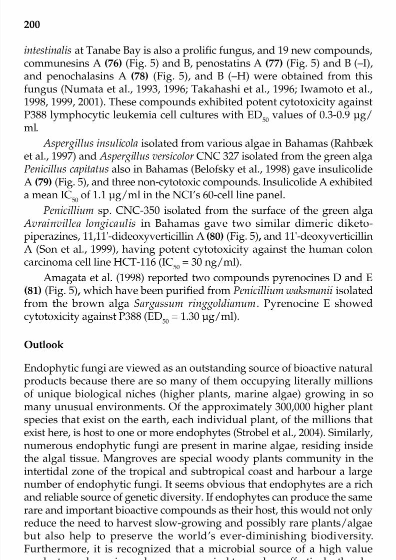
7/23/2019 Novel Therapeutic Agents From Plants
http://slidepdf.com/reader/full/novel-therapeutic-agents-from-plants 211/464
200
intestinalis at Tanabe Bay is also a prolific fungus, and 19 new compounds,communesins A (76) (Fig. 5) and B, penostatins A (77) (Fig. 5) and B (–I),and penochalasins A (78) (Fig. 5), and B (–H) were obtained from thisfungus (Numata et al., 1993, 1996; Takahashi et al., 1996; Iwamoto et al.,1998, 1999, 2001). These compounds exhibited potent cytotoxicity againstP388 lymphocytic leukemia cell cultures with ED
50values of 0.3-0.9 µg/
ml.
Aspergillus insulicola isolated from various algae in Bahamas (Rahbæket al., 1997) and Aspergillus versicolor CNC 327 isolated from the green algaPenicillus capitatus also in Bahamas (Belofsky et al., 1998) gave insulicolideA (79) (Fig. 5), and three non-cytotoxic compounds. Insulicolide A exhibited
a mean IC50 of 1.1 µg/ml in the NCI’s 60-cell line panel.Penicillium sp. CNC-350 isolated from the surface of the green alga
Avrainvillea longicaulis in Bahamas gave two similar dimeric diketo-piperazines, 11,11'-dideoxyverticillin A (80) (Fig. 5) , and 11'-deoxyverticillinA (Son et al., 1999), having potent cytotoxicity against the human coloncarcinoma cell line HCT-116 (IC
50= 30 ng/ml).
Amagata et al. (1998) reported two compounds pyrenocines D and E
(81) (Fig. 5) , which have been purified from Penicillium waksmanii isolatedfrom the brown alga Sargassum ringgoldianum. Pyrenocine E showedcytotoxicity against P388 (ED
50= 1.30 µg/ml).
Outlook
Endophytic fungi are viewed as an outstanding source of bioactive natural
products because there are so many of them occupying literally millionsof unique biological niches (higher plants, marine algae) growing in somany unusual environments. Of the approximately 300,000 higher plantspecies that exist on the earth, each individual plant, of the millions thatexist here, is host to one or more endophytes (Strobel et al., 2004). Similarly,numerous endophytic fungi are present in marine algae, residing insidethe algal tissue. Mangroves are special woody plants community in theintertidal zone of the tropical and subtropical coast and harbour a large
number of endophytic fungi. It seems obvious that endophytes are a richand reliable source of genetic diversity. If endophytes can produce the samerare and important bioactive compounds as their host, this would not onlyreduce the need to harvest slow-growing and possibly rare plants/algae but also help to preserve the world’s ever-diminishing biodiversity.Furthermore, it is recognized that a microbial source of a high valueproduct may be easier and more economical to produce effectively, thereby
reducing its market price.The review of the literature indicates that novel chemotypes directed
© 2009 by Taylor & Francis Group, LLC

7/23/2019 Novel Therapeutic Agents From Plants
http://slidepdf.com/reader/full/novel-therapeutic-agents-from-plants 212/464
201
against cancer have come from the microbial source, which is poorlyinvestigated. Although the work on the utilization of endophytes has just begun, but it has already become obvious that they represent a promisingsource of novel metabolite.
As most of the plant diversity is found in tropical countries, there will be a great need of culture collection of this group of fungi with trainedmycologists having classical and molecular background. The collectionwill help not only in getting novel secondary metabolites for variouspharmaceutical and agricultural applications but also for applications like biotransformation, enzyme production etc.
References
Alquati, S.A.B. 1999. Paracoccidioides brasiliensis não ocorre na forma endofíticaem gramíneas da região endêmica de Botucatu, SP, Brasil, Ms. Thesis,Botucatu, UNESP.
Amagata, T., K. Minoura and A. Numata. 1998. Cytotoxic metabolites produced by a fungal strain from a Sargassum algae. Journal of Antibiotics. 51: 432-434.
Amna, T., S.C. Puri, V. Verma, J.P. Sharma, R.K. Khajuria, J. Musarrat, M. Spitellerand G.N. Qazi. 2006. Bioreactor studies on the endophytic fungusEntrophospora infrequens for the production of an anticancer alkaloidcamptothecin. Canadian Journal of Microbiology. 52(3): 189-196.
Arnold, E.A. 2005. Diversity and Ecology of Fungal Endophytes in TropicalForests. pp. 267-288. In: S.K. Deshmukh and M.K. Rai (eds.) The Bio-Diversityof fungi—Their Role in Human Life. Science Publishers, Inc Enfield, USA.
Bashyal, B.P., E.M.K. Wijeratne, S.H. Faeth and A.A.L. Gunatilaka. 2005.Globosumones A-C, cytotoxic orsellinic acid esters from the Sonoran Desertendophytic fungus Chaetomium globosum. Journal of Natural Products 68(5):724-728.
Bérdy, J. 2005. Bioactive Microbial Metabolites A Personal View. Journal of Antibiotics. 58(1): 1-26.
Bettucci, L., R. Alonso and S. Tiscornia. 1999. Endophytic mycobiota of healthytwigs and the assemblage of species associated with twig lesions of
Eucalyptus globosus and E. grandis in Uruguay. Mycological Research 103:468-472.Belofsky, G.N., P.R. Jensen, M.K. Renner and W. Fenical. 1998. New cytotoxic
sesquiterpenoid nitrobenzoyl esters from a marine isolate of the fungusAspergillus versicolor. Tetrahedron 54: 1715–1724.
Brady, S.F., M.P. Singh, J.E. Janso and J. Clardy. 2000. Cytoskyrins A and B, newBIA active bisanthraquinones isolated from an endophytic fungus. OrganicLetters 2(25): 4047-4049.
Brossi, A. and M. Suffness. 1990. The Alkaloids: Antitumor Bisindole Alkaloidsfrom Catharanthus roseus (L.) Academic press, San Diego, USA, pp. 133-204.
Chabner, B.A., C.J. Allegra, G.A. Curt and P. Calabresi. 1996. Antineoplastic agents© 2009 by Taylor & Francis Group, LLC
7/23/2019 Novel Therapeutic Agents From Plants
http://slidepdf.com/reader/full/novel-therapeutic-agents-from-plants 213/464
202
pp. 1233-1287. In: Goodman and Gilman’s The Pharmacological Basis of Therapeutics. McGraw-Hill, New York, USA.
Carroll, G. and F.E. Carroll. 1978. Studies on the incidence of coniferous needleendophytes in the Pacific Northwest. Canadian Journal of Botany 56: 3034-
3043.Chen, H., Y. Lin, G. Cen, G. Hu and L. Wang. 2006. Catalytic transfer
hydrogenation of mycoepoxydiene. Chemistry of Natural Compounds. 42:407-409.
Chen, H.Y., P. Qi, Y.C. Lin, G.Y. Chen, L. Wang and L.L.P. Vrijoed. 2005.Preparation of hexahydromycoepoxydiene by catalytic hydrogenation of mycoepoxydiene. Zhongshan Daxue Xuebao, Ziran Kexueban 44(3): 122-123.
Cueto, M., P.R. Jensen and W. Fenical. 2000. N -Methylsansalvamide, a cytotoxiccyclic depsipeptide from a marine fungus of the genus Fusarium .Phytochemistry 55: 223–226.
Dahanukar, S.A., R.A. Kulkarni and N.N. Rege. 2000. Pharmacology of medicinalplants and natural products. Indian Journal of Pharmacology 32: 81-118.
De Bary A. 1866. Morphologie und Physiologie der Pilze, Flechten undMyxomyceten. In: Wilh.Hofmeister. (ed.) Handbuch der physiologischenBotanik. Engelmann, XII. Leipzig, Germany.
Devarajan, P.T., T.S. Suryanarayanan and V. Geetha. 2002. Endophytic fungiassociated with the tropical seagrass Halophila ovalis (Hydrocharitaceae).Indian Journal of Marine Sciences 31: 73-74.
Ding, G., Y.C. Song, J.R. Chen, C. Xu, Hui M., Ge, X.T. Wang and R.X. Tan. 2006.Chaetoglobosin U, a cytochalasan alkaloid from endophytic Chaetomium globosum IFB-E019. Journal of Natural Products. 69(2): 302-304.
Dreyfuss, M.M. and I.H. Chapela. 1994. Potential of fungi in the discovery of novel, low-molecular weight pharmaceuticals. pp. 49-80. In: V.P. Gullo (ed.)The Discovery of Natural Products with Therapeutic Potential. Butterworth-
Heinemann, Boston, USA.Filip, P., R.W.S. Weber, O. Sterner and T. Anke. 2003. Hormonemate, a new
cytotoxic and apoptosis-inducing compound from the endophytic fungus Hormonema dematioides. I. Identification of the producing strain, and isolationand biological properties of hormonemate. Zeitschrift fuer Naturforschung,C: Journal of Biosciences 58: 547-552.
Garo, E., C.M. Starks, P.R. Jensen, W. Fenical, E. Lobkovsky and J. Clardy. 2003.Trichodermamides A and B, cytotoxic modified dipeptides from the marine-
derived fungus Trichoderma virens. Journal of Natural Producs 66: 423-426.Ge, H.M., Y.C. Song, C.Y. Shan, Y.H. Ye and R.X. Tan. 2005. New and cytotoxicanthraquinones from Pleospora sp. IFB-E006, an endophytic fungus inImperata cylindrica. Planta Medica 71(11): 1063-1065.
Hallmann, J., A. Quadt-Hallmann, W.F. Mahaffee and J.W. Kloepper. 1997.Bacterial endophytes in agriculture crops. Canadian Journal of Microbiology43: 895-914.
Hartwell, J.L. 1982. Plants used against cancer. A survey. Quarterman Publications,Lawrence, MA, USA.
Hormazabal, E., G. Schmeda-Hirschmann, L. Astudillo, J. Rodriguez and C.Theoduloz. 2005. Metabolites from Microsphaeropsis olivacea, an endophytic© 2009 by Taylor & Francis Group, LLC

7/23/2019 Novel Therapeutic Agents From Plants
http://slidepdf.com/reader/full/novel-therapeutic-agents-from-plants 214/464
203
fungus of Pilgerodendron uviferum. Zeitschrift fuer Naturforschung, C: Journalof Biosciences 60(1/2): 11-21.
Isaka, M., A. Jaturapat, K. Rukseree, K. Danwisetkanjana, M. Tanticharoen andY. Thebtaranonth. 2001. Phomoxanthones A and B, Novel Xanthone Dimers
from the Endophytic Fungus Phomopsis Species. Journal of Natural Products64(8): 1015-1018.
Isaka, M., C. Suyarnsestakorn, M. Tanticharoen, P. Kongsaeree and Y.Thebtaranonth. 2002. Aigialomycins A-E, new resorcyclic macrolides fromthe marine mangrove fungus Aigialus parvus. Journal of Organic Chemistry67: 1561-1566.
Iwamoto, C., K. Minoura, S. Hagishita, K. Nomoto and A. Numata. 1998.Penostatins F-I, novel cytotoxic metabolites from a Penicillium species
separated from an Enteromorpha marine alga. Journal of the Chemical Society,Perkin Transaction I 449-456.Iwamoto, C., K. Minoura, T. Oka, T. Ohta, S. Hagishita and A. Numata. 1999.
Absolute stereostructures of novel cytotoxic metabolites, penostatins A-E,from a Penicillium species separated from an Enteromorpha alga. Tetrahedron55: 14353-14368.
Iwamoto, C., T. Yamada, Y. Ito, K. Minoura and A. Numata. 2001. Cytotoxiccytochalasans from a Penicillium species separated from a marine alga.Tetrahedron 57: 2997-3004.
Kapoor, L.D. 1990. Handbook of Ayurvedic Medicinal Plants. CRC Press. BocaRaton, Fl., USA, pp. 149-150.
Klemke, C., S. Kehraus, A.D. Wright and G.M. Koenig. 2004. New secondarymetabolites from the marine endophytic fungus Apiospora montagnei. Journalof Natural Products 67(6): 1058-1063.
Kralj, A., S. Kehraus, A. Krick, E. Eguereva, G. Kelter, M. Maurer, A. Wortmann,H.H. Fiebig and G.M. Konig. 2006. Arugosins G and H: prenylatedpolyketides from the marine-derived fungus Emericella nidulans var. acristata.
Journal of Natural Products 69(7): 995-1000.Krishnamurthy, Y.L. and T.V. Hemalatha. 2003. Isolation of endophytic fungi
from some grasses. Journal of Mycology and Plant Pathology 33: 305-306.Lee J.C., G.A. Strobel, E. Lobkovsky and J. Clardy. 1996. Torreyanic acid: a
selectively cytotoxic quinone dimer from the endophytic fungus Pestalotiopsismicrospora. Journal of Organic Chemistry 61(10): 3232-3233.
Li, H.J., J.H. Yao, Y.G. Chen, Y.C. Lin and L.L.P. Vrijmoed. 2003. Novel ceramidesfrom a mangrove endophytic fungus No. 2524. Zhongshan Daxue Xuebao,
Ziran Kexueban 42(6): 132-133.Li, H.J., Y.C. Lin, L.L.P. Vrijmoed and E.B.G. Jones. 2004. A new cytotoxic sterolproduced by an Endophytic fungus from Castaniopsis fissa at the South Chinasea coast. Chinese Chemical Letters 15(4): 419-422.
Li, J.Y., G.A. Strobel, R.S. Sidhu, W.M. Hess and E. Ford. 1996. Endophytic taxolproducing fungi from Bald Cypress Taxodium distichum. Microbiology 142 ,2223-2226.
Li, J.Y., R.S. Sidhu, E. Ford W.M. Hess and G.A. Strobel. 1998. The induction of taxol production in the endophytic fungus Periconia sp . from Torreya grandifolia. Journal of Industrial Microbiology. 20: 259-264.
Lin, Y., J. Wang, X. Wu, S. Zhou, L.L.P. Vrijmoed and E.B.G. Jones. 2002. A novel© 2009 by Taylor & Francis Group, LLC

7/23/2019 Novel Therapeutic Agents From Plants
http://slidepdf.com/reader/full/novel-therapeutic-agents-from-plants 215/464
204
compound enniatin G from the mangrove fungus Halosarpheia sp. strain #732from the South China Sea. Australian Journal of Chemistry 55: 225-227.
Lin, X., Y. Huang, M. Fang, J. Wang, Z. Zheng and W. Su. 2005. Cytotoxic andantimicrobial metabolites from marine lignicolous fungi, Diaporthe sp. FEMS
Microbiol Lett. 251(1): 53-58.Liu, J. and B. Yu. 2004. Isolation of endophytic fungi from Camptotheca acuminata
and the screening method of antitumor secondary metabolite produced bythe fungi. Zhiwu Ziyuan Yu Huanjing Xuebao 13(4): 6-10.
Mahesh, B., M.V. Tejesvi, M.S. Nalini, H.S. Prakash, K.R. Kini, V. Subbiah andH.S. Shetty. 2005. Endophytic mycoflora of inner bark of Azadirachta indicaA. Juss Current Science. 88: 218-219.
Maria, G.L. and K.R. Sridhar. 2003. Endophytic fungal assemblage of two
halophytes from west coast mangrove habitats, India. Czech Mycology. 55,241-251.Numata, A., C. Takahashi, Y. Ito, K. Minoura, T. Yamada, C. Matsuda and K.
Nomoto. 1996. Penochalasins, a novel class of cytotoxic cytochalasans froma Penicillium species separated from a marine alga: structure determinationand solution conformation. Journal of the Chemical Society, PerkinTransaction 1 239-245.
Numata, A., C. Takahashi, Y. Ito, T. Takada, K. Kawai, Y. Usami, E. Matsumura,M. Imachi, T. Ito and T. Hasegawa. 1993. Communesins, cytotoxic metabolitesof a fungus isolated from a marine alga. Tetrahedron Letters. 34: 2355-2358.
Pamphile, J.A. and J.L. Azevedo. 2002. Molecular characterization of endophyticstrains of Fusarium verticillioides (= Fusarium moniliforme) from maize (Zea Mays L.). Word Journal of Microbiology and Biotechnology 18: 391-396.
Petrini, O. 1986. Taxonomy of endophytic fungi in aerial plant tissues. pp. 175-187. In: N.J. Fokkema and J.Van den Heuvel (eds) Microbiology of thePhyllosphere. Cambridge University Press, Cambridge, UK.
Petrini, O. 1991. Fungal endophytes of tree leaves. pp. 179-197. In: J.H. Andrews
and S.S. Hirano (eds). Microbial ecology of leaves. Springer-Verlag, NewYork, USA.
Petrini, O., U. Hake and M.M. Dreyfuss. 1990. An analysis of fungal communitiesfrom fruticose lichens. Mycologia 82: 444-451.
Pornpakakul, S., J. Liangsakul, N. Ngamrojanavanich, S. Roengsumran, P.Sihanonth, J. Piapukiew. E. Sangvichien, S. Puthong and A. Petsom. 2006.Cytotoxic activity of four xanthones from Emericella variecolor, an endophyticfungus isolated from Croton oblongifolius. Archives of Pharmcal Research.
29(2): 140-144.Potmeisel, M. and H. Pinedo. 1995. Camptothecins: New Anticancer Agents. CRCPress. Boca Raton, Florida, USA.
Puri, S.C., V. Verma, T. Amna, G.N. Qazi and M. Spiteller. 2005. An EndophyticFungus from Nothapodytes foetida that produces Camptothecin. Journal of Natural Products 68(12): 1717-1719.
Rahbæk, L., C. Christophersen, J. Frisvad, H.S. Bengaard, S. Larsen and B.R.Rassing. 1997. Insulicolide A: a new nitrobenzoyloxy-substitutedsesquiterpene from the marine fungus Aspergillus insulicola. Journal of Natural Products 60: 811–813.
Ratnayake, A.S., W.Y. Yoshida, S.L. Mooberry and T.K. Hemscheidt. 2001a.© 2009 by Taylor & Francis Group, LLC

7/23/2019 Novel Therapeutic Agents From Plants
http://slidepdf.com/reader/full/novel-therapeutic-agents-from-plants 216/464
205
Nomofungin: a new microfilament disrupting agent. Journal of OrganicChemistry 66(26): 8717-8721.
Ratnayake, A.S., W.Y. Yoshida, S.L. Mooberry and T.K. Hemscheidt. 2001b. Thestructure of microcarpalide, a microfilament disrupting agent from an
endophytic fungus. Organic Letters 3(22): 3479-3481.Redecker, D., R. Kodner and L.E. Graham. 2000. Glomalean fungi from the
Ordovician. Science 289: 1920-1921.Rygiewicz, P.T. and C.T. Andersen. 1994. Mycorrhizae alter quality and quantity
of carbon allocated below ground. Nature. 369: 58-60.Shen, L., Y.H. Ye, X.T. Wang, H.L. Zhu, C. Xu, Y.C. Song, H. Li and R.X. Tan.
2006a. Structure and total synthesis of aspernigerin: a novel cytotoxicendophyte metabolite. Chemistry European Journal. 12 (16): 4393-4396.
Shen, L., R.H. Jiao, Y.H. Ye, X.T. Wang, C. Xu, Y.C. Song, H.L. Zhu and R.X. Tan.2006b. Absolute configuration of new cytotoxic and other bioactivetrichothecene macrolides. Chemistry Europian Journal 12(21): 5596-5602.
Silva, G.H., H.L. Teles, H.C. Trevisan, V.S. Bolzani, M.C.M. Young, L.H. Pfenning,M.N. Eberlin, R. Haddad, C.M. Costa-Neto and A.R. Araujo. 2005. New bioactive metabolites produced by Phomopsis cassiae, an endophytic fungusin Cassia spectabilis. Journal of the Brazilian Chemical Society 16(6B): 1463-1466.
Son, B.W., P.R. Jensen, C.A. Kauffman and W. Fenical. 1999. New cytotoxicepidithiodioxopiperazines related to verticillin A from a marine isolate of the fungus Penicillium. Natural Product Letters 13(3): 213-222.
Song, Y.C., H. Li, Y.H. Ye, C.Y. Shan, Y.M. Yang and R.X. Tan. 2004. Endophyticnaphthopyrone metabolites are co-inhibitors of xanthine oxidase, SW1116cell and some microbial growths. FEMS Microbiol Lett. 241(1): 67-72.
Stierle, A., G.A. Strobel and D. Stierle. 1993. Taxol and taxane production byTaxomyces andreanae. Science 260: 214-216.
Stierle, D.B., A.A. Stierle and T. Bugni. 2003. Sequoiamonascins a-d: novel
anticancer metabolites isolated from a redwood endophyte. Journal of Organic Chemistry. 68(12): 4966-4969.
Strobel, G., X. Yang, J. Sears, R. Kramer, R.S. Sidhu and W.M. Hess. 1996. Taxolfrom Pestalotiopsis microspora, an endophytic fungus of Taxus wallichiana.Microbiology 142: 435-440.
Strobel G.A., W.M. Hess, J.Y. Li, E. Ford, J. Sears, R.S. Sidhu and B. Summerell.1997a. Pestalotiopsis guepinii, a taxol-producing endophyte of the wollemipine, Wollemia nobilis. Australian Journal of Botany 45 (6): 1073-1082.
Strobel G.A., R. Torczynski and A. Bollon. 1997b. Acremonium sp. A leucinostatinA producing endophyte of European yew (Taxus baccata). Plant Science(Shannon, Ireland) 128(1): 97-108.
Strobel, G., W.M. Hess, G. Baird, E. Ford, J.Y. Li and R.S. Sidhu. 2001. Stegoleriumkukenani gen, et sp nov an endophytic taxol producing fungus from theRoraima and Kukenan tepuis of Venezuela, Mycotaxon 78: 353-361.
Strobel, G.A., B. Daisy, U. Castillo and J. Harper. 2004. Natural products fromendophytic micro-organisms. Journal of Natural Products. 67(2): 257-68.
Suffness, M. 1995. Discovery and development of taxol. pp. 3-25. In: Taxol: Scienceand Applications, CRC Press, Boca Raton, Fl.,USA.
Takahashi, C., A. Numata, Y. Ito, E. Matsumura, H. Araki, H. Iwaki and K.© 2009 by Taylor & Francis Group, LLC

7/23/2019 Novel Therapeutic Agents From Plants
http://slidepdf.com/reader/full/novel-therapeutic-agents-from-plants 217/464
206
Kushida. 1994a. Leptosins, antitumor metabolites of a fungus isolated froma marine alga. Journal of the Chemical Society, Perkin Transaction 1 1859–1864.
Takahashi, C., A. Numata, E. Matsumura, K. Minoura, H. Eto, T. Shingu, T. Ito
and T. Hasegawa. 1994b. Leptosins I and J, cytotoxic substances produced by a Leptosphaeria sp. physico-chemical properties and structures. Journal of Antibiotics 47: 1242-1249.
Takahashi, C., Y. Takai, Y. Kimura, A. Numata, A. Shigematsu and H. Tanaka.1995a. Cytotoxic metabolites from a fungal adherent of a marine alga.Phytochemistry 38: 155-158.
Takahashi, C., K. Minoura, T. Yamada, A. Numata, K. Kushida, T. Shingu, S.Hagishita, H. Nakai, T. Sato and H. Harada. 1995b. Potent cytotoxic
metabolites from a Leptosphaeria species. structure determination andconformational analysis. Tetrahedron 51 3483-3498.Takahashi, C., A. Numata, T. Yamada, K. Minoura, S. Enomoto, K. Konishi, M.
Nakai, C. Matsuda and K. Nomoto. 1996. Penostatins, novel cytotoxicmetabolites from a Penicillium species separated from a green alga.Tetrahedron Letters 37: 655-658.
Tan, L.T., X.C. Cheng, P.R. Jensen and W. Fenical. 2003. Scytalidamides A and B,new cytotoxic cyclic heptapeptides from a marine fungus of the genusScytalidium. Journal of Organic Chemistry 68: 8767-8773.
Tan, R.X. and W.X. Zou. 2001. Endophytes: a rich source of functional meta- bolites. Natural Product Reports. 18: 448-459.
Usami, Y., S. Aoki, T. Hara and A. Numata. 2002. New dioxopiperazine meta- bolites from a Fusarium species separated from a marine alga. Journal of Antibiotics 55(7): 655-659.
Wagenaar, M.M. and J. Clardy. 2001. Dicerandrols, new antibiotic and cytotoxicdimers produced by the fungus Phomopsis longicolla isolated from anEndangered Mint. Journal of Natural Products 64(8): 1006-1009.
Wagenaar, M.M., J. Corwin, G.A. Strobel and J. Clardy. 2000. Three newcytochalasins produced by an endophytic fungus in the genus Rhinocladiella. Journal of Natural Products 63(12): 1692-1695.
Wang, F.W., Y.H. Ye, J.R. Chen, X.T. Wang, H.L. Zhu, Y.C. Song and R.X. Tan.2006a. Neoplaether, a new cytotoxic and antifungal endophyte metabolitefrom Neoplaconema napellum IFB-E016. FEMS Microbiol Lett. 261(2): 218-223.
Wang, J., G. Li, H. Lu, Z. Zheng, Y. Huang and W.Su. 2000. Taxol fromTubercularia sp. strain TF5, an endophytic fungus of Taxus mairei. FEMS
Microbiology Letters 193(2): 249-253.Wang, S., X.M. Li, F. Teuscher, D.L. Li, A. Diesel, R. Ebel, P. Proksch and B.G.Wang. 2006b. Chaetopyranin, a Benzaldehyde Derivative, and Other RelatedMetabolites from Chaetomium globosum, an Endophytic Fungus Derived fromthe Marine Red Alga Polysiphonia urceolata. Journal of Natural Products 69:1622-1625.
Xu, Q., J. Wang, Y. Huang, Z. Zheng, S. Song, Y. Zhang and W. Su. 2004.Metabolites from mangrove endophytic fungus Dothiorella sp. ActaOceanologica Sinica 23(3): 541-547.
Yamada, T., C. Iwamoto, N. Yamagaki, T. Yamanouchi, K. Minoura, T. Yamori,Y. Uehara, T. Andoh, K. Umemura and A. Numata. 2002. Leptosins M-N
1,
© 2009 by Taylor & Francis Group, LLC

7/23/2019 Novel Therapeutic Agents From Plants
http://slidepdf.com/reader/full/novel-therapeutic-agents-from-plants 218/464

7/23/2019 Novel Therapeutic Agents From Plants
http://slidepdf.com/reader/full/novel-therapeutic-agents-from-plants 219/464
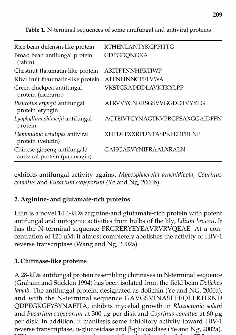
7/23/2019 Novel Therapeutic Agents From Plants
http://slidepdf.com/reader/full/novel-therapeutic-agents-from-plants 220/464
7/23/2019 Novel Therapeutic Agents From Plants
http://slidepdf.com/reader/full/novel-therapeutic-agents-from-plants 221/464
210
cowpea (Vigna unguiculata) (Ye et al., 2000c), pinto bean (Phaseolus vulgariscv. ‘Pinto Bean’) (Ye and Ng, 2002c), and ricebean (Delandia umbellata) (Yeand Ng, 2002a). The chitinase-like protein from pinto beans augmentsmacrophage production of nitric oxide (Ye and Ng, 2002c), while itscounterpart from rice beans stimulates splenocyte proliferation (Ye et al.,2001b). These chitinase-like proteins exhibit a weaker antifungal actionon Rhizoctonia solani than dolichin. They all demonstrate weak cell-freetranslating inhibiting activity and some HIV-1 reverse transcriptaseinhibitory activity (Ye et al., 2000c; Ye and Ng, 2002c). The translation-inhibiting activity may account for, at least in part, the antifungal activity.
The pinto bean chitinase-like protein phasein A, with the N-terminal
sequence CDVGSVISASLFEQQLKHRNGPAPARGDR, has a more potentantifungal activity toward Fusarium oxysporum than the 32-kDa novel pinto bean antifungal protein (Phasein B) with the N-terminal sequenceGARKDDHAKLUFLLKDIEYO, and approximately equipotent to the 32-kDa protein toward Physalospora piricola (Ye and Ng, 2002c). Activitytowards the various fungi are demonstrable at 60 µg per disk.
A 15-kDa antifungal protein, with an N-terminal sequence EQFGKQ
AGMALQPNG similar to those of chitinases, has been isolated from Panaxnotoginseng (sanchi ginseng) roots. It demonstrates potent antifungal activityagainst Fusarium oxysporum at 20 µg (Lam and Ng, 2001a).
Two chitinases, chitinase A and chitinase B, both 28 kDa in molecularsize, have been isolated from maize (Zea mays) seeds. They show 87%homology in amino acid sequence. Their N-amino acid sequences areMANAPRILAIGLLALLAAAGPAAAQN and QLVALGLALLCAVA
GPAAAQN, respectively. Chitinase A is more active than chitinase Btoward Trichoderma reesei, Alternaria solani, and Fusarium oxysporum. The binding constant of chitinase A against N, N’, N”, N’’’-tetracetylchitotetrose is considerably lower than that of chitinase B while chitinaseA has a higher specific activity than chitinase B (Huynh et al. 1992a).
The chive ( Allium tuberosum) chitinase-like protein, with an N-terminalsequence EQHGSQAGGALHPGXLHYSKYGGYGGTTPDYYGDGQQshowing a striking similarity to chitinases from leek ( Allium porrum) and
garlic ( A. sativum), exerts antifungal activity against a variety of fungi suchas Rhizoctonia solani, Fusarium oxysporum, Mycosphaerella arachidicola andBotrytis cinerea. The IC
50value of its antifungal activity against B. cinerea
was 0.2 µM. Its antifungal activity is stable over a wide range of pH values(1.6 to 12.3) and at temperatures up to 60ºC for 5 minutes. It has mitogenicactivity toward mouse splenocytes but exerts cytotoxicity on breast cancercells (Lam et al., 2000).
© 2009 by Taylor & Francis Group, LLC

7/23/2019 Novel Therapeutic Agents From Plants
http://slidepdf.com/reader/full/novel-therapeutic-agents-from-plants 222/464
211
4. Chitin-binding proteins
A 30-amino acid peptide with 6 Cys and 7 Gly residues, manifestingsequence similarity to the chitin-binding domain of chitin-binding
chitinases and a more avid binding to chitin than chitin-binding chitinases,has been purified from intercellular washing fluid from sugar beet (Betavulgaris L.) leaves. It exhibits antifungal activity against the devastatingleaf pathogen Cercospora beticola (Sacc.) (Nielsen et al., 1997). The mRNAexpression of this peptide is restricted to young flowers and leaves thatare young and mature. However, no induction of expression of the peptideor its mRNA has been noted during infection with C. beticola or following
treatment with dichloroisonicotinic acid, which is well-known to induceresistance in plants.
A 4244 Da chitin-binding protein, with an amino acid sequence and acysteine/glycine rich chitin-binding domain typical of many chitin- binding proteins, has been isolated from Ginkgo biloba leaves. It exertsantifungal activity against Alternaria alternata, Fusarium graminearum, F.moniliforme and Pellicularia sasakii (Huang et al., 2000).
An ethylene-inducible chitin-binding protein, with antifungal activitytoward several crop fungal pathogens, has been purified from guilder rose( Hydrangea macrophylla) leaves (Yang and Gong, 2002).
An antifungal protein, designated as CBP20, inducible by wound andtobacco mosaic virus, has been purified from tobacco (Nicotiana tabacumsamsun NN) leaves (Ponstein et al., 1994). It lyses the germ tubes and/orslows the growth of Trichoderma viride and Fusarium oxysporum. It has anN-terminal chitin-binding domain and a C-terminal domain similar totobacco and tomato pathogenesis-related proteins.
5. Cyclophilin-like proteins
A 18-kDa cyclophilin-like antifungal protein from black-eyed peas Vignaunguiculata L., with the N-terminal sequence FDMTAGPQPAGRIVFEGFADMVGRTAVN, exhibits stronger antifungal activity toward Mycosphaerella
arachidicola than Botrytis cinerea at 60 µg per disk (Ye and Ng et al., 2001a).Another cyclophilin-like antifungal protein from mungbean (Vigna radiata)seeds with a similar N-terminal sequence, designated as mungin, inhibitsproliferation of mouse splenocytes. It does not inactivate HIV-1 reversetranscriptase although it attenuates the activities of α- and β-glucosidases.It inhibits the fungi Rhizoctonia solani, Coprinus comatus, Mycosphaerellaarachidicola, Botrytis cinerea and Fusarium oxysporum at 60 µg per disk (Yeand Ng, 2000a).
© 2009 by Taylor & Francis Group, LLC

7/23/2019 Novel Therapeutic Agents From Plants
http://slidepdf.com/reader/full/novel-therapeutic-agents-from-plants 223/464

7/23/2019 Novel Therapeutic Agents From Plants
http://slidepdf.com/reader/full/novel-therapeutic-agents-from-plants 224/464
213
spruce. It potently inhibits fungal growth in Botrytis cinerea, Mycosphaerellaarachidicola, Fusarium oxysporum, Coprinus comatus, and Rhizoctonia solaniwith an IC
50of 0.25 µM, 6.5 µM, 3.6 µM, 3.4 µM and 8.7 µM, respectively.
It also exerts moderate antibacterial activity against Staphylococcus aureus,Pseudomonas aeruginosa and Escherichia coli. It attenuates the mitogenicresponse in mouse splenocytes and inhibits HIV-1 reverse transcriptase(Wang and Ng, 2000b).
9. Glucanases
A glucanase and two chitinases have been prepared from chickpea (Cicer
arietinum) (Vogelsang and Barz, 1993). Novel antifungal peptidesdesignated cicerin and arietin have been isolated from chickpea seeds (Yeet al., 2002). An antifungal peptide named cicerarin with the N-terminalsequence VKSTGRADDDLAVKTKYLPP has been purified from greenchickpea seeds (Chu et al., 2003).
10. Hemagglutinins/lectins
A homodimeric 67-kDa hemagglutinin from red kidney bean (Phaseolusvulgaris) seeds, with the N-terminal sequence ANQTSFNFQRFDCETNLILQRDATVSSKGQLRLTNVNDNPEEPTVDNLGRA, demonstrates moderateantifungal activity against Fusarium oxysporum and Rhizoctonia solani at60 µg per disk (Ye et al., 2001a). Antifungal activity in stinging nettle lectin(Broekaert et al., 1989) and potato tuber lectin (Gozia et al., 1995) haspreviously been reported.
11. Lipid transfer proteins
A 9.4-kDa peptide with the N-terminal sequence ALSCGTVSGNLAACAGYV was isolated from seeds of Brassica campestris L. var. purpureaBailey. The IC
50of its antifungal activity against M. arachidicola and F.
oxysporum were 4.5 µM and 8.3 µM, respectively. A 10-kDa protein,designated as Ace-AMP1, with antifungal and antibacterial activity towardGram-positive bacteria, and sequence resemblance to plant lipid transferproteins, has been purified from onion seeds. Its N-terminal sequenceis QMICPRVNRIVIPCVAYGL. The protein is incapable of effectingphospholipid transfer from liposomes to mitochondria, unlike radish andmaize nonspecific lipid transfer proteins. It has only 76% of the conservedamino acid residues in known plant nonspecific lipid transfer proteins.Its antifungal activity is hardly affected by the presence of cations at
physiological concentrations. On the contrary, the antifungal activity of radish lipid transfer protein is observable only at low cation concentrations
© 2009 by Taylor & Francis Group, LLC

7/23/2019 Novel Therapeutic Agents From Plants
http://slidepdf.com/reader/full/novel-therapeutic-agents-from-plants 225/464
214
(Terras et al., 1992). Maize and wheat lipid transfer proteins have little orno antimicrobial activity (Cammue et al., 1995). The solution structure of Ace-AMP1 has been elucidated (Tassin et al., 1998).
12. Miraculin-like proteins
A 38-kDa antifungal protein, with an N-terminal sequence APNVAPDVGHADLRA similar to miraculin and antifungal activity toward Fusariumoxysporum, has been isolated from sugar snap (Pisum sativum var.macrocarpon) legumes (Ye et al., 2000b).
13. Peroxidase
A 37-kDa peroxidase, with antifungal activity against Botrytis cinerea,Fusarium oxysporum and Mycosphaerella arachidicola and an N-terminalsequence LDNPAGFKGDKANGMLKMGNLCFLAKA, has been purifiedfrom French bean (Phaseolus vulgaris cv. ‘French Bean’) legumes (Ye andNg, 2002d). It has been shown that pathogen induces peroxidase
production in barley coleoptile (Kristensen et al., 1999).
14. Protease inhibitors
A Bowman-Birk type trypsin-chymotrypsin inhibitor from broad bean (Vicia faba) seeds, with the N-terminal sequence GDKVKSACCDTTLLKKKEHPPPRR suppresses mycelial growth in Mycosphaerella arachidicola, Fusariumoxysporum and Botrytis cinerea (Ye et al., 2001b). The IC
50
value of itsantifungal activity toward B. cinerea was 51 µM. It inhibits HIV-1 reversetranscriptase with an IC
50of 40 µM and demonstrates mitogenic activity
toward murine splenocytes.
A 24-kDa cystine protease inhibitor from pearl millet (Pennisetum glaucum) exhibits potent antifungal activity against Trichoderma reesei, andClaviceps, Helminthosporium, Curvularia, Alternaria and Fusarium species(Joshi et al., 1998).
15. Ribonucleases
From Chinese ginseng (Panax ginseng) roots an antifungal protein withribonuclease activity has been isolated. The protein, designated aspanaxagin, has two identical 26-kDa subunits with the N-terminalsequence GAHGARPYNIFRAALXRALN. Its HIV-1 reverse transcriptase
inhibiting activity can be heightened by succinylation using succinicanhydride (Ng and Wang, 2001). A heterodimeric ribonuclease with
© 2009 by Taylor & Francis Group, LLC

7/23/2019 Novel Therapeutic Agents From Plants
http://slidepdf.com/reader/full/novel-therapeutic-agents-from-plants 226/464

7/23/2019 Novel Therapeutic Agents From Plants
http://slidepdf.com/reader/full/novel-therapeutic-agents-from-plants 227/464
216
oxysporum. Regalado and Ricardo (1996) purified and characterized athaumatin-like protein and a chitinase from intercellular fluid of Lupinusalbus leaf, stem and root tissues. Thaumatin-like proteins, induced byinfection with Ascochyta rabiei , have been isolated from intercellularwashing fluid of chickpea (Cicer arietinum L.) leaves (Hanselle et al., 2001).
A 27-kDa thaumatin-like antifungal protein has been reported fromDiospyros texana fruits (Vu and Huynh, 1994). A 21-kDa thaumatin-likeantifungal protein from kiwi fruits, with an N-terminal sequenceATFNFINNCPFTVWAAAVPG and inhibitory activity on Botrytis cinerea andHIV-1 reverse transcriptase (Wang and Ng, 2002b), has been isolated. TheIC
50values of its antifungal activity against B. cinereus and M. arachidicola
were respectively 0.43 µM and 8 µM.Barley ( Hordeum vulgare) thaumatin-like proteins exert antifungal
activity against Trichoderma viride and Candida albicans. The activity issynergistic with that of nikkomycin Z, a nucleoside-peptide inhibitor of fungal chitin synthesis (Hejgaard et al., 1991).
A thaumatin-like protein, a β-1,3-glucanase precursor, and anantifungal protein, are co-induced by the signaling compound jasmonic
acid in rice (Oryza sativa) leaf tissues (Rakwal et al., 1999). A 22-kDathaumatin-like protein has been purified from maize seeds (Huynh et al.,1992a,b).
The mRNA level of thaumatin-like protein is maximal in the ripe cherry(Prunus avium L.) fruits. However, antifungal activity of the protein is notobserved (Fils-Lycaon et al., 1996).
The 25-kDa flax seed antifungal protein suppresses the growth of
Alternaria solani and synergizes the antifungal action of nikkomycin Ztoward Candida albicans. It shows marked sequence homology to otherpathogenesis-related antifungal proteins (Bergmeyer et al., 1992).
A 30-kDa thaumatin-like antifungal protein with the N-terminalsequence AKITFTNNHPRTIWP has been purified from chestnut (Casta-nopsis chinensis) seeds (Chu and Ng, 2003a). It exerts antifungal activityagainst Fusarium oxysporm with an IC
50of 0.5 µM. Another thaumatin-like
protein was isolated from seeds of the chestnut Castanea mollisima (Chuand Ng, 2003b). It exerts antifungal activity with an IC50
of 0.83 µM againstFusarium oxysporum, 6.48 µM against Mycosphaerella arachidicola and 9.21µM against Physalospora piricola.
Dendrocin is an antifungal protein from bamboo shoots with a N-terminal sequence TTLTLHNLCPYPVWWLVTPNNGGYPIIDNTPVVLGsimilar to thaumatin-like proteins (Wang and Ng, 2003a). The IC
50values
of its antifungal activity toward B. cinera, F. oxysporum and M. arachidicolawere, respectively, 1.8, 1.4 and 5.1 µM.
© 2009 by Taylor & Francis Group, LLC

7/23/2019 Novel Therapeutic Agents From Plants
http://slidepdf.com/reader/full/novel-therapeutic-agents-from-plants 228/464

7/23/2019 Novel Therapeutic Agents From Plants
http://slidepdf.com/reader/full/novel-therapeutic-agents-from-plants 229/464
218
From the pinto bean a novel 32-kDa antifungal protein withpronounced macrophage-stimulating activity has been reported (Ye andNg, 2002c). An antifungal peptide designated as angularin, 8 kDa inmolecular weight, and exhibiting antifungal activity against Mycosphaerellaarachidicola and Botrytis cinerea, has been isolated from red beans (Ye andNg, 2002b). Two 4 to 5 kDa antifungal peptides from Ceylon spinach(Basella rubra) seeds produce an antifungal action against Botrytis cinerea, Mycosphaerella arachidicola and Fusarium oxysporum (Wang and Ng, 2001d).β-basrubin has the N-terminal sequence KIMAKPSKFYBQLRGR and α- basrubin has the N-terminal sequence GADFQECMKEHSQKQHQHQ.Both antifungal peptides are active against B. cinerea, F. oxysporum and M.
arachidicola at 3.2 µg per disk. There is no activity toward C. comatus andR. Solani.
Snakin-1, an antimicrobial peptide from potato tubers, synergizes withpotato defensin PTH1 in antifungal action against Botrytis cinerea andantibacterial action against Clavibacter michiganensis sub sp. sepedonicus. Itspattern of gene expression suggests that it is a component of constitutivedefense barriers, especially those of storage and reproductive organs
(Segura et al., 1999).Four highly basic 20-amino acid peptides with 2 disulfide bonds from
Impatiens balsamina seeds have a growth-retarding effect on various fungiand bacteria. The predicted precursor of these antimicrobial peptides hasa novel structure composed of repeated alternating basic mature peptidedomains and acidic propeptide domains (Tailor et al., 1997).
Alocasin is a 11-kDa protein from giant taro (Alocasia macrorhiza)
rhizomes. It inhibits only B. cinerea out of the several fungi tested (Wangand Ng, 2003b).
The black soybean produces a 25-kDa antifungal protein withantimitogenic and HIV-1 reverse transcriptase inhibiting activities (Ngaiand Ng, 2003a ). Fabin from the broad bean possesses an N-terminalsequence GDPGDQNGKA similar to glucanase and calcyon (Ng and Ye,2003). It demonstrates, mitogenic and translation-inhibiting activities, andalso antifungal activity against B. cinerea, F. oxysporum, M. arachidicola and
R. solani at 60 µg per disk.
Two antimicrobial peptides from Mirabilis jalapa seeds, designated asMj-AMP1 and Mj-AMP2, are closely related in sequence and exhibit activityagainst gram-positive bacteria and a large number of fungi. Mj-AMP2, being more potent in antifungal activity, has an IC
50of 0.5-20 µg/ml
compared with the value of 6-30 µg/ml for Mj-AMP1. They show sequencehomology to mu-agatoxin, a class of insecticidal neurotoxic peptides from
spider venom (Cammue et al., 1992).The in vitro antifungal activity of Mirabilis jalapa and Amaranthus© 2009 by Taylor & Francis Group, LLC

7/23/2019 Novel Therapeutic Agents From Plants
http://slidepdf.com/reader/full/novel-therapeutic-agents-from-plants 230/464
219
caudatus antimicrobial peptides, purified from transgenic tobacco plantsexpressing their precursor proteins, is about the same as that of theauthentic proteins. Nevertheless, the transgenic plants do not acquire ahigher resistance against infection with either Botrytis cinerea or Alternarialongpipes (De Bolle et al., 1996).
Structure-function relationship in the case of Amaranthus candatusantimicrobial peptide 2 has been examined. The replacement of anyaromatic acid by alanine and the mutation of any aromatic acid to alanine,and the mutation of tyrosine 20 to tryptophan, ensue in a decrease in chitinaffinity. The mutation of phenylanine 18 to a larger aromatic residueincreases the affinity for chitin (Muraki et al., 2000).
A 4-kDa basic peptide without sequence similarity to thionins has beenpurified from maize (inbred B73) kernels. It retards spore germination andhyphal elongation in the maize pathogens F. moniliforme and F. graminearum, a bacterial pathogen of maize, and several bacteria (Duvicket al., 1992).
During seed germination in cheeseweed (Malva parviflora), cigar tree(Catalpa speciosa) and wheat, a larger number of antifungal proteins appear,
and the antifungal activity shifts from small molecules to larger molecules.Random proteolytic digestion of seed protein does not generate antifungalproteins. This increased production of antifungal protein may play acritical role in plant protection during early developmental stages (Wanget al., 2002).
A subtractive hybridization technique has been employed to clonecDNAs representing genes that show increased expression during leaf
senescence in Brassica napus. One of the genes products is an antifungalprotein (Buchanan-Wollaston and Ainsworth, 1997).
The three-dimensional solution structure of Raphanus sativus antifungalprotein 1 (Rs-AFP1) has been investigated with the use of 1H NMR. Themolecule adopts a compact globular fold comprised of an α-helix and atriple-stranded β-sheet with the middle strand connected by two disulfide bridges to the α-helix. A type via β-turn links β-strands 2 and 3. The loop between β-strand 1 and the α-helix is relatively well-defined. The structuralfeatures have all the characteristics of the cysteine-stabilized α-, β motif (Fant et al., 1998).
The crystal structure of zeamatin, antifungal protein from Zea mays,has been reported (Batalia et al., 1996). Like maize zeamatin, protein Rand S from barley grains inhibit fungal growth in synergism withnikkomycin Z, a nucleoside-peptide inhibitor of fungal chitin synthesis(Hejgaard, 1991).
© 2009 by Taylor & Francis Group, LLC
7/23/2019 Novel Therapeutic Agents From Plants
http://slidepdf.com/reader/full/novel-therapeutic-agents-from-plants 231/464

7/23/2019 Novel Therapeutic Agents From Plants
http://slidepdf.com/reader/full/novel-therapeutic-agents-from-plants 232/464
221
with non-transgenic controls (Oldach et al., 2001).
The solution structure of Aspergillus giganteus antifungal protein has been determined. Its folding comprises five antiparallel β-strands
connected in –1, –1, +3, +1, topology and highly twisted, defining a smalland compact β barrel stabilized by four internal disulfide bridges. Acationic site constituted by up to three lysine side chains next to ahydrophobic stretch, both at the protein surface, may form a potentialphospholipid binding site which would furnish the basis of its antifungalactivity (Campos-Olivas et al., 1995).
A 12-kDa protein, with sequence homology to Aspergillus giganteus andantifungal activity, has been purified from the culture medium of Penicilliumchrysogenum (Marx et al., 1995).
An antifungal peptide, with 58 amino acids and 6 Cys residues, has been isolated from the culture medium of Aspergillus niger. It exhibits acertain extent of sequence homology to cysteine-rich antifungal peptidesreported from the seeds of Sinapsis alba and Arabidopsis thaliana or theextracellular media of Aspergillus giganteus and Penicillium chrysogenumsome.Potent inhibitory activity against yeast and filamentous algae has been
observed (Gun Lee et al., 1999).Cryptocandin is a lipopeptide with a molecular weight of about 1.1
kDa from the endophytic fungus Cryptosporiopsis cf. quercina, chemicallyrelated to fungal antimycotics including echinocandins and pneumocan-dins. Its structure is glutamine (1)-3-hydroxy-4-hydroxy methylproline(2)-4,5-dihydroxyornithine(3)-palmitic acid-threonine(4)-4-hydroxy-proline(5)-3,4-dihydroxy-homotyrosine(6) where the first amino acid (1) is joined to
the 6th
amino acid (6) to form a cyclic structure. It is inhibitory towardCandida albicans, Trichophyton mentagrophytes, T. rubrum, Sclerotina sclero-tiorum and Botrytis cinerea (Strobel et al., 1999).
Viridin is a 65-kDa antifungal protein isolated from the culture mediumof the mold Trichoderma viride (Hao et al., 1999). The same group of investigators have isolated an antifungal protein in addition to α-sarcinfrom Aspergillus giganteus (Hao et al., 1998). The antifungal protein inhibitshyphal growth in Verticillium dahliae.
The cDNA encoding the antifungal protein KP4 from Ustilago maydisinfecting virus has been inserted behind the maize ubiquitin promoter andtransferred to varieties of wheat that are susceptible to stinking smut (Tilletiatritici) disease. KP4-transgenic wheat plants have a higher resistanceagainst stinking smut (Clausen et al., 2000).
Cloning, sequence analysis and expression of the gene encodingantifungal proteins from Aspergillus giganteus and Raphanus sativus have
been achieved (Alves et al., 1994; Wnendt et al., 1994).
© 2009 by Taylor & Francis Group, LLC

7/23/2019 Novel Therapeutic Agents From Plants
http://slidepdf.com/reader/full/novel-therapeutic-agents-from-plants 233/464
222
IV. ANTIFUNGAL PEPTIDES FROM BACTERIA
Antifungal peptides with inhibitory activity toward Alternaria brassiciolahave been demonstrated in the fermentation broth of Bacillus subtilis CL2T
(Leifert et al., 1995).
Iturin A and surfactin are antifungal lipopeptides from Bacillus subtilis.The purification protocol comprises solid-phase extraction on C18 gel andreversed-phase chromatography employing a biocompatible Pep RPC HR5/5 column with an FPLC system (Razafindralambo et al., 1993).
A lipopeptide, AFC-BC11, largely associated with the cell membrane,has been characterized from a soil isolate of Burkholderia cepacia BC11. It
accounts for the ability of BC11 to effectively control the damping-off of cotton caused by the fungal pathogen Rhizoctonia solani (Kang et al., 1998).
Tensin is an antifungal cyclic lipopeptide from Pseudomonas fluorescens.It causes a reduction in radial mycelial extension but an increase in branching and resette formation in Rhizoctonia solani (Nielsen et al., 2000).
Viscosinamiole is a new cyclic depsipeptide with surfactant andantifungal activities from Pseudomonas fluorescens. Its structure is β-hydroxy-
decanoyl-L-leucine(1)-D-glutamine(2)-D-allo-threonine(3)-D-valine(4)-L-leucine(5)-D-serine(6)-L-leucine(7)-D-serine(8)-L-isoleucine(9) in whichamino acid residue no. 3 is joined to amino acid residue no. 9 to form acyclic structure. It shows inhibitory activity toward the phytopathogenicfungi Pythium ultimum and Rhizoctonia solani in vitro as well as in planta(Nielsen et al., 1999).
Pseudomycin A from liquid culture of Pseudomonas syringae is a 1.2
kDa peptide made up of hydroxyaspartic acid, aspartic acid, diamino- butyric acid, lysine, arginine and serine. Prominent activity against thehuman pathogen Candida albicans is observed (Harrison et al., 1991).
Ecomycins are antimycotic peptides with a molecular weight of about1.1 kDa, isolated from Pseudomonas viridiflava, a plant-associated bacterium.They show activity against a wide range of phytopathogenic fungi (Milleret al., 1998).
A chitin-binding protein with antifungal activity has been purifiedfrom the culture filtrate of Streptomyces tendae Tu 901. It exhibits a molecularweight of 9.86 kDa and synergistic interaction with the chitin synthetaseinhibitor nikkomycin on growth of Aspergillus species. The chitin-bindingprotein demonstrates strong binding to the surface of germinating conidiaand to tips of growing hyphae, resulting in large spherical conidia, swollenhyphae and atypical branching (Bormann et al., 1999).
© 2009 by Taylor & Francis Group, LLC

7/23/2019 Novel Therapeutic Agents From Plants
http://slidepdf.com/reader/full/novel-therapeutic-agents-from-plants 234/464
223
V. ANTIFUNGAL PROTEINS FROM INSECTS
The full sequence of cecropin A is MNFVRILSFVFALVLALGAVSAAPEPRWKLFKKIEKVGRNVRDGLIKAGPAIAVIGGAKSLGK Cecropin A—
derived peptides, smaller than cecropin A which is a naturally occurringpeptide with a significant role in the insect immune response, have beensynthesized by Cavallavin et al. (1998) and tested for antifungal activity.An 11-amio peptide corresponding to the N-terminal amphipathic α-helixdomain of cecropin A demonstrates strong antifungal activity againstPhytophthora infestans (IC
50= 2 µM).
Antifungal proteins have also been isolated from insects. Cicadin is a
6.5-kDa antifungal peptide from dried juvenile cicadas with the N-terminalsequence NEYHGFVDKANNENKRKKQQGRDDFVVKPNNFANRRRKDDYNENYYDDVDAADVV. It exerts potent antifungal activity withIC
50values at nanomolar concentrations (Wang and Ng, 2002c). The
purification and cDNA cloning of an antifungal protein from thehemolymph of Holotrichia diomphalia larvae have been achieved (Lee et al.,1995).
Drosomycin, induced by septic injury of the fruitfly Drosophilamelanogaster (Fehlbaum et al., 1994), is a 44-residue antifungal peptide. Itssolution structure involves a three-stranded β-sheet and an α-helix, withfour disulfide bridges maintaining the protein global fold. It shows 41%sequence similarity with Raphanus sativus antifungal protein 2 (Rs-AFP2).Structural analysis of drosomycin and Rs-AFP2 disclosed the sharing bythe two antifungal proteins of a hydrophobic cluster located at the proteinsurface in which a lysine residue is embedded. The active site of
drosomycin has been proposed based on the structural similarity betweenthe two antifungal proteins and the results regarding the antifungal activityof Rs-AFP2 mutants (Landon et al., 1997, 2000).
A synthetic cDNA of Drosophila melanogaster has been expressed inSaccharomyces cerevisiae, using the mating factor alpha gene andsimultaneously overexpressed KEX2 gene to increase the yield of fullyexpressed drosomycin. Drosomycin has been shown by Edman degra-
dation and mass spectrometry to have sequence similarity to plantdefensins and the same array of intramolecular S-S bonds (Michaut et al.,1996).
Heliomicin from the lepidopteran Heliothis virescens possesses bothantifungal and antibacterial activities (Lamberty et al., 2001).
An antifungal protein has been purified from the hemolymph of thirdinstar larvae of Sarcophaga peregrina (Iijima et al., 1993). It inhibits the
growth of Candida albicans.Tenecin 3 is an antifungal protein from the insect Tenebrio molitor which
© 2009 by Taylor & Francis Group, LLC

7/23/2019 Novel Therapeutic Agents From Plants
http://slidepdf.com/reader/full/novel-therapeutic-agents-from-plants 235/464
224
inhibits the growth of Candida albicans. Its full sequence is MKTFVICLILVVAVSAAPDHHDGHLGGHQTGHGGGGGGGH2GGGGGGHLGGHGGGGPGGHLGGHGGGIGGTGGQQHGQHGPGTQAGHQGGYKTHGH.Its structural characteristics have been studied. It has a random structuralpropensity that is not significantly affected by temperature, pH, organicsolvents and sodium dodecyl sulfate micelles (Lee et al., 1999a). It acquireshigher antifungal activity after conjugation with polyethylene glycol butit does not interact with mammalian cells (Lee et al., 1996, 1999a, 1999b).
VI. ANTIFUNGAL PEPTIDES FROM MAMMALS
Hepecidin 20 and hepecidin 25 are two cysteine-rich peptides originatingfrom the liver and found in the urine. They manifest antifungal activityagainst Aspergillus fumigatus, A. niger and Candida albicans as well as bacteria (Park et al., 2001).
Mechanisms of action of some antifungal proteins
Investigations on the mechanism of antifungal action for some of theaforementioned proteins and peptides have been carried out. Thaumatin-like proteins may destabilize fungal membranes and bind and/orhydrolyze fungal β-1,3-glucans. Some thaumatin-like proteins like tobaccoosmotin stimulate a mitogen-activated protein kinase signal translationmechanism to induce changes in the fungal wall that increase toxicity (Yunet al., 1998; Grenier et al., 1999). Chitinases may hydrolyze chitin, apolymer of N-acetylglucosamine and an important component of the fungal
wall (Graham and Sticklen, 1994). However, the mechanisms of action of many other antifungal proteins and peptides await clarification.
VII. PLANT ANTIVIRAL PROTEINS
Antiviral activity has been detected in the extracts of many plants (Chessin,1995; Verma et al., 1995a,b). In some cases the antiviral principle has beenisolated and chemically identified (Bonness et al., 1994; Verma et al.,1995a,b). In addition to alkaloids, flavonoids, furocoumarins, hypericins,lignans, tannins, polysaccharides, terpenoids and thiophenes (Hudson,1995; Ng et al., 1997), plants produce antiviral proteins (Bol et al., 1995;Gera et al., 1995; Mitra, 1995). Ribosome inactivating proteins (RIPs)represent the best-studied group of plant antiviral proteins. The list of RIPsthat have been purified and characterized has been expanding (Battelliand Stirpe, 1995; Irwin, 1995; Zipf, 1995). Type 1 RIPs, which are single-
chained proteins with a molecular mass close to 30 kDa, have been isolatedfrom the following families: Asparagaceae, Caryophyllaceae, Cucur-
© 2009 by Taylor & Francis Group, LLC

7/23/2019 Novel Therapeutic Agents From Plants
http://slidepdf.com/reader/full/novel-therapeutic-agents-from-plants 236/464
225
bitaceae, Euphorbiaceae, Nyctaginaceae, Phytotaccaceae and Poaceae (Nget al., 1992; Shaw et al., 1994; Battelli and Stirpe, 1995) Type 1 RIPs have been shown to inhibit infection caused by plant viruses including Africancassava mosaic virus, alfafa mosaic virus, cucumber mosaic virus, potatovirus X, potato virus Y, southern bean mosaic virus, tobacco mosaic virus,and tobacco necrosis virus. Animal viruses including herpes simplex virus-1 influenza virus and poliovirus are inhibited by type 1 RIPs. Some type 1RIPs are capable of inhibiting HIV-1 replication and the activities of theenzymes crucial to the life cycle of the HIV-1 including reverse trans-criptase, protease and integrase (Au et al., 2000). Type 2 RIPs, composedof an RIP chain and a lectin chain, have been purified from members of
the Euphorbiaceae, Fabaceae, Passifloraceae, Sambucaceae and Viscaceaefamilies (Battelli and Stirpe, 1995; Chen et al., 2002). Type 2 RIPs inhibittobacco mosaic virus (Battelli and Stirpe, 1995).
The in planta antiviral activity of the type 2 RIPs from Sambucus nigra(Chen et al., 2002) is attributed to their polynucleoticle-adenosineglycosylase activity toward tobacco mosaic virus RNA (Vandenbussche etal., 2004).
Expression of a Phytolacca insularis antiviral protein (ribosomeinactivating protein) gene is increased considerably in leaves aftermechanical wounding and treatment with jasmonic acid and abscisic acid(Song et al., 2000). RIP is also induced by stress in the common ice plant Mesembryanthemum crystallinum (Rippmann et al., 1997).
RIPs are characterized by the ability to inhibit protein synthesis byrabbit reticulocyte lysate. They exhibit RNA N-glycosidase activity and
consequently depurinate, RNA (Bolognesi et al., 1990, 1997; Taylor andIrvin, 1990; Barbieri et al., 1992; Kataoka et al., 1992; Parente et al., 1993;Olivieri et al., 1996; Desmyter et al., 2003). The DNase (Day et al., 1998)and RNase (Wang et al., 2003) activities of RIPs have been shown to bedue to contaminants. Polynucleotide: adenosine glycosidase is the soleactivity of RIPs on DNA (Barbieri et al., 2000).
Expression of RIP gene has been found in abundance in leaves of Dianthus sinensis (Cho et al., 2000). RIPs have been isolated from different
tissues of the same plant (Battelli and Stirpe, 1995). Different forms of RIPmay exist in the same plant tissue e.g. Phytolacca americana seeds (Desvoyeset al., 1995; Honjo et al., 2002; Vivanco et al., 1999).
Native polleweed (Phytolacca americana) antiviral protein has been usedclinically as the active moiety of immunoconjugate against cancer andAIDS. Large-scale expression and purification of biologically activerecombinant pokeweed antiviral protein was first achieved by Rajamohan
et al. (1999). It has been demonstrated that an intact active site, as well asan intact C-terminal, are required for toxicity of pokeweed antiviral protein
© 2009 by Taylor & Francis Group, LLC

7/23/2019 Novel Therapeutic Agents From Plants
http://slidepdf.com/reader/full/novel-therapeutic-agents-from-plants 237/464
226
and its depurination of tobacco ribosomes in vivo. However, an intact activesite, but not an intact C-terminus, is crucial to the antiviral activity,indicating a dissociation of antiviral activity from toxicity (Tumer et al.,1997).
Resistance to a plant DNA virus, African cassava mosaic virus, had been achieved by virus-induced expression of the RIP dianthin intransgenic Nisotiana denthamiana plants (Hong et al., 1996).
Monoclonal antibodies against pokeweed antiviral protein have beenraised. They represent a useful tool for studying the mechanisms of RIP biosynthesis and plant protection involving RIPs (Desvoyes et al., 1995).Some lectins manifest inhibitory activity against viruses including HIV-1and bovine immunodeficiency virus and HIV-1 reverse transcriptase (Nget al., 1997; Ooi et al., 2000; Wang and Ng, 2001e).
VIII. ANTIBACTERIAL PROTEINS
Some of the ribosome inactivating proteins and antifungal proteins exhibitantibacterial activity. Two RIPs have been isolated and characterized from
the roots of the Andean crop Mirabilis expansa radiobacter, A. tumefaciensPseudomonas syringae (Vivanco et al., 1999).
Some pathogenic bacteria such as Shigella dysenteriae and certain strainsof E. coli. produce type 2 RIPs called Shiga toxins that are made up of asingle A chain and a pentamer of receptor-binding B chains Shiga toxinsdisplay an IC
50value of 0.8 nM against E. coli ribosomes (Suh et al., 1998).
Future perspectives and conclusions
Some of the antifungal proteins and peptides have been shown to possesstranslation-inhibiting, immunomodulatory (Wang and Ng, 2000a; Ye et al.,2001a,b, 2002c) and HIV-1 enzyme inhibiting (Ng et al., 2002) activities, just like ribosome inactivating proteins. Ribosome inactivating proteins alsoexhibit antifungal activity albeit weaker than that of other antifungalproteins.
In the foregoing account an impression can be gathered that antifungalproteins and peptides of different structures are produced by plants,animals, fungi and bacteria. Some of the antifungal proteins inhibitenzymes essential to the life cycle of human immunodeficiency virus (Nget al., 2002). Table 1 presents the N-terminal sequences of some of theantifungal and antiviral proteins reported in the literature. Differentantifungal proteins and peptides may exhibit different specificities in their
antifungal action, and may differ in their inhibitory potencies toward thesame fungus. Continued quest for more antifungal proteins and peptides
© 2009 by Taylor & Francis Group, LLC

7/23/2019 Novel Therapeutic Agents From Plants
http://slidepdf.com/reader/full/novel-therapeutic-agents-from-plants 238/464

7/23/2019 Novel Therapeutic Agents From Plants
http://slidepdf.com/reader/full/novel-therapeutic-agents-from-plants 239/464
228
inactivating proteins depurinate ribosomal RNA at multiple sites. Biochem J. 286: 1-4.
Barbieri, L., P. Valbonesi, F. Righi, G. Zuccheri, F. Monti, P. Gorini, B. Samori andF. Stirpe. 2000. Polynucleotide: adenosine glycosidase is the sole activity of
ribosome-inactivating proteins on DNA. J Biochem (Tokyo) 128: 883-889.Batalia, M.A., A.F. Monzingo, S. Ernst, W. Roberts and J.D. Robertus. 1996. The
crystal structure of the antifungal protein zeamatin, a member of thethaumatin-like, PR-5 protein family, Nat. Struct. Biol. 3: 19-23.
Battelli, M.G. and F. Stirpe. 1995. Ribosome inactivating proteins from plants.pp. 39-64. In: M. Chessin, D. DeBorde and A. Zipf. [eds.]. Antiviral Proteinsin Higher Plants. CRC Press, Boca Raton, FL., USA.
Bergmeyer, J.R., C.E. Smith and Q.K. Huynh. 1992. Isolation and characterization
of a 25 kDa antifungal protein from flax seeds, Biochem. Biophys. Res.Commun. 31: 480-487.Bloch, C., Jr., S.U. Patel, F. Baude, M.J. Zuelebil, M.D. Carr, P.J. Sadler and J.M.
Thornton. 1998. 1H NMR structure of an antifungal gamma-thionin proteinSI alpha 1: similarity to scorpion toxins, Proteins 32: 334-349.
Bol, J.F. and H.J.M. Linthorst. 1995. Putative role of pathogenesis-related proteinsin the antiviral response. pp. 147-160. In: M. Chessin, D. DeBrode and A.Zipf. [eds.] Antiviral Proteins from Higher Plants. CRC Press, Boca Raton,FL., USA.
Bolognesi, A., L. Barbieri, A. Abbondanza, A.I. Falasca, D. Carnicelli, M.G. Battelliand F. Stirpe. 1990. Purification and properties of new ribosome-inactivatingproteins with RNA N-glycosidase activity. Biochim. Biophys. Acta 1087: 293-302.
Bolognesi, A., L. Polito, F. Olivieri, P. Valbonesi, L. Barbieri, M.G. Battelli, M.V.Carusi, E. Benvenuto, F. Del Vecchio Blanco, A. Di Marco, A. Parente, M. DiLoreto and F. Stirpe. 1997. New ribosome inactivating proteins withpolynucleotide: adenosine glycosidase and antiviral activities from Basella
rubra L. and Bougainvillea spectabilis Willd. Planta 203: 422-429.Bonness, M.S., M.P. Ready, J.D. Irvin and T.J. Mabry. 1994. Pokweweed antiviral
protein inactivates pokeweed ribosomes: implications for the antiviralmechanism. Plant J. 5: 173-183.
Bormann, C., D. Baier, I. Horr, C. Raps, J. Berger, G. Jung and H. Schwarz. 1999.Characterization of a novel, antifungal, chitin-binding protein fromStreptomyces tendae Tu901 that interferes with growth polarity, J. Bacteriol.181: 7421-7429.
Broekaert, W.F., J. Van Parijs, F. Leyns, H. Joos and W.J. Peumans. 1989. A chitin- binding lectin from stinging nettle rhizomes with antifungal properties, Sci.245: 1100-1102.
Buchanan-Wollaston, V. and C. Ainsworth. 1997. Leaf senescence in Brassica napus:cloning of senescence related genes by subtractive hydridisation, Plant Mol.Biol. 33: 821-834.
Cammue, B.P., M.F. De Balee, F.R. Terras, P. Proost, J. Van Damme, S.B. Rees, J.Vanderleyden and W.F. Broekaert. 1992. Isolation and characterization of anovel class of plant antimicrobial peptides from Mirabilis jalapa L. seeds, J.Biol. Chem. 267: 2228-2233.
Cammue, B.P.A., K. Thevissen, M. Hendriks, K. Eggermont, I.J. Goderis, P. Proost,© 2009 by Taylor & Francis Group, LLC

7/23/2019 Novel Therapeutic Agents From Plants
http://slidepdf.com/reader/full/novel-therapeutic-agents-from-plants 240/464
229
J. Van Damme, R.W. Osborn, F. Guerbette, J.C. Kader and W.F. Broekaert.1995. A potent antimicrobial protein from onion seeds showing sequencehomology to plant lipid transfer proteins, Plant Physiol. 109: 445-455.
Campos-Olivas, R., M. Bruix, J. Santoro, J. Lacadena, A. Martinez del Pazo, J.G.
Gavilanes and M. Rico. 1995. NMR solution structure of the antifungalprotein from Aspergillus giganteus: evidence for cysteine pairing isomerism,Biochem. 34: 3009-3021.
Caruso, C., C. Capovale, G. Chilosi, F. Vacca, L. Bertini, P. Magro, E. Poerio andV. Buonocore. 1996. Structural and antifungal properties of a pathogenesis-related protein from wheat kernel, J. Protein Chem. 15: 35-44.
Caruso, C., M. Nobile, L. Leonardi, L. Bertini, V. Buonocore and C. Caporale.2001. Isolation and amino acid sequence of two new PR-4 proteins from
wheat, J. Protein Chem. 20: 327-335.Cavallavin, L., D. Andreu and B. San Segundo. 1998. Cecropin A-derived peptidesare potent inhibitors of fungal plant pathogens, Mol. Plant Microbe Interact.11: 218-227.
Chen, Y., F. Vandenbussche, P. Rouge, P. Proost, W.J. Peumans and E.J. VanDamme. 2002. A complex fruit-specific type-2 ribosome inactivating proteinfrom elderberry (Sambucus nigra) is correctly processed and assembled intransgenic tobacco plants. Eur. J. Biochem. 269: 2897-2906.
Chessin, M. 1995. Endogenous antiviral proteins and self-recognition. pp. 95-107. In: M. Chessin, D. DeBrode D. and A. Zipf. [eds.] Antiviral Proteins inHigher Plants. CRC Press, Boca Raton, FL., USA.
Cho, H.J., S.J. Lee, S. Kim and B.D. Kim. 2000. Isolation and characterization of cDNAs encoding ribosome inactivating proteins from Dianthus sinensis L.Mol. Cells 10: 135-141.
Chu, K.T. and T.B. Ng. 2003a. Isolation of a large thaumatin-like antifungal proteinfrom seeds of the Kweilin chestnut Castanopsis chinensis. Biochem. Biophys.Res. Commun. 301: 364-370.
Chu, K.T. and T.B. Ng. 2003b. Mollisin, an antifungal protein from the chestnutCastanea mollisima, Planta Med. 69: 809-813.
Chu, K.T., K.H. Liu and T.B. Ng. 2003. Cicerarin, a novel antifungal peptide fromthe green chickpea, Peptides 24: 659-663.
Clausen, M., R. Krauter, G. Schachermayr, I. Potrykus and C. Sautter. 2000.Antifungal activity of a virally encoded gene in transgenic wheat, Nat.Biotechnol. 18: 446-449.
Day, P.J., J.M. Lord and L.M. Roberts. 1998. The deoxyribonuclease activity
attributed to ribosome-inactivating proteins is due to contamination. Eur JBiochem. 258: 540-545.De Bolle, M.F., R.W. Osborn, I.J. Goderis, L. Noe, D. Aland, C.A. Hart, S.
Torrekens, F. Van Leuven and W.F. Broekaert. 1996. Antimicrobial peptidesfrom Mirabilis jalapa and Amaranthus caudatus: expression, processing,localization and biological activity in transgenic tobacco, Plant Mol. Biol. 31:993-1008.
Desmyter, S., F. Vandenbussche, Q. Hao, P. Proost, W.J. Peumans and E.J. VanDamme. 2003. Type-1 ribosome-inactivating protein from iris bulbs: a usefulagronomic tool to engineer virus resistance, Plant Mol. Biol. 51: 567-576.
Desvoyes, B., P. Dulieu, J.L. Poyet and P. Adami. 1995. Production and© 2009 by Taylor & Francis Group, LLC

7/23/2019 Novel Therapeutic Agents From Plants
http://slidepdf.com/reader/full/novel-therapeutic-agents-from-plants 241/464

7/23/2019 Novel Therapeutic Agents From Plants
http://slidepdf.com/reader/full/novel-therapeutic-agents-from-plants 242/464
231
genimivirus infection by virus-induced expression of dianthin in transgenicplants, Virol. 220: 1191-27.
Honjo, E., D. Dong, H. Motoshima and K. Watanabe. 2002. Genomic clonesencoding two isoforms of pokeweed antiviral protein in seeds (PAP-S1 and
S2) and the N-glycosidase activities of their recombinant proteins onribosome and DNA in comparison with other isoforms, J. Biochem. (Tokyo)131: 225-231.
Huang, X., W.J. Xie and Z.Z. Gong. 2000. Characteristics and antifungal activityof a chitin binding protein from Ginkgo biloba, FEBS Lett. 478: 123-126.
Hudson, J.B. 1995. The phytochemical approach to antiviral chemotherapy. pp.161-174. In: M. Chessin, D. De Borde D. and A. Zipf. [eds.] Antiviral Proteinsin Higher Plants. CRC Press, Boca Raton, FL., USA.
Huynh, Q.K., J.R. Borgmeyer and J.F. Zobel. 1992a. Isolation and characterizationof a 22 kDa protein with antifungal properties from maize seeds, Biochem.Biophys. Res. Commun. 182: 1-5.
Huynh, Q.K., C.M. Hironaka, E.B. Levine, C.E. Smith, J.R. Borgmeyer and D.M.Shah. 1992b. Antifungal proteins from plants. Purification, molecular cloning,and antifungal properties of chitinases from maize seeds, J. Biol. Chem. 267:6635-6640.
Iijima, R., S. Kurata and S. Natori. 1993. Purification, characterization, and cDNAcloning of an antifungal protein from the hemolymph of Sarcophaga peregrina(flesh fly) larvae, J. Biol. Chem. 268: 12055-12061.
Irwin, J.D. 1995. Antiviral proteins from Phytolacca. pp. 65-94. In: M. Chessin, D.De Borde. and A. Zipf. [eds.] 1995. Antiviral Proteins in Higher Plants. CRCPress, Boca Raton, FL., USA.
Joshi, B.N., M.N. Sainani, K.B. Bastawade, V.S. Gupta and P.K. Ranjekar. 1998.Cysteine protease inhibitor from pearl millet: a new class of antifungalprotein, Biochem. Biophys. Res. Commun. 246: 382-287.
Kang, Y., R. Carlson, W. Tharpe and M.A. Schell. 1998. Characterization of genes
involved in biosynthesis of a novel antibiotic from Burkholderia cepacia BC11and their role in biological control of Rhizoctonia solani, Appl. Environ.Microbiol. 64: 3939-3947.
Kataoka, J., N. Habaka, M. Miyano, C. Masuta and A. Koicvai. 1992. Adeninedepurination and inactivation of plant ribosomes by an antiviral protein of Mirabilis jalapa (MAP), Plant Mol. Biol. 20: 1111-1l19.
Koo, J.C., S.Y. Lee, H.J. Chun, Y.H. Cheong, J.S. Choi, S. Kawabata, M. Miyagi,S. Tsunasawa, K.S. Ha, D.W. Bae, C.D. Han, B.L. Lee and M.J. Cho. 1998.
Two hevein homologs isolated from the seed of Pharbitis nil L. exhibit potentantifungal activity, Biochim. Biophys. Acta 1382: 80-90.Kristensen, A.K., J. Burnstedt, J.E. Nielsen, J.D. Kreiberg, J.D. Mikkelsen, P.
Roepstorff and K.K. Nielsen. 2001. Partial characterization and localizationof a novel type of antifungal protein (IWF6) isolated from sugar beet leaves,Plant Sci. 159: 29-38.
Kristensen, B.K. H. Bloch and S.K. Rasmuseen. 1999. Barley coleoptileperoxidases. Purification, molecular cloning and induction by pathogens,Plant Physiol. 120: 501-512.
Kwon, S.Y., C.S. An, J.R. Liu and K.H. Pack. 1997. A ribosome inactivating proteinfrom Amaranthus viridis. Biosci. Biotechnol. Biochem. 61: 1613-1614.© 2009 by Taylor & Francis Group, LLC

7/23/2019 Novel Therapeutic Agents From Plants
http://slidepdf.com/reader/full/novel-therapeutic-agents-from-plants 243/464
232
Lacadena, J., A. del Pozo. Martinez, M. Gasset, B. Patino, R. Campos-Olvias, C.Vazquez, A. Martinez-Ruis, J.M. Mancheno, M. Onaderra and J.G. Gavilanes.1992. Characterization of the antifungal protein secreted by the mould Aspergillus giganteus, Arch. Biochem. Biophys. 324: 273-281.
Lam, S.K. and T.B. Ng. 2001a. Isolation of a small chitinase-like antifungal proteinfrom Panax notoginseng (sanchi ginseng) roots, Int. J. Biochem. Cell. Biol:287-92.
Lam, S.K. and T.B. Ng. 2001b. Isolation of a novel thermolabile heterodimericribonuclease with antifungal and antiproliferative activities from roots of the sanchi ginseng Panax notoginseng, Biochem. Biophys. Res. Commun. 285:419-423.
Lam, S.K. and T.B. Ng. 2001c. First simultaneous isolation of a ribosome
inactivating protein and an antifungal protein from a mushroom (Lyophyllumshimeiji) together with evidence for synergism of their antifungal effects,Arch. Biochem. Biophys. 393: 271-280.
Lam, S.S., H.X. Wang and T.B. Ng. 1998. Purification and characterization of novelribosome inactivating proteins, alpha- and beta-pisavins, from seeds of thegarden pea Pisum sativum. Biochem. Biophys. Res. Commun. 253: 135-142.
Lam, Y.W., H.X. Wang and T.B. Ng. 2000. A robust cysteine-deficient chitinase-like antifungal protein from inner shoots of the edible chive Alliumtuberosum, Biochem. Biophys. Res. Commun. 279: 74-80.
Lamberty, M., S. Ades, S. Uttenweiler-Joseph, G. Brookhart, D. Bushey, J.A.Hoffmann and P. Bulet. 2001. Insect immunity—isolation from thelepidopteran Heliothis virescens of a novel insect defensin with potentantifungal activity, J. Biol. Chem. 274: 9320-9326.
Landon, C., P. Sodano, C. Hetru, J. Hoffmann and M. Ptak. 1997. Solutionstructure of drosomycin, the first inducible antifungal protein from insects,Protein Sci. 6: 1878-1884.
Landon, C., A. Pajon, F. Volvelle and P. Sodano. 2000. The active site of
drosomycin, a small insect antifungal protein, delineated by comparison withthe modeled structure of Rs-AFPx, a plant antifungal protein, J. Peptide Res.56: 231-238.
Leah, R., H. Tommerup, I. Svendsen and J. Mundy. 1991. Biochemical andmolecular characterization of three barley seed proteins with antifungalproperties, J. Biol. Chem. 246: 1564-1573.
Lee, K.M., D.H. Kim, Y.H. Lee, B.S. Choi, J.H. Chung and B.L. Lee. 1999a.Antifungal activities of recombinant antifungal protein by conjugation with
polyethylene glycol, Mol. Cells. 9: 410-416.Lee, S.Y., H.J. Moon, S. Kurata, S. Natori and B.L. Lee. 1995. Purification andcDNA cloning of an antifungal protein from the hemolymph of Holotrichiadiomphalia larvae, Biol. Pharm. Bull. 18: 1049-1052.
Lee, Y.J., T.J. Chung, C.W. Park, Y. Hahn, J.H. Chung, B.L. Lee, D.M. Han, Y.H. Jung, S. Kim and Y. Lee. 1996. Structure and expression of the tenecin 3gene in Tenebrio molitor, Biochem. Biophys. Res. Commun: 218, 6-11.
Lee, Y.T., D.H. Kim, J.Y. Suh, J.H. Chung, B.L. Lee, Y. Lee and B.S. Choi. 1999b.Structural characteristics of tenecin 3, an insect antifungal protein, Biochem.Mol. Biol. Int. 47: 369-376.
Leifert, C., H. Li, S. Chidburee, S. Hampson, S. Workman, D. Sigee, H.A. Epton© 2009 by Taylor & Francis Group, LLC

7/23/2019 Novel Therapeutic Agents From Plants
http://slidepdf.com/reader/full/novel-therapeutic-agents-from-plants 244/464
233
and A. Harbour. 1995. Antibiotic production and biocontrol activity byBacillus subtilis CL27 and Bacillus pumilus CL45, J. Appl. Bacteriol. 78: 97-108.
Lin, P., L. Xia and T.B. Ng. 2007. First isolation of an antifungal lipid transfer
protein from seeds of a Brassica species. Peptides 28: 1514-1519.Liu, Y., J. Luo, C. Xu, F. Ren, C. Peng, G. Wu and J. Zhao. 2000. Purification,
characterization and molecular cloning of the gene of a seed-specific,antimicrobial protein from pokeweed, Plant Physiol. 122: 1015-1024.
Marx, F., H. Haas, M. Reindl, G. Stoffler, F. Lottspeich and B. Redl. 1995. Cloning,structural organization and regulation of expression of the Penicilliumchrysogenum paf gene encoding an abundantly secreted protein withantifungal activity, Gene 167: 167-171.
Michaut, L., P. Fehlbaum, M. Mariatte, A. Van Dorsselaer, J.M. Reichhart and P.Bulet. 1996. Determination of the disulfide array of the first inducibleantifungal peptide from insects: drosomycin from Drosophila melanogaster,FEBS Lett. 395: 6-10.
Miller, C.M., R.V. Miller, D. Garton-Kenny, B. Redgrave, J. Sears, M.M. Condron,D.B. Teplow and G.A. Strobel. 1998. Ecomycins, unique antimycotics fromPseudomonas viridiflava, J. Appl. Microbiol. 84: 937-944.
Mitra, A. 1995. Induced plant antiviral proteins. pp. 109-118. In: M. Chessin, D.De Borde and A. Zipf. [eds.] Antiviral Proteins in Higher Plants. CRC Press,Boca Raton, FL., USA.
Muraki, M., H. Morii and K. Harata. 2000. Chemically prepared hevein domainseffect on C-terminal truncation and the mutagenesis of aromatic residueson the affinity for chitin, Protein Engin. 13: 385-389.
Nakaya, K., K. Omata, I. Okahashi, Y. Nakamura, H. Kolckenbrock and N.Ulbrich. 1990. Amino acid sequence and disulfide bridges of an antifungalprotein isolated from Aspergillus giganteus, Eur. J. Biochem. 193: 31-38.
Neumann, G.M., R. Condron and G.M. Polya. 1996. Purification and mass
spectrometry-based sequencing of yellow mustard (Sinapis alba L.) 6 kDaproteins. Identification as antifungal proteins, Int. J. Pept. Protein Res. 47:437-446.
Ng, T.B. and H.X. Wang. 2001. Panaxagin, a new protein from Chinese ginsengpossesses anti-fungal, anti-viral, translation-inhibiting and ribonucleaseactivities, Life Sci. 68: 739-749.
Ng, T.B. and A. Parkash. 2002. Hispin, a novel ribosome inactivating proteinwith antifungal activity from hairy melon seeds, Protein Exp. Purif. 26: 211-
217.Ng, T.B. and X.Y. Ye. 2003. Fabin, a novel calcyon-like and glucanase-like proteinwith mitogenic, antifungal and translation-inhibitory activities from broad beans, Biol. Chem. 384: 811-815.
Ng, T.B., W.Y. Chan and H.W. Yeung. 1992. Proteins with abortifacient, ribosomeinactivating, immunomodulatory, antitumor and anti-AIDS activities fromCucurbitaceae plants, Gen. Pharmacol. 23: 575-590.
Ng, T.B., B. Huang, W.P. Fong and H.W. Yeung. 1997. Anti-HIV natural productswith special emphasis on HIV reverse transcriptase inhibitors, Life Sci. 61:933-949.
Ng, T.B., T.K. Au, T.L. Lam, X.Y. Ye and C.C. Wan. 2002. Inhibitory effects of © 2009 by Taylor & Francis Group, LLC

7/23/2019 Novel Therapeutic Agents From Plants
http://slidepdf.com/reader/full/novel-therapeutic-agents-from-plants 245/464

7/23/2019 Novel Therapeutic Agents From Plants
http://slidepdf.com/reader/full/novel-therapeutic-agents-from-plants 246/464
235
stressed rice (Oryza sativa L.) leaf tissues by two-dimensional polyacrylamidegel electrophoresis: induction of pathogenesis-related and cellular protectantproteins by jasmonic acid, UV irradiation and copper chloride, Electro-phoresis 20: 3472-3478.
Razafindralambo, H., M. Paquot, C. Hbid, P. Jacques, J. Destain and P. Thonart.1993. Purification of antifungal lipopeptides by reversal-phase high-performance liquid chromatography. J. Chromatogr. 639: 81-85.
Regalado, A.P. and C.P. Ricardo. 1996. Study of the intercellular fluid of healthyLupinus albus organs. Presence of a chitinase and a thaumatin-like protein,Plant Physiol. 110: 227-232.
Rippmann, J.F., C.B. Michalowski, D.E. Nelson, H.J. Bohnert. 1997. Induction of a ribosome-inactivating protein upon environmental stress, Plant Mol Biol.
35: 701-709.Roberts, W.K. and C.P. Selitrennikoff. 1986. Isolation and partial characterizationof two antifungal proteins from barley, Biochim. Biophys. Acta 880: 161-170.
Schaaper, W.M., G.A. Posthuma, H.H. Plasman, L. Sijtsma, F. Fant, F.A.Borremans, K. Thevisen, W.F. Broekaert, R.H. Meloen and A. vanAmerongen. 2001. Synthetic peptides derived from the beta2-beta3 loop of Raphanus sativus antifungal protein 2 that mimic the active site, J. PeptideRes. 57: 409-418.
Segura, A., M. Moreno, F. Madueno, A. Molina and F. Garcia-Olmedo. 1999.Snakin-1, a peptide from potato that is active against plant pathogens, Mol.Plant Microbe Interact. 12: 16-23.
Shao, F., Z. Hu, Y.M. Xiong, Q.Z. Huang, C.G. Wang, R.H. Zhu and D.C. Wang.1999. A new antifungal peptide from the seeds of Phytolacca americana:characterization, amino acid sequence and cDNA-cloning, Biochim. Biophys.Acta. 1430: 262-268.
Shaw, P.C., W.L. Chan, H.W. Yeung and T.B. Ng. 1994. Trichosanthin—a protein
with multiple pharmacological properties, Life Sci. 55: 253-262.Song, S.K., Y. Choi, Y.H. Moon, S.G. Kim, Y.D. Choi and T.S. Lee. 2000. Systemic
induction of a Phytolacca insularis antiviral protein gene by mechanicalwounding, jasmonic acid, and abscisic acid, Plant Mol. Biol. 43: 439-450.
Strobel, G.A., R.V. Miller, C. Martinez-Miller, M.M. Condron, D.B. Teplow andW.M. Hess. 1999. Cryptocandin, a potent antimycotic from the endophyticfungus Cryptospoiopsis cf, quercina, Microbiol. 145: 1919-1926.
Suh, J.K., C.J. Hovde and J.D. Robertus. 1998. Shiga toxin attacks bacterial
ribosome as effectively as eucaryotic ribosomes, Biochem. 37: 9394-9398.Tailor, R.H., D.P. Acland, S. Attenborough, B.P. Cammue, I.J. Evans, R.W. Osborm, J.A. Ray, S.B. Rees and W.F. Broekaert. 1997. A novel family of small cysteine-rich antimicrobial peptides from seed of Impatiens balsamina is derived froma single precursor protein, J. Biol. Chem. 272: 24480-24487.
Tam, J.P., Y.A. Lu, J.L. Yang and K.W. Chiu. 1999. An unusual structural motif of antimicrobial peptides containing end-to-end macrocyle and cystine-knotdisulfides, Proc. Natl. Acad. Sci. USA 96: 8913-8918.
Tassin S., W.F. Broekaert, D. Marion, D.P. Acland, M. Ptak, F. Vovelle and P. Sodano.1998. Solution structure of Ace-AMP1, a potent antimicrobial proteinextracted from onion seeds. Structural analogies with plant nonspecific lipid© 2009 by Taylor & Francis Group, LLC

7/23/2019 Novel Therapeutic Agents From Plants
http://slidepdf.com/reader/full/novel-therapeutic-agents-from-plants 247/464
236
transfer proteins, Biochem. 37: 3623-3627.Taylor, B.E., J.D. Irvin. 1990. Depurination of plant ribosomes by pokeweed
antiviral protein, FEBS Lett. 273: 144-146.Terras, F.P., I.J. Goderis, F. Van Leuven, J. Vander leyden, B.P. Cammue and W.F.
Broekaert. 1992. In vitro antifungal activity of a radish (Raphanus saticus L.)seed protein homologous to nonspecific lipid transfer proteins, Plant Physiol.100: 1055-1058.
Tumer, N.E., D.J. Hwang and M. Bonness. 1997. C-terminal deletion mutant of pokeweed antiviral protein inhibits viral infection but does not depurinatehost ribosomes, Proc. Natt. Acad. Sci. 94: 3866-3871.
Vandenbussche, F., S. Desmyter, M. Ciani, P. Proost, W.J. Peumans and E.J. VanDamme. 2004. Analysis of the in planta antiviral activity of elderberry
ribosome-inactivating proteins, Eur. J. Biochem. 271: 1508-1515.Verma, H.N., H.N. Varsha and V.K. Baranwal. 1995a. Endogenous virus inhibitorsfrom plants: their physical and biological properties. pp. 1-21. In: M. Chessin,D. DeBorde and A. Zipf. [eds.]. Antiviral Proteins in Higher Plants. CRCPress, Boca Raton, FL., USA.
Verma, H.N., H.N. Varsha and V.K. Baranwal. 1995b. Agricultural role of endogenous antiviral substances of plant origin. pp. 23-38. In: M.Chessin,D. DeBorde D. and A. Zipf. [eds.]. Antiviral Proteins in Higher Plants. CRCPress, Boca Raton, FL., USA.
Vila, L., V. Lacadena, P. Fontanet, A. de Pozo Martinez and B. San Segundo. 2001.A protein from the mold Aspergillus giganteus is a potent inhibitor of fungalplant pathogens, Mol. Plant Microbe Interact. 14: 1327-1331.
Vivanco, J.M., B.J. Savary and H.E. Flores. 1999. Characterization of two noveltype 1 ribosome inactivating proteins from the storage roots of the Andeancrop Mirabilis expansa, Plant Physiol. 119: 1447-1456.
Vogelsang, R. and W. Barz. 1993. Purification, characterization and differentialhormonal regulation of a β-1, 3-glucanase and two chitinases from chickpea
(Cicer arietinum L.), Planta 189: 60-69.Vu, L. and Q.K. Huynh. 1994. Isolation and characteization of a 27 kDa antifungal
protein from the fruits of Diospyros texana, Biochem. Biophys. Res. Commun.202: 666-672.
Walker, M., P. Shinn, S. Brooks, E. Buehler, Q. Chao, F. Dunn, S. Khan, C. Kim,H. Altafi, R. Araujo, L. Conn, A.B. Conway, A. Gonzalez, N.F. Hansen, L.Huizar, I. Kremenetskaia, C. Lenz, J. Li, S. Liu, S. Luros, D. Rowley, J.Schwartz, M. Toriumi, V. Vysotskaia, G. Yu, R.W. Davis, N.A. Federspiel, A.
Theologis and J.R. Ecker. 1999. Genomic sequence for Arabidopsis thalianaBAC T19E23 from chromosome I, Sequence submitted to NCBI.www.pubmed.com.
Wang, H.X. and T.B. Ng. 2000a. Quinqueginsin, a novel protein with anti-humanimmunodeficiency virus, antifungal, ribonuclease and cell-free translation-inhibitory activities from American ginseng roots, Biochem. Biophys. Res.Commun. 269: 203-208.
Wang, H.X. and T.B. Ng. 2000b. Ginkbilobin, a novel antifungal protein fromGinkgo biloba seeds with sequence similarity to embryo-abundant protein,Biochem. Biophys. Res. Commun. 279: 407-411.
Wang, H.X. and T.B. Ng. 2001a. Purification of chrysancorin, a novel antifungal© 2009 by Taylor & Francis Group, LLC

7/23/2019 Novel Therapeutic Agents From Plants
http://slidepdf.com/reader/full/novel-therapeutic-agents-from-plants 248/464
237
protein with mitogenic activity from garland chrysanthemum seeds, Biol.Chem. 382: 947-951.
Wang, H.X. and T.B. Ng. 2001b. Isolation of a novel deoxyribonuclease withantifungal activity from Asparagus officinalis seeds, Biochem. Biophys. Res.
Commun. 289: 120-124.Wang, H.X. and T.B. Ng. 2001c. Purification of allivin, a novel antifungal protein
from bulbs of the round-cloved garlic, Life Sci. 70: 357-365.Wang, H.X. and T.B. Ng. 2001d. Novel antifungal peptides from Ceylon spinach
seeds, Biochem. Biophys. Res. Commun. 288: 765-770.Wang, H.X. and T.B. Ng. 2001e. Examination of lectins, polysaccharopeptide,
polysaccharides, alkaloid, cumarin and trypsin inhibitors for inhibitoryactivity against human immunodeficiency virus reverse transcriptase and
glycohydrolases, Planta Med. 67: 669-672.Wang, H.X. and T.B. Ng. 2002a. Isolation of lilin, a novel arginine- and glutamate-rich protein with potent antifungal and mitogenic activities from lily bulbs,Life Sci. 70: 1075-1084.
Wang, H.X. and T.B. Ng. 2002b. Isolation of an antifungal thaumatin-like proteinfrom kiwi fruits, Phytochem. 61: 1-6.
Wang, H.X. and T.B. Ng. 2002c. Isolation of cicadin, a novel and potent antifungalpeptide from dried juvenile cicadas, Peptides 23: 7-11.
Wang, H.X and T.B. Ng. 2003a. Dendrocin, a distinctive antifungal protein from bamboo shoots, Biochem. Biophys. Res. Commun. 307: 75-755.
Wang, H.X and T.B. Ng. 2003b. Alocasin, an antifungal protein from rhizomesof the giant taro Alocasia macrorrhiza, Protein Exp. Purif. 28: 9-14.
Wang, H.X. and T.B. Ng. 2004. Eryngin, a novel antifungal peptide from fruiting bodies of the mushroom Pleurotus eryngii, Peptides 25: 1-5.
Wang, H.X., T.B. Ng, C.H.K. Cheng and W.P. Fong. 2003. Contamination of ribosome inactivating proteins with ribonucleases, separated by affinitychromatography on Red Sepharose, Prep. Biochem. Biotechnol. 33: 101-111.
Wang, H.X., T.B. Ng and Q.H. Liu. 2004. Alveolarin, a novel antifungalpolypeptide from the wild mushroom Polyporus alveolaris, Peptides 25: 693-696.
Wang, X., R.S. Thomas, J.A. Carroll and K.L. Duffin. 2002. Temporal generationof multiple antifungal proteins in primed seeds, Biochem. Biophys. Res.Commun. 292: 236-242.
Wnendt, S., N. Ulbrich and U. Stahl. 1994. Molecular cloning, sequence analysisand expression of the gene encoding an antifungal protein from Aspergillus
giganteus, Curr. Genet. 25: 519-523.Wong, R.N., T.B. Ng, S.H. Chan, T.X. Dong and H.W. Yeung. 1992. Characteri-zation of Mirabilis antiviral protein—A ribosome inactivating protein from Mirabilis jalapa L, Biochem. Int. 28: 585-593.
Yang, Q. and Z.Z. Gong. 2002. Purification and characterization of an ethylene-induced antifungal protein from leaves of guilder rose ( Hydrangeamacrophylla), Protein Expr. Purif. 24: 76-82.
Ye, X.Y. and T.B. Ng. 2000a. Mungin, a novel cyclophilin-like antifungal proteinfrom the mungbean, Biochem. Biophys. Res. Commun. 273: 1111-1115.
Ye, X.Y. and T.B. Ng. 2000b. Hypogin, a novel antifungal peptide from peanutswith sequence similarity to peanut allergen, J. Peptide Res. 57: 330-336.© 2009 by Taylor & Francis Group, LLC

7/23/2019 Novel Therapeutic Agents From Plants
http://slidepdf.com/reader/full/novel-therapeutic-agents-from-plants 249/464

7/23/2019 Novel Therapeutic Agents From Plants
http://slidepdf.com/reader/full/novel-therapeutic-agents-from-plants 250/464

7/23/2019 Novel Therapeutic Agents From Plants
http://slidepdf.com/reader/full/novel-therapeutic-agents-from-plants 251/464
240
manifested the most potent xanthine oxidase inhibitory activity. Theresidual fraction showed little activity. The first two fractions reduced theoxidative damage induced by tert-butyl hydroperoxide in rat primaryhepatocytes as evidenced by malondialdehyde formation (Tseng et al.,1997).
Arbutin (4-hydroxyphenyl-β glucopyranoside, C12
H16
O7) (Fig. 1)
Arbutin was the major antioxidant compound in the aqueous extract of Rhodococcum vitis-(L.) Avrorin. It inhibited the increase in serum alanineaminotransferase and glutathione S-transferase activities and lipid
peroxidation in liver homogenate induced by galactosamine (Myagmar etal., 2004).
Fig. 1. Chemical structure of Arbutin.
Benzophenanthridine alkaloids
Vavreckova et al. (1996) reported the inhibition of 5- and 12-lipoxygenase by Chelidonium majus benzophenanthridine alkaloids.
Carnosic acid and carnosol (Fig. 2)
Carnosic acid from rosemary (Rosmarinus officinalis L.) scavenged freeradicals as measured by the 2,2-diphenyl-1-picryhydrazyl assay andinhibited linoleic acid oxidation as measured by the beta-carotene assay(Wellwood and Cole, 2004). The use of ultrasound for extraction of carnosicacid using butanone, ethyl acetate and ethanol as solvents improved the
O
HO
O
OH
O
O
H
H
OH
© 2009 by Taylor & Francis Group, LLC

7/23/2019 Novel Therapeutic Agents From Plants
http://slidepdf.com/reader/full/novel-therapeutic-agents-from-plants 252/464

7/23/2019 Novel Therapeutic Agents From Plants
http://slidepdf.com/reader/full/novel-therapeutic-agents-from-plants 253/464
242
Esculetin (6, 7-dihydroxycoumarin, CaH6O4) (Fig. 4) from A. scoparia isa lipoxygenase inhibitor. It inhibited Ras-mediated cell proliferation andattenuates vascular restenosis after angioplasty in rats (Pan et al., 2003).Esculetin from Alchemilla speciosa had antimutagenic activity (Schimmerand Eschelbach, 1997).
The coumarin xanthotoxol from Cnidium monnieri fruits had potentantioxidant activity. On the other hand, columbianetin, bergapten and
angelicin from Cnidium monnieri fruits had only slight antioxidant activity(Ng et al., 2000).
Ecdysteroids
Ecdysone (Fig. 5) is (2β, 3β, 5β, 20R)–2, 3, 14, 20, 22, 25 hexahydro-xycholesten-7-en-6-one. Ecdysteroids possess an ecdysone skeleton.
Fig. 4. Chemical structure of Esculetin.
O
O
HO
HO
OH
CH 3
OH
CH3
CH3
OH
CH3
HO
HO
O
R
H3C
Fig. 5. Chemical structure of α-ecdysone.
© 2009 by Taylor & Francis Group, LLC

7/23/2019 Novel Therapeutic Agents From Plants
http://slidepdf.com/reader/full/novel-therapeutic-agents-from-plants 254/464
243
From the Chinese herb Serratula strangulata, four ecdysteroids including20-hydroxyecdysone (E1), 25-deoxy-11,20-dihydroxyecdysone (E2), 24-(2-hydroxyethyl)-20-hydroxyecdysone (E3) and 20-hydroxyecdysone-20,22-monoacetonide (E4) were isolated. They protected human erythrocytesagainst oxidative hemolysis induced by a water-soluble inhibitor, 2,2’-azobis 2-aminopropane hydrochloride, and inhibited rat hepaticmicrosome peroxidation (Cai et al., 2002).
Flavonoids
Flavonoids are polyphenolic compounds found in many fruits and
vegetables. Flavonoids (apin, luteolin-, apigenin-glycosides) and essentialoils (apiol, miriszticin) are components of the parsley (Petroselinum crispum(Mill.) Hill) fruit that contribute to its antioxidant activity (Fejes et al., 1998).
Baicalein (5,6,7-trihydroxyflavone) from the root of the Chinese herbScutellaria baicalensis Georgi exhibited antioxidant, anti-inflammatory and12-lipoxygenase inhibitory activities. It selectively inhibited the nitric oxide-dependent apoptotic pathway of activated brain microglia by suppressing
production of cytotoxic nitric oxide (Suk et al., 2003). Baicalin inhibitedproduction of eotaxin, an eosinophilic chemokine associated witheosinophil recruitment to allergic inflammation sites (Nakajima et al., 2001).
Purearin (8-(β-D-glucopyranosyl-7-hydroxyl-3-(4-hydroxyphenyl-4H-1-benzopyran-4-one) (Fig. 6) is the main flavonoid in the root of the wildleguminous creeper Purearia lobatai. Both purearin and the crude extractof P. lobata roots inhibited the steady-state chemiluminscent reaction basedon horseradish peroxidase and a luminol-oxidant-enhancer reagent, albeitwith different mechanisms, suggesting the presence of active compoundsother than puararin in the crude extract. The crude extract showed strongantioxidant activity (Guerra et al., 2000).
Firenzuoli et al. (2004) reported that dietary consumption of flavonoids
Fig. 6. Chemical structure of Purearin.
O
R1
R2O
R1
OH
O
© 2009 by Taylor & Francis Group, LLC

7/23/2019 Novel Therapeutic Agents From Plants
http://slidepdf.com/reader/full/novel-therapeutic-agents-from-plants 255/464
244
had an inverse relationship with the later development of cancer andcoronary heart disease and that citrus species, Ginkgo biloba, hawthorn,soy, St. John’s Wort, Silybum marirum, tea and Vaccinium mirtillis containedflavonoids. Kolaviron, a biflavonoid fraction of Garcinia kola, reducedoxidative damage to DNA in the liver (Farombi et al., 2004). Theantioxidant principles in the Nigerian herb G. kola that inhibited lipidperoxidation in rat liver homogenate were tentatively identified asisoflavones (Adegoke et al., 1998).
The flavonoids baicalin, luteolin-7-glucuronide-6’-methyl ester andrutin from S. baicalensis, Ixeris denticulate f. pinnatipartia and Sophora japonica,respectively, had potent antioxidant activity. In contrast, naringin from
Drynaria fortunei rhizomes had only low activity (Ng et al., 2000).
Gallic acid (Fig. 7)
The structure of catechin is (2R, 3S)-2-(3,4-dihydroxyphenyl)-3,4-dihydro-1(2H)-benzopyran-3.5.7-triol (Fig. 8).
Epigallocatechin-3-gallate (EGCG), which is (2R3R)-2-(3,4,5-
trihydroxyphenyl)-3,4-dihydro-1(2H)-benzopyran-3,5,7-triol 3-(3,4,5-
Fig. 7. Chemical structure of Gallic acid.
OH
OH
OH
HO
O
Fig. 8. Chemical structure of Catechin.
O
OH
OH
OH
OH
HO
OH
© 2009 by Taylor & Francis Group, LLC

7/23/2019 Novel Therapeutic Agents From Plants
http://slidepdf.com/reader/full/novel-therapeutic-agents-from-plants 256/464
245
trihydroxybenzoate), a constituent of green tea, inhibited proliferation of cultured rabbit lens epithelial cells by inducing apoptosis. The findingsopened up the possibility of using EGCG for the prevention of postcapsularopacity (Huang et al., 2000).
Gallic acid found in the medicinal herb Limonium wrightii from theOkinawa islands scavenged 1,1-diphenyl-2-picrylhydrazyl, superoxideanions and hydroxyl radicals. It suppressed pH or bol-12-myristateacetate-stimulated production of reactive oxygen species in polymorpho-nuclear leukocytes. It also inhibited lipid peroxidation in rat liver micro-somes and reduced CCl
4-induced hepatotoxicity (Aniya et al., 2002).
One of the antioxidant compounds isolated from the aqueous extractof dried rose (Rosa rugosa Thunb.) flowers by ion exchange chromatographyon CM-cellulose and gel filtration on Sephadex G75 was a derivative of gallic acid (3,4,5-trihydroxybenzoic acid). Another two included apolysaccharide and a polysaccharide-peptide gallic acid, which demons-trated the most potent antioxidative activity.
Gypenosides
Gypenosides, (Fig. 9) which are saponins (steryl glycosides) from theChinese herb Gynostemma pentaphyllum, reduced the content of superoxideanions and H
2O
2in human neutrophils, and inhibited chemical-induced
lipid peroxidation in liver microsomes and vascular endothelial cells. Thepigments also reduced the oxidative stress in human monocytes andmurine macrophages (Li et al., 1993).
Fig. 9. Chemical structure of dammarane-type gypenosides (where R1 = glucose,
rhamnose; R2 = glucose, rhamnose, etc.; R3 = CH3, CH2OH, CHO; R4 = H,CH
2OH; and R5 = glucose, xylose, etc.
H3CO-R5
CH3
CH3
R4
R3
H3C CH3
R1-O
R2-O
CH3
© 2009 by Taylor & Francis Group, LLC

7/23/2019 Novel Therapeutic Agents From Plants
http://slidepdf.com/reader/full/novel-therapeutic-agents-from-plants 257/464
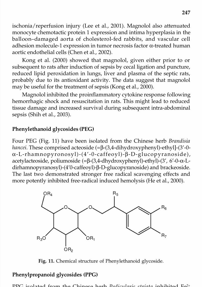
7/23/2019 Novel Therapeutic Agents From Plants
http://slidepdf.com/reader/full/novel-therapeutic-agents-from-plants 258/464
247
ischonia/reperfusion injury (Lee et al., 2001). Magnolol also attenuatedmonocyte chemotactic protein 1 expression and intima hyperplasia in the balloon–damaged aorta of cholesterol-fed rabbits, and vascular celladhesion molecule-1 expression in tumor necrosis factor α-treated humanaortic endothelial cells (Chen et al., 2002).
Kong et al. (2000) showed that magnolol, given either prior to orsubsequent to rats after induction of sepsis by cecal ligation and puncture,reduced lipid peroxidation in lungs, liver and plasma of the septic rats,probably due to its antioxidant activity. The data suggest that magnololmay be useful for the treatment of sepsis (Kong et al., 2000).
Magnolol inhibited the proinflammatory cytokine response followinghemorrhagic shock and resuscitation in rats. This might lead to reducedtissue damage and increased survival during subsequent intra-abdominalsepsis (Shih et al., 2003).
Phenylethanoid glycosides (PEG)
Four PEG (Fig. 11) have been isolated from the Chinese herb Brandisia
hancei. These comprised acteoside (=β-(3,4-dihydroxyphenyl)-ethyl]-(3’-0-α-L-rhamnopyronosyl)-(4’-0-caffeoyl)-β-D-glucopyranoside),acetylacteoside, poliumoside (=β-(3,4-dhydroxyphenyl)-ethyl)-(3’, 6’-0-α-L-dirhamnopyranosyl)-(4’0-caffeoyl)-β-D-glucopyranoside) and brackeoside.The last two demonstrated stronger free radical scavenging effects andmore potently inhibited free-radical induced hemolysis (He et al., 2000).
Fig. 11. Chemical structure of Phenylethanoid glycoside.
R6
R7
R5
OO
OR1
O R2
OR4
R3O
Phenylpropanoid glycosides (PPG)
PPG isolated from the Chinese herb Pedicularis striata inhibited Fe2+
dependent lipid peroxidation through chelation of Fe2+ ions and underphysiological conditions the chelates formed are sufficiently stable (Li etal., 1997).
© 2009 by Taylor & Francis Group, LLC

7/23/2019 Novel Therapeutic Agents From Plants
http://slidepdf.com/reader/full/novel-therapeutic-agents-from-plants 259/464
248
Proanthocyanidins from grape seeds
Grape seed proanthocyanidins and Scutellaria baicalensis were synergisticin their ability to scavenge reactive oxygen species (Shao et al., 2004). Thus
lower dosages of each of them can be used and possible side effects due tohigh dosages can be avoided.
Salvianolic acid B
Salvianolic acid B from the Chinese herb Salvia miltiorrhiza and theaqueous methanolic extract of S. miltiorrhiza attenuated the expression of
vascular adhesion molecule-1 and intercellular cell adhesion molecule-1in human aortic endothelial cells treated with tumor necrosis factor-alpha.This activity, together with their anti-inflammatory and low densitylipoprotein-inhibiting activities, may explain their usefulness for thetreatment of atherosclerosis. All aforementioned activities are related totheir antioxidant activities (Chen et al., 2001). A salvianolic acid B-enrichedfraction of S. miltiorrhiza induced neointimal cell apoptosis in a rabbitangioplasty model (Hung et al., 2001).
Schisandrin B (Fig. 12)
It is a dibenzocyclooctadiene derivative from Fructus schisandrae. It hadprotective effects against cerebral oxidative stress (Ko and Lam, 2002).
Fig. 12. Chemical structure of Schisandrin B.
CH3
H
OH
CH3
H3CO
H3CO
OCH3
H3CO
H3CO
OCH3
© 2009 by Taylor & Francis Group, LLC

7/23/2019 Novel Therapeutic Agents From Plants
http://slidepdf.com/reader/full/novel-therapeutic-agents-from-plants 260/464
249
Sodium tanshinone sufonate
It is a derivative of tanshinone IIA (=1,6-dimethylphenanthro(1,2-b) furan-10, 11-dione) isolated from S. miltiorrhiza rhizomes. It reduced the size of
myocardial infarcts, and prolonged the survival of cultured humansaphenous vein endothelial cells but not human ventricular myocytes invitro upon exposure to xanthine oxidase-generated oxyradicals. The datasuggest that it may protect the vascular endothelium (Wu et al., 1993).
Tetrandrine (Fig. 13)
It is an alkaloid (+)-(S)-3-phenyl-1’-(phenylmethyl)-(3,4’-bipperidine)-2,6-dione-HCl, isolated from the Chinese herb Stephania tetrandra, its free radicalscavenging activity contributed to its inhibitory activity on nuclear factor-kappa B activation by phorbol 12-myristate 13-acetate (Ye et al., 2000).
Fig. 13. Chemical structure of Tetrandrine.
Thonningianin
Two ellagitannins, designated as thonningianins A and B, were isolatedfrom the African herb Thonningia sanguinea. They exhibited potent freeradical scavenging activity against 1,1-diphenyl-2picryhydrazyl asevidenced by electron spin resonance analysis (Ohtani et al., 2000).Thonningianin A inhibited human glutathione S-transferase, an enzymeinvolved in resistance of tumor cells against chemotherapeutics (Gyamfiet al., 2004).
N
O
N
CH3
H3C
O
OCH3
OCH3
H3CO
OCH3
© 2009 by Taylor & Francis Group, LLC

7/23/2019 Novel Therapeutic Agents From Plants
http://slidepdf.com/reader/full/novel-therapeutic-agents-from-plants 261/464
250
Trilinolein (C57
H98
O6)
Trilinolein (Fig. 14), which is (1β)-6,6’,7,12-tetramethoxy-2,2’-dimethyl- berbaman isolated from sanchi ginseng (Panax notoginseng), had myocardial
protective effects related to its antioxidant activity (Chan and Tomlinson,2000). It reduced norepinephrine-induced protein synthesis via inhibitionof cardiomyocyte production of reactive oxygen species (Liu et al., 2004a).Angiotensin II-stimulated protein synthesis, beta-myosin heavy chainpromoter activity, intracellular reactive oxygen species production, andH
2O
2-activated phosphorylation of mitogen-activated protein kinases, were
all inhibited by trilinolein and the antioxidant N-acetylcysteine (Liu et al.,
2004b). Trilinolein counteracted the free radical damage associated withatherogenesis, and ischemial reperfusion induced myocardial damage. Thefindings constitute a scientific basis for the traditional use of Panaxnotoginseng for the treatment of circulatory problems (Chan et al., 2002).Trilinolein increased the activity of superoxide dismutase in cultured rat brain astrocytes (Chiu et al., 1999). It preserved heart mitochondrialultrastructure after exposure to ischemia and reduced oxygen free radicalproduction by leukocytes (Chan et al., 1996).
Fig. 14. Chemical structure of Trilinolein.
Other compounds
The lignan 4’-demethyldeoxypodophyllotoxin, the alkaloid tetra-hydropalmatine (5,8,13,13a-tetrahydro-2,3,9,10-tetramethoxy-6H-dibenzo[a, g] qurnolizine) and the bisbenzyl erianin (2-methoxy-5-[2-(3,4,5-trimethoxy-phenyl)-ethyl]-phenol) from Sinopodophyllum emodii (Wall) Ying,Stephania sinica Diels and Dendrobium chrysotoxum Lindl respectivelyexhibited potent antioxidant activity in both lipid peroxidation and
hemolysis assays. The terpene tanshinone I from S. miltiorrhiza inhibitedlipid peroxidation but not hemolysis (Ng et al., 2000).
O
O
O
O
O
O
© 2009 by Taylor & Francis Group, LLC

7/23/2019 Novel Therapeutic Agents From Plants
http://slidepdf.com/reader/full/novel-therapeutic-agents-from-plants 262/464
251
Different compounds from Aster tataricus Linn. f.
A number of compounds were isolated from the medicinal plant Aster tataricus including shionone, epifriedelinol, quercetin 2-(3,4-
dihydroxyphenyl-3,5,7-trihydroxy-chromen-4-one), (Fig. 15) kaempferol(3,5,7-trihydroxy-4’-methoxyflavone) (Fig. 16), scopoletin (7-hydroxy-6-methoxy-2H-chromen-2-one) (Fig. 17), emodin (3-methyl-1,6,8-trihydroxy-anthraquinone) (Fig. 18), aurantiamide acetate (N-benzoyl-phenylalanyl-phenylalaninol acetate) and 1,7-dihydroxy-6-methyl-anthraquinone. Thecompounds were compared with regard to their ability in inhibitinghemolysis of rat erythrocytes induced by 2’-2’ azobis (2-amidinoproane)
dihydrochloride, lipid peroxidation using a FeSO4-ascorbic acid system,and generation of superoxide radicals using a phenazine methosulfate-
Fig. 15. Chemical structure of Quercetin.
Fig. 16. Chemical structure of Kaempferol.
O
OH
O H
OH
OOH
HO
O
OH
OH
OOH
HO
© 2009 by Taylor & Francis Group, LLC

7/23/2019 Novel Therapeutic Agents From Plants
http://slidepdf.com/reader/full/novel-therapeutic-agents-from-plants 263/464
252
nicotinamide adenine dinucleotide system. Effects on the Fe-bleomycin-induced DNA damage reflected pro-oxidant activity. Quercetin andkaempferol were most potent in inhibiting hemolysis, lipid peroxidation,and superoxide radical generation. Scopoletin and emodin were similar
to quercetin and kaempferol in inhibiting superoxide radical generationand second to them in inhibiting lipid peroxidation. Aurantiamide acetateexhibited some inhibitory activity toward superoxide radical generation.1,7-dihydroxy-6-methyl-anthraquinone exerted an inhibitory activity onlyon superoxide radical generation. Shionone and epifriedelinol did notdisplay any antioxidant activity. Quercetin and kaempferol, but not theremaining compounds, exhibited some pro-oxidant activity (Ng et al.,2003).
Herbal extracts
Achyrocline satureoides (Lam.) DC.
The aqueous extract of this South American herb inhibited the oxidationof human low density lipoprotein (Gugliucci and Menini, 2002). Both
aqueous and methanolic extracts inhibited the generation of thiobarbituricacid-reactive substances in rat liver homogenate (Desmarchelier et al., 1998).
Fig. 18. Chemical structure of Emodin.
Fig. 17. Chemical structure of Scopoletin.
O OHO
H3CO
O
O
OH OH
OHH3C
© 2009 by Taylor & Francis Group, LLC

7/23/2019 Novel Therapeutic Agents From Plants
http://slidepdf.com/reader/full/novel-therapeutic-agents-from-plants 264/464

7/23/2019 Novel Therapeutic Agents From Plants
http://slidepdf.com/reader/full/novel-therapeutic-agents-from-plants 265/464
254
Crataegus pinnatifida var. psilosa fruits
This is a medicinal herb used for making a soft drink in Taiwan. The hotwater extract of its dried fruits are composed of flavonoids (6.9%),
procyanidins (2.2%), catechin (0.5%) and epicatechin (0.2%), chemicalstructures are very common and can be found in many papers, exhibitedan antioxidant action (Chu et al., 2003).
Curcuma longa L.
Its antioxidant activity, together with its other activities including anti-
inflammatory, anticarcinogenic and antimicrobial activities, have beenreviewed (Anonymous, 2001a).
Desmotrichum fimbriatum Blume
The aqueous ethanolic extract of this ayurvedic herb ameliorated theperoxidative damage caused in mice by cold water swim stress(Chakrabarty et al., 2001).
Dimocaepus longan L.
An aqueous extract of the herb demonstrated hydroxyl radical-scavengingactivity as revealed by a gas chromatography-mass spectrometry method based on the Fenton reaction system (Wang and Smythe, 2003).
Drynaria fortumei [Kunze] J. SM
The water extract of this Chinese herb lowered the level of intracellularreactive oxygen species in rat osteoblasts, and protected osteoblasts fromH
2O
2-induced death (Liu et al., 2001).
Echinacea spp.
It had antioxidant as well as anti-inflammatory activities (Rininger et al.,2000).
Epimedium spp.
Its extract increased erythrocyte and hepatic soperoxide dismutase activityand erythrocyte glutathione peroxidase activity, while it reduced hepatic
lipoperoxide and cardiac muscle lipofuscin in aged animals (Zeng et al.,1997).
© 2009 by Taylor & Francis Group, LLC

7/23/2019 Novel Therapeutic Agents From Plants
http://slidepdf.com/reader/full/novel-therapeutic-agents-from-plants 266/464
255
Fructus corni (Cornus officinalis SIEB et Zucc.)
It reduced oxidative stress in macrophages and endothelial cells (Peng etal., 1998).
Ginkgo biloba L.
Its extract inhibited tumor necrosis factor α-induced reactive oxygen speciesproduction, transcription factor activation, and cell adhesion molecule inhuman aortic endothelial cells (Chen et al., 2003). Ginkgo biloba is knownfor improving mental sharpness by increasing blood flow to the brain
(Gajewski and Hensch, 1999).
Juglans sinensis (D.C.) DODE
Ahn et al. (2002) found that J. sinensis extract exerted a protective effectagainst HgCl
2-induced acute renal failure. This effect might be due to its
antioxidant activity since it counteracted lipid peroxidation in renal corticalslices brought about by HgCl
2.
Mesona procumbens Hemsl.
The antimutagenic activity of an aqueous extract of M. procumbens might be caused by polyphenolic compounds and ascorbic acid (Yen et al., 2001).
Panax ginseng C. A. Meyer
Its major active constituents, the ginsenosides, (steryl glycosides),displayed antioxidant activity (Keifer and Pantuso, 2003).
Peucedanum japonicum Thunb.
This Japanese herb exhibited antioxidant and antiproliferative activities.Its preventive effect on the initial stage of colon carcinogenesis is related
to inhibition of the incidence of aberrant crypt foci and β-caterinaccumulated crypts by scavenging of free radicals in the colon mucosa,and to the modulation of cell proliferation in premalignant lesions.(Morioka et al., 2004).
Picrorhiza kurroa Royle ex Benth
This Ayurvedic herb possessed antioxidative activity among other activitiessuch as hepatoprotective, immunomodulating and anticholestalic activities
© 2009 by Taylor & Francis Group, LLC

7/23/2019 Novel Therapeutic Agents From Plants
http://slidepdf.com/reader/full/novel-therapeutic-agents-from-plants 267/464
256
(Anonymous, 2001b). Apocynin 1-(4-hydroxy-3-methoxyacetophenone)from its roots inhibited platelet aggregation and production of inflam-matory mediators by lung macrophages (Engels et al., 1992).
Pluchea sagittalis (Lam.) Cabera
The aqueous extract of this South American plant exerts a potent anti-oxidant action on blood leukocytes and exhibits strong anti-inflammatoryactivity (Perez-Garcia et al., 1996).
Podophyllum hexandrum (Syn. P. emodi.)
It is a Himalayan herb with antitumor and radioprotective activities. Pre-irradiation treatment with the herb produced a rise in intestinal superoxidedismutase activity but no changes in hepatic catalase activity in the post-irradiation period. The herb had a protective action upon exposure to lethalwhole-body irradiation (Mittal et al., 2001).
Polygonum multiflorum var. hypoleucum
The antimutagenic activity of its root extract has been demonstrated bythe Tradescantia micronucleus assay. This activity was ascribed to theantioxidant action of P. multiflorum enhancement of DNA repair or radicalelimination from irradiated cells (Zhang et al., 1999).
Rhaponticum uniflorum (L.) DC.
This Chinese herb inhibited lipid peroxide generation and improvedmembrane fluidity of smooth muscle cells. It also made atheroscleroticlesions less severe (Lu, 1993).
Rhodiola rosea L.
This plant had adaptogenic characteristics. Its biologically active
constituents, also mainly found in the rhizome, included rosarin, rosarin,rosin, salidroside and tyrosol. All showed some antioxidant activity andprevented the cardiovascular system from stress and arrhythmias(Kucinskaite et al., 2004). Rhodiola reduced hypoxia-induced oxidativeinjury (Ip et al., 2001).
Rhubarb (Rheum officinale Baillon)
Iizuka et al. (2004) have shown the antioxidant activity of rhubarbconstituents toward low density lipoprotein.
© 2009 by Taylor & Francis Group, LLC

7/23/2019 Novel Therapeutic Agents From Plants
http://slidepdf.com/reader/full/novel-therapeutic-agents-from-plants 268/464
257
Rubus chingi (j)
An aqueous extract of its fruits protected primary rat liver cells againsttert-butyl hydroperoxide induced oxidative stress (Yau et al., 2002).
Salvia lavandulaefolia Vahl. (Spanish sage)
Antioxidant activity, as evidenced by inhibition of bovine brain liposomeperoxidation, was detected in the extract (Perry et al., 2001).
Salvia miltiorrhiza var. miltiorrhiza
Xia et al. (2003) observed that antioxidant therapy with a crude extract of S. miltiorrhiza reduced the rise of plasma endothelin-1, thromboxane B2and malondialdehyde after cardiopulmonary bypass in patients withcongenital heart disease. An extract of S. miltiorrhiza increased vitamin Econtent of LDL and reduced atherosclerosis in cholesterol-fed rabbits (Wuet al., 1998).
Salvia officinalis L. (sage)
Its antioxidant activity was reported by Dauksas et al. (2001).
Serratula spp.
Bathori et al. (2004) demonstrated the inhibitory effect of aqueous methanol
extracts of S. coronata, S. tinctoria and S. wolffii on lipid peroxidation. Theflavonoid-containing fraction of S. coronata had a higher inhibitory activitythan the corresponding ecdysteroid-containing fraction.
Sutherlandia frutescens L.
This South African herb is used to treat cancer by the natives and recentlyto treat AIDS patients aiming at improving their health, demonstrated
antioxidant activity (Tai et al., 2004). This African medicinal herbThonningia sanguinea contains thonningianin A which has antioxidantactivity (Gyamfi and Aniya, 2002).
Withania somnifera L.
This Ayurvedic herb exhibited antioxidative as well as a host of other
activities including anti-inflammatory, antitumor, anti-stress, immuno-modulating, hemopoietic and rejuvenating activities (Mishra et al., 2000).
© 2009 by Taylor & Francis Group, LLC

7/23/2019 Novel Therapeutic Agents From Plants
http://slidepdf.com/reader/full/novel-therapeutic-agents-from-plants 269/464
258
Other herbs
Zhu et al. (2004) reported that many traditional herbs used for treatingcardiovascular disorders, including Camelia sinensis, Herba leonuri, Radix
Achyranthis bidentate, Rhizoma ligustici and Salvia miltiorrhiza containednonphenolic compounds that could inhibit oxidative reactions mediated by the inflammatory oxidants, peroxynitrite, hypochlorous acid andhydroxy radical and also iron-dependent lipid peroxidation.
Bartlett and Eperjesi (2004) revised the use of the following nutrientsin ocular disease: Ginkgo biloba, the carotenoids, vitamins A, B, C and E,minerals zinc and selenium, lutein and zeaxanthin. The nutrients are
known to have antioxidant activity or are associated with antioxidantenzymes. The inclusion of vitamins C and E and lutein/zeaxanthin innutritional supplement was recommended.
Farombi et al. (2004) noted that black currant juice displayedantioxidant activity that could not be entirely accounted for by the presenceof vitamin C.
Ichikawa et al. (2003) showed that shengmai san, a traditional Chinese
medicine formula composed of Panax ginseng, Ophiopogon japonicus andSchisandra chinensis, was able to prevent cerebral oxidative damage in rats.
The antioxidative activity of aqueous infusions of sage (Salvia officinalisL.) originated from both polyphenolic and nonpolyphenolic substances(Matsingou et al., 2003).
Chinese red yeast rice displayed anti-atherosclerotic activity (Heber etal., 2001).
Akamatsu et al. (1997) studied the antioxidant action of keigai-rengyo-to, an oral Japanese Kampo (herb) medicine on neutrophil functions. Theresults indicated that its clinical efficacy on acne may be partly attributedto its antioxidant action on infiltrated neutrophils.
Kahkonen et al. (1999) reported high antioxidant activity thatcorrelated with a high phenolic content in berries, especially crowberryand aronia. Apple extracts exhibited potent antioxidant activity despite a
low phenolic content. The medicinal plants bog-rosemary, heather,meadowsweet, and willow herb, and the nonedible plant materialsincluding birch phloem, pine bark and cork, spruce needles and willow bark showed high antioxidant activity. Beetroot peel and potato peel alsodemonstrated potent antioxidant activity.
Liu and Ng (2000) examined aqueous extracts of 12 Chinese herbs forantioxidant activity, including Andrographis paniculata, Arctium lappa, Coptis
chinensis, Fritillaria cirrhosa, Lonicera japonica, Paeonia suffruticosa, Parischinensis, Pinella ternata, Prunella vulgaris, Prunus persica, Senecio scandens
© 2009 by Taylor & Francis Group, LLC

7/23/2019 Novel Therapeutic Agents From Plants
http://slidepdf.com/reader/full/novel-therapeutic-agents-from-plants 270/464
259
and Sophora flavescens. The extracts of C. chinensis, P. suffruticosa, P. vulgarisand S. scandens were the most potent in inhibiting rat erythrocyte hemolysisand in scavenging superoxide and hydroxyl radicals.
CONCLUSIONS AND FUTURE PERSPECTIVES
Many herbs exhibit antioxidant activity. In some cases the chemicalstructure of the active principle has been elucidated. In addition toantioxidant activity, the compounds may possess related activities suchas anti-inflammatory and cardioprotective activities. The antioxidantconstituents in many herbs remain to be chemically characterized and it
is likely that some of these compounds are novel. The bulk of results fromanimal experiments indicate the beneficial effects of antioxidants on thehealth status although data regarding human populations, including thosearising from multi-national collaborative efforts that involved studies of different centers, are less unequivocal. Nevertheless, intake of antioxidantssuch as vitamins C and E and melatonin as dietary supplements and foodsand herbs rich in antioxidants such as kiwi fruits, blueberries, strawberries
and nuts may protect against some diseases such as cardiovascular,neurodegenerative and malignant diseases.
Acknowledgements
The excellent secretarial assistance of Miss Fion Yung and Miss Kathy Lauis deeply appreciated.
References
Adegoke, G.O., M.V. Kumar, K. Sambaiah and B.R. Lokesh. 1998. Inhibitory effectof Garcinia kola on lipid peroxidation in rat liver homogenate. Indian J. Exp.Biol. 36: 907-910.
Ahn, C.B., C.H. Song, W.H. Kim and Y.K. Kim. 2002. Effects of Juglans sinensisDode extract and antioxidant on mercury chloride-induced acute renal failure
in rabbits, J. Ethnopharmacol. 82: 45-49.Akamatsu, H., Y. Asada and T. Horio. 1997. Effect of keigai-rengyo-to, a Japanese
kampo medicine, on neutrophil functions: a possible mechanism of actionof keigai-rengyo-to in acne, J. Int. Med. Res. 25: 255-265.
Albu, S., E. Joyce, L. Paniwnyk, J.P. Lorimer and T.J. Mason. 2004. Potential forthe use of ultrasound in the extraction of antioxidants from Rosmarinusofficinalis for the food and pharmaceutical industry, Ultrason Sonochem. 11:261-265.
Aniya, Y., C. Miyagi, A. Nakandakari, S. Kamiya, N. Imaizumi and T. Ichiba.2002. Free radical scavenging action of the medicinal herb Limonium wrightii
© 2009 by Taylor & Francis Group, LLC
7/23/2019 Novel Therapeutic Agents From Plants
http://slidepdf.com/reader/full/novel-therapeutic-agents-from-plants 271/464
260
from the Okinawa islands, Phytomedicine. 9: 239-244.Aniya, Y., M. Shimabukuro, M. Shimoji, M. Kohatsu, M.A. Gyamfi, C. Miyagi,
D. Kunii, F. Takayama and T. Egashira. 2000. Antioxidant and hepato-protective actions of the medicinal herb Artemisia campestris from the
Okinawa Islands, Biol. Pharm. Bull. 23: 309-312.Anonymous. 2001a. Curcuma longa (turmeric). Monograph, Altern. Med. Rev. 6:
Suppl: S62-6.Anonymous. 2001b. Picrorhiza kurroa. Monograph, Altern. Med. Rev. 6: 319-321.Bartlett, H., and F. Eperjesi. 2004. An ideal ocular nutritional supplement?
Ophthalmic. Physiol. Opt. 24: 339-349.Bathori, M., I. Zupko, A. Hunyadi, E. Gacsne-Baitz, Z. Dinya and P. Forgo. 2004.
Monitoring the antioxidant activity of extracts originated from various
Serratula species and isolation of flavonoids from Serratula coronata,Fitoterapia. 75: 162-167.Cai, Y.J., J.Q. Dai, J.G. Fang, L.P. Ma, L.F. Hou, L. Yang and Z.L. Liu. 2002.
Antioxidative and free radical scavenging effects of ecdysteroids fromSerratula strangulata, Can. J. Physiol. Pharmacol. 80: 1187-1194.
Chakrabarty, M., G.K. Datta, S. Ghosh, and P.K. Debnath. 2001. Induction of antioxidative enzyme by the ayurvedic herb Desmotrichum fimbriatum Bl. inmice, Indian J. Exp. Biol. 39: 485-486.
Chan, P. and B. Tomlinson. 2000. Antioxidant effects of Chinese traditionalmedicine: focus on trilinolein isolated from the Chinese herb sanchi (Panax pseudoginseng), J. Clin. Pharmacol. 40: 457-461.
Chan, P., C.S. Niu, J.T. Cheng, C.W. Tsao, S.K. Tsai and C.Y. Hong. 1996. Trilinoleinpreserves mitochondria ultrastructure in isolated rat heart subjected toglobal ischemia through antioxidant activity as measured by chemi-luminescence, Pharmacology. 52: 216-225.
Chan, P., G.N. Thomas and B. Tomlinson. 2002. Protective effects of trilinoleinextracted from Panax notoginseng against cardiovascular disease, Acta
Pharmacol. Sin. 23: 1157-1162.Chen, J.W., Y.H. Chen, F.Y. Lin, Y.L. Chen and S.J. Lin. 2003. Ginkgo biloba extract
inhibits tumor necrosis factor-alpha-induced reactive oxygen speciesgeneration, transcription factor activation, and cell adhesion moleculeexpression in human aortic endothelial cells, Arterioscler. Thromb. Vasc. Biol.23: 1559-1566.
Chen, Y.H., S.J. Lin, J.W. Chen, H.H. Ku and Y.L. Chen. 2002. Magnolol attenuatesVCAM-1 expression in vitro in TNF-alpha-treated human aortic endothelial
cells and in vivo in the aorta of cholesterol-fed rabbits, Br. J. Pharmacol. 135:37-47.Chen, Y.H., S.J. Lin, H.H. Ku, M.S. Shiao, F.Y. Lin, J.W. Chen and Y.L. Chen.
2001. Salvianolic acid B attenuates VCAM-1 and ICAM-1 expression in TNF-alpha-treated human aortic endothelial cells, J. Cell Biochem. 82: 512-521.
Chiu, J.H., C.T. Ho, Y.H. Wei, W.Y. Lui and C.Y. Hong. 1997. In vitro and in vivoprotective effect of honokiol on rat liver from peroxidative injury, Life Sci.61: 1961-1971.
Chiu, W.T., P. Chan, S.S. Liao, J.R. Liou and J.T. Cheng. 1999. Effect of trilinoleinon the activity and gene expression of superoxide dismutase in cultured rat brain astrocytes, Neurosci. Lett. 269: 17-20.© 2009 by Taylor & Francis Group, LLC

7/23/2019 Novel Therapeutic Agents From Plants
http://slidepdf.com/reader/full/novel-therapeutic-agents-from-plants 272/464
261
Choi, H.R., J.S. Choi, Y.N. Han, S.J. Bae and H.Y. Chung. 2002. Peroxynitritescavenging activity of herb extracts, Phytother. Res. 16, 364-367.
Chu, C.Y., M.J. Lee, C.L. Liao, W.L. Lin, Y.F. Yin and T.H. Tseng. 2003. Inhibitoryeffect of hot-water extract from dried fruit of Crataegus pinnatifida on low-
density lipoprotein (LDL) oxidation in cell and cell-free systems, J. Agric.Food Chem. 51: 7583-7588.
Dauksas, E., P.R. Venskutonis, V. Povilaityte and B. Sivik. 2001. Rapid screeningof antioxidant activity of sage (Salvia officinalis L.) extracts obtained bysupercritical carbon dioxide at different extraction conditions, Nahrung. 45:338-341.
Desmarchelier, C., J. Coussio and G. Ciccia. 1998. Antioxidant and free radicalscavenging effects in extracts of the medicinal herb Achyrocline satureioides
(Lam.) DC. (“marcela”), Braz. J. Med. Biol. Res. 31: 1163-1170.Engels, F., B.F. Renirie, B.A. Hart, R.P. Labadie and F.P. Nijkamp. 1992. Effects of apocynin, a drug isolated from the roots of Picrorhiza kurroa, on arachidonicacid metabolism, FEBS Lett. 305: 254-256.
Farombi, E.O., M. Hansen, G. Ravn-Haren, P. Moller and L.O. Dragsted. 2004.Commonly consumed and naturally occurring dietary substances affect biomarkers of oxidative stress and DNA damage in healthy rats, Food Chem.Toxicol. 42: 1315-1322.
Fejes, S., A. Kery, A. Blazovics, A. Lugasi, E. Lemberkovics, G. Petri and E. Szoke.1998. Investigation of the in vitro antioxidant effect of Petroselinum crispum(Mill.) Nym. ex A. W. Hill, Acta Pharm. Hung. 68: 150-156.
Fejes, S., A. Blazovics, A. Lugasi, E. Lemberkovics, G. Petri and A. Kery. 2000. Invitro antioxidant activity of Anthriscus cerefolium L. (Hoffm.) extracts, J.Ethnopharmacol. 69: 259-265.
Firenzuoli, F., L. Gori, A. Crupi and D. Neri. 2004. Flavonoids: risks or therapeuticopportunities? Recent. Prog. Med. 95: 345-351.
Gajewski, A. and S.A. Hensch. 1999. Ginkgo biloba and memory for a maze,
Psychol. Rep. 84: 481-484.Guerra, M.C., E. Speroni, M. Broccoli, M. Cangini, P. Pasini, A. Minghett, N. Crespi-
Perellino, M. Mirasoli, G. Cantelli-Forti and M. Paolini. 2000. Comparison between chinese medical herb Purearia lobata crude extract and its mainisoflavone purearin antioxidant properties and effects on rat liver CYP-catalysed drug metabolism, Life Sci. 67: 2997-3006.
Gugliucci, A. and T. Menini. 2002. Three different pathways for human LDLoxidation are inhibited in vitro by water extracts of the medicinal herb
Achyrocline satureoides, Life Sci. 71: 693-705.Gyamfi, M.A. and Y. Aniya. 2002. Antioxidant properties of thonningianin A,isolated from the African medicinal herb, Thonningia sanguinea, Biochem.Pharmacol. 63: 1725-1737.
Gyamfi, M.A., I.I. Ohtani, E. Shinno and Y. Aniya. 2004. Inhibition of glutathioneS-transferases by thonningianin A, isolated from the African medicinal herb,Thonningia sanguinea, in vitro, Food Chem. Toxicol. 42: 1401-1408.
He, Z.D., K.M. Lau, H.X. Xu, P.C. Li and P. Pui-Hay But. 2000. Antioxidant activityof phenylethanoid glycosides from Brandisia hancei, J. Ethnopharmacol. 71:483-486.
Heber, D. 2001. Herbs and atherosclerosis, Curr. Atheroscler. Rep. 3: 93-96.© 2009 by Taylor & Francis Group, LLC

7/23/2019 Novel Therapeutic Agents From Plants
http://slidepdf.com/reader/full/novel-therapeutic-agents-from-plants 273/464
262
Hoult, J.R. and M. Paya. 1996. Pharmacological and biochemical actions of simplecoumarins: natural products with therapeutic potential, Gen. Pharmacol. 27:713-722.
Huang, W., S. Li, J. Zeng, Y. Liu, M. Wu and M. Zhang. 2000. Growth inhibition,
induction of apoptosis by green tea constituent (-)-epigallocatechin-3-gallatein cultured rabbit lens epithelial cells, Yan Ke Xue Bao. 16: 194-198.
Hung, H.H., Y.L. Chen, S.J. Lin, S.P. Yang, C.C. Shih, M.S. Shiao and C.H. Chang.2001. A salvianolic acid B-rich fraction of Salvia miltiorrhiza induces neointimalcell apoptosis in rabbit angioplasty model, Histol. Histopathol. 16: 175-183.
Ichikawa, H., X. Wang and T. Konishi. 2003. Role of component herbs inantioxidant activity of shengmai san—a traditional Chinese medicine formulapreventing cerebral oxidative damage in rat, Am. J. Chin. Med. 31: 509-521.
Iizuka, A., O.T. Iijima, K. Kondo, H. Itakura, F. Yoshie, H. Miyamoto, M. Kubo,M. Higuchi, H. Takeda and T. Matsumiya. 2004. Evaluation of Rhubarb usingantioxidative activity as an index of pharmacological usefulness, J.Ethnopharmacol. 91: 89-94.
Ip, S.P., C.T. Che and P.S. Leung. 2001. Association of free radicals and the tissuerenin-angiotensin system: prospective effects of Rhodiola, a genus of Chineseherb, on hypoxia-induced pancreatic injury, J. Pharmacol. JOP: 16-25.
Kahkonen, M.P., A.I. Hopia, H.J. Vuorela, J.P. Rauha, K. Pihlaja, T.S. Kujala andM. Heinonen. 1999. Antioxidant activity of plant extracts containing phenoliccompounds, J. Agric. Food Chem. 47: 3954-3962.
Kiefer, D. and T. Pantuso. 2003. Panax ginseng, Am. Fam. Physician. 68: 1539-1542.
Ko, K.M. and B.Y. Lam. 2002. Schisandrin B protects against tert-butylhydro-peroxide induced cerebral toxicity by enhancing glutathione antioxidantstatus in mouse brain, Mol. Cell Biochem. 238: 181-186.
Kong, C.W., K. Tsai, J.H. Chin, W.L. Chan and C.Y. Hong. 2000. Magnololattenuates peroxidative damage and improves survival of rats with sepsis,
Shock. 13: 24-28.Kucinskaite, A., V. Briedis and A. Savickas. 2004. Experimental analysis of
therapeutic properties of Rhodiola rosea L. and its possible application inmedicine, Medicina (Kaunas). 40: 614-619.
Lee, Y.M., G. Hsiao, H.R. Chen, Y.C. Chen, J.R. Sheu and M.H. Yen. 2001.Magnolol reduces myocardial ischemia/reperfusion injury via neutrophilinhibition in rats, Eur. J. Pharmacol. 422: 159-167.
Li, J., R.C. Ge, R.L. Zheng, Z.M. Liu and Z.J. Jia. 1997. Antioxidative and chelating
activities of phenylpropanoid glycosides from Pedicularis striata, ZhongguoYao Li Xue Bao. 18: 77-80.Li, L., L. Jiao and B.H. Lau. 1993. Protective effect of gypenosides against
oxidative stress in phagocytes, vascular endothelial cells and livermicrosomes, Cancer Biother. 8: 263-272.
Lin, S.C., T.C. Chung, C.C. Lin, T.H. Ueng, Y.H. Lin, S.Y. Lin and L.Y. Wang.2000. Hepatoprotective effects of Arctium lappa on carbon tetrachloride- andacetaminophen-induced liver damage, Am. J. Chin. Med. 28: 163-173.
Liu, F. and T.B. Ng. 2000. Antioxidative and free radical scavenging activities of selected medicinal herbs. Life Sci. 66: 725-735.
Liu, H.C., R.M. Chen, W.C. Jian and Y.L. Lin. 2001. Cytotoxic and antioxidant© 2009 by Taylor & Francis Group, LLC

7/23/2019 Novel Therapeutic Agents From Plants
http://slidepdf.com/reader/full/novel-therapeutic-agents-from-plants 274/464
263
effects of the water extract of the traditional Chinese herb gusuibu (Drynaria fortunei) on rat osteoblasts, J. Formos. Med Assoc. 100: 383-388.
Liu, J.C., P. Chan, J.J. Chen, H.M. Lee, W.S. Lee, N.L. Shih, Y.L. Chen, H.J. Hongand T.H. Cheng. 2004a. The inhibitory effect of trilinolein on norepinephrine-
induced beta-myosin heavy chain promoter activity, reactive oxygen speciesgeneration, and extracellular signal-regulated kinase phosphorylation inneonatal rat cardiomyocytes, J. Biomed. Sci. 11: 11-18.
Liu, J.C., T.H. Cheng, H.M. Lee, W.S. Lee, N.L. Shih, Y.L. Chen, J.J. Chen and P.Chan. 2004b. Inhibitory effect of trilinolein on angiotensin II-inducedcardiomyocyte hypertrophy, Eur. J. Pharmacol. 484: 1-8.
Lu, Y.C. 1993. Lipid peroxide, membrane fluidity of smooth muscle cells andatherosclerosis, Zhonghua Bing Li Xue Za Zhi. 22: 42-45.
Martin, D., A.I. Rojo, M. Salinas, R. Diaz, G. Gallardo, J. Alam, De C.M. Galarreta,and A. Cuadrado. 2004. Regulation of heme oxygenase-1 expression throughthe phosphatidylinositol 3-kinase/Akt pathway and the Nrf2 transcriptionfactor in response to the antioxidant phytochemical carnosol, J. Biol. Chem.279: 8919-8929.
Matsingou, T.C., N. Petrakis, M. Kapsokefalou and A. Salifoglou. 2003.Antioxidant activity of organic extracts from aqueous infusions of sage, J.Agric. Food. Chem. 51: 6696-6701.
Miller, A.L. 1998. Botanical influences on cardiovascular disease, Altern. Med. Rev.3: 422-431.
Mishra, L.C., B.B. Singh and S. Dagenais. 2000. Scientific basis for the therapeuticuse of Withania somnifera (ashwagandha): a review, Altern. Med. Rev. 5: 334-346.
Mittal, A., V. Pathania, P.K. Agrawala, J. Prasad, S. Singh and H.C. Goel. 2001.Influence of Podophyllum hexandrum on endogenous antioxidant defencesystem in mice: possible role in radioprotection, J. Ethnopharmacol. 76: 253-262.
Morioka, T., M. Suzui, V. Nabandith, M. Inamine, Y. Aniya, T. Nakayama, T.Ichiba, H. Mori and N. Yoshimi. 2004. The modifying effect of Peucedanum japonicum, a herb in the Ryukyu Islands, on azoxymethane-induced colonpreneoplastic lesions in male F344 rats, Cancer Lett. 205: 133-141.
Myagmar, B.E., E. Shinno, T. Ichiba and Y. Aniya. 2004. Antioxidant activity of medicinal herb Rhodococcum vitis-idaea on galactosamine-induced liver injuryin rats, Phytomedicine. 11: 416-423.
Nakajima, T., M. Imanishi, K. Yamamoto, J.C. Cyong and K. Nirai. 2001.
Inhibitory effect of baicalein, a flavonoid in Scutellaria root, on eotaxinproduction by human dermal fibroblasts, Planta Med. 67: 132-135.Ng, T.B., F. Liu and Z.T. Wang. 2000. Antioxidative activity of natural products
from plants, Life Sci. 66: 709-723.Ng, T.B., F. Liu, Y. Lu, C.H. Cheng and Z. Wang. 2003. Antioxidant activity of
compounds from the medicinal herb Aster tataricus, Comp. Biochem.Physiol. C Toxicol. Pharmacol. 136: 109-115.
Ng, T.B., J.S. He, S.M. Niu, L. Zhao, Z.F. Pi, W. Shao and F. Liu. 2004. A gallic acidderivative and polysaccharide with antioxidative activity from rose (Rosarugosa) flowers, J. Pharm. Pharmacol. 56: 537-545.
Ohtani, I.I., N. Gotoh, J. Tanaka, T. Higa, M.A. Gyamfi and Y. Aniya. 2000.© 2009 by Taylor & Francis Group, LLC

7/23/2019 Novel Therapeutic Agents From Plants
http://slidepdf.com/reader/full/novel-therapeutic-agents-from-plants 275/464
264
Thonningianins A and B, new antioxidants from the African medicinal herbThonningia sanguinea, J. Nat. Prod. 63: 676-679.
Osato, J.A., L.A. Santiago, G.M. Remo, M.S. Cuadra and A. Mori. 1993.Antimicrobial and antioxidant activities of unripe papaya, Life Sci. 53: 1383-
1389.Pan, S.L., Y.W. Huang, J.H. Guh, Y.L. Chang, C.Y. Peng and C.M. Teng. 2003.
Esculetin inhibits Ras-mediated cell proliferation and attenuates vascularrestenosis following angioplasty in rats, Biochem. Pharmacol. 65: 1897-1905.
Peng, Q., Z. Wei and B.H. Lau. 1998. Fructus corni attenuates oxidative stress inmacrophages and endothelial cells, Am. J. Chin. Med. 26: 291-300.
Perez-Garcia, F., E. Marin, S. Canigueral and T. Adzet. 1996. Anti-inflammatoryaction of Pluchea sagittalis: involvement of an antioxidant mechanism, Life
Sci. 59: 2033-2040.Perry, N.S., P.J. Houghton, J. Sampson, A.E. Theobald, S. Hart, M. Lis-Balchin, J.R. Hoult, P. Evans, P. Jenner, S. Milligan and E.K. Perry. 2001. In-vitro activityof S. lavandulaefolia (Spanish sage) relevant to treatment of Alzheimer’sdisease, J. Pharm. Pharmacol. 53: 1347-1356.
Rahman, K. 2003. Garlic and aging: new insights into an old remedy, AgeingRes. Rev. 2: 39-56.
Rininger, J.A., S. Kickner, P. Chigurupati, A. McLean and Z. Franck. 2000.Immunopharmacological activity of Echinacea preparations followingsimulated digestion on murine macrophages and human peripheral bloodmononuclear cells, J. Leukoc. Biol. 68: 503-510.
Schimmer, O. and H. Eschelbach. 1997. Esculetin in Alchemilla speciosa: identificationand antimutagenic properties, Pharmazie. 52: 476-478.
Shao, Z.H., T.L. Vanden Hoek, C.Q. Li, P.T. Schumacker, L.B. Becker, K.C. Chan,Y. Qin, J.J. Yin and C.S. Yuan. 2004. Synergistic effect of Scutellaria baicalensisand grape seed proanthocyanidins on scavenging reactive oxygen speciesin vitro, Am. J. Chin. Med. 32: 89-95.
Shih, H.C., Y.H. Wei and C.H. Lee. 2003. Magnolol alters cytokine response afterhemorrhagic shock and increases survival in subsequent intraabdominalsepsis in rats, Shock. 20: 264-268.
Shirwaikar, A., A.P. Somashekar, A.L. Udupa, S.L. Udupa and S. Somashekar.2003. Wound healing studies of Aristolochia bracteolata Lam. with supportiveaction of antioxidant enzymes, Phytomedicine. 10: 558-562.
Skakun, N.L. and E.I. Blikhar. 1986. Effect of antioxidants on lipid peroxidationand on liver function in patients with pulmonary tuberculosis, Farmakol.
Toksikol. 49: 112-114.Suk, K., H. Lee, S.S. Kang, G.J. Cho and W.S. Choi. 2003. Flavonoid baicaleinattenuates activation-induced cell death of brain microglia, J. Pharmacol. Exp.Ther. 305: 638-645.
Tai, J., S. Cheung, E. Chan and D. Hasman. 2004. In vitro culture studies of Sutherlandia frutescens on human tumor cell lines, J. Ethnopharmacol. 93: 9-19.
Tseng, T.H., E.S. Kao, C.Y. Chu, F.P. Chou, H.W. Lin Wu and C.J. Wang. 1997.Protective effects of dried flower extracts of Hibiscus sabdariffa L. againstoxidative stress in rat primary hepatocytes, Food Chem. Toxicol. 35: 1159-1164.© 2009 by Taylor & Francis Group, LLC

7/23/2019 Novel Therapeutic Agents From Plants
http://slidepdf.com/reader/full/novel-therapeutic-agents-from-plants 276/464
265
Vavreckova, C., I. Gawlik and K. Muller. 1996. Benzophenanthridine alkaloidsof Chelidonium majus; I. Inhibition of 5- and 12-lipoxygenase by a non-redoxmechanism, Planta Med. 62: 397-401.
Wang, C.J., J.M. Wang, W.L. Lin, C.Y. Chu, F.P. Chou and T.H. Tseng. 2000.
Protective effect of Hibiscus anthocyanins against tert-butyl hydroperoxide-induced hepatic toxicity in rats, Food Chem. Toxicol. 38: 411-416.
Wang, P., Z. Zhang, X. Ma, Y. Huang, X. Liu, P. Tu and T. Tong. 2003. HDTIC-1and HDTIC-2, two compounds extracted from Astragali Radix, delayreplicative senescence of human diploid fibroblasts, Mech. Ageing. Dev. 124:1025-1034.
Wang, X. and G.A. Smythe. 2003. Assessment of hydroxyl radical generationand radical scavenging activity of Chinese medicinal herbs using GC-MS,
Redox. Rep. 8: 223-228.Wellwood, C.R. and R.A. Cole. 2004. Relevance of carnosic acid concentrationsto the selection of rosemary, Rosmarinus officinalis (L.), accessions foroptimization of antioxidant yield, J. Agric. Food Chem. 52: 6101-6107.
Wu, T.W., L.H. Zeng, K.P. Fung, J. Wu, H. Pang, A.A. Grey, R.D. Weisel and J.Y.Wang. 1993. Effect of sodium tanshinone IIA sulfonate in the rabbitmyocardium and on human cardiomyocytes and vascular endothelial cells,Biochem. Pharmacol. 46: 2327-2332.
Wu, Y.J., C.Y. Hong, S.J. Lin, P. Wu and M.S. Shiao. 1998. Increase of vitamin Econtent in LDL and reduction of atherosclerosis in cholesterol-fed rabbits by a water-soluble antioxidant-rich fraction of Salvia miltiorrhiza, Arterioscler.Thromb. Vasc. Biol. 18: 481-486.
Xia, Z., J. Gu, D.M. Ansley, F. Xia and J. Yu. 2003. Antioxidant therapy with Salviamiltiorrhiza decreases plasma endothelin-1 and thromboxane B2 aftercardiopulmonary bypass in patients with congenital heart disease, J. Thorac.Cardiovasc. Surg. 126: 1404-1410.
Yau, M.H., C.T. Che, S.M. Liang, Y.C. Kong and W.P. Fong. 2002. An aqueous
extract of Rubus chingii fruits protects primary rat hepatocytes against tert- butyl hydroperoxide induced oxidative stress. Life Sci. 72: 329-338.
Ye, J., M. Ding, X. Zhang, Y. Rojanasakul and X. Shi. 2000. On the role of hydroxylradical and the effect of tetrandrine on nuclear factor—kappaB activation by phorbol 12-myristate 13-acetate, Ann. Clin. Lab. Sci. 30: 65-71.
Yen, G.C., P.D. Duh and Y.L. Hung. 2001. Contributions of major components tothe antimutagenic effect of Hsian-tsao ( Mesona procumbens Hemsl.), J. Agric.Food Chem. 49: 5000-5004.
Zeng, N., X. Meng, Y. Zhang, X. Lai, J. Zheng, L. Chen and C. Ren. 1997.Antioxidative effect of constituents of herba Epimedii (ESPS), ZhongguoZhong Yao Za Zhi. 22: 46-48, 63. Chinese.
Zhang, H., B.S. Jeong and T.H. Ma. 1999. Antimutagenic property of an herbalmedicine, Polygonum multiftorum Thunb. detected by the Tradescantiamicronucleus assay, J. Environ. Pathol. Toxicol. Oncol. 18: 127-130.
Zhu, Y.Z., S.H. Huang, B.K. Tan, J. Sun, M. Whiteman and Y.C. Zhu. 2004.Antioxidants in Chinese herbal medicines: a biochemical perspective, Nat.Prod. Rep. 21: 478-489.
© 2009 by Taylor & Francis Group, LLC

7/23/2019 Novel Therapeutic Agents From Plants
http://slidepdf.com/reader/full/novel-therapeutic-agents-from-plants 277/464
Abbreviations
AIDS (acquired immune deficiency syndrome); COX (cyclooxygenase); ER(estrogenic receptor); HBsAg (hepatitis B surface antigen); HBV (hepatitisB virus); HCV (hepatitis C virus); HIV (human immunodeficiency virus);ICAM-1 (intercellular adhesion molecule-1); IFN-γ (interferon-γ ); LDL (low-density lipoprotein); LOX (lipoxygenase); LTB
4(leukotriene B
4); NF-κ B
(nuclear factor-κ B); NO (nitric oxide); NOS (nitric oxide synthase); PGE2
(prostaglandin E2); PAP (pokeweed antiviral protein); PG (prostaglandin);TGF-β (transforming growth factor-β); TNF-α (tumor necrosis factor-α);TXB
2(tromboxane B
2).
INTRODUCTION
Are medicinal plants effective in the treatment of human diseases? The
answer is a resounding ‘yes’ The ancestral use of medicinal plants to treatdifferent pathologies is, in itself, a strong argument for phytotherapy. As ascientific field, however, the study of medicinal plants has often given riseto misinformation as some of the observed results of treatment with thesesubstances may, in fact, be due to a placebo rather than a pharmacologicaleffect. For this reason, the clear differentiation between the ‘activities’ andthe ‘effects’ of medicinal plants against diseases and pathologies isessential for establishing the differences between the uses of medicinal
plants in folk medicine, experimental pharmacological studies and clinicaltrials.
Bioactivity of Medicinal Plants:Progress and Perspectives10
José-Luis Ríos and Rosa A. SendraDepartment of Pharmacology, Faculty of Pharmacy,University of Valencia, 46100 Burjassot (Valencia, Spain).E-mail: [email protected]
© 2009 by Taylor & Francis Group, LLC

7/23/2019 Novel Therapeutic Agents From Plants
http://slidepdf.com/reader/full/novel-therapeutic-agents-from-plants 278/464
267
Folk medicine and medicinal plants
The use of medicinal plants for healing is as old as humankind itself. Thereare many historical references to the use of plants to treat or prevent
afflictions and diseases, with countless documents confirming the use of medicinal plants in different countries, cultures and civilizations inEurope, Asia, America and Africa over the last 5000 years. But evenprehistoric man knew about the healing powers of plants, as evidence fromthe archaeological excavations in Shanidian (Iraq) show. The findingsthere indicate that some 60,000 years before our civilization came to be,Neanderthal man already had a rudimentary knowledge of the use of
medicinal plants. One of the most relevant documents on medicinal plants,however, dates from about 1555 BC and makes reference to about 160 crudedrugs, their applications and the various prescriptions used during thefirst Egyptian dynasties from 3300 to 2600 BC (De Pasquale, 1984).
In Europe, knowledge concerning materia medica and medicinal plantsevolved greatly in both the Greek and Roman eras, with Hippocrates,Galen and Dioscorides being the most representative figures, but the useof medicinal plants subsequently decreased, partly due to the new-gainedknowledge of active principles and the birth of pharmacochemistry. Theisolation of natural products from plants and the studies of Magendie andClaude Bernard on the mechanism of action of various drugs furtherreduced the interest in the therapeutic use of medicinal plants and theiractive extracts, relegating their use to popular remedies and folk medicinein some rural regions of the more developed countries. In Asia, however,the use of medicinal plants against diseases was maintained and improved
upon with new developments in phytotherapy. It is perhaps for this reasonthat the two leading countries in phytotherapy today are China, with itsworld-renowned traditional medicine, and India, famous for its Ayurvedicmedicine.
Natural products were, of course, the main focus of both physiologyand pharmacology when these two sciences were in their initial phases.Atropine, cocaine, morphine, tubocurarine and other alkaloids allowed
researchers to establish the physiological mechanisms of endogenousmediators and the mode of action of drugs. More recently, capsaicin andcannabinoids have been the object of mechanist studies which haverevealed several previously unknown biochemical, physiological andpharmacological effects of these substances. Capsaicin, the pungentingredient of chilli peppers, has thus proven to be an extremely relevantnatural product as the study of this compound has shed light on thesensory pharmacology, physiology and biochemistry of the vanilloid
receptor TRPV1 (Szolcsányi, 2004; Cortright and Szallasi, 2004). In the caseof cannabinoids, the receptors CB1 and CB2 have become attractive targets
© 2009 by Taylor & Francis Group, LLC

7/23/2019 Novel Therapeutic Agents From Plants
http://slidepdf.com/reader/full/novel-therapeutic-agents-from-plants 279/464
268
(Pavlopoulos et al., 2006) for designing therapeutic ligands for potentialtreatments for obesity and the prevention of comorbid metabolic disorders(Antel et al., 2006), gastrointestinal diseases (Massa and Monory, 2006),liver diseases (Mallat and Lotersztajn, 2006) and diabetes (Lebovitz, 2006).
Research in medicinal plant activity: past, present and future
Upon the discovery of natural products as active compounds from crudedrugs and of their potential use in medicine, research on medicinal plants began to focus on medicinal chemistry in order to discover the potentialactive compounds of such products, along with their possible derivatives
and the various structure-activity relationships. This type of research ledto the synthesis of derivatives such as aspirin, which was widely used asan analgesic, an anti-inflammatory and, more recently, an anti-thrombosisagent. Over time, more and more synthetic drugs were introduced as aresult of studies that focused on increasing the activity of knowncompounds while simultaneously reducing the non-desirable side effectsand toxicity. In this way, drugs such as verapamil gave rise to a new groupof active chemicals that served as relaxants of smooth muscle, which inturn led to another new generation of drugs, the calcium antagonists.Likewise, research on opioids produced new agonists and antagonists thatwere used as central analgesics; it also led to the isolation of monacholinK, later dubbed lovastatin, from the fungus Monascus purpureus Went, whichthen opened the way to a new class of hypocholesterolemiant drugs, theinhibitors of β-hydroxy-β-methylglutaryl CoA (HMG-CoA) reductase(Robbers and Tyler, 1999).
Clearly then, a great motivation for the increase in research onmedicinal plants in the past few decades has been to obtain newcompounds for use as drugs. In addition, other interesting studies have been carried out to gain insight into the pharmacological activity of plantextracts. Thus, numerous research groups all over the world have studiedthe antimicrobial, anticancer, anti-inflammatory, immunostimulant andother activities of extracts from medicinal plants. These studies have tended
to use different criteria; for example, some researchers select plantsaccording to randomized criteria while others use a pharmacological ortherapeutic criterion. These latter experiments permit a scientific evaluationof the medicinal use of plants by assessing their pharmacological andtoxicological effects. Some studies even shed light on the mode of action of the compounds present in a given plant. Still other researchers study themore representative compounds present in the active extract, which oftenleads to further studies of plants with related phytochemicals. Research
on phenolics from green tea and cocoa, for instance, established theantioxidant properties of these compounds along with their consequent
© 2009 by Taylor & Francis Group, LLC

7/23/2019 Novel Therapeutic Agents From Plants
http://slidepdf.com/reader/full/novel-therapeutic-agents-from-plants 280/464
7/23/2019 Novel Therapeutic Agents From Plants
http://slidepdf.com/reader/full/novel-therapeutic-agents-from-plants 281/464
270
extracts is essential for evaluating their potential use in medicine. In fact,this is the usual way of establishing the efficacy and clinical safety of aplant extract. The present progress of phytotherapy in the Western worldis essentially due to recent studies that confirm the efficacy and safety of many medicinal plants against certain pathologies. For example, theaforementioned Ginkgo biloba has been widely studied in clinical trials thathave demonstrated its effectiveness in treating cerebral insufficiency andperipheral occlusive disease. Indeed, it was found to be basically as effectiveas standard drugs in clinical studies of dementia and was more effectivethan a placebo. In other neurosensory disturbances such as tinnitus,however, its effect was not significantly different from that of a placebo
(ESCOP, 2003). A meta-analysis of ginkgo for intermittent claudicationassessed eight double-blind, randomized clinical trials and suggested asignificant, but modest increase in pain-free walking distance whencompared with a placebo. Another systematic review identified ninedouble-blind, placebo-controlled, randomized clinical trials of ginkgo forsymptomatic treatment of dementia and suggested that the substance iseffective in delaying the clinical deterioration of patients and in bringing
about symptomatic improvement. Moreover, in this review ginkgo wasfound to be an effective treatment against tinnitus (Ernst, 2005). Otherclinical trials demonstrated the efficacy of garlic against hyper-cholesterolaemia and hypertension, but the efficacy of ginseng for physicaland psychomotor performance, cognitive function, immunomodulation,diabetes and herpes simplex has not been demonstrated and remainsuncertain (Ernst, 2005).
Another relevant plant extract is the standardized hydroalcoholicextract of St. John’s wort ( Hypericum perforatum L.), which has beensubjected to over 30 clinical studies involving more than 15,000 patients.It has been demonstrated to be effective against mild to moderate, but notsevere depression. The studies were performed with both a placebo and astandard antidepressant as a control (ESCOP, 2003).
THE PRESENT AND FUTURE OF MEDICINAL PLANTS
Thus, while many plants are used in folk medicine against differentpathologies, questions remain. Are they really effective? What kind of pathologies are clearly modified by the use of medicinal plants?. Areplants effective against cancer or acquired immune deficiency syndrome(AIDS)? Some of these questions are answered in this section, in whichwe review studies on the effectiveness of medicinal plants and their extracts
against different pathologies such as infectious diseases, cancer,inflammatory processes or metabolic pathologies. From Table 1, which lists
© 2009 by Taylor & Francis Group, LLC

7/23/2019 Novel Therapeutic Agents From Plants
http://slidepdf.com/reader/full/novel-therapeutic-agents-from-plants 282/464

7/23/2019 Novel Therapeutic Agents From Plants
http://slidepdf.com/reader/full/novel-therapeutic-agents-from-plants 283/464

7/23/2019 Novel Therapeutic Agents From Plants
http://slidepdf.com/reader/full/novel-therapeutic-agents-from-plants 284/464
273
focus shows that in the last 5 years about 850 research papers have beenpublished on this subject, 111 of which were reviews. However, many arestudies on cytotoxicity against selected cells and the activity is not welldefined. This fact underscores the need for a careful choice of method,extract concentration and product to be tested. Thus, for studying theactivity of a medicinal plant extract, the initial concentration should beadequately established and the protocol to determine potential activeprinciples should be clearly defined. Another problem is that while in vitroscreening provides useful information on the cytotoxicity and relativepotency of an extract or isolated compound, the activity does not alwayscarry over in vivo (McLaughlin, 1991). The problem is that in vivo bioassays
are comparatively more expensive, time-consuming and complicated. Inaddition, they are sometimes not reproducible and may be statisticallyunreliable. They are also extract-depleting, which makes them unsuitablefor the screening of antitumor activity (McLaughlin, 1991).
Do medicinal plants thus have potential as anticancer agents? Yes, but only as a source of active compounds. In this vein, natural productsare an extremely interesting source of antineoplastic agents. One example
is camptothecin, isolated from Camptotheca acuminata Decaisne, and itsderivatives (topotecan, irinotecan and others), which are potent antitumorand DNA topo I inhibitory agents. Another interesting compound ispodophyllotoxin from Podophyllum peltatum, which inhibits mitosis byreversibly binding to tubulin and inhibiting microtubule assembly whileits analogue inhibits the essential enzyme DNA topo II, thereby increasingDNA cleavage. Other compounds that have been isolated from medicinalplants are vinblastine and vincristine from Catharanthus roseus (L.) G. Don,which are major drugs in the treatment of Hodgkin’s lymphoma and acutechildhood leukemia, respectively. Their synthetic derivatives have led tovarious analogues such as vindesine, which shows activity against othertumors, exhibits less toxicity and has fewer side effects. Other compoundswith only one mode of action include the aforementioned paclitaxel, amitotic inhibitor that promotes the assembly of microtubules. Thiscompound has been used selectively to treat breast and ovarian cancer (Lee,
1999; Mukherjee et al., 2001).Recently, Kimura (2005) reviewed the antitumoral and antimetastatic
effects of various natural products, describing the properties of stilbenederivatives such as resveratrol and cassiagrol A (stilbene dimer), whichare isolated from Polygonum and Cassia species. These derivatives werefound to display both effects in different in vitro and in vivo modelsthrough the inhibition of tumor-induced neovascularization. Moreover, two
chalcone derivatives from Angelica keiskei Koidzumi also inhibited tumorgrowth and metastasis in tumor-bearing mice through the inhibition of
© 2009 by Taylor & Francis Group, LLC

7/23/2019 Novel Therapeutic Agents From Plants
http://slidepdf.com/reader/full/novel-therapeutic-agents-from-plants 285/464
274
tumor-induced neovascularization and/or the inhibition of immunesuppression caused by tumors (Kimura, 2005). Of all the natural products,however, the sesquiterpene lactones are one of the groups with the mostpotential as anticancer drugs. Several different studies on these lactoneshave established a correlation between the in vitro effects and the structure-activity relationship. The active compounds all have a common functionalstructure, an α-methylene-γ -lactone group, and they act on cell signallingpathways such as nuclear transcription factor-κ B (NF-κ B) and mitogen-activated protein kinases (MAPK). They may thus be promising anticanceragents with potential applications in both cancer chemotherapy andchemoprevention (Zhang et al., 2005).
Chemoprevention, especially against skin cancer is another possibleapplication for medicinal plants as anticancer agents. Ultraviolet radiationfrom the sun is not only responsible for the majority of skin damage, butalso for both the initiation and the promotion of the various phases of skincancer. Taking this into account, photochemoprevention by means of natural products could ameliorate the adverse effects of ultravioletradiation on the skin. Of special interest are the natural antioxidants
obtained from green tea (Camellia sinensis), turmeric (Curcuma longa L.), milkthistle (Silybum marianum (L.) Gaertner), garlic ( Allium sativum L.), ginkgo(Ginkgo biloba), red clover (Trifolium pratense L.) and soy (Glycine max (L.)Merrill). The antioxidant compounds that have been obtained from theseplants include epigallocatechin-3-gallate, epigallocatechin and epicatechingallate from tea; curcumin from turmeric; silymarin from milk thistle;genistein from soy, red clover and ginkgo; and resveratrol, apigenin,carotenoids, α-tocopherol and caffeic and ferulic acids from various
sources. All of these isolated compounds prevent ultraviolet-mediatedcarcinogenesis through different mechanisms, but in general, when appliedas sunscreens, they may reduce ultraviolet-generated, radical oxygenspecies-mediated photodamage, immunosuppression and skin cancer inhumans (F’guyer et al., 2003). The possible use of medicinal plants andtheir products in radioprotection was reviewed by Arora et al. (2005), whocompiled a series of species used in Ayurveda to treat free radical-mediated
ailments. They selected Centella asiatica L., Ginkgo biloba, Hippophaerhamnoides L., Mentha piperita L., Ocimum sanctum L., Panax ginseng C.A.Meyer, Podophyllum hexandrum Royle and Tinospora cordifolia Miers exHook. f. & Thomson, which were found to exhibit an array of biologicalactivities that could be relevant for mitigating ionizing radiation-induceddamage in mammalian systems. However, no clinical trials have beendeveloped to establish their effect on human health.
© 2009 by Taylor & Francis Group, LLC

7/23/2019 Novel Therapeutic Agents From Plants
http://slidepdf.com/reader/full/novel-therapeutic-agents-from-plants 286/464
275
Medicinal plants with anti-inflammatory activity
Different plant extracts have been subjected to specific studies to establishtheir efficacy and toxicity in complementary and alternative medical
therapies for rheumatic conditions. Of these, Harpagophytum procumbensL. may have an application in the treatment of low back pain whileTripterygium wilfordii Hook. f. and Uncaria tomentosa (Willd) DC seem to beinteresting for the treatment of rheumatoid arthritis and Urtica dioica L.and Salix alba L. are effective against osteoarthritis. However, Tanacetum parthenium L., which inhibited the expression of the intercellular adhesionmolecule (ICAM)-1, tumor necrosis factor (TNF)-α, interferon (IFN)-γ , and
Iκ B kinase and also decreased T cell adhesion, had no demonstrable effectin the treatment of rheumatoid arthritis in human studies (Setty and Sigal,2005).
Some medicinal plants contain compounds with specific activityagainst pro-inflammatory enzymes such as cyclooxygenase (COX),lipoxygenase (LOX) and nitric oxide synthase (NOS). Indeed, several of these plants served as a springboard for the synthesis of new compoundswith higher activity, such as salicylic acid. Recent studies havedemonstrated the mechanism of action of many known medicinalplants, opening the way to possible new heads of series for novel anti-inflammatory drugs. One example is parthenolide, obtained on the basisof feverfew (Tanacetum parthenium), which inhibits the expression of COX-2, thereby preventing prostaglandin (PG) generation. Another example isyomogin from Artemisia princeps Pamp., which inhibits nitric oxide (NO)production by suppressing NOS expression (Sautebin, 2000). Flavonoids
are another source of interesting active compounds for the treatment of inflammatory diseases. Some of these compounds, such as citrusflavonoids, inhibit TNF-α release (Habtemariam, 2000) while others inhibitthe various lipoxygenases. Thus, while baicalein specifically inhibits 5-LOX, cirsiliol is a specific inhibitor of 12-LOX (Middleton et al., 2000) andquercetin inhibits 15-LOX (Sadik et al., 2003). Such compounds could serveas new and specific models for obtaining selective inhibitors of inflammatory diseases and other pathologies such as thrombosis,osteoporosis, cancer and viral infections (Nijveldt et al., 2001).
In 2004, Gagnier et al. reviewed the effect of devil’s claw ( Harpa- gophytum procumbens) on osteoarthritis and low back pain and concludedthat there is limited evidence for the efficacy of the plant’s ethanol extractat doses containing less than 30 mg harpagoside per day in the treatmentof knee and hip osteoarthritis. Moderate evidence exists of the effectivenessof the use of root powder with 60 mg of harpagoside in the treatment of
osteoarthritis of the spine, hip and knee as well as for the use of an aqueousextract at a daily dose of 100 mg of harpagoside in the treatment of acute
© 2009 by Taylor & Francis Group, LLC

7/23/2019 Novel Therapeutic Agents From Plants
http://slidepdf.com/reader/full/novel-therapeutic-agents-from-plants 287/464
276
exacerbations of chronic, non-specific low back pain and for the use of anaqueous extract at 60 mg of harpagoside for short-term treatments of chronic, non-specific low back pain. Finally, there is strong evidence thatthe use of an aqueous extract at a daily dose equivalent to 50 mg of harpagoside is effective in the treatment of acute exacerbation of chronic,non-specific low back pain. Review of systematic trials concluded that theefficacy of devil’s claw products is not transferable from product to product,and that extracts containing at least 50 mg harpagoside in the daily dosagewere more active than products containing less, as was the case with theethanolic extracts (Chrubasik et al., 2003).
Recently, two high quality trials examined the effects of devil’s claw
in the treatment of low back pain and found strong evidence that dailydoses standardized to 50 or 100 mg of harpagoside were better than aplacebo or rescue medication for short-term improvements in pain. Anotherhigh quality trial demonstrated a relative equivalence between devil’s clawand 12.5 mg per day of the selective COX-2 inhibitor rofecoxib (Gagnier etal., 2006). Although experimental studies in vitro seem to indicate that Harpagophytum procumbens inhibits eicosanoid biosynthesis (Setty and
Sigal, 2005), clinical studies in healthy human volunteers indicated thatthere was no significant effect on eicosanoid mediators such as PGE
2,
tromboxane B2
(TXB2), 6-ketoPGF
1α and leukotriene B
4(LTB
4) after three
weeks of a daily treatment with 4 × 500 mg of powdered roots standardizedwith 3% iridoids. The study concluded that Harpagophytum procumbens didnot act by a mechanism characteristic of anti-arthritic drugs of the non-steroidal, anti-inflammatory type. In addition, although the active principleof Harpagophytum procumbens has yet to be identified, harpagoside is usedas a marker for standardization (Gagnier et al., 2004).
Trials examining the effects of Salix alba on low back pain foundmoderate evidence that daily doses standardized to 120 mg or 240 mgsalicin were better than a placebo or rescue medication for short-termimprovements in pain, with a relative equivalence to 12.5 mg per day of rofecoxib (Gagnier et al., 2006).
Aloe vera L. is one of the best-known herbal remedies for the treatmentof different diseases, especially skin diseases, but in a double-blind,placebo-controlled study of a commercial gel in the treatment of slight tomoderate psoriasis vulgaris, only a modest effect was obtained; indeed, itwas no better than the placebo effect. However, the high response rate forthe placebo indicates a possible effect for this in its own right, which thusreduces the observed therapeutic effect by comparison (Paulsen et al., 2005).These types of results are frequent, perhaps because in some cases the
placebo may actually contain substances with effects similar to those of the pharmacological agent used in the experiment. In this particular case,
© 2009 by Taylor & Francis Group, LLC

7/23/2019 Novel Therapeutic Agents From Plants
http://slidepdf.com/reader/full/novel-therapeutic-agents-from-plants 288/464
277
a weak hydrating effect could be enough to cause a good to mediumresponse against the inflammatory pathology. Other specific studies haveclearly demonstrated the positive effects of polyunsaturated fatty acids andantioxidants on psoriasis when administered in the diet (Wolters, 2005).Some fatty acid derivatives such as eicosapentaenoic acid (C20:5n3) fromfish (mackerel and herring) act as a competitive inhibitor of arachidonicacid conversion to PGE
2and LTB
4. Since high levels of arachidonic acid
and LTB4
have been measured in the skin and erythrocyte membranes of patients with psoriasis, eicosapentaenoic acid has been found to reducethe inflammation in psoriatic skin by means of a competitive mechanismwith arachidonic acid in its metabolization to LTB
4. In addition, the acid
seems to raise superoxide anion liberation in psoriatic dermal fibroblasts,a process which is implicated in the inflammatory mechanism of psoriasis.Patients with psoriasis exhibit several markers of oxidative stress and asufficient amount of antioxidants may thus be helpful in preventing animbalance of oxidative stress and antioxidant defence in psoriasis(Wolters, 2005).
Plants with antimicrobial and antiviral activities
In the past few decades, the search for new anti-infectious agents hasoccupied many research groups in the field of ethnopharmacology.Recently, Ríos and Recio (2005) reviewed the number of articles publishedon the antimicrobial activity of medicinal plants in PubMed during theperiod between 1966 and 2004 and found 422 articles. In these studies,the antimicrobial activity of plant extracts was attributable to different types
of chemical compounds, including alkaloids, flavonoids, sesquiterpenelactones, diterpenes, triterpenes or naphtoquinones, among others. Someof the plants are extensively used not only in folk medicine, but also inphytotherapy. Some examples include the use of bearberry ( Arctostaphylosuva-ursi L.) and cranberry juice (Vaccinium macrocarpon Ait.) to treat urinarytract infections, the use of lemon balm ( Melissa officinalis L.), garlic ( Alliumsativum) and tea tree ( Melaleuca alternifolia (Maiden et Betch) Cheel) as broad-spectrum antimicrobial agents and the use of the latter for treatingacne and infectious skin problems (Ríos and Recio, 2005).
Use of essential oils to treat different diseases constitutes a widely usedsystem of natural product therapy. Aromatherapy, for instance, is the useof concentrated essential oils extracted from herbs, flowers and other partsof plants to treat diseases ranging from skin infections to anxiety (Cookeand Ernst, 2000). Among the various uses of such oils, the principal oneis in relation to different types of respiratory, urinary tract and skin
infections. Essential oils obtained from frankincense, petitgrain, bergamot,sweet orange, lemongrass, eucalyptus, jasmine, juniper, lavender, tea tree,
© 2009 by Taylor & Francis Group, LLC

7/23/2019 Novel Therapeutic Agents From Plants
http://slidepdf.com/reader/full/novel-therapeutic-agents-from-plants 289/464
278
cajeput, niaouli, melissa, peppermint, basil, geranium pine, pepper,patchouli, rose, rosemary, savoury and benzoin all have a relevance indermatology. However, the number of controlled clinical trials has beensmall (Stevensen, 1998). Of all the oils tested, tea tree oil is the mostthoroughly and best studied from a clinical point of view. Indeed, itsproperties against onychomycosis have been clearly demonstrated, alongwith its effects against different bacteria. However, several grave cutaneousside effects after treatment with this essential oil have also been observed(Mevorah et al., 2003).
The activity of medicinal plant extracts against viruses in vitro has been widely assayed by various research groups. However, the results of
these studies must be taken with a pinch of salt, as in vitro studies areoften invalid in the case of viral infections. For example, from Jassim andNaji’s (2003) review of screening studies of medicinal plants againstvarious viruses it became clear that compounds requiring concentrationsof 250 µg/ml to be effective cannot be considered as future therapeuticagents. Nevertheless, the data from these studies are useful as aspringboard for the synthesis of new antiviral agents through structural
modification of the active compounds isolated from extracts. Indeed, thereis a wide spectrum of chemical compounds which exhibit antiviral activity,including coumarins, flavonoids, tannins, alkaloids, lignans, terpenes,naphtoquinones, anthraquinones, polysaccharides, proteins and peptides(Jassim and Naji, 2003).
Several parameters must be set when studying antiviral activity asdifferent results are often described for different methods of extraction, for
example. Thus, different studies have examined the aqueous extracts of plants such as Acacia nilotica L. and Maytenus senegalensis (Lam.) Exell, themethanol extracts of species such as Dodonaea angustifolia L. and theacetone extracts in the case of Combretum paniculatum Vent. (Jassim andNaji, 2003). There are also clear differences depending on the season of collection. For instance, Phytolacca americana L. gives three differentpokeweed antiviral proteins (PAP) depending on the season in whichthe leaves are collected; thus, spring, early summer and late summer give
PAP-I, PAP-II and PAP-III, respectively. These proteins cause concentrationdependent depurination of genomic human immunodeficiency virus(HIV)-1 RNA (Rajamohan et al., 1999).
Many contradictory results have been obtained in studies onPhyllanthus amarus Schumach. & Thonn., a medicinal plant used in Indiaas an antiviral agent. Different trials have demonstrated that standardizedextracts containing 20 mg of geraniin have no effect on the levels of
hepatitis B surface antigen (HBsAg) and that the plant powder does notreduce the duration of jaundice in hepatitis B virus (HBV) patients.
© 2009 by Taylor & Francis Group, LLC

7/23/2019 Novel Therapeutic Agents From Plants
http://slidepdf.com/reader/full/novel-therapeutic-agents-from-plants 290/464
279
However, in vitro, the Phyllanthus amarus extracts inhibited the secretion of HBsAg and suppressed mRNA transcription of the HBV by means of aspecific mechanism of action (Jassim and Naji, 2003). These results justifythe necessity of running clinical trials to confirm this activity in vivo.
With respect to the efficacy of such extracts as antiviral agents, severalinteresting clinical trials have been published all ratifying their use in folkmedicine and confirming the potential use of plant extracts against viralinfections. Of the studies reviewed, the most relevant involves the study of five patients with chronic hepatitis C who were treated for one year witha standardized extract of Viscum album L. The treatment reduced hepatitisC virus (HCV) production by 6 to 20-fold in two of the patients, thus
normalizing their liver functions and improving their quality of life withoutside effects (Tusenius et al., 2001).
In a similar fashion, andrographolide from Andrograhis paniculata(Burm. f.) Wall ex Nees was studied in a clinical trial as a potential anti-HIV agent on 13 HIV positive patients and 5 HIV negative healthyvolunteers. At the end of the trial there was a significant increase in theCD4+ lymphocyte level of the HIV-infected patients (Calabrese et al., 2000).
A double-blind, placebo-controlled, randomized trial on 66 patientswith recurrent herpes simplex virus (HSV) infections was performed witha cream containing a standardized leaf extract of Melissa officinalis. Theresults demonstrated the activity of the extract against herpes simplexlabialis without cytotoxic side reactions (Koytchev et al., 1999), but newstudies on herpes simplex infections of the genital mucosa and HSV-2,which invades the sciatic nerve ganglia, could be of interest for future trials
(Jassim and Naji, 2003).Coneflower (Echinaceae species) has also been used for the treatmentof genital herpes. Although some pharmaceutical products containingEchinacea purpurea (L.) Moench extract have been reported to be antiviralagents against HSV-1 and HSV-2, there is insufficient clinical evidence tosupport the use of this medicinal plant in the treatment of herpes (Perfectet al., 2005). Other medicinal plants such as Eleutherococcus senticosus (Rupr.& Maxim.) Maxim. and Aloe vera, have also been described as active
against the herpes virus; however, while the first may be useful in reducingthe severity, duration and frequency of outbreaks, the duration of treatmentseems excessive compared with that of the standard treatment. It is thusonly useful for suppressive rather than episodic therapy. The clinicalstudies realized with the latter species showed a reduction of symptomresolution as compared to the placebo effect; however, these studies wereonly performed on men. Without similar studies on genital herpes among
women, the efficacy of this plant cannot be clearly and completelyestablished (Perfect et al., 2005).
© 2009 by Taylor & Francis Group, LLC
7/23/2019 Novel Therapeutic Agents From Plants
http://slidepdf.com/reader/full/novel-therapeutic-agents-from-plants 291/464
280
In 2003, Kalemba and Kunicka reviewed antimicrobial propertiesagainst some bacteria and fungi of essential oils and their constituentsand cited more than 500 reports. The essential oils from thyme (Thymusvulgaris L.), origanum (Origanum vulgare L.), mint ( Mentha piperita),cinnamon (Cinnamomum cassia Blume), salvia (Salvia officinalis L.) and clove(Syzygium aromaticum (L.) Merrill et L.M. Perry) were found to possess thestrongest antimicrobial properties among many tested. One year later,Martin and Ernst (2004) published a systematic review on antifungalherbal preparations that were tested in controlled clinical trials. Of them,tea tree oil preparations were tested in four randomized clinical trials andsome positive outcomes were attributed to the intervention in all trials. In
addition, two trials of Solanum species and one trial oil of bitter orangepreparations were compared with conventional treatments. In all casesencouraging results were reported, but the authors conclude that there arefew controlled clinical trials of herbal antifungal medicines, and only teatree oil present some promise and solicit further investigations in rigorousclinical trials.
Medicinal plants and digestion
Phytotherapy for the digestive apparatus is quite popular in Germany andFrance, where medicinal plants are widely used for treating digestiontroubles, chronic constipation and diarrhoea. In many cases the symptomsimprove with dietary measures alone, but in some cases an adjuvanttreatment may be of interest. In the case of diarrhoea, the inclusion of starchfrom rice, maize or potatoes in the diet serves as a rehydration measure
while for constipation the use of laxatives with a different mechanism, e.g.a bulk-forming agent or a stimulant laxative, could be of interest. Examplesof the former include ispaghula (Plantago ovata Forssk.), psyllium (Plantago psyllium L.) and flaxseed (Linum usitatissimum L.), which all have a highpercentage of fibre and polysaccharides that swell in the gastrointestinaltract. In the case of the latter, plants with anthraquinone-derivatives areknown to increase peristalsis in the colon, reducing both transit time andthe reabsorption of water from the colon. These kinds of principles,however, tend to produce colic and gripping pains due to the spasticcontractions of the smooth muscle (Heinrich et al., 2004). Plants containingthese principles include senna (Cassia senna L. and C. angustifolia Vahl),frangula (Rhamnus frangula L.), cascara (R. purshiana DC) and rhubarb(Rheum palmatum L.).
The treatment of chronic constipation comprises one of the most used—and abused—applications of medicinal plants. Recently, Ramkumar and
Rao (2005) reviewed the efficacy and safety of traditional medical therapiesfor chronic constipation and found that there is little evidence to justify
© 2009 by Taylor & Francis Group, LLC

7/23/2019 Novel Therapeutic Agents From Plants
http://slidepdf.com/reader/full/novel-therapeutic-agents-from-plants 292/464
281
the use of stimulant laxatives such as Senna and other anthraquinones.The studies that exist are limited in number and power and flawed in theirdesign and/or conduct, all of which reduces their validity and safety.Moreover, there is little evidence to support a recommendation for oragainst the use of the modality itself. In the case of psyllium or ispaghula,their seeds do seem to improve stool frequency, but different conclusionswere reached, with some clinical trials suggesting that total gut transitimproved while others observed no difference with respect to a placebo.In summary, the reviewers concluded that the use of ispaghula for chronicconstipation is supported by a fair level of evidence because most of theresults show some kind of benefit, but the strength of this conclusion is
limited by the number, quality and consistency of the individual studies.Their recommendation for using this medicinal plant in the treatment of constipation was therefore only moderate.
Milk thistle (Silybum marianum) is used to treat viral infections, cirrhosisof the liver and to protect the liver against toxins. Diverse clinical trialshave shown conflicting evidence of its efficacy; the authors thus recommendthat patients should be informed that while there is insufficient evidence
to prove the effectiveness of milk thistle in the treatment of viral hepatitisand alcoholic liver disease, at least it appears to be safe. The evidence forcytoprotection is likewise limited, but the available data suggest some benefits. As for the extract’s anticarcinogenic properties, clinical trials arecurrently under way (Rainone, 2005).
Licorice (Glycyrrhiza glabra L.) has been widely used in Europe at leastsince the 4th century BC. It was used even earlier in ancient Assyria, Egypt,China and India. Its use against diseases of the respiratory, gastrointestinal,cardiovascular and urogenital systems as well as against various skindiseases is widely documented from the time of the ancient Greeks to thepresent day. The comparison of its medical uses in different cultures hasdemonstrated the validity of specific indications in different ethnic groupswith different cultural backgrounds (Fiore et al., 2005). However, some of these properties, such as its use in gastric ulcer disease, have yet to beproven in clinical trials. In addition, other newly discovered properties of
licorice should be included in future clinical research, including its antiviraleffects against different respiratory viruses, its effect on allergic/inflammatory skin diseases and its anti-atherogenic properties. However,all studies should take into account the mineralocorticoid properties of this extract. These arise from its inhibition of cortisol metabolism andconstitute the principal non-desirable effect of licorice, as they may increasesodium and decrease potassium levels as well as cause water retention,all of which could affect the arterial tension in many patients withhypertension (Fiore et al., 2005). Licorice is thus not a totally innocuoussubstance and its use should be carefully controlled.
© 2009 by Taylor & Francis Group, LLC

7/23/2019 Novel Therapeutic Agents From Plants
http://slidepdf.com/reader/full/novel-therapeutic-agents-from-plants 293/464
282
Medicinal plants and metabolism
Changes in the metabolism of lipids or glucose causes metabolicalterations, many of which give rise to diseases such as atherosclerosis or
diabetes. In the case of the latter, one specific type, namely diabetesmellitus, is a metabolic disorder of the endocrine system which is commonthroughout the world. Of the various kinds of diabetes, type 2 ornoninsulin-dependent diabetes mellitus could, in part, be treated withmedicinal plants. Still, while there are many references to studies of thistype, not enough clinical trials have been reported in the literature. Li etal. (2004) reviewed the natural medicines traditionally used in China for
the therapy of diabetes mellitus while Mukherjee et al. (2006) reviewed themedicinal plants with hypoglycaemic potentials used in traditional Indianmedicine. In the former, the authors compiled a list of the 29 medicinesmost frequently used in anti-diabetic compound recipes, along with another22 traditional Chinese medicines often used in anti-diabetic compoundrecipes, 13 traditional Chinese medicines or natural products commonlyused in diets to control and/or treat diabetes and its complications and18 medicinal plants with outstanding anti-diabetic potential (Li et al., 2004).
Claims for the effects of these plants and plant extracts are based on theirtraditional use and on experimental pharmacology data from studies withanimals, but there is no data from clinical studies in humans. In the reviewcarried out by Mukherjee et al. (2006), 65 species and 7 phytoconstituentswith hypoglycaemic potential are included. Some are currently beinginvestigated in clinical trials, but there are no specific references to thesestudies.
Plant sterols and stanols (saturated sterols) are a potential alternativefor lowering plasma cholesterol levels as they have been found to reduceplasma total- and LDL-cholesterol. This is probably due to the fact that,despite their similar chemical structures, plant sterols have poor intestinalabsorption as compared to cholesterol. Other biological activities of phytosterols, including their effects on lecithin:cholesterol acyltransferaseactivity, bile acid synthesis, oxidation and uptake of lipoproteins, hepaticand lipoprotein lipase activities and the coagulation system, have all beenlinked to their anti-atherogenic properties (Moghadasian, 2000).
Silymarin, a mixture of flavonolignans from milk thistle (Silybummarianum), is used in the treatment of liver diseases because of its hepato-protective activity, which is considered to involve antioxidative andmembrane stabilizing effects. Liver injury is often reflected as a secondaryhyperlipoproteinemia, which may lead to development of atherosclerosis,particularly when associated with hypercholesterolaemia. It is thought that
silymarin may be of some benefit with regard to liver lipid metabolism andthe regulation of plasma lipoproteins (Skottova and Krecman, 1998).
© 2009 by Taylor & Francis Group, LLC

7/23/2019 Novel Therapeutic Agents From Plants
http://slidepdf.com/reader/full/novel-therapeutic-agents-from-plants 294/464
283
Panax ginseng has been described as a stimulant of both carbohydrateand lipid metabolism. The reports, however, are often contradictory,perhaps due to differences in methods of extraction, subsequent treatments,or even the season of collection (Gillis, 1997). In addition, some of the activeprinciples, namely the panaxans, have been identified and found to elicithypoglycaemia in both normal and diabetic mice. In addition, adenosine,a carboxylic acid and a peptide were all found to inhibit catecholamine-induced lipolysis in rat epididymal fat pads (Ng and Yeung, 1985).However, clinical trials did not corroborate these results.
Garlic ( Allium sativum) exhibits hypolipidemic, antiplatelet andprocirculatory effects. Although not all of garlic’s active ingredients are
known and allicin-like transient components are not directly active, ampleresearch suggests that an allicin-free garlic preparation standardized witha bioavailable component such as S-allylcysteine is active and that variouseffects of garlic may be attributed to it. Furthermore, various chemicalconstituents in garlic products, including nonsulphur compounds suchas saponins, may contribute to the essential biological activities of the plant(Amagase, 2006). Several garlic compounds can suppress LDL oxidation
in vitro. This is important because when oxidized (not in its native state),LDL promotes vascular dysfunction by several means, namely by exertingdirect cytotoxicity toward endothelial cells, by increasing chemotacticproperties for monocytes, by transforming macrophages to foam cells viascavenger-receptors and by enhancing the proliferation of various celltypes. Indeed, short-term garlic supplementation in human subjects wasshown to cause an increased resistance of LDL to oxidation and aconsequent reduction of both atherogenesis and risk for cardiovascular
diseases (Lau, 2001).In a randomized, double-blind, placebo-controlled intervention study,
we showed that aged garlic extract supplementation was effective inlowering plasma concentration of total cholesterol by 7% and LDLcholesterol by 10% in hypercholesterolaemic men compared with subjectstaking a placebo. Further experimental studies have suggested thathydrophilic and hydrophobic compounds of garlic actually inhibit
cholesterol synthesis (Yeh and Liu, 2001). However, on the basis of numerous rigorously designed controlled studies, there is little unequivocalevidence for the lipid-lowering properties of garlic preparations (Bertholdand Sudhop, 1998; Spigelski and Jones, 2001).
Medicinal plants in the treatment of heart disease,hypertension and vascular diseases
Crude drugs used in diseases of the cardiovascular system are of greatrelevance in phytotherapy. Prevention of hypercholesterolaemia with the
© 2009 by Taylor & Francis Group, LLC

7/23/2019 Novel Therapeutic Agents From Plants
http://slidepdf.com/reader/full/novel-therapeutic-agents-from-plants 295/464
284
aid of plant extracts could prevent atherosclerosis while the use of medicinal plants is a good complement to diet in the prevention andtreatment of hypertension. In fact, within the field of phytotherapy, thenumber of studies on medicinal plants that exert activity on thecardiovascular system is quite significant, as can be seen in Table 3.
Table 3. Experimental researches on some relevant cardiovascularpathologies published from 1969 to 2006 (April) Source: PubMed.
Medicinal plants & General Reviews
Arteriosclerosis 161 20
Heart disease 1522 144
Hypertension 557 83
Vascular diseases 1394 142
The main cause of heart and vascular diseases is atherosclerosis andits associated risk factors. These include hypertension and hyper-cholesterolemia, which can activate several pro-inflammatory enzyme
systems including those of xanthine oxidase, NADH/NADPH oxidase,and myeloperoxidase. Upon activation, these systems produce reactiveoxygen species that can have an effect on NO and LDL, as well as on theendothelia. Polyphenols from cocoa (flavanols and procyanidins) canstimulate NO production and reduce xanthine oxidase and myelo-peroxidase activity, but they also suppress the production of pro-inflammatory cytokines and 15-LOX activity, enhance the production of
anti-inflammatory cytokines, and positively modulate the effect of transforming growth factor (TGF)-β and TNF-α. These mechanisms are inpart modulated by nuclear factor-κ B (NF-κ B) gene transcription, which isitself modified by the antioxidant activity of polyphenols. This is due tothe fact that NF-κ B is a transcription factor sensitive to oxidant stimuliand to changes in the cellular thiol redox state (Keen et al., 2005).
Antioxidants from plants may also affect numerous intracellularsignalling cascades and influence the cardiovascular system by enhancing
vascular function and decreasing platelet activity. Some foods and plantextracts provide a high concentration of compounds with antioxidantcapacity; these include tea, red wine, blueberries, garlic, strawberries andcocoa (Keen et al., 2005). Green tea (Camellia sinensis (L.) Kuntze) containsa high level of polyphenols and as such was found to improve endo-thelium-dependent, flow-mediated dilatation of the brachial artery inpatients with coronary artery disease (Achike and Kwan, 2003).
Flavonoids are also a source of medicinal agents, some of which actagainst cardiovascular disease. These substances may have an effect on
© 2009 by Taylor & Francis Group, LLC

7/23/2019 Novel Therapeutic Agents From Plants
http://slidepdf.com/reader/full/novel-therapeutic-agents-from-plants 296/464
285
the contraction of cardiac and vascular smooth muscle cells (Middleton etal., 2000). In general, they have antioxidant effects, which could preventthe endothelial damage induced by oxidative agents by inhibiting lipidperoxidation and its consequent effects (Woodman and Chan, 2004).
The use of hawthorn (Crataegus oxyancantha L. syn. C. laevigata (Poiret)DC) as an adjuvant in the treatment of heart failure and as an anti-arrhythmic drug is accepted by numerous specialists. Hydroalcoholicpreparations of hawthorn (Crataegus oxyancantha, and other species) leavesand flowers are recommended for declining cardiac performancecorresponding to functional capacity class II as defined by the New YorkHeart Association (NYHA). This category applies to patients with cardiac
disease that causes a slight limitation of physical activity and which maymanifest itself in symptoms such as fatigue, palpitations, dyspnoea oranginal pain (ESCOP, 2003). The more relevant compounds found inhawthorn are flavonoids, proanthocyanidins, triterpenes and organic acid(phenylpropanoid derivatives), which confer cardiotonic, anti-arrhythmic,hypotensive, hypolipidemic and antioxidative properties to this plant(Chang and Zuo, 2002). To date, clinical studies seem to confirm the efficacy
of this plant in decreasing heart rate, improving tolerance to exercise andpressure/heart rate products and lowering serum lipids in patients withNYHA stage II heart failure or with hyperlipidemia, but no significant effecton diastolic blood pressure has been observed (Chang and Zuo, 2002). Therecommended daily dose of hawthorn is 160-900 mg of a native water-ethanol extract of the leaves or flowers, equivalent to 30-169 mg of epicatechin or 3.5-19.8 mg of flavonoids, administered in two or three doses(Rigelsky and Sweet, 2002). An international, multi-centre, prospective
clinical study including a large number of NYHA class II and III heartfailure patients is currently underway to test hawthorn’s long-termtherapeutic effects (Chang, 2005).
The use of medicinal plants to treat haemorrhoids and varicose veinsis well documented and accepted in traditional and official medicines from both Western and Eastern cultures. Clinical trials have demonstrated thatoral supplementation with gotu kola (Centella asiatica), horse chestnut
( Aesculus hippocastanum L.), butcher’s broom (Ruscus aculeatus L.), witchhazel ( Hamamelis virginiana L.) or bioflavonoid mixtures may prevent time-consuming, painful and expensive complications of varicose veins andhaemorrhoids, with clear subjective and objective improvements insymptoms (MacKay, 2001). In the case of horse chestnut, its most relevantactive compound, aescin, has been widely studied and its pharmacologicalproperties demonstrated; the results justify the therapeutic use of thismedicinal plant for treating various venous disorders. In fact, a number of different pharmacological experiments and clinical trials have demons-trated the anti-oedematous, anti-inflammatory and venotonic properties of
© 2009 by Taylor & Francis Group, LLC

7/23/2019 Novel Therapeutic Agents From Plants
http://slidepdf.com/reader/full/novel-therapeutic-agents-from-plants 297/464
286
both aescin and hippocastanus extracts (Sirtori, 2001). The most relevantstudies focus on their therapeutic activity and efficacy in chronic venousinsufficiency; indeed, of the considerable number of trials that have beencarried out with oral horse chestnut extract and aescin, almost all haveconcluded that these substances are both effective and well-tolerated andthat they improve upon the results obtained with flavonoids. In addition,oral horse chestnut extract and aescin have been proven effective againsthaemorrhoids and post-operative oedema (Sirtori, 2001).
Standardized extracts from ginkgo (Ginkgo biloba) leaves are used foracute ischaemic stroke. Zeng et al. (2005) recently reviewed the clinicaltrials published on this subject and concluded that there was no convincing
evidence from trials of sufficient methodological quality to support theroutine use of ginkgo extract to promote recovery after stroke. The authorsproposed that high-quality, large-scale, randomized, controlled trials areneeded to test the efficacy of ginkgo against acute ischaemic stroke.
Medicinal plants as a source of phytoestrogens
Phytoestrogens are a family of plant compounds that act like estrogens inanimal cells and bodies. Phytoestrogens are found in different vegetables,including some commonly included in the diet, and they have beencorrelated with the incidence of many diseases, including coronary heartdisease, breast cancer and endometrial and ovarian cancers. They havealso been related to menopausal symptoms. From a chemical point of view,phytoestrogens are found in two different structural groups: flavonoidsand lignans. The flavonoid group of phytoestrogens includes coumestans,
prenylated flavonoids and isoflavones, among others. In the lignan group,only a reduced number of compounds with estrogenic properties have beenreported. Vegetables rich in phytoestrogens derived from flavonoidsinclude soybeans and their derivatives (soyflour, tempeh, tofu), while thelignan group comprises flaxseed and its derivatives. Other foods containingthese compounds are lentils, seaweed, oat bran, kidney beans, wheat, garlic,squash, asparagus, pears, rye and plums. Most of these are common in
the normal diet, but some of them are used as medicinal agents (Knightand Eden, 1996; Mazur and Adlercreutz, 1998; Tham et al., 1998), to obtainenriched extracts for treatment of menopausal symptoms in women. Theseinclude different species widely used in phytotherapy such as soy (Glycinemax); red clover (Trifolium pratense) and other Trifolium species, which arerich in isoflavones (Figure 1); alfalfa ( Medicago sativa L.); species of Brassicawith coumestanes (Figure 2); hops ( Humulus lupulus L.), which containprenylflavonoids and chalcones; Vitis vinifera L. and some species of
conifers with estilbenes (resveratrol) as well as flaxseed and cereals withlignans (Figure 3). In the case of isoflavones, coumestanes and flavonoids,
© 2009 by Taylor & Francis Group, LLC

7/23/2019 Novel Therapeutic Agents From Plants
http://slidepdf.com/reader/full/novel-therapeutic-agents-from-plants 298/464
287
Fig. 1. Phytoestrogens derived from isoflavone structure.
OHO
OCH3
O
Formononetin Daidzein
OHO
OH
O
Equol
OHO
OH
Genistein
OHO
OH
OOH
Fig. 2. Phytoestrogens derived from coumestane structure.
Coumestrol
OHO
OH
O
O
Matairesinol Secoisolariciresinol
H3CO
HO
O
O
OH
OCH3
H3CO
HO
OH
OCH3
OH
OH
HO
O
O
OH
HO
OH
OH
OH
Enterolactone Enterodial
Fig. 3. Phytoestrogens derived from lignan structure.
© 2009 by Taylor & Francis Group, LLC

7/23/2019 Novel Therapeutic Agents From Plants
http://slidepdf.com/reader/full/novel-therapeutic-agents-from-plants 299/464
288
Fig. 4. Chemical structure of 17β-Estradiol.
OH
HO
17β-Estradiol
the effects are directly mediated by the compounds themselves, which actvia estrogen receptors (Tham et al., 1998). However, lignans such asmatairesinol and secoisolariciresinol are first metabolized to the activeprinciples enterodiol and enterolactone, which are responsible for thepharmacological activity (Ríos et al., 2002). All the active compoundsmentioned here show similarities to 17β-estradiol (Figure 4), theendogenous ligand of the estrogenic receptor (ER) in mammals.
Endogenous estrogens might be competitively displaced at the ER byphytoestrogens, which would thus act as an anti-estrogen (Viereck et al.,
2005). The isoflavones, for example, have less affinity than estradiol forthe ER and in general, their affinity for binding to ERβ is 30 times higherthan their affinity for ERα (Barkhem et al., 1998). Since ERβ are concentratedin the central nervous system, bones and the vascular wall, phytoestrogenswill have a greater effect in these organs, while in the breast andendometrial tissues, where ERα are predominant, they will have less of an effect. In any case, as a consequence of their effects, phytoestrogens have
been shown to improve the symptoms in postmenopausal women,especially the lipidic balance, thereby lowering cardiovascular risk. Theseeffects are increased by the antioxidant properties of isoflavones (Andersonet al., 1995). However, the effects on bone and cancer have not yet beendemonstrated in clinical trials; therefore, new and well-designed studiesneed to be carried out.
Black cohosh (Cimicifuga racemosa (L.) Nutt., syn. Actaea racemosa L.)has been used to treat the physiological signs of menopause for over 100
years (Mahady, 2005). Clinical trials on the effects of black cohosh extractson menopausal symptoms have yielded positive results, with recentstudies showing not only excellent efficacy against classic menopausalcomplaints, but also osteoprotective properties. Moreover, the extracts weredeemed safe even when the dosage was increased threefold. Furthermore,several studies suggest that Cimicifuga racemosa extracts might help controlthe physiological and psychological problems typically found during
menopause, such as hot flashes, profuse sweating, insomnia, and anxiety(Mahady, 2005; Viereck et al., 2005). However, the methodology used to
© 2009 by Taylor & Francis Group, LLC

7/23/2019 Novel Therapeutic Agents From Plants
http://slidepdf.com/reader/full/novel-therapeutic-agents-from-plants 300/464
289
date lacks rigour and further clinical studies are needed. Moreover, sometransient adverse events have been observed and recent experimental datasuggest that cimicifuga extracts are, in fact, not estrogenic (Mahady, 2005).
Medicinal plants and benign prostatic hyperplasia
There are various pathologies and dysfunctions that specifically affect men,such as benign prostatic hyperplasia, prostatitis, prostate cancer and erectiledysfunction, together with other problems that affect men and women alike,such as urolithiasis or urinary tract infections. In this section we will reviewthe specific developments in the treatment of benign prostatic hyperplasia
with medicinal plant extracts. Benign prostatic hyperplasia is a non-malignant enlargement of the prostate that can lead to obstructive andirritative lower urinary tract symptoms. The pharmacological use of phytotherapy for the treatment of lower urinary tract symptoms associatedwith benign prostatic hyperplasia has been growing steadily in the pastfew years.
There is a great deal of evidence of the positive effects on prostate
pathologies of diets with low levels of saturated fats but rich in fibre,antioxidants, phytoestrogens and phytosterols (Dreikorn, 2005). Thesedata, in part, support the use of medicinal plants against prostatepathologies, but good results have been observed specifically with sawpalmetto (Serenoa repens Barthram, syn. Sabal serrulata Nutral ex Schultes)and African prune tree (Pygeum africanum Hook, syn. Prunus africana Hookf. Kalkm.). Still other plants are commonly used to treat prostate problems
in folk medicine or phytotherapy, including African potato ( Hypoxis rooperiT. Moore, roots), stinging nettle (Urtica dioica, roots), rye (Secale cereale L.,pollen), and pumpkin (Cucurbita pepo L., seeds). The active fractionsgenerally contain phytosterols, triterpenoids, lipids and fatty acids, butother compounds have been identified, such as phytoestrogens, lectins, andpolysaccharides (Dreikorn, 2005).
The n-hexane lipidosterolic extract of saw palmetto has been includedin a large number of clinical trials and its efficacy in the treatment of benignprostatic hyperplasia has been demonstrated in placebo-controlled,comparative clinical studies. However, its mechanism of action has notyet been clearly described, although an anti-androgenic action, an anti-inflammatory effect and an antiproliferative influence through theinhibition of growth factors are probably all implicated (Buck, 2004).Specific studies have demonstrated that the lipophilic extracts from sawpalmetto produce an inhibition of 5α-reductase in a dose-dependent and
non-competitive manner. Moreover, the extract inhibits the receptor bindingof androgens, α
1-receptor binding and eicosanoid synthesis (ESCOP, 2003).
© 2009 by Taylor & Francis Group, LLC

7/23/2019 Novel Therapeutic Agents From Plants
http://slidepdf.com/reader/full/novel-therapeutic-agents-from-plants 301/464
290
Clinical trials have demonstrated that a standardized preparation of African prune tree may be a useful treatment option for men with lowerurinary tract symptoms consistent with benign prostatic hyperplasia.However, the studies were small in size and short in duration; they alsoused varied doses and preparations and rarely reported outcomes usingstandardized, validated measures of efficacy. Additional placebo-controlled trials are therefore necessary to test the efficacy of African prunetree extract on lower urinary tract symptoms related to benign prostatichyperplasia (Wilt et al., 1998).
Medicinal plants and the central nervous system
Many medicinal plants have been shown to be effective against variousafflictions related to the central nervous system. Tea, coffee, cola andguarana are stimulants due to their caffeine content, while the stimulantsephedra and coca contain ephedrine and cocaine, respectively. Opium, of course, is an analgesic and a depressant due to its morphine content.However, many species with specific activity have no one defined active
principle. For example, there are several studies on valerian or passionflower in which some concrete constituents have been isolated, but theirspecific activity has not been demonstrated.
Recently, several acetylcholinesterase inhibitors were introduced in thepharmacotherapeutic treatment of Alzheimer’s disease. Of the naturalproducts used to treat this malady, galantamine has shown good potential.This substance, which is found in different Amarillidaceae plants, has beendemonstrated to have significant benefits on cognitive and global measuresrelevant in dementia (Lleó et al., 2006). These results have opened the fieldto potential new medicinal plants with acetylcholinesterase inhibitors.
Valerian (Valeriana officinalis L.) and St. John’s wort ( Hypericum perforatum) are the two medicinal plants most widely used against troublesand diseases affecting the nervous system. Valerian root extract is usedfor its hypnotic and sedative properties to provide relief of temporary mildnervous tension and/or difficulty in falling asleep (ESCOP, 2003). Its effects
have been widely demonstrated in different experimental in vivo studiesand corroborated in various clinical trials. The extract was found to reducethe pre-sleep phase and improve sleep duration. Moreover, latency timewas reduced and sleep phases increased with no alteration to REM sleep(Houghton, 1999).
Hydroethanolic extracts from St. John’s wort are indicated to treatepisodes of mild depressive disorders or mild to moderate depressive
episodes. Other preparations are recommended to treat mild depressionand support emotional balance (ESCOP, 2003). The clinical efficacy of
© 2009 by Taylor & Francis Group, LLC

7/23/2019 Novel Therapeutic Agents From Plants
http://slidepdf.com/reader/full/novel-therapeutic-agents-from-plants 302/464
291
various standardized extracts in the treatment of depression has beendemonstrated in 38 controlled clinical trials and two recent meta-analyses.One conclusion of these studies was that the safety of the extracts wasmore favourable than that of synthetic antidepressants. However,pharmacokinetic interactions may occasionally occur as a result of changesin the activity of drug-metabolizing and drug-transporting proteins.Consequently, St. John’s wort preparations should not be taken con-currently with other antidepressants, coumarin-type anticoagulants, theimmunosuppressants cyclosporine and tacrolimus, protease and reversetranscriptase inhibitors or antineoplastic agents (Schulz, 2006).
Another medicinal plant widely used for treating Alzheimer’s disease
is ginkgo (Ginkgo biloba), several standardized extracts of which are usedin official medicine. Although the efficacy of ginkgo is low, it has a highdegree of safety (Lleó et al., 2006). It is mostly used to reduce or preventmemory deterioration due to ageing and milder forms of dementia,including Alzheimer’s disease (Heinrich et al., 2004).
Different trials and systematic reviews have demonstrated the efficacyof ginkgo for the symptomatic treatment of dementia and suggested that it
is effective in delaying the clinical deterioration of patients or in bringingabout symptomatic improvements (Ernst, 2005). The same author reacheda similar conclusion concerning the use of ginkgo extracts in the treatmentof tinnitus, concluding that the evidence was favourable althoughmethodological limitations make clear conclusions difficult.
Free radicals or oxidative injury is one of the causes of humanneurological diseases; therefore, therapies that make use of free radical
scavengers have been postulated to prevent, delay or ameliorate thesepathologies. However, clinical trials with various antioxidants have not been conclusive and more specific studies need to be carried out (Delantryand Dichter, 2000).
Medicinal plants and modification of the immune response
The use of medicinal plants for enhancing the immune response has been
reported by different authors (Labadie et al., 1989). However, the degree of effectiveness of many such plants has not been demonstrated. The principalspecies that have been studied in this field to date are Echinacea purpurea(purple coneflower), E. angustifolia DC (narrow-leaf coneflower), E. pallidaNutt. (pale purple coneflower), and Astragalus membranaceus (Fisch.) Bunge,all of which have been used for centuries for treating the common cold,coughs, bronchitis and inflammation of the mouth and pharynx.
Echinacea is the most common immunostimulant plant used inphytotherapy, and its properties have been affirmed by various in vivo and
© 2009 by Taylor & Francis Group, LLC

7/23/2019 Novel Therapeutic Agents From Plants
http://slidepdf.com/reader/full/novel-therapeutic-agents-from-plants 303/464
292
in vitro experiments. Such studies have demonstrated a wide range of effects, including increases in phagocytosis, chemotaxis and oxidative burst of both neutrophils and macrophages. Moreover, it is toxic againstvarious tumor cells and cells infected with the parasite Leishmania enriettiior the yeast Candida albicans. However, since the results vary when differentpreparations or species are used, it is necessary to run these experimentswith a defined species and standardized preparations. The clinical trialsto date have not been conclusive, probably due to the aforementionedproblems in methodology, but the properties of Echinacea seem to betherapeutic, not prophylactic (Percival, 2000).
Astragalus membranaceus and other closely related species have also been
shown to have immunostimulant properties and they are widely used intraditional Chinese medicine to improve the immunological responseagainst different infections and cancer. Their activity has been related tothe polysaccharide fraction, and two active compounds have beenidentified (astragalan I and II); however, other compounds such asastragalosides may also be implicated (Ríos and Waterman, 1997). Severalclinical studies carried out in China have demonstrated the plant’s
immunostimulant properties in colds and upper respiratory infections(Heinrich et al., 2004).
Medicinal plants and human exercise performance
Adaptogens or inducers of states of non-specifically increased resistanceare the agents that exert effects on both sick and healthy individuals bycorrecting any dysfunctions without producing unwanted side effects
(Davydov and Krikorian, 2000). Various medicinal plants are used to thisend, including Cordyceps sinensis (Berkeley) Saccardo, Eleutherococcussenticosus, Lepidium meyenii Walp., Panax ginseng, P. notoginseng F.H. Chenex C.Y. Wu & K.M. Feng, P. qinquefolius L., Pfaffia paniculata (Mart) O. Kuntze,Rhodolia crenulata (Hook. & Thomson) H. Ohba, Schizandra chinensis, Smilaxofficinalis Kunth. and Withania somnifera (L.) Dunal (Bucci, 2000; Davydovand Krikorian, 2000). Of these, Eleutherococcus senticosus and Panax ginseng
are those that have been studied the most and whose effects have beendocumented the best.
Some medicinal plants, especially ginseng, have been reported to haveaphrodisiac properties. Indeed, in a clinical study of patients with erectiledysfunction, changes in early detumescence and erectile parameters suchas penile rigidity and girth, libido and patient satisfaction were signifi-cantly higher in ginseng-treated patients than in both the reference groupand those given a placebo. This effect is related to the release of NO from both endothelial cells and perivascular nitrergic nerves, with thecompound responsible for this release most likely being ginsenoside Rg3
© 2009 by Taylor & Francis Group, LLC

7/23/2019 Novel Therapeutic Agents From Plants
http://slidepdf.com/reader/full/novel-therapeutic-agents-from-plants 304/464
293
(Gillis, 1997; Achike and Kwan, 2003).
A second group of herbs and plant extracts are used as ergogenic aidsto enhance long-term endurance, increase muscular strength and boost
athletic performance (Bucci, 2000). This group includes adaptogenic plantsas well as species with caffeine (Coffea arabica L., Camellia sinensis, Paulliniacupana Mart. and Ilex paraguariensis Saint Hilaire) and plants withephedrine and related alkaloids (Ephedra sinica Stapf, Citrus aurantium L.and Sida cordifolia L.) (Bucci, 2000). The combination of plant extracts withcaffeine and ephedrine or p-synephrine produces improvements inphysical performance that are higher than those observed for caffeine orephedrine alone. This combination may also reproduce short-term
thermogenic and metabolic effects that lead to the loss of body fat (Bucci,2000).
Perspectives
Could biotechnology be applied to medicinal plants and their activeprinciples? This seems to be a valid possibility to avoid the typical
problems associated with obtaining such substances. As mentioned above,medicinal plants are a promising source for the development of drugs, andmany types of active ingredients from plant sources have been studied inorder to clarify the relationship between their chemical structure and theiractivity; however, the limited quantity of active compounds in a given plantor plant organ, along with the low growth rate of many plants leads toproblems in the development of this type of research. In such cases, plantcell cultures may be a useful technology in solving these problems as theywould secure a stable supply of the active compounds without damage tonatural plant resources. This was the method followed for the efficientproduction of the anticancer drug paclitaxel. Taxus cell suspension cultureswere used because these cell lines were found to produce paclitaxel andrelated taxanes upon being subjected to specific external stimuli. Byapplying the data from both the two-stage culture and the high-densityculture, a large-scale culture process was developed which afforded a stable
paclitaxel production in the range of 140-295 mg/l (Tabata, 2006).Vinblastine and the related alkaloid vincristine from Catharanthus
roseus are some of the best-studied principles from medicinal plants. Dueto their pharmaceutical importance and their low content in the plant, amodel system was developed for biotechnological studies on the secondaryplant metabolism, in which the alkaloid biosynthetic pathway can bemanipulated genetically by ‘metabolic engineering’ to achieve higher
production levels of Catharanthus alkaloids (Van der Heijden et al., 2004).The study of natural products from fungi could be of even greater
© 2009 by Taylor & Francis Group, LLC

7/23/2019 Novel Therapeutic Agents From Plants
http://slidepdf.com/reader/full/novel-therapeutic-agents-from-plants 305/464
294
interest for the application of biotechnology. Recently, Agaricus blazei Murilland Ganoderma lucidum (Curtis Fr.) P. Karst. have been used for thetreatment of cancer and several active substances isolated from them haveserved as antitumor and antimetastatic agents (Kimura, 2005). These activesubstances obtained from basidiomycete could be produced by means of biotechnology.
While the use of new medicinal plants is welcome, the reintroductionof known plants on which research was halted for various reasons is of great interest for therapies based on natural products and plant extractsderived from natural sources. Such is the case of cannabis (Cannabis sativavar indica) and cannabinoids. The selection of adequate non-psychotropic
extracts or structural modifications of the active principles could be usefulfor the introduction of new active substances and extracts. This is the caseof nabilone as an anti-emetic and of benzopyranoperidine, levonantradoland ajulemic acid as analgesics. The preparation of adequate extracts withreduced side effects could be effective in reducing nausea and vomiting,in stimulating appetite and in reducing different types of pain in thetreatment of multiple sclerosis, spinal cord injuries, Gilles de la Tourette’s
syndrome, epilepsy, glaucoma, Parkinson’s disease and dystonia (BenAmar, 2006). Several clinical trials on patients with these ailments haveproduced good results, especially in the treatment of refractory emesis.However, further clinical trials—well-designed, carefully executed andpowered for efficacy—are essential for defining the specific role of cannabionoids, as well as for defining the best administration route inorder to maximize the beneficial effects of each preparation and minimizeundesirable reactions (Ben Amar, 2006).
Finally, the effective use of medicinal plants and phytomedicines fortreating certain pathologies demands the use of a defined protocol for thepreparation of extracts, well-established standardization methods,pharmacological studies of the elaborated phytomedicine and not merelyof the active plant, specific clinical trials and good, systematic reviews onthe utilization of these substances (Tyler, 1999). One of the main stumbling blocks to this type of research is the difficulty in and high cost of
developing clinical trials to test the effects of medicinal plants and plantextracts. In most cases, the larger entities that market drugs are willing tomake a substantial investment in clinical studies, but only for their ownproducts. Meanwhile, smaller companies do not have the financialresources to sponsor this kind of research. Another problem in thedevelopment of clinical trials is that associated with standardization andthe determination of phytoequivalence. The fact is that many medicinalplants such as chamomile or echinaceae have no one defined activeprinciple. In this case, a group of principles, the essential oils (chamomileand valerian) or flavonoids (ginkgo), for example, is taken to be the
© 2009 by Taylor & Francis Group, LLC

7/23/2019 Novel Therapeutic Agents From Plants
http://slidepdf.com/reader/full/novel-therapeutic-agents-from-plants 306/464
295
reference for the sake of the quality control. In the case of St. John’s wortextracts, the European Pharmacopoeia refers to hypericin as a markeralthough the active principle has not been defined. In addition, bioavailability studies are recommended in order to gain insight into the behaviour of an extract after its administration. Solving these problems,together with the adequate use of appropriate excipients and diluents,could improve the therapeutic properties of many phytomedicines (Tyler,1999).
Conclusions
Since their reintroduction as a valid therapeutic system in Westernmedicine, the activity and safety of medicinal plants have been widelystudied. More recently, pharmacological studies have demonstrated thespecific properties of a great number of these plants. However, thephytotherapy of the future should be based on the efficacy of medicinalplants together with their extracts and products. For this purpose, theinclusion of greater numbers of plants in possible clinical trials is desirable.But such trials need be of a sufficient size and duration to detect importantdifferences in clinically relevant endpoints. They also need to usestandardized symptom scale scores. Thus, selection of randomizedcontrolled trials involving a higher number of patients, double-blindedexperiments, and the use of standardized extracts with standard drugs asa positive control should all be established practice for the selection of phytomedicines for their therapeutic use.
References
Achike, F.I. and C.Y. Kwan. 2003. Nitric oxide, human diseases and the herbalproducts that affect the nitric oxide signalling pathway. Clin. Exp. Pharmacol.Physiol. 30, 605-615.
Amagase, H. 2006. Clarifying the real bioactive constituents of garlic. J. Nutr.136, 716S-725S.
Anderson, J.W., B.M. Johnstone and M.E. Cook-Newell. 1995. Meta-analysis of the effects of soy protein intake on serum lipids. N. Eng J Med 333, 276-282.
Antel, J., P.C. Gregory and U. Nordheim. 2006. CB1 cannabinoid receptorantagonists for treatment of obesity and prevention of comorbid metabolicdisorders. J. Med. Chem. 49, 4008-4016.
Arora, R., D. Gupta, R. Chawla, R. Sagar, A. Sharma, R. Kumar, J. Prasad, S.Singh, N. Samanta and R.K. Sharma. 2005. Radioprotection by plantproducts: present status and future prospects. Phytother. Res. 19, 1-22.
Barckem, T., B. Carlsson, Y. Nilsson, E. Enmark, J. Gustafsson and S. Nilsson.1998. Differential response of estrogen receptor alpha and estrogen receptor
© 2009 by Taylor & Francis Group, LLC

7/23/2019 Novel Therapeutic Agents From Plants
http://slidepdf.com/reader/full/novel-therapeutic-agents-from-plants 307/464
296
beta to partial estrogen agonists/antagonists. Mol. Pharmacol. 54, 105-112.Ben Amar, M. 2006. Cannabinoids in medicine: a review of their therapeutic
potential. J. Ethnopharmacol. 105, 1-25.Berthold, H.K. and T. Sudhop. 1998. Garlic preparations for prevention of
atherosclerosis. Curr. Opin. Lipidol. 9, 565-569.Bucci, L.R. 2000. Selected herbals and human exercise performance. Am. J. Clin.
Nutr. 72 (supl.), 624S-636S.Buck, A.C. 2004. Is there a scientific basis for the therapeutic effects of Serenoa
repens in benign prostatic hyperplasia? Mechanism of action. J. Urol. 172,1792-1799.
Calabrese, C., S.H. Berman, J.G. Babish, X. Ma, L. Shinto, M. Dorr, K. Wells, C.A.Wenner and L.J. Standish. 2000. A phase I trial of andrographolide in HIV
positive patients and normal volunteers. Phytother. Res. 14, 333-338.Chang, W.T. 2005. Hawthorn: potential roles in cardiovascular disease. Am. J.
Chin. Med. 33, 1-10.Chang, Q. and Z. Zuo. 2002. Hawthorn. J. Clin. Pharmacol. 42, 605-612.Chrubasik, S., C. Conradt and A. Black. 2003. The quality of clinical trials with
Harpagophytum procumbens. Phytomedicine 10, 613-623.Cooke, B. and E. Ernst. 2000. Aromatherapy: A systematic review. Br. J. Gen
Pract. 50, 493-496.
Cordell, G.A. 2000. Biodiversity and drug discovery. A symbiotic relationship.Phytochemistry 55, 463-480.Cordell, G.A., A.D. Kinghorn and J.M. Pezzuto. 1993. Separation, structure
elucidation, and bioassay of cytotoxic natural products. pp. 195-219. In: S.M.Colegate and R.J. Molyneux (eds.) Bioactive Natural Products. Detection,Isolation and Structural Determination CRC Press, Boca Raton, FL., USA.
Cortright, D.N. and A. Szallasi. 2004. Biochemical pharmacology of the vanilloidreceptor TRPV1. Eur. J. Biochem. 271, 1814-1819.
Davydov, M. and A.D. Krikorian. 2000. Eleutherococcus senticosus (Rupr. &Maxim.) Maxim. (Araliaceae) as an adaptogen: a closer look. J. Ethno-pharmacol. 72, 345-393.
De Pasquale, A. 1984. Pharmacognosy: the oldest modern science. J. Ethno-pharmacol. 11, 1-16.
Delantry, N. and M.A. Dichter. 2000. Antioxidant therapy in neurologic disease.Arch. Neurol. 57, 1265-1270.
Dreikorn, K. 2005. Complementary and alternative medicine in urology. Br. J.Urol. Int. 96, 1177-1184.
Ernst, E. 2005. The efficacy of herbal medicine an overview. Fundam. Clin.Pharmacol. 19, 405-409.
European Scientific Cooperative on Phytotherapy (ESCOP). Monographs. TheScientific Foundation for Herbal Medicinal Products. 2003. 2nd edition,Thieme, Stuttgart, Germany.
Fiore, C., M. Eisenhut, E. Ragazzi, G. Zanchin and D. Armanini. 2005. A historyof the therapeutic use of liquorice in Europe. J. Ethnopharmacol. 99, 317-324.
F’guyer, S., F. Afaq and H. Mukhtar. 2003. Photochemoprevention of skin cancer by botanical agents. Photodermatol. Photoimmunol. Photomed. 19, 56-72.
© 2009 by Taylor & Francis Group, LLC

7/23/2019 Novel Therapeutic Agents From Plants
http://slidepdf.com/reader/full/novel-therapeutic-agents-from-plants 308/464
297
Gagnier, J.L., S. Chrubasik and E. Manheimer. 2004. Harpagophytum procumbensfor osteoarthritis and low back pain: A systematic review. BMC Compl.Altern. Med. 4: 13. DOI: 10.1186/1472-6882-4-13.
Gagnier, J.J., M. van Tulder, B. Berman and C. Bombardier. 2006. Herbal medicine
for low back pain. Cochrane Database Syst. Rev. (2) Art. No: CD004504.DOI: 10.1002/14651858.CD004504.pub3.
Gillis, C.N. 1997. Panax ginseng pharmacology: a nitric oxide link? Biochem.Pharmacol. 54, 1-8.
Habtemariam, S. 2000. Natural inhibitors of tumor necrosis factor-α production,secretion and function. Planta Med. 66, 303-313.
Heinrich, M., J. Barnes, S. Gibbons and E.M. Williamson. 2004. Fundamentals of Pharmacognosy and Phytotherapy. Churchill Livingstone, Edinburg, pp. 200-
205, 233, 243-244.Houghton, P.J. 1999. The scientific basis for the reputed activity of valerian. J.
Pharm. Pharmacol. 51, 505-512. Jassim, S.A.A. and M.A. Naji. 2003. Novel antiviral agents: a medicinal plant
perspective. J. Appl. Microbiol. 95, 412-427.Kalemba, D. and A. Kunicka. 2003. Antibacterial and antifungal properties of
essential oils. Curr. Med. Chem. 10, 813-829.Keen, C.A., R.R. Holt, P.I. Oteiza, C.G. Fraga and H.H. Schmitz. 2005. Cocoa
antioxidants and cardiovascular health. Am. J. Clin. Nutr. 81 (suppl.), 198S-303S.Kimura, Y. 2005. New anticancer agents: in vitro and in vivo evaluation of the
antitumor and antimetastatic actions of various compounds isolated frommedicinal plants. In Vivo 19, 37-60.
Knight, D.C. and J.A. Eden. 1996. A review of the clinical effects of phytoestrogens. Obstet. Ginecol. 87, 897-904.
Koytchev, R., R.G. Alken and S. Dundarov. 1999. Balm mint extract (Lo-701) for
topical treatment of recurring herpes labialis. Phytomedicine 6, 225-230.Labadie, R.P., J.M. van der Nat, J.M. Simons, B.H. Kroes, S. Kosasi, A.J.J. vanden Berg, L.A. ‘tHart, W.G. van der Sluis, A. Abeysekera, A. Bamunuarachchiand K.T.D. De Silva. 1989. An ethnopharmacognostic approach to the searchfor immunomodulators of plant origin. Planta Med. 55, 339-348.
Lau, B.H. 2001. Suppression of LDL oxidation by garlic. J. Nutr. 131, 985S-988S.Lee, K.H. 1999. Novel antitumor agents from higher plants. Med. Res. Rev. 19,
569-596.Lebovitz, H.E. 2006. Therapeutic options in development for management of
diabetes: pharmacologic agents and new technologies. Endocr. Pract. 12(Suppl 1) 142-147.
Li, W.L., H.C. Zheng, J. Bukuru and N. De Kimpe. 2004. Natural medicines usedin the traditional Chinese medical system for therapy of diabetes mellitus. J. Ethnopharmacol. 24, 1-21.
Lleó, A., S.M. Greenberg and J.H. Growdon. 2006. Current pharmacotherapyfor Alzheimer’s disease. Annu. Rev. Med. 57, 513-533.
MacKay, D. 2001. Hemorrhoids and varicose veins: a review of treatment option.
Altern. Med. Rev. 6, 126-140.Mahady, G.B. 2005. Black cohosh ( Actaea/Cimicifuga racemosa): review of the
© 2009 by Taylor & Francis Group, LLC

7/23/2019 Novel Therapeutic Agents From Plants
http://slidepdf.com/reader/full/novel-therapeutic-agents-from-plants 309/464
298
clinical data for safety and efficacy in menopausal symptoms. TreatEndocrinol. 4, 177-184.
Mallat, A. and S. Lotersztajn. 2006. Endocannabinoids as novel mediators of liverdiseases. J. Endocrinol. Invest. 29 (3 Suppl), 58-65.
Martin, K.W. and E. Ernst. 2004. Herbal medicines for treatment of fungal infec-tions: a systematic review of controlled clinical trials. Mycoses. 47, 87-92.
Massa, F. and K. Monory. 2006. Endocannabinoids and the gastrointestinal tract. J. Endocrinol. Invest. 29 (3 Suppl), 47-57.
Mazur, W. and H. Adlercreutz. 1998. Natural and anthropogenic environmentaloestrogens: the scientific basis for risk assessment. Pure Appl. Chem. 70,1759-1776.
McLaughlin, J.L. 1991. Crown gall tumors on potato discs and brine shrimp
lethality: two simple bioassays for higher plant screening and fractionation.pp. 1-32. In: K. Hostettmann. (ed). Methods in Plant Biochemistry: Assaysfor bioactivity. Academic Press, London, UK.
Mevorah, B., E. Orion, H. Matz and R. Wolf. 2003. Cutaneous side effect of alternative therapy. Dermatol. Ther. 16, 141-149.
Middleton, E., C. Kandaswami and T.C. Theoharides. 2000. The effects of plantflavonoids on mammalian cells: implication for inflammation, heart disease,and cancer. Pharmacol. Rev. 52, 673-751.
Moghadasian, M.H. 2000. Pharmacological properties of plant sterols in vivo andin vitro observations. Life Sci. 67, 605-615.Mukherjee, A.K., S. Basu, N. Sarkar and A.C. Ghosh. 2001. Advances in cancer
therapy with plant based natural products. Curr. Med. Chem. 8, 1467-1486.Mukherjee, P.K., K. Maiti, K. Mukerjee and P.J. Houghton. 2006. Leads from
Indian medicinal plants with hypoglycemic potentials. J. Ethnopharmacol.106, 1-28.
Ng, T.B. and H.W. Yeung. 1985. Hypoglycemic constituents of Panax ginseng.
Gen. Pharmacol. 16, 549-552.Nijveldt, R.J., E. van Nood, D.E.C. van Hoorn, P.G. Boelens, K. van Norren andP.A.M. van Leeuwen. 2001. Flavonoids: A review of probable mechanismsof action and potential applications. Am. J. Clin. Nutr. 74, 418-425.
Pavlopoulos, S., G.A. Thakur, S.P. Nikas and A. Makriyannis. 2006. Cannabinoidreceptors as therapeutic targets. Curr. Pharm. Des. 12, 1751-1769.
Paulsen, E., L. Korsholm and F. Brandrup. 2005. A double-blind, placebo-controlled study of a commercial Aloe vera gel in the treatment of slight tomoderate psoriasis vulgaris. J. Eur. Acad. Dermatol. Venereol. 19, 326-331.
Percival, S.S. 2000. Use of Echinacea in medicine. Biochem. Pharmacol. 60, 155-158.
Perfect, M.M., N. Bourne, C. Ebel and S.L. Rosenthal. 2005. Use of complementaryand alternative medicine for the treatment of genital herpes. Herpes 12, 38-41.
Rainone, F. 2005. Milk thistle. Compl. Altern. Med. 72, 1285-1288.Rajamohan, F., T.K. Venkatachalam, J.D. Irvin and F.M. Uckun. 1999. Pokeweed
antiviral protein isoforms PAP-I, PAP-II and PAP-III depurinate RNA of
human immunodeficiency virus (HIV)-1. Biochem. Biophys. Res. Commun.260, 453-458.
© 2009 by Taylor & Francis Group, LLC

7/23/2019 Novel Therapeutic Agents From Plants
http://slidepdf.com/reader/full/novel-therapeutic-agents-from-plants 310/464
299
Ramkumar, D. and S.S.C. Rao. 2005. Efficacy and safety of traditional medicaltherapies for chronic constipation: systematic review. Am. J. Gastroenterol.100, 396-971.
Raskin, I., D.M. Ribnicky, S. Komarnytsky, N. Ilic, A. Poulev, N. Borisjuk, A.
Brinker, D.A. Moreno, C. Ripoll, N. Yakoby, J.M. O’Neal, T. Cornwell, I.Pastor and B. Fridlender. 2002. Plants and human health in the twenty-firstcentury. Trends Biotechnol. 20, 522-531.
Rigelsky, J.M. and B.V. Sweet. 2002. Hawthorn: pharmacology and therapeuticuses. Am. J. Health Syst. Pharm. 59, 417-422.
Ríos, J.L. and P.G. Waterman. 1997. A review of the pharmacology and toxicologyof Astragalus. Phytother Res. 11, 411-418.
Ríos, J.L. and M.C. Recio. 2005. Medicinal plants and antimicrobial activity. J.
Ethnopharmacol. 100, 80-84.Ríos, J.L., R.M. Giner and J.M. Prieto. 2002. New finding on the bioactivity of
lignans. pp. 183-292. In: Atta-Ur-Rahman (ed.) Studies in Natural ProductsChemistry: Bioactive Natural Products. Elsevier, Amsterdam, TheNetherlands.
Robbers, J.E. and V.E. Tyler. 1999. Tyler’s Herbs of Choice. The Therapeutic Useof Phytomedicinals. Haworth Herbal Press, New York, USA, pp. 125-127.
Sadik, C.D., H. Sies and T. Scheve. 2003. Inhibition of 15-lipoxygenases by
flavonoids: strucutre-activity relations and mode of action. Biochem.Pharmacol. 65, 773-781.Sautebin, L. 2000. Prostaglandins and nitric oxide as molecular targets for anti-
inflammatory therapy. Fitoterapia 71, S48-S57.Setty, A.R. and L.H. Sigal. 2005. Herbal medications commonly used in the
practice of rheumatology: mechanisms of action, efficacy, and side effects.Semin. Arthritis Rheum. 34, 773-784.
Schulz, V. 2006. Safety of St. John’s Wort extract compared to synthetic
antidepressants. Phytomedicine 13, 199-204.Sirtori, C.R. 2001. Aescin: pharmacology, pharmacokinetics and therapeuticprofile. Pharmacol. Res. 44, 183-193.
Skottova, N. and V. Krecman. 1998. Silymarin as a potential hypocholes-terolaemic drug. Physiol. Res. 47, 1-7.
Spigelski, D. and P.J. Jones. 2001. Efficacy of garlic supplementation in loweringserum cholesterol levels. Nutr. Rev. 59, 236-241.
Stevensen, C.J. 1998. Aromatherapy in dermatology. Clin. Dermatol. 16, 689-694.
Stickel, F. and H.K. Seitz. 2000. The efficacy and safety of comfrey. Public HealthNutr. 3(4A), 501-508.
Szolcsányi, J. 2004. Forty years in capsaicin research for sensory pharmacologyand physiology. Neuropeptides 38, 377-384.
Tabata, H. 2006. Production of paclitaxel and the related taxanes by cell suspensioncultures of Taxus species. Curr. Drug Targets. 7, 453-461.
Tham, D.M., C.D. Gardner and W.L. Haskell. 1998. Potential health benefits of dietary phytoestrogens: A review of the clinical, epidemiological, and
mechanistic evidence. J. Clin. End. Metabol. 83, 2223-2235.Tusenius, K.J., J.M. Spoek and C.W. Kramers. 2001. Iscador Qu for chronic
© 2009 by Taylor & Francis Group, LLC

7/23/2019 Novel Therapeutic Agents From Plants
http://slidepdf.com/reader/full/novel-therapeutic-agents-from-plants 311/464
300
hepatitis C: an exploratory study. Complement. Ther. Med. 9, 12-16.Tyler, V.E. 1999. Phytomedicines: Back to the future. J. Nat. Prod. 62, 1589-1592.Van der Heijden, R., D.I. Jacobs, W. Snoeijer, D. Hallard and R. Verpoorte. 2004.
The Catharanthus alkaloids: pharmacognosy and biotechnology. Curr. Med.
Chem. 11, 607-628.Viereck, V., G. Emons and W. Wittke. 2005. Black cohosh: just another
phytoestrogen? Trends Endocrinol. Metabol. 16, 214-221.Wilt, T., A. Ishani, R. Mac Donald, I. Rutks and G. Stark. 1998. Pygeum africanum
for benign prostatic hyperplasia (Cochrane Review). Cochrane DatabaseSyst. Rev. (1). CD001044. DOI:10.1002/14651858.CD001044.
Woodman, O.L. and E.C.H. Chan. 2004. Vascular and anti-oxidant actions of flavonols and flavones. Clin. Exp. Pharmacol. Physiol. 31, 786-790.
Wolters, M. 2005. Diet and psoriasis: experimental data and clinical evidence. Br. J. Dermatol. 153, 706-714.
Yeh, Y.Y. and L. Liu. 2001. Cholesterol-lowering effect of garlic extracts andorganosulfur compounds: human and animal studies. J. Nutr. 131, 989S-993S.
Zeng, X., M. Liu, Y. Yang, Y. Li and K. Asplund. 2005. Ginkgo biloba for acuteischaemic stroke. Cochrane Database Syst. Rev. 19, DE003691.
Zhang, S., Y.K. Won, C.N. Ong and H.M. Shen. 2005. Anti-cancer potential of sesquiterpene lactones: bioactivity and molecular mechanisms. Curr. Med.
Chem. Anti-Cancer Agents 5, 239-249.
© 2009 by Taylor & Francis Group, LLC

7/23/2019 Novel Therapeutic Agents From Plants
http://slidepdf.com/reader/full/novel-therapeutic-agents-from-plants 312/464
Introduction
Indian hemp (Cannabis sativa L. var. indica) from the Cannabaceae familyhas been used and cultivated since ancient times for therapeutic or ritualpurposes. The Indian hemp plant (Figure 1) was originally considered asa distinct species but came to be regarded as a variety of C. sativa, thecommon European hemp, which exhibits a variety of ecotypes giving riseto plants with differing chemical compositions. One of the earliest historical
accounts of the medicinal use of Cannabis is found in the Chinesepharmacopoeia from 200 A.D. (Lambert and Fowler, 2005). Throughouthuman history Cannabis herba (marijuana) has been used because of itspsychomodulatory effects, but also to treat pain, nausea, fever, infections,stimulation of appetite, and for gynaecological disorders. The Chineseemperor Huang Ti apparently adviced taking Cannabis for the relief of muscle cramps, and rheumatic and menstrual pain as early as 2600 B.C.
(Di Marzo et al., 2004). The Roman physician Claudius Galenus [129-199A.D.] was the first to mention the narcotic effect of Cannabis (Tschirch, 1910).Much has been written about the psychotropic effects of Cannabis flowerresin (hashish), e.g. about its use as a psychomimetic drug by the Assassins(hashishin, meaning user of Cannabis resin). Led by Hasan Al Sabah, the‘Old Man of the Mountain’, the Assassins were a secret society activeduring the Middle Ages and based at the mountain fortress of Alamut,located in modern-day Iran (Bromberg and Rodgers, 1946). Members of this
society extensively consumed Cannabis to relax and pacify after warfare,and apparently also to subordinate the novices (Tschirch, 1910). The
Cannabimimetics:The Pharmacological Potential ofCannabinoid Receptor Ligands11 Jürg GertschInstitute of Pharmaceutical Sciences, ETH Zurich, Wolfgang-
Pauli-Str. 10, CH-8093 Zürich, Switzerland. E-mail: [email protected]
© 2009 by Taylor & Francis Group, LLC
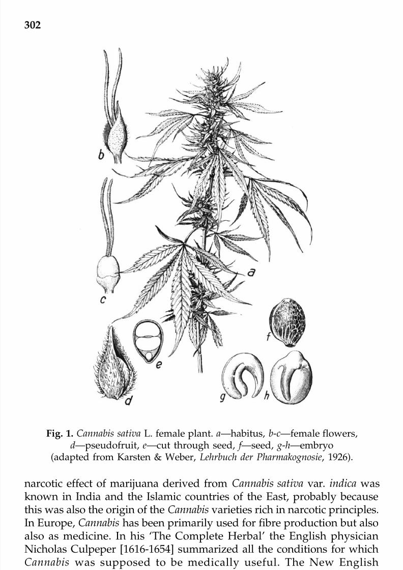
7/23/2019 Novel Therapeutic Agents From Plants
http://slidepdf.com/reader/full/novel-therapeutic-agents-from-plants 313/464
302
Fig. 1. Cannabis sativa L. female plant. a—habitus, b-c—female flowers,d—pseudofruit, e—cut through seed, f —seed, g-h—embryo
(adapted from Karsten & Weber, Lehrbuch der Pharmakognosie, 1926).
narcotic effect of marijuana derived from Cannabis sativa var. indica wasknown in India and the Islamic countries of the East, probably becausethis was also the origin of the Cannabis varieties rich in narcotic principles.In Europe, Cannabis has been primarily used for fibre production but alsoalso as medicine. In his ‘The Complete Herbal’ the English physicianNicholas Culpeper [1616-1654] summarized all the conditions for whichCannabis was supposed to be medically useful. The New EnglishDispensatory of 1764 recommended applying hemp roots to the skin for
inflammation, a remedy that was already popular in Eastern Europe. TheEdinburgh New Dispensary of 1794 included a long description of the
© 2009 by Taylor & Francis Group, LLC

7/23/2019 Novel Therapeutic Agents From Plants
http://slidepdf.com/reader/full/novel-therapeutic-agents-from-plants 314/464
303
effects of hemp and stated that the oil was useful in the treatment of coughs,venereal disease, and urinary incontinence. But Cannabis did not becomepopular as a medicine until the middle of the 19th century. During its earlyheyday, from 1840 to approximately 1900, more than 100 papers werepublished in the Western medical literature, recommending Cannabis forvarious illnesses and discomforts (Mikuriya, 1973). Little is known of therecreational use of Cannabis in Europe prior to its introduction as a narcotic
by Napoleon’s soldiers returning from Egypt where, at that time, marijuanawas smoked mainly by the underprivileged (Kimmens, 1977). In the 19th
century Cannabis was increasingly consumed by artists and intellectualsas a mind opening entheogen (Baudelaire, 1858). Famous for recreational
Cannabis experimentation was the ‘club of haschischins’ founded byBaudlaire, Gautier, and Rimbaud on one of the Seine isles in France(Gautier, 1843). Since the 1960s marijuana has become the most widespreadillicit drug of abuse in the world.
Despite major technological and conceptual advances in biochemicalpharmacology during the second half of the 20th century, the progress of the knowledge on Cannabis pharmacology was rather slow. Moreover, the
initial focus in Cannabis research was almost exclusively on thepsychotropic effects, and the concept of the peripheral cannabinoidreceptors was unknown. The psychoactive principle of Cannabis wasdescribed as an oil mixture and was referred to as cannabinol (Tschirch,1910). In the early 1940s cannabidiol and cannabinol, the first purecannabinoids, were isolated and investigated chemically. Since thesecompounds were evidently not the psychoactive principles of marijuana(female flowers of C. sativa), despite of cannabinol being able to act on
cannabinoid receptors as found later, they were largely ignored. In 1964,Gaoni and Mechoulam finally identified ∆9-tetrahydrocannabinol (∆9-THC)as the major psychoactive principle in Cannabis, ending a search for almost60 years. The fascinating history of the chemical research on plantcannabinoids has been reviewed (Mechoulam and Hanus, 2000; Russo,2007) and is not a subject of this chapter. Until the 1980s, the termcannabinoids represented, by definition, the group of typical C21
compounds present in Cannabis sativa L., their carboxylic acids, andtransformation products also referred to as phytocannabinoids (Lambertet al., 2005). The most important phytocannabinoids are ∆9-THC, ∆8-THC,cannabidiol, and cannabinol (Figure 2). Despite the knowledge about ∆9-THC as the major bioactive principle its cellular target was not identifiedfor more than 25 years. Early cannabinoid research mainly focused on
behavioural effects in animals, and structure-activity relationship studiesemployed the dog ataxia test, experiments with rhesus monkeys, or the
‘cannabinoid tetrad test battery’ which is generally performed in mice(Martin, 1986). The lack of a biological target protein for ∆9-THC during
© 2009 by Taylor & Francis Group, LLC

7/23/2019 Novel Therapeutic Agents From Plants
http://slidepdf.com/reader/full/novel-therapeutic-agents-from-plants 315/464
304
that time of research led to much controversy between scientists as somespeculated that non-specific toxic effects on cellular membranes couldaccount for the psychomimetic action of the lipophilic compound ∆9-THC.
Since the discovery that ∆9-THC and related cannabinoids act onspecific physiological receptors in the human body, and the subsequentelucidation of the mammalian endogenous cannabinoid system, the fieldof cannabinoid research has continually expanded. The first report of aspecific cannabinoid receptor, which binds ∆9-THC was published in 1988(Devane et al., 1988). This initial study was performed with the tritiated
Pfizer compound CP-55,940 (2), a ring-opened and less lipophilic analogueof ∆9-THC which competitively binds to the same receptors as ∆9-THC(Howlett et al., 1990). Tissue binding studies with [3H]CP-55,940 suggestedthat the cannabinoid receptor density was greatest in the limbic and corticalareas, the basal ganglia and cerebellum, and that it gradually decreasedinto the brainstem and spinal cord (Herkenham et al., 1990, 1991; Mailleuxand Vanderhaeghen, 1992). Subsequent to this, the first cannabinoid
receptor, the cannabinoid type-1 (CB1), was cloned from a rat brain cDNAlibrary using a probe derived from the sequence of bovine substance-Kreceptor (Matsuda et al., 1990). After the cloning of the CB
1receptor
subsequent DNA sequence homology studies led to the discovery of asecond cannabinoid receptor (the so-called type-2 or CB
2receptor) from
the myelomonocytic leukemia cell line HL60 (Munro et al., 1993).
Cannabinoid receptors belong to the class A rhodopsin-like G-protein-coupled receptor (GPCR) family. The CB
1receptor, which has been cloned
from human, cat, rat, and mouse is predominantly but not exclusivelyfound in the nervous system, while the CB
2receptor, which has been cloned
OH
O
H
HOH
O
H
H
OH
O
OH
HO
Fig. 2. Major chemical constitutents (classical cannabinoids)of Cannabis sativa L. var. indica.
© 2009 by Taylor & Francis Group, LLC

7/23/2019 Novel Therapeutic Agents From Plants
http://slidepdf.com/reader/full/novel-therapeutic-agents-from-plants 316/464
305
from human, mouse, and rat is mainly expressed on immune cells and inthe spleen (Cabral and Marciano-Cabral, 2005; Mackie, 2005). Even thoughthe classification of CB
1as central and CB
2as peripheral cannabinoid
receptors is generally correct it is important to note that the CB1
receptorcan be strongly expressed in certain peripheral cells like immune cells andthat the CB
2receptor is potentially also present in neurons of the brain
(Gong et al., 2006). The cannabinoid receptors show approximately 48%overall identity with 68% identity in the transmembrane regions (Howlettet al., 2002), and are coupled to the G
i/G
ofamily of G-proteins (Howlett,
2005). Signal transduction pathways regulated by cannabinoid receptorsinclude the inhibition of adenylate cyclase and stimulation of phospho-
lipase C (vide infra).The presence of cannabinoid receptors in mammalian cells was
indicative of the existence of an endogenous cannabinoid system. One of the first clues to the existence of cannabinoid receptors was provided bystudies showing that incubation of neuroblastoma cells with cannabinoidsincluded a decrease of cyclic adenosine monophosphate (cAMP) in thecells, suggesting that the putative receptors were negatively coupled to
adenylate cyclase. N -arachidonoyl ethanolamine, the first endogenousligand described for the cannabinoid receptors, was discovered in the early1990s (Devane et al., 1992; Di Marzo et al., 1994; Thomas et al., 1996). Thisarachidonic acid derivative was called ‘anandamide’, referring to theSanskrit term ‘ananda’, meaning bliss. Anandamide, which is synthesizedon demand activates both CB
1(K
i < 50 nM) and CB
2(K
i < 250 nM) receptors
and is rapidly eliminated through a two-step process consisting of aputative carrier-mediated transport followed by metabolization by the
membrane-bound serine hydrolase, referred to as fatty acid amide hydrolase(FAAH) (Ligresti et al., 2005). It soon became apparent that anadamide wasonly one member of a whole family of endocannabinoids and that otherendogenous cannabinoids, including 2-arachidonoyl glycerol (2-AG)together with CB
1and CB
2receptors constituted a major neurochemical
and possibly also immunomodulatory system in mammalian physiology(Mechoulam et al., 1998; Klein et al., 2003; Di Marzo et al., 2007). The search
for endogenous ligands for the cannabinoid receptors has led to thediscovery of several polyunsaturated compounds derived from fatty acids,mostly arachidonic acid (Kogan and Mechoulam, 2006). However, if endocannabinoids are defined as being part of distinct synthetic andmetabolic pathways, and capable to bind to the cannabinoid receptorsselectively at physiological concentrations, only anandamide and 2-AGare true endogenous cannabinoids. The physiological role of anandamideis still unclear and its action appears to be associated to areas as distinct
as schizophrenia and reproduction. Moreover, the question remainswhether the numerous effects reported for anandamide merely reflect its
© 2009 by Taylor & Francis Group, LLC

7/23/2019 Novel Therapeutic Agents From Plants
http://slidepdf.com/reader/full/novel-therapeutic-agents-from-plants 317/464
306
action on CB1
and CB2
receptors or whether other yet unknown targets mayalso play a role.
The discovery that anandamide binds to cannabinoid receptors and
acts as an endogenous mimic of ∆9
-THC led to a pronounced interest inother fatty acid amides as cannabinoid receptor ligands (Appendino et al.,2006a, 2006b; Di Marzo, 2007). As reviewed recently, several groups haveexplored the synthesis and biological evaluation of anandamide analogues,obtained by modifications of the fatty acid chain and of the ethanolaminehead group (Mahadevan and Razdan, 2005). Based on the structure of anandamide efforts have been undertaken to design ligands with improved
binding selectivity for individual cannabinoid receptors and with a better
pharmacokinetic profile (Kogan and Mechoulam, 2006). Our recent findingthat the medicinal plant purple coneflower (Echinacea purpurea and E.angustifolia), which is indicated for the treatment of upper respiratoryinfections and the common cold, produces N -alkyl amides which are high-affinity functional CB
2receptor-specific ligands (K
i ~ 50 nM) has led us to
search for other CB2 binding compounds within this natural product family.
Since fatty acid amides are relatively abundant in plants, these hitherto
neglected natural products may be a feasible source for new cannabinoidlead structures and model compounds to study CB2
receptor signaltransduction. In our screening for CB
2binding compounds we have recently
identified distinct CB2
binding N -alkyl amides from different plant taxa aswell as the ubiquitous sesquiterpene beta-caryophyllene that also exertscannabimimetic effects both in vitro and in vivo (Gertsch et al., 2006; Gertschet al., 2008).
In this chapter the pharmacological relevance of cannabinoid receptorligands is reviewed, with a special focus on recent developments in thefield of CB
2cannabinoid receptor drug discovery.
CB1
receptor pharmacology
The pharmacology of cannabinoid receptors, especially CB1
receptors, has been reviewed extensively (Klein et al., 2003; Di Marzo et al., 2004; Howlett,
2005; Lambert and Fowler, 2005; Mackie, 2006). The CB1 cannabinoidreceptor is highly conserved (for a review see Lutz, 2002), and it has beencloned from several species, including zebra fish and amphibian newt.Interestingly, it seems to be absent in insects and sponges, and cannabinoidreceptors are thus believed to have evolved in the last common ancestorsof bilaterians, with secondary loss occuring in insects and other clades(McPartland et al., 2005). In mammals CB
1receptors are not only expressed
in the brain but also testis, ileum, urinary bladder, the vas deferens, andon immune cells of the monocytic lineage (unpublished observation).Alternatively spliced forms of the CB
1cannabinoid receptor have been
© 2009 by Taylor & Francis Group, LLC

7/23/2019 Novel Therapeutic Agents From Plants
http://slidepdf.com/reader/full/novel-therapeutic-agents-from-plants 318/464
307
found (Ryberg et al., 2005), though their role is still unknown. In the CNS,CB
1receptors are predominantely expressed on axons and nerve terminals,
and functional evidence also supports their expression on somata (Howlettet al., 2002). In man, the CB
1
receptor is responsible for the ‘high’ frommarijuana smoking produced by centrally available ∆9-THC. Cannabinoidreceptors are G-protein coupled receptors (GPCR), which are linked toadenylate cyclase (ADC) and a variety of other second messengers andsignalling components. Ligation of CB
1has been shown to not only
suppress ADC but also to activate it (Rhee et al., 1998). CB1
receptors alsoinhibit presynaptic N- and P/Q-type calcium channels and activateinwardly rectifiying potassium channels (Mackie et al., 1995; Twitchell et
al., 1997). Smoked doses of 20 mg of ∆9-THC will produce increases of 20to 50 beats per minute in heart rate, impairment of short term memory andconcentration, possibly mood changes such as euphoria, altered perceptionof time, hunger, and other effects. At higher doses, marijuana interfereswith motor coordination and complex task functioning. Pharmacologicaleffects from inhaled ∆9-THC and ∆8-THC begin almost immediately. ∆9-THCplasma levels peak 10 to 20 minutes post inhalation and begin to decline.Effects last for two to three hours. Studies indicate that marijuana andalcohol potentate one another more than either drug alone. Thus, smalleramounts of ethanol and marijuana may increase toxic effects. Peak levelsof marijuana metabolites in the urine occur about five hours post dose andthus are not useful in predicting the degree of intoxication. Knockoutanimals have been obtained for the CB
1receptor by several groups (Zimmer
et al., 1999; Ledent et al., 1999). The CB1
receptor predominantely mediatesthe antimimetic action of Cannabis (Mechoulam, 1986) and agonists, such
as ∆9-THC (Dronabinol, MarinolTM) are used to reduce vomiting induced by anticancer chemotherapy (Plasse, 1991). Of all potential drugs affectingthe endocannabinoid system, CB
1receptor antagonists have probably
received the most attention because of the recently marketed andsubsequently withdrawn drug AcompliaTM (rimonabant) (1), also knownas SR141716, indicated for adipositas (obesity). The drug was developed
based on the observation that marijuana consumption stimulates theappetite. Thus, the rationale for using a CB
1
antagonist is conceptuallysimple as blocking the receptor might decrease the appetite. In fact, CB
1
antagonist decrease weight, but not for quite these reasons as the weightloss involves metabolic effects in hepatocytes and adipocytes (for a reviewsee Mackie, 2006). Clinical studies with CB
1antagonists are generally
perceived as encouraging and rimonabant (1) at a daily dose of 20 mgtogether with a hypocaloric diet leads to sustained weight loss of anaverage of 7 kg versus a weight loss of 2 kg in the placebo treated group
(Van Gaal et al., 2005). Although this may appear as a modest result,rimonabant also led to significant improvements in lipid profiles and CB
1
© 2009 by Taylor & Francis Group, LLC

7/23/2019 Novel Therapeutic Agents From Plants
http://slidepdf.com/reader/full/novel-therapeutic-agents-from-plants 319/464
308
antagonists have been shown to block the increase in fatty acid synthaseexpression induced by a high-fat diet (Osei-Hyiaman et al., 2005). Previuosto this finding, a molecular connection between the endocannabinoidsystem and fatty acid synthesis had already been established (Osei-Hyiaman et al., 2003). In adipocytes, CB
1activation has been associated to
lipoprotein lipase activity (Cota et al., 2003).
For some time it was assumed that the endocannabinoid system mayregulate addictive behaviour (for a review see Mackie, 2006). From a largenumber of animal experiments, there appears to be a reciprocal relation-ship between the motivational or rewarding aspects of endogenous opioidsand the endocannabinoids (Corchero et al., 2004). In fact, the rewarding
properties of opioids are absent in CB1 knockout mice (Cossu et al., 2001).During the trials with 1 the link between the CB
1receptor and addictive
behaviour could be confirmed (Gallate et al., 2004). Whether CB1
anta-gonists should be considered in the treatment of craving disorders dependson the overall behavioural effects of CB
1antagonists. The apparent
involvement of the CB1
receptors in addiction, such as nicotine and alcohol,has been the subject of many studies. CB
1antagonists block nicotine-
induced conditioned place preference and dopamine release in the nucleusacumbens (Valjent et al., 2002). Moreover, CB1
receptor activation has beenreported to enhance alcohol consumption while blocking or deletion of CB
1
decreases alcohol consumption (Gallate et al., 2004). Thus, other potentialapplications of CB
1antagonists are treatment of nicotine and alcohol
addiction. The recent clinical approval and worldwide marketing of AcompliaTM will now enable clinicians to determine whether CB
1anta-
gonists confirm the promising therapeutic value for the treatment of craving
disorders. Due to the possible role of CB1 receptors in the regulation of anxiety, memory extinction, and schizophrenia there is a theoreticalconcern that chronic CB
1blockage might be accompanied by significant
mood disorders. Although clinical experience with AcompliaTM is merely based on two years human trial only modest gastrointestinal problems,dizziness, and arthralgias have been reported (Van Gaal et al., 2005). Sincethe CB
1receptor is likely to play multiple roles, such as control of inflam-
mation in the CNS and regulation of bone development (vide infra), its blockage may well have long-term side effects.
In a recent article Teixeira-Clerc et al. (2006) reported that CB1
receptorsare strongly induced in fibrogenic liver cells, such as stellate cells of thecirrhotic human liver and in stellate cells of mice treated with carbontetrachloride, a known fibrinogenic stimulus. The antifibrinogenic effectof CB
1blockage could be attributed to decreased proliferation and increased
apoptosis of activated stallate cells, probably related to a reduction in thehepatic levels of the dominant fibrinogenic cytokine transforming growthfactor-β-1 (TGF-β1). These findings raise the possibility of using CB
1© 2009 by Taylor & Francis Group, LLC

7/23/2019 Novel Therapeutic Agents From Plants
http://slidepdf.com/reader/full/novel-therapeutic-agents-from-plants 320/464
309
antagonists to treat liver cirrhosis (Kunos et al., 2006).
The effects of ∆9-THC which are mediated via CB1
activation have beenextensively studied and characterized. Nonetheless, data about the
detrimental effects of ∆9
-THC are contradictory. The abuse of marijuanaand its purified products has been assosiated with modulation of cognitionand profound psychological changes. Although the psychotropic effectsof ∆9-THC are generally accepted there is a controversy about itsaddictiveness. Promising results in the use of ∆9-THC in cancer patients,particularly in the relief of nausea and vomiting caused by chemotherapyhave led to the development of ∆9-THC based medications, such asdronabinol (synthetic ∆9-THC), which is used for the treatment of nausea
and vomiting. In the US, together with its use to stimulate the appetite of AIDS patients, ∆9-THC is also indicated for the treatment of the symptomsof multiple sclerosis. Related to the medicinal use of Cannabis is the recentintroduction of the standardized Cannabis sativa extract SativexTM, whichcontains ∆9-THC, cannabinol, the non-psychoactive cannabidiol (Smith,2004). Moreover, SativexTM also contains full terpenoid content, such asmono and sesquiterpenes. Since ∆9-THC and cannabinol not only activates
CB1 but also CB2 receptors (vide infra) it is not known whether the positivepharmacological effects of these compounds are mediated by CB1, CB
2, or
a combination of both. Since CB2
receptor agonists have been shown toproduce many of the beneficial effects reported for ∆9-THC, but lack thepsychotropic side effects, cannabimimetics with CB
2selectivity over CB
1
are currently being developed for therapeutic use.
CB2
receptor pharmacology
Overall, the exact physiological role of CB2
receptors remains to be fullydefined. However, several intriguing preclinical studies suggest that CB
2
agonists may be clinically useful for distinct disease areas (Figure 3). Infact, the potential use of CB
2receptor agonists has been strongly
emphasized in different recent reviews (Lambert and Fowler, 2005; Mackie,2006).
In addition to its role in the cellular immune system (e.g. resolution of inflammation) (Klein et al., 2003), the CB
2receptor has become an attractive
target for drug discovery in areas as distinct as chronic pain, athero-sclerosis, and osteoporosis (vide infra). Recent studies also highlight thepotential of CB
2receptor specific ligands in the treatment of cancer,
including tumors of the immune system (Alberich Jorda et al., 2004;Sarfaraz et al., 2005; www.pharmoscorp.com) and brain tumors likegliomas (Blázquez et al., 2008). However, despite a wealth of preclinicaldata only the specific synthetic CB
2agonist cannabinor (PRS-211,375) (3)
from Pharmos Ltd. is in phase II clinical trial as an analgesic for pain and© 2009 by Taylor & Francis Group, LLC

7/23/2019 Novel Therapeutic Agents From Plants
http://slidepdf.com/reader/full/novel-therapeutic-agents-from-plants 321/464
310
inflammation (www.pharmoscorp.com). This compound has also beenfound to be efficacious in preclinical models of multiple sclerosis,rheumatoid arthritis, and inflammatory liver damage (www.pharmoscorp.com; Lavon et al., 2003). In light of such developments it is notsurprising that several laboratories now focus on the elaboration of newCB
2ligands. Therefore, not only CB
1-binding compounds, like the
successful phase III Sanofi CB1
antagonist AcompliaTM (vide supra) or theclinically used unselective CB
1/CB
2agonists MarinolTM (dronabinol), and
CesametTM (nabilone), but also compounds specifically directed at the CB2
receptor are promising candidates for drug discovery. At present, potentantagonists for both receptors, as well as four major structural classes of
agonists have been identified (vide infra). While an extensive amount of structure-activity relationship (SAR) information has been developed forCB
1ligands (vide supra), the SAR for CB
2ligands is only now emerging in
the literature (Lambert and Fowler, 2005; Reggio, 2005).
A particularly attractive feature of selective CB2
agonists as potentialtherapeutics is the fact that they are devoid of psychomimetic activity. It iswell known that CB
1receptor activation typically leads to analgesia in
different models (Hohmann et al., 2004) and Cannabis sativa and its extractshave been used to treat painful conditions for centuries. At the same timeit is now also clear that selective CB
2agonists are likewise effective in
chronic pain models such as models of neurophatic pain, peripheralinflammatory pain, and also in some pain sensitization models (Ibrahimet al., 2003; Hohmann et al., 2004; Ibrahim et al., 2006). The sites of CB
2
analgesic action remain unclear, although the skin is likely to be one siteas it has recently been shown that CB
2agonists enhance β-endorphin
release from keratinocytes (Ibrahim et al., 2005). According to Mackie (2006),the studies on the analgesic effects of CB
2agonists in neurophathic pain
Fig. 3. Cannabinoid receptors have been associated with distinctpathophysiologies and have therefore been discussed
as possible targets for a range of diseases.
Cirrhosis
Neurodegeneration
CB1 receptor
Adipositas
Craving disorders
Pain
Schizophrenia
Cancer
Nausea / Vomiting
CB2 receptor
Osteoporosis
Atherosclerosis
?Pain
Inflammation
Neurodegeneration Cancer
Chronic liver injury
© 2009 by Taylor & Francis Group, LLC

7/23/2019 Novel Therapeutic Agents From Plants
http://slidepdf.com/reader/full/novel-therapeutic-agents-from-plants 322/464
311
models following nerve injury suggest that the analgesia is mediated viaspinal microglia (Wotherspoon et al., 2005).
That CB2
receptors are involved in bone growth has been impressively
shown by the observation that CB2 knockout mice show markedlydecreased bone mass compared to wild type (Ofek et al., 2006). Moreover,the finding that in humans a silent single nucleotide polymorphism in CB
2
correlates strongly with osteoporosis in women (Karsak et al., 2005) seemsto substantiate the role of this receptor in bone formation. The therapeuticpossibilities of these findings are further evidenced by the observation thatthe CB
2agonist HU308 (4) decreases bone loss following ovariectomy in
mice (Ofek et al., 2006). The combination of these results and those of the
efficacy of CB2 agonists in chronic pain associated to inflammation isinteresting because anti-inflammatory drugs are currently associated todetrimental effects on bone density (Mackie, 2006). However, the exact roleof CB
2receptors during osteoclastogenesis and for osteoblast development
remains to be elucidated as the available data are somewhat contradictory because also stimulation of osteoclast activity via both CB
1and CB
2has
been reported (Idris et al., 2005). Currently, it is not clear whether an agonist
or an inverse agonist leads to inhibition of osteoclastogenesis. CB2 ligandsmay have therapeutic utility in chronic inflammatory diseases. As wasdemonstrated recently, low doses of ∆9-THC (hCB
2K
i= 36.4 ± 10 nM) slows
the extension of atherosclerotic lesions in ApoE-/- mice via a CB2
receptor-mediated mechanism (Steffens et al., 2005). CB
2mediated signaling may
also be involved in neurodegeneration associated with plaque developmentin Alzheimer’s disease (Ramirez et al., 2005). Another study suggests thatCB
2receptors may play a role in the inflammatory response accompanying
retroviral encephalitis, as may e.g. occur after HIV infection (Benito et al.,2005). These findings clearly show that CB
2receptor pharmacology has
developed into an exciting new area of research and that CB2
specificligands may eventually provide new drugs to treat old diseases. However,many fundamental questions remain to be answered and the CB
2signaling
pathways in both immune and neuronal cells are far from beingunderstood. Specific CB
2agonists, inverse agonists, and antagonists will
be required as tools for the elucidation of the molecular mechanisms andsignals involved in CB2
pharmacology. Any new class of CB2
bindingcompound may therefore provide new insights into the workingmechanism of this receptor.
Current CB2
receptor ligands
In general, cannabinoid ligands are classified as (a) classical cannabinoids
related to ∆9-THC, (b) the non-classical cannabinoids related to 2, (c) theaminoalkylindoles related to WIN 55,212-2 (5), and (d) the eicosanoids
© 2009 by Taylor & Francis Group, LLC

7/23/2019 Novel Therapeutic Agents From Plants
http://slidepdf.com/reader/full/novel-therapeutic-agents-from-plants 323/464
312
related to the endocannabinoids anandamide (6) and 2-AG (7) (Lambertand Fowler, 2005). Additional compounds, such as JTE907 (8) and BAY38-7271 (9), which cannot be allocated into any of the standard classeshave also been described (see Figures 4 and 5). Although many canna-
binoid receptor ligands show little, or at best modest, selectivity for eitherreceptor, a number of synthetic compounds are known which havesignificant selectivity for the CB
2receptor (Figure 6). Prominent examples
include HU308 (4), AM1241 (10), the 1-methoxy ∆8-THC derivatives, 1-methoxy ∆9-THC-DMH (L759633) (11), 1-methoxy ∆9(11)-THC-DMH(L759656) (12), plus a number of 1-deoxy ∆9-THC analogues. In particular,1-deoxy-3-(1',1'-dimethylbutyl) ∆9-THC (JWH-133) (13) shows two hundred-
fold selectivity for the CB2 receptor over CB1. In addtion, indol derivativessuch as 1-propyl-2-methyl-3-(1-naphthoyl)indole (JWH-015) (14) or 1-(2,3-dichlorobenzoyl)-2-methyl-3-(2-[1-morpholino]ethyl)-5-methoxyindole(L768242) (15) have been reported as CB
2-specific cannabimimetics. Efforts
have been made to develop structure-activity relationships (SAR) at CB2
for cannabimimetic indoles, but with limited success. Derivatives bearinga cyclohexane ring ortho to the phenolic hydroxyl group of the phenolrepresent another major class of cannabinoid ligands. These compoundsare related to the ring-opened pyran derivatives. Efforts have been madeto find CB
2selective agonists among the series and 4 is just one prominent
example. There are also reports of CB2
receptor agonists with monocycliccore structures. E.g., the tetrazine derivative 16 exhibited 90-fold selectivityfor CB
2and 2-imino-1,3-thiazolidine 17 showed more than 500-fold
selectivity (Adam et al., 2006). A pyridone analogue 18 of the bicyclic CB1
agonist CP47,597 showed CB2
selective activity (CB2 K
i= 56 nM) (Adam et
al. 2006). Pyridine derivatives, such as 19 have been disclosed by Glaxo,and 19 was more than 100-fold selective for CB
2over the CB
1receptor
(Adam et al., 2006).
Several CB2
antagonists have been reported (Griffin et al., 1999; Pertweeet al., 1995; Huffman, 2005). Although the Sanofi compound SR144528 (20)is the most widely used CB
2antagonist/reverse agonist it does not always
display typical antagonist behaviour (Rhee and Kim, 2002) and blocking
experiments using this compound could thus be misleading. For example,we have recently reported on the unexpected strong anti-inflammatoryeffects of 20 (Raduner et al., 2006), which were probably not the result of an inverse agonist action at CB
2receptors. Although less employed, the
compound JTE-907 (8) is another CB2
antagonist/reverse agonist (Iwamuraet al., 2001; Ueda et al., 2005). The aminoalkylindole compound AM630(21) appears to behave as a classical antagonist (52), but more studies areneeded to show its utility to specifically block CB
2signals without
triggering unrelated signal transduction events. In spite of the number of CB
2-specific receptor ligands known (and their structural diversity), to the© 2009 by Taylor & Francis Group, LLC

7/23/2019 Novel Therapeutic Agents From Plants
http://slidepdf.com/reader/full/novel-therapeutic-agents-from-plants 324/464
313
Fig. 4. Structures of CB receptor ligands discussed I.
AcompliaTM
(1)
Cl
N N
O
NH
Cl
Cl
OH
OH
HO CP-55,940 (2)
OH
O
H
H
SN
NH
Cannabinor (3)
O
O
HO
HU308 (4)
O
N
O
N
O
WIN 55,212-2 (5)
OHNH
O
Anandamide (6)
2-AG (7)
O
O
OH
OH
NH
O
O
O
O
NH
O
O
JTE907 (8)
O O
S
O
O
CF3HO
BAY 38 -7271 (9)
© 2009 by Taylor & Francis Group, LLC

7/23/2019 Novel Therapeutic Agents From Plants
http://slidepdf.com/reader/full/novel-therapeutic-agents-from-plants 325/464
314
Fig. 5. Structures of CB receptor ligands discussed II.
Fig. 4 continued. Structures of CB receptor ligands discussed I.
N
N
O
I
NO2
AM1241 (10)
O
O
H
H
L759633 (11)
O
O
H
H
L759656 (12) JWH-133 (13)
O
H
H
O
N
JWH-015 (14)
N
O
N
O
O
Cl
Cl
L768242 (15)
N
N N
N
CO2Et
CO2Et
16
N
S
N
OSEt
17
© 2009 by Taylor & Francis Group, LLC

7/23/2019 Novel Therapeutic Agents From Plants
http://slidepdf.com/reader/full/novel-therapeutic-agents-from-plants 326/464
315
Fig. 5 continued. Structures of CB receptor ligands discussed II.
N
O
OH
18
N
HN
O
NHN Cl
CF3
19
NH
O
N
NCl
SR144528 (20)
O
N
I
N
O
O
AM630 (21)
N -alkyl amide A1 (22)
NH
O
N -alkyl amide A2 (23)
NH
O
O
NH
N -alkyl amide A3 (24)
© 2009 by Taylor & Francis Group, LLC

7/23/2019 Novel Therapeutic Agents From Plants
http://slidepdf.com/reader/full/novel-therapeutic-agents-from-plants 327/464
316
Fig. 6. Binding affinity and selectivity (CB2
vs. CB1) of different cannabinoid
receptor ligands. White dots represent K i-values for CB
1receptors and the dark
dots the K i-values for CB
2receptors. Echinacea compounds A1 (22) and A2 (23)
show similar K i-values as the CB2 antagonist AM630 (21).
best of our knowledge, only the CB2
receptor agonist 3 from Pharmos Ltd.has entered trials for clinical development (www.pharmoscorp.com), andGlenmark Pharmaceuticals has recently anounced clinical trial with a theirCB
2-specific lead compound (www.glenmarkpharma.com).
Plant N -alkyl amides as new CB2
cannabimimetics
Although not as widespread as other classes of natural products fatty N -alkyl amides are relatively abundant in plants and they have been foundin a variety of families, including the Asteraceae, Brassicaceae, Legumi-nosae, Piperaceae, and Rutaceae (Chapman, 2004). These endogenous lipidconstituents, which often occur in just trace concentrations, are believedto regulate a wide range of plant physiological processes, including growth
and root development (Ramirez-Chavez et al., 2004). Moreover, naturalfatty acid amides are often insecticidal (Molina-Torres et al., 2004) and theirpresence is thus likely to provide an evolutionary advantage. It has beenshown that plants produce volatile fatty acid amides, such as linolenoyl-L-glutamine in response to elicitors in oral secretions of caterpillars (Laitet al., 2003). Other fatty acid N -alkyl amides frequently found in plantsare N -acylethanolamines (derived from fatty acids 12:0; 14:0; 16:0, and18:2) (Chapman et al., 1999), but despite their structural similarity to
anandamide (ethanolamide head group) they have not been shown so farto possess affinities to cannabinoid receptors. Nevertheless, and although
J T E 9 0 7
J W H 1 3 3
G W 4
0 5 8 3 3
L 7 5 9
6 3 3
J W H 1 3 9
A M 6 3 0
E c h i n a c e a A 1
E c h i n a c e a A 2
T H C
A M 1 2 4 1
H U 3 0 8
S R 1 4 4 5 2 8
2 - A G
A n a n d a m
i d e
0.1
1
10
100
1000
10000
K i v a l u e ( n M )
( d i s p l a c e m e n t o f [ 3 H ] C P - 5 5 , 9 4 0 )
© 2009 by Taylor & Francis Group, LLC

7/23/2019 Novel Therapeutic Agents From Plants
http://slidepdf.com/reader/full/novel-therapeutic-agents-from-plants 328/464
317
anandamide itself has never been isolated from plant sources, it has beensuggested that plant N -acylethanolamines modulate the endocannabinoidsystem in mammals by down-regulating FAAH, which leads to a longerhalf-life of the endogenous ligand anandamide (Di Marzo et al., 1998;Cravatt and Lichtman, 2003). We have recently investigated the pharma-cological profile of extracts and individual constituents of the medicinalplant Echinacea purpurea and we were able to show that certain fatty acidN -isobutyl amides modulate TNF-α expression in monocytes via CB
2
receptors (Gertsch et al., 2004). These findings were confirmed in anindependent study which demonstrated that the interaction of N-isobutylamides and N -methylbutyl amides from Echinacea angustifolia with the
endocannabinoid system in a rodent model (Woelkart et al., 2005). Togetherwith our most recent observation that dodeca-2E,4E,8Z,10Z-tetraenoic acidN -isobutyl amide (22) and dodeca-2E,4E-dienoic acid N -isobutyl amide (23)from Echinacea directly and specifically interact with the human CB
2
receptor (K i ~ 50 nM) and thereby elicit discrete functional responses
(Raduner et al., 2006), these insights may lead to a revived interest in plantfatty acid amides other than N -acylethanolamines. Although N -alkylamides from the medicinal plant taxon Echinacea were first reported aspharmacologically active entities already more than 10 years ago (Muller-
Jakic et al., 1994), the discovery that members of this compound family bind to CB
2receptors for the first time provides a mechanistic rationale for
some of the pharmacological effects commonly associated with themedicinal plant Echinacea. In contrast to anandamide and 2-AG manyplant N -alkyl amides produce a strong tingling effect, which is distinctfrom capsaicin and not directly related to the vanilloid receptor TRPV1
(Greger, 1988). The tingling of these compounds was shown to beassociated to activation of tactile and thermal trigeminal neurons (Bryantand Mezine, 1999), but its underlying molecular mechanism remainsunclear. 22 and 23 bind to CB
2with ca. 30-50 fold selectivity over the CB
1
receptor (Raduner et al., 2006). In addition, binding to CB2
was also shownto be highly selective relative to a large number of other receptors in a
broad-based receptor screen, including 49 receptors of pharmacologicalrelevance. Encouraged by these findings, we have now initiated thescreening of different lipophilic plant extracts known to contain N -alkylamides, in order to search for other CB
2receptor binding compounds. E.g.,
N -alkyl amides similar to those of Echinacea can e.g. be found in Spilanthesand Xanthoxylum species. Rather satisfactorily, a number of different N -alkyl amide-rich plant extracts have alrady shown reproducible K
i values
in the lower ng/ml range. As the structural diversity of naturally coccuringplant N -alkyl amides is limited and only few head groups are known in
plants (Figure 7), we have also started to investigate synthetic analogs of these natural products. Preliminary data show that certain N -benzyl
© 2009 by Taylor & Francis Group, LLC

7/23/2019 Novel Therapeutic Agents From Plants
http://slidepdf.com/reader/full/novel-therapeutic-agents-from-plants 329/464
318
amides, which also naturally occur, have moderate affinity for CB2
receptorsif they are equipped with an apropriate acyl tail (unpublished data). Incontrast to anandamide, N -alkyl amides, such as the ones from Echinaceaare not metabolised by FAAH (Woelkart et al., 2005) and can be found inhuman plasma after oral ingestion with C
maxvalues in the lower ng/mL
range and a typical Tmax
of < 1h (Woelkart et al., 2005; Matthias et al., 2005).This difference in metabolic stability is likely a consequence of structural
differences in the acyl tail. While the arachidonic acid part of anandamideleads to a very low metabolic stability in vitro and in vivo (Lin et al., 1998;Cravatt and Lichtman, 2003), Echinacea constituents are mainly derivedfrom undeca and dodecanoic acid, with varying degrees of unsaturationand different double bond configurations in the acyl part. Althoughnatural N -alkyl amides as such are not drug-like structures, e.g. they donot conform to the Lipinski rule of five (Lipinski et al., 2001), they may
nonetheless have the potential to serve as novel scaffolds for thedevelopment of analogs with a better pharmacokinetic profile and/or asnew tool compounds to study CB
2receptor pharmacology.
The CB2
receptor-bound conformation of N -alkyl amides
It has not been possible so far to determine the bioactive conformation of anandamide bound to the CB
2receptor or even that of the ligand-free
receptor by experimental methods. Attempts to determine the bioactiveconformation of anandamide by computational methods using homology
Fig. 7. Major plant derived N -amides which are found ashead groups in fatty acid N -amides.
NR
O
N
O
R
O
N
O
R
N
O
R
O
NH
R
O
NH
R
O
R
NH
O
R
R
O
NH
R
R
R
O
NH
NH
OHNH
R
O
© 2009 by Taylor & Francis Group, LLC

7/23/2019 Novel Therapeutic Agents From Plants
http://slidepdf.com/reader/full/novel-therapeutic-agents-from-plants 330/464
319
models based on the structure of rhodopsin A proved to be difficult due tothe high degree of flexibility of the anandamide arachidonic acid part,which contains four nonconjugated double bonds. In contrast, dockingstudies with 22 and 23 revealed a putative binding site in the CB
2
receptor,which is located adjacent to helices III, V, VI, and VII, near to theextracellular side of the 7TM bundles (Figure 8) (Raduner et al., 2006).According to this model the amide head group points into the hydrophilicpocket, which is formed by the side chains of Asp189 and Tyr190. Thephenolic hydroxyl group of and the latter is engaged in a H-bondinteraction with the amide hydrogen of 22 and 23, while the phenyl ringitself is involved in π-π interactions with the C2-C3 double bond of the alkyl
amide (Figure 9). Far less clear is the CB2 receptor interaction of the acyltail. Preliminary SAR data in our laboratory (Figure 10) show that
Fig. 8. Putative CB2 receptor binding conformation of N -alkyl amides22 and 23 based on a CB2
homology model (Raduner et al., 2006).
Fig. 9. N -isobutyl amide core structure showing the acyl tail and polarhead (open boxes) and the central parts involved in CB
2receptor
binding-interaction (shadowed boxes).
O
NH
© 2009 by Taylor & Francis Group, LLC

7/23/2019 Novel Therapeutic Agents From Plants
http://slidepdf.com/reader/full/novel-therapeutic-agents-from-plants 331/464
320
Fig. 10. Preliminary SAR of the acyl tail in N -isobutyl amides with
regard to hCB2 receptor affinity showing relative K i-values.
configuration of the double bonds and the degree of flexibility of the acylchain (Matovic et al., 2007) are important for binding, e.g. compound 24does not bind to cannabinoid receptors (Raduner et al., 2006). Obvioulsythe above model can only be considered as putative and will have to bevalidated experimentally. This could e.g. involve investigation of the effects
of mutations of Tyr190 in CB2 on the binding of compounds 22 and 23.
CB2
receptor signal transduction
Although Gi/G
omediated responses have been well established for the CB
1
receptor it has only recently been shown that CB2
receptors also signal viaG
o-proteins in monocytic cells. In fact, Sugiura et al. (2000) showed that
the endocannabinoid 2-AG not only inhibits cAMP formation but also
induces internal Ca2+ signals in a CB2-dependent way. This CB2-dependentstimulation of Ca2+ transients has been confirmed in our laboratory
R
O
NH
O
NH
E
E Z
Z
E
Z
Z
R1
E
R2
R3
R 4
R5
K i > 10000 nM
K i < 100 nM
K i > 4000 nM
K i > 9000 nM
K i > 10000 nM
K i < 100 nM
© 2009 by Taylor & Francis Group, LLC

7/23/2019 Novel Therapeutic Agents From Plants
http://slidepdf.com/reader/full/novel-therapeutic-agents-from-plants 332/464
321
(Raduner et al., 2006). Related studies have shown that the CB2
receptor islinked to phospholipase C, possibly via G
o-proteins, and that anandamide
can induce Ca2+ transients in pulmonary endothelial cells (Zoratti et al.,2003). In contrast to 2-AG and N -alkyl amides from Echinacea, theethanolamine compound anandamide does not modulate intracellular Ca2+
transients in monocytes. As monocytes/macrophages are the relevanttargets of CB
2specific ligands this unexpected finding may also be relevant
in a physiological setting. Moreover, interactions of the two endo-
Fig. 11. Model of the molecular effects on N -alkyl amides and cannabinoids onthe expression of cytokines in T-cells. The T
H1-T
H2 shift currently observed is (at
least in the case of N -alkyl amides and anandamide) associated to differentialmodulation of CD3 and CD28 pathways, leading to increased IL-3, IL-4, IL-5
and IL-10 expression (Raduner et al., 2006). Modulation of Ca2+ transients bycannabinomimetics is not mediated by CB
2receptors but TRAPC.
GsGs
CD28CD3TCR
PLCγ
Ca2+
Calcineurin
IP3PKC
NF-κBNF-AT
Gi
MAPKs
(JNK)
AP-1
ADC
cAMP
DAG
CB2
CB2?
PTKs
Super-induction / no modulation
Inhibition / no modulation
IL-3, IL-4, IL-5, IL-10, TNF-
?
N -alkyl amides
Cannabinoids
N -alkyl amides
CannabinoidsN -alkyl amidesCannabinoids
T-cell
Zap70
Erk
PMA
?TRPC
?
© 2009 by Taylor & Francis Group, LLC

7/23/2019 Novel Therapeutic Agents From Plants
http://slidepdf.com/reader/full/novel-therapeutic-agents-from-plants 333/464
322
cannabinoids with the CB2
receptor are likely to activate different signalingpathways by yet unknown mechanisms.
Whether cannabinoid ligand-induced Ca2+ transients in T-cells are also
mediated via CB2 receptors (Figure 11) remains debatable. It has beenreported that ∆9-THC and cannabinol robustly elevate Ca2+ in restinghuman and murine T cells in a CB
2-dependent way, whereas 2 does not
influence Ca2+ transients (Rao et al., 2004). It may well be that in T-cellsCa2+ signaling is influenced by another yet unknown receptor. Recently,the Kaminski laboratory has proposed that classic tricyclic cannabinoidswith structural similarity to ∆9-THC elicit a robust influx of Ca2+ in T cellsthrough receptor-operated cation channels in a manner sensitive to
cannabinoid receptor antagonists, but independent of the CB1 and CB2
receptors (Rao and Kaminski, 2006). According to this hypothesis, thecanonical transient receptor potential (CTRP) could be involved in thiseffect. Independent of these speculations, we believe that the endo-cannabinoids anandamide and 2-AG distinctly interact with cannabinoidreceptors in distinctly different ways, despite the fact that they bind to thesame or closely overlapping binding sites. As the N -alkyl amides 22 and
23 show the Ca2+
signaling effects of 2-AG and the antiinflammatory effectsof anandamide, this class of natural products could also serve as modelcompounds to study CB
2receptor interactions and signal transduction
events.
Immunomodulatory effects of cannabinoids and N -alkyl amides from Echinacea
Anandamide and N -alkyl amides from Echinacea show potent effects onLPS-induced TNF-α expression in whole blood assays (Figure 12).Although LPS recognizing CD14+ mononuclear cells strongly express theCB
2receptor we found that the anti-inflammatory effect is not exclusively
mediated via cannabinoid receptors (Raduner et al., 2006). Thus,anandamide inhibits the LPS-induced expression of pro-inflammatorycytokines IL-1β, IL-12 and TNF-β at low nanomolar concentrations (IC
50<
50 nM), in spite of its relatively moderate K i value (hCB2) of 218 ± 148 nM,suggesting that the effect could either be a consequence of low CB2
receptoroccupancy or could be mediated by another target. Based on data for 24,which is structurally related to 22 and lacks CB
2receptor affinity, but still
shows the typical anti-inflammatory effects (Raduner et al., 2006), wecurrently favour the latter explanation. In contrast to IL-1β, IL-12 and TNF-α, the constitutive expression of IL-6 was up-regulated by CB
2agonists in
the same ex vivo whole blood model in a CB2
receptor-dependent way, as
indicated by competition experiments with the reverse antagonistSR144528 (Raduner et al., 2006). N -alkyl amides from Echinacea also
© 2009 by Taylor & Francis Group, LLC

7/23/2019 Novel Therapeutic Agents From Plants
http://slidepdf.com/reader/full/novel-therapeutic-agents-from-plants 334/464
323
inhibited LPS-induced activation of mouse RAW264.7 macrophages andthus exert anti-inflammatory effects (Chen et al., 2005). In studies withhuman subjects, lung aveolar macrophages removed from marijuanasmokers were compromised in their ability to produce TNF-α, granulocyte/macrophage colony stimulation factor and IL-6 in response to LPSstimulation (Baldwin et al., 1997). It seems that cannabinoids can inhibitthe production of TNF-β and other cytokines in several different modelsand by several different mechanisms, not all of which depend on
interaction with cannabinoid receptors (for a review see Klein, 2005). Whilesuppressing the production of cytokines, cannabinoids have been shown
Fig. 12 Model of the intracellular effects of N -alkyl amides and 2-AG inmonocytes/macrophages (Mφ). The LPS stimuli mediated by CD14/TLR4appear to be inhibited in a CB
2-independent way, but intracellular Ca2+ is
increased by 2-AG and N -alkyl amides via CB2
receptors.
GsGs
CD14TLR4
PLCs
Ca2+
IP3 IKK
NF-κB
Gi
MAPKs
AP-1
ADC
cAMP
DAG
CB2
CB2?
IRAK
Inhibition
IL-1β, IL-6, IL-8, TNF-α
?
Monocyte / MΦ
N -alkyl amides2-AG
MyD88
TRAFF
TAK
?
PKC
N -alkyl amides2-AG N -alkyl amides
Cannabinoids
?
© 2009 by Taylor & Francis Group, LLC

7/23/2019 Novel Therapeutic Agents From Plants
http://slidepdf.com/reader/full/novel-therapeutic-agents-from-plants 335/464
324
to increase the production of cytokines (including TNF-α, IL-1, IL-6, andIL-10) when they are administered together with bacteria or antigens (Kleinet al., 1993; Klein, 2005) or, in some cases, even when administered alone(Kishimoto et al., 2004). Therefore, cannabinoids in vivo could eithersuppress or enhance the production of these pro-inflammatory mediators,depending on either the nature of the stimulus or the type of canna-
bimimetic used.
N -alkyl amides from Echinacea have recently been shown to inhibitIL-2 expression in T helper-cells in the lower µM range (Sasagawa et al.,2006), which is an effect typical for cannabinoids (17). We have shownthat N -alkyl amides from Echinacea at concentrations achievable upon oral
administration in humans (lower nM range) have a TH-biasing effect, inwhich T
H1-cell activity is suppressed and T
H2-cell activity is increased.
The same effect has been shown for cannabinoids and this generallysupports the potential use of cannabimimetics in the treatment of chronicinflammatory diseases. In the case of N -alkyl amides we could show thatthe T
H2 shift (i.e. the expression of the corresponding cytokines) is
associated to superstimulation of CD28 and inhibition of CD3 sginalling
(Figure 11). Overall, N -alkyl amides show similar immuno-modulatoryeffects as cannabinoids and therefore qualify to be classified ascannabimimetics.
Anti-inflammatory cannabinoids in diet—towards a betterunderstanding of CB
2receptor action?
The endocannabinoid system is an ancient lipid signalling network which
in mammals modulates neuronal functions, inflammatory processes, andis involved in the aetiology of certain human lifestyle diseases, such asCrohn’s disease, atherosclerosis, and osteoarthritis. The system is able todown-regulate stress-related signals that lead to chronic inflammation andcertain types of pain, but it is also involved in causing inflammation-associated symptoms, depending on the physiological context. Thecannabinoid type-2 (CB
2) receptor, which unlike the CB
1receptor does not
induce central side effects, has been shown to be a promising therapeutictarget. While CB1
receptor antagonists/inverse agonists are of therapeuticvalue, CB
2receptor ligands including agonists are of pharmacological
interest. Although the endocannabinoid system is known to be involvedin the regulation of energy homoeostasis and metabolism (mainly viaCB
1receptors) there was hitherto no direct link between food intake
and cannabinoid receptor activation. Our recent finding that beta-caryophyllene, a ubiquitous lipohilic plant natural product, selectively
binds to the CB2 receptor and acts as a full agonist is unexpected (Gertschet al., 2008). Maybe even more unexpected is that oral administration of
© 2009 by Taylor & Francis Group, LLC

7/23/2019 Novel Therapeutic Agents From Plants
http://slidepdf.com/reader/full/novel-therapeutic-agents-from-plants 336/464
325
this dietary compound exerts potent antiinflamamtory effects in wild typemice but not in CB
2receptor (Cnr2-/-) knockout mice. Like other CB
2ligands
also beta-caryophyllene inhibits the pathways triggered by activation of the toll-like receptor complex CD14/TLR4/MD2, which typically lead tothe expression of proinflammatory cytokines (IL-1b, IL-6; IL-8 and TNF-alpha) and promotes a TH
1immune response. Here, the CB
2receptor-
dependent effect of beta-caryophyllene on LPS-triggered activation of thekinases Erk1/2 and JNK1/2 are further discussed with respect to thepossibility that both CB
2inverse agonists and agonists, independent of
their G-protein signalling, may block LPS-triggered activation of MAPKs,leading to inhibition of proinflammatory cytokine expression and
attenuation of inflammation.Apart from the well-known psychomimetic action of the classical
cannabinoid ∆9-tetrahydrocannabinol (THC) mediated by centrallyexpressed cannabinoid CB
1receptors, it is now generally accepted that the
endocannabinoid system also occupies regulatory roles in peripheraltissues (Di Marzo, 2008; Kunos and Osei-Hyiaman, 2008; Klein andNewton, 2007). The cannabinoid CB
2receptor, which is involved in the
tuning of inflammatory processes (Wolf and Ulrich, 2008; Wright et al.,2008; Lotersztajn et al., 2008) is able to mediate cellular signals that inmany cases lead to attenuation of inflammation (vide supra). Theendogenous CB receptor ligands, the arachidonic acid derivativesanandamide and 2-arachidonoylglycerol (2-AG), which are the majorendocannabinoids known, partially or fully activate CB receptors in arather non-selective manner and controlled by strict biosynthesis anddegradation (Di Marzo, 2008).
Anandamide and 2-AG have been shown to inhibit the inflammatoryprocesses triggered upon activation of the toll-like receptor complex CD14/TLR4/MD2 (i.e. LPS and carrageenan stimulation) (Klegeris et al., 2003;Gallily et al., 2000). The same effect has also been reported for other CB
2
receptor agonists like JWH133 (Duncan et al., 2008), HU-308 (Hanus etal., 1999), and N -alkylamides (Raduner et al., 2006). Paradoxically, alsoCB
2receptor inverse agonists like JTE-907 (Ueda et al., 2005) SR144528
(Ueda et al., 2005; Oka et al., 2005), and Sch.336
(Lunn et al., 2006) showthis effect in the same or similar models. As both CB
2receptor inverse
agonists (Lunn et al., 2008) and agonists block identical pathways in anapparently CB
2receptor-dependent manner, mediators other than cAMP
or calcium transients should be involved in this mechanism. In our study(Gertsch et al., 2008) treatment of primary monocytes/macrophages withthe CB
2receptor agonist beta-caryophyllene leads to inhibition of LPS-
stimulated phosphorylation of the kinases Erk1/2 and JNK1/2 but not p38,despite the fact that the phosphorylation of these kinases is increased bythis compound and JWH133 in resting (non-stimulated) monocytes/
© 2009 by Taylor & Francis Group, LLC

7/23/2019 Novel Therapeutic Agents From Plants
http://slidepdf.com/reader/full/novel-therapeutic-agents-from-plants 337/464
326
macrophages. Thus, the context of differentially activated signallingpathways determines the way signals are integrated. Erk1/2 has beenshown to be either activated or inhibited by different GPCRs, both via G-protein dependent and independent mechanisms, involving intracellularcalcium transients (Chuderland et al., 2008) the b-arrestin scaffold(Gripentrog et al., 2008) and ubiquitination (Shenoy et al., 2005) JNK1/2is a stress-activated protein kinase that can be modulated by GPCRs.Protein tyrosine kinases are thought to be involved in the regulation of signal transmission from GPCRs to activation of the JNK/SAPK kinasepathway (Dikic and Blaukat, 1999). To inhibit LPS-stimulated Erk1/2 and
JNK phosphorylation (i.e. inhibition of their pathways) could be achieved
by blocking MyD88, TRAF6 and IKK-γ , which would lead to reducedtranscription of proinflammatory cytokines and metalloproteinases. Thequestion remains how CB
2receptor ligands as diverse as inverse agonists
and full agonists do the same thing in an apparently receptor-dependentfashion and whether in all cases MAPKs are involved. A direct inhibitionof NF-κ B, which is the primary signal in LPS-induced gene expression, ismore unlikely because this factor is inhibited only at higher µM-concentrations of cannabinoids. How exactly does the CB
2
receptormodulate MAPKs in immune cells? There are different possible mecha-nisms, such as hitherto neglected CB
2pathways like the ras-MAPK
pathway or even an indirect action via TNF-α expression. Interestingly,accumulation of TNF-α transcripts, but inhibition of protein expressionhas been shown after treatment with both CB
2selective inverse agonists
and agonists (Puffenbarger et al., 2000; Gertsch et al., 2004). On the sameline, inhibition of the JNK pathway in monocytes/macrophages will
inhibit TNF-α translation (Swantek et al., 1997). Moreover, TNF-αstimulated pathways could be inhibited indirectly, such that the pro-inflammatory feedback will shut down. In fact, there is good evidence thatconstitutive TNF-α is inhibited by the endocannabinoid system and thatthe CB
2receptor is involved (Rajesh et al., 2008; Mechoulam et al., 2007).
Moreover, TNF-α can activate the endocannabinoid system in adiposetissue (Kempf et al., 2007). As TNF-α triggers the activation of Ras, p38MAPK, ERK1/2, SAPK/JNK and Akt pathways and ultimately pro-inflammatory cytokine expression and cellular proliferation and migration,it is also possible that the autocrine action of TNF-α may be inhibited bycannabinoids. The CB
2agonists JWH-133 and HU-308 dose-dependently
attenuate the effects of TNF-α (Batkai et al., 2007; Rajesh et al., 2007). Onthe other hand, activation of the CB
2receptor by JWH133 in macrophages
has been shown to activate Erk1/2 which leads to expression of IL-10 andthus a counteraction to proinflammatory gene expression (Correa et al.,
2005). The CB2 receptor has also been found to be a potential target toprevent atherosclerosis (Steffens et al., 2005), in part via TNF-α signalling
© 2009 by Taylor & Francis Group, LLC

7/23/2019 Novel Therapeutic Agents From Plants
http://slidepdf.com/reader/full/novel-therapeutic-agents-from-plants 338/464
327
(Pacher et al., 2008). Because CB2
receptor activation attenuates TNF-α-induced endothelial cell activation, transendothelial migration of monocytes, and monocyte/neutrophil-endothelial adhesion, and decreasesTNF-α-induced proliferation and migration of human coronary vascularsmooth muscle cells (Rajesh et al., 2007), in addition to inhibitingconstitutive and LPS-stimulated TNF-α expression, the CB
2receptor may
be an essential pleiotropic modulator of TNF-α signal transduction.Providing that the multifunctional cytokine TNF-α is involved in numerouspathophysiological processes and the endocannabinoid system candynamically regulate the levels and effects of this cytokine, more researchshould be dedicated to the molecular understanding of CB receptor
signalling in the immune system.Unfortunately, the pharmacological tool box for CB
2receptors is still
very limited, the results with selective agonists and antagonists oftenconfusing and the conclusions drawn from such experiments potentiallyerroneous. For example, it would be wrong to say that 2-AG does mediateintracellular calcium in HL60 cells in a CB
2receptor-independent manner
because the selective CB2
antagonist/inverse agonist AM630 cannot block
this effect. The CB2 inverse agonist SR144528 can exactly do this (Oka etal., 2005). In some experiments AM630 and SR144528 are able to block theanti-inflammatory effects of CB
2agonists; in other experiments SR144528
is antiinflammatory. Thus, the classical concept of agonists and antagonists,“one lock and one key”, does not sufficiently describe cannabinoid GPCRsignalling. Even more puzzling is that CB
2receptor-selective agonists like
HU308 increase the inflammation caused by allergens in the skin, whileCB
2
antagonists and CB1
agonists inhibit inflammation (Karsak et al., 2007),thus pointing to the possibility that different cell types mediate distinctsignals. The apparent involvement of CB
receptors and Erk1/2 and JNK
MAPKs in the production of inflammatory signals and skin cancerdevelopment after UVB irradiation seems to confirm this (Zheng et al., 2008).
Even though we do obviously not yet understand the underlyingmolecular mechanisms of these observations, it can be said that in mostcases systemic administration of CB
2receptor-selective ligands leads to
attenuation of systemic inflammation in different animal models. It needsto be emphasized, however, that it is currently impossible to determine themolecular mechanism of a ligand in vivo and whether agonists are doingwhat antagonists should do and vice versa. One hint in that direction comesfrom data showing that the CB
1antagonist SR141716A acts like a partial
agonist in vivo (Smith et al., 2000).
In the case of the dietary natural product beta-caryophyllene, a full
CB2 receptor-selective agonist in vitro, potent antiinflammatory canna- bimimetic effects are observed (Gertsch et al., 2008). Intriguingly, the lowest
© 2009 by Taylor & Francis Group, LLC

7/23/2019 Novel Therapeutic Agents From Plants
http://slidepdf.com/reader/full/novel-therapeutic-agents-from-plants 339/464
328
oral dose tested (5 mg/kg) of this widespread and apparently non-toxiccompound, which is also an FDA-approve food additive, was the mosteffective. Maybe this strengthens the hypothesis that beta-caryophylleneis indeed a dietary cannabinoid, thus inferring that by eating thiscompound the endocannabinoid system may be modulated in a beneficialway, possibly through modulation of TNF-α pathways, maybe even to sucha degree that certain lifestyle diseases could be prevented. The finding thatoral beta-caryophyllene is more effective in mice than JWH-133 despite itssignificantly lower CB
2receptor affinity is encouraging. However, further
studies will have to determine whether this natural product is also bioavailable in humans and in more general terms, how CB
2receptor
ligands exactly modulate inflammation in vivo.
Conclusions
Cannabis sativa and/or its constitutents show a variety of beneficial effectsdespite of the clear risk factor associated with CB
1receptor activation. In
particular, direct activation of CB1
by ∆9-THC may act pro-psychotic,possibly via increased dopamine signaling in the nucleus accumbes(Lupica and Riegel, 2005). Current data regarding the medicinal potentialof cannabinoids and cannabimimetics thus suggest that CB
1antagonists
and CB2
agonists are interesting pharmacophores. Even though at leastthree putative cannabinoid receptors independent of CB
1and CB
2are
discussed (Mackie and Stella, 2006), the overwhelming data on CB1
andCB
2-dependent pharmacological effects clearly show that these receptors
are pharmacological targets. The recently postulated new cannabinoid
receptor GPR55 may have physiological roles, such as modulation of painreception (Staton et al., 2008), but too little is known to make a conclusionabout the therapeutic potential of this receptor. CB
2agonists have the
potential to treat diseases related to chronic inflammation, pain, and boneformation, and CB
1antagonists to metabolic disorders and potentially also
liver cirrhosis. To date, a limited number of different cannabinoid ligandsare available, most of which are derived from either ∆9-THC (classical),CP55940 (non-classical), aminoakylindol (WIN55212) or eicosanoids(endogenous cannabinoids).
Natural products other than plant-derived cannabinoids comprise ahitherto neglected source for the discovery of potential new ligands forcannabinoid receptors. Morevoer, in light of the diversity of plant secondarymetabolites traditional plant cannabinoids may not be the only class of natural products with affinities to cannabinoid receptors. Our discoverythat plant N -alkyl amides and beta-caryophyllene exert CB
2-specific
cannabimimetic effects clearly confirms this assumption. While potent bioavailable CB
1agonists, like ∆9-THC, may have been discovered during
© 2009 by Taylor & Francis Group, LLC

7/23/2019 Novel Therapeutic Agents From Plants
http://slidepdf.com/reader/full/novel-therapeutic-agents-from-plants 340/464
329
the course of human experimentation with psychoactive plants, agonistsdirected at CB
2and CB
1antagonists may have remained undetected. The
discovery of new natural non-cannabinoid cannabimimetics couldtherefore contribute to the development of more effective drugs for distinctdiseases. Natural product chemists have thus a unique opportunity toparticipate in the existing cannabinoid-receptor directed drug discoveryand find new natural products which bind to and either activate or blockcannabinoid receptors. This is best summarized by a recent quote byVincenzo Di Marzo (2007). ‘Thus, the exciting promenade from plantnatural products to animal physiology and back might soon become a truetreasure hunt.’
References
Adam, J., P.M. Cowley, T. Kiyoi, A.J. Morrison and C.J.W. Mort. 2006. RecentProgress in Cannabinoid Research. Progr. in Med. Chem. 44, 208-313.
Alberich Jorda, M., N. Rayman, M. Tas, S.E. Verbakel, N. Battista, K. van Lom,B. Lowenberg, M. Maccarrone and R. Delwel. 2004. The peripheral
cannabinoid receptor CB2, frequently expressed on AML blasts, eitherinduces a neutrophilic differentiation block or confers abnormal migrationproperties in a ligand-dependent manner. Blood 104, 526-534.
Appendino, G., A. Minassi, L. Berton, A.S. Moriello, M.G. Cascio, L. De Petrocellisand V. Di Marzo. 2006a. Oxyhomologues of anandamide and relatedendolipids: chemoselective synthesis and biological activity. J. Med. Chem.49, 2333-2338.
Appendino, G., M.G. Cascio, S. Bacchiega, A.S. Moriello, A. Minassi, A. Thomas,
R. Ross, R. Pertwee, L. De Petrocellis and V. Di Marzo. 2006b. First “hybrid”ligands of vanilloid TRPV1 and cannabinoid CB2 receptors and non-polyunsaturated fatty acid-derived CB2-selective ligands. FEBS Lett. 580, 568-574.
Baldwin, G.C., D.P. Tashkin, D.M. Buckley, A.N. Park, S.M. Dubinett and M.D.Roth. 1997. Marijuana and cocaine impair alveolar macrophage function andcytokine production. Am. J. Respir. Crit. Care Med. 156, 1606-1613.
Bátkai, S., D. Osei-Hyiaman, H. Pan, O. El-Assal, M. Rajesh, P. Mukhopadhyay,F. Hong, J. Harvey-White, A. Jafri, G. Haskó, J.W. Huffman, B. Gao, G. Kunosand P. Pacher. 2007. Cannabinoid-2 receptor mediates protection againsthepatic ischemia/reperfusion FASEB J., 21, 1788-1800.
Baudelaire, Ch. 1858. Le Haschisch—de L’idéal artificiel, in: La RevueContemporaine 30, France.
Benito, C., W.K. Kim, I. Chavarria, C.J. Hillard, K. Mackie, R.M. Tolon, K. Williamsand J. Romero. 2005. A glial endogenous cannabinoid system is upregulatedin the brains of macaques with simian immunodeficiency virus-inducedencephalitis. J. Neurosci. 25, 2530-2536.
Blázquez, C., M. Salazar, A. Carracedo, M. Lorente, A. Egia, L. González-Feria,A. Haro, G. Velasco and M. Guzmán. 2008. Cannabinoids inhibit glioma cell
© 2009 by Taylor & Francis Group, LLC

7/23/2019 Novel Therapeutic Agents From Plants
http://slidepdf.com/reader/full/novel-therapeutic-agents-from-plants 341/464
330
invasion by down-regulating matrix metalloproteinase-2 expression. CancerRes. 68, 1945-1952.
Bromberg, W. and T.C. Rodgers. 1946. Marihuana and aggressive crime, in:American J. Psych. 102, 825-827.
Bryant, B.P. and I. Mezine. 1999. Alkylamides that produce tingling paresthesiaactivate tactile and thermal trigeminal neurons. Brain Res. 842, 452-460.
Cabral, G.A. and F. Marciano-Cabral. 2005. Cannabinoid receptors in microgliaof the central nervous system: immune functional relevance. J. Leukoc. Biol.78, 1192-1197.
Chapman, K.D. 2004. Occurrence, metabolism, and prospective functions of N-acylethanolamines in plants. Prog. Lipid Res. 43, 302-327.
Chapman, K.D., B. Venables, R. Markovic, R.W. Blair Jr and C. Bettinger. 1999.
N-Acylethanolamines in seeds. Quantification Of molecular species and theirdegradation upon imbibition Plant Physiol. 120, 1157-1164.Chen, Y., T. Fu, T. Tao, J. Yang, Y. Chang, M. Wang, L. Kim, L. Qu, J. Cassady, R.
Scalzo and X. Wang. 2005. Macrophage activating effects of new alkamidesfrom the roots of Echinacea species. J. Nat. Prod. 68, 773-776.
Chuderland, D., G. Marmor, A. Shainskaya and R. Seger. 2008. Calcium-mediatedinteractions regulate the subcellular localization of extracellular signal-regulated kinases. J. Biol. Chem., 283, 11176-11188.
Corchero, J., J. Manzanares and J.A. Fuentes. 2004. Cannabinoid/opioid crosstalkin the central nervous system. Critical Rev. Neurobiol. 16, 159-172.
Correa, F., L. Mestre, F. Docagne and C. Guaza. 2005. Activation of cannabinoidCB2 receptor negatively regulates IL-12p40 production in murinemacrophages: role of IL-10 and ERK1/2 kinase signaling. Br. J. Pharmacol.,145, 441-448.
Cossu, G., C. Ledent, L. Fattore, A. Imperato, G.A. Bohme et al. 2001. CannabinoidCB1 receptor knockout mice fail to self-administer morphine but not otherdrugs of abuse. Behav. Brain Res. 118, 61-65.
Cota, D., G. Marsicano, M. Tschop, Y. Grubler, C. Flachskamm et al. 2003. Theendogenous cannabinoid system affects energy balance via central orexigenicdrive and peripheral lipogenesis. J. Clin. Invest. 112, 423-431.
Cravatt, B.F. and A.H. Lichtman. 2003. Fatty acid amide hydrolase: an emergingtherapeutic target in the endocannabinoid system. Curr. Opin. Chem. Biol.7, 469-475.
Devane, W.A., F.A. Dysarz, M.R. Johnson, L.S. Melvin and A.C. Howlett. 1988.Determination and characterization of a cannabinoid receptor in rat brain.
Mol. Pharmacol. 34, 605-613.Devane, W.A., L. Hanus, A. Breuer, R.G. Pertwee, L.A. Stevenson, G. Griffin, D.Gibson, A. Mandelbaum, A. Etinger and R. Mechoulam. 1992. Isolation andstructure of a brain constituent that binds to the cannabinoid receptor.Science 258, 1946-1949.
Di Marzo, V., A. Fontana, H. Cadas, S. Schinelli, G. Cimino, J.C. Schwartz and D.Piomelli. 1994. Formation and inactivation of endogenous cannabinoidanandamide in central neurons. Nature 372, 686-691.
Di Marzo, V., N. Sepe, L. De Petrocellis, A. Berger, G. Crozier, E. Fride and R.Mechoulam. 1998. Trick or treat from food endocannabinoids? Nature 396,636-637.© 2009 by Taylor & Francis Group, LLC

7/23/2019 Novel Therapeutic Agents From Plants
http://slidepdf.com/reader/full/novel-therapeutic-agents-from-plants 342/464
331
Di Marzo, V., M. Bifulco and L. De Petrocellis. 2004. The endocannabinoid systemand its therapeutic exploitation. Nat. Rev. Drug Discov. 3, 771-784.
Di Marzo, V. 2008. Targeting the endocannabinoid system: to enhance or reduce?Nat. Rev. Drug Discov. 7, 438-455.
Di Marzo, V. 2008. Endocannabinoids: synthesis and degradation. Rev. Physiol.Biochem. Pharmacol. 160, 1-24.
Di Marzo, V., T. Bisogno and L. De Petrocellis. 2007. Endocannabinoids and relatedcompounds: walking back and forth between plant natural products andanimal physiology. Chem Biol. 14, 741-756.
Dikic, I. and A. Blaukat. 1999. Protein tyrosine kinase-mediated pathways in Gprotein-coupled receptor signaling. Cell. Biochem. Biophys. 30, 369-387.
Duncan, M., A. Mouihate, K. Mackie, C.M. Keenan, N.E. Buckley, J.S. Davison,
K.D. Patel, Q.J. Pittman and K.A. Sharkey. 2008. Cannabinoid CB2 receptorsin the enteric nervous system modulate gastrointestinal contractility inlipopolysaccharide-treated rats. Am J. Physiol. Gastrointest. Liver Physiol.ahead of print.
Gallate, J.E., P.E. Mallet and I.S. McGregor. 2004. Combined low dose treatmentwith opioid and cannabinoid receptor antagonists synergistically reducesthe motivation to consume alcohol in rats. Psychopharmacology 173, 210-216.
Gallily, R., A. Breuer and R. Mechoulam. 2000. 2-Arachidonylglycerol, anendogenous cannabinoid, inhibits tumor necrosis factor-alpha productionin murine macrophages, and in mice. Eur. J. Pharmacol. 406, R5-7.
Gautier, T. 1843. Le Club des Haschischines, in: Feuilleton de la Presse Médicale.Gertsch, J., R. Schoop, U. Kuenzle and A. Suter. 2004. Echinacea alkylamides
modulate TNF-alpha gene expression via cannabinoid receptor CB2 andmultiple signal transduction pathways. FEBS Lett. 577, 563-569.
Gertsch, J., S. Raduner and K.H. Altmann. 2006. J. Recept. Sign. Transd. 26, 1-26.Gertsch, J., M. Leonti, S. Raduner, I. Racz, J.Z. Chen, X.Q. Xie, K.H. Altmann, M.
Karsak and A. Zimmer. 2008. Beta-caryophyllene is a dietary cannabinoid.Proc. Natl. Acad. Sci. USA. 105, 9099-9104.
Gong, J.P., E.S. Onaivi, H. Ishiguro, Q.R. Liu, P.A. Tagliaferro, A. Brusco and G.R.Uhl. 2006. Cannabinoid CB2 receptors: immunohistochemical localizationin rat brain. Brain Res. 1071, 10-23.
Greger, H. 1988. Comparative Phytochemistry of Alkamides. Elsevier,Amsterdam, Oxford, New York.
Griffin, G., E.J. Wray, Q. Tao, S.D. McAllister, W.K. Rorrer, M.M. Aung, B.R. Martin
and M.E. Abood. 1999. Evaluation of the cannabinoid CB2 receptor-selectiveantagonist, SR144528: further evidence for cannabinoid CB2 receptor absencein the rat central nervous system. Eur. J. Pharmacol. 377, 117-125.
Gripentrog, J.M. and H.M. Miettinen. 2008. Formyl peptide receptor-mediatedERK1/2 activation occurs through G(i) and is not dependent on beta-arrestin1/2. Cell Signal., 20, 424-431.
Hanus, L., A. Breuer, S. Tchilibon, S. Shiloah, D. Goldenberg, M. Horowitz, R.G.Pertwee, R.A. Ross, R. Mechoulam and E. Fride. 1999. HU-308: a specificagonist for CB(2), a peripheral cannabinoid receptor. Proc Natl Acad SciUSA., 96, 14228-14233.
Herkenham, M., A.B. Lynn, M.D. Little, M.R. Johnson, L.S. Melvin, B.R. de Costa© 2009 by Taylor & Francis Group, LLC

7/23/2019 Novel Therapeutic Agents From Plants
http://slidepdf.com/reader/full/novel-therapeutic-agents-from-plants 343/464
332
and K.C. Rice. 1990. Cannabinoid receptor localization in brain. Proc Natl.Acad. Sci. USA, 87, 1932-1936.
Herkenham, M., A.B. Lynn, M.R. Johnson, L.S. Melvin, B.R. de Costa and K.C.Rice. 1991. Characterization and localization of cannabinoid receptors in rat
brain: a quantitative in vitro autoradiographic study. J. Neurosci. 11, 563-583.
Hohmann, A.G., J.N. Farthing, A.M. Zvonok and A. Makriyannis. 2004. Selectiveactivation of cannabinoid CB2 receptors suppresses hyperalgesia evoked by intradermal capsaicin. J. Pharmacol. Exp. Ther. 308, 446-453.
Hohmann, A.G., R.L. Suplita, N.M. Bolton, M.H. Neely, D. Fegley, R. Mangieri, J.F. Krey, J.M. Walker, P.V. Holmes, J.D. Crystal, A. Duranti, A. Tontini, M.Mor, G. Tarzia and D. Piomelli. 2005. An endocannabinoid mechanism for
stress-induced analgesia. Nature 435, 1108-1112.Howlett, A.C. 2005. Cannabinoid receptor signalling. Handb. Exp. Pharmacol.168, 53-79.
Howlett, A.C., M. Bidaut-Russell, W.A. Devane, L.S. Melvin, M.R. Johnson andM. Herkenham. 1990. The cannabinoid receptor: biochemical, anatomicaland behavioral characterization. Trends Neurosci. 13, 420-423.
Howlett, A.C., F. Barth, T.I. Bonner, G. Cabral, P. Casellas, W.A. Devane, C.C.Felder, M. Herkenham, K. Mackie, B.R. Martin, R. Mechoulam and R.G.Pertwee. 2002. International Union of Pharmacology, XXVII. Classificationof cannabinoid receptors. Pharmacol. Rev. 54, 161-202.
Huffman, J.W. 2005. CB2 receptor ligands. Mini Rev. Med. Chem. 5, 641-649.Ibrahim, M.M., H. Deng, A. Zvonok, D.A. Cockayne, J. Kwan, H.P. Mata, T.W.
Vanderah, J. Lai, F. Porreca, A. Makriyannis and T.P. Malan Jr. 2003.Activation of CB2 cannabinoid receptors by AM1241 inhibits experimentalneuropathic pain: pain inhibition by receptors not present in the CNS. Proc.Natl. Acad. Sci. USA 100, 10529-10533.
Ibrahim, M.M., F. Porreca, J. Lai, P.J. Albrecht, F.L. Rice, A. Khodorova, G. Davar,
A. Makriyannis, T.W. Vanderah, H.P. Mata and T.P. Malan Jr. 2005. CB2cannabinoid receptor activation produces antinociception by stimulatingperipheral release of endogenous opioids. Proc. Natl. Acad. Sci. USA 102,3093-3098.
Ibrahim, M.M., M.L. Rude, N.J. Stagg, H.P. Mata, J. Lai, T.W. Vanderah, F. Porreca,N.E. Buckley, A. Makriyannis and T.P. Malan. Jr. 2006 CB2 cannabinoidreceptor mediation of antinociception. Pain 122, 36-42.
Idris, A.I., R.J. van ‘t Hof, I.R. Greig, S.A. Ridge, D. Baker, R.A. Ross and S.H.
Ralston. 2005. Regulation of bone mass, bone loss and osteoclast activity bycannabinoid receptors. Nat. Med. 11, 774-779.Iwamura, H., H. Suzuki, Y. Ueda, T. Kaya and T. Inaba. 2001. In vitro and in
vivo pharmacological characterization of JTE-907, a novel selective ligandfor cannabinoid CB2 receptor. J. Pharmacol. Exp. Ther. 296, 420-425.
Karsak, M., M. Cohen-Solal, J. Freudenberg, A. Ostertag, C. Morieux, U. Kornak, J. Essig, E. Erxlebe, I. Bab, C. Kubisch, M.C. de Vernejoul and A. Zimmer.2005. Cannabinoid receptor type 2 gene is associated with humanosteoporosis. Hum. Mol. Genet. 14, 3389-3396.
Karsak, M., E. Gaffal, R. Date, L. Wang-Eckhardt, J. Rehnelt, S. Petrosino, K.Starowicz, R. Steuder, E. Schlicker, B. Cravatt, R. Mechoulam, R. Buettner, S.© 2009 by Taylor & Francis Group, LLC

7/23/2019 Novel Therapeutic Agents From Plants
http://slidepdf.com/reader/full/novel-therapeutic-agents-from-plants 344/464
333
Werner, V. Di Marzo, T. Tüting and A. Zimmer. 2007. Attenuation of allergiccontact dermatitis through the endocannabinoid system. Science, 316, 1494-1497.
Kempf, K., J. Hector, T. Strate, B. Schwarzloh, B. Rose, C. Herder, S. Martin and
P. Algenstaedt. 2007. Immune-mediated activation of the endocannabinoidsystem in visceral adipose tissue in obesity. Horm. Metab. Res. 39, 596-600.
Kimmens, Andrew C. (ed.) 1977. Tales of Hashish: A Literary Look at the HashishExperience. William Morrow, New York, USA.
Kishimoto, S., Y. Kobayashi, S. Oka, M. Gokoh, K. Waku and T. Sugiura. 2004. 2-Arachidonoylglycerol, an endogenous cannabinoid receptor ligand, inducesaccelerated production of chemokines in HL-60 cells. J. Biochem. 135, 517-524.
Klegeris, A., C.J. Bissonnette and P.L. McGeer. 2003. Reduction of humanmonocytic cell neurotoxicity and cytokine secretion by ligands of thecannabinoid-type CB2 receptor. Br. J. Pharmacol. 139,775-786.
Klein, T.W. 2005. Cannabinoid-based drugs as anti-inflammatory therapeutics.Nat. Rev. Immunol. 5, 400-411.
Klein, T.W., C. Newton, R. Widen and H. Friedman. 1993. Delta 9-tetrahydro-cannabinol injection induces cytokine-mediated mortality of mice infectedwith Legionella pneumophila. J. Pharmacol. Exp. Ther. 267, 635-640.
Klein, T.W., C. Newton, K. Larsen, L. Lu, I. Perkins, L. Nong and H. Friedman.2003. The cannabinoid system and immune modulation. J. Leukoc. Biol. 74,486-496.
Klein, T.W. and C.A. Newton. 2007. Therapeutic potential of cannabinoid-baseddrugs. Adv Exp Med Biol., 601, 395-413.
Kogan, N.M. and R. Mechoulam. 2006. The chemistry of endocannabinoids. J.Endocrinol. Invest. 29, 3-14.
Kunos, G., D. Osei-Hyiaman, S. Batakai and B. Gao. 2006. Cannabinoids hurt,heal cirrhosis. Nat. Med. 6, 608-610.
Kunos, G. and D. Osei-Hyiaman. 2008. Endocannabinoids and liver disease. IV.Endocannabinoid involvement in obesity and hepatic steatosis. Am. J. Physiol.Gastrointest. Liver Physiol., 294, G1101-1104.
Lait, C.G., H.T. Alborn, P.E. Teal and J.H. Tumlinson 3rd. 2003. Rapid biosynthesisof N-linolenoyl-L-glutamine, an elicitor of plant volatiles, by membrane-associated enzyme(s) in Manduca sexta. Proc. Natl. Acad. Sci. USA. 100, 7027-7032.
Lambert, D.M. and C.J. Fowler. 2005. The endocannabinoid system: drug targets,
lead compounds, and potential therapeutic applications., J. Med. Chem. 48,5059-5087.Lavon, I., T. Sheinin, S. Meilin, E. Biton, A. Weksler, G. Efroni, A. Bar-Joseph, G.
Fink and A. Avraham. 2003. A novel synthetic cannabinoid derivative inhibitsinflammatory liver damage via negative cytokine regulation. Mol.Pharmacol. 64, 1334-1341.
Ledent, C., O. Valverde, G. Cossu, F. Petitet, J.F. Aubert, F. Beslot, G.A. Bohme,A. Imperato, T. Pedrazzini, B.P. Roques, G. Vassart, W. Fratta and M.Parmentier. 1999. Unresponsiveness to cannabinoids and reduced addictiveeffects of opiates in CB1 receptor knockout mice. Science 283, 401-404.
Ligresti, A., M.G. Cascio and V. Di Marzo. 2005. Endocannabinoid metabolic© 2009 by Taylor & Francis Group, LLC

7/23/2019 Novel Therapeutic Agents From Plants
http://slidepdf.com/reader/full/novel-therapeutic-agents-from-plants 345/464
334
pathways and enzymes. Curr. Drug. Targets CNS Neurol. Disord. 4, 615-623.
Lin, S., A.D. Khanolkar, P. Fan, A. Goutopoulos, C. Qin, D. Papahadjis and A.Makriyannis. 1998. Novel analogues of arachidonylethanolamide
(anandamide): affinities for the CB1 and CB2 cannabinoid receptors andmetabolic stability. J. Med. Chem. 41, 5353-5361.
Lipinski, C.A., F. Lombardo, B.W. Dominy and P.J. Feeney. 2001. Experimentaland computational approaches to estimate solubility and permeability indrug discovery and development settings. Adv. Drug Deliv. Rev. 46, 3-26.
Lotersztajn, S., F. Teixeira-Clerc, B. Julien, V. Deveaux, Y. Ichigotani, S. Manin, J.Tran-Van-Nhieu, M. Karsak, A. Zimmer, and A. Mallat. CB2 receptors asnew therapeutic targets for liver diseases. Br. J. Pharmacol. 153, 286-289.
Lunn, C.A., J.S. Fine, A. Rojas-Triana, J.V. Jackson, X. Fan, T.T. Kung, W.Gonsiorek, M.A. Schwarz, B. Lavey, J.A. Kozlowski, S.K. Narula, D.J. Lundell,R.W. Hipkin and L.A. Bober. 2006. A novel cannabinoid peripheralcannabinoid receptor-selective inverse agonist blocks leukocyte recruitmentin vivo. J. Pharmacol. Exp. Ther. 316, 780-788.
Lunn, C.A., E.P. Reich, J.S. Fine, B. Lavey, J.A. Kozlowski, R.W. Hipkin, D.J.Lundell and L. Bober. 2008. Biology and therapeutic potential of cannabinoidCB2 receptor inverse agonists. Br. J. Pharmacol. 153, 226-239.
Lupica, C.R. and A.C. Riegel. 2005. Endocannabinoid release from midbraindopamine neurons: A potential substrate for cannabinoid receptor antagonisttreatment of addiction. Neuropharmacology. 48, 1105-1116.
Lutz, B. 2002. Molecular biology of cannabinoid receptors. Prostaglandins Leukot.Essent. Fatty Acids. 66, 123-42.
Mackie, K. 2005. Distribution of cannabinoid receptors in the central andperipheral nervous system. Handb. Exp. Pharmacol. 168, 299-325.
Mackie, K. 2006. Cannabinoid receptors as therapeutic targets. Annu. Rev.Pharmacol. Toxicol. 46, 101-122.
Macie, K. and N. Stella. 2006. Cannabinoid receptors and endocannabinoids:Evidence for new players. AAPs J. 8, E298-E306.
Mackie, K., Y. Lai, R. Westernbroek and R. Mitchell. 1995. Cannabinoids activatean inwardly rectifying potassium conductance and inhibit Q-type calciumcurrents in AtT20 cells transfected with rat brain cannabinoid receptor. J.Neurosci. 15, 6552-6561.
Mahadevan, A. and R.K. Razdan. 2005. Further advances in the synthesis of endocannabinoid-related ligands. AAPS J. 7, E496-502.
Mailleux P. and J.J. Vanderhaeghen. 1992. Localization of cannabinoid receptorin the human developing and adult basal ganglia. Higher levels in thestriatonigral neurons. Neurosci. Lett. 148, 173-176.
Malan, T.P. Jr, M.M. Ibrahim, H. Deng, Q. Liu, H.P. Mata, T. Vanderah, F. Porrecaand A. Makriyannis. 2001. CB2 cannabinoid receptor-mediated peripheralantinociception. Pain 93, 239-245.
Martin, B. 1986. Cellular effects of cannabinoids. Pharmacol. Rev. 38, 45-74.Matovic, N, A. Matthias, J. Gertsch, S. Raduner, K.M. Bone, R.P. Lehmann and
J.J. DeVoss. 2007. Stereoselective synthesis, natural occurence and CB2receptor binding affinities of alkylamides from herbal medicines such asEchinacea sp. Org. Bioorg. Chem. In press.© 2009 by Taylor & Francis Group, LLC

7/23/2019 Novel Therapeutic Agents From Plants
http://slidepdf.com/reader/full/novel-therapeutic-agents-from-plants 346/464
335
Matsuda, L.A., S.J. Lolait, M.J. Brownstein, A.C. Young and T.I. Bonner. 1990.Structure of a cannabinoid receptor and functional expression of the clonedcDNA. Nature, 346, 561-564.
Matthias, A., R.S. Addison, K.G. Penman, R.G. Dickinson, K.M. Bone and R.P.
Lehmann. 2005. Echinacea alkamide disposition and pharmacokinetics inhumans after tablet ingestion. Life Sci. 77, 2018-2029.
McKallip, R.J., C. Lombard, M. Fisher, B.R. Martin, S. Ryu, S. Grant, P.S.Nagarkatti and M. Nagarkatti. 2002. Targeting CB2 cannabinoid receptorsas a novel therapy to treat malignant lymphoblastic disease. Blood 100, 627-634.
McPartland, J.M., J. Agraval, D. Gleeson, K. Heasman and M. Glass. 2005.Cannabinoid receptors in invertebrates, J. Evol. Biol. 19, 366-373.
Mechoulam, R. 1986. Interview with Prof. Raphael Mechoulam, codiscoverer of THC, Interview by Stanley Einstein. Int. J. Addict. 21, 579-587.
Mechoulam, R., E. Fride and V. Di Marzo. 1998. Endocannabinoids. Eur. J.Pharmacol. 359, 1-18.
Mechoulam, R. and L. Hanus. 2000. A historical overview of chemical researchon cannabinoids. Chem. Phys. Lipids 108, 1-13.
Mechoulam, R. and E. Shohami. 2007. Endocannabinoids and traumatic braininjury. Mol Neurobiol. 36, 68-74.
Mikuriya, T.H. (ed) 1973. Marijuana: Medical Papers, 1839-1972 MediComp.Oakland, USA.Molina-Torres, J., C.J. Salazar-Cabrera, C. Armenta-Salinas and E. Ramirez-
Chavez. 2004. Fungistatic and bacteriostatic activities of alkamides from Heliopsis longipes roots: affinin and reduced amides. J. Agric. Food Chem.52, 4700-4704.
Muller-Jakic, B., W. Breu, A. Probstle, K. Redl, H. Greger and R. Bauer. 1994. Invitro inhibition of cyclooxygenase and 5-lipoxygenase by alkamides from
Echinacea and Achillea species. Planta Med. 60, 37-40.Munro, S., K.L. Thomas and M. Abu-Shaar. 1993. Molecular characterization of aperipheral receptor for cannabinoids. Nature 365, 61-65.
Ofek, O., M. Karsak, N. Leclerc, M. Fogel, B. Frenkel, K. Wright, J. Tam, M. Attar-Namdar, V. Kram, E. Shohami, R. Mechoulam, A. Zimmer and I. Bab. 2006.Peripheral cannabinoid receptor, CB2, regulates bone mass. Proc. Natl. Acad.Sci. USA 103, 696-701.
Oka, S., S. Yanagimoto, S. Ikeda, M. Gokoh, S. Kishimoto, K. Waku, Y. Ishimaand T. Sugiura. 2005. Evidence for the involvement of the cannabinoid CB2receptor and its endogenous ligand 2 arachidonoylglycerol in 12-O-tetradecanoylphorbol-13-acetate-induced acute inflammation in mouse ear. J. Biol. Chem., 280, 18488-18497.
Osei-Hyiaman, D., M. Depetrillo, P. Pacher, J. Liu, S. Radaeva et al. 2003.Endocannabinoid action at hepatic CB1 receptors regulates fatty acidsynthesis: role in diet-induced obesity. J. Clin. Invest. 115, 1298-1305.
Osei-Hyiaman, D., M. DePetrillo, P. Pacher, J. Liu, S. Radaeva, S. Batkai, J. Harvey-White, K. Mackie, L. Offertaler, L. Wang and G. Kunos. 2005. Endocanna-
binoid activation at hepatic CB1 receptors stimulates fatty acid synthesis andcontributes to diet-induced obesity. J. Clin. Invest. 115, 1298-1305.
© 2009 by Taylor & Francis Group, LLC

7/23/2019 Novel Therapeutic Agents From Plants
http://slidepdf.com/reader/full/novel-therapeutic-agents-from-plants 347/464
336
Pacher, P. and Z. Ungvári. 2008. Pleiotropic effects of the CB2 cannabinoid receptoractivation on human monocyte migration: implications for atherosclerosisand inflammatory diseases. Am. J. Physiol. Heart Circ. Physiol., 294, H1133-4.
Pertwee, R., G. Griffin, S. Fernando, X. Li, A. Hill and A. Makriyannis. 1995.AM630, a competitive cannabinoid receptor antagonist. Life Sci. 56, 1949-1955.
Plasse, T.F. 1991. Clinical use of dronabinol. J. Clin. Oncol. 9, 2079-2080.Puffenbarger, R.A., A.C. Boothe and G.A. Cabral. 2000. Cannabinoids inhibit LPS-
inducible cytokine mRNA expression in rat microglial cells. Glia, 29, 58-69.Raduner, S., A. Majewska, J.Z. Chen, X.Q. Xie, J. Hamon, B. Faller, K.H. Altmann
and J. Gertsch. 2006. Alkylamides from Echinacea are a new class of canna-
binomimetics. Cannabinoid type 2 receptor-dependent and -independentimmunomodulatory effects. J. Biol. Chem. 281, 14192-14206.Rajesh, M., H. Pan, P. Mukhopadhyay, S. Bátkai, D. Osei-Hyiaman, G. Haskó, L.
Liaudet, B. Gao and P. Pacher. 2007. Cannabinoid-2 receptor agonist HU-308 protects against hepatic ischemia/reperfusion injury by attenuatingoxidative stress, inflammatory response, and apoptosis. J Leukoc Biol. 82,1382-1389.
Rajesh, M., P. Mukhopadhyay, S. Bátkai, G. Haskó, L. Liaudet, J.W. Huffman, A.Csiszar, Z. Ungvari, K. Mackie, S. Chatterjee and P. Pacher. 2007. CB2-receptor stimulation attenuates TNF-alpha-induced human endothelial cellactivation, transendothelial migration of monocytes, and monocyte-endothelial adhesion. Am. J. Physiol. Heart Circ. Physiol., 293, H2210-2218.
Rajesh, M., P. Mukhopadhyay, G. Haskó, J.W. Huffman, K. Mackie and P. Pacher.2008. CB2 cannabinoid receptor agonists attenuate TNF-alpha-inducedhuman vascular smooth muscle cell proliferation and migration. Br. J.Pharmacol. 153, 347-357.
Ramirez, B.G., C. Blazquez, T. Gomez del Pulgar, M. Guzman and M.L. de
Ceballos. 2005. Prevention of Alzheimer’s disease pathology by canna- binoids: neuroprotection mediated by blockade of microglial activation. J.Neurosci. 25, 1904-1913.
Ramirez-Chavez, E., J. Lopez-Bucio, L. Herrera-Estrella and J. Molina-Torres. 2004.Alkamides isolated from plants promote growth and alter root developmentin Arabidopsis. Plant Physiol. 134, 1058-1068.
Rao, G.K., W. Zhang and N.E. Kaminski. 2004. Cannabinoid receptor-mediatedregulation of intracellular calcium by delta(9)-tetrahydrocannabinol in resting
T cells. J. Leukoc. Biol. 75, 884-892.Rao, G.K. and N.E. Kaminski. 2006. Cannabinoid-mediated elevation of intracellular calcium: a structure-activity relationship. J. Pharmacol. Exp. Ther.317, 820-829.
Reggio, P.H. 2005. Cannabinoid receptors and their ligands: ligand-ligand andligand-receptor modeling approaches. Handb. Exp. Pharmacol. 168, 247-281.
Rhee, M.H. and S.K. Kim. 2002. SR144528 as inverse agonist of CB2 cannabinoidreceptor. J. Vet. Sci. 3, 179-184.
Rhee, M.H., M. Bayewitch, T. Avidor-Reiss, R. Levy and Z. Vogel. 1998.Cannabinoid receptor activation differentially regulates the various adenylylcyclase isozymes. J. Neurochem. 71, 1525-1534.© 2009 by Taylor & Francis Group, LLC

7/23/2019 Novel Therapeutic Agents From Plants
http://slidepdf.com/reader/full/novel-therapeutic-agents-from-plants 348/464
337
Russo, E.B. 2007. History of cannabis and its preparations in saga, science, andsobriquet. Chem Biodivers. 4, 1614-1648.
Ryberg, E., H.K. Vu, N. Larsson, T. Groblewski, S. Hjorth, T. Elebring, S. Sjorgenand P.J. Greasley. 2005. Identification and characterisation of a novel splice
variant of the human CB1 receptor. FEBS Lett. 579, 259-264.Sarfaraz, S., F. Afaq, V.M. Adhami and H. Mukhtar. 2005. Cannabinoid receptor
as a novel target for the treatment of prostate cancer. Cancer Res. 65, 1635-1641.
Sasagawa, M., N.B. Cech, D.E. Gray, G.W. Elmer and C.A. Wenner. 2006.Echinacea alkylamides inhibit interleukin-2 production by Jurkat T cells. Int.Immunopharmacol. 6, 1214-1221.
Shenoy, S.K. and R.J. Lefkowitz. 2005. Receptor-specific ubiquitination of beta-
arrestin directs assembly and targeting of seven-transmembrane receptorsignalosomes. J. Biol. Chem. 280, 15315-15324.Smith, P.F. 2004. GW-1000. GW Pharmaceuticals. Curr. Opin. Investig. Drugs 5,
748-754.Smith, S.R., C. Terminelli and G. Denhardt. 2000. Effects of cannabinoid receptor
agonist and antagonist ligands on production of inflammatory cytokinesand anti-inflammatory interleukin-10 in endotoxemic mice. J. Pharmacol.Exp. Ther., 293, 136-150.
Staton, P.C., J.P. Hatcher, D.J. Walker, A.D. Morrison, E.M. Shapland, J.P. Hughes,E. Chong, P.K. Mander, P.J. Green, A. Billinton, M. Fulleylove, H.C. Lancaster, J.C. Smith, L.T. Bailey, A. Wise, A.J. Brown, J.C. Richardson, I.P. Chessell.2008. The putative cannabinoid receptor GPR55 plays a role in mechanicalhyperalgesia associated with inflammatory and neuropathic pain. Pain,ahead of print.
Steffens, S., N.R. Veillard, C. Arnaud, G. Pelli, F. Burger, C. Staub, M. Karsak, A.Zimmer, J.L. Frossard and F. Mach. 2005. Low dose oral cannabinoid therapyreduces progression of atherosclerosis in mice. Nature 434, 782-786.
Sugiura, T., S. Kondo, S. Kishimoto, T. Miyashita, S. Nakane, T. Kodaka, Y. Suhara,H. Takayama and K. Waku. 2000. Evidence that 2-arachidonoylglycerol butnot N-palmitoylethanolamine or anandamide is the physiological ligand forthe cannabinoid CB2 receptor. Comparison of the agonistic activities of various cannabinoid receptor ligands in HL-60 cells. J. Biol. Chem. 275, 605-612.
Swantek, J.L., M.H. Cobb and T.D. Geppert. 1997. Jun N-terminal kinase/stress-activated protein kinase (JNK/SAPK) is required for lipopolysaccharide
stimulation of tumor necrosis factor alpha (TNF-alpha) translation:glucocorticoids inhibit TNF-alpha translation by blocking JNK/SAPK. Mol.Cell. Biol. 17, 6274-6282.
Teixeira-Clerc, F., B. Julien, P. Grenard, J. Tran Van Nhieu, V. Deveaux, L. Li, V.Serriere-Lanneau, C. Ledent, A. Mallat and S. Lotersztajn. 2006. CB1cannabinoid receptor antagonism: a new strategy for the treatment of liverfibrosis. Nat. Med. 12, 608-610.
Tschirch, A. 1910. Handbuch der Pharmakognosie. Vol. II, Leipzig.Thomas, B.F., I.B. Adams, S.W. Mascarella, B.R. Martin and R.K. Razdan. 1996.
Structure-activity analysis of anandamide analogs: relationship to acannabinoid pharmacophore. J. Med. Chem. 39, 471-479.© 2009 by Taylor & Francis Group, LLC

7/23/2019 Novel Therapeutic Agents From Plants
http://slidepdf.com/reader/full/novel-therapeutic-agents-from-plants 349/464
338
Twitchell, W., S. Brown and K. Mackie. 1997. Cannabinoids inhibit N- and P/Q-type calcium channels in cultured rat hipocampal neurons. J. Neurophysiol.78, 43-50.
Ueda, Y., N. Miyagawa, T. Matsui, T. Kaya and H. Iwamura. 2005. Involvement
of cannabinoid CB(2) receptor-mediated response and efficacy of cannabinoid CB(2) receptor inverse agonist, JTE-907, in cutaneousinflammation in mice. Eur. J. Pharmacol. 520, 164-171.$
Valjent, E., J.M. Mitchell, M.J. Besson, J. Caboche and R. Maldonado. 2002.Behavioural and biochemical evidence for interactions between Delta 9-tetrahydrocannabinol and nicotine. Br. J. Pharmacol. 135, 564-578.
Van Gaal, L.F., A.M. Rissanen, A.J. Scheen, O. Ziegler and S. Rossner. 2005. Effectsof the cannabinoid-1 receptor blocker rimonabant on weight reduction and
cardiovascular risk factors in overnight patients: 1-year experience from theRIO-Euro study. Lancet 365, 1389-1397.Woelkart, K., W. Xu, Y. Pei, A. Makriyannis, R.P. Picone and R. Bauer. 2005. The
endocannabinoid system as a target for alkamides from Echinaceaangustifolia roots. Planta Med. 71, 701-705.
Woelkart, K., C. Koidl, A. Grisold, J.D. Gangemi, R.B. Turner, E. Marth and R.Bauer. 2005. Bioavailability and pharmacokinetics of alkamides from theroots of Echinacea angustifolia in humans. J. Clin. Pharmacol. 45, 683-689.
Wolf, S.A. and O. Ullrich. 2008. Endocannabinoids and the brain immune system:new neurones at the horizon? J Neuroendocrinol. 20 Suppl 1: 15-19.
Wotherspoon, G., A. Fox, P. McIntyre, S. Colley, S. Bevan and J. Winter. 2005.Peripheral nerve injury induces cannabinoid receptor 2 protein expressionin rat sensory neurons. Neuroscience 135, 235-245.
Wright, K.L., M. Duncan and K.A. Sharkey. 2008. Cannabinoid CB2 receptors inthe gastrointestinal tract: a regulatory system in states of inflammation. Br J Pharmacol., 153, 263-270.
http://www.pharmoscorp.com/development/cannabinor.html Accessed online
by the corresponding author on the 23. August 2006.Zimmer, A., A.M. Zimmer, A.G. Hohmann, M. Herkenham and T.I. Bonner. 1999.
Increased mortality, hypoactivity, and hypoalgesia in cannabinoid CB1receptor knockout mice. Proc. Natl. Acad. Sci. USA, 96, 5780-5785.
Zheng, D., A.M. Bode, Q. Zhao, Y.Y. Cho, F. Zhu, W.Y. Ma and Z. Dong. 2008.The cannabinoid receptors are required for ultraviolet-induced inflammationand skin cancer development. Cancer Res., 68, 3992-3998.
Zoratti, C., D. Kipmen-Korgun, K. Osibow, R. Malli and W.F. Graier. 2003.
Anandamide initiates Ca(2+) signaling via CB2 receptor linked tophospholipase C in calf pulmonary endothelial cells. Br. J. Pharmacol. 140,1351-1362.
© 2009 by Taylor & Francis Group, LLC

7/23/2019 Novel Therapeutic Agents From Plants
http://slidepdf.com/reader/full/novel-therapeutic-agents-from-plants 350/464
Introduction
About 60 to 70% of the patients suffering from painful osteoarthritis orlow back pain may benefit from herbal medicines. The advantage of treatment with herbal medicines over synthetic pain killers is the broadermechanism of action (Table 1a and 1b) and of lower risk for adverse events(see below).
The active principle of herbal medicines
Herbal medicine contains many co-active compounds. The effect of a herbalmedicine is the sum of the action of all the active compounds which is
Anti-inflammatory Herbal Medicinesfor the Control of Pain12 Julia Elodie Vlachojannis
1
, Rujee K. Duke2
,Van H. Tran3 , Colin C. Duke3 andSigrun Chrubasik4
1Orthodontic Department, Columbia University, 630 West168th, VC 9, Rm 219B, NYC 10032, NY, Tel.: 001-212 3050698; Fax: 001-212 305 4609, E-mail: [email protected]
2Department of Pharmacology, University of Sydney, NSW2006, Australia. Tel.: 612 9351 6204; Fax: 612 9351 3868,
E-mail: [email protected] Chemistry, Faculty of Pharmacy, University
of Sydney, NSW 2006, Australia. Tel.: 612 9351 6076, 6129351 2321; Fax: 612 9351 4391, E-mail: [email protected], [email protected]
4Institut für Rechtsmedizin, University of Freiburg, Albertstr.9, 79104 Freiburg, Germany. Tel.: 0041-788517357; Fax:0041-432437564, E-mail: [email protected]
freiburg.de
© 2009 by Taylor & Francis Group, LLC

7/23/2019 Novel Therapeutic Agents From Plants
http://slidepdf.com/reader/full/novel-therapeutic-agents-from-plants 351/464
340
called ‘the active principle’. When the combined effect of a mixture of compounds is equal or greater than the sum of individual effects, thephenomenon is called synergism. However, if the overall effect is less thanthat predicted from individual compounds, the phenomenon is called‘antagonism’. The active principle of the herb is concentrated in thepreparation by solvent extraction from various parts of the plants includingroots, leaves, flowers and fruits. Polar solvent is used to extract primarilyhydrophilic compounds and nonpolar solvent for lipophilic compounds.Aqueous and alcoholic extracts (high and medium polarity) differ thereforein their profile of action, so that the results of clinical studies obtainedwith aqueous extracts cannot be transferred to extracts prepared with other
solvents except their active principle has been demonstrated to beessentially similar (Chrubasik and Roufogalis, 2003). For example, the activeprinciples of Harpagophytum procumbens extracts prepared with water or60% ethanol differ considerably. The ethanolic extract contains—fortechnical reasons—at the most 50% of the harpagoside content of theaqueous extract DoloteffinR (Sporer and Chrubasik, 1999).
Targets for the active principle of herbal medicines
The mechanisms of inflammation and cartilage destruction are shown inFigure 1. The inflammatory pathways include arachidonic acid cascades,vascular permeation induced by nitric oxide (formed from argininecatalysed by inducible nitric oxide synthase (iNOS) and cytokines actingas chemotaxis agents and by activation of COX-2 enzyme. Cartilagedestruction is largely a result of protein matrix hydrolysis by the proteolytic
enzymes including elastase, hyaluronidase and matrix-metalloproteinases(MMPs). MMP activity is inhibited by MMP inhibitors including tissueinhibitor of metalloproteinase (TIMP). Cartilage destruction is also induced by cytokines and oxygen radicals.
Arachidonic acid is released by hydrolysis of the cell membrane lipid by phospholipase A2 at the site of injury and converted to the inflam-matory mediators leukotrienes (LTs) by lipoxygenase enzymes (LOXs),
thromboxanes (TXs) and prostaglandins (PGs) by the cyclooxygenaseenzymes (COXs). Linoleic acid (LA) and γ -linolenic acid (GLA) which belong to the ω -6 fatty acid family are the dietary source of arachidonicacid. α-Linolenic acid, stearidonic acid, eicosapentaenoic acid (EPA) anddocosahexaenoic acid (DHA) belong to the ω -3 fatty acid family. The
biosynthesis of leukotrienes (LTs), thromboxanes (TXs) and prostaglandins(PGs) from the ω -3 and ω -6 fatty acid family utilizes the same series of
enzymes (not shown).Herbal medications interact to various extents with the inflammatory
© 2009 by Taylor & Francis Group, LLC

7/23/2019 Novel Therapeutic Agents From Plants
http://slidepdf.com/reader/full/novel-therapeutic-agents-from-plants 352/464
341
T a b l e 1
a . E f f e c t m e c h a n i s m s u
g g e s t e d f r o m i n v i t r o s t u d i e s ( G L A g a m m a - l i n o l e n i c a c i d )
I n h i b i t i o n o f
A n t i o x i d a t i v e
C O X - 1
C O X - 2
L O X
C y t o k i n e s
E l a s t a s e *
e f f e c t
H y a l u r o n i d a s e
A v o c a d o s o y b e a n
N o t
H e n r o t i n e t a l . ,
N o t
M a u v i e l e t a l . , 1 9 9 1 ;
K u t e t a l . , 1 9 9 8 K i m e t a l . , 2 0 0 0 ;
P e r s e a a m e r i c a n a
i n v e s t i g a t e d
2 0 0 3
i n v e s t i g a t e d
K u t - L a s s e r r e
A u e t a l . , 2 0 0 7
G l y c i n e m a x
e t a l . , 2 0 0 1 ;
H e n r o t i n e t a l . , 2 0 0 3 ;
A n d r i a m a n a l i j a o n a
e t a l . , 2 0 0 6
B l a c k
c u r r e n t
P h a m e t a l . ,
N o t
N o t
G a r b a c k i e t a l . ,
N o t
C o s t a n t i n o
R i b e s
n i g r u m
1 9 8 6 ;
i n v e s t i g a t e d
i n v e s t i g a t e d
2 0 0 5
i n v e s t i g a t e d
e t a l . , 1 9 9 3
G a r b a c
k i e t a l . ,
N o t
2 0 0 2
i n v e s t i g a t e d
C a t ’ s
c l a w * *
A g u i l a r e t a l . ,
A g u i l a r e t a
l . ,
N o t
S a n d o v a l e t a l . ,
N o t
S a n d o v a l e t a l . ,
U n c a r i a s p e c i e s
2 0 0 2
2 0 0 2
i n v e s t i g a t e d
2 0 0 0 , 2 0 0 2 ;
i n v e s t i g a t e d
2 0 0 0 ;
A l l e n - H a l l e t a l . ,
S a n d o v a l e t a l . ,
2 0 0 7 ;
2 0 0 2 ;
M i l l e r e t a l . , 2 0 0 6 ;
G o n c
a l v e s e t a l . ,
A g u i l a r e t a l . , 2 0 0 2
2 0 0 5 ; P i l a r s k i , 2 0 0 6
D e v i l ’ s c l a w *
N o a c t i v i t y
F i e b i c h e t a
l . ,
L o e w e t a l . ,
F i e b i c h e t a l . , 2 0 0 1 ;
* B o j e e t a l . ,
K a s z k i n e t a l . ,
H a r p a g o p h y t u m
F i e b i c h
e t a l . ,
2 0 0 1 ;
2 0 0 1
C h r u b a s i k e t a l . ,
2 0 0 3
2 0 0 4 ;
p r o c u
m b e n s
2 0 0 1
C h r u b a s i k e t a l . ,
2 0 0 2 ;
H u a n
g e t a l . ,
2 0 0 2 ;
H u a n g e t a l . , 2 0 0 6 ;
2 0 0 6
K u n d u e t a l . , 2 0 0 5 ;
C h r u b a s i k e t a l . , 2 0 0 6
C o n t d .
© 2009 by Taylor & Francis Group, LLC

7/23/2019 Novel Therapeutic Agents From Plants
http://slidepdf.com/reader/full/novel-therapeutic-agents-from-plants 353/464
342
T a b l e 1 a c o n t i n u e
d
I n h i b i t i o n o f
A n t i o x i d a t i v e
C O X - 1
C O X - 2
L O X
C y t o k i n e s
E l a s t a s e *
e f f e c t
H y a l u r o n i d a s e
N a e t a l . , 2 0
0 4 ;
H u a n g e t a l . ,
2 0 0 6
G i n g
e r
W u e t a l . , 1 9 9 3
T j e n d r a p u t r a
K i u c h i e t a l . ,
F r o n d o z a e t a l . ,
* T s u k a h a r a
W a n g
e t a l . ,
Z i n g i b e r
N u r t j a h j a -
e t a l . , 2 0 0 1
1 9 9 2
2 0 0 4 ;
e t a l . , 2 0 0 6
2 0 0 3 ;
o f f i c i n a l i s
T j e n d r a p u t r a
F r o n d o z a
K i m e t a l . , 2 0 0 4
K u o e t a l . , 2 0 0 5 ;
e t a l . , 2
0 0 3
e t a l . , 2 0 0 4 ;
A s n a n i e t a l . ,
K i m e t a l . , 2 0 0 4 ;
2 0 0 7 ;
K i u c h i e t a l . , 1 9 9 2
C a o e t a l . , 1 9 9 3
N e t t l
e h e r b
E l H a o
u a r i
N o t
O b e r t r e i s
O b e r t r e i s e t a l . ,
M e l z i g
O z e n
e t a l . , 2 0 0 3 ;
e t a l . , 2
0 0 6 ;
i n v e s t i g a t e d
e t a l . , 1 9 9 6 ( a )
1 9 9 6 ( b ) ;
e t a l . , 2 0 0 1
K a n t e r e t a l . ,
O b e r t r e i s e t a l . ,
T e u c h e r e t a l . , 1 9 9 6 ;
2 0 0 5 ;
1 9 9 6 ( a )
R i e h e m a n n
H a r p
u t e t a l . ,
e t a l . , 1 9 9 9 ;
2 0 0 5
K l i n g e l h ö f e r
e t a l . , 1 9 9 9 ;
S c h u l z e - T a n z i l
e t a l . , 2 0 0 2
R o s e
h i p a n d s e e d
J ä g e r e
t a l . ,
J ä g e r e t a l . ,
N o a c t i v i t y
N o t
R e n n e r t
W e n z
i g e t a l . ,
R o s a
c a n i n a
2 0 0 7 ;
2 0 0 7
W e n z i g e t a l . ,
i n v e s t i g a t e d
e t a l . , 2 0 0 2
2 0 0 7
W e n z i g
e t a l . ,
2 0 0 7
2 0 0 7
C o n t d .
© 2009 by Taylor & Francis Group, LLC

7/23/2019 Novel Therapeutic Agents From Plants
http://slidepdf.com/reader/full/novel-therapeutic-agents-from-plants 354/464
343
T a b l e 1 a c o n t i n u e
d
I n h i b i t i o n o f
A n t i o x i d a t i v e
C O X - 1
C O X - 2
L O X
C y t o k i n e s
E l a s t a s e *
e f f e c t
H y a l u r o n i d a s e
S a l a i
g u g g a l
S i e m o n
e i t e t a l . , N o a c t i v i t y
A m m o n e t a l . ,
R o y e t a l . ,
S a f a y h i e t a l . ,
P r o - o
x i d a t i v e
B o s w
e l l i a s e r r a t a
2 0 0 7
A m m o n e t
a l . ,
1 9 9 1 , 1 9 9 3 ;
2 0 0 5 , 2 0 0 6 ;
1 9 9 7
A l t m a n e t a l . ,
1 9 9 3 ;
P o e c k e l e t a l . ,
G a y a t h r i e t a l . ,
2 0 0 4 ;
S i e m o n e i t e
t a l . ,
2 0 0 6
2 0 0 7 ;
G l a s e
r e t a l . ,
2 0 0 7
T a k a d a e t a l . , 2 0 0 6
1 9 9 9
S e e d
o i l s w i t h
I v e r s o n
e t a l . ,
N o t
I v e r s o n e t a l . ,
D e L u c a e t a l . , 1 9 9 9 ;
J i a n g e t a l . ,
N o t
G L A
1 9 9 2
i n v e s t i g a t e d
1 9 9 2
F u r s e e t a l . ,
1 9 9 6
i n v e s
t i g a t e d
B o r a g o o f f i c i n a l i s
2 0 0 1 , 2 0 0 2 ;
O e n o t h e r a b i e n n i s
D o o p e r e t a l . ,
R i b e s
n i g r u m
2 0 0 3
W i l l o
w b a r k
K h a y y a l e t a l . ,
K r i v o y e t a
l . ,
W u r m e t a l . ,
F i e b i c h a n d
K u p p s a m y
K a h k
o n e n
S a l i x
s p e c i e s
2 0 0 5
2 0 0 1 ;
1 9 8 2 ;
C h r u b a s i k ,
e t a l . , 1 9 9 0
e t a l . , 1 9 9 9 ;
F i e b i c h &
K h a y y a l e t a l . ,
2 0 0 4 ;
R o h n
e r t e t a l . ,
C h r u b a s i k ,
2 0 0 4 ; 2 0 0 5
K h a y y a l e t a l . ,
1 9 9 8 ;
K h a y y a l e t
a l . ,
2 0 0 5
K h a y
y a l e t a l . ,
2 0 0 5
2 0 0 5
* a n t i c o n v u l s i v e a c t i o n ( M
a h o m e d a n d O j e w o l e
, 2 0 0 6 ) .
* * i
n t e r a c t i o n w i t h 5 - H T 2
r e c e p t o r s ( J ü r g e n s e n e t a l . , 2 0 0 5 ) .
© 2009 by Taylor & Francis Group, LLC

7/23/2019 Novel Therapeutic Agents From Plants
http://slidepdf.com/reader/full/novel-therapeutic-agents-from-plants 355/464
344
cascade (COX-1 and/or COX-2 and/or LOX) (Table 1a), but experimentaldata also indicate interaction with the production of cytokines, themediators of cartilage destruction (Table 1a and b). Other mechanisms of action include elastase or hyaluronidase inhibition, antioxidativeeffectiveness (Table 1a) and other still unidentified effects that maycontribute to the overall analgesic and joint protective effects.
Classification and effectiveness of herbal medicines in clinical use
We classify the anti-inflammatory herbal medicines as follows: (1) self-treatment according to traditional use, (2) food supplements and, (3)medicinal products. Their evidence of effectiveness has recently been
summarized in a systematic review (Chrubasik et al., 2007a). Currently,there are not sufficient safety data of anti-inflammatory herbal medicines
InflammationChemotaxis
Vascular permeation
Cartilage destruction
Oxygen radicalsElastase HyaluronidaseOther proteolytic enzymes
LTs PGs
LA GLA DGLA
Cell membrane
Cytokines
Arginine
Matrix-Metalloproteinases
MMPInhibitors(TIMP)
X
Phospholipase A2
LOXCOX-1 Constitutive
COX-2 Inducible
Activation
ω -6 Fatty acids
NOiNOS
Arachidonic acid
Fig. 1. Inflammatory targets for herbal medicinal products. The schemeillustrates the mechanism for inflammation induced by various agentsincluding leukotrienes and prostaglandins from arachidonic acidcascade, matrix-metalloproteinases, nitric oxide (NO) from oxidation of arginine catalysed by iNOS (inducible nitric oxide synthase) andcytokines, and for the cartilage destruction induced by oxygen radicals,elastase, hyaluronidase, other proteolytic enzymes and cytokines.
Linoleic acid (LA) and γ -linolenic (GLA) are consumed with nutrition.
© 2009 by Taylor & Francis Group, LLC

7/23/2019 Novel Therapeutic Agents From Plants
http://slidepdf.com/reader/full/novel-therapeutic-agents-from-plants 356/464
345
T a b l e 1 b . I n t e r a c t i o n o f h e r b a l m e d i c i n e s w i t h c y t o k i n e r e l e a s
e
I n h i b i t i o n o f C y t p o k i n e s
I n t e r l e u k i n - 1 ß
T N F - α
N F - κ B
M
M P s
O t h
e r s
A v o c a d o s o y b e a n
M a u v i e l e t a l . ,
A u
e t a l . , 2 0 0 7
G a b a y e t a l . ,
H e n r o
t i n e t a l . ,
H e n r o t i n e t a l . ,
P e r s e a a m e r i c a n a
1 9 9 1 ;
2 0 0
7 ;
1 9 9 8 ;
1 9 9 8 , 2 0 0 3
G l y c i n e m a x
H e n
r o t i n e t a l . ,
G a b a y e t a l . ,
G a b a y
e t a l . ,
2 0 0 6 ; A l t i n e l
2 0 0 3 ;
2 0 0
7
2 0 0 7
e t a l . , 2 0
0 7 ;
A n d
r i a m a n a l i j a o n a
B o u m e d
i e n e
e t a l . , 2 0 0 6 ;
e t a l . , 1 9
9 9 ;
A u e t a l . , 2 0 0 7
A n d r i a m
a n a l i j a o n a
e t a l . , 2 0
0 6
B l a c k
c u r r e n t
N o t
i n v e s t i g a t e d
N o
t i n v e s t i g a t e d
N o
t i n v e s t i g a t e d
N o t i n
v e s t i g a t e d
G a r b a c k
i e t a l . ,
R i b e s
n i g r u m
2 0 0 5
C a t ’ s
c l a w * *
A l l e
n - H a l l e t a l . ,
S a n
d o v a l e t a l . ,
A g u i l a r e t a l . ,
N o t i n
v e s t i g a t e d
M i l l e r e t a l . , 2 0 0 6
U n c a r i a s p e c i e s
2 0 0 7
2 0 0
0 ;
2 0 0
2
S a n
d o v a l e t a l . ,
2 0 0
2 ;
A l l
e n - H a l l e t a l . ,
2 0 0
7
D e v i l ’ s c l a w *
C h r u b a s i k e t a l . ,
F i e b i c h e t a l . , 2 0 0 1 ;
H u
a n g e t a l . ,
N o t i n
v e s t i g a t e d
N o t i n v e s t i g a t e d
H a r p a g o p h y t u m
2 0 0 2
C h r u b a s i k e t a l . ,
2 0 0
6
p r o c u
m b e n s
2 0 0
2
G i n g
e r
N o t
i n v e s t i g a t e d
F r o
n d o z a e t a l . ,
F r o
n d o z a e t a l . ,
N o t i n
v e s t i g a t e d
N o t i n v e s t i g a t e d
Z i n g i b e r o f f i c i n a l i s
2 0 0
4 ;
2 0 0
4
L a n t z e t a l . , 2 0 0 7
C o n t d .
© 2009 by Taylor & Francis Group, LLC

7/23/2019 Novel Therapeutic Agents From Plants
http://slidepdf.com/reader/full/novel-therapeutic-agents-from-plants 357/464
346
T a b l e 1 b c o n t i n u e
d
I n h i b i t i o n o f C y t p o k i n e s
I n t e r l e u k i n - 1 ß
T N F - α
N F - κ B
M
M P s
O t h
e r s
N e t t l
e h e r b
O b e
r t r e i s e t a l . ,
O b e r t r e i s e t a l . ,
R i e
h e m a n n
S c h u l z e - T a n z i l
R i e h e m a n n e t a l . ,
1 9 9 6 ( b ) ;
1 9 9
6 ( b ) ;
e t a l . , 1 9 9 9
e t a l . ,
2 0 0 2
1 9 9 9 ;
T e u c h e r e t a l . ,
T e u
c h e r e t a l . ,
K l i n g e l h
o e f e r
1 9 9 6
1 9 9
6
e t a l . , 1 9
9 9
R o s e
h i p a n d s e e d
N o t
i n v e s t i g a t e d
N o
t i n v e s t i g a t e d
N o
t i n v e s t i g a t e d
N o t i n
v e s t i g a t e d
N o t i n v e s t i g a t e d
R o s a
c a n i n a
S a l a i
g u g g a l
G a y
a t h r i e t a l . , 2 0 0 7
G a y a t h r i e t a l . , 2 0 0 7
T a k
a d a e t a l . ,
R o y e t a l . , 2 0 0 6
R o y e t a
l . , 2 0 0 5 ;
B o s w
e l l i a s e r r a t a
2 0 0
6
G a y a t h r i e t a l . ,
2 0 0 7
S e e d
o i l s w i t h G L A
F u r s e e t a l . , 2 0 0 1 ,
D e L u c a e t a l . , 1 9 9 9 ;
n o t i n v e s t i g a t e d
n o t i n
v e s t i g a t e d
N o t i n v e s t i g a t e d
2 0 0 2 ;
D o o p e r e t a l . , 2 0 0 3 ;
D e L
u c a e t a l . , 1 9 9 9 ;
F u r s e e t a l . , 2 0 0 1
D o o
p e r e t a l . , 2 0 0 3
W i l l o
w b a r k
F i e b
i c h & C h r u b a s i k , N o
t i n v e s t i g a t e d
N o
t i n v e s t i g a t e d
N o t i n
v e s t i g a t e d
K h a y y a l e t a l . , 2 0 0 5
S a l i x
s p e c i e s
2 0 0 4 ;
K h a
y y a l e t a l . , 2 0 0 5
T N
F — t u m o r n e c r o s i s f a c t o r ; N F — n u c l e a r f a c t o r ; M M P s — m e t a l l o p r o t e i n a s e s ; O t h e r s — o t h e r c y t o k i n e s .
© 2009 by Taylor & Francis Group, LLC

7/23/2019 Novel Therapeutic Agents From Plants
http://slidepdf.com/reader/full/novel-therapeutic-agents-from-plants 358/464
347
available to recommend them during pregnancy or lactation. Externallyused anti-inflammatory herbal medicines are not considered in thischapter.
Anti-inflammatory herbal medicines for self-treatment according to traditional use
The cost of self-treatment with anti-inflammatory herbal medicines is lowcompared with synthetic anti-inflammatory drugs. Nettle (Urtica dioica) and blackcurrant leaf (Ribes nigrum) are listed in the European ScientificCooperative on Phytotherapy (ESCOP) monographs (N.N., 2003). From our
own experience, we recommend that ginger (Zingiberis officinalis) is addedto the list.
Urtica dioica L.
The active principle of the herb or leaf of Urtica dioica (herba or foliumUrticae dioicae) contains a monohydroxy polyunsaturated fatty acid (13-HOTrE), phenylcarbonic acids (e.g. caffeoylmalic acid, flavonoids(principally kaempferol, isorhamnetin, quercetin and their 3-rutinosidesand 3-glucosides) (Figure 2) and other compounds not yet identified.
The active principle of nettle herb interacts with the arachidonic acidmetabolism but also with the release of cytokines (Table 1a and 1b). Aproprietary nettle extract as well as 13-HOTrE suppressed in vitro theexpression of several matrix metalloproteinases (Schulze-Tanzil et al., 2002)
Fig. 2. The structures of the active principle 13-HOTrE andco-active compounds in nettle herb or leaf.
O
OH
O
OH
OH
O
O
R1
R2
R1 R2Quercetin H H
Quercetin 3-O -rutinoside H β-D-rutinosyl
Quercetin 3-O -β-D-glucoside H β-D-glucosylIsorhamnetin Me H
Isorhamnetin 3-O -rutinoside Me β-D-rutinosyl
Isorhamnetin 3-O -β-D-glucoside Me β-D-glucosyl
O
OH
OR
OH
OH OR
Kaempferol H
Kaempferol 3-O -rutinoside β-D-rutinosyl
Kaempferol 3-O -β-D-glucoside β-D-glucosyl
Me
OH
COOH13-HOTrE
O
OH
OH
O COOH
COOH
Caffeoylmalic acid
© 2009 by Taylor & Francis Group, LLC

7/23/2019 Novel Therapeutic Agents From Plants
http://slidepdf.com/reader/full/novel-therapeutic-agents-from-plants 359/464
348
known to be involved in cartilage destruction. The ESCOP monographrecommends nettle herb as adjuvant in the symptomatic treatment of osteoarthritis, rheumatoid arthritis, and/or rheumatic conditions(Anonymous, 2003). For internal use in adults, 15 ml of fresh juice up tothree times daily is recommended, but from our own observations, this dosewas not effective in the treatment of pain.
A pilot study indicated that 50 g of a nettle leaf stew (heated up to notmore than 80°C) helped to save on additional consumption of diclofenacin patients suffering from acute knee osteoarthritis (Chrubasik et al., 1997).Tea (aqueous extract) was ineffective in alleviating osteoarthritic pain orpain in the lower back (own observation). Urtica urens may also be of help
but contains a lower quantity of co-active ingredients (flavonoids,phenylcarbonic acids). In Germany, there are commercially availableproprietary lipophilic nettle extracts, but their clinical effectiveness havenot been demonstrated so far.
It is recommended that the plastic gloves are worn when harvestingand handling nettle leaves. After careful washing, the leaves should becut into small pieces, then mixed with any hot vegetable at the end of the
cooking time. Specific adverse events include allergy in few subjects(Anonymous, 2003) and occasionally minor gastrointestinal complaints.
Ribes nigrum L.
The ESCOP monograph (Anonymous, 2003) recommends the leaf of Ribesnigrum (ribis nigri folium) as adjuvant in the treatment of rheumaticconditions. Characteristic compounds of the active principle in black-currant leaf include mono- and diglycosides of quercetin and kaempferol,mainly isoquercitrin and rutin, monomeric flavanols (mainly gallocatechinand epigallocatechin), proanthocyanidins and hydroxycinnamic acidderivatives (Figure 3). Moreover, unsaturated fatty acids (Dobson et al.,2002) and proanthocyanidins belong to the active principle.
While interactions with the arachidonic acid cascade, cytokines andantioxidative activity have been demonstrated (Table 1a and 1b), interaction
with other targets (Figure 1) remains to be shown. The dried leaves should be consumed as an infusion (20-50 g/litre infused for 15 minutes), 250-500 ml daily. Due to its diuretic action, blackcurrant leaf should not betaken concurrently with diuretics indicated for cardiac or renal insuffi-ciency except on medical advice. There are no clinical data available onthe anti-inflammatory effect of blackcurrant leaf, however animal experi-ments indicate that Ribes nigrum acts as anti-inflammatory drug (Declume,
1989).
© 2009 by Taylor & Francis Group, LLC

7/23/2019 Novel Therapeutic Agents From Plants
http://slidepdf.com/reader/full/novel-therapeutic-agents-from-plants 360/464
349
Zingiber officinalis L.
There is no doubt that ginger contains anti-inflammatory active principle(Chrubasik et al., 2005a). Crude ginger contains up to 9% lipids orglycolipids largely made up with linoleic acid (Figure 4) which wouldcontribute to the anti-inflammatory effect, and about 5 to 8% oleoresin.
The pungent principle, accounting for 25% of the oleoresin, consists
mainly of gingerols. [6]-gingerol (the main gingerol) is more pungent than[8]-gingerol or [10]-gingerol (Figure 4) (Chrubasik et al., 2005a). Gingerols
O
O
OH
R1
OH
OH
OH O
R2
R1 R2
Quercitin 3-O -β-D-glucofuranoside H β-D-glucofuranosyl
Quercitin 3-O -β-D-rutinoside H β-D-rutinosyl
Myricitin 3-O -β-D-glucofuranoside OH β-D-glucofuranosyl
OOH
OH
OH
OH
OH
OOH
OH
OH
OH
OH
OH
OH
R
R
S
R
R
Proanthocyanidin
O
OR
OH
OH
OH
R1
OH
+
2
R1 R2
Cyanidin 3-O -β-D-glucopyranoside H β-D-glucopyranosyl
Cyanidin 3-O -β-D-rutinoside H β-D-rutinosyl
Delphinidin 3-O -β-D-glucopyranoside H β-D-glucopyranosyl
Delphinidin 3-O -β-D-rutinoside H β-D-rutinosyl
OOH
OH
OH
OH
OH
OH
R
S
Gallocatechin
OOH
OH
OH
OH
OH
OH
R
R
Epigallocatechin
OO
OH
O
OH
OH
OH
R
OH
S R
S
R S
R
1-O -(E)-p -Coumaroyl-β-D-Glucopyranose H
1-O -(E)-Caffeoyl-β-D-glucopyranose OH
1-O -(E)-Feruloyl-β-D-glucopyranose OMe
Fig. 3. Structures of co-active compounds of the active principlein blackcurrant leaf.
© 2009 by Taylor & Francis Group, LLC

7/23/2019 Novel Therapeutic Agents From Plants
http://slidepdf.com/reader/full/novel-therapeutic-agents-from-plants 361/464
350
together with the minor constituents including the corresponding ketones(gingerdiones), alcohols (gingerdiols) and deoxygenated products(paradols) contribute to the active principle in fresh ginger, and also thecorresponding dehydrated product (shogaol) to the active principle in thedried rhizome (Figure 4).
Raw ginger (zingiberis rhizoma) squeezed and added to fruit juice mayalleviate a headache or other pain (own observation). The pungentprinciple in ginger may produce gastric pain after oral intake of gingerpreparations. Specific adverse events have not yet been described duringuse of ginger as a pain-relieving herbal medicine. However, the anti-inflammatory clinical effectiveness of ginger has not yet been assessedsystematically. For some extracts, not commercially available in Europe,moderate effectiveness in the treatment of osteoarthritc pain has beendemonstrated (Chrubasik et al., 2007a).
Food supplements
Food supplements are commercial herbal medicinal products with no claimfor health benefit. To claim benefit in the treatment of osteoarthritis or low back pain, these medicines need to undergo governmental regulations thatare associated with high cost. However, there is clinical evidence available,that a powder from rose hip and seed, and γ -linolenic acid containing seed-
oils (see below) are of help in saving on the consumption of synthetic anti-inflammatory drugs.
Fig. 4. The structures of gingerols—co-active compoundsof the active principle of ginger.
O
OH
MeO
OH
(CH2)n
CH3
Gingerols (n=1-4, 6, 8, 10)
n6-Gingerol 4
8-Gingerol 610-Gingerol 8
OH
MeO(CH
2)n
OH OH
CH3
Gingerdiols (n=1-4, 6, 8, 10)
n6-Gingerdiol 48-Gingerdiol 610-Gingerdiol 8
COOH
Linoleic acid
O
OH
MeO(CH
2)n
CH3
Paradols (n=1-4, 6, 8, 10)
n6-Paradol 48-Paradol 610-Paradol 8
O
OH
MeO(CH
2)n
CH3
Shogaols (n=1-4, 6, 8, 10)
n6-Shogaol 4
8-Shogaol 610-Shogaol 8
O
OH
MeO(CH
2)n
O
CH3
Gingerdiones (n=1-4, 6, 8, 10)
n6-Gingerdione 48-Gingerdione 610-Gingerdione 8
© 2009 by Taylor & Francis Group, LLC

7/23/2019 Novel Therapeutic Agents From Plants
http://slidepdf.com/reader/full/novel-therapeutic-agents-from-plants 362/464
351
Rosa canina L. subspecies lito
A systematic review on a powder from rose hip and seed (rosaepseudofructus cum fructibus) found moderate evidence of effectiveness in
the treatment of painful osteoarthritis. Two clinical studies of good qualityshowed superiority of the proprietary product LitozinR over placebo(Chrubasik et al., 2006a). Recently, a randomized double-blind studyindicated that this powder is also effective in rheumatoid arthritis
Fig. 5. Structures of co-active compounds of the anti-inflammatoryactive principle in rose hip.
Me
Me
OH
H H
MeH
H Me
MeCH2
Me
HOOC
R
S
R
S
R
R
R
R
Betulinic acid
Me
Me
HMeMe
OH
COOH
Me MeH
MeHR
R S
R S
R S
R
S
Ursolic acid
Me Me
MeMe
H
Me
HMeMe
OH
COOH
HR
R
S
R S
R
S S
Oleanolic acid
OOH
OH
OH
R1
OH
R2
+
Anthocyanins
OO OH
OH OH
H
OH
R S
Ascorbic acid
COOH α-Linolenic acid
R1 R2Pelargonidin H HCyanidin OH HPeonidin OMe HDelphinidin OH OHPetunidin OMe OHMalvidin Me Me
© 2009 by Taylor & Francis Group, LLC

7/23/2019 Novel Therapeutic Agents From Plants
http://slidepdf.com/reader/full/novel-therapeutic-agents-from-plants 363/464
352
Fig. 6. The structure of γ -linolenic acid.
(Rossnagel et al., 2007). A survey was undertaken to find out whether thepowder is effective in the treatment of low back pain and the results weresurprising as 60% of the patients were responders (Chrubasik et al., inpress). The anti-inflammatory active principle is lipophilic and contains agalactolipid, linoleic acid (Figure 4) and α-linolenic acid and, triterpeneacids including betulinic, oleanolic and ursolic acids, ascorbic acid,flavonoids and anthocyanins (Figure 5). Interaction with COX-1 and COX-2 has recently been demonstrated (Jäger et al., 2007). In contrast to theextract prepared from rose hip peels (rosae pseudofructus sine fructibus),a proprietary powder from rose hip and seed had no impact onlipoxygenase. Extracts from both, rose hip and rose hip and seed exerted
anti-oxidative activity (Wenzig et al., 2007).Similar to the aqueous extract from Harpagophytum procumbens, the
treatment of powder from rose hip and seed may require up to 3 to 4 monthsuntil the maximum analgesic effect is achieved. Depending on the intensityof the complaints, up to 10 g powder per day may be required. Occasionalallergy was observed as sole specific adverse event. Some patients maysuffer minor gastrointestinal adverse effect. In case of constipation, 300 to
500 ml water or other liquids should be consumed when taking thepowder. The volume of plant fibres increases by absorption of the liquidand may act as a laxative (like other plant fibres such as linseed (Linumustitatissimum) or psyllium (Plantago psyllia). In case of irritable bowelsyndrome with diarrhea, the plant fibres should not be taken with a largeamount of liquid. A 2-hours gap between intake of the powder and othermedications is recommended, to minimize the risk of interaction in theabsorption of concomitant medications.
γ -Linolenic acid containing seed oil
The seed oil of Oenothera biennis L., Borago officinalis L. and Ribes nigrum L.contains γ -linolenic acid (Figure 6), which interacts with the mediators of inflammation and cartilage destruction (Table 1a and 1b). However, up to3 g seed oil per day is required for an impact on rheumatic complaints
(Ernst and Chrubasik, 2000). Long-term studies are urgently required thatprove clinical effectiveness in the treatment of painful osteoarthritis andlow back pain.
COOH
© 2009 by Taylor & Francis Group, LLC

7/23/2019 Novel Therapeutic Agents From Plants
http://slidepdf.com/reader/full/novel-therapeutic-agents-from-plants 364/464
353
Herbal medicinal products
a) Preparations of Harpagophytum procumbens (Burch.) DC.
More than 20 studies investigated the effectiveness of preparations of thesecondary roots of Harpagophytum procumbens (Burch.) DC. (harpagophytiradix). The ESCOP monograph (N.N., 2003) recommends devil’s claw assymptomatic treatment of painful osteoarthritis and relief of low back pain.The daily dose should be based on up to 9 g crude drug, equivalent to upto 100 mg of harpagoside (Figure 7). Other compounds of the active prin-ciple include harpagide, procumbide and phenolic glycosides (Figure 7).
Fig. 7. Structures of co-active compounds of the active principle of Harpagophytum procumbens.
O
O
O
OH
OH
OH
OH
H
MeH
OH
OH
O
S
S S R
S R
S S
R S S
Procumbide
O
OH
OHMe
O
O O
OH
OH
OH
OHH
OH
O
O
OHOH
Acteoside
R R
R R R
S
S
S R R
OH
OH
O
O
O O
O
OH
OHOH
Me
OH
OHH
O
OH
OH
R R
R R R
S
S
S R R
Isoacteoside
O
O
O
O
OH
OH
OH
H
MeH
OH
OH
O
O
OH
S
S S R
S R
S S
R S S
6'-O-(p-Coumaroyl)procumbide
O
O
O
OH
OH
OH OH
O
OOH
HMe
H
OH
Harpagoside
S S R
S R
S
S
R
R S
O
O
O
OH
OH
OH OH
OHH
MeH
OH
OH
Harpagide
S S R
S R
S
S
R
R S
© 2009 by Taylor & Francis Group, LLC

7/23/2019 Novel Therapeutic Agents From Plants
http://slidepdf.com/reader/full/novel-therapeutic-agents-from-plants 365/464
354
Although the active principle has not yet been characterized in detail,a broad mechanism of action has been demonstrated including interactionwith the arachidonic acid cascade and cytokine release (Table 1a and 1b).
Duration of treatment should last at least 2-3 months. If symptomspersist, medical advice should be sought. Evidence of clinical effectivenessof Harpagophytum procumbens in the treatment of osteoarthritis and/or low
back pain has been summarized in six systematic reviews (Chrubasik etal., 2007a). A confirmatory study supported by many exploratory studiesof good quality demonstrated that aqueous extract with more than 50 mgharpagoside in the daily dosage (e.g. DoloteffinR) is effective in alleviatingmusculoskeletal pain. Likewise, a confirmatory and several exploratory
studies demonstrated effectiveness of the cryoground powder HarpadolR
(daily supply of harpagoside 60 mg/day) in the treatment of osteoarthriticpain.
However, studies with a confirmatory design were not able todemonstrate clinical effectiveness of an extract prepared with 60% ethanol(daily supply of harpagoside up to 30 mg/day).
Dose-dependent effectiveness was shown for aqueous extract: the larger
the dose, the greater the effectiveness. In addition, onset of full effectivenessmay require up to 3 to 4 months administration as suggested by the ESCOPmonograph (N.N., 2003) and also shown in two surveys over one yearduration (Chrubasik et al., 2005b, 2007b). Sensitive people may suffer fromgastrointestinal complaints due to the bitter principle in devil’s claw—theco-active iridoid glycosides (Figure 7). A systematic review on the adverseevents indicated that the Harpagophytum medicinal products used in
the dose recommended by the ESCOP monograph are well tolerated(Vlachojannis et al., 2008). In addition, a study has shown an increase ingene expression of tissue inhibitor of metalloproteinases in a canineosteoarthritis model for Harpagophytum extract suggesting the likelihoodof possible cartilage-damage-preventative effect (Chrubasik et al., 2006b).
b) Preparations from Salix species L.
Willow bark (salicis cortex) consists of the whole or fragmented dried barkof young branches or whole dried pieces of current year twigs of variousspecies of the genus Salix including S. purpurea, S. daphnoides and S. fragilis.The drug contains not less than 1.5% of total salicylic derivatives,expressed as salicin (Figure 8).
It is generally thought that salicin metabolites are the main co-activecompound of the active principle in willow bark. However, a daily dose of
willow bark extract providing 240 mg of salicin corresponds only to a doseof about 100 mg of salicylic acid. Therefore other compounds are mainly
© 2009 by Taylor & Francis Group, LLC

7/23/2019 Novel Therapeutic Agents From Plants
http://slidepdf.com/reader/full/novel-therapeutic-agents-from-plants 366/464
355
responsible to explain the potent anti-inflammatory and analgesic effectsof willow bark (Schmid, 1998), e.g. flavonoids (naringenin and otherglucosides), flavanols (catechin), polyphenols and hydroxycinnamic acidderivatives including p-methyoxycinnamyl, p-coumaroyl, feruloylglucosides (Figures 3 and 8). It remains to be established which compounds
contribute to the willow bark broad mechanism of action (Table 1a and1b).
The ESCOP monograph (N.N., 2003) recommends willow bark for therelief of low back pain, symptomatic relief of mild osteoarthritic andrheumatic complaints. The daily dose should be based on the intake of upto 240 mg of total salicin. In case of sensitivity of salicylates, intake of willow bark preparations should be avoided.
The ethanolic extract AssalixR
with 120 bis 240 mg salicin in the dailydosage was investigated in several clinical studies of good quality, one of which demonstrated dose-dependent efficacy (Chrubasik et al., 2000). Arecent study was not able to confirm clinical effectiveness in the treatmentof osteoarthritic and rheumatoid arthritic pain (Biegert et al., 2004)—forvarious reasons. The more intensive the pain, a higher dose is necessary.Our study (author SC) showed that the extract AssalixR with 720 mg salicinper day was sufficient to relieve pain in patients with disk prolapses.
However, safety data are necessary before such high doses can berecommended for the treatment of skeletal pain.
Fig. 8. Co-active compounds of the active principle of Salix species.
OH
O
OH
OH
OOH
OH
R S
R S
S
Salicin
O
O
OH
OH
OOH
OH
O
OOH
Salicortin
R S
R S
S
O
O
OH
OH
OOH
OH
O OHR
S
R S
S
Salicin salicylate
OO
OH
O
OH
OH
OH
OMe
S R
S
R S
1-O -(E)-p -methoxycinnamyl-β-D-glucopyranose
OOH
OH
OH
OH
OH
R
S
Catechin
OO
O O
OH
R1
R2
R1 R2
Naringenin 7-O -β-D-glucopyranoside H β-D-glucopyranosylNaringenin 5-O -β-D-glucopyranoside β-D-glucopyranosyl H
O
OH
O
OH
O
OH
OH
OH
OH
OH
OH
OH
OH
Polyphenol
© 2009 by Taylor & Francis Group, LLC

7/23/2019 Novel Therapeutic Agents From Plants
http://slidepdf.com/reader/full/novel-therapeutic-agents-from-plants 367/464
356
Willow bark extract does not damage the stomach mucosa or influencethrombocyte aggregation. Thus, it is not an acetylsalicylic acid substitutefor cardioprotection. Specific adverse events other than occasional allergieshave not yet been observed.
c) Preparations of Uncaria tormentosa (AUBLET) J. GMEL.
One exploratory study shows, that a standardized preparation containing0.75 mg of pentacyclic oxindole alkaloids (Figure 9), KrallendornR (uncariaerhizoma) was effective in patients suffering from inflammatory rheumaticcomplaints (Mur et al., 2002). More studies are urgently required to prove
clinical effectiveness and the tolerability of the extract in the treatment of osteoarthritic and low back pain.
Fig. 9. The structure of pentacyclic oxindole alkaloids(co-active compounds) of Uncaria tormentosa.
d) Avocado soybean fraction PiascledineR
In France, intake of the unsaponifiable fraction of avocado (Persea americanaMILL.) and soybean (Glycine max WILD.) is very popular amongstosteoarthritic sufferers. The fruit of the avocado consists of a large numberof lipophilic compounds and is a rich source of monounsaturated foodlipids and essential fatty acids (linoleic acid and α-linolenic acid). Theactive principles of avocado are linoleic acid (Figure 4) and α-linolenicacid (Figure 5) and other constituents in the unsaponifiable fraction. COX-
2 inhibition and inhibition of hyaluronidase were demonstrated in vitroas well as interaction with cytokine release (Table 1a and 1b). Several
NH
N
O
OH
Me
H
H
O
MeO
R
R
S
S
S
uncarine A
NH
N
O
OH
Me
H
H
O
MeO
S
R
S
S
S
uncarine C
NH
N
O
OH
Me
H
H
O
MeO
R
S
R
S
S
uncarine B
NH
N
O
OH
Me
H
H
O
MeO
S
R
S
S
S
uncarine D
NH
N
O
OH
Me
H
H
O
MeO
S
S
S
S
S
uncarine E
NH
N
O
OH
Me
H
H
O
MeO
R
R
S
S
S
uncarine F
© 2009 by Taylor & Francis Group, LLC
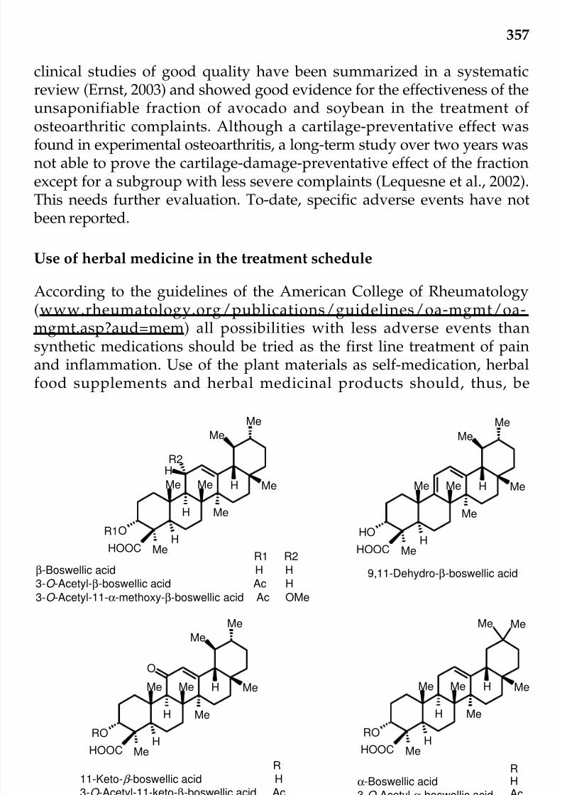
7/23/2019 Novel Therapeutic Agents From Plants
http://slidepdf.com/reader/full/novel-therapeutic-agents-from-plants 368/464
357
clinical studies of good quality have been summarized in a systematicreview (Ernst, 2003) and showed good evidence for the effectiveness of theunsaponifiable fraction of avocado and soybean in the treatment of osteoarthritic complaints. Although a cartilage-preventative effect wasfound in experimental osteoarthritis, a long-term study over two years wasnot able to prove the cartilage-damage-preventative effect of the fractionexcept for a subgroup with less severe complaints (Lequesne et al., 2002).This needs further evaluation. To-date, specific adverse events have not been reported.
Use of herbal medicine in the treatment schedule
According to the guidelines of the American College of Rheumatology(www.rheumatology.org/publications/guidelines/oa-mgmt/oa-mgmt.asp?aud=mem) all possibilities with less adverse events thansynthetic medications should be tried as the first line treatment of painand inflammation. Use of the plant materials as self-medication, herbalfood supplements and herbal medicinal products should, thus, be
Fig. 10. The structures of boswellic acids—co-active compoundsof the active principle in Salai guggal.
Me
H
Me
HMeMe Me
H
O
HOOC
Me
Me
HR2
R1
R1 R2
β-Boswellic acid H H
3-O -Acetyl-β-boswellic acid Ac H
3-O -Acetyl-11-α-methoxy-β-boswellic acid Ac OMe
MeH
Me
HMeMe Me
OH
HOOC
Me
Me
9,11-Dehydro-β-boswellic acid
MeH
Me
HMeMe Me
H
RO
HOOC
Me
Me
O
MeH
Me
HMeMe Me
H
RO
HOOC
Me Me
RHAc
α-Boswellic acid
3-O -Acetyl-α-boswellic acid
R
11-Keto- β -boswellic acid H
3-O -Acetyl-11-keto-β-boswellic acid Ac
© 2009 by Taylor & Francis Group, LLC

7/23/2019 Novel Therapeutic Agents From Plants
http://slidepdf.com/reader/full/novel-therapeutic-agents-from-plants 369/464
358
recommended before synthetic medications are advised. It is suggested thatif no improvement occurs within three to four months intake of a herbalmedication, other herbal preparations or a combination of herbal medicinesshould be tried. So far the evidence of effectiveness of herbal medicinalproducts is strong for preparations from Harpagophytum procumbens andavocado soybean, moderate for ginger and rose hip and, conflicting forwillow bark. For preparations from Salai guggal (Boswellia serrata ROXB.ex COLEBR.), no evidence of effectiveness is available, although theoreti-cally it may alleviate osteoarthritic complaints (Table 2; Chrubasik et al.,2007a). Boswellic acids (Figure 10) have been shown to specifically inhibit5-lipoxygenase and were identified as a part of the active principle. Future
research may provide an improved extract from this herbal medication toimprove osteoarthritic conditions.
Table 2. Number of systematic reviews and their conclusions on theevidence of effectiveness of medicinal product from various herbs
(after Chrubasik et al., 2007a)
No. Evidence
Devil’s claw 5 Strong
Avocado soybean 1 Strong
Ginger 1 Moderate
Rose hip and seed 1 Moderate
Willow bark Moderate
Salai guggal 1 Insufficient
for other preparations 6 Insufficient
Future perspectives
Herbal extracts have been shown to be more potent in alleviating pain andinflammation than individually isolated compounds. In addition they areassociated with less adverse events than isolated/synthetic compounds
probably due to the lower dose in the active principle and the broadermechanism of action of the co-active compounds. Future research shouldfocus on the preparation of improved extracts that concentrate the activeprinciple and free them of inactive compounds especially compounds thatare less well tolerated or those that cause adverse events. Plant culturesmay provide starting materials with higher content of co-active compounds.Aspects of good manufacturing practice and good laboratory practice area matter of course.
Randomized controlled studies according to good clinical practiceneeds to be carried out to increase the body of evidence for the effectiveness
© 2009 by Taylor & Francis Group, LLC

7/23/2019 Novel Therapeutic Agents From Plants
http://slidepdf.com/reader/full/novel-therapeutic-agents-from-plants 370/464
359
of herbal medicines in the treatment of pain and inflammation. Surveillanceand pilot studies should be followed by confirmatory studies that provethe efficacy of the herbal medicinal products. A comparison of herbalmedicinal products in homogenous patient populations may enable aranking of the products in different osteoarthritic conditions.
Currently, there are not sufficient safety data available for any of theherbal medicines for pain and inflammation. Preclinical studies areurgently needed to rule out any harm, especially for chronic conditionwhich requires treatment over longer periods. Likewise, data are scarceand very much needed to assure safety during pregnancy and lactation.
Conclusion
Treatment with herbal medicinal products is another treatment option withless adverse events compared to synthetic medications and should beconsidered as first-line treatment in the sense of the American College of Rheumatology that treatments with lower risks should be preferred. In caseof musculoskeletal pain, three herbal anti-inflammatory options are
available. The first option includes an inexpensive self-treatment with nettleherb (herba urticae), blackcurrant leaf (ribes nigri folium) or raw ginger(zingiberis rhizoma). For pain relief, nettle leaf may be rubbed onto theaffected area or a stew of nettle herb may be consumed. A tea from blackcurrant leaf or freshly squeezed ginger may also help to save onsynthetic analgesic medications.
The second option are food supplements with moderate evidence of effectiveness such as the rose hip powder LitozinR and the γ -linolenic acidcontaining seed oils from borage (Borago officinalis L.), evening primrose(Oenothera biennis L.) or blackcurrant (Ribes nigrum L.). Confirmatory studiesthat prove the effectiveness in the treatment of painful osteoarthritis havenot yet been carried out.
The third option comprises the anti-inflammatory herbal medicinalproducts with good clinical evidence of effectiveness including DoloteffinR,an aqueous extract from devil’s claw ( Harpagophytum procumbens (Burch.)
DC. and the unsaponifiable fraction PiascledineR from avocado andsoybean. For other anti-inflammatory herbal medicinal products, theevidence is conflicting: for the ethanolic extract AssalixR from willow bark(Salix purpurea L. subspecies daphnoides) or insufficient: for the extractKrallendornR from cats’s claw (Uncaria guianensis (AUBLET) J. GMEL.) orfor preparations from Salai guggal (Boswellia serrata).
Confirmatory studies to increase the body of evidence of effectiveness
as well as safety data and research on the mechanism of action are urgentlyneeded for increased treatment acceptance of herbal medications.
© 2009 by Taylor & Francis Group, LLC

7/23/2019 Novel Therapeutic Agents From Plants
http://slidepdf.com/reader/full/novel-therapeutic-agents-from-plants 371/464
360
References
Aguilar, J.L., P. Rojas, A. Marcelo, A. Plaza, R. Bauer, E. Reininger, C.A. Klaasand I. Merfort. 2002. Anti-inflammatory activity of two different extracts of
Uncaria tomentosa (Rubiaceae). Journal of Ethnopharmacology 81: 271-6.Allen-Hall, L., P. Cano, J.T. Arnason, R. Rojas, O. Lock and R.M Lafrenie. 2007.
Treatment of THP-1 cells with Uncaria tomentosa extracts differentiallyregulates the expression if IL-1beta and TNF-alpha. Journal of Ethnopharma-cology 109: 312-7.
Altinel, L., Z.K. Saritas, K.C Kose, K. Pamuk, Y. Aksoy and M. Serteser. 2007.Treatment with unsaponifiable extracts of avocado and soybean increasesTGF-beta1 and TGF-beta2 levels in canine joint fluid. Tohoku Journal of
Experimental Medicine 211: 181-6.Altmann, A., D. Poeckel, L. Fischer, M. Schubert-Zsilavecz, D. Steinhilber and O.
Werz. 2004. Coupling of boswellic acid-induced Ca2+ mobilisation and MAPKactivation to lipid metabolism and peroxide formation in human leucocytes.British Journal of Pharmacology 141: 223-32.
Ammon, H.P., T. Mack, G.B. Singh and H. Safayhi. 1991. Inhibition of leukotrieneB4 formation in rat peritoneal neutrophils by an ethanolic extract of thegum resin exudate of Boswellia serrata. Planta Medica 57: 203-7.
Ammon, H.P., H. Safayhi, T. Mack and J. Sabieraj. 1993. Mechanism of antiinflammatory actions of curcumine and boswellic acids. Journal of Ethnopharmacology 38: 113-9.
Andriamanalijaona, R., H. Benateau, P.E. Barre, K. Boumediene, D. Labbe, J.F.Compere and J.P. Pujol. 2006. Effect of interleukin-1beta on transforminggrowth factor-beta and bone morphogenetic protein-2 expression in humanperiodontal ligament and alveolar bone cells in culture: modulation byavocado and soybean unsaponifiables. Journal of Periodontology 77: 1156-66.
Anonymous, 2003. ESCOP Monographs. Thieme Press, Exeter, Stuttgart, NewYork.
Asnani, V. and R.J. Verma. 2007. Antioxidative effect of rhizome of Zinziberofficinale on paraben-induced lipid peroxidation: an in vitro study. ActaPoloniae Pharmaceutica 64: 35-7.
Au, R.Y., T.K. Al-Talib, A.Y. Au, P.V. Phan and C.G. Frondoza. 2007. Avocadosoybean unsaponifiables (ASU) suppress TNF-alpha, IL-1beta, COX-2, iNOSgene expression, and prostaglandin E(2) and nitric oxide production in
articular chondrocytes and monocyte/macrophages. OsteoarthritisCartilage. 15: 1249-55.
Biegert, C., I. Wagner, R. Ludtke, I. Kotter, C. Lobmuller, I. Gunaydin, K. Taxisand L. Heide. 2004. Efficacy and safety of willow bark extract in thetreatment of osteoarthritis and rheumatoid arthritis: results of 2 randomizeddouble-blind controlled trials. The Journal of Rheumatology 31: 2121-30.
Boje, K., M. Lechtenberg and A. Nahrstedt. 2003. New and known iridoid- andphenylethanoid glycosides from Harpagophytum procumbens and their in
vitro inhibition of human leukocyte elastase. Planta Medica 69: 820-5.Boumediene, K., N. Felisaz, P. Bogdanowicz, P. Galera, G.B. Guillou and J.P. Pujol.1999. Avocado/soya unsaponifiables enhance the expression of transforming© 2009 by Taylor & Francis Group, LLC

7/23/2019 Novel Therapeutic Agents From Plants
http://slidepdf.com/reader/full/novel-therapeutic-agents-from-plants 372/464
361
growth factor beta1 and beta2 in cultured articular chondrocytes. ArthritisRheumatism 42: 148-56.
Cao, Z.F., Z.G. Chen, P. Guo, S.M. Zhang, L.X. Lian, L. Luo and W.M. Hu. 1992.Scavenging effects of ginger on superoxide anion and hydroxyl radical.
Zhongguo Zhong Yao Za Zhi. 18: 750-1, 764.Chrubasik, S. and B. Roufogalis. 2003. Bioequivalence of herbal medicines. New
Zealand J Pharm 53: 39-44.Chrubasik, S., W. Enderlein, R. Bauer and W. Grabner. 1997. Evidence for
antirheumatic effectiveness of stewed herba urticae dioicae in acute arthritis:a pilot study. Phytomedicine 4: 105-108.
Chrubasik, S., E. Eisenberg, E. Balan, T. Weinberger, R. Luzzati and C. Conradt.2000. Treatment of low back pain exacerbations with willow bark extract: a
randomized double-blind study. The American Journal of Medicine 109: 9-14.Chrubasik, S., B. Fiebich, A. Black and S. Pollak. 2002. Treating low back pain
with an extract of Harpagophytum that inhibits cytokine release. European Journal of Anaesthesiology 19: 209.
Chrubasik, S., M. Pittler and B. Roufogalis. 2005a. Zingiberis rhizoma: A compre-hensive review on the ginger effect and efficacy profiles. Phytomedicine 12:684-701.
Chrubasik, S., O. Künzel, J. Thanner, C. Conradt and A. Black. 2005b. A one-year follow-up after a pilot study with Doloteffin R for low back pain.Phytomedicine 11: 470-77.
Chrubasik, C., R.K. Duke and S. Chrubasik. 2006a. The evidence for clinicalefficacy of rose hip and seed: a systematic review. Phytother Res. 20: 1-3.
Chrubasik, J.E., E. Lindhorst, E. Neumann, U. Gerlach, M. Faller-Marquardt, T.Torda, U. Müller-Ladner and S. Chrubasik. 2006b. Potential molecular basisof the chondroprotective effect of Harpagophytum procumbens. Phytomedicine.13: 598-600.
Chrubasik, J.E., B.D. Roufogalis and S. Chrubasik. 2007a. Evidence of effectivenessof herbal anti-inflammatory drugs in the treatment of painful osteoarthritisincluding chronic low back pain. Phytotherapy Research 21: 675-683.
Chrubasik, S., C. Chrubasik, O. Künzel and A. Black. 2007b. Patient-perceived benefit during one year of treatment with DoloteffinR. Phytomedicine 14:371-6.
Chrubasik, C., L. Wiesner, A. Black, U. Müller-Ladner and S. Chrubasik. In press.A one-year survey on the use of a powder from Rosa canina lito in acute
exacerbations of chronic pain. Phytotherapy Research.Costatino, L., G. Ratselli, T. Rossi, M. Bertoldi and A. Albasini. 1993. Activiteantilipoperoxydante e’extraits polyphenoliques de Ribes nigrum L. PlantesMedicinales et Phytotherapy 26: 207-14.
DeLuca, P., R.G. Rossetti, C. Alavian, P. Karim and R.B. Zurier. 1999. Effects of gammalinolenic acid on interleukin-1 beta and tumor necrosis factor-alphasecretion by stimulated human peripheral blood monocytes: studies in vitroand in vivo. Journal of Investigative Medicine 47: 246-50.
Declume, C. 1989. Anti-inflammatory evaluation of a hydroalcoholic extract of black currant leaves (Ribes nigrum). Journal of Ethnopharmacology 27: 91-8.© 2009 by Taylor & Francis Group, LLC
7/23/2019 Novel Therapeutic Agents From Plants
http://slidepdf.com/reader/full/novel-therapeutic-agents-from-plants 373/464
362
Dobson, G. 2002. Leaf lipids of Ribes nigrum: a plant containing 16:3, alpha-18:3,gamma-18:3 and 18:4 fatty acids. Biochemical Society Transactions 28: 583-6.
Dooper, M.M., B. van Riel, Y.M. Graus and L. M’Rabet. 2003. Dihomo-gamma-linolenic acid inhibits tumor necrosis factor-alpha production by human
leucocytes independently of cyclooxygenase activity. Immunology 110: 348-57.
El Haouari, M., M. Bnouham, M. Bendahou, M. Aziz, A. Ziyyat, A. Legssyer andH. Mekhfi. 2006. Inhibition of rat platelet aggregation by Urtica dioica leavesextracts. Phytotherapy Research 20: 568-72.
Ernst E. and S. Chrubasik. 2000. Phytoanalgesics for rheumatic conditions: asystematic review of randomized placebo-controlled, double-blind trials.Rheumatic Disease Clinics 26: 13-27.
Ernst, E. 2003. Avocado-soybean unsaponifiables (ASU) for osteoarthritis—asystematic review. Clinical Rheumatology 22: 285-8.Fiebich, B.L., M. Heinrich, K.O. Hiller and N. Kammerer. 2001. Inhibition of TNF-
alpha synthesis in LPS-stimulated primary human monocytes by Harpagophytum extract Stei Hap 69. Phytomedicine 8: 28-30.
Fiebich, B.L. and S. Chrubasik. 2004. Effects of an ethanolic Salix extract on therelease of selected inflammatory mediators in vitro. Phytomedicine 11: 135-8.
Frondoza, C.G., A. Sohrabi, A. Polotsky, P.V. Phan, D.S. Hungerford and L.Lindmark. 2004. An in vitro screening assay for inhibitors of proinflamma-tory mediators in herbal extracts using human synoviocyte cultures. In VitroCellular & Developmental Biology—Animal 40: 95-101.
Furse, R.K, R.G. Rossetti and R.B. Zurier. 2001. Gammalinolenic acid, anunsaturated fatty acid with anti-inflammatory properties, blocks amplificationof IL-1 beta production by human monocytes. Journal of Immunology 67:490-6.
Furse, R.K., R.G. Rossetti, C.M. Seiler and R.B. Zurier. 2002. Oral administration
of gammalinolenic acid, an unsaturated fatty acid with anti-inflammatoryproperties, modulates interleukin-1beta production by human monocytes. Journal of Clinical Immunology 22: 83-91.
Gabay, O., M. Gosset, A. Levy, C. Salvat, C. Sanchez, A. Pigenet, A. Sautet, C. Jacques and F. Berenbaum. 2008. Stress-induced signaling pathways in hyalinchondrocytes: inhibition by Avocado-Soybean Unsaponifiables (ASU).Osteoarthritis Cartilage 16: 373-384.
Garbacki, N., L. Angenot, C. Bassleer, J. Damas and M. Tits. 2002. Effects of
prodelphinidins isolated from Ribes nigrum on chondrocyte metabolism andCOX activity. Naunyn Schmiedebergs Archives of Pharmacology 365: 434-41.
Garbacki, N., M. Kinet, B. Nusgens, D. Desmecht and J. Damas. 2005.Proanthocyanidins, from Ribes nigrum leaves, reduce endothelial adhesionmolecules ICAM-1 and VCAM-1. Journal of Inflammation (Lond) 2: 9.
Gayathri, B., N. Manjula, K.S. Vinaykumar, B.S. Lakshmi and Balakrishnan. 2007.Pure compound from Boswellia serrata extract exhibits anti-inflammatoryproperty in human PBMCs and mouse macrophages through inhibition of TNF-alpha, IL-1beta, NO and MAP kinases. International Immunopharma-cology 7: 473-82.© 2009 by Taylor & Francis Group, LLC

7/23/2019 Novel Therapeutic Agents From Plants
http://slidepdf.com/reader/full/novel-therapeutic-agents-from-plants 374/464
363
Glaser, T., S. Winter, P. Groscurth, H. Safayhi, E.R. Sailer, H.P. Ammon, M. Schabetand M. Weller. 1999. Boswellic acids and malignant glioma: induction of apoptosis but no modulation of drug sensitivity. British Journal of Cancer80: 756-65.
Goncalves, C., T. Dinis and M.T. Batista. 2005. Antioxidant properties of proanthocyanidins of Uncaria tomentosa bark decoction: a mechanism foranti-inflammatory activity. Phytochemistry 66: 89-98.
Harput, U.S., I. Saracoglu and Y. Ogihara. 2005. Stimulation of lymphocyteproliferation and inhibition of nitric oxide production by aqueous Urtica dioicaextract. Phytotherapy Research 19: 346-8.
Henrotin, Y.E., A.H. Labasse, J.M. Jaspar, D.D. De Groote, S.X. Zheng, G.B.Guillou and J.Y. Reginster. 1998. Effects of three avocado/soybean
unsaponifiable mixtures on metalloproteinases, cytokines and prostaglandinE2 production by human articular chondrocytes. Clinical Rheumatology 17:31-9.
Henrotin, Y.E., C. Sanchez, M.A. Deberg, N. Piccardi, G.B. Guillou, P. Msika and J.Y. Reginster. 2003. Avocado/soybean unsaponifiables increase aggrecansynthesis and reduce catabolic and proinflammatory mediator production by human osteoarthritic chondrocytes. Journal of Rheumatology 30: 1825-34.
Henrotin, Y.E., M.A. Deberg, J.M. Crielaard, N. Piccardi, P. Msika and C. Sanchez.2006. Avocado/soybean unsaponifiables prevent the inhibitory effect of osteoarthritic subchondral osteoblasts on aggrecan and type II collagensynthesis by chondrocytes. Journal of Rheumatology 33: 1668-78.
Huang, T.H., V.H. Tran, R.K. Duke, S. Tan, S. Chrubasik, B.D. Roufogalis andC.C. Duke. 2006. Harpagoside suppresses lipopolysaccharide-induced iNOSand COX-2 expression through inhibition of NF-kB activation. Journal of Ethnopharmacology 104: 149-55.
Iverson, L., K. Fogh and K. Kragbelle. 1992. Effect of dihomogammalinolenic
acid and its 15-lipoxygenase metabolite on eicosanoid metabolism by humanmononuclear leukocytes in vitro: Selective inhibition of the 5-lipoxygenasepathway. Archives of Dermatological Research 284: 222-226.
Jäger, A.K., I.M. Eldeen and J. van Staden. 2007. COX-1 and -2 activity of rosehip. Phytotherapy Research 21: 1251-1252.
Jiang, W.G., M.C. Puntis, D.F. C. Horrobin, M.B. and Hallett. 1996. Inhibition of neutrophil respiratory burst and cytokine priming by gamma-linolenic acid.British Journal of Surgery 83: 659-64.
Jürgensen, S., S. Dalbo, P. Angers, A.R. Santos and R.M. Ribeiro-do-Valle. 2005.Involvement of 5-HT2 receptors in the antinociceptive effect of Uncariatomentosa. Pharmacology Biochemistry and Behavior 81: 466-77.
Kahkonen, M.P., A.I. Hopia, H.J. Vuorela, J.P. Rauha, K. Pihlaja, T.S. Kujala andM. Heinonen. 1999. Antioxidant activity of plant extracts containing phenoliccompounds. Journal of Agriculture and Food Chemistry 47: 3954-62.
Kanter, M., O. Coskun and M. Budancamanak. 2005. Hepatoprotective effects of Nigella sativa L and Urtica dioica L on lipid peroxidation, antioxidant enzymesystems and liver enzymes in carbon tetrachloride-treated rats. World Journal of Gastroenterology 11: 6684-8.
Kaszkin, M., K.F. Beck, E. Koch, C. Erdelmeier, S. Kusch, J. Pfeilschifter and D.© 2009 by Taylor & Francis Group, LLC

7/23/2019 Novel Therapeutic Agents From Plants
http://slidepdf.com/reader/full/novel-therapeutic-agents-from-plants 375/464
364
Loew. 2004. Downregulation of iNOS expression in rat mesangial cells byspecial extracts of Harpagophytum procumbens derives from harpagoside-dependent and independent effects. Phytomedicine 11: 585-95.
Khayyal, M.T., M.A. El-Ghazaly, D.M. Abdallah, S.N. Okpanyi, O. Kelber and D.
Weiser. 2005. Mechanisms involved in the anti-inflammatory effect of astandardized willow bark extract. Arzneimittelforschung 55: 677-87.
Kim, O.K., A. Murakami, D. Takahashi, Y. Nakamura, K. Torikai, H.W. Kim andH. Ohigashi. 2000. An avocado constituent, persenone A, suppressesexpression of inducible forms of nitric oxide synthase and cyclooxygenasein macrophages, and hydrogen peroxide generation in mouse skin.Bioscience, Biotechnology and Biochemistry 64: 2504-7.
Kim, S.O., K.S. Chun, J.K. Kundu and Y.J. Surh. 2004. Inhibitory effects of [6]-
gingerol on PMA-induced COX-2 expression and activation of NF-kappaBand p38 MAPK in mouse skin. Biofactors 21: 27-31.Kiuchi, F., S. Iwakami, M. Shibuya, F. Hanaoka and U. Sankawa. 1992. Inhibition
of prostaglandin and leukotriene biosynthesis by gingerols and diarylhepta-noids. Chemical & Pharmaceutical Bulletin (Tokyo) 40: 387-91.
Klingelhöfer, S., B. Obertreis, S. Quast and B. Behnke. 1999. Antirheumatic effectof IDS 23, a stinging nettle leaf extract, on in vitro expression of T helpercytokines. Journal of Rheumatology 26: 2517-22.
Krivoy, N., E. Pavlotzky, S. Chrubasik, E. Eisenberg and G. Brooks. 2001. Effectof Salicis Cortex extract on human platelet aggregation. Planta Medica 67:209-213.
Kundu, J.K., K.S. Mossanda, H.K. Na and Y.J. Surh. 2005. Inhibitory effects of the extracts of Sutherlandia frutescens (L.) R. Br. and Harpagophytum procumbens DC. on phorbol ester-induced COX-2 expression in mouse skin:AP-1 and CREB as potential upstream targets. Cancer Letter 218: 21-31.
Kuo, P.C., A.G. Damu, C.Y. Cherng, J.F. Jeng, C.M. Teng, E.J. Lee and T.S. Wu.2005. Isolation of a natural antioxidant, dehydrozingerone from Zingiber
officinale and synthesis of its analogues for recognition of effective antioxidantand antityrosinase agents. Archives of Pharmacal Research 28: 518-28.
Kuppsamy, U.R., H.E. Khoo and N.P. Das. 1990. Structure-activity studies of flavonoids as inhibitors of hyaluronidase. Biochemical pharmacology 140:397-401.
Kut, C., A. Assoumou, M. Dridi, M. Bonnefoix, B. Gogly, B. Pellat, G.B. Guillouand G. Godeau. 1998. Morphometric analysis of human gingival elastic fibresdegradation by human leukocyte elastase protective effect of avocado and
soybean unsaponifiables (ASU). Pathologie-Biologie (Paris) 46: 571-6.Kut-Lasserre, C., C.C. Miller, A.L. Ejeil, B. Gogly, M. Dridi, N. Piccardi, B. Guillou,B. Pellat and G. Godeau. 2001. Effect of avocado and soybean unsaponifiableson gelatinase A (MMP-2), stromelysin 1 (MMP-3), and tissue inhibitors of matrix metalloproteinase (TIMP-1 and TIMP-2) secretion by humanfibroblasts in culture. Journal of Periodontology 72: 1685-94.
Lesquesne, M., F. Maheu, C. Cadet and R.L. Dreiser. 2002. Structural effect of avocado/soybean unsaponifiables on joint space loss in osteoarthritis of thehip. Arthritis & Rheumatism 47: 50-8.
Lantz, R.C., G.J. Chen, M. Sarihan, A.M. Sólyom, S.D. Jolad and B.N.Timmermann. 2007. The effect of extracts from ginger rhizome on© 2009 by Taylor & Francis Group, LLC

7/23/2019 Novel Therapeutic Agents From Plants
http://slidepdf.com/reader/full/novel-therapeutic-agents-from-plants 376/464
365
inflammatory mediator production. Phytomedicine 14: 123-8.Loew, D., J. Mollerfeld, A. Schrodter, S. Puttkammer and M. Kaszkin. 2001.
Investigations on the pharmacokinetic properties of Harpagophytum extractsand their effects on eicosanoid biosynthesis in vitro and ex vivo. Clinical
Pharmacology & Therapeutics 69: 356-64.Mahomed I.M. and J.A. Ojewole. 2006. Anticonvulsant activity of Harpagophytum
procumbens DC [Pedaliaceae] secondary root aqueous extract in mice. BrainResearch Bulletin 69: 57-62.
Mauviel, A., G. Loyau and J.P. Pujol. 1991. Effect of unsaponifiable extracts of avocado and soybean (Piascledine) on the collagenolytic action of culturesof human rheumatoid synoviocytes and rabbit articular chondrocytestreated with interleukin-1. Revue du Rhumatisme et des Maladies Osteoartic
58: 241-5.Melzig, M.F., B. Löser and S. Ciesielski. 2001. Inhibition of neutrophil elastaseactivity by phenolic compounds from plants. Pharmazie. 56: 967-70.
Miller, M.J., S. Ahmed, P. Bobrowski and T.M. Haqqi. 2006. The chrondoprotectiveactions of a natural product are associated with the activation of IGF-1production by human chondrocytes despite the presence of IL-1beta. BMCComplementary and Alternative Medicine 7; 6: 13.
Mur, E., F. Hartig, G. Eibl and M. Schirmer. 2002. Randomized double blind trialof an extract from the pentacyclic alkaloid-chemotype of Uncaria tomentosafor the treatment of rheumatoid arthritis. Journal of Rheumatology 29: 678-81.
Na, H.K., K.S. Mossanda, J.Y. Lee and Y.J. Surh. 2004. Inhibition of phorbol ester-induced COX-2 expression by some edible African plants. Biofactors 21(1-4): 149-53.
Nurtjahja-Tjendraputra, E., A.J. Ammit, B.D. Roufogalis, V.H. Tran and C.C. Duke.2003. Effective anti-platelet and COX-1 enzyme inhibitors from pungentconstituents of ginger. Thrombosis Research 111: 259-65.
Obertreis, B., K. Giller, T. Teucher, B. Behnke and H. Schmitz. 1996a. Anti-inflammatory effect of Urtica dioica folia extract in comparison to caffeic malicacid. Arzneimittelforschung 46: 52-6.
Obertreis, B., T. Ruttkowski, T. Teucher, B. Behnke and H. Schmitz. 1996b. Ex-vivo in-vitro inhibition of lipopolysaccharide stimulated tumor necrosisfactor-alpha and interleukin-1 beta secretion in human whole blood byextractum Urticae dioicae foliorum. Arzneimittelforschung 46: 389-94.
Ozen, T. and H. Korkmaz. 2003. Modulatory effect of Urtica dioica L. (Urticaceae)
leaf extract on biotransformation enzyme systems, antioxidant enzymes,lactate dehydrogenase and lipid peroxidation in mice. Phytomedicine 10:405-15.
Pham, H.C., N. Ifansyah, R. Chahine, A. Mounayar-Chalfoun, J. Gleye and C.Moulis. 1986. Comparative effects of total flavonoids extracted from Ribesnigrum leaves, rutin and isoquercetrin on biosynthesis and release of prostaglandins in the ex-vivo rabbit heart. Prostaglandins, Leukotrienes andMedicine 22: 295-300.
Pilarski, R., H. Zielinski, D. Ciesiolka and K. Gulewicz. 2006. Antioxidant activityof ethanolic and aqueous extracts of Uncaria tomentosa (Willd.) DC. Journalof Ethnopharmacology 104: 18-23.© 2009 by Taylor & Francis Group, LLC

7/23/2019 Novel Therapeutic Agents From Plants
http://slidepdf.com/reader/full/novel-therapeutic-agents-from-plants 377/464
366
Poeckel, D., L. Tausch, N. Kather, J. Jauch and O. Werz. 2006. Boswellic acidsstimulate arachidonic acid release and 12-lipoxygenase activity in humanplatelets independent of Ca2+ and differentially interact with platelet-type12-lipoxygenase. Molecular Pharmacology 70: 1071-8.
Rennert, B. and M.F. Melzig. 2002. Free fatty acids inhibit the activity of Clostridium histolyticum collagenase and human neutrophil elastase. PlantaMedica 68: 767-9.
Riehemann, K., B. Behnke and K. Schulze-Osthoff. 1999. Plant extracts fromstinging nettle (Urtica dioica), an antirheumatic remedy, inhibit theproinflammatory transcription factor NF-kappaB. FEBS Letters 442: 89-94.
Rohnert, U., W. Schneider and E.F. Elster. 1998. Superoxide-dependent andindependent nitrite formation from hydroxylamine: inhibition by plant
extracts. Journal of Biosciences 53: 241-49.Rossnagel, K., S. Roll, A. Wagner, O. Mune, J. Erlendsson, A. Kharazmi, H.Sörensen, S.N. Willich and K. Winther. 2007. Can patients with rheumatoidarthritis benefit from the herbal remedy rose-hip? Annals of the RheumaticDiseases 66 Supplement 2: 603.
Roy, S., S. Khanna, H. Shah, C. Rink, C. Phillips, H. Preuss, G.V. Subbaraju, G.Trimurtulu, A.V. Krishnaraju, M. Bagchi, D. Bagchi and C.K. Sen. 2005.Human genome screen to identify the genetic basis of the anti-inflammatoryeffects of Boswellia in microvascular endothelial cells. DNA Cell Biology 24:244-55.
Roy, S., S. Khanna, A.V. Krishnaraju, G.V. Subbaraju. T. Yasmin, D. Bagchi andC.K. Sen. 2006. Regulation of vascular responses to inflammation: induciblematrix metalloproteinase-3 expression in human microvascular endothelialcells is sensitive to antiinflammatory Boswellia. Antioxidants and RedoxSignaling 8: 653-60.
Safayhi, H., B. Rall, E.R. Sailer and H.P. Ammon. 1997. Inhibition by boswellicacids of human leukocyte elastase. The Journal of Pharmacology and
Experimental Therapeutics 281: 460-3.Sandoval, M., R.M. Charbonnet, N.N. Okuhama, J. Roberts, Z. Krenova, A.M.
Trentacosti and M.J. Miller. 2000. Cat’s claw inhibits TNFalpha productionand scavenges free radicals: role in cytoprotection. Free Radical Biology &Medicine 29: 71-8.
Sandoval, M., N.N. Okuhama, X.J. Zhang, L.A. Condezo, J. Lao, F.M. Angeles,R.A. Musah, P. Bobrowski and M.J. Miller. 2002. Anti-inflammatory andantioxidant activities of cat’s claw (Uncaria tomentosa and Uncaria guianensis)
are independent of their alkaloid content. Phytomedicine 9: 325-37.Schmid, B. 1998. Behandlung von Cox- und Gonarthrosen mit einemTrockenextrakt aus Salix purpurea x daphnoides, Thesis. University of Tübingen, Tübingen, Germany.
Schulze-Tanzil, G., S.P. de, B. Behnke, S. Klingelhoefer, A. Scheid and M. Shakibaei.2002. Effects of the antirheumatic remedy hox alpha—a new stinging nettleleaf extract—on matrix metalloproteinases in human chondrocytes in vitro.Histology and Histopathology 17: 477-85.
Siemoneit, U., B. Hofmann, N. Kather, T. Lamkemeyer, J. Madlung, L. Franke,G. Schneider, J. Jauch, D. Poeckel and O. Werz. 2008. Identification andfunctional analysis of cyclooxygenase-1 as a molecular target of boswellic© 2009 by Taylor & Francis Group, LLC
7/23/2019 Novel Therapeutic Agents From Plants
http://slidepdf.com/reader/full/novel-therapeutic-agents-from-plants 378/464
367
acids. Biochemical Pharmacology 75: 503-513.Sporer, F. and S. Chrubasik. 1999. Präparate aus der Teufelskralle ( Harpagophytum
procumbens). Zeitschrift Phytotherapie 20: 235-6.Takada, Y., H. Ichikawa, V. Badmaev and B.B. Aggarwal. 2006. Acetyl-11-keto-
beta-boswellic acid potentiates apoptosis, inhibits invasion, and abolishesosteoclastogenesis by suppressing NF-kappa B and NF-kappa B-regulatedgene expression. Journal of Immunology 176: 3127-40.
Teucher, T., B. Obertreis, T. Ruttkowski and H. Schmitz. 1996. Cytokine secretionin whole blood of healthy subjects following oral administration of Urticadioica L. plant extract. Arzneimittelforschung 46: 906-10.
Tjendraputra, E., V.H. Tran, D. Liu-Brennan, B.D. Roufogalis and C.C. Duke. 2001.Effect of ginger constituents and synthetic analogues on cyclooxy- genase-2
enzyme in intact cells. Bioorganic Chemistry 29: 156-63.Tsukahara, K., H. Nakagawa, S. Moriwaki, Y. Takema, T. Fujimura and G.Imokawa. 2006. Inhibition of ultraviolet-B-induced wrinkle formation by anelastase-inhibiting herbal extract: implication for the mechanism underlyingelastase-associated wrinkles. International Journal of Dermatology 45: 460-8.
Vlachojannis, J., B. Roufogalis and S. Chrubasik. 2008. Systematic Review on thesafety of Harpagophytum preparations for osteoarthritic and low back pain.Phytotherapy Research 22: 149-52.
Wang, C.C., L.G. Chen, L.T. Lee and L.L. Yang. 2003. Effects of 6-gingerol, anantioxidant from ginger, on inducing apoptosis in human leukemic HL-60cells. In Vivo 17: 641-5.
Wenzig, E.M., U. Widowitz, O. Kunert, S. Chrubasik, F. Bucar, E. Knauder and R.Bauer (in press). Phytochemichal composition and in vitro pharmacologicalactivity of two rose hip (Rosa canina L.) preparations. Phytomedicine.
Wu, H., D.J. Ye, Y.Z. Zhao and S.L. Wang. 1993. Effect of different preprations of ginger on blood coagulation time in mice. Zhongguo Zhon Yao Za Zhi 18:
147-9.Wurm, G., J. Baumann and U. Geres. 1982. Beeinflussung des Arachidon-
säurestoffwechsels durch Flavonoide. Deutsche Apotheker Zeitung 122:2062-8.
© 2009 by Taylor & Francis Group, LLC

7/23/2019 Novel Therapeutic Agents From Plants
http://slidepdf.com/reader/full/novel-therapeutic-agents-from-plants 379/464
Introduction
For millions of years nature has involved in chemical evolution through anatural combinatorial synthesis. Plants are very interesting and usefulsources of molecules for the discovery of new medicinal products.
One of the most important fundamental objectives of metabolicengineering or metabolomics is the augmentation of cellular phenotype,
for instance metabolite over-production, by the introduction of geneticcontrols.
Metabolic Engineering for theFabrications of PharmaceuticallyCentral Metabolites fromMicroorganisms and Higher Plants13Mahmud Tareq Hassan Khan1, 2, * and
Arjumand Ather3, 4
1PhD School of Molecular and Structural Biology, andDepartment of Pharmacology, Institute of Medical Biology,University of Tromsø, Tromsø 9037, Norway.
2Pharmacology Research Lab., Faculty of Pharmaceutical
Sciences, University of Science & Technology, Chittagong,Bangladesh.3Plant Biotechnology Division, International Center for
Chemical and Biological Sciences, University of Karachi,Karachi, Pakistan.
4Present address: Institute for Chemistry, University of Tromsø, Tromsø 9037, Norway.
*Corresponding address: PhD School of Molecular and
Structural Biology, and Department of Pharmacology,Institute of Medical Biology, University of Tromsø, 9037Tromsø, Norway. Tel.: (Off): +47 776 46755; Cell: +47 97794171; Fax: +47 776 45310; E-mail: [email protected]; [email protected]
© 2009 by Taylor & Francis Group, LLC

7/23/2019 Novel Therapeutic Agents From Plants
http://slidepdf.com/reader/full/novel-therapeutic-agents-from-plants 380/464
369
The era of recombinant approaches to metabolic engineering of naturalproducts has a very definable beginning.
The completion of sequencing of the several genomes including the
human genome has resulted in the increased number of availability of genes for metabolic engineering. In parallel, increased number of databasesand in silico (bioinformatic) tools to handle these genomes even in a largescale, also help to develop more precisely the metabolomics.
In this chapter some general and technical aspects of metabolomicsare reviwed in the framework of designing novel leads or producing largeamounts of pharmaceutically as well as other industrially importantcompounds, which are complicated to manufacture using usual syntheticapproaches and less amount present in nature. Recent advancements inthe field of metabolomics are also reviewed.
Metabolic engineering has proven to be a precious instrument for thelarge production of several pharmaceutically as well as industriallyrelevant molecules from microorganisms as well from higher plants.Current development of studies employing transgenic and/or recombinanttechnologies have opened up opportunities of the metabolic engineering
of biosynthetic pathways, leading to the growth of manufacturing high-value secondary metabolites, even at the commercial level.
By definition ‘metabolomics’ is the “systematic study of the uniquechemical fingerprints that specific cellular processes leave behind”—specifically, the study of their small-molecule metabolite profiles (Daviss,2005). The word ‘metabolome’ corresponds to the set of all metabolites ina particular living organism; which are the final products (e.g., secondary
metabolites, metabolic intermediates, hormones, proteins, or any signalingmolecules, etc.) of its’ gene expression. Although the messenger RNA(mRNA) gene expression data and proteomic analyses do not tell thecomplete chronicle of what possibly may be occurring in a cell, metabolicprofiling is able to offer an immediate ‘snapshot’ of the physiology of thatparticular cell. One of the most important challenges of systems biology isto assimilate ‘proteomics’, ‘transcriptomics’, and ‘metabolomics’information to present a further inclusive representation of the whole living
organisms. The word metabonomics’ is also used, particularly in thecontext of drug toxicity assessment. There is some disagreement over theexact differences between ‘metabolomics’ and ‘metabonomics’ ; in general,the term ‘metabolomics’ is more commonly used (Anonymous, 2005).
The chemical evolution of nature can be regarded as a naturalcombinatorial synthesis for millions of years. During this period, higherplants evolved natural products with remarkable pharmacological
activities. Compounds obtained from natural sources such as micro-organisms and plants make up as much as 40% of the drugs in current
© 2009 by Taylor & Francis Group, LLC

7/23/2019 Novel Therapeutic Agents From Plants
http://slidepdf.com/reader/full/novel-therapeutic-agents-from-plants 381/464
370
use, either directly or through synthetic modification, and have a prominentplace in the history of drug discovery (Hutchinson, 1998).
Plants manufacture a huge array of pharmaceutically significant
molecules. Many of these are derived from plants which are complicatedto cultivate, or grow exceptionally slowly. In the past there has been acomprehensive study for the potential to manufacture these kinds of mole-cules by using different techniques of metabolomics, like cell culture, etc.
During the molecular evolution, plants have developed very efficientapproaches to magnetize pollination or seed-dispersing animals (such as,
birds or insects), for shielding themselves against UV-light and fordefending themselves against herbivores, microorganisms and otherplants. In natural growth conditions, plants compete for space, light, waterand nutrients. The major strategies used are based on the production of secondary metabolites, including, essential oils, which are fragrantproducts active either as attractant for pollinations or as a repellent. Thepollination is also favoured by colourants like molecules such as,flavonoids, anthocyanins and carotenoids. Flavonoids are also involvedin the absorption of UV-radiation (Meijkamp et al., 1999) and in many
defensive processes.The term ‘metabolic engineering’ coined by J.E. Bailey (1991),
exemplifies a broad array of manipulations and experimental advances toimprove the productivity of a desired metabolite by an organism (Strohl,2001). Included under the banner of metabolic engineering are examplesof improvement in productivity and/or yield, improvement of substrateuptake, broadening the substrate range of an organism, improvement of
the suitability of an organism in fermentations (i.e., ‘fitness’ ), analysis and
Fig. 1. Different tools and steps involved in the metabolic andreverse metabolic engineering.
© 2009 by Taylor & Francis Group, LLC

7/23/2019 Novel Therapeutic Agents From Plants
http://slidepdf.com/reader/full/novel-therapeutic-agents-from-plants 382/464
371
modification of metabolic flux, elimination of metabolic overflow,elimination of unwanted or competing metabolic pathways, improvementof the organism to resist noxious conditions such as solvents, and so forth(Stephanopoulos, 1999).
The very different varieties of biochemical pathways and productionof secondary metabolites is the basis for the well-known feature of plants,which are a tremendous source for isolation of molecules and of greatinterest for the discovery of new medicinal agents, considered less riskythan those of microbial or animal origin (Shanks and Morgan, 1999).However, it should be pointed out that the beneficial medicinal effects of plant material might be also due to the endogenous combination of several
secondary products. On the other hand, approaches involved in geneticengineering of medicinal plants have been ascribed to increase yield of pharma-ceutically important single secondary metabolites (Briskin, 2000).
Table 1 presents some plant-derived pharmaceutical products,including the source plant species, their price in US$ and their therapeuticindications.
The plant molecular biology has evolved since long time ago from
studies of single gene functions useful to extrapolate basic principles in avery simplistic manner, to more analysis of processes known to beregulated by a multitude of tightly interconnected and even interdependentfactors. By definition, the modern plant molecular science, and in particularits applied component plant biotechnology requires a holistic, multi-disciplinary and comprehensive approach to achieve its objective (Sasakiand Christou, 2004).
The enhanced classification of the biomolecular complexes, branching,and the knowledge of the interactive metabolic pathways of manyorganisms is feasible with advances in genome sequencing, transcriptomeand proteome analysis and may provide more suitable starting platformsfor engineering of secondary metabolite production. Indeed, it is knowledgeand exploitation of the dynamic nature of gene expression and enzymeregulation that may prove to be highly useful in metabolic and combi-natorial engineering approaches. In combination with additional tech-
niques for metabolic pathway modification, such as directed evolution, thegoals of improved secondary metabolite production is the employment of novel products which can be readily achieved (Mijts and Schmidt-Dannert,2003).
Over the past six years, several review papers have been published onthe subject of metabolic engineering of natural products (Strohl, 2001).
In this chapter some of the recent advancements in this field and the
general and technical aspects of metabolomics (metabolic engineering) arepresented in the context of designing novel lead molecules or to produce
© 2009 by Taylor & Francis Group, LLC

7/23/2019 Novel Therapeutic Agents From Plants
http://slidepdf.com/reader/full/novel-therapeutic-agents-from-plants 383/464
372
Table 1. Pharmaceutical products from natural sources (mainly from plants)(modified from Rao and Ravishankar 2002)
Plants Compounds Therapeutic uses Price
(US$/kg)
Aconitum spp. Acinitine Antiarhythmic n/aa
Artemisia annua Artemisinin Antimalarial 400
Coptis japonica Berberine Intestinal ailment 3,250
Capsicum frutescens Capsaicin Counter-irritant 750
Camptotheca acuminata Camptothecin Antitumor 432,000
Castanospermum australe Castanospermine Glycoside inhibitor n/aa
Catharenthus roseus Vincristine Antileukemic 2,000,000
Catharenthus roseus Vinblastine Antileukemic 1,000,000
Catharenthus roseus Ajmalicine Antihypertensive 37,000
Cinchona ledgeriana Quinine Antimalarial 500
Colchium autumnale Colchicine Antitumor 35,000
Coleus forskolii Forskolin Bronchial asthma n/aa
Digitalis lanata Digoxin Cardiac stimulant 3,000
Dioscorea deltoidea Diosgenin Steroidal precursor 1,000
Lithospermum erythrorhizon Shikonin Antibacterial 4,500
Orchrosia elliptica Ellipticine Antitumor 240,000
Papavar somniferum Codeine Sedative 17,000
Papavar somniferum Morphine Sedative 340,000
Panax ginseng Ginsenosides Health tonic n/aa
Podophyllum petalum Podophyllotoxin Antitumor n/aa
Sanguinaria canadensis Sanguinarine Antiplaque 4,800
Taxus brevifolia Taxol Anticancer 600,000
Notes: anot available.
huge amounts of pharmaceuticals as well as other industrially importantcompounds which are difficult to develop even with usual synthetic
approaches.
Reasons for considering biotechnology in metabolic engineering
The biotechnological approaches generally consist of growth of cells,tissues, organs or the entire organisms by growing them in vitro and geneticmanipulation of them to get desired compounds. The development of the
micropropagation methods for a number of medicinal plant species has been already reported and needs to be extended (Naik, 1998).
© 2009 by Taylor & Francis Group, LLC

7/23/2019 Novel Therapeutic Agents From Plants
http://slidepdf.com/reader/full/novel-therapeutic-agents-from-plants 384/464
373
Cryopreservation of cells is an area of great interest in the preservationof medicinal plants and it has already been used in several plant species.The development and adoption of plant cell culture and organ culturemethods have led to the production of plant products on a large scale (Raoand Ravishankar, 2002).
The fundamental ambition of metabolic engineering is the enhance-ment of cellular phenotype, such as metabolite over-production by theintroduction of genetic controls. At this point, metabolic engineeringendeavours have considered the properties of the overall metabolic network,in sharp distinction to the single-gene focus that characterizes emblematicapplications of genetic engineering, as well as the biotechnology (Alper et
al., 2005).Table 2 shows different issues related to the biotechnological advances
in the usual projects found on plant-derived secondary metabolites. Table3 presents some of the classes and sub-classes of compounds isolated andreported so far from the higher plant cultures in vitro.
Table 2. Partial list of the most important issues of biotechnology
in the production of secondary metabolites from plants(from Rao and Ravishankar, 2002)
Main issues Related issues
Plant cells, tissues and organ cultures Cell culture
Shoot culture
Root culture
Scale-up of cultures
Transgenic plants/organisms Metabolic engineering
Heterologous expression
Generation of molecularcomplexes
Micro-propagation of medicinal plants Endangered plants
High-yielding varieties
Metabolically engineered plants
Newer sources Algae
Other photosynthetic marine forms
Safety considerations Propagation in the cell environment
Effects on biodiversityEffects onhealth
© 2009 by Taylor & Francis Group, LLC

7/23/2019 Novel Therapeutic Agents From Plants
http://slidepdf.com/reader/full/novel-therapeutic-agents-from-plants 385/464
374
Table 3. Examples of the chemical classes and sub-classes of compounds isolated from higher plant cultures in vitro
(modified from Stockigt et al., 1995)
Chemical classes of compounds
Phenylpropanoids Alkaloids Terpenoids Quinones Steroids
Anthocyanins Acridines Monoterpenes Anthro- Cardiacquinones glycosides
Coumarins Betalaines Sesquiterpenes Benzo- Pregneno-quinones lone
derivatives
Flavonoids Quinolizidines Diterpenes Naphtho-quinones
Hydroxy- Furono- Triterpenescinnamoyl quinonesderivatives
Isoflavonoids Harringtonines Carotenes
Lignans Isoquinolines
Phenolenones Indoles
Proanthocyanidins Purines
Stilbenes Pyridines
Tanins Tropanealkaloids
Major issues and new trends in metabolic engineering
Genetic modification by plant transformation allows stable alterations of the biochemical processes involved in important traits such as yield,nutritional quality and resistance to diseases. The achievements inmodification of secondary metabolism in plants have been reviewed,including alternative approaches to change the secondary metabolism(Hallard, 2000).
There are at least three distinct uses of metabolic engineering(metabolomics) in plants:
1. Increase or decrease of the amount of metabolites and productionof ‘novel’ compounds;
2. Improving quality traits of food and fodder or alter traits of horticultural plants;
3. Increasing the resistance of plants against pest and diseases.
1. In the first strategy of metabolomics the aim is to increase the levelof certain compounds, like medicinal products, colourants, flavours and
© 2009 by Taylor & Francis Group, LLC

7/23/2019 Novel Therapeutic Agents From Plants
http://slidepdf.com/reader/full/novel-therapeutic-agents-from-plants 386/464
375
fragrances, for economically important production. For example,strictosidine synthase (Str) has been over-expressed in Catharanthus roseusleading to the production of 200 mg/ml terpenoid indole alkaloids (Canelet al., 1998). Another interesting example is that of the stable transgenichairy root clone of Hyoscymus muticus, over-expressing the hyscyamine 6β-hydroxylase (H6βH); this transformation ultimately leads to the produc-tion of 17 mg/ml scopolamine, which was about 100 times more than thewild-type clones (Jouhikainen et al., 1999). In another example, a newpathway was created by introducing a gene encoding a biosyntheticenzyme like Tdc (tryptophan decarboxylase) from Catheranthus roseus, intobacco resulting in a high production of tryptamine (up to 1 mg/gm fresh
weight) (Songstad et al., 1990).2. The second approach consists of development of dietary values of
the foodstuff by activating pathways leading to health endorsingmolecules. For instance, following genetic engineering approaches the pro-vitamin A (β-carotene) biosynthetic pathway has been activated into riceendosperm, in order to facilitate the production of vitamin A in the rice,reducing a serious health problem due to vitamin A deficiency in many
countries using rice as the chief staple-diet (Hallard, 2000).3. The third issue is the use of metabolomics of secondary metabolism
to increase or broaden the spectrum of disease resistance. An example isthe introduction of the biosynthesis of novel phytoalexins or structuralvariants of the naturally occurring phytoalexins, or by modifyingexpression of transcriptional regulators of phytoalexin pathways. Forinstances, the introduction of stilbene synthase gene in tobacco and tomato
has increased the resistance against Botrytis and Phytophthora infection,respectively. This protection was due to the production of the phytoalexin-resveratrol (Hallard, 2000).
Genetically modified medicinal plants
Metabolic engineering of natural products is a science that has been builton the goals of traditional strain improvement with the availability of
modern molecular biological technologies. In the past 15 years, the stateof the art in metabolic engineering of natural products has advanced fromthe first proof-of-principle experiment based on minimal known geneticsto a commonplace event using highly specific and sophisticated genemanipulation methods. With the availability of genes, host organisms,vector systems, and standard molecular biological tools, it is expected thatmetabolic engineering will be translated into industrial reality (Strohl,
2001).The genetic transformation allowing plants technologies are versatile
© 2009 by Taylor & Francis Group, LLC

7/23/2019 Novel Therapeutic Agents From Plants
http://slidepdf.com/reader/full/novel-therapeutic-agents-from-plants 387/464
376
platform for cultivar improvement as well as for studying gene function(Hansen and Wright, 1999).
Transgene integration in plants transformed by either Agrobacterium-
mediated or direct DNA delivery methods occurs via illegitimaterecombination (Sasaki and Christou, 2004). Recent investigations aimedat genetically dissecting transgene integration mechanism(s) have providednew insights into the process and are expected to fast track the elucidationof factors that control stable and predictable transgene expression (Sasakiand Christou, 2004).
It is surprising that relatively few metabolic pathways have beenmodulated using recombinant DNA technology. One reason for this is thatsuch pathways must be studied in the context of the whole cell, rather thanat the single pathway level, and that even the simplest modifications canhave unpredictable consequences throughout the complete system (Sasakiand Christou, 2004).
Genetic engineering of a secondary metabolic pathway aims to eitherincrease or decrease the quantity of a single compound or group of interconnected compounds. To decrease the production of a certain
unwanted (or of a group of) compound(s) several approaches have beenreported. An enzymatic step in the pathway can be knocked out, forexample, by reducing the level of the corresponding mRNA via antisense,or RNA interference (RNAi) technologies, or by over-expressing anantibody against the enzyme (Verpoorte and Memelink, 2002). Theantisense approach has been successfully used for changing flower colours(Verpoorte and Memelink, 2002). In this case other approaches include
diversion of the flux into a competitive pathway or an increase in thecatabolism of the target compound (Verpoorte and Memelink, 2002).
NPTII is one of the first widely used dominant selectable markers ineukaryotes. It encodes neomycin phosphotransferase (NPT) conferringresistance to amynoglycosides (for structure see Figure 2). This family of
HOH2C
HO
H2N OH
O
H2N
HO
NH2
O
H2NCH2NH2
OH
OH
O
O
Kanamycin B
Fig. 2. The amynoglycoside structure of the antibiotic kanamycin B.
© 2009 by Taylor & Francis Group, LLC

7/23/2019 Novel Therapeutic Agents From Plants
http://slidepdf.com/reader/full/novel-therapeutic-agents-from-plants 388/464
377
antibiotics, which includes kanamycin, neomycin and gentamycin, interactin the cytosol of prokaryotes and eukaryotes with at least three ribosomalproteins and with specific bases within the decoding region of the smallerribosomal RNA subunit, resulting in inhibition of protein synthesis andincreased frequency of induced translational errors. These activitiesdetermine in fact the observed toxicity of the selective media upon theviability of the plant tissues (Cucu et al., 2002).
Cucu and co-workers (2002) performed an indirect and mediatedgenetic transformation of Atropa belladona using the disarmed Agrobacteriumtumefaciens pTi carrying the NPTII gene for neomycin phosphotransferase.Its expression into the plant host genome conferred the kanamycin
resistance trait which has been easily detected by both in vitro andmolecular-genotype (PCR) and phenotype (protein) assays. The obtainedgenotype and phenotype profiles may confirm both the integration andthe expression of the NPTII gene into the A. belladonna genome (Cucu et al.,2002).
More often, the goal is to increase the production of certain compoundsin the normal producing plant species (Verpoorte and Memelink, 2002).
The overproduction or enhancement of desired metabolites, however, islargely a different issue than the production of novel metabolites.Overproduction can be addressed by increasing the critical precursorpool(s), by either adding, modifying, or deleting regulatory genes, byaltering promoter, terminator, and/or regulatory sequences, increasing thecopy number of genes for (a) bottleneck reaction(s), or removing competing,unneeded pathways (Strohl, 2001).
A good example of pathway-specific metabolic engineering comes fromthe detailed metabolic flux analysis of precursors required for cephamycinand cephalosporin biosynthesis in Streptomyces clavuligerus by Malmberget al. (1993 and 1995). Finding that α-aminoadipic acid (α-AAA) was alimiting precursor, they cloned an extra copy of the lysine ε-amino-transferase (LAT) gene into the chromosome of S. clavuligerus, achieving both increased enzyme activity and significantly greater levels of thedesired product cephamycin C (Malmberg et al., 1993; Malmberg et al.,
1995; Khetan et al., 1996).Over the past five years, the field of metabolic engineering of natural
products has seen an exponential proliferation of efforts from an ever-expanding cadre of laboratories. Additionally, industrial efforts have also
been rejuvenated, largely due to the interest of small biotechnologycompanies and the maturation of the technology used to develop suchcompounds (Strohl, 2001).
© 2009 by Taylor & Francis Group, LLC

7/23/2019 Novel Therapeutic Agents From Plants
http://slidepdf.com/reader/full/novel-therapeutic-agents-from-plants 389/464
378
Technologies for genetic transformation of plants
Genetic transformation of plants are possible using different DNA deliverytechnologies, the most successful of which (Hallard, 2000) are (a) the
introduction of genes via a binary vector transferred by Agrobacterium, and(b) direct delivery strategies.
Agrobacterium is a plant pathogenic bacterium occurring in the soil thathas the ability to transfer part of its DNA into the genome of the plant.Two species of Agrobacterium are of particular importance in plant tissueculture, A. tumefaciens (which causes the formation of an undifferentiatedtumor, known as ‘crown gall tumor’) and A. rhizogenes (which induces
the formation of fast-growing plagiotropic, highly branched root tissue,known as ‘hairy root’). Both of these phenotypes are the results of the abilityof the plasmid of Agrobacterium to incorporate several of its genes into thegenome of host plant (Hallard, 2000).
As far as founded gene transfer approaches for direct uptake of DNA by protoplasts, several methods were developed and reported (Kofer et al.,1998; Hallard, 2000). Since DNA is not lipophilic, it is relatively incapable
of passing through the plasma membrane without some treatment, toincrease permeability such as with polyethylene glycol (PEG). The PEGmethod for transformation allows the insertion of the DNA in the plastome.In case of plasmid transformation, this can be accomplished by biolisticand PEG transformation technologies (Kofer et al., 1998; Hallard, 2000).
Furthermore, DNA uptake into protoplasts can also be inducedelectrically (electroporation) in order to produce temporary pores in theplasma membrane (Hallard, 2000).
Some other alternative DNA delivery methods have been reviewed bySongstad et al. (1995). They reviewed recent updates of silicon carbidefibers, electroporation, electrophoresis, microinjection, etc., and associatedadvantages and disadvantages (Hallard, 2000).
Genomic tools
Up to April 1999, the sequences for more than 150 different gene clustersencoding natural product biosynthesis pathways had been placed into thepublic data bases (at The National Center for Biotechnology Information,NCBI) (Strohl, 2001). At that point, the prokaryotes were represented by115 gene clusters, of which approximately half encoded Type I or Type IIpolyketide biosynthesis pathways, and another 400 encoded non-ribosomalpeptide biosynthesis pathways (NRPS mediated) (see Table 4) (Strohl,
2001).With the recent advances in sequencing brought about with automated
© 2009 by Taylor & Francis Group, LLC

7/23/2019 Novel Therapeutic Agents From Plants
http://slidepdf.com/reader/full/novel-therapeutic-agents-from-plants 390/464
379
sequences such as, the ABI Prism 3100 Genetic Analyzer, which is capableof handling a through-put of 184,320 bases per day, genomes are beingsequenced today at nearly the rate that natural product gene clusters were
being sequenced just 5 years ago. Thus, the database and number of genesin it is expected to grow exponentially over the next few years. This wealthof information, as well as the sequences themselves, will allow moreaccurate and informative bioinformatic analyses, as well as a much largerpool of genes that can be used to manipulate natural product biosyntheticpathways (Strohl, 2001).
Table 4. Analysis of gene clusters encoding the biosynthesis of natural
products in microorganisms (from Strohl, 2001)
DNA source encoding Number of gene clusters
Prokaryotic pathways
Total 115
Type I polyketides 22
Type II polyketides 25
Non-ribosomal peptide synthetases (NRPS) 40
Aminoglycosides 11
Other 17
Eukaryotic pathways
Total 41
Type I iterative polyketide synthases (IPS-I) 15
Non-ribosomal peptide synthetases (NRPS) 13
Isoprenoid 12
Other 1
In a review Cameron and Chaplen in 1997 stated that, the completesequencing of several microbial genomes has resulted in the increasedavailability of genes for metabolic engineering. The number of databases
and computational tools to deal with this information has also increased.This development has stimulated, and will continue to stimulate, advancesin metabolic engineering. Specific recent advances include improvementof pathways for aromatic metabolites, the development of a more completeunderstanding of the effect of bacterial hemoglobin on cell performance,the development of NMR-based methods for the monitoring of intracellularmetabolites and metabolic flux, and the application of metabolic control
analysis and metabolic flux analysis to a variety of systems (Cameron andChaplen, 1997).
© 2009 by Taylor & Francis Group, LLC

7/23/2019 Novel Therapeutic Agents From Plants
http://slidepdf.com/reader/full/novel-therapeutic-agents-from-plants 391/464
380
Combinatoral biosynthesis for new drug discovery
Combinatorial biosynthesis involves interchanging genes responsible forsecondary metabolism between antibiotic-producing microorganisms to
create unnatural gene combinations or hybrid genes if only part of a geneis exchanged (Hutchinson, 1998).
It is possible to make novel metabolites by utilizing both theapproaches, due to the effect of a new enzyme on a metabolic pathway orto the formation of proteins with new enzymatic properties. The methodhas been particularly successful with the polyketide synthase (PKS) genes:derivatives of medically important macrolide antibiotics and unusual
polycyclic aromatic compounds have been produced by novel combinationsof the type I and type II PKS genes, respectively. Recent extensions of theapproach to include deoxy-sugar (DOS) biosynthesis genes have expandedthe possibilities for making new microbial metabolites and discoveringvaluable drugs through the genetic engineering of bacteria (Hutchinson,1998).
The PKS genes, which direct the development of an especially large
family of secondary metabolites (O’Hagan, 1991), were the first genes usedto clone homologous genes representing a specific class of microbialnatural products (Malpartida et al., 1987). The DOS biosynthesis genes alsohave been employed in the same manner (Stockmann and Piepersberg,1992); in fact, many other types of secondary metabolism genes can be usedin a similar fashion as a fast way to access the gene cluster (Hutchinson,1998). The availability of the first cluster of antibiotic production genescloned triggered research to discover whether the genes could be expressed
in another microorganism. This was successful (Malpartida and Hopwood,1984) and led to the important investigation of whether genetically hybrid
bacteria containing one or more genes from two different organisms thatallow biosynthetically related metabolites to produce novel ones(Hutchinson, 1998).
Metabolic engineering of lycopene biosynthesis: a case study
Lycopene (for chemical structure see Figure 3), a carotenoid in the samefamily as β-caroteno, is what gives tomatoes pink, grapefruit, apricots, redoranges, watermelon, rosehips, and guava their red color. It is not merelya pigment. It is a powerful antioxidant that has been shown to neutralizefree radicals, especially those derived from oxygen, thereby conferringprotection against prostate cancer, breast cancer, atherosclerosis, andassociated coronary artery disease. It reduces LDL (low-density lipoprotein)
oxidation and helps reduce cholesterol levels in the blood (Helmenstine,2005).
© 2009 by Taylor & Francis Group, LLC

7/23/2019 Novel Therapeutic Agents From Plants
http://slidepdf.com/reader/full/novel-therapeutic-agents-from-plants 392/464
381
Very recently Alper and co-workers (2005) identified the generesponsible for producing lycopene in Escherichia coli. The authorsconstructed a triple knockout gene which can ultimately exhibited a nearly
40% increase over an engineered, high producing parental strain (Alperet al., 2005).
For this purpose Alper et al. (2005) utilized genome-wide stoichio-metric flux balance analysis as an aid in discovering putative genesimpacting network properties and cellular phenotype. In the case of lycopene biosynthesis in E. coli, they constructed and tested correspondingsingle, double and triple gene knockouts.
Metabolic engineering in industry
The large-scale production of antibiotics, anticancer agents, and otherimportant drugs from microbial fermentations has been a cornerstone of the pharmaceutical industry since the development of penicillin in the1940s. Since natural microbes do not produce compounds of pharma-ceutical interest in large scale for commercial purposes, strain improvement
became a critical part of drug development process (Vinci and Byng, 1999;Parekh et al., 2000; Baltz, 2001). Metabolic engineering technology forindustrial microorganisms is under development to create rational, morereliable, and more cost-effective approaches to strain improvement. Strainimprovement is a critical component of the drug development process, yetthe genetic basis for high production by industrial microorganisms is stilla mystery (Reeves et al., 2004).
Interestingly, the traditional mutate-and-screen method of strainimprovement that was developed in the 1940s for the penicillin strain hasnot changed significantly over the years (Queener and Lively, 1986; vanNistelrooij et al., 1998). This is because the method is technically simple toperform and has been successful at generating improved strains. Today’spenicillin strain, for example, has been improved 3000-fold over the strainused in the early 1940s (van Nistelrooij et al., 1998).
Reeves et al. (2004) began searching of genetic modifications criticalfor high-level antibiotic production. The model system was used for
Fig. 3. The structural features of lycopene.
Lycopene
© 2009 by Taylor & Francis Group, LLC

7/23/2019 Novel Therapeutic Agents From Plants
http://slidepdf.com/reader/full/novel-therapeutic-agents-from-plants 393/464
382
erythromycin production, in the unicellular actinomycete, Aeromicrobiumerythreum. A tagged-mutagenesis approach allowed reverse engineering of improved strains, revealing two genes, mutB and cobA, in the primarymetabolic branch for methylmalonyl-CoA utilization. Knockouts in thesegenes created a permanent metabolic switch in the flow of methylmalonyl-CoA, from the primary branch into a secondary metabolic branch, drivingerythromycin overproduction. The model provides insights into theregulation and evolution of secondary metabolism (Reeves et al., 2004).
‘Inverse/Reverse’ metabolic engineering
In 1996 Bailey and co-workers defined ‘inverse metabolic engineering’ as, aparticular host organism which is engineered to obtain improved properties
by transferring the desired phenotype from another organism in which itis functional (Baily et al., 1996). Even when the transferred phenotype isnot expressed in the host organism identically as in the original, theobserved ‘complications’ can assist in obtaining information about the hostthat might lead to better identifying targets for genetic and metabolicmodification (Lai and Klapa, 2004).
For bacteria that produce pharmaceuticals, industrial classical strainimprovement programs have successfully generated mutant strains with10-100X higher product titers (Lal et al., 1996), and these strains can beused for reverse engineering studies (Lum et al., 2004). For instance,transcript levels in production cultures of wild-type and classicallyimproved strains of the actinomycete bacteria Saccharopolyspora erythraeaand Streptomyces fradiae were monitored using microarrays of the sequenced
actinomycete S. coelicolor. Sac. erythraea and S. fradiae synthesize thepolyketide antibiotics erythromycin and tylosin, respectively, and theclassically improved strains contain unknown overproduction mutations(Lum et al., 2004).
Lum and co-workers stated that, the study of ‘reverse engineering’addresses two key drawbacks of the traditional approach (Lum et al., 2004).These are:
(1) Traditional strain improvement programs are laborious andrequire long and unpredictable durations, typically many years;
(2) The random mutations in traditionally improved strains respon-sible for high titers cannot be identified readily, making over-production mechanisms inaccessible for new strain engineeringefforts.
By analyzing several thousands of genes in parallel, genomic
technologies, especially the microarray-type technologies, should greatlyfacilitate the characterization of existing over-producer strains and generate
© 2009 by Taylor & Francis Group, LLC

7/23/2019 Novel Therapeutic Agents From Plants
http://slidepdf.com/reader/full/novel-therapeutic-agents-from-plants 394/464
383
information to more efficiently engineer new strains that synthesize highyields of natural products (Lum et al., 2004).
Genome-wide or large-scale methodologies employed in functional
genomics such as DNA sequencing, transcription profiling, proteomics,and metabolite profiling have become important tools in many metabolicengineering strategies. These techniques allow the identification of geneticdifferences and insight into their cellular effects. In the field of inversemetabolic engineering mapping of differences between strains with adifferent degree of a certain desired phenotype and subsequent identi-fication of factors conferring that phenotype are essential parts. Therefore,the tools of functional genomics in particular have the potential to promote
and expand inverse metabolic engineering. In a scientific report Bro andNielsen (2004) stated the use of functional genomics methods in inversemetabolic engineering, and discussed the identification of targets formetabolic engineering with low fold changes using these techniques.
Conclusions and recommendations
In conclusion it can be stated that higher plants are still an importantsource of natural products with interesting and new pharmacologicalactivities. In addition, other groups of organisms are worth investigating,such as lower animals or the marine floras.
Plant cell and transgenic hairy root cultures are promising potentialalternative sources for the production of high-value secondary metabolitesof commercial relevance. Recent developments in transgenic research haveopened up the possibility of the metabolic engineering of biosyntheticpathways to produce high-value secondary metabolites (Rao andRavishankar, 2002).
New genomic tools could be used to characterize intermediate strainsin classical strain improvement programs, identifying strains that likelyharbor different over-production mechanisms (Lal et al., 1996).
For improved and quicker strain development, scientists are nowchoosing ‘reverse metabolic engineering’ approaches. These approaches
also address the major drawbacks of the classical ‘metabolic engineering’techniques to save time, money, etc. So those still following the so called‘classical’ approaches should consider the ‘reverse’ approaches.
Acknowledgements
One of us (M.T.H.K.) is the recipient of a grant from MCBN-UNESCO (grant
no. 1056).
© 2009 by Taylor & Francis Group, LLC

7/23/2019 Novel Therapeutic Agents From Plants
http://slidepdf.com/reader/full/novel-therapeutic-agents-from-plants 395/464
384
References
Alper, H., Y.S. Jin, J.F. Moxley and G. Stephanopoulos. 2005. Identifying genetargets for the metabolic engineering of lycopene biosynthesis in Escherichia
coli. Metab Eng. 7(3): 155-64.Anonymous. 2005. http://en.wikipedia.org/wiki/Metabolomics#endnote_
title_definition.Bailey, J.E. 1991. Toward a science of metabolic engineering. Science 252, 1668-
1675.Bailey, J.E., A. Sburlati, V. Hatzimanikatis, K. Lee, W.A. Renner and P.S. Tsai. 1996.
Inverse metabolic engineering: a strategy for directed genetic engineeringof useful phenotypes. Biotechnol. Bioeng. 52: 109-121.
Baltz, R.H. 2001. Genetic methods and strategies for secondary metabolite yieldimprovement in actinomycetes. Antonie Van Leeuwenhoek, 79: 251-259.
Briskin, D.P. 2000. Medicinal plants and phytomedicines. Linking plant biochemistry and physiology to human health. Plants Physiology, 124: 507-514.
Bro, C. and J. Nielsen. 2004. Impact of ‘ome’ analyses on inverse metabolicengineering. Metab. Eng. 6: 204-211.
Cameron, D.C. and F.W.R. Chaplen. 1997. Developments in metabolic
engineering. Current Opinion in Biotechnology, 8: 175-l80.Canel, C., M.I. Lopes Cardoso and S. Whitmer. 1998. Effects of over-expressionof strictosidine synthase and tryptophan decarboxylase on alkaloidproduction by cell cultures of Catharanthus roseus. Planta, 205: 414-419.
Cucu, N., G. Negoianu-Tenea and L. Gavrila. 2002. Genetically modified medicinalplants. II. Transfer and expression of a marker kanamycine resistance genein Atropa belladonna plants. Roum. Biotechnol. Lett., 7(4): 869-874.
Daviss, B. 2005. Growing pains for metabolomics, The Scientist, 19(8): 25-28.Hallard, D.A.C. 2000. Transgenic plant cells for the production of indole alkaloids.
PhD Thesis. Leiden University, Leiden, The Netherlands, pp. 1-16.Hansen, G. and M.S. Wright. 1999. Recent advances in the transformation of
plants. Trends in plant science, 4(6): 226-231.Helmenstine, A.M. 2005. Biochemsirty of lycopene, http://chemistry.about.com
/cs/biochemistry/a/aa050401a.htm.Hutchinson, C.R. 1998. Combinatorial biosynthesis for new drug discovery.
Current Opinion in Microbiology, 1: 319-329. Jouhikainen, K., L. Lindgren and T. Jokelainen. 1999. Enhancement of
scopolamine production in Hyoscyamus muticus L. hairy root cultures bygenetic engineering. Planta, 208: 545-551.
Khetan, A., L.H. Malmberg, D.H. Sherman and W.S. Hu. 1996. Metabolicengineering of cephalosporin biosynthesis in Streptomyces clavuligerus. Ann.N.Y. Acad. Sci. 782: 17-24.
Kofer, W., C. Eibl and K. Steinmuller. 1998. PEG-mediated plasmid transformationin higher plants—Review. In Vitro Cell Develop Biol Plant. 34: 303-309.
Lai, K. and M.I. Klapa. 2004. Alternative pathways of galactose assimilation: could
inverse metabolic engineering provide an alternative to galactosemicpatients? Metabolic Engineering 6: 239-244.Lal, R., R. Khanna, H. Kaur, M. Khanna, N. Dhingra, S. Lal, K.H. Gartemann, R.
© 2009 by Taylor & Francis Group, LLC

7/23/2019 Novel Therapeutic Agents From Plants
http://slidepdf.com/reader/full/novel-therapeutic-agents-from-plants 396/464
385
Eichenlaub and P.K. Ghosh. 1996. Engineering antibiotic producers toovercome the limitations of classical strain improvement programs. Crit.Rev. Microbiol. 22: 201-255.
Lum, A.M., J. Huang, C.R. Hutchinson and C.M. Kao. 2004. Reverse engineering
of industrial pharmaceutical-producing actinomycete strains using DNAmicroarrays. Metabolic Engineering, 6: 186-196.
Malmberg, L.-H., W.-S. Hu and D.H. Sherman. 1993. Precursor flux controlthrough targeted chromosomal insertion of the lysine ε-aminotransferase(LAT) gene in cephamycin C biosynthesis. J. Bacteriol. 17: 6916-6924.
Malmberg, L.-H., W.-S. Hu and D.H. Sherman. 1995. Effects of enhanced lysine εaminotransferase activity on cephamycin biosynthesis in Streptomycesclavuligerus. Appl. Microbiol. Biotechnol. 44: 198-205.
Malpartida, F. and D.A. Hopwood. 1984. Molecular cloning of the whole biosynthetic pathway of a Streptomyces antibiotic and its expression in aheterologous host. Nature, 309: 462-464.
Malpartida, F., S.E. Hallam, H.M. Kieser, H. Motamedi, C.R. Hutchinson, M.J.Butler, D.A. Sugden, M. Warren, C.R. Bailey and C. McKillop . 1987.Homology between Streptomyces genes coding for synthesis of differentpolyketides used to clone antibiotic biosynthetic genes. Nature, 325: 818-21.
Meijkamp B., R. Aerts and J. van de Staaij. 1999. Effects of UV-B on the secondarymetabolites in plants. pp. 77-99. In: J. Rozema (ed.), Stratospheric ozonedepletion: the effect of enhanced UV-B radiation on terrestrial ecosystems.Vol. 5, Backhuys Publishers, Leiden, The Netherlands.
Mijts, B.N. and C. Schmidt-Dannert. 2003. Engineering of secondary metabolitepathways. Curr. Opin. Biotech. 14: 597-602.
Naik, G.R. 1998. Micropropagation studies in medicinal and aromatic plants. pp.50-6. In: I.A. Khan and A. Khanun (eds). Role of biotechnology in medicinaland aromatic plants. Hyderabad: Ukaz Publications, Hyderabad, India.
O’Hagan, D. 1991. The Polyketide MetabofitesMetabolites. Ellis Horwood series
in organic chemistry. Ellis Horwood, New York, USA. pp. 1-176.Parekh, S., V.A. Vinci and R.J. Strobel. 2000. Improvement of microbial strains
and fermentation processes. Appl. Microbiol. Biotechnol. 54: 287-301.Queener, S.W. and D.H. Lively. 1986. Screening and selection for strain
improvement, pp. 155-169. In: A.L. Demam and N.A. Solomon (eds.) Manualof Industrial Microbiology and Biotechnology. (eds.) American Society forMicrobiology, Washington, D.C., USA.
Rao, S.R. and G.A. Ravishankar. 2002. Plant cell cultures: Chemical factories of
secondary metabolites. Biotech. Adv. 20: 101-153.Reeves, A.R., W.H. Cernota, I.A. Brikun, R.K. Wesley and J.M. Weber. 2004.Engineering precursor flow for increased erythromycin production inAeromicrobium erythreum. Metab. Eng. 6: 300-312.
Sasaki, T. and P. Christou. 2004. Plant biotechnology, Editorial overview. CurrentOpinion in Biotechnology, 15: 117-119.
Shanks, J.V. and J. Morgan. 1999. Plant ‘hairy root’ culture. Current Opinion inBiotechnology, 10(2): 151-155.
Songstad, D.D., V. De Luca and N. Brisson. 1990. High levels of tryptamineaccumulation in transgenic tobacco expressing tryptophan decarboxylase.Plan Physiol, 94: 1410-1413.© 2009 by Taylor & Francis Group, LLC

7/23/2019 Novel Therapeutic Agents From Plants
http://slidepdf.com/reader/full/novel-therapeutic-agents-from-plants 397/464
386
Songstad, D.D., D.A. Somers and R.J. Griesbach. 1995. Advances in alternativeDNA delivery techniques. Plant Cell, Tissue and Organ Culture 40: 1-15.
Stephanopoulos, G. 1999. Metabolic fluxes and metabolic engineering. Metab.Eng. 1: 1-11.
Stockigt, J., P. Obitz, H. Flakenhagen, R. Lutterbach and R. Endress. 1995. Naturalproducts and enzymes from plant cell cultures. Plant Cell Tissue Org Cult43: 914-20.
Stockmann, M. and W. Piepersberg. 1992. Gene probes for the detection of 6-deoxyhexose metabolism in secondary metabolite-producing strepto-mycetes. FEMS Microbiol Lett, 69: 185-189.
Strohl, W.R. 2001. Biochemical Engineering of Natural Product BiosynthesisPathways. Metab. Eng. 3: 4-14 and references cited therein.
van Nistelrooij, H.J.M., J. Krijgsman, E. De Vroom and C. Oldenhof. 1998.Penicillin update: Industrial. pp. 85-91. In: R.I. Mateles (ed.), Penicillin: AParadigm for Biotechnology. Candida Corp., Chicago, IL, USA.
Verpoorte, R. and J. Memelink. 2002. Engineering secondary metaboliteproduction in plants. Current Opinion in Biotechnology, 13: 181-187, andreferences there in.
Vinci, V.A. and G. Byng. 1999. pp. 103-113. In: A.L. Demain, J.E. Davies, R.M.Atlas, G. Cohen, C.L. Hershberger, W-S. Hu, D.H. Sherman, R.C. Willsonand J.H.D. Wu. (eds.). Manual of Industrial Microbiology and Biotechnology,2nd Edition. Am. Soc. Microbiol., Washington, DC, USA.
© 2009 by Taylor & Francis Group, LLC

7/23/2019 Novel Therapeutic Agents From Plants
http://slidepdf.com/reader/full/novel-therapeutic-agents-from-plants 398/464
Introduction
Cancer is still the second-leading cause of death in industrializedcountries. The fate of many cancer patients, for whom cure of their disease
is not a reality, is becoming ever more of an issue. There are unprecedentedefforts to discover new treatment, and the knowledge of cancer hasincreased dramatically over the past four decades. Natural products are avaluable resource for drug discovery and development.
The recent developments in drug discovery from natural resources can be traced back to fundamental observations in the first half of the 20th
century. The term ‘allelopathy’ coined by the plant physiologist HansMolisch in 1937 describes the effect of products of a donor plant on abioticand biotic environmental factors. In line with this term is the name‘allelochemics’ coined by Whittaker and Feeny in 1971 for compounds bywhich organisms of one species affect the growth, health, behavior orpopulation biology of other species, excluding nutrients and vitamin-likecompounds. Typically, allelochemicals are secondary metabolites, whichare—in contrast to primary metabolites—not essential for the nutrition of plants. Secondary metabolites are regarded as defense against competitors,
herbivores and pathogens and signal compounds to attract insects forreproduction. Therefore, secondary metabolites represent an important part
Pharmacogenomics of Biotic andAbiotic Natural Products Derivedfrom Traditional Chinese Medicinefor Cancer Therapy14Thomas Efferth1 and Bernd Kaina2
1Correspondence address: Pharmaceutical Biology (C015),German Cancer Research Center, Im Neuenheimer Feld280, 69120 Heidelberg, Germany. Tel.: 49-6221-423426; Fax:49-6221-423433, E-mail: [email protected]
2Institute of Toxicology, University of Mainz, Mainz, Germany
© 2009 by Taylor & Francis Group, LLC

7/23/2019 Novel Therapeutic Agents From Plants
http://slidepdf.com/reader/full/novel-therapeutic-agents-from-plants 399/464
388
of the plants’ life strategies to maintain survival and reproductive fitness.Fortunately, many secondary metabolites of terrestrial and marineorganisms exert pharmacological features. It is, however, quite clear thatthe pharmacological activities of secondary metabolites does not representan altruistic behavior of other organisms towards humans but rather apleasing side effect.
Although medicinal herbs gradually lost importance in the course of chemistry’s progress in industrialized countries during the 20th century,the current thriving revival of phytotherapy is followed by an increasingscientific interest in bioactive compounds as lead drugs for semi-syntheticmodification.
It comes as no surprise that natural products belong to the majorplayers in cancer research, since a considerable portion of anti-tumor agentscurrently used in the clinic are of natural origin. Drugs of different classesare part of the armory to fight the war against cancer, e.g., Vinca alkaloids(vincristine, vinblastine, vindesine, vinorelbine), taxanes (paclitaxel,docetaxel), epipodophyllotoxins (etoposide, teniposide), camptothecin andits derivatives (topotecan, irinothecan), anthracyclines (doxorubicin,
daunorubicin, epirubicin, idarubicin), and others (Zhang et al., 2000;Duflos et al., 2002; Hartmann and Lipp, 2006; Pommier, 2006; Cortés-Funesand Coronado, 2007; Levêque and Jehl, 2007). Over the past years, therewas a major shift in the development of cancer drugs, from screening of cytotoxic drugs to the development of molecular targeted drugs. Theconceptual idea is that the knowledge of the mechanism(s) of a drugprovides a better approach to reach improved clinical results on the basis
of patients’ molecular characteristics.These facts lead to pursue the search for new, bioactive lead structures
with an anti-tumor activity by a strategy of bioactivity-guided isolationfrom plants used in traditional folk medicines. Medicinal plants are usedin many tribes all over the world and are subjects of ethnobotany andethnopharmacology (Heinrich and Bremner, 2006). It can be expected thatthe search for bioactive plant compounds is more successful in medicinalplants than a search across all plant species. Traditional Chinese medicine(TCM) commands a unique position amongst traditional medicines, sinceits enormous variety of drugs of plant origin is founded on more than 5000years of documented tradition. An elaborated system has been developedand many written documents and textbooks have been handed down overthe millennia. This might imply that inactive plants and recipes havevanished over the centuries and that the materia medica of modern TCM isenriched with bioactive plants. This may significantly improve prospects
for identifying novel active constituents from TCM (Lee, 1999). Based onthis rationale, we investigated natural products derived from TCM during
© 2009 by Taylor & Francis Group, LLC

7/23/2019 Novel Therapeutic Agents From Plants
http://slidepdf.com/reader/full/novel-therapeutic-agents-from-plants 400/464
389
the past dozen of years (Efferth et al., 1996, 2001, 2002a, 2002b, 2003, 2004;Dell’Eva et al., 2004; Efferth, 2007; Efferth et al., 2007). Genome-wide
microarray analyses provide an attractive approach to identify genesinvolved in the response of tumor cells to natural products. For example,we have investigated artemisinin and six derivatives thereof and found asignificant correlation of the sensitivity of these compounds to genesregulating tumor angiogenesis (Anfosso et al., 2006). Indeed, one of themechanisms of artemisinins towards tumors is the inhibition of angiogenesis as shown by us and others (Chen et al., 2003, 2004; Dell’Eva
et al., 2004).TCM comprises medicinal products from plants, animals and minerals,
acupuncture, and other practices. In the present overview, we exemplarilyfocus on each one example from medicinal plants, animals, and minerals,in order to demonstrate the conceptual feasibility, how natural productsderived from TCM can be investigated with cutting-edge technologies frommolecular biology and pharmacogenomics.
Fig. 1. TCM products are sold on traditional medicinal markets in Chinasince many centuries. Today, they represent a valuable resource for thedevelopment of molecular targeted and individualized treatment options
for cancer patients.
© 2009 by Taylor & Francis Group, LLC

7/23/2019 Novel Therapeutic Agents From Plants
http://slidepdf.com/reader/full/novel-therapeutic-agents-from-plants 401/464
390
Berberine from the Chinese goldthreat (Coptis chinensis Franch)
Berberine is the active principle of the Chinese Goldthreat (Coptis chinensis)and a number of other herbs. Berberine is used in traditional Chinese
medicine for the treatment of dysentery and infectious diarrhea, and otherdiseases (Birdsall and Kelly, 1997). In addition to its anti-microbial activity(Amin et al., 1969; Ghosh et al., 1985; Sun et al., 1988; Lesnau et al., 1990;Nakamoto et al., 1990), a number of other pharmacological actions werereported including this term which is already included in antimicrobial,immuno-stimulant, anti-tumor, anti-motility, anti-parasitic, and hypotensiveproperties, and relaxant effects on the Corpus cavernosum in erectile
dysfunction (Simeon et al., 1989; Lau et al., 2001; Drewes et al., 2003).Considering this broad spectrum of pharmacological activities, it isnoteworthy that berberine is relatively nontoxic to humans (Rabbani et al.,1987).
In addition to the role of ABC transporters for berberine resistance of human tumor cells, they are also important for berberine’s function inplants and microorganisms. For instance, berberine is synthesized in theroots of Coptis japonica and is transported by the ABC transporter, Cjmdr1,to the rhizome of the plant, where berberine protects from anti-microbialattacks (Shitan et al., 2003). Gram-negative plant pathogens have developedan effective permeability barrier against plant antimicrobials including berberine by a membrane which restricts the penetration of amphipathiccompounds and by MDR pumps which extrude toxins across this barrier(Tegos et al., 2002). Berberis plants produce berberine as well as an ABCtransporter inhibitor, 5-methoxy-hydnocarpin, to disable the bacterial
resistance mechanisms against berberine (Stermitz et al., 2000).In a recent investigation, we showed that berberine exerts profound
cytotoxic activity towards tumor cell lines (Efferth et al., 2005a). Thecorrelation of IC
50values for berberine of 60 NCI cell lines with those for
daunorubicin, vincristine, paclitaxel, and other investigational drugs pointsto a possible involvement of berberine in the multidrug resistancephenotype. Given the relevance of ABC transporters for multidrug resistance
(Gillet et al., 2004, 2007), a systematic analysis of the role of ABC transportergene family for resistance to berberine has been undertaken. Significantrelationships between the IC
50values for berberine to the expression of
several ABC transporters in the 60 NCI cell lines indicate that they might be candidate resistance genes for berberine. Among different ABCtransporters, the ABCB1 ( MDR1) gene raised our interest. We validated theresults obtained by correlation analyses by using a cell model, which over-expresses this ABC transporter. Indeed, we found that ABCB1 ( MDR1)
confers resistance to berberine, which is in accord to previous results of ours and others (Lin et al., 1999; Efferth et al., 2002b; Pan et al., 2002).
© 2009 by Taylor & Francis Group, LLC
7/23/2019 Novel Therapeutic Agents From Plants
http://slidepdf.com/reader/full/novel-therapeutic-agents-from-plants 402/464
391
The response of tumor cells to berberine and protoberberine isdetermined by multiple other factors in addition to ABC transporters, i.e.,activator protein 1 (AP-1), telomerase, DNA topoisomerase II, andnucleoplasmin/B23 (Fukada et al., 1999; Wu et al., 1999; Kang and Chung,2002). For this reason, we performed COMPARE and hierarchical clusteranalyses of microarray-based mRNA expression values for 9706 genes of the 60 cell lines of the National Cancer Institute (NCI), USA, in an effort togain deeper insight into the multi-factorial nature of cellular response to berberine. Apart from genes with still an unknown function, we identifiedgenes from different functional groups to be correlated with cellularresponse to berberine, e.g., signal transducers ( MMP5, GRB14, FLJ13052),
interleukins (IL-15), and others (GLS, CD2AP, ADCY8, NK4, SDHB, GC20).Although none of these genes have been assigned to drug sensitivity, theresults obtained from this study suggest that these genes may alsocontribute to berberine resistance. Further studies are warranted to clarifytheir causative relevance for cellular drug response. A recent study reportedmRNA expression levels of 11,000 genes by microarray technology inrelationship to cellular sensitivity to berberine (Iizuka et al. 2003). Wecompared our set of genes with that of these authors. None of the genesidentified in the present study was among the genes identified by Iizukaet al. (2003). Two explanations might account for the discrepancy in bothmicroarray-based investigations: first, while our approach focused on thecorrelation of mRNA expression levels in 60 NCI cell lines of different tumortypes (leukemia, colon cancer, lung cancer, breast cancer, ovarian cancer,CNS tumors, prostate cancer, and renal cancer) and IC
50values for
berberine, Iizuka et al. (2003) correlated gene expression of 8 pancreatic
cell lines with response to berberine. The different tumor types used in both studies may lead to different gene expression profiles. Second, theselection criteria in our approach were very stringent. We selected onlygenes with the highest COMPARE indices and the smallest probability tocorrelate erroneously with IC
50values for berberine by false discovery rate
(FDR) calculation. Other genes identified by Iizuka et al. (2003) might alsocorrelate significantly with IC
50values for berberine in our approach,
however with smaller COMPARE correlation coefficients. Furthermore, theevaluation of gene expression in the study of Iizuka et al. (2003) and inthe present investigation was performed on a subset of 11,000 and 9706genes, respectively, which represents only a fraction of an estimated totalnumber of 25,000 genes in the human genome. As a result, there may bemany genes not included that may play an important role in determiningcell sensitivity to berberine.
© 2009 by Taylor & Francis Group, LLC
7/23/2019 Novel Therapeutic Agents From Plants
http://slidepdf.com/reader/full/novel-therapeutic-agents-from-plants 403/464
392
Cantharidin from the Chinese blister beetle ( Mylabris phalerata Pallas)
Cantharidin is the active principle of the Chinese blister beetle ( Mylabris phalerata) which exerts profound cytotoxicity towards tumor cells. Topical
application of cantharidin has a long tradition in Asian medicine for thetreatment of warts caused by Molluscum contagiosum virus (MCV) infections(Moed et al., 2001), while the use of cantharidin to treat pediatric MCVinfections in Western academic medicine has been found effective(Silverberg, 2003; Smolinski and Yan, 2005). Blister beetles and cantharidinare also used in China and Vietnam to treat cancer (Wang, 1989). Despiteits usefulness, the potential poisonous effects of cantharidin would have
fatal consequences in the event of careless mistakes in the use of thiscompound (Karras et al., 1996; Swingle et al., 2006). Not only is exact dosagerequired for the use of cantharidin itself, but also for raw preparations of blister beetles to be used in traditional medicines.
Cantharidin and norcantharidin (a demethylated cantharidin deriva-tive, which also has clinical potential) are protein phosphatase 1 (PP1)and protein phosphatase 2A (PP2A) inhibitors (Li and Casida, 1992;Honkanen, 1993; McCluskey et al., 2001; Rauh et al., 2007). This activityappears necessary for the growth inhibition activity against tumor cells of these compounds. Protein phosphatases are involved, among others, inthe regulation of multiple cellular processes including apoptosis, signaltransduction pathways, cell cycle progression, glucose metabolism andcalcium transport (Wera and Hemmings, 1995). Thus, although the biochemical target of cantharidin and norcantharidin is known, the criticalmolecular pathways by which cantharidin and norcantharidin cause
growth inhibition and cell death are unclear (Wu et al., 2001; Chen et al.,2005; Huan et al., 2006).
In a recent analysis, we showed that cantharidin treatment causes DNAstrand breaks in CCRF-CEM cells (Efferth et al., 2005b). DNA strand breakshave also been documented in oral cancer KB cells after norcantharidintreatment (Kok et al., 2003). A correlation between an increase in the mRNAlevel for several DNA damage repair genes in the 60 cell line panel and
resistance to cantharidin argues for DNA damage as a mechanisticcomponent of cantharidin-induced apoptosis. This is consistent with a rolefor p53 in cantharidin-induced apoptosis seen in this study because oneof the major functions of p53 is to induce apoptosis when DNA damageexcesses a threshold (Norbury and Zhivotovsky, 2004); p53 also plays arole in norcantharidin-induced apoptosis in glioblastoma cells (Hong etal., 2000). Phosphorylation of p53 stabilizes the protein (Norbury andZhivotovsky, 2004), thus inhibition of phosphatases that target p53 (see
below) may enhance the ability of p53 to exert its effect. The ability of Bcl-2 to protect against cantharidin-induced apoptosis seen in this study
© 2009 by Taylor & Francis Group, LLC

7/23/2019 Novel Therapeutic Agents From Plants
http://slidepdf.com/reader/full/novel-therapeutic-agents-from-plants 404/464
393
indicates that the DNA damage-triggered mitochondrial pathway is alsoinvolved. Mitochondrial dysfunction and activation of caspases involvedin the intrinsic (mitochondrial) pathway of apoptosis have been seen inother cell types after cantharidin or norcantharidin treatments as well(Chen et al., 2002).
The cross-resistance of the oxidative stress resistant WEHI7.2 variantsto cantharidin suggests that cantharidin treatment causes oxidative stresswhich plays a role in cantharidin-induced apoptosis. Analogs of cantharidin increase xanthine oxidase activity which would increaseintracellular ROS (Tsauer et al., 1997). It is, therefore, tempting to speculatethat oxidative stress is involved in the induction of DNA damage by
cantharidin. Increase of endogenous ROS level has repeatedly been shownto cause significant DNA breakage (Slupphaug et al., 2003).
Protection against oxidative stress, an increased level of Bcl-2, or thepresence of wild-type p53 have a modest affect on cantharidin-inducedtoxicity. Mutational inactivation of Polβ, but not of ERCC1, key enzymesof base excision repair and nucleotide excision repair pathways,respectively, exerted an effect on cantharidin cytotoxicity. This suggests
that cantharidin induces non-bulky DNA lesions that are repaired by baseexcision repair, but not by nucleotide excision repair. Lesions induced byoxidative stress are rather repaired by base excision repair and DNA strand break repair pathways (involving homologous and non-homologous end- joining) (Christmann et al., 2003). This suggests the possibility that multiplemechanisms are responsible for cantharidin-induced toxicity. In this study,for example, p53 status affected the IC
50of cantharidin; however, cells with
mutated p53 still died. A p53-independent mechanism of cantharidin-induced cytotoxicity has been seen in hepatoma cells (Chen et al., 2002).
In another investigation, we performed COMPARE analyses of the IC50
values for cantharidin and the microarray-based mRNA expression of 9706genes in the 60 NCI cell lines to produce scale indices of correlationcoefficients (Efferth, 2005). By this approach, 21 candidate genes wereidentified. The mRNA expression of these genes was subjected tohierarchical cluster analysis. The distribution of cell lines resistant orsensitive to cantharidin in the dendrogram was significantly differentindicating that cellular response to cantharidin is predictable by thesegenes. Another conclusion from this study is that these candidate genesmight contribute to the molecular action of cantharidin towards cancercells. While the specific functions of the proteins encoded by the 21 genesidentified by our approach are different, it is intriguing that many of them are in one way or another involved in DNA damage response, DNA
repair, and/or apoptosis. Since cantharidin is an inhibitor of proteinphosphatases 1 (PP1) and 2A (PP2A), the regulatory subunit 13B of PP1,
© 2009 by Taylor & Francis Group, LLC
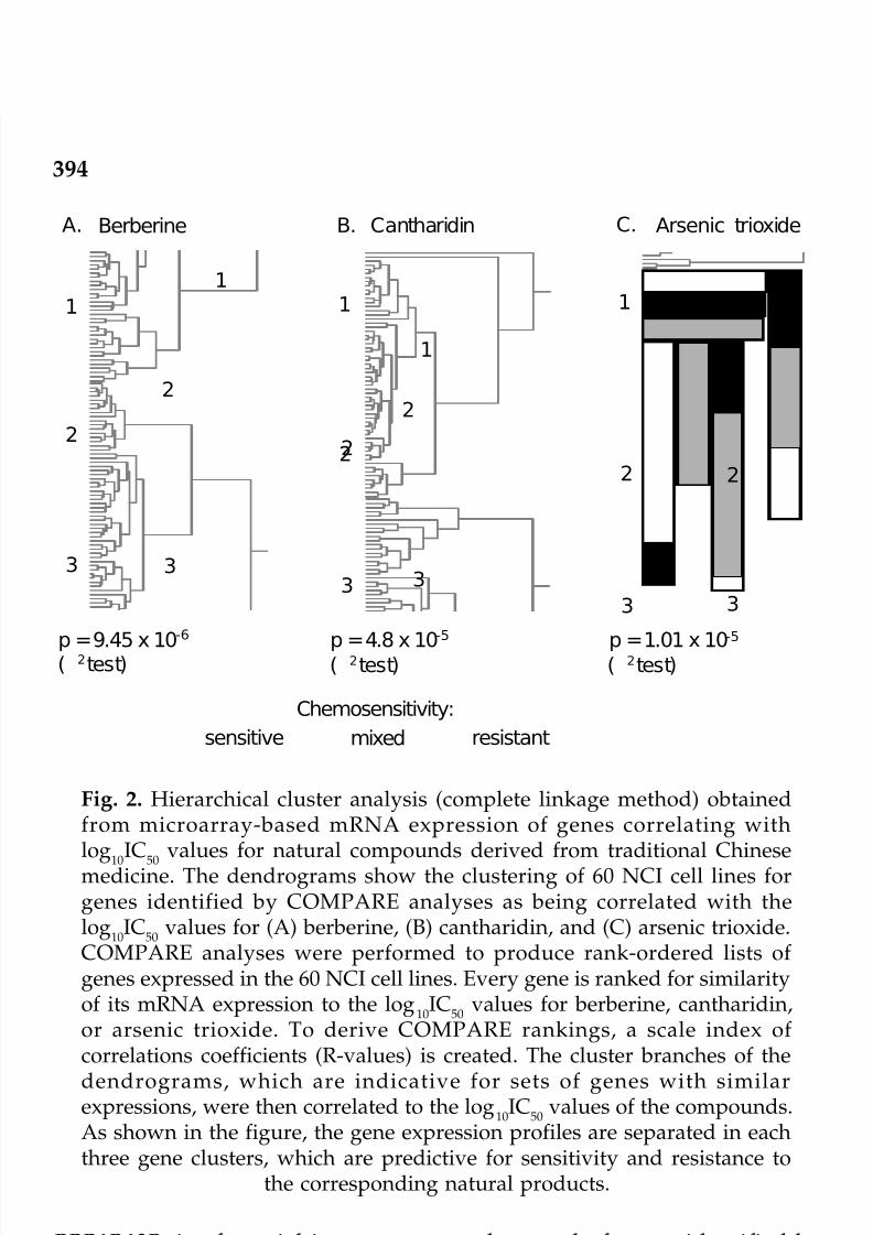
7/23/2019 Novel Therapeutic Agents From Plants
http://slidepdf.com/reader/full/novel-therapeutic-agents-from-plants 405/464
394
PPP1R13B, is of special interest among the panel of genes identified byour approach. PPP1R13B plays a central role in the regulation of apoptosisvia its interaction with the tumor suppressor gene p53. It regulates p53 byenhancing DNA binding activity and trans-activation function of p53 onthe promoters of pro-apoptotic genes in vivo (Samuels-Lev et al., 2001). A
role of PP1 in the repair of UV-induced DNA lesions (Hermann et al., 2002)and the induction of cytosine arabinoside-induced apoptosis was shown
Fig. 2. Hierarchical cluster analysis (complete linkage method) obtainedfrom microarray-based mRNA expression of genes correlating withlog
10IC
50values for natural compounds derived from traditional Chinese
medicine. The dendrograms show the clustering of 60 NCI cell lines forgenes identified by COMPARE analyses as being correlated with thelog
10IC
50values for (A) berberine, (B) cantharidin, and (C) arsenic trioxide.
COMPARE analyses were performed to produce rank-ordered lists of genes expressed in the 60 NCI cell lines. Every gene is ranked for similarityof its mRNA expression to the log
10IC
50values for berberine, cantharidin,
or arsenic trioxide. To derive COMPARE rankings, a scale index of correlations coefficients (R-values) is created. The cluster branches of thedendrograms, which are indicative for sets of genes with similarexpressions, were then correlated to the log
10IC
50values of the compounds.
As shown in the figure, the gene expression profiles are separated in eachthree gene clusters, which are predictive for sensitivity and resistance to
the corresponding natural products.
11
1
11
2
22
2
2 2
3 333
3 3
A. Berberine B. Cantharidin C. Arsenic trioxide
sensitive mixed resistant
Chemosensitivity:
p =9.45 x 10⁻⁶ p =4.8 x 10⁻⁵ p =1.01 x 10⁻⁵
χ 2( test) χ 2( test) χ 2( test)
1
2
© 2009 by Taylor & Francis Group, LLC

7/23/2019 Novel Therapeutic Agents From Plants
http://slidepdf.com/reader/full/novel-therapeutic-agents-from-plants 406/464
395
(Wang et al., 2001). It is, therefore, reasonable to hypothesize thatPPP1R13B also has specific functions in cantharidin-induced DNA repairand apoptosis. PP1 is one of four major serine/threonine proteinphosphatases, and new protein phosphatases are still emerging. They arecrucial regulators of many cellular functions by altering the phos-phorylation of target proteins. The level of phosphorylation is in a well-controlled balance by the opposing actions of protein kinases and proteinphosphatases. The PP1 holoenzyme consists of catalytic and regulatorysubunits. Up to now, four catalytic (a, b, c, d) and more than a dozen of regulatory subunits are known. The regulatory subunits modulate thesubstrate specificity and target the holoenzyme to specific sub-cellular
localizations.A recent study reported the up- or down-regulation of mRNA
expression levels of 2087 genes by microarray technology after treatmentwith cantharidin (Zhang et al., 2004). We compared our set of genes withthat of these authors. None of the 21 genes identified in the present studywas among the genes identified by Zhang et al. (2004). However, someclosely related genes were found with both approaches. Whereas we found
the regulatory subunit 13B of protein phosphatase 1 (PPP1R13B), Zhanget al. (2004) found regulatory subunit 5 of PP1 (PPP1R5). The β2 noncatalytic subunit of AMP-activated protein kinase (PRKAB2) was identified
by us and the γ 1 subunit of this enzyme by Zhang and coworkers (2004).In our approach, mRNA expression of cytochrome b5 (CYB5) correlated toIC
50for cantharidin, Zhang and colleagues (2004) found an altered
expression of a DNA sequence which contains the 3' end of the gene forflavohemoprotein b5 and b5R cytochrome b-type NAD(P)H oxido-
reductase. Furthermore, heat shock-binding protein 1 (HSBP1) wasassociated with cantharidin sensitivity in our investigation, while heatshock protein 1 (HSPA1B) was linked to cantharidin treatment in Zhang’sstudy. The small overlap in both microarray-based investigations issurprising at first sight. Two explanations could be attributed to this: First,while our approach focused on the correlation of baseline mRNAexpression levels in 60 NCI cell lines and IC
50values for cantharidin, Zhang
et al. (2004) treated a single cell line (HL60) with a single concentration of cantharidine (25 µM). Those are different biological conditions leading todifferent gene expression profiles. Second, the selection criteria in ourapproach were very stringent. We selected 21 out of 9706 genes with thehighest COMPARE indices and the smallest probability to correlateerroneously with IC
50values for cantharidin by a false discovery rate
algorithm. It is probable that many other genes identified by Zhang et al.(2004) also correlate significantly with IC
50values for cantharidin in our
approach, however with smaller COMPARE correlation coefficients.
© 2009 by Taylor & Francis Group, LLC

7/23/2019 Novel Therapeutic Agents From Plants
http://slidepdf.com/reader/full/novel-therapeutic-agents-from-plants 407/464
396
Arsenic trioxide
Arsenic is a natural semimetal in soil, water and air. It exists as red arsenic(As
2S
2), yellow arsenic (As
2S
3), white arsenic (As
2O
3, arsenic trioxide),
phenylarsine oxide (C6H5AsO), and as salts of sodium, potassium andcalcium (Miller et al., 2002). Since ancient times arsenic was used as amedicinal agent, i.e., in Greek, Roman, and Chinese medicine (Klaassen,1996). Arsenic was appreciated as Fowler’s Solution for many diseases inthe 18th and 19th century, i.e., syphilis, cancer, ulcers, etc. (Haller, 1975). Inthe 20th century, Paul Ehrlich, the founder of modern chemotherapy, foundthe arsenical salvarsan which was the standard therapy against syphilis
for decades (Chan and Huff, 1997). On the other hand, arsenic compoundscan be poisonous (Knowles and Benson, 1983). The revival of arsenic inmodern medicine was initiated by Chinese scientists showing dramaticregression rates of acute promyelocytic leukemia (PML) by arsenic trioxide(Shen et al., 1997). These findings were subsequently corroborated inclinical studies in the U.S.A. (Soignet et al., 2001).
Various molecular determinants of the biological effect of arsenictrioxide have been elucidated. It promotes the degradation of the oncogenicfusion protein of the PML and retinoic acid receptor α (RARα) genes inacute promyelocytic leukemia, resulting in induction of cellular differen-tiation (Chen et al., 1996; Soignet et al., 2001). Apoptosis is selectivelyinduced in malignant cells through enhancement of reactive oxygen speciesand activation of caspases (Huang et al., 1999; Anderson et al., 2002;Hayashi et al., 2002; Liu et al., 2003). Cells can arrest in the G1 or G2/Mphases of the cell cycle after treatment with arsenic trioxide (Liu et al., 2003).
Tumor angiogenesis is targeted by arsenic trioxide through inhibition of vascular epithelial growth factor production (Roboz et al., 2000).
As cellular responses of tumor cells to arsenic trioxide seem to be multi-factorial, we aimed to get a comprising insight into molecular predictorsof sensitivity or resistance to arsenic trioxide (Efferth and Kaina, 2004).For this reason, we mined the NCI’s microarray database. Out of 9706genes, 47 genes whose mRNA expression in 60 tumor cell lines correlated
with highest correlation coefficients to inhibition concentration 50% (IC50)values were selected by COMPARE analysis. These genes were subjectedto hierarchical cluster analysis and cluster image mapping to reveal,whether the expression profiles of these genes are useful to predictsensitivity or resistance of cell lines to arsenic trioxide.
While the specific functions of the proteins encoded by the 47 genesidentified by our approach are different, it is intriguing that many of themare in one way or another involved in apoptosis. This may be taken as aclue that arsenic trioxide impairs the apoptotic machinery and thatapoptosis is an important mechanism of arsenic trioxide’s cytotoxicity.
© 2009 by Taylor & Francis Group, LLC

7/23/2019 Novel Therapeutic Agents From Plants
http://slidepdf.com/reader/full/novel-therapeutic-agents-from-plants 408/464
397
Some of the genes identified in the present investigation and genes closelyrelated to them have previously been associated with the action of arsenicals.
We found that the mRNA expression of chemokine-like super familymember 4 and growth factor receptor-bound protein 7 correlated with theIC
50values for arsenic trioxide. Interestingly, using a microarray approach
Wu et al. (2003) found that several cytokines and growth factors weredifferentially expressed in arsenic-exposed healthy individuals.
The identification of keratin 8 as a predictor of cellular response toarsenic trioxide corresponds to recent findings of Ramirez et al. (2000).These authors observed that arsenite causes DNA-protein cross-links withcytokeratins due to their high content of sulfhydryl groups. Microarrayanalyses pointed to various keratins (K8, K15, K18, K19) after treatment of cells with arsenic (Yih et al., 2002; Bae et al., 2003).
Another gene found by our approach, thioredoxin reductase I, isinhibited by arsenicals (Lin et al., 2001). Thioredoxin is a regulator of reactive oxygen species that also affects apoptosis by apoptosis-signal-regulating kinase I in an at least partially p53-dependent manner (Ma et
al., 2002; Anestal and Arner, 2003).Our result that aldehyde dehydrogenase 3A2 might contribute to
resistance to arsenic trioxide is in accord with the alteration of hepaticmitochondrial aldehyde dehydrogenase activity by sodium arsenate(Fowler et al., 1982).
While a relationship of COX-1 has not been documented previously,COX-2 expression is known to be elevated by arsenite through stimulation
of NFκ B activity (Tsai et al., 2002). NFκ B contributes to arsenic-inducedapoptosis (Mathas et al., 2003).
The correlation of mRNA expression of the UBE1 gene in the presentinvestigation implies that the proteasome degradation pathway may playa role for resistance to arsenic trioxide. This view is supported by recentmicroarray data with arsenic sulfide pointing to the closely related UBE1Lgene (Wang et al., 2003).
In conclusion, the data presented here not only provide expressionprofiles of genes that predict cellular sensitivities and resistance to arsenictrioxide, but may also serve to generate testable hypothesis on molecularmechanisms of cytotoxicity in tumor cells.
Conclusion and perspectives
Using natural products exemplarily taken from medicinal plants, animals,and minerals, we showed how molecular biological and pharmacogenomicapproaches can be used to predict sensitivity or resistance of tumor cells
© 2009 by Taylor & Francis Group, LLC

7/23/2019 Novel Therapeutic Agents From Plants
http://slidepdf.com/reader/full/novel-therapeutic-agents-from-plants 409/464
398
to these agents. This approach also enables the identification of genespossibly involved in molecular pathways responsible for the killing of cancer cells.
The knowledge of molecular mechanisms of cancer cells increaseddramatically over the past years. This is a fertile ground to search for themodes of action of natural products. It can be assumed that a wealth of information will be generated in the years to come to unravel the molecularmechanisms of known and novel compounds derived from traditionalChinese medicine. The final goal is to develop novel drugs for cancertreatment. Nevertheless, even after the sequencing of the human genomeat the beginning of the new millennium we do not understand the full
range of molecular mechanisms of carcinogenesis. Apart from DNAalterations, transcriptional and translational mechanisms (alternativesplicing) as well as posttranslational modifications (phosphorylation,glycosylation, acetylation, methylation, ubiquitinylation etc.) must be takeninto account elucidating the spectrum of carcinogeneic and tumorpromoting mechanisms. The impact of these events and their interactionwith natural products needs further to be investigated.
Although many compounds from natural sources have been identifiedin the past decades and successful protection is possible now, personalassessment, whether a cancer treatment would be successful or not, remainsstill difficult to predict.
Novel ‘-omics’ technologies (genomics, proteomics, metabonomics) andpharmacogenetics (single nucleotide polymorphisms) may help to definepatterns specifically linked to therapy response of tumors and prognosis
of survival of patients. On the other hand, it is to be expected that thisnew dimension of molecular analyses will raise even more questions thanit can answer.
A new discipline at the horizon is systems biology. It attempts tointegrate the vast amount of data generated by the ‘-omics’ technologiesand to generate not only models to describe mechanisms and diseases, butalso to develop predictive bioinformatic models for carcinogenic riskassessment at the level of each single individual. This will also be the dawn
of a new era in natural product research.
References
Amin, A.H., T.V. Subbaiah and K.M. Abbasi. 1969. Berberine sulfate: antimicrobialactivity, bioassay, and mode of action. Canadian Journal of Microbiology15, 1067-1076.
Anderson, K.C., L.H. Boise, R. Louie and S. Waxman. 2002. Arsenic trioxide inmultiple myeloma: rationale and future directions. Cancer Journal 8, 12-25.© 2009 by Taylor & Francis Group, LLC

7/23/2019 Novel Therapeutic Agents From Plants
http://slidepdf.com/reader/full/novel-therapeutic-agents-from-plants 410/464
399
Anestal, K. and E.S. Arner. 2003. Rapid induction of cell death by selenium-compromised thioredoxin reductase 1 but not by the fully active enzymecontaining selenocysteine. Journal of Biological Chemistry 278, 15966-15972.
Anfosso, L., T. Efferth, A. Albini and U. Pfeffer. 2006. Microarray expression
profiles of angiogenesis-related genes predict tumor cell response toartemisinins. Pharmacogenomics Journal 6, 269-278.
Bae, D.S., R.J. Handa, R.S. Yang and J.A. Campain. 2003. Gene expression patternsas potential molecular biomarkers for malignant transformation in humankeratinocytes treated with MNNG, arsenic, or a metal mixture. ToxicologicSciences 74, 32-42.
Birdsall, T.C. and G.S. Kelly. 1997. Berberine: Therapeutic potential of an alkaloidfound in several medicinal plants. Alternative Medicine Research 2, 94-103.
Chan, P.C. and J. Huff. 1997. Arsenic carcinogenesis in animals and in humans:mechanistic, experimental, and epidemiological evidence. Journal of Environmental Sciences and Health Part C Environmental Carcinogenesisand Ecotoxicology Reviews C15, 83-122.
Chen, G.Q., J. Zhu, X.G. Shi, J.N. Ni, H.J. Zhong, G.Y. Si, X.L. Jin, W. Tang, X.S.Li, S.M. Xong, Z.X. Shen, G.L. Sun, J. Ma, P. Zhang, T.D. Zhang, C. Gazin, T.Naoe, S.J. Chen, Z.Y. Wang and Z. Chen. 1996. In vitro studies on cellularand molecular mechanisms of arsenic trioxide (As
2O
3) in the treatment of
acute promyelocytic leukemia: As2O
3induces NB4 cell apoptosis with
downregulation of Bcl-2 expression and modulation of PML-RARα/PMLproteins. Blood 88, 1052-1061.
Chen, H.H., H.J. Zhou and X. Fang. 2003. Inhibition of human cancer cell linegrowth and human umbilical vein endothelial cell angiogenesis byartemisinin derivatives in vitro. Pharmacological Research 48, 231-236.
Chen, H.H., H.J. Zhou, W.Q. Wang and G.D. Wu. 2004. Antimalarial dihydroarte-misinin also inhibits angiogenesis. Cancer Chemotherapy and Pharmacology53, 423-432.
Chen, Y.J., C.J. Shieh, T.H. Tsai, C.D. Kuo, L.T. Ho, T.Y. Liu and H.F. Liao. 2005.Inhibitory effect of norcantharidin, a derivative compound from blister beetles, on tumor invasion and metastasis in CT26 colorectal adenocarcinomacells. Anticancer Drugs 16, 293-299.
Chen, Y.N., J.C. Chen, S.C. Yin, G.S. Wang, W. Tsauer, S.F. Hsu and S.L. Hsu.2002. Effector mechanisms of norcantharidin-induced mitotic arrest andapoptosis in human hepatoma cells. International Journal of Cancer 100,158-165.
Christmann, M., M.T. Tomicic, W.P. Roos and B. Kaina. 2003. Mechanisms of human DNA repair: an update. Toxicology 193, 3-34.Cortés-Funes, H. and C. Coronado. 2007. Role of anthracyclines in the era of
targeted therapy. Cardiovascular Toxicology 7, 56-60.Dell’Eva, R., U. Pfeffer, R. Vene, L. Anfosso, A. Forlani, A. Albini and T. Efferth.
2004. Inhibition of angiogenesis in vivo and growth of Kaposi’s sarcomaxenograft tumors by the anti-malarial artesunate. Biochemical Pharma-cology 68, 2359-2366.
Drewes, S.E., J. George and F. Khan. 2003. Recent findings on natural productswith erectile-dysfunction activity. Phytochemistry 62, 1019-1025.
Duflos, A., A. Kruczynaski and J.M. Barret. 2002. Novel aspects of natural and© 2009 by Taylor & Francis Group, LLC

7/23/2019 Novel Therapeutic Agents From Plants
http://slidepdf.com/reader/full/novel-therapeutic-agents-from-plants 411/464
400
modified vinca alkaloids. Current Medicinal Chemistry of Anticancer Agents2, 55-70.
Efferth, T. 2005. Microarray-based prediction of cytotoxicity of tumor cells tocantharidin. Oncology Reports 13, 459-463.
Efferth, T. 2007. Willmar Schwabe Award 2006: antiplasmodial and antitumoractivity of artemisinin—from bench to bedside. Planta Medica 73, 299-309.
Efferth T. and B. Kaina. 2004. Microarray-based prediction of cytotoxicity of tumorcells to arsenic trioxide. Cancer Genomics and Proteomics 1, 363-370.
Efferth, T., G. Rücker, M. Falkenberg, D. Manns, A. Olbrich, U. Fabry and R.Osieka. 1996. Detection of apoptosis in KG-1a leukemic cells treated withinvestigational drugs. Arzneimittelforschung 46, 196-200.
Efferth, T., H. Dunstan, A. Sauerbrey, H. Miyachi and C.R. Chitambar. 2001. The
anti-malarial artesunate is also active against cancer. International Journalof Oncology 18, 767-773.Efferth, T., M. Marschall, X. Wang, S.M. Huong, I. Hauber, A. Olbrich, M.
Kronschnabl, T. Stamminger and E.S. Huang. 2002a. Antiviral activity of artesunate towards wild-type, recombinant, and ganciclovir-resistant humancytomegaloviruses. Journal of Molecular Medicine 80, 233-242.
Efferth, T., M. Davey, A. Olbrich, G. Rücker, E. Gebhart and R. Davey. 2002b.Activity of drugs from traditional Chinese medicine toward sensitive andMDR1- or MRP1-overexpressing multidrug-resistant human CCRF-CEMleukemia cells. Blood Cells, Molecules and Diseases 28, 160-168.
Efferth, T., A. Sauerbrey, A. Olbrich, E. Gebhart, P. Rauch, H.O. Weber, J.G.Hengstler, M.E. Halatsch, M. Volm, K.D. Tew, D.D. Ross and J.O. Funk. 2003.Molecular modes of action of artesunate in tumor cell lines. MolecularPharmacology 64, 382-394.
Efferth, T., A. Benakis, M.R. Romero, M. Tomicic, R. Rauh, D. Steinbach, R. Hafer,T. Stamminger, F. Oesch, B. Kaina and M. Marschall. 2004. Enhancement of cytotoxicity of artemisinins toward cancer cells by ferrous iron. Free Radicals
in Biology and Medicine 37, 998-1009.Efferth, T., T. Chen, B. Kaina and G. Wang. 2005a. Molecular determinants of
response of tumor cells to berberine. Cancer Genomics and Proteomics 2,115-124.
Efferth, T., R. Rauh, S. Kahl, M. Tomicic, H. Boechzelt, M.E. Tome, M.M. Briehl,R. Bauer and B. Kaina. 2005b. Molecular modes of action of cantharidin intumor cells. Biochemical Pharmacology 69, 811-818.
Efferth, T., P.C. Li, V.S. Konkimalla and B. Kaina. 2007. From traditional Chinese
medicine to rational cancer therapy. Trends in Molecular Medicine 13, 353-361.Fowler, B.A., J.S. Woods, K.S. Squibb and N.M. Davidian. 1982. Alteration of
hepatic mitochondrial aldehyde dehydrogenase activity by sodium arsenate:the relationship to mitochondrial—microsomal oxidative interactions.Experimental and Molecular Pathology 37, 351-357.
Fukuda, K., Y. Hibiya, M. Mutoh, M. Koshiji, S. Akao and H. Fujiwara. 1999.Inhibition of activator protein 1 activity by berberine in human hepatomacells. Planta Medica 65, 381-383.
Ghosh, A.K., F.K. Bhattacharyya and D.K. Ghosh. 1985. Leishmania donovani:amastigote inhibition and mode of action of berberine. Experimental© 2009 by Taylor & Francis Group, LLC

7/23/2019 Novel Therapeutic Agents From Plants
http://slidepdf.com/reader/full/novel-therapeutic-agents-from-plants 412/464
401
Parasitology 60, 404-413.Gillet, J.P., T. Efferth, D. Steinbach, J. Hamels, F. de Longueville, V. Bertholet and
J. Remacle. 2004. Microarray-based detection of multidrug resistance inhuman tumor cells by expression profiling of ATP-binding cassette
transporter genes. Cancer Research 64, 8987-8993.Gillet, J.P., T. Efferth and J. Remacle. 2007. Chemotherapy-induced resistance by
ATP-binding cassette transporter genes. Biochimica and Biophysica Acta 1775:237-262.
Haller, J.S. 1975. Therapeutic mule: the use of arsenic in the nineteenth centurymateria medica. Pharmacy in History 17, 87-100.
Hartmann, J.T. and H.P. Lipp. 2006. Camptothecin and podophyllotoxinderivatives: inhibitors of topoisomerase I and II—mechanisms of action,
pharmacokinetics and toxicity profile. Drug Safety 29, 209-230.Hayashi, T., T. Hideshima, M. Akiyama, P. Richardson, R.L. Schlossman, D.Chauhan, N.C. Munshi, S. Waxman and K.C. Anderson. 2002. Arsenic trioxideinhibits growth of human multiple myeloma cells in the bone marrowmicroenvironment. Molecular Cancer Therapy 1, 851-860.
Heinrich, M. and P. Bremner. 2006. Ethnobotany and ethnopharmacy—their rolefor anti-cancer drug development. Current Drug Targets 7, 239-245.
Herman, M., Y. Ori, A. Chagnac, T. Weinstein, A. Korzets, D. Zevin, T. Malachiand U. Gafter. 2002. DNA repair in mononuclear cells: role of serine/threonine phosphatases. Journal of Laboratory and Clinical Medicine 140,255-262.
Hong, C.Y., S.C. Huang, S.K. Lin, J.J. Lee, L.L. Chueh, C.H. Lee, J.H. Lin and M.Hsiao. 2000. Norcantharidin-induced post-G(2)/M apoptosis is dependenton wild-type p53 gene. Biochemical and Biophysical Research Communi-cations 276, 278-285.
Honkanen, R.E. 1993. Cantharidin, another natural toxin that inhibits the activityof serine/threonine protein phosphatases types 1 and 2A. FEBS Letters 330,
283-286.Huan, S.K., H.H. Lee, D.Z. Liu, C.C. Wu and C.C. Wang. 2006. Cantharidin
induced cytotoxicity and cyclooxygenase 2 expression in human bladdercarcinoma cell line. Toxicology 223, 136-143.
Huang, X.J., P.H. Wiernik, R.S. Klein and R.E. Gallagher. 1999. Arsenic trioxideinduces apoptosis of myeloid leukemia cells by activation of caspases. MedicalOncology 16, 58-64.
Iizuka, N., M. Oka, K. Yamamoto, A. Tangoku, K. Miyamoto, T. Miyamoto, S.
Uchimura, Y. Hamamoto and K. Okita. 2003. Identification of common ordistinct genes related to antitumor activities of a medicinal herb and its majorcomponent by oligonucleotide microarray. International Journal of Cancer107, 666-672.
Kang, M.R. and I.K. Chung. 2002. Down-regulation of DNA topoisomeraseIIalpha in human colorectal carcinoma cells resistant to a protoberberinealkaloid, berberrubine. Molecular Pharmacology 61, 879-884.
Karras, D.J., S.E. Farrell, R.A. Harrigan, F.M. Henretig and L. Gealt. 1996.Poisoning from “Spanish fly” (canthardin). American Journal of EmergencyMedicine 14, 478-483.
Klaassen, C.D. 1996. Heavy metals and heavy-metal antagonists. pp. 1649-1672.© 2009 by Taylor & Francis Group, LLC

7/23/2019 Novel Therapeutic Agents From Plants
http://slidepdf.com/reader/full/novel-therapeutic-agents-from-plants 413/464
402
In: J.G. Hardman, A.G. Gilman and L.E. Limbird (eds) Goodman andGilman’s The Pharmacological basis of therapeutics. McGraw-Hill, New York,USA.
Knowles, P.C. and A.A. Benson. 1983. The biogeochemistry of arsenic. Trends in
Biochemical Sciences 8, 178-180.Kok, S.H., C.Y. Hong, M.Y. Kuo, C.H. Lee, J.J. Lee, I.U. Lou, M.S. Lee, M. Hsiao
and S.K. Lin. 2003. Comparisons of norcantharidin cytotoxic effects on oralcancer cells and normal buccal keratinocytes. Oral Oncology 39, 19-26.
Lau, C.W., Y.Q. Yao, Z.Y. Chen, W.H. Ko and Y. Huang. 2001. Cardiovascularactions of berberine. Cardiovascular Drug Reviews 19, 234-244.
Lee, K.H. 1999. Novel antitumor agents from higher plants. Medical ResearchReviews 19, 569-596.
Lesnau, A., J. Hils, G. Pohl, G. Beyer, M. Janka and L.T. Hoa. 1990. Antiviralactivity of berberine salts [Article in German]. Pharmazie 45, 638-639.Levêque, D. and F. Jehl. 2007. Molecular pharmacokinetics of catharanthus (vinca)
alkaloids. Journal of Clinical Pharmacology 47, 579-588.Li, Y.M. and J.E. Casida. 1992. Cantharidin-binding protein: identification as
protein phosphatase 2A. Proceedings of the National Academy of Scienceof the United States of America 89, 11867-11870.
Lin, H.L., T.Y. Liu, C.W. Wu and C.W. Chi. 1999. Berberine modulates expressionof mdr1 gene product and the responses of digestive track cancer cells toPaclitaxel. British Journal of Cancer 81, 416-422.
Lin, S., L.M. Del Razo, M. Styblo, C. Wang, W.R. Cullen and D.J. Thomas. 2001.Arsenicals inhibit thioredoxin reductase in cultured rat hepatocytes. ChemicalResearch and Toxicology 14, 305-311.
Liu, Q., S. Hilsenbeck and Y. Gazitt. 2003. Arsenic trioxide-induced apoptosis inmyeloma cells: p53-dependent G1 or G2/M cell cycle arrest, activation of caspase-8 or caspase-9, and synergy with APO2/TRAIL. Blood 101, 4078-4087.
Ma, X., J. Hu, D.J. Lindner and D.V. Kalvakolanu. 2002. Mutational analysis of human thioredoxin reductase 1. Effects on p53-mediated gene expressionand interferon and retinoic acid-induced cell death. Journal of BiologicalChemistry 277, 22460-22468.
Mathas, S., A. Lietz, M. Janz, M. Hinz, F. Jundt, C. Scheidereit, K. Bommert andB. Dörken. 2003. Inhibition of NF-κ B essentially contributes to arsenic-induced apoptosis. Blood 102, 1028-1034.
McCluskey, A., S.P. Ackland, E. Gardiner, C.C. Walkom and J.A. Sakoff. 2001.
The inhibition of protein phosphatases 1 and 2A: a new target for rationalanti-cancer drug design? Anticancer Drug Design 16, 291-303.Miller, W.H. Jr., H.M. Schipper, J.S. Lee, J. Singer and S. Waxman. 2002.
Mechanisms of action of arsenic trioxide. Cancer Research 62, 3893-3903.Moed, L., T.A. Shwayder and M.W. Chang. 2001. Cantharidin revisited: a blister-
ing defense of an ancient medicine. Archives of Dermatology 137, 1357-1360.Nakamoto, K., S. Sadamori and T. Hamada. 1990. Effects of crude drugs and
berberine hydrochloride on the activities of fungi. Journal of Prosthetricsand Dentistry 64, 691-694.
Norbury, C.J. and B. Zhivotovsky. 2004. DNA damage-induced apoptosis.Oncogene 23, 2797-808.© 2009 by Taylor & Francis Group, LLC

7/23/2019 Novel Therapeutic Agents From Plants
http://slidepdf.com/reader/full/novel-therapeutic-agents-from-plants 414/464
403
Pan, G.Y., G.J. Wang, X.D. Liu, J.P. Fawcett and Y.Y. Xie. 2002. The involvementof P-glycoprotein in berberine absorption. Pharmacology and Toxicology91, 193-197.
Pommier, Y. 2006. Topoisomerase I inhibitors: camptothecins and beyond. Nature
Reviews Cancer 6, 789-802.Rabbani, G.H., T. Butler, J. Knight, S.C. Sanyal and K. Alam. 1987. Randomized
controlled trial of berberine sulfate therapy for diarrhea due toenterotoxigenic Escherichia coli and Vibrio cholerae. Journal of InfectiousDiseases 155, 979-984.
Ramirez, P., L.M. Del Razo, M.C. Gutierrez-Ruiz and M.E. Gonsebatt. 2000.Arsenite induces DNA-protein crosslinks and cytokeratin expression in theWRL-68 human hepatic cell line. Carcinogenesis 21, 701-706.
Rauh, R., S. Kahl, H. Boechzelt, R. Bauer, B. Kaina and T. Efferth. 2007. Molecular biology of cantharidin in cancer cells. Chinese Medicine 2, 8.Roboz, G.J., S. Dias, G. Lam, W.J. Lane, S.L. Soignet, R.P. Warrell Jr. and S. Rafii.
2000. Arsenic trioxide induces dose- and time-dependent apoptosis of endothelium and may exert an antileukemic effect via inhibition of angiogenesis. Blood 96, 1525-1530.
Samuels-Lev, Y., D.J. O’Connor, D. Bergamaschi, G. Trigiante, J.K. Hsieh, S.Zhong, I. Campargue, L. Naumovski, T. Crook and X. Lu. 2001. ASPPproteins specifically stimulate the apoptotic function of p53. Molecular Cell8, 781-794.
Shen, Z.X., G.Q. Chen, J.H. Ni, X.S. Li, S.M. Xiong, Q.Y. Qiu, J. Zhu, W. Tang,G.L. Sun, K.Q. Yang, Y. Chen, L. Zhou, Z.W. Fang, Y.T. Wang, J. Ma, P. Zhang,T.D. Zhang, S.J. Chen, Z. Chen and Z.Y. Wang. 1997. Use of arsenic trioxide(As
2O
3) in the treatment of acute promyelocytic leukemia (APL): II. Clinical
efficacy and pharmacokinetics in relapsed patients. Blood 89, 3354-3360.Shitan, N., I. Bazin, K. Dan, K. Obata, K. Kigawa, K. Ueda, F. Sato, C. Forestier
and K. Yazaki. 2003. Involvement of CjMDR1, a plant multidrug-resistance-
type ATP-binding cassette protein, in alkaloid transport in Coptis japonica.Proceedings of the National Academy of Science of the United States of America 100, 751-756.
Silverberg, N. 2003. Pediatric molluscum contagiosum: optimal treatmentstrategies. Paediatric Drugs 5, 505-512.
Simeon, S., J.L. Rios and A. Villar. 1989. Pharmacological activities of proto- berberine alkaloids. Plant Medicine and Phytotherapy 23, 202-250.
Slupphaug, G., B. Kavli and H.E. Krokan. 2003. The interacting pathways for
prevention and repair of oxidative DNA damage. Mutation Research 531,231-251.Smolinski, K.N. and A.C. Yan. 2005. How and when to treat molluscum
contagiosum and warts in children. Pediatric Annals 34, 211-221.Soignet, S.L., S.R. Frankel, D. Douer, M.S. Tallman, H. Kantarjian, E. Calleja, R.M.
Stone, M. Kalaycio, D.A. Scheinberg, P. Steinherz, E.L. Sievers, S. Coutre, S.Dahlberg, R. Ellison and R.P. Warrell. Jr. 2001. United States multicenter studyof arsenic trioxide in relapsed acute promyelocytic leukemia. Journal of Clinical Oncology 19, 3852-3860.
Stermitz, F.R., P. Lorenz, J.N. Tawara, L.A. Zenewicz and K. Lewis. 2000. Synergyin a medicinal plant: antimicrobial action of berberine potentiated by 5'-© 2009 by Taylor & Francis Group, LLC

7/23/2019 Novel Therapeutic Agents From Plants
http://slidepdf.com/reader/full/novel-therapeutic-agents-from-plants 415/464
404
methoxyhydnocarpin, a multidrug pump inhibitor. Proceedings of theNational Academy of Science of the United States of America 97, 1433-1437.
Sun, D., H.S. Courtney and E.H. Beachey. 1988. Berberine sulfate blocks adherenceof Streptococcus pyogenes to epithelial cells, fibronectin, and hexadecane.
Antimicrobial Agents and Chemotherapy 32, 1370-1374.Swingle, M., L. Ni and R.E. Honkanen. 2006. Small-molecule inhibitors of ser/
thr protein phosphatases: specificity, use and common forms of abuse.Methods in Molecular Biology 365, 23-38.
Tegos, G., F.R. Stermitz, O. Lomovskaya and K. Lewis. 2002. Multidrug pumpinhibitors uncover remarkable activity of plant antimicrobials. AntimicrobialAgents and Chemotherapy 46, 3133-3141.
Tsai, S.H., Y.C. Liang, L. Chen, F.M. Ho, M.S. Hsieh and J.K. Lin. 2002. Arsenite
stimulates cyclooxygenase-2 expression through activating Iκ B kinase andnuclear factor κ B in primary and ECV304 endothelial cells. Journal of CellularBiochemistry 84, 750-758.
Tsauer, W., J.G. Lin, P.Y. Lin, F.L. Hsu and H.C. Chiang. 1997. The effects of cantharidin analogues on xanthine oxidase. Anticancer Research 17, 2095-2098.
Wang, G.S. 1989. Medical uses of mylabris in ancient China and recent studies. Journal of Ethnopharmacology 26, 147-162.
Wang, H., S. Liu, X. Lu, X. Zhao, S. Chen and X. Li. 2003. Gene expression profilechanges in NB4 cells induced by realgar. Chinese Medical Journal (EnglishEdition) 16, 1074-1077.
Wang, R.H., C.W. Liu, V.I. Avramis and N. Berndt. 2001. Protein phosphatase1alpha-mediated stimulation of apoptosis is associated with dephosphory-lation of the retinoblastoma protein. Oncogene 20, 6111-6122.
Wera, S. and B.A. Hemmings. 1995. Serine/threonine protein phosphatases.Biochemical Journal 311, 17-29.
Wu, H.L., C.Y. Hsu, W.H. Liu and B.Y. Yung. 1999. Berberine-induced apoptosis
of human leukemia HL-60 cells is associated with down-regulation of nucleophosmin/B23 and telomerase activity. International Journal of Cancer81, 923-929.
Wu, L.T., J.G. Chung, J.C. Chen and W. Tsauer. 2001. Effect of norcantharidin onN-acetyltransferase activity in HepG2 cells. American Journal of ChineseMedicine 29, 161-172.
Wu, M.M., H.Y. Chiou, I.C. Ho, C.J. Chen and T.C. Lee. 2003. Gene expressionof inflammatory molecules in circulating lymphocytes from arsenic-exposed
human subjects. Environmental and Health Perspectives 111, 1429-1438.Yih, L.H., K. Peck and T.C. Lee. 2002. Changes in gene expression profiles of human fibroblasts in response to sodium arsenite treatment. Carcinogenesis23, 867-876.
Zhang, C.H. et al. 2000. Enhanced paclitaxel production induced by thecombination of elicitors in cell suspension cultures of Taxus chinensis.Biotechnology Letters 22, 1561–1564.
Zhang, J.P., K. Ying, Z.Y. Xiao, B. Zhou, Q.S. Huang, H.M. Wu, M. Yin, Y. Xie,Y.M. Mao and Y.C. Rui. 2004. Analysis of gene expression profiles in humanHL-60 cell exposed to cantharidin using cDNA microarray. International Journal of Cancer 108, 212-218.© 2009 by Taylor & Francis Group, LLC

7/23/2019 Novel Therapeutic Agents From Plants
http://slidepdf.com/reader/full/novel-therapeutic-agents-from-plants 416/464
1. Introduction
For decades the introduction of very effective drugs has revolutionized thetreatment of tuberculosis. In recent years, however, the emerging multipledrug resistant (MDR) and extensively drug-resistant (XDR) strains and HIVepidemics have become a major threat and thus calls for an urgent searchfor new and effective treatment for this deadly disease. The potential spreadof these drug- resistant strains was exemplified by the concern of international health agencies when it was discovered that an individualinfected with the extremely drug-resistant tuberculosis (XDR-TB) had flown
on two transatlantic flights from the U.S. to Europe and returning viaMontreal.
http://news.yahoo.com/s/ap/20070717/ap_on_he_me/tuberculosis_infection
The genus Mycobacterium (Mycobacteriaceae) is highly diverse with 85species known. Some cause human and animal diseases and others areomnipresent in nature. The three most common human mycobacterialdiseases are the primary pulmonary disease, tuberculosis, and two skin
diseases, leprosy ( M. leprae) and buruli ulcer ( M. ulcerans) (Okunade et al.,2004).
Plant Derived AntimycobacterialMetabolites: An Overview15Adewole L. Okunade
1
andMemory P.F. Elvin-Lewis2
1Center for Human Nutrition, Division of Geriatrics andNutritional Science, Department of Internal Medicine,Washington University School of Medicine, St. Louis, MO63110, U.S.A. Tel.: 314 747 4052; Fax: 314 362 8032; E-mail:[email protected]
2Department of Biology, One Brookings Drive, Washington
University, St. Louis, MO 63130, U.S.A. Tel. 314 362 6059;E-mail: [email protected]
© 2009 by Taylor & Francis Group, LLC

7/23/2019 Novel Therapeutic Agents From Plants
http://slidepdf.com/reader/full/novel-therapeutic-agents-from-plants 417/464
406
Tuberculosis (TB) is one of the oldest and most pervasive diseases inhistory. TB is caused by Mycobacterium tuberculosis (MTB), and to a lesserdegree M. bovis and M. africanum, and continues to be a major disease of global importance infecting at least one third, or two billion, of the world’spopulation (Dye et al., 1999; WHO, 2004). It is a highly infective airborneand chronic bacterial disease usually infecting the lungs, although otherorgans are sometimes involved. Most healthy individuals are able to controlthe infection with a vigorous immune response, halting the progression of the disease, but not necessarily eradicating the organism (McKinney, 2000).Most cases are asymptomatic but can be reactivated under certaindebilitating circumstances that impair the immune system such as
malnutrition, diabetes, malignancy, and especially, AIDS (acquiredimmunodeficiency syndrome). For example, in sub-Saharan countries over50% of AIDS related deaths have been attributed to tuberculosis (WHO,2002). Also in these compromised cases, individuals are also susceptibleto infection from Mycobacterium species from ubiquitous sources. Late stageAIDS patients are particularly susceptible to the ubiquitous M. avium sp. Avium, also referred to as the Mycobacterium avium complex (MAC). Somemembers of this group are also known to cause skin ulcers. This was onceconsidered a rare zoonotic nodular wound infection of bird handlers butthe disease in immunocompromised persons can result in pulmonaryinfections, lymphadenopathy and disseminated disease. Pulmonarydisease can be caused by other ubiquitous mycobacteria such as M. fortuitum, M. kansasii, M. intracellularae, M. xenopi and M. scrofulaceum(Czajkowska et al., 2002). Of these, M. kansasii disease is associated moreclosely with levels of immunosuppression and progression of Human
Immunodeficiency Virus (HIV) infection than are infections caused by MTB(Canueto-Qintero et al., 2003; Okunade et al., 2004).
In the early 20th century a vaccine made from an avirulent strain of M.bovis called Bacille Calmette-Guérin (BCG) was introduced as a prophylacticstrategy to fight TB. It is still utilized in many Western and Asian countries,in spite of the fact that its efficacy is variable and immunity tends not to be permanent. In this respect the U.S has opted to detect and treat latent
tuberculosis rather than employing this approach which also invalidatesthe use of the diagnostic skin-test. By the mid 50s the use of Streptomyces- based antibiotics (streptomycin, rifampin) and other chemotherapeuticantimycobacterial agents (pyrazinamide, ethambutol, isoniazid) becameepicentral to the treatment of TB. Often used in combination, to preventthe development of drug resistance during prolonged drug therapy, theseagents can dramatically reduce the incidence of the disease, particularlywhen treatment is carefully supervised (Mitnick et al., 2003). Depending
upon the severity of the disease, and because of the persistence of sequestered non-replicating bacteria in lung granulomas, successful
© 2009 by Taylor & Francis Group, LLC

7/23/2019 Novel Therapeutic Agents From Plants
http://slidepdf.com/reader/full/novel-therapeutic-agents-from-plants 418/464
407
treatment usually takes up to six months or more (Boshoff and Barry, 2005).
However, in the early 1990s it was becoming evident that thispromising trend was being reversed by a number of factors that continue
to promote the emergence of high rates of pulmonary disease and thedevelopment of antibiotic resistant strains (Drug-resistant strains—MDR)(Okunade et al., 2004). These factors include their introduction byimmigrants from high prevalence nations to developed ones, the activetransmission in overcrowded environments such as prisons, hospitals andhomeless shelters, particularly in relation to the increase in crack cocaineand intravenous drug use, the HIV/AIDS pandemic (vide infra), in additionto wherever poor nutrition, ineffective diagnosis and poor treatment
regimens has promoted their spread (Fatkenheuer et al., 1999). Theprevalence of these strains is generally higher in southeastern Asia, sub-Sahara Africa and Eastern Europe. Nevertheless in communities whereaggressive control, prevention, disease surveillance and follow-upstrategies are employed along with the use of the WHO recommendeddirectly observed treatment short course (DOTS) a reduction in active casescan be achieved.
When ubiquitous mycobacteria are involved (usually in immunecompromised individuals such as those with AIDS) treatments areparticularly challenging and often require the use of a macrolide antibiotic(either clarithromycin or azithromycin) along with one or more otherantimycobacterial agents, such as ciprofloxacin, ethambutol, and rifabutin.Unfortunately, rifabutin and clarithromycin can interact with proteaseinhibitors, and rifabutin with some non-nucleoside reverse transcriptaseinhibitors, making treatment of AIDS patients more difficult (Pablos-Mendez, 1998; WHO, 2003; Okunade et al., 2004).
Despite the availability of effective chemotherapy and moderatelyprotective vaccine, world events and the misuse of these drugs has led tothe resumption of TB and the emergence of multiple drug-resistant strains(MDR), which unlike many other bacteria, are a result of step-wisemutations to individual drugs (Pauli et al., 2005). A review on theepidemiology, chemotherapy and mechanism of resistance is presented by
Ducati et al. (2006). Clearly there is a critical need to develop new effectiveagents, a task which could be facilitated by achieving a greater under-standing of the molecular mechanisms behind drug activity (McKinney,2000; Cox et al., 2003; Scherman et al., 2003).
The advancement in bioassay technology over the years has led to thedevelopment of relatively time-saving, shorter incubation period assaytechniques with lower risk of contamination (Heifets, 1986, 1988; Ashtekar
et al., 1987; Chung et al., 1995; Collins and Franzblau, 1997; Ma et al., 2001).A recent mini-review by Pauli et al. (2005) provides a comprehensive
© 2009 by Taylor & Francis Group, LLC

7/23/2019 Novel Therapeutic Agents From Plants
http://slidepdf.com/reader/full/novel-therapeutic-agents-from-plants 419/464
408
account of the developments in both in vitro and in vivo antituberculosis bioassays. Most are based on the inhibitory in vitro activity against Mycobacterium pertaining to C6 (glycolytic) metabolism (Pauli et al., 2005)and cell wall synthesis (Scherman et al., 2003). The type of organisms thatare used depends upon the facilities that are available to the investigators.For example, the virulent strain such as M. tuberculosis H
37Rv, which is
representative of drug-resistant clinical isolates, can only be used understrict containment and protective protocols utilizing a biosafety level 3laboratory (BL-3). An alternative option is to work in a BL-2 cabinet withthe slow growing, avirulent strains such as M. tuberculosis H
37Ra, and the
vaccine strains M. bovis BCG which are more closely aligned to drug
susceptibility and genetic composition than the rapidly growingsaprophytic strains like, M. fortuitum, M. smegmatis, M. avium, M. phlei, Maurum (Pauli et al., 2005). Of the latter, M. fortuitum, is currently the speciesof choice as an alternative screening model (Gillespie et al., 2001). However,there is also the need to eradicate the tubercle bacilli’s anaerobic phasewhich allows it to persist in a non-replicating state during standard andprolonged TB chemotherapy regimens (Okunade et al., 2004). While assaysare known to target this activity (Pauli et al., 2005), they have yet to beapplied in any significant way to identifying compounds that couldfundamentally shorten the treatment of the disease.
Nonetheless, over the past decade these technologies have led to a surgein the number of compounds that have been added into the scientificliterature as possible antimycobacterial agents. Naturally occurring purecompounds as well as extracts from higher and lower forms of plants,microorganisms, fungi, and marine organisms have indicated that
inhibitory activity against MTB is widespread in nature. Many compoundsisolated using preliminary functional assays have been provided frominvestigators interested in phytochemical biodiversity. Usually, the potentialpharmaceutical worth of these compounds remains unknown since datato show that these compounds are adversely affecting mycobacterialsurvival mechanisms in humans, or have been derived from medicinalplants is lacking (Okunade et al., 2004).
Hundreds of plants and plant products have been identified as havingantimycobacterial properties. A number of comprehensive reviews onantimycobacterial activity of plant species and natural products haveappeared in the literature (Newton et al., 2000; Copp, 2003; Okunade etal., 2004; Jachak and Jain, 2006; Copp and Pearce, 2006) in addition tothose limited to regional and/or of medicinal plant importance fromEthiopia (Asres et al., 2001), Indian (Rivero-Cruz et al., 2005; Gautam etal., 2007) or the Peruvian Amazon (Lewis et al., 1999; Lewis, 2003). Othersare associated with specific types of compounds such as the antimyco- bacterial activity of plant terpenoids (Cantrell et al., 2001) or antimyco-
© 2009 by Taylor & Francis Group, LLC

7/23/2019 Novel Therapeutic Agents From Plants
http://slidepdf.com/reader/full/novel-therapeutic-agents-from-plants 420/464
409
bacterial compounds derived from lichens, algae, fungi or marine sources(König et al., 2000; Muller, 2001; Mayer et al., 2007). There continues to bea renewed effort at isolating and assaying compounds from these sourcesmore importantly because natural products are a proven template for thedevelopment of new drugs (Cragg et al., 1997; Newman et al., 2000, 2003;Kinghorn, 2001). Nonetheless, challenges exist in making this effortworthwhile in regards to sourcing, the structural complexity of themolecules, the opportunity to synthetic follow up on promising leads, andthe need to elicit data on mammalian cytotoxic activity vs their anti-TBpotential (Pauli et al., 2005).
While by no means inclusive, this overview will highlight the different
structural features of the antimycobacterial natural products primarily fromplants, emphasizing the structural diversities of naturally occurringcompounds with antimycobacterial properties at minimal inhibitoryconcentrations (MICs) of <100 µg/mL. This will include active compoundsfrom different classes, such as alkaloids, terpenoids, coumarins/chromones, peptides and phenolics. Mention will also be made wherenecessary of some synthetic analogues with proven antimycobacterial
activities especially against certain Mycobacterium species associated withAIDS. Even though the focus will mainly be on terrestrial plants, in fewcases, mention will be made of compounds from marine organisms as wellas from lichens, algae and fungi. Noteworthy among the examples citedare the number of isolates that have been derived from traditionalpharmacopeias, often with corollary uses for respiratory illness,tuberculosis, and its related symptoms such as bloody sputa, fever, cough,scrofula (affecting neck lymph nodes) and others associated with tropical
skin ulcers that are frequently caused by M. ulcerans, M. leprae, as well asubiquitous Mycobacteria or related Nocardia. Certain related orders inflowering plants, are known to share similar bioreactivities, suggesting acommonality of certain compounds. These are often found in the samephylogenetic group (10 in all) made up of subclasses and/or monophyleticclades. The taxa in Table 1 are presented within phylogenetic groupings(Lewis and Elvin-Lewis, 2003).
Aliphatics and simple aromatics
Two recent reviews (Copp, 2003; Copp and Pearse, 2007) presented a goodnumber of antimycobacterial compounds from these classes of compounds.Simple aliphatic compounds like 3-nitropropionic acid isolated from fungiand gamma lactones and polyacetylenes from a variety of plant sourcesare covered in these reviews. The polyynes 1-6 from the Devil’s Club,
Oplopanax horridus, inhibit the growth of MTB and. M. avium at 10 µg/disk in a disk diffusion assay (Kobaisy et al., 1997). The antibiotic polyyne
© 2009 by Taylor & Francis Group, LLC

7/23/2019 Novel Therapeutic Agents From Plants
http://slidepdf.com/reader/full/novel-therapeutic-agents-from-plants 421/464
410
T
a b l e 1 . T r a d i t i o n a l s o u
r c e s o f p o t e n t i a l l y u s e f u l a n t i t u b e r c u l a r c o m p o u n d s
P G *
F a m i l y /
S p e c i e s
C o m m o n n a m e
T
r i b e / R e g i o n
R e l e v a n t m e d i c i n a l u
s e
C o m p o u n d #
1
A n n o n a c e a e
C a n
a n g a o d o r a t a
D w a r f Y l a n g -
S o u t h E a s t A s i a ,
R e s p i r a t o r y : f l o w e r s , a s t h m a , f e v e r
5 3
( L a
m . ) H o o k . f .
Y l a n g T r e e
T h a i l a n d , A u s t r a l i a
( P e
r r y , 1 9 8 0 )
a n d
T h o m s o n
A n n o n a c e a e
C l i e s t o p h o l i s p a t e n s
W e
s t A f r i c a
R e s p i r a t o r y : T B i n C o n g o
; f e v e r : l e a f
5 1
( B e
n t h . ) E n g l .
i n f u s i o n ( B u r k i l l , 1 9 8 5 )
a n d
D i e l s
A n n o n a c e a e
P o l y a l t h i a c e r a s o i d e s
S o u
t h E a s t A s i a ,
R e s p i r a t o r y : b a r k a n d w o
o d
7 ,
5 5
( R o
x b . ) B e n t h . a n d
T h a i l a n d
d e c o c t i o n ( P e r r y , 1 9 8 0 )
H o
o k . f . e x B e d d .
1
C a n e l l a c e a e
W a r b u r g i a u g a n d e n s i s
E a s t A f r i c a
R e s p i r a t o r y : i n n e r b a r k ; a l s o f o r
9 8
S p r a g u e
p a i n ; F e v e r ( m a l a r i a ) ( K o
k w a r o ,
1 9 7
6 ) .
1
L a u r a c e a e
L a u
r u s n o v o c a n a r i e n s i s
L a u r e l o i l
M a
d i e r a I s l a n d s
A n
t i i n f e c t i v e : O i l ( R i v e r a
a n d O b o n ,
9 1 ,
9 2
R i v
a s M a r t . , L o u s a
1 9 9
5 )
P i p e r a c e a e
P i p e r s a r m e n t o s u m ,
S E
A s i a
R e s p i r a t o r y : r o o t l e t s c h e w e d f o r
1 4 - 2 0
R o x b .
c o u
g h s a n d a s t h m a ( P e r r y
, 1 9 8 0 )
P . s a n c t u m ( M i q . )
M e
s o A m e r i c a ,
R e s p i r a t o r y : a s t h m a , T . B ( M a t a e t a l . ,
S c h
l t d l . e x C . D C .
M e
x i c o N .
2 0 0
4 ) ; M e d i c i n a l : r o o t s c a r m i n a t i v e
P . p e d i c e l l a t u m C . D C .
T h a i l a n d
( R u
k a c h a i s i r i k u l e t a l . , 2 0 0 4 )
A c a n t h a c e a e
S t r o b i l a n t h e s c u s e a
C h i n a / T a i w a n ,
F e v
e r : r o o t s a n d l e a v e s ( A
B a r e f o o t
3 6
( N e e s ) K u n t z e
I n d
i a
D o c t o r s M a n u a l . R u n n i n g
P r e s s I S B N
C o n t d .
© 2009 by Taylor & Francis Group, LLC

7/23/2019 Novel Therapeutic Agents From Plants
http://slidepdf.com/reader/full/novel-therapeutic-agents-from-plants 422/464
411
T a b l e 1
c o n t i n u e d
P G *
F a m i l y /
S p e c i e s
C o m m o n n a m e
T
r i b e / R e g i o n
R e l e v a n t m e d i c i n a l u
s e
C o m p o u n d #
0 - 9
1 4 2 9 4 - 9 2 - X ) R e s p i r a t o r y : c o u g h
( S i v a r a j a n a n d B a l a c h a n d
r a n , 1 9 9 4 )
P l u m b a g i n a c e a e
P l u m b a g o z e y l a n i c a L .
C i t r a k a h
I n d
i a
S k i n : a s i r r i t a n t ( S i v a r a j a n
a n d
1 4 2
B a l a c h a n d r a n , 1 9 9 4 )
4
C l u s i a c e a e
C a l o p h y l l u m l a n i g e r u m
M a n g o s t e e n
M a
l a y s i a
U n
k n o w n ; a n t i i n f e c t i v e : ( C .
7 8
M i q .
i n o p h y l l u m L . ) f o r s k i n u l c
e r s ( P e r r y ,
1 9 8
0 )
C l u s i a c e a e
G a r
c i n i a m a n g o s t a n a L .
M a n g o s t e e n
S E
A s i a
F e v
e r : l e a v e s ; a n t i i n f e c t i v
e : r o o t
8 6 ,
8 5
d e c o c t i o n f o r s k i n i n f e c t i o
n s , u l c e r s
( P e
r r y , 1 9 8 0 )
E u p h o r b i a c e a e
C e l a e n o d e n d r o n
U n
k n o w n
7 6
m e x
i c a n u m S t a n d l .
E u p h o r b i a c e a e
C r o t o n k o n g e n s i s
C h i n a
U n
k n o w n , ( C . t i g l i u m L ) :
s e e d s
1 1 4 ,
1 1 5
G a g n e p .
R e s p i r a t o r y
E u p h o r b i a c e a e
E u p
h o r b i a l a g a s c a e
S E
E u r o p e
S k i n : u l c e r s , c a n c e r s , t u m
o r s , w a r t s
1 3 3
S p r e n g .
( H a r t w e l l , 1 9 6 9 )
E u p h o r b i a c e a e
P e d
i l a n t h u s
S l i p p e r f l o w e r ,
S E A s i a , O c e a n i a ,
M e
d i c i n a l : s k i n l e s i o n s ( c a
u s t i c )
9 9 - 1 0 4
t i t h y m a l o i d e s ( L . )
J a p a n e s e
T h a i l a n d , S o u t h
( J o h n s o n , 1 9 9 9 )
P o i t .
P o i n t s e t t a
a n d
M e s o A m e r i c a
C o n t d .
© 2009 by Taylor & Francis Group, LLC

7/23/2019 Novel Therapeutic Agents From Plants
http://slidepdf.com/reader/full/novel-therapeutic-agents-from-plants 423/464
412
T a b l e 1
c o n t i n u e d
P G *
F a m i l y /
S p e c i e s
C o m m o n n a m e
T
r i b e / R e g i o n
R e l e v a n t m e d i c i n a l u
s e
C o m p o u n d #
E u p h o r b i a c e a e
S a p i u m h a e m a t o p -
U n
k n o w n , ( S a p i u m s e b i f e r u m ( L . )
1 1 6
s e r m
u m M ü l l . A r g .
R o x b ) A n t i i n f e c t i v e : s k i n
u l c e r s
1 1 3
S . i n d i c u m
F a b a c e a e
B a u
h i n i a p u r p u r e a
K a n c a n a r a h
T h a i l a n d I n d i a
R e s p i r a t o r y : c o u g h ; s k i n :
u l c e r s ,
1 7 1 - 1 7 3
W a l l .
l e p
r o s y ; g l a n d u l a r s w e l l i n g s
F a b a c e a e
D e r
r i s i n d i c a ( L a m . )
E a s t a n d S E A s i a
A n
t i i n f e c t i v e : s k i n u l c e r s
( S i v a r a j a n
5 7 - 6 7
B e n
n e t
a n d
B a l a c h a n d r a n , 1 9 9 4 )
F a b a c e a e
G l y c y r r h i z a i n f l a t a
S i n k i a n g L i c o r i c e , C h i n a
R e s p i r a t o r y : T C M — r o o t s
f o r c o u g h ,
5 6
B a t
a l i n
G a n c a o
a s t h m a
F l a c o u r t i a c e a e
C a s e a r i a g r e w i i f o l i a
T h a i l a n d
F e v e r : b a r k a n d f l o w e r d
e c o c t i o n s
1 0 5 - 1 1 0
V e n t .
( C o
p p a n d P e a r c e , 2 0 0 6 )
J u g l a n d a c e a e
E n g
e l h a r d i a
T a i w a n
U n
k n o w n
1 4 3
r o x b u r g h i a n a W a l l .
4
M o r a c e a e
A r t o c a r p u s r i g i d u s
P e r u p u t
S E
A s i a , M a l a y s i a ,
A n
t i n f e c t i v e : w o u n d s ( a n
t i i n f e c t i v e ) ;
7 1
B l u
m e
T h a i l a n d
F e v
e r : ( A . r u b r o v e n i u s W a
r b . ) ( P e r r y ,
1 9 8
0 )
P o l y g o n a c e a e
R u p r e c h t i a t r i f l o r a
A r g e n t i n a , B r a z i l ,
U n
k n o w n
1 3 3 - 1 3 5
G r i s e b .
P a r a g u a y
C o n t d .
© 2009 by Taylor & Francis Group, LLC

7/23/2019 Novel Therapeutic Agents From Plants
http://slidepdf.com/reader/full/novel-therapeutic-agents-from-plants 424/464
413
T a b l e 1
c o n t i n u e d
P G *
F a m i l y /
S p e c i e s
C o m m o n n a m e
T
r i b e / R e g i o n
R e l e v a n t m e d i c i n a l u
s e
C o m p o u n d #
R h a m n a c e a e
C o l u b r i n a r e t u s a
B u c k t h o r n
U n
k n o w n : o t h e r s p e c i e s S k i n
1 3 8 ,
1 3 9
( P i t t i e r ) R . S . C o w a n
D i s
e a s e s ( P e r r y , 1 9 8 0 ; M o
e r m a n ,
1 9 9
9 )
Z y g o p h y l l i a c e a e
L a r r e a d i v a r i c a t a C a v .
C r e o s o t e B u s h
M e
x i c o , A r g e n t i n a
I n f l a m m a t i o n : a n t i - r h e u m
a t i c
1 5 3
( P e
d e r n e r a e t a l . , 2 0 0 6 )
5
C o m b r e t a c e a e
C o m
b r e t u m m o l l e
E a s t A f r i c a , U p p e r
R e s p i r a t o r y : l e a f . ( K o k w a r o , 1 9 7 6 )
1 6 1
R . B r . e x G . D o n
V o l t a
R e s p i r a t o r y : d e c o c t i o n l e a f y t w i g s
b r o
n c h i t i s ( B u r k i l l , 1 9 8 5 )
R u t a c e a e
A m y r i s e l e m i f e r a L .
S E , U . S . A . ,
R e s p i r a t o r y : i n f l u e n z a ; f e
v e r
2 2
M e
s o A m e r i c a ,
( J o h n s o n , T C R C E t h n o b o t a n y D e s k
C a r i b b e a n
R e f e r e n c e )
R u t a c e a e
C l a u s e n a e x c a v a t a
S E
A s i a , I n d i a ,
F e v e r : r o o t ; a n t i i n f e c t i v e :
s k i n u l c e r s
3 7 - 4 0 ,
7 9 ,
8 0
B u r m . f .
T h a i l a n d , C h i n a
( C .
a n i s u m - o l e n s B l a n c o . ) M e r r .
d e c o c t i o n o f r o o t a n d f r u i t : c o u g h
w i t h f e v e r ( P e r r y , 1 9 8 0 )
5
R u t a c e a e
E v o
d i a r u t i c a r p a
F r u c t o s E v o d i a e
C h i n a , K o r e a
M e
d i c i n a l : t r a d i t i o n a l C h i n e s e
3 0 - 3 4
( A . J u s s . ) B e n t h .
R u t a e c a r p a e
M e
d i c i n e ( T C M ) ; f r u i t f o r
e p i g a s t r i c
p a i n , g a s t r o i n t e s t i n a l c o m
p l a i n t s
( B e
n s k y a n d G a m b l e , 1 9 9
3 )
C o n t d .
© 2009 by Taylor & Francis Group, LLC

7/23/2019 Novel Therapeutic Agents From Plants
http://slidepdf.com/reader/full/novel-therapeutic-agents-from-plants 425/464
414
T a b l e 1 c o n t i n u e d
P G *
F a m i l y /
S p e c i e s
C o m m o n n a m e
T
r i b e / R e g i o n
R e l e v a n t m e d i c i n a l u
s e
C o m p o u n d #
5
R u t a c e a e
G a l i p e a o f f i c i n a l i s
A n g o s t u r a
V e n e z u e l a
F e v
e r : b a r k
2 6 - 2 9
J . H
a n c o c k ( c o r r e c t
c i t a
t i o n a s G . c u s p a r i a
A . S t . - H i l . e x D C )
5
R u t a c e a e
L u n
a s i a a m a r a B l a n c o
S E
A s i a ,
M e
d i c i n a l : t o p i c a l a n t i i n f e
c t a n t ,
2 3 - 2 5
N e w G u i n e a
t r o p i c a l u l c e r s ( P r e s c o t t e t a l . , 2 0 0 7 )
5
R u t a c e a e
M i t r a c a r p u s s c a b e r ,
W e
s t A f r i c a
M e
d i c i n a l , a n t i i n f e c t i v e , l e p r o s y
3 5
Z u c c [ s y n .
M . v i l l o s u s
( D a l z i e l , 1 9 3 6 )
( S w
. ) D C ]
6
A p o c y n a c e a e
T a b e r n a e m o n t a n a
M i l k w o o d
W e s t I n d i e s
F e v
e r : l a t e x ( M o r t o n , 1 9 8
1 )
4 7
c i t r i f o l i a L .
M e
s o A m e r i c a
6
A s c l e p i a d i a c e a e
C r y
p t o l e p i s
W .
A f r i c a
F e v
e r ( I w u e t a l . , 1 9 9 9 )
4 1 - 4 3
s a n g u i n o l e n t a
( L i n d l . ) S c h l t r .
6
B i g n o n e a c e a e
I n c a r v i l l e a d i s s e c t i f o l i o l a
C h i n a
U n
k n o w n
8 8
Q . S
. Z h a o
6
B i g n o n i a c e a e
T a b e b u i a G o m e s e x
S o u
t h A m e r i c a
M a
n y s p e c i e s m e d i c i n a l e
. g . ,
1 4 4
D C
.
T . a
v e l l a n e d a e ( P a u d ’ A r c o
) f o r f e v e r ,
s k i n u l c e r s , p a i n , a n t i - i n f e
c t i v e . C
o n t d .
© 2009 by Taylor & Francis Group, LLC

7/23/2019 Novel Therapeutic Agents From Plants
http://slidepdf.com/reader/full/novel-therapeutic-agents-from-plants 426/464
415
T a b l e 1
c o n t i n u e d
P G *
F a m i l y /
S p e c i e s
C o m m o n n a m e
T
r i b e / R e g i o n
R e l e v a n t m e d i c i n a l u
s e
C o m p o u n d #
6
E b e n a c e a e
E u c
l e a n a t a l e n s i s
S o u
t h A f r i c a
R e s p i r a t o r y : c o u g h , T B . A
r n o l d a n d
1 4 7
A . D C .
G u
l u m i a n , 1 9 8 4
6
L a m i a c e a e
A j u
g a r e m o t a W a l l .
C h i n a , E . A f r i c a
L e a v e s : F e v e r : ( K a r e r u e t
a l . , 2 0 0 7 )
1 3 3
e x B e n t h .
A n
t i i n f e c t i v e : ( G l o v e r e t a l . , 1 9 6 6 )
S k i n : u l c e r s e t c . ( L i n d s a y a n d
H e p p e r , 1 9 7 8 )
6
L a m i a c e a e
L e u c a s v o l k e n s i i G ü r k e
K e n y a
U n
k n o w n , ( L . m a r t i n s e n s i s ( J a c q . )
1 1 1
A i t . f : R e s p i r a t o r y , f e v e r , w o u n d s
( B u
r k i l l , 1 9 8 5 )
6
L a m i a c e a e
S a l v i a m u l t i c a u l i s V a h l
S W
A s i a
U n
k n o w n
1 1 2
6
P r i m u l a c e a e
P r i m u l a o b c o n i c a
G e r m a n p r i m r o s e C h i n a
S k i
n i r r i t a n t , a l l e r g e n
1 4 1
H a n c e
6
R u b i a c e a e
M o r i n d a c i t r i f o l i a L .
N o n i , I n d i a n
P o l y n e s i a ,
A n
t i i n f e c t i v e : s k i n u l c e r s , i m m a t u r e
1 1 1 ,
1 3 0 - 1 3 2
M u l b e r r y
P h i l l i p i n e s
f r u
i t T B , r e s p i r a t o r y ( S a l u
d e s e t a l . ,
2 0 0
2 )
6
R u b i a c e a e
P r i s m a t o m e r i s f r a g r a n s
T h a i l a n d
P . t e t r a n d r a f o r b r o n c h i t i s
1 4 5 ,
1 4 6
E . T
. G e d d e s
( P e
r r y , 1 9 8 0 )
6
S o l a n a c e a e
P h y
s a l i s a n g u l a t a L .
A m
a z o n i a ,
A n
t i - i n f l a m m a t o r y R e s p i r a t o r y ;
1 4 0
W A f r i c a
( B u
r k h i l l , 1 9 9 7 v 4 )
C o n t d .
© 2009 by Taylor & Francis Group, LLC

7/23/2019 Novel Therapeutic Agents From Plants
http://slidepdf.com/reader/full/novel-therapeutic-agents-from-plants 427/464
416
T a b l e 1
c o n t i n u e d
P G *
F a m i l y /
S p e c i e s
C o m m o n n a m e
T
r i b e / R e g i o n
R e l e v a n t m e d i c i n a l u
s e
C o m p o u n d #
6
S o l a n a c e a e
S o l a n u m s o d o m a e u m
A p p l e o f S o d o m ,
N o
r t h A f r i c a
R e s p i r a t o r y : c o u g h ( U p h o f , 1 9 6 8 ) ,
2 1
D r e g e i n D C .
N i g h t s h a d e
t u b
e r c u l o s i s
6
T h e o p h r a s t a c e a e C l a v i j a p r o c e r a B . S t å h l
A g u a r u n a J i v a r o :
A n
t i i n f e c t i v e : s k i n u l c e r ( R o j a s e t a l . ,
1 2 3
P e r u
2 0 0
6 ) .
6
V e r b e n a c e a e
L a n
t a n a h i s p i d a K u n t h
M e
x i c o
R e s p i r a t o r y : T B b r o n c h i t i s , c o u g h .
1 1 9 - 1 2 0
( J i m
e n e z - A r e l l a n e s e t a l . ,
2 0 0 7 )
7
A p i a c e a e
A n e t h u m g r a v e o l e n s L .
D i l l
C o s m o p o l i t a n
R e s p i r a t o r y : s e e d o r P l a n t d e c o c t i o n -
6 ,
8 1 ,
8 2
G r i p p e i n b a b i e s ( C h e v a l l i e r , 1 9 9 6 )
a r o
m a t i c h e r b a n d s p i c e i n c o o k e r y
( S t a v r i a n d G i b b o n s , 2 0 0 5
)
7
A p i a c e a e
P e u
c e d a n u m o s t r u t h i u m
M a s t e r w o r t
E u r a s i a
R e s p i r a t o r y : r o o t e x p e c t o
r a n t
7 7
( L . ) W . K o c h
( C h
i e j , 1 9 8 4 )
A r a l i a c e a e
O p l o p a n a x h o r r i d u s
D e v i l ’ s C l u b
N W
A m e r i n d i a n s
R e s p i r a t o r y : b a r k a n d r o o t s f o r T B .
1 - 5
T o r
r e y a n d G r a y
a n d n e i g h b o r i n g
R e s p i r a t o r y i l l n e s s e s , b l o o d y s p u t a ,
e x .
M i q u e l
F i r s t N a t i o n t r i b e s
( K o
b a i s y e t a l . , 1 9 9 7 ; M o e r m a n , 1 9 9 9 ;
L a n t z e t a l . , 2 0 0 4 )
7
A s t e r a c e a e
I n u l a h e l e n i u m L .
E l e c a m p a n e I n u l a N E
A m e r i n d i a n s
R e s p i r a t o r y : r o o t a n d / o r
l e a f
9 6 ,
9 7
a n d n e i g h b o r i n g
d e c o c t i o n s f o r t u b e r c u l o s
i s , o t h e r
F i r s t N a t i o n t r i b e s
r e s p i r a t o r y c o n d i t i o n s .
( M o e r m a n , 1 9 9 9 )
C o n t d .
© 2009 by Taylor & Francis Group, LLC

7/23/2019 Novel Therapeutic Agents From Plants
http://slidepdf.com/reader/full/novel-therapeutic-agents-from-plants 428/464
417
T a b l e 1
c o n t i n u e d
P G *
F a m i l y /
S p e c i e s
C o m m o n n a m e
T
r i b e / R e g i o n
R e l e v a n t m e d i c i n a l u
s e
C o m p o u n d #
7
A s t e r a c e a e
H a p l o p a p p u s s o n o r i e n s i s
G o l d e n b u s h ,
M e
x i c o
U n
k n o w n : o t h e r s p e c i e s h e r b a l t e a s
6 8 - 7 0
( A . G r a y ) S . F . B l a k e
C o m m o n
a n d m e d i c i n a l
c o c k l e b u r
7
A s t e r a c e a e
I o s t e p h a n e h e t e r o p h y l l a
M a n s o
M e
x i c o
R e s p i r a t o r y : r o o t d e c o c t i o n
1 5 2
( C a
v . ) B e n t h e x H e m s l
( S a
n d o v a l e t a l . , 2 0 0 5 )
7
V a l e r i a n a c e a e
V a l e r i a n a l a x i f l o r a D C .
C h i l i
U n
k n o w n : s o m e N A s p e c i e s u s e d f o r
1 1 7 ,
1 1 8
r e s p i r a t o r y ( V . s i t c h e n e n s i s B o n g .
R o o t d e c o c t i o n f o r T B ) , s k
i n u l c e r s
b y
A m e r i n d i a n s a n d F i r s t
N a
t i o n p e o p l e s ( M o e r m a n
, 1 9 9 9 )
9
L i l i a c e a e
A l l i u m n e a p o l i t a n u m
W h i t e G a r l i c
S . E u r o p e ,
R e s p i r a t o r y : c o l d s , s o r e t h r o a t s a n d
4 4 ,
4 5
C i r
i l l o
N . A f r i c a ,
c o u
g h s
M i d d l e E a s t
9
L i l i a c e a e
C h l o r o p h y t u m
I n d
i a
U n
k n o w n : o t h e r s p e c i e s :
8 3 ,
8 4
i n o r n a t u m K e r . G a w l
a n t i i n f e c t i v e ( S i v a r a j a n a n d
B a l a c h a n d r a n , 1 9 9 4 )
* P h y l o g e n e t i c g r o u p . # C
o m p o u n d n u m b e r .
© 2009 by Taylor & Francis Group, LLC

7/23/2019 Novel Therapeutic Agents From Plants
http://slidepdf.com/reader/full/novel-therapeutic-agents-from-plants 429/464
418
Fig. 1. Aliphatics and simple aromatics.
© 2009 by Taylor & Francis Group, LLC

7/23/2019 Novel Therapeutic Agents From Plants
http://slidepdf.com/reader/full/novel-therapeutic-agents-from-plants 430/464
419
falcarindiol 6 was also isolated from Dill ( Anethum graveolens) and shownto exhibit strong activity against a panel of fast growing mycobacteriaspecies M. fortuitum, M. phlei, M. aurum, M. smegmatis and M. abscessuscultured on Columbia blood agar (Oxoid) and supplemented with 7%defibrinated horse blood (Oxoid). The MIC values range from 2-4 µg/mL.(Stavri and Gibbons, 2005). Octadecatrynoic acid 7 from the roots of
Polyathia cerasoides showed antimycobacterial activity against the drugresistant strain MTB H
37Ra in the Microplate Alamar Blue Assay (MABA)
with MIC value of 6.25 µg/mL (Kanokmedhakul et al., 2007).
Phytochemical studies of fresh roots of Piper sarmentosum and leavesof P. sanctum resulted in the isolation of amides and aromatic alkenes withantimycobacterial activities. Compounds 8-13 from P. sanctum significantlyinhibit the growth of drug sensitive strain of MTB (H
37Rv, ATTC27294) in
MABA (Mata et al., 2004). Compound 13 was the most potent with MICvalue of 4 µg/ml but was also the most cytotoxic of the compounds. Both
Fig. 1 continued. Aliphatics and simple aromatics.
© 2009 by Taylor & Francis Group, LLC

7/23/2019 Novel Therapeutic Agents From Plants
http://slidepdf.com/reader/full/novel-therapeutic-agents-from-plants 431/464
420
tetradecane derivative 8 and the hexadecane derivative 9 have MIC valueof 6.25 µg/mL, z-piperolide 11 has an MIC of 64 µg/mL and the othertwo compounds, 10 and 12, both have MIC of 32 µg/mL Using the sameassay, the amides 15-20 as well as the aromatic alkene 14 from P.sarmentosum exhibited antimycobacterial activity against MTB H
37Ra with
MIC values ranging from 25- 50 µg/mL (Tuntiwachwuttikul et al., 2006).
2. Alkaloids
Many plant alkaloids are well known therapeutic agents. Some of theknown naturally-occurring alkaloids and analogs have been found to
possess antimycobacterial activities. The indoloquinazolinone alkaloidtryptanthrin 36 from the Chinese/Taiwanese medicinal plant Strobilanthescusia, found in a number of preparations for respiratory infections, wasstudied extensively as result of its potent activity against MTB (H
37Rv) (MIC
1 µg/mL) and MAC (MIC 2 µg/mL) in Bactec studies, and M. smegmatis(MIC 4 µg/mL) in agar-dilution test (Mitscher and Baker, 1998). Simpleplant alkaloids with antimycobacterial activities include pyrrole alkaloidsolsodomine 21 isolated from Solanum sodomaeum (El Sayed et al., 1998).This compound was active against the AIDS pathogen Mycobacteriumintracellulare (MIC 10 µg/mL). The oxazole alkaloid texalin 22 isolated from Amyris elemifera showed a modest activity against MTB H37Rv and twoother related species, M. kansasii and M. avium with MIC value of 25 µg/mL in the Middlebrook 7H11 agar medium and Bactec 460-TB radiometricassay (Rastogi et al., 1998). Quinoline alkaloids 23-25 from Lunasia amarainhibit the MTB H
37Rv in a Bactec 460 assay. Both 23 and 24 have MIC
value of 16 µg/mL while 24 have MIC value of 33 µg/mL (Aguinaldo etal., 2007). Ethanol extracts of Angostura back, Galipea officinalis affordedquinoline alkaloids 26-29 that were active against 10 strains of MTB withMICs ranging from 6.25-50 µg/ml (Houghton et al., 1999). The quinolones30-34 isolated from Evodia ruticarpa have also been reported to haveantimycobacterial activities. MICs range from 2-32 µg/mL against a panelof fast growing mycobacteria including M. fortuitum, M. smegmatis and M. phlei (Adams et al., 2005) The azaanthraquinone 35 from Mitracarpus scaberis a good inhibitor of M. intracellulare (MIC 6.25 µg/mL, Okunade et al.,1999). The carbazoles 37-40 obtained from the rhizomes and roots of Clausena excavata (Rutaceae) all have modest activity against MTB (MIC50-100 µg/mL) (Sunthikawinsakul et al., 2003). The indoloquinolinealkaloid 41-43 from Cryptolepis sanguinolenta showed activity against M. fortuitum (Gillespie et al., 2001). The hydrochloride of cryptolepine 41, awell known DNA intercalator, also has a good activity against a related
M. bovis (MIC 12.5 µg/mL) (Gibbons et al., 2003). 42 has an MIC value of 31 µg/mL against M. fortuitum while the dimer 43 has a value of 6.25 µg/
© 2009 by Taylor & Francis Group, LLC

7/23/2019 Novel Therapeutic Agents From Plants
http://slidepdf.com/reader/full/novel-therapeutic-agents-from-plants 432/464
421
Fig. 2. Alkaloids.
© 2009 by Taylor & Francis Group, LLC
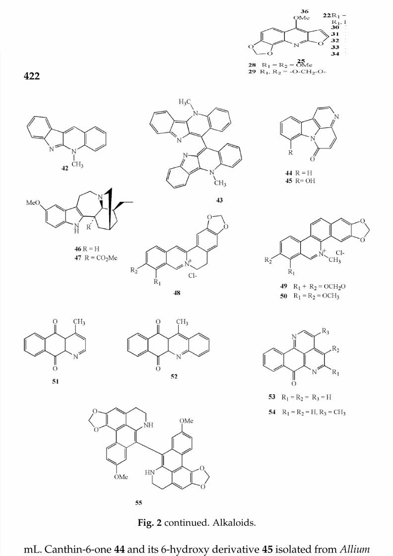
7/23/2019 Novel Therapeutic Agents From Plants
http://slidepdf.com/reader/full/novel-therapeutic-agents-from-plants 433/464
422
mL. Canthin-6-one 44 and its 6-hydroxy derivative 45 isolated from Alliumneapolitanum significantly inhibits the growth of a panel of fast-growingMycobacterial species of M. fortuitum, M. smegmatis, M. phlei and M. abcessus.MICs range from 8-32 µg/mL. (O’Donnell and Gibbons, 2007). Otherstructurally complex indole alkaloids with modest antimycobacterialactivities (MICs 50-100 µg/mL) include ibogaine 46 and voacangine 47
isolated from Tabernaemontana citrifolia (Rastogi et al., 1998). The bis- benzylisoquinoline alkaloid berberine 48 is found in numerous genera
Fig. 2 continued. Alkaloids.
© 2009 by Taylor & Francis Group, LLC

7/23/2019 Novel Therapeutic Agents From Plants
http://slidepdf.com/reader/full/novel-therapeutic-agents-from-plants 434/464
423
including species of Coptis and Berberis (Hsieh et al., 2007) inhibits thegrowth of M. smegmatis at MIC of 25 µg/ml and M. intracellulare (MIC >100µg/ml) (Okunade et al., 1994; Mitscher and Baker, 1998; Gentry et al., 1998).The structurally related benzo[c]phenanthridine alkaloid chlerythrine 49and sanguinarine 50 are other examples that demonstrated appreciableactivity against MTB H
37Rv, M. avium, M. bovis BCG and M. smegmatis with
MIC of 12.5 µg/mL. It is believed that the iminium ion improves thelipophilicity and hence increases the bioavailability of the alkaloids to theorganisms. Synthetic analogs 52 and 54 of the naturally occurringcleistopholine 51 (from Cleistopholis patens, Annonaceae) and sampangine53 (from Cananga odorata, Annonaceae) strongly inhibit the AIDS pathogen
M. intracellularae (MIC 52, 1.56 µg/mL; 54, 0.39 µg/mL equal to or less thanthose recorded for the control drug rifampin (Peterson et al., 1992). Thedimeric aporphine alkaloid from bidebiline E 55 from Polyalthia cerasoidesalso showed antimycobacterial activity against MTB H
37Ra (MIC 6.25 µg/
mL). (Kanokmedhakul et al., 2007).
3. Flavonoids, coumarins, chromones and xanthones
These plant pigments are very widely distributed in the plant kingdomand have a wide range of biological activities. A good number of them have been reported to exhibit a modest activity against MTB (Copp, 2003;Okunade et al., 2004 and Copp and Pearce, 2006). The structure-activityrelationships of a series of non-natural flavonoids were reported by Lin etal. (2002). They observed that chalcones are more active than the flavonesand flavanones probably because they are more geometrically constrained.
The authors also reported that the 2’-hydroxy substitution of chalcone andflavonoids enhances the antimycobacterial activity. The licochalcone 56isolated from the root of Chinese licorice Glycyrrhiza inflata (Friis-Moller etal., 2002) was very active against MTB (MIC 5-10 µg/mL), M. bovis (MIC10-20 µg/mL) and M. bovis BCG (MIC 5-10 µg/mL) but less potent towards M. intracellulare (MIC 20-80 µg/mL) and M. avium (MIC >80 µg/mL). Aseries of flavonoid compounds 57-67 from Derris indica exhibitedantimycobacterial activity against MTB H
37
Ra with MIC values between6.25 and 100 µg/mL (Koysomboon et al., 2006). The flavonols 68-70 isolatedfrom Haplopappus sonoriensis (Asteraceae) inhibited the growth of MTBH37Rv by 33, 98 and 48% respectively at a concentration of 100 µg/mL(Murillo et al., 2003). The flavonoid artonin F 71 was the most potent of the compounds isolated from Artocarpus rigidus that showed antimyco- bacterial activity against MTB H
37Ra with MIC 6.25 µg/mL (Namdaung et
al., 2006). Others compounds from this plant with moderate activities in
Microplate Alamar Blue assay, include flavonoids 72, 73 (MIC 25 and 50µg/mL respectively), chromone artorigidusin74 and xanthone 75 both
© 2009 by Taylor & Francis Group, LLC
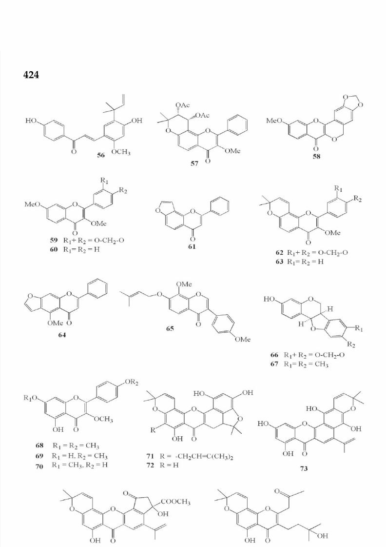
7/23/2019 Novel Therapeutic Agents From Plants
http://slidepdf.com/reader/full/novel-therapeutic-agents-from-plants 435/464
424
Fig. 3. Flavonoids, coumarins, chromene and xanthone.© 2009 by Taylor & Francis Group, LLC

7/23/2019 Novel Therapeutic Agents From Plants
http://slidepdf.com/reader/full/novel-therapeutic-agents-from-plants 436/464
425
Fig. 3 continued. Flavonoids, coumarins, chromene and xanthone.
© 2009 by Taylor & Francis Group, LLC

7/23/2019 Novel Therapeutic Agents From Plants
http://slidepdf.com/reader/full/novel-therapeutic-agents-from-plants 437/464
426
with MIC value of 12.5 µg/mL. 71-73 also show antiplasmodial activitiesand all five compounds (71-75) are cytotoxic to one or two cancer cell lines
but 71 is not toxic at 20 µg/mL. Biflavonoid 76 previously isolated fromthe Mexican plant Celaenodendron mexicanum (Castañeda et al., 1992)showed a mild activity against MTB H
37Rv (MIC 64 µg/mL) (Rivero-Cruz
et al., 2005). Coumarins are also known to possess anti TB activities. Theanti HIV agent ostruthin 77 from Peucedanum ostruthium (Apiaceae) withthe prenyl group at C-6 demonstrates potent activity against differentstrains of the rapidly growing mycobacteria, M. aurum, M. fortuitum, M. phlei and M. smegmatis, with MIC values of 3.4-6.7 µM, analogous to theMIC value obtained for the control isoniazid (Schinkovitz et al., 2003). The
presence of the prenyl group appears to be important to the activity of thecoumarins, since it increases the lipophilicity and hence the bioavailabilityto the organism as earlier observed in some sesquiterpenes (Cantrell et al.,2001). The pyranocoumarin (+) calanolide 78 also known as an anti HIVagent from Calophyllum lanigerum exhibits a strong activity towards MTBH
37Rv (MIC 3.3 µg/mL) and an array of drug resistant strains with MIC
values ranging from 8-16 µg/mL. Preliminary mechanistic studies showedthat this compound swiftly inhibits RNA and DNA synthesis followed byan inhibition of protein synthesis. Other analogs of this compound are alsoshown to exhibit similar antimycobacterial activity (Xu et al., 2004). Thepyranocoumarins dentatin 79 and nor-dentatin 80 from Clausena excavataare modestly active against MTB H
37Ra (MIC 50, 100 µg/mL respectively).
The related natural xanthoxyletin 79B was inactive and hence it is believedthat the prenyl at the C-8 position is essential for the antimycobacterialactivity. The 5-O prenylated furanocoumarins 81, 82 from dill— A. graveolus
also demonstrated moderate activity against a panel of rapidly growingmycobacteria, M. phlei, ATCC 11758, M. aurum Pasteur Institute 104482, M. smegmatis ATCC 14468 and M. abscessus ATCC 19977 with MIC valuesranging from 32-64 µg/mL (Stavri and Gibbons, 2005). The homo-isoflavanone, 3-(4-methoxybenzyl)-7,8-methylenedioxy-chroman-4-one 83and the dihydrobenzopyran 84 isolated from Chlorophytum inornatumexhibit modest antimycoabacterial activities. 83 was active against M. phlei(MIC 16 µg/mL) and M. aurum (MIC 32 µg/mL) while 84 was only activeagainst M. aurum (MIC 64 µg/mL) (O’Donnell et al., 2006). A series of prenylated antimycobacterial xanthones typified by compounds 85, 86 havealso been reported from Garcinia mangostana with MICs ranging from 6.25-25 µg/mL (Suksamrarn et al., 2003).
4. Terpenoids
This group is subdivided into monoterpenes, sesquiterpenes, diterpenesand triterpenes. The antimycobacterial activity of this class of compounds
© 2009 by Taylor & Francis Group, LLC

7/23/2019 Novel Therapeutic Agents From Plants
http://slidepdf.com/reader/full/novel-therapeutic-agents-from-plants 438/464
427
has been comprehensively reviewed by Cantrell et al., 2001; Copp, 2003and Copp and Pearce, 2006. No less than 300 examples of naturallyoccurring terpenoids were reported to have moderate to high antimyco- bacterial activity against MTB and more compounds are being added tothe literature. A few examples of the subdivisions will be discussed hereand the reader is directed to the earlier reviews. A few monoterpenes have been shown to possess antimycobacterial properties. This includes geraniol87 which is moderately active against MTB (MIC 64 µg/mL) (Rajab et al.,1998). Chen et al. (2003) reported that the monoterpene dissectol 88 isolatedfrom Incarvillea dissectifoliola exhibited mild activity towards MTB 90-221387in a disk diffusion assay. Also the bornyl ester 89 from Piper aff. pedicellatum
exhibited a significant activity against the H37Ra strain of MTB H37Ra (MIC25 µg/mL) (Rukachaisirikul et al., 2004).
Sesquiterpenoids particularly the sesquiterpene lactones exhibit a widerange of biological activities and very many of them have been shown tohave highly significant antimycobacterial activities. Farnesol 90 have beenreported to show activity against MTB H
37Rv with MIC of 8 µg/mL.
Sesquiterpenoid lactones of the germacranolide, guaianolide and
eudesmanolide are known to be anti-tubercular with MICs ranging from 2to 128 µg/mL (Cantrell et al., 2001). The germacranolide costunolide 91and dehydrocostuslactone 92 which respectively constitutes 3.8% and 1.5%of the oil expressed from the ripe fruit of Laurus novocanariensis (Laurel oil)exhibit the growth of an array of drug-resistant strains of MTB H
37Ra with
a MIC of 6.25 and 12.5 µg/mL, respectively (Luna-Herrera et al., 2007).Using the fluorometric Alamar Blue microassay, the authors showed thatthe two compounds acted synergistically amplifying the antimycobacterial
activity in contrast to either used alone. Other plant derived germacronolidesesquiterpenes like parthenolide 93, epoxy costunolide 94 and santamarine95 are all moderately active against MTB H
37Rv (MIC 16, 64, 64 µg/mL,
respectively) (Fischer et al., 1998). The eudesmanolide sesquiterpenelactones alantolactone 96 and isoalantolactone 97 were isolated from theroot extract of Inula helenium. Both compounds exhibited antimycobacterialactivity against MTB H
37Rv with MIC values of 32 µg/mL. It has been
suggested that the α-methylene-γ -lactone ring as well as the presence of an additional alkylating site together with moderate to high lipohilicityenhance the in vitro activity of these group of compounds (Fischer et al.,1998 from Copp, 2003; Cantrell et al., 1999a, 2001). The East African plantWarburgia ugandensis afforded the antimycobacterial sesquiterpene lactonemuzigadial 98 that exhibits moderate activities towards a panel of antimycobacterial strains, M. avium and M. phlei, M. fortuitum and M.smegmatis (MIC 16-64 µg/mL ) (Wube et al., 2005).
From the white latex of the Thai plant Pedilanthus tithymaloides wererecently isolated six new poly-O-acylated jatrophane diterpenoids 99- 103
© 2009 by Taylor & Francis Group, LLC

7/23/2019 Novel Therapeutic Agents From Plants
http://slidepdf.com/reader/full/novel-therapeutic-agents-from-plants 439/464
428
Fig. 4. Terpenoids.
© 2009 by Taylor & Francis Group, LLC

7/23/2019 Novel Therapeutic Agents From Plants
http://slidepdf.com/reader/full/novel-therapeutic-agents-from-plants 440/464
429
Fig. 4 continued. Terpenoids.
© 2009 by Taylor & Francis Group, LLC

7/23/2019 Novel Therapeutic Agents From Plants
http://slidepdf.com/reader/full/novel-therapeutic-agents-from-plants 441/464
430
Fig. 4 continued. Terpenoids.
© 2009 by Taylor & Francis Group, LLC

7/23/2019 Novel Therapeutic Agents From Plants
http://slidepdf.com/reader/full/novel-therapeutic-agents-from-plants 442/464
431
with antimycobacterial activities against MTB H37Ra with MIC valuesranging from 12.5 to 100 µg/mL (Mongkolvisut and Sutthivaiyakit, 2007).All the compounds except 100 also demonstrate antimalarial activities Theclerodane diterpenes 104-109 isolated from the bark of Casearia grewiifoliaa plant widely distributed in Thailand exhibited antimycobacterial activityagainst MTB H
37Ra. The MIC values are 12.5 µg/mL for all the compounds
except 108 which has a MIC value of 25 µg/mL. The compounds also arecytotoxic towards some cancer cell lines (Kanokmedhakul et al., 2005a).The diterpenoid (E)-phytol 110, is one of the antimycobacterial constituentsisolated from Morinda citrifolia (Saludes et al., 2002) and the mainantimycobacterial constituent of the Kenyan Shrub Leucas volkensii (Rajab
et al., 1998). It is active against MTB H37Rv with a MIC value of 0.95-3.8µg/mL. The nor-diterpenoid 12-demethylmulticauline 111 has a MIC of 0.46 µg/mL comparable with rifampin and even less than that forethambutol (Cantrell et al., 2001). This compound was the most potentagainst the H
37Rv strain of MTB of the three other aromatic norditerpenoids
and two abietane diterpenoids earlier isolated from the roots of Salviamulticaulis (Ulubelen et al., 1997). A good number of phobol esters havealso been reported to show moderate to good antimycobacterial activities.One of the phobol esters 112 from the tree Sapium indicum (Euphorbiaceae),exhibits activity against MTB H
37Ra (MIC 3.12 µg/mL) comparable to the
positive control, kanamycin sulfate, but not as active as isoniazid. Thestereochemistry at position 4 is very important to the activity of thiscompound (Chumkaew et al., 2003). The secokauranes 113 and 114 fromCroton kongensis (Euphorbiaceae) (Thongtan et al., 2003) show significantactivity towards MTB H
37Ra with MIC values of 25 and 6,25 µg/mL
respectively.Hopane terpenoids, 115 from Sapium haematospermum (Woldemichael
et al., 2004) and betulin 116, and betulinic acid 117 isolated from Valerianalaxiflora (Gu et al., 2004) respectively, inhibit the growth of MTB H
37Rv
(ATCC 27294) with MIC values of 13.4, 30, 62.1 µg/mL. Oleananetriterpenes with antimycobacterial activities were also isolated from theseplants. The oleanane triterpenes 118, 119 as well as the ubiquitous
oleanolic acid from Lantana hispida, exhibit moderate activity against MTBH37
Rv (MIC 50 µg/mL 118, 119 and 25 µg/mL for oleanolic acid). Thesecompounds were also tested against a series of drug-resistant variants of H37Rv strains with similar results. The 3β,25-epoxy oleanane triterpenoids120 and 121 isolated and from Lippia turbinata and from other Lantanaspecies (Barre et al., 1997; Watcher et al., 2001; Okunade et al., 2004) exhibitantimycobacterial activity against MTB H
37Rv with MIC values of 38, 36
µg/mL respectively (Watcher et al., 2001). Of particular importance is the
18β,28-Epoxy oleanane triterpene, Aegicerin, 122 from Clavija procera thatwas tested against 37 different sensitive and resistant strains of MTB H
37Rv
© 2009 by Taylor & Francis Group, LLC

7/23/2019 Novel Therapeutic Agents From Plants
http://slidepdf.com/reader/full/novel-therapeutic-agents-from-plants 443/464
432
and showed activity with MIC ranging between 1.6 and 3.12 µg/mL (Rojaset al., 2006). The epoxy oleanane 123 isolated from the Junella tridens andseveral other oleanane and ursane triterpenes including the well knownoleanolic acid 124 and ursolic acid 125 exhibit growth inhibition activitiestowards MTB H
37Rv. MIC values for 123-125 are respectively 64, 50 and
15 µg/mL (Caldwell et al., 2000; Copp, 2003). The cycloartane triterpenoids126-128 exhibit modest activity towards MTB H
37Rv (MIC 12.5, 8.0, 8.0 µg/
mL respectively). 127 and 128 also show weak activities towards the verocell line (Cantrell et al., 1996; Saludes et al., 2002).
5. Steroids and saponins
The plant Morinda citrifolia (Rubiaceae), afforded among others sterols thatare strongly active against MTB H
37Rv with MIC values ranging from < 2
to 32 µg/mL (Okunade et al., 2004). Thalia multiflora, an aquatic plant fromSouth America afforded nine sterols and other classes of compounds. Fourof the sterols 129-132 show strong activities (MIC 1.0-4.0 µg/mL) againstMTB H
37Rv (ATCC27294) (Gutierrez-Lugo et al., 2005). The naturally
Fig. 5. Steriods/saponins.
© 2009 by Taylor & Francis Group, LLC

7/23/2019 Novel Therapeutic Agents From Plants
http://slidepdf.com/reader/full/novel-therapeutic-agents-from-plants 444/464
433
Fig. 5 continued. Steriods/saponins.
occurring ergosterol peroxide 133 isolated from Ajuga remota (Cantrell etal., 1999b) and Euphorbia lagascae (Duarte et al., 2007) was shown to possessantimycobacterial activity. 133 and other peroxides 134, 135 from theArgentinian plant Ruprechtia triflora (Woldemichael et al., 2003) were alsoreported to have activity against MTB H
37Rv with MIC value ranging from
2-4 µg/mL. Fusidic acid, 136, isolated from the fungus Fusidium coccneum(Godtfredsen et al., 1962) is a widely used antibiotic that has been shown
© 2009 by Taylor & Francis Group, LLC

7/23/2019 Novel Therapeutic Agents From Plants
http://slidepdf.com/reader/full/novel-therapeutic-agents-from-plants 445/464
434
to inhibit the growth of 30 clinical isolates of MTB (MIC 4, and 32-64 µg/mL) and M. kansasii (MIC 2-64 µg/mL ) (Hoffner et al., 1990; Cantrell etal., 1996; Witzig and Frazblau, 1993). Rugutt and Rugutt (2002) studiedthe relationships between molecular properties and antimycobacterialactivities of nine steroids including fusidic acid. They suggested that themaximum antimycobacterial activity seemed to depend on hydrophobicityand type of substituents on the phytyl moiety on the steroidal backbone.The saponin 137 (Figure 5) obtained from the gorgonian octocoral Eunicea pinta, (Shi et al., 2002) the jujubogenin saponin 138, as well as 139 fromthe stems of Colubrina retusa (Rhamnaceae) (ElSohyl et al., 1999), alsoexhibited good antimycobacterial activity. Compound 137 was active
against MTB H37Rv (MIC values 6.25 µg/mL) while 138 and 139 were activeagainst M. intracellulare with MIC values of 50 and 10 µg/mL respectively.The seco-steroids phasalin B, 140 from Physalis angulata (Solanaceae)(Januario et al., 2002) show moderate activity against MTB H
37Rv (MIC 32
µg/mL). Woldemichael et al. (2003) also reported some sterols fromRuprechtia triflora (Polygonaceae) with moderate to very good antimyco- bacterial activity against MTB.
6. Quinones, phenols and polyphenols
The simple quinone Primin (2-methoxy-6-pentylcyclohexa-2,5-diene-1,4-dione) 141, a natural compound occuring in Primula obconica and someother plant species is a sensitizer and the source of allergic contactdermatitis, primrose dermatitis in some humans. This compound has broadspectrum antiparasitic activities and moderate activity against MTB H
37Rv
(ATTC27294) (MIC 60 µg/mL; Tasdemir et al., 2006). Plumbagin 142 fromPlumbago zeylanica, 3-methoxjuglone 143 from Engelhardia roxburghiana andlapachol 144 commonly found in most Tabebuia (Biginoniaceae) family allexhibit antimycobacterial activities (Mossa et al., 2004; Tran et al., 2004). Asynthetically derived plumbagin 142 was shown to be active against M.avium and M. smegmatis (MIC value in each case is 12.5 µg/mL). 143 wasactive against MTB H
37Rv with an MIC value of 0.2 µg/mL while lapachol
144 exhibited activity towards M. smegmatis (MIC 50 mg/mL) Anthra-quinones 145, 146 isolated from Prismatomeris fragrans (Kanokmedhakulet al., 2005b) exhibit modest activity against MTB H37Ra (MIC 25 and 50µg/mL respectively). A bisnaphthoquinonoid, diospyrin 147, isolated fromEuclea natalensis showed a modest activity against drug-susceptible and-resistant strains of MTB H
37Rv (MIC 100 µg/mL). The synthetic amino-
acetate derivative 147b of diospyrin dimethyl ether showed improvedactivities (MIC 10-50 µg/mL) (Lall et al., 2003).
The isolated deoxy preussomerins palmarumycin 148 believed to beisolated from fungi associated on dried fruits of Diospyros ehretioides as
© 2009 by Taylor & Francis Group, LLC

7/23/2019 Novel Therapeutic Agents From Plants
http://slidepdf.com/reader/full/novel-therapeutic-agents-from-plants 446/464
435
Fig. 6. Quinones, phenols/polyphenols.
© 2009 by Taylor & Francis Group, LLC

7/23/2019 Novel Therapeutic Agents From Plants
http://slidepdf.com/reader/full/novel-therapeutic-agents-from-plants 447/464
436
well as napthaquinone palmarumycin JC2 149 from this plant exhibitedactivity towards MTB H37Ra (MIC, 148, 1.56-3.12 µg/mL; 149, 6.25 µg/mL) (Prajoubklanga et al., 2005). The thiocarbamate 150 analog of thenaturally occurring sesquiterpene-phenol aureols 151 from the JamaicanSponge Smenospongia aurea, was active against Mycobacterium tuberculosis
(H37Rv). 150 completely inhibited the growth at a concentration of 6.25 µg/mL while the natural compound 151 showed only 31% inhibition at the
Fig. 6 continued. Quinones, phenols/polyphenols.
© 2009 by Taylor & Francis Group, LLC

7/23/2019 Novel Therapeutic Agents From Plants
http://slidepdf.com/reader/full/novel-therapeutic-agents-from-plants 448/464
437
same concentration (Hu et al., 2002). Phenol 152 from Iostephane heterophyllaand 153 from Larrea divaricata exhibited moderate activity against MTB(Rivero-Cruz et al., 2005) MIC 16 and 50 µg/mL respectively). The tricyclicdaiphenol ether engelhardione 154 has a potent activity against MTBH
37Rv with an MIC of 0.2 µg/mL (Lin et al., 2005). The lichenous fungal
metabolites preussomerins 155-160 from Microsphaeropsis sp were alsoreported to have antimycobacterial activity against MTB H
37Ra with MIC
values ranging from 3.12 to 25 µg/mL (Seephonkai et al., 2002;Prajoubklang et al., 2005,). The antimycobacterial activity of punicalagin161 a polyphenol from Combretum molle (Combretaceae) was evaluatedusing two strains of MTB, typus humanus and an unidentified patient
strain. It is the only tannin so far associated with antimycobacterial activityhowever the activity is very weak with MIC value greater than 200 µg/mL(Asres et al., 2001).
7. Peptides
Most of the peptides reported to possess antimycobacterial activities areobtained from either marine plant/organisms, fungi or bacterial sources.This is well covered in the reviews by Copp (2003), Okunade et al. (2004),Copp and Pearce (2007) and Kahalalide A 162 was isolated by Hamannet al. (1996) in Hawaii from the marine mollusk Elysia rufescens and itsalgal diet Bryopsis sp. It is the simplest of a family of cyclic peptidesidentified from this extract, and showed activity against Mycobacteriumtuberculosis, inhibiting 83% of bacterial growth at 12.5 µg/mL (El Sayed,2000). Kahalalide A does not contain obviously reactive functional groups
and it is not cytotoxic to various tumor cell lines, suggesting a selectiveantibacterial target. This compound has recently been synthesized througha solid phase combinatorial chemistry (Bourel-Bonnet et al., 2005).Viomycin (tuberactinomycin B) 163, a well-studied member of the familyof basic cyclic peptide was once prescribed for the treatment of tuberculosis,and has been shown to block translocation during protein biosynthesis. Ithas a MIC value of 0.32 µg/mL when tested against M. bovis BCG ATTC27291 (Oliva et al., 1998). The gene cluster encoding viomycin biosynthesiswas identified and cloned from Streptomyces vinaceus (Yin et al., 2003). Thecyclodepsipeptides enniatins 164-166 from fungi and marine sources have been reported to demonstrate modest to high antimycobacterial activityagainst MTB with MIC values ranging from 3.12 to 6.25 µg/mL. (Nilanontaet al., 2003) and are mostly not cytotoxic. Syringomycin 167, a highlyfunctionalized cyclic peptide shows a high antimycobacterial activityagainst a related model M. smegmatis with MIC of 1.50 µg/mL comparable
or even lower than those obtained for the primary drugs and secondarydrugs. The antibacterial thiazole peptides 168-170 isolated from the
© 2009 by Taylor & Francis Group, LLC

7/23/2019 Novel Therapeutic Agents From Plants
http://slidepdf.com/reader/full/novel-therapeutic-agents-from-plants 449/464
438
Fig. 7. Peptides.
© 2009 by Taylor & Francis Group, LLC

7/23/2019 Novel Therapeutic Agents From Plants
http://slidepdf.com/reader/full/novel-therapeutic-agents-from-plants 450/464
439
Fig. 7 continued. Peptides.
© 2009 by Taylor & Francis Group, LLC

7/23/2019 Novel Therapeutic Agents From Plants
http://slidepdf.com/reader/full/novel-therapeutic-agents-from-plants 451/464
440
saprophytic Nocardia sp. (ATCC 202099) fermentation broth extractionexhibit very potent activity against MTB, M. avium and a variety of gram-positive pathogens (Pucci et al., 2004). Nocathiacin 1 168 exhibits activityagainst MTB (ATCC 35828, MIC < 0.008 µg/mL), M.avium (A26778, MIC0.06 µg/mL) and M. avium (A26640, MIC 0.25 µg/mL).
8. Miscellaneous
Boonphong et al. (2007) recently isolated a number of dihydrobenzoxepinstypified by compounds 171-173 and other compounds from Bauhinia purpurea possessing antimycobacterial activities in the microplate Alamar
Blue assay. The dihydrobenzoxepin 172 has the highest activity with MICof 24 µM against MTB H
37Ra. However, all the compounds are cytotoxic
Fig. 8. Miscellanous.
© 2009 by Taylor & Francis Group, LLC

7/23/2019 Novel Therapeutic Agents From Plants
http://slidepdf.com/reader/full/novel-therapeutic-agents-from-plants 452/464
441
against KB and BC cell lines. Lariatins A 174, and B 175 inhibit the growthof M. smegmatis with respective MIC values of 3.13 and 6.25 µg/mL in agardilution method, only lariatin A inhibits the growth of MTB H
37Rv in liquid
microdilution method. Lariatins A and B were separated and purified fromthe culture broth of Rhodococcus jostii KO1B0171, which was isolated fromsoil aggregates collected in Yunnan, China (Iwatsuki et al., 2007).
Conclusion
The goal of identifying suitable new TB drugs is multifaceted and thecurrent focus in drug discovery efforts is to identify compounds which are
bactericidal, are mechanistically different from those currently used so thatcross-resistance does not develop, and are effective against non-replicating,persistent types so as to shorten the duration of therapy. They should also be suitable for oral daily dosing with few toxic side reactions, and be costeffective for global applications. (Goldman, 2007; Personal communication).
While broad screening efforts have identified a multitude of bioreactiveand structurally unique compounds, most have been published before even
preliminary toxicity testing has taken place. Furthermore when they have been subject to additional testing for their toxicity, bioavailability andpotency profiles in animal models their promise as possible new TB drugshas been disappointing. These results are not unique to this endeavor andhave been commonplace in the pharmaceutical industry whenever thisapproach has been undertaken in the past.
Under-appreciated for their potential are those unique compounds
which have been derived from ethnomedical leads. This is particularly trueof plants used as traditional medicines for the treatment of respiratoryinfections and even tuberculosis. Noteworthy among these would be thoseremedies, which are ingested and possibly low in toxicity, rather thaninhaled or utilized in other ways e.g., rubifacient. Too frequently their bioreactive structures have been published before adequate testing hasshown their parameters of dosage, safety and efficacy, particularly for drugresistant and recalcitrant types of infections. This has resulted in many
promising candidates being lost to drug discovery programs of thepharmaceutical industry who would mandate that patent protection is anecessary prerequisite to development.
To overcome this issue with certain viable candidates already in thepublic domain, semi-synthesis of known bioreactive molecules might be away of rescuing the potential of these candidates. Moreover, these strategiesmay not only reduce their toxicity, but also improve their potency and
bioavailability. In this regard, a good number of phytochemicals have thepotential as lead compounds but so far only a few have been studied
© 2009 by Taylor & Francis Group, LLC

7/23/2019 Novel Therapeutic Agents From Plants
http://slidepdf.com/reader/full/novel-therapeutic-agents-from-plants 453/464
442
beyond preliminary functional testing. While inexpensive preliminarytoxicity studies in vitro have their limitations, they nonetheless are usefulin providing needed information as to whether or not additional costlypreclinical animal trials are warranted. Furthermore there is always therisk that promising results elicited in preclinical studies may not beapplicable to humans (Goldman, 2007; Personal Communication).
The carbazole alkaloids are examples of lead compounds beingderived from an ethnomedical source. These have shown moderate butconsistent activity across the literature and are good candidates for furtherstudies (Ma et al., 2005). Notwithstanding a few number of plant derivedcompounds have been tested in vivo. Two examples discussed below
include the alkaloid tryptanthrin 36 that has been well studied because of its potency, simple structure and ease of synthesis. It has been shown thatwhen a panel of multiply drug-resistant strains was utilized its potencywas even superior to that of isoniazid. Structural-activity studies have also been carried out. One of the analogs, compound PA 005 was tested in vivoin infected mice (rBCGlux) with unsatisfactory results and hence was notpursued further. It was effective against rBCGlux in macrophages at 0.23
mg/L comparative to isoniazid but inferior to it when tested at 0.06 or0.015 µg/mL. It was modestly effective when given orally at 50 mg/kg toinfected mice (rBCGlux) (Mitscher and Baker, 1998). Another example isthe pyranocoumarin (+)-calanolide A 78 Efficacy evaluations in macro-phages revealed that the compound significantly inhibited intracellularreplication of M. tuberculosis H
37Rv at concentrations below the MIC
observed in vitro. Preliminary mechanistic studies indicated that it rapidlyinhibits RNA and DNA synthesis followed by an inhibition of proteinsynthesis (Xu et al., 2004). Using 78 as a template, a library of compounds(over 60) has been produced and screened against MTB H
37Ra. Seven of
these have MIC less than 12.8 µg/mL and four of them were bactericidal(Xu et al., 2006).
To be able to take more of these compounds beyond the preliminaryscreening there is no doubt that more research money is needed, with readyaccess to specialty testing laboratories being a primary consideration. It is
hoped that the agencies funding this research will encourage researchersto consider needed additional research before they rush their discoveryinto publication.
As recommended earlier, (Elvin-Lewis, 2006), the low cost, provisionalpatent mechanism in the United States should provide Americanresearchers with needed protection during the period of a year whentoxicity and other mechanistic assays are being conducted. Investigators
in other countries should be aware of national or international patent lawswhich can afford needed protection during this critical period.
© 2009 by Taylor & Francis Group, LLC

7/23/2019 Novel Therapeutic Agents From Plants
http://slidepdf.com/reader/full/novel-therapeutic-agents-from-plants 454/464
443
Acknowledgement
The authors will like to thank Dr. R.C. Goldman of the Program Office,Therapeutics Research Program, NIH (DHHS/NIH/NIAID/Division of
AIDS) for useful discussions and Elsevier Publishers for copyrightpermission.
References
Adams, W., A.A. Wube, F. Bucar, R. Bauer, O. Kunert and F. Haslinger. 2005.
Quinolone alkaloids from Evodia rutaecarpa: A potent new group of antimycobacterial compounds. Int J. Antimicrob. Agents 26(3) 262-264.
Aguinaldo, A.M., V.M. Dalangin-Mallar, A.P. Macabeo, L.T. Byrne, F. Abe, T.Yamauchi and S.G. Franzblau. 2007. Quinoline alkaloids from Lunasia amarainhibit Mycobacterila tuberculosis H37Rv in vitro. Int. J. Antimicrob. Agents.29 (6), 744-746.
Arnold, H.-J. and M. Gulumian. 1984. Pharmacopoeia of traditional medicine inVenda. J. Ethnopharmacol, 12, 35-74.
Ashtekar, D.R., F. Fernandes, B.G. Khadse and M.V. Shirodkar. 1987. A rapidmethod for the evaluation of new antituberculous agents. Chemotherapy(Basel, Switzerland) 33(1), 22-27.
Asres, K., F. Bucar, S. Edelsbrunner, T. Kartnig, G. Hoger and W. Thiel. 2001.Investigations on antimycobacterial activity of some Ethiopian medicinalplants. Phytother. Res, 15, 323 326.
Baeew, J.T., B.F. Bowden, J.C. Coll, J. De Jesus, V.E. De La Fuente, G.C. Janairoand C.Y. Ragasa. 1997. A bioactive triterpene from Lantana camara.Phytochem 45, 321-324.
Bensky, D. and A. Gamble. 1993. Chinese Herbal Medicine: Materia Medica,Revised edition. Eastland Press, Inc. Seattle, USA.
Boonphong, S., P. Puangsombat, A. Baramee, C. Mahidol, S. Ruchirawat and P.Kittakoop. 2007. Bioactive Compounds from Bauhinia purpurea PossessingAntimalarial, Antimycobacterial, Anti-Inflammatory, and Cytotoxic Activities. J. Nat. Prod. 70, 795-801.
Boshoff, H.I. and C.E. Barry. 2005. Tuberculosis—metabolism and respiration inthe absence of Growth. Nat Rev Microbial. 3(1): 70-80.
Bourel Bonnet, L., K.V. Rao, M.T. Hamann and A. Ganesan. 2005. Solid-PhaseTotal Synthesis of Kahalalide A and Related Analogues. J. Med. Chem. 48(5),1330-35.
Burkill, H.M. 1985. The Useful Plants of West Tropical Africa, Royal BotanicGardens, Kew, U.K. Vol 1, 1995; Vol 3, 1997; Vol 4, 2000; Vol 5.
Burkill, H.M. 1997. The Useful Plants of West Tropical Africa. The Royal BotanicGarden, Kew, UK.
Caldwell, C.G., S.G. Franzblau, E. Suarez and B.N. Timmermann. 2000. Oleanane
triterpenes from Junellia tridens. J. Nat. Prod. 63, 1611-1614.Cantrell, C.L., T. Lu, F.R. Fronczek, N.H. Fischer, L.B. Adams and S.G. Franzblau.
© 2009 by Taylor & Francis Group, LLC

7/23/2019 Novel Therapeutic Agents From Plants
http://slidepdf.com/reader/full/novel-therapeutic-agents-from-plants 455/464
444
1996. Antimycobacterial cycloartanes from Borrichia frutescens. J. Nat Prod.59: 1131-1136.
Cantrell, C.L, M.S. Rajab, S.G. Franzblau, F.R. Fronczek and N.H. Fischer. 1999b.Antimycobacterial ergosterol-5,8-endoperoxide from Ajuga remota. Planta
Med 65(8): 732-4.Cantrell, C.L, L. Abate, F.R. Fronczek, S.G. Franzblau, L. Quijano and N.H. Fischer.
1999a. Antimycobacterial eudesmanolides from Inula helenium and Rudbeckiasubtomentosa. Planta Med. 65(4), 351-355.
Cantrell, C.L., S.G. Franzblau and N.H. Fischer. 2001. Antimycobacterial plantterpenoids. Planta Med. 67, 685-694.
Canueto-Quintero J, F.J. Caballero-Granado, M. Herrero-Romero, A.Domínguez-Castellano, P. Martín-Rico, E.V. Verdú, D.S. Santamaría, R.C.
Cerquera, M. Torres-Tortosa, Grupo Andaluz para el Estudio de lasEsfermedades Infecciosas. 2003. Epidemiological, clinical, and prognosticdifferences between the diseases caused by Mycobacterium kansasii and Mycobacterium tuberculosis in patients infected with HIV: a multicenter study.Clin Infect Dis. 37: 584-90.
Castañeda, P., M.R.E. García, B.E. Hernández, B.A. Torres, A.L. Anaya and R.Mata. 1992. Effects of some compounds isolated from Celaenodendronmexicanum Standl (Euphorbiaceae) on seeds and phytopathogenic fungi. J.Chem. Ecol. 18: 1025-1037.
Chen, W., Y. Shen and J. Xu. 2003. Dissectol A, an unusual monoterpene glycosidefrom Incarvillea dissectifoliola. Planta Medica 69, 579-582.
Chevallier. A. 1996. The Encyclopedia of Medicinal Plants Dorling Kindersley.London ISBN 9-780751-303148.
Chiej, R. 1984. Encyclopedia of Medicinal Plants. MacDonald ISBN 0-356-10541.Chumkaew, P., C. Karalai, C. Ponglimanont and K. Chantrapromma. 2003.
Antimycobacterial activity of phorbol esters from the fruits of Sapiumindicum. J. Nat. Prod. 66, 540-543.
Chung, G.A.C., Z. Aktar, S. Jackson and K. Duncan. 1995. High-throughput screenfor detecting antimycobacterial agents. Antimicrob. Agents Chemother. 39,2235-38.
Collins, L. and S.C. Franzblau. 1997. Microplate alamar blue assay versus BACTEC460 system for high-throughput screening of compounds against Mycobacterium tuberculosis and Mycobacterium avium. Antimicrob. AgentsChemother. 41, 1004-1009.
Copp, B.R. and A.N. Pearce. 2007. Natural product growth inhibitors of
Mycobacterium tuberculosis. Nat. Prod. Rep. 24, 278-297.Copp, Brent R. 2003. Antimycobacterial natural products. Nat. Prod. Rep, 20,535-557.
Cragg, G.M., D.J. Newman and K.M. Snader. 1997. Natural products in drugdiscovery and development. J. Nat. Prod. 60, 52-60.
Cox, H., S. Hargreaves and G. Ismailov. 2003. Effect of multidrug resistance onglobal tuberculosis control. Lancet 362, 1858-1859.
Czajkowska, M., E. Augustynowicz-Kopeæ, Z. Zwolska, I. Bestry, M.Martusewicz-Boros, M. Marzinek, E. Radzikowska, P. Remiszewski, B.Roszkowska-Sliz, J. Szopiñski, J. Za l eska, J. Zych and E. Rowiñska-Zakrzewska. 2002. Pulmonary Mycobacterioses—frequency of occurrence,© 2009 by Taylor & Francis Group, LLC

7/23/2019 Novel Therapeutic Agents From Plants
http://slidepdf.com/reader/full/novel-therapeutic-agents-from-plants 456/464
445
clinical spectrum and predisposing factors. Pneumonol Alergol Pol 70: 550-560.
Dalziel, J.M. 1936. Useful Plants of West Tropical Africa: The Crown Agents forOverseas Governments and Administrations. London, UK.
Duarte, N., M-J. U. Ferreira, M. Martins, M. Viveiros and L. Amaral L. 2007.Antibacterial Activity of Ergosterol Peroxide against Mycobacteriumtuberculosis: Dependence upon System and Medium Employed. Phytother.Res. 21, 601-604.
Ducati, R.G, A. Ruffino-Netto, L.A. Basso and D.S. Santos. 2006. Mem Inst OswalsoCruz, Rio de Janiero 101 (7), 697-714.
Dye, C., S. Scheele, P. Dolin, V. Pathania and M.C. Raviglione. 1999. Consensusstatement. Global burden of tuberculosis: estimated incidence, prevalence,
and mortality by country. WHO global surveillance and monitoring project. JAMA, 282, 677-686.El Sayed, K.A., M.T. Hamann, H.A. Abd El-Rahaman and A.M. Zaghloul. 1998.
New Pyrrole Alkaloids from Solanum sodomaeum. J. Nat Prod 61, 848-850.El Sayed, K.A., P. Bartyzel, X. Shen, T.L. Perry, J.K. Zjawiony and M.T. Hamann.
2000. Marine natural products as antituberculosis agents. Tetrahedron, 56(7),949-53.
ElSohly, H.N., S. Danner, X.-C. Li, A.C. Nimrod and A.M. Clark. 1999. Newantimycobacterial saponin from Colubrina retusa. J. Nat. Prod. 62, 1341-1342.
Elvin-Lewis, M. 2006. Evolving concepts related to achieving benefit sharing forCustodians of traditional knowledge. www. Ethnobotanyjournal.org/vol4/i1547-3465-04-071.pdf
Fatkenheuer, G, H. Taelman, P. Lepage, A. Schwenk and R. Wenzel. 1999. Thereturn of Tuberculosis. Diagn Microbiol Infect Dis. 34, 139-146.
Fischer, N.H., T. Lu, C.L. Cantrell, L. Quijano and S.G. Franzblau. 1998.Antimycobacterial evaluation of germacranolides. Phytochem. 49, 559.
Friis-Moller, A., M. Chen, K. Fuursted, S.B. Christensen and A. Kharazmi. 2002.
In vitro antimycobacterial and antilegionella activity of licochalcone A fromChinese licorice roots. Planta Med. 68, 416-419.
Gautam, R., A. Saklani and S.M. Jachak. 2007. Indian medicinal plants as a sourceof antimycobacterial agents. J. Ethnopharmacol 110, 200-234.
Gentry, E.J., H.B. Jampani, A. Keshavarz-Shokri, M.D. Morton, D.V. Velde, H.Telikepalli, L.A. Mitscher, R. Shawar, D. Humble and W. Baker. 1998.Antitubercular Natural Products: Berberine from the Roots of Commercial Hydrastis canadensis Powder. Isolation of Inactive 8-Oxotetrahydro-
thalifendine, Canadine, β -Hydrastine, and Two New Quinic Acid Esters,Hycandinic Acid Esters-1 and -2. J, Nat. Prod. 61, 1187.Gibbons, S., F. Fallah and C.W. Wright. 2003. Cryptolepine hydrochloride: A
potent antimycobacterial alkaloid derived from Cryptolepis sanguinolenta.Phytother. Res. 17, 434-436.
Gillespie, S.H., E.D. Morrisseyl and D. Everett 2001. A comparison of the bactericidal activity of quinolone antibiotics in a Mycobacterium fortuitummodel. J. Med. Microbiol. 50, 565-570.
Godtfredsen, W.O., S. Jahnsen, H. Lorck, K. Roholt and L. Tybring. 1962. Fusidicacid: A new antibiotic. Nature, 987.
Gu, J.-Q., Y. Wang, S.G. Franzblau, G. Montenegro, D. Yang and B.N.© 2009 by Taylor & Francis Group, LLC

7/23/2019 Novel Therapeutic Agents From Plants
http://slidepdf.com/reader/full/novel-therapeutic-agents-from-plants 457/464
446
Timmermann. 2004. Antitubercular constituents of Valeriana laxiflora. PlantaMedica 70(6), 509-514.
Glover, P.E., J. Stewart and M.D. Gwynne. 1996. Masai and Kipsigis notes onEast African plants, Part III—Medicinal uses of plants. East African agricultural
and forestry journal, 32, 2, 200-207.Gutierrez-Lugo, M.-T., Y. Wang, S.G. Franzblau, E. Suarez and B.N. Timmermann.
2005. Antitubercular Sterols from Thalia multiflora Horkel ex Koernicke.Phytother Res. 19, 876-880.
Hamann, M.T., C. Otto, P.J. Scheuer and D.C. Dunbar. 1996. Kahalalides: bioactivepeptides from a marine mollusk Elysia rufescens and its algal diet Bryopsissp. J. Org. Chem. 61, 6594-6600.
Hartwell, J.L. 1969. Plants used against cancer. A survey. Lloydia 62, pp. 153-205.
Heifets, L.B. 1986. Rapid automated methods (BACTEC System) in clinicalmycobacteriology. Semin. Respir. Infect. 1, 242-249.
Heifets, L. 1988. Qualitative and quantitative drug-susceptibility tests in myco- bacteriology. Am. Rev. Respir. Dis. 137(5), 1217-1222.
Hoffner, S.E, B. Olsson-Liljquest, K.J. Rydgård, S.B. Svenson and G. Källenius.1990. Susceptibility of mycobacteria to fusidic acid. Eur J Clin Microbiol InfectDis. 9(4): 294-7.
Houghton, P.J., T.Z. Woldemariam, Y. Watanabe and M. Yates. 1999. Activity
against Mycobacterium tuberculosis of alkaloid constituents of angostura bark, Galipea officinalis. Planta Med. 65, 250-254.Hsieh Y.S., W.H. Kuo, T.W. Lin, H.R. Chang, T.H. Lin, P.N. Chen and S.C. Chu.
2007. Protective Effects of Berberine against Low-Density Lipoprotein (LDL)Oxidation and Oxidized LDL-Induced Cytotoxicity on Endothelial Cells. J.Agric Food Chem. 55(25) 10437.
Hu, J.-F., J.A. Schetz, M. Kelly, J.N. Peng, K.K. Ang, H. Flotow, C.Y. Leong, S.B.Ng, A.D. Buss, S.P. Wilkins and M.T. Hamann. 2002. New antiinfective and
human 5-HT2 receptor binding natural and semisynthetic compounds fromthe Jamaican sponge Smenospongia aurea. J. Nat. Prod. 65(4), 476-480.Iwatsuki, M., R. Uchida, Y. Takakusagi, A. Matsunoto, C.L. Jiang, Y. Takahashi,
M. Arai, S. Kobayashi, M. Matsumoto, J. Inokoshi, H. Tomoda and S. Omura.2007. Lariatins, Novel Anti-mycobacterial Peptides with a Lasso Structure,Produced by Rhodococcus jostii KO1-B0171. J. Antibiot. (Tokyo) 60(6) 357-363.
Iwu, M.W., A.R. Duncan and C.O. Okunji. 1999. New antimicrobials of plantorigin. pp. 457-462. In: J. Janick (ed.). Perspectives on new crops and newuses. ASHS Press, Alexandria, VA, USA.
Jachak, S.M. and R. Jain. 2006. Current Status of Target-Based AntimycobacterialNatural Products. Anti-Infective Agents in Medicinal Chemistry, 5, 123-133Bentham Science Publishers Ltd.
Januário, A.H., E.R. Filho, R.C. Pietro, S. Katshima, D.N. Sato and S.C. Franca.2002. Antimycobacterial physalins from Physalis angulata L. (Solanaceae).Phytother. Res. 16, 445-448.
Jimenez-Arellanes, A., M. Meckes, J. Torres and J. Luna-Herrera. 2007.
Antimycobacterial triterpenoids from Lantana hispida (Verbenaceae). J.Ethnopharmacol 111, 202-5.
© 2009 by Taylor & Francis Group, LLC

7/23/2019 Novel Therapeutic Agents From Plants
http://slidepdf.com/reader/full/novel-therapeutic-agents-from-plants 458/464
447
Johnson, T. 1999. CRC Ethnobotany Desk Reference. CRC Press. Boca Raton,Florida ISBN 084931187X.
Kanokmedhakul, S., K. Kanokmedhakul, T. Kanarsa and M. Buayairaksa. 2005a.New Bioactive Clerodane Diterpenoids from the bark of Casearia grewiifolia.
J. Nat. Prod. 68, 183-185.Kanokmedhakul, K., S. Kanokmedhakul and R. Phatchana. 2005b. Biological
activity of Anthraquinones and Triterpenoids from Prismatomeris fragrans. J.Ethnopharmacol 100, 284-8.
Kanokmedhakul, S., K. Kanokmedhakul and R. Lekphrom. 2007. BioactiveConstituents of the Root of Polyathia cerasoides. J. Nat. Prod. 70(9), 1536-8
Kareru, P.G., G.M. Kenji, A.N. Gachanja, J.M. Keriko and G. Mungai. 2007.Traditional medicine among the Embu and Mbeere peoples of Kenya.
African Journal of Traditional, Complementary and Alternative Medicines 4(1), 75-86.
Kinghorn, A.D. 2001. Pharmacognosy in the 21st century. Journal of Pharmacyand Pharmacology 53, 135-148.
Kobaisy, M., Z. Abramowski, L. Lermer, G. Saxena, R.E.W. Hancock and G.H.N.Towers. 1997. Antimycobacterial Polyynes of Devil’s Club (Oplopanaxhorridus) a North American Native Medicinal Plant. J. Nat. Prod. 60(11),1210-1213.
Kobayashi, J., J.F. Cheng, H. Nakamura, Y. Ohizumi, Y. Hirata, T. Sasaki, T. Ohtaand S. Nozoe. 1988. Ascididemin, a novel pentacyclic aromatic alkaloid withpotent antileukemic activity from the Okinawan tunicate Didemnum sp.Tetraheron Lett. 29, 1177-80.
Kokwaro, J.O. 1976. Medicinal Plants of East Africa. East African Lit. Bureau,Kampala, Nairobi, Dar Es Salaam.
König, G.M., A.D. Wright and S.G. Franzblau. 2000. Assessment of antimyco- bacterial activity of a series of mainly marine derived natural products. Planta
Med. 66, 337-342.Koysomboon, S., I. van Altena, S. Kato and K. Chantrapromma. 2006.Antimycobacterial flavonoids from Derris indica. Phytochem. 67, 1034-1040.
Lall, N., M. Das Sarma, B. Hazra and J.J.M. Meyer. 2003. Antimycobacterialactivity of diospyrin derivatives and a structural analogue of diospyrinagainst Mycobacterium tuberculosis in vitro. Journal of AntimicrobialChemotherapy, 51, 435-438.
Lantz, T.C., K. Swerhun and N.J. Turner. 2004. Devil’s Club (Oplopanax horridus):An Ethnobotanical Review. Herbalgram 62: 33-45.
Lewis, W.H. 2003. Pharmaceutical discoveries based on ethnomedicinal plants:1985 to 2000 and beyond. Econ. Bot. 57, 126-134.
Lewis, W.H. and M.P.F. Elvin-Lewis. 2003. Medical Botany—Plants AffectingHuman Health John Wiley and Sons Inc., USA. ISBN 0-471-62882-4.
Lewis, W.H., G. Lamas, A. Vaisberg, D.G. Corley, C. Sarasara, J. Albán, R. Apanu,R. Castro, C. Crandall, M. Elvin-Lewis, W. Fritz, G. Hammond, P. Klueh, B.Millán, A.L. Okunade, V. Pacheco, O. Tovar and G. Yarupaitan. 1999. Peruvianmedicinal plant sources of new pharmaceuticals (ICBG). Pharmaceut. Biol.
37, Suppl., 66-83.Lin, W.-Y., C.-F. Peng, I.-L. Tsai, J.J. Chen, M.-J. Cheng and I.-S. Chen. 2005.
© 2009 by Taylor & Francis Group, LLC

7/23/2019 Novel Therapeutic Agents From Plants
http://slidepdf.com/reader/full/novel-therapeutic-agents-from-plants 459/464
448
Antitubercular constituents from the roots of Engelhardia roxburghiana. PlantaMed 71, 171-5.
Lin, Y.-M., Y. Zhou, M.T. Flavin, L.-M. Zhou, W. Nie and F.C. Chen. 2002.Chalcones and flavonoids as anti-Tuberculosis agents. Bioorg. Med. Chem
10(8), 2795-2802.Lindsay, R.S. and F.N. Hepper. 1978. Medicinal plants of Marakwet, Kenya. Kew,
Royal Botanical Gardens, UK.Luna-Herrera, J., M.C. Costa, H.G. Gonzalez, A.I. Rodrigues and P.C. Castilho.
2007. Synergistic antimycobacterial activities of sesquiterpene lactones fromLaurus spp. J. Antimicrob Chemother. 59, 548-522.
Ma, C., R.J. Case, Y. Wang, H-J Zhang, G.T. Tan, N.V. Hung, N.M. Cuong, S.G.Franzblau, D.D. Soejarto, H.H.S. Fong and G.F. Pauli. 2005. Anti-Tuberculosis
constituents from the stem bark of Micromelum hirsutum. Planta Med. 71:261-267.Ma, Y., R.J. Stern, M.S. Scherman, V.D. Vissa, W. Yan, V.C. Jones, F. Zhang, S.G.
Franzblau. W.H. Lewis and M.R. McNeil. 2001. Drug targeting Mycobacteriumtuberculosis cell wall synthesis: genetics of dTDP-rhamnose synthetic enzymesand development of a microtiter plate-based screen for inhibitors of conversion of dTDP-glucose to dTDP-rhamnose. Antimicrob. AgentsChemother. 45, 1407-16.
Mata, R., I. Morales, O. Perez, I. Rivero-Cruz, L. Acevedo, I. Enriquez-Mendoza,R. Bye, S. Franzblau and B. Timmermann. 2004. AntimycobacterialCompounds from Piper sanctum. J. Nat. Prod. 67(12) 1961-1968.
Mayer, A.M., A.D. Rodriguez, R.G. Berlinck and M.T. Hamann. 2007. Marinepharmacology 2003-4: marine compounds with antibacterial, anticoagulant,antifungal, anti-inflammatory, antimalarial, antiplatelet, antituberculosis andantiviral activities; affecting the cardiovascular, immune and nervous systemsand other miscellaneous mechanisms of action. Comp. Biochem Physiol CToxicol Pharmacol 145(4) 553-81.
McKinney, J.D. 2000. In vivo veritas: The search for TB drug targets goes live.Nature Medicine, New York 6, 1330-1333.
Mitnick, C., J. Bayona, E. Palaceous, S. Shin, J. Furin, F. Alcántara, E. Sánchez, M.Sarria, M. Becerr, M.C. Fawzi, S. Kapiga, D. Neuberg, J.H. Maguire, J.Y. Kimand P. Farmer. 2003. Community-based therapy for multidrug-resistanttuberculosis in Lima, Peru. New Eng J Med 348: 119-128.
Mitscher L.A. and W.R. Baker. 1998. Tuberculosis: A Search for Novel TherapyStarting with Natural Products. Pure Appl. Chem 70, 365.
Moerman, D.E. 1999. Native American Ethnobotany. Timber Press, Portland,Oregon. USA. p. 927.Mongkolvisut W. and S. Sutthivaiyakit. 2007. Antimalarial and Antituberculous
Poly-O-acylated Jatrophane Diterpenoids from Pedilanthus tithymaloides. J.Nat. Prod. 70(9): 1434-8.
Morton, J.F. 1981. Atlas of Medicinal Plants of Middle America: Bahamas toYucatan. Charles C. Thomas. Springfield. Ill. USA. p. 1420.
Mossa, J.S., F.S. El-Feraly and I. Muhammad. 2004. Antimycobacterial Constituentsfrom Juniperus, procera, Ferula communis and Plumbago zeylanica and their InVitro Synergistic Activity with Isonicotinic Acid Hydrazide. Phytother Res.18, 934-937.© 2009 by Taylor & Francis Group, LLC

7/23/2019 Novel Therapeutic Agents From Plants
http://slidepdf.com/reader/full/novel-therapeutic-agents-from-plants 460/464
449
Muhammad, I., J.S. Mossa, M.A. Al-Yahya, A.F. Ramadan and F.S. El-Feraly. 1995.Further antibacterial diterpenes from the bark and leaves of Juniperus proceraHochst. ex Endl. Phytother. Res. 9, 584-588.
Müller, K. 2001. Pharmaceutically relevant metabolites from lichens. Appl
Microbiol Biotechnol 56, 9-16.Murillo, J.I., R. Encarnacion-Dimayuga, J. Malmstrom, C. Christophersen and
S.G. Franzblau. 2003. Antimycobacterial flavones from Haplopappussonorensis. Fitoterapia. 74, 226-230.
Namdaung, U., N. Aroonrerk, S. Suksamrarn, K. Danwisetkanjana, J.Saenboonrueng, W. Arjchomphu and A. Suksamrarn. 2006. BioactiveConstituents of the Root Bark of Artocarpus rigidus subsp rigidus. Chem.Pharm. Bull., 54(10), 1433-36.
Newman, D.J., G.M. Cragg and K.M. Snader. 2000. The influence of naturalproducts upon drug discovery. Nat. Prod. Rep. 17, 215-234.Newman, D.J., G.M. Cragg and K.M. Snader. 2003. Natural products as sources
of new drugs over the period 1981-2002. J. Nat. Prod 66, 1022-1037.Newton, S.M., C. Lau and C.W. Wright. 2000. A review of antimycobacterial
natural products. Phytother. Res. 14, 303-322.Nilanonta, C., M. Isaka, R. Chanphen, N. Thong-orn, M. Tanticharoen and Y.
Thebtaranonth. 2003. Unusual enniatins produced by the insect pathogenicfungus Verticillium hemipterigenum: isolation and studies on precursor-directed biosynthesis. Tetrahedron. 59, 1015-1020.
O’Donnell, G. and S. Gibbons. 2007. Antibacterial Activity of Two Canthin-6-oneAlkaloids from Allium neapolitanum. Phytother. Res. 21, 653-657.
O’Donnell, G., F. Bucar and S. Gibbons. 2006. Phytochemistry and antimyco- bacterial activity of Chlorophytum inornatum Phytochem. 67, 178-182.
Okunade, A.L., C.D. Hufford, M.D. Richardson, J.R. Peterson and A.M. Clark.1994. Antimicrobial properties of alkaloids from Xanthorhiza simplicissima. J.Pharm Sci 83, 404-406.
Okunade, A.L., A.M. Clark, C.D. Hufford and B.O. Oguntimein. 1999.Azaanthraquinone an antimicrobial alkaloid from Mitrcarpus sxaber PlantaMed 65, 447-8.
Okunade, A.L. and W.H. Lewis. 2004. Oleanene constituents of Lantana cujabensisFitoterapia 75, 327-331.
Okunade, A.L., M.P. Elvin-Lewis and W.H. Lewis. 2004. Natural antimycobacterialmetabolites: current status. Phytochemistry 65, 1017-1032.
Oliva, B., A. Comanducci and I. Chopra. 1998. Antibacterial Spectra of Drugs
used for Chemotherapy of Mycobacterial infections. Tubercle and LungDisease, 79(20), 107-9.Pablos-Mendez, A., M.C. Raviglione, A. Laszlo, N. Binkin, H.L. Rieder, F. Bustreo,
D.L. Cohn, C.S. Lambregts-van Weezenbeek, S.J. Kim, P. Chaulet and P.Nunn. 1998. Global surveillance for antituberculosis-drug resistance, 1994-1997. World Health Organization-International Union against tuberculosisand lung disease working group on anti-tuberculosis drug resistancesurveillance. New Engl. J. Med. 338, 1641-1649.
Pauli, G.F., J. Case, T. Inui, Y. Wang, S. Cho, N.H. Fischer and S.G. Franzblau.2005. New perspectives on natural products in TB drug research. LifeSciences 78(5), 485-494.© 2009 by Taylor & Francis Group, LLC

7/23/2019 Novel Therapeutic Agents From Plants
http://slidepdf.com/reader/full/novel-therapeutic-agents-from-plants 461/464
450
Pedernera, A.M., T. Guardia, C.G. Calderón, A.E. Rotelli, N.E. de la Rocha, S.D.Genaro and L.E. Pelzer. 2006. Anti-ulcerogenic and anti-inflammatory activityof the methanolic extract of Larrea divaricata Cav. in rat. J Ethnopharmacol.105(3), 415-20.
Prajoubklanga, A., B. Sirithunyaluga, P. Charoenchaib, R. Suvannakadb, R.Suvannakad, N. Sriubolmas, S. Piyamongkol, P. Kongsaeree and P.Kittakoop. 2005. Bioactive Deoxypreussomerins and Dimeric Naphtho-quinones from Diospyros ehretioides Fruits: Deoxypreussomerins May NotBe Plant Metabolites But May Be from Fungal Epiphytes or Endophytes.Chemistry and Biodiversity. 2, 1358-67.
Prescott T.A., I.H. Sadler, R. Kiapranis and S.K. Maciver. 2007. Lunacridine fromLunasia amara is a DNA intercalating topoisomerase II inhibitor J.
Ethnopharmacol 109, 289-294.Perry, L.M. 1980. Medicinal Plants of East and Southeast Asia: AttributedProperties and Uses. MIT Press, Cambridge, MA. p. 19, 174.
Peterson, J.R., J.K. Zjawiony, S. Liu, C.D. Hufford, A.M. Clark and R.D. Rogers.1992. Copyrine alkaloids: synthesis spectroscopic characterization, andantimycotic/antimycobacterial activity of A- and B-ring-functionalizedsampangines. J. Med. Chem. 35, 4069-4077.
Pucci, M.J., J.J. Bronson, J.F. Barrett, K.L. DenBleyker, L.F. Discotto, J.C. Fung-Tomc and Y. Ueda. 2004. Antimicrobial Evaluation of Nocathiacins, a ThiazolePeptide Class of Antibiotics. Antimicrob. Agents Chem. 48(10) 2697-3701.
Rajab, M.S., C.L. Cantrell, S.G. Franzblau and N.H. Fischer. 1998. Antimyco- bacterial activity of (E)-phytol and derivatives. A preliminary structure-activity study. Planta Med. 64, 2-4.
Rastogi, N., J. Abaul, K.S. Goh, A. Devallois, E. Philogène and P. Bourgeois. 1998.Antimycobacterial activity of chemically defined natural substances fromthe Caribbean flora in Guadeloupe. FEMS Immunol. Med. Microbiol. 20, 267-273.
Rivera, D. and C. Obón. 1995. The ethnopharmacology of Madeira and PortoSanto islands, a review. J Ethnopharmac 46: 73-93.
Rivero-Cruz, I., L. Acevedo, J.A. Guerrero, S. Martínez, R. Bye, R. Pereda-Miranda, S. Franzblau, B.N. Timmermann and R. Mata. 2005. Antimyco- bacterial agents from Mexican medicinal plants. Journal of Pharm Pharmacol.57, 1117-26.
Rojas, R., L. Caviedes, J.C. Aonte, A.J. Visberg, W.H. Lewis, G. Lamas, C. Sarasara,R.H. Gilman and G.B. Hammond. 2006. Aegicerin, the First Oleanane
Triterpene with Wide-Ranging Antimycobacterial Activity, Isolated fromClavija procera J. Nat. Prod. 69, 845-846.Rugutt, J.K. and K.J. Rugutt. 2002. Relationships between molecular properties
and antimycobacterial activities of steroids. Nat. Prod. Lett. 16(2) 107-113.Rukachaisirikul, T., S. Prabpai, P. Kongsaeree and A. Suksamrarn. 2004. (+)-Bornyl
piperate, A new monoterpene ester from Piper aff. pedicellatum roots. Chem.Pharm. Bull. 52, 760-1.
Saludes, J.P., M.J. Garson, S.G. Franzblau and A.M. Aguinaldo. 2002.Antitubercular constituents from the hexane fraction of Morinda citrifolia Linn.(Rubiaceae). Phytother. Res. 16, 683-685.
Sandoval, E., R.A. Bye and M.I. Ríos. Aguilar. 2005. Microscopic analysis and© 2009 by Taylor & Francis Group, LLC

7/23/2019 Novel Therapeutic Agents From Plants
http://slidepdf.com/reader/full/novel-therapeutic-agents-from-plants 462/464
451
Histochemical observations of the medicinal root of Iostephane heterophylla.Bol. Soc Bot. M»x 77, 65-73.
Scherman, M.S., K.A. Winans, R.J. Stern, V. Jones, C.R. Bertozzi and M.R. McNeil.2003. Drug targeting Mycobacterium tuberculosis cell wall synthesis:
development of a microtiter plate-based screen for UDP-galactopyranosemutase and identification of an inhibitor from a uridine-based library.Antimicrob. Agents Chemother. 47, 378-382.
Schinkovitz, A., S. Gibbons, M. Stavri, M.J. Cocksedge and F. Bucar. 2003.Ostruthin: An antimycobacterial coumarin from the roots of Peucedanumostruthium. Planta Med. 69, 369-371.
Seephonkai, P., M. Isaka, P. Kittakoop, P. Palittapongarnpim, S. Kamchon-wongpaisan, M. Tanticharoen and Y. Thebtaranonth. 2002. Evaluation of
antimycobacterial, antiplasmodial and cytotoxic activities of preussomerinsisolated from the lichenicolous fungus Microsphaeropsis sp. BCC 3050. PlantaMed. 68: 45-48.
Shevyakov, S.V., O.I. Davydova, A.S. Kiselyov, D.V. Kravchenko, A.V.Ivachtchenko and M. Krasavin. 2006. Natural products as templates for bioactive compound libraries: Part 2. Novel modifications of vasicine(peganine) core via efficient and regioselective generation of 3-lithiodeoxy-vasicine and its stereoselective addition to aliphatic ketones. Nat. Prod Res.20(9), 871-881.
Shi, Y.-P., A.D. Rodríguez, C.L. Barnes, J.A. Sánchez, R.G. Raptis and P. Baran.2002. New terpenoid constituents from Eunicea pinta. J. Nat. Prod. 65, 1232-1241.
Sivarajan, V.V. and I. Balachandran. 1994. Ayurvedic drugs and their plant sources.International Science Publisher, Lebanon, NH 576pp.
Stavri, M. and S. Gibbons. 2005. The Antimycobacterial Constituents of Dill( Anethum graveolens). Phytother. Res. 19, 938-941.
Suksamrarn, S., N. Suwannapoch, W. Phakhodee, J. Thanuhiranlert, P.
Ratananukul, N. Chimnoi and A. Suksamrarn. 2003. Antimycobacterialactivity of prenylated xanthones from the fruits of Garcinia mangostana. ChemPharm Bull. 51, 857-9.
Sunthitikawinsakul, A., N. Kongkathip, B. Kongkathip, S. Phonnakhu, J.W. Daly,T.F. Spande, Y. Nimit and S. Rochanaruangrai. 2003. Coumarins andcarbazoles from Clausena excavata exhibited antimycobacterial and antifungalactivities. Planta Med. 69(2), 155-157.
Tasdemir, D., R. Brunb, V. Yardley, S.G. Franzblau and P. Ruedi. 2006. Antituber-
culotic and Antiprotozoal Activities of Primin, a Natural Benzoquinone: Invitro and in vivo Studies. Chemistry and Biodiversity, 3 1230-7.Thongtan, J., P. Kittakoop, N. Ruangrungsi, J. Saenboonrueng and Y.
Thebtaranonth. 2003. New antimycobacterial and antimalarial 8,9-secokaurane diterpenes from Croton kongensis. J. Nat. Prod. 66, 868-70.
Tran, T., E. Saheba, A.V. Arcerio, V. Chavez, Q-Y Li, L.E. Martinez and T.P. Primm.2004. Quinones as antimycobacterial agents. Bioorg. Med. Chem. 12, 4809-4813.
Tuntiwachwuttikul, P., P. Phansa, Y. Pootaeng-on and W.C. Taylor. 2006. ChemicalConstituents of the Roots of Piper Sarmentosum. Chem Pharm Bull. 54(2)149-51.© 2009 by Taylor & Francis Group, LLC

7/23/2019 Novel Therapeutic Agents From Plants
http://slidepdf.com/reader/full/novel-therapeutic-agents-from-plants 463/464
452
Ulubelen, A., G. Topcu and C.B. Johansson. 1997. Norditerpenoids andDiterpenoids from Salvia multicaulis with Antituberculous Activity. J. Nat.Prod 60, 1275-1280.
Uphof, J.C.T. 1968. Dictionary of Economic Plants. J. Cramer Publisher in Lehre.
Wachter G.A., S. Valcic, S.G. Franzblau, E. Suarez and B.N. Timmermann. 2001.Antitubercular activity of triterpenoids from Lippia turbinata. J. Nat. Prod64, 37-41.
Wealth of India: Raw Material. 1969. Vol. 8, CSIR, New Delhi, India, pp. 295-298.Witzig R.S. and S.G. Franzblau. 1993. Susceptibility of Mycobacterium kansasii
to ofloxacin, sparfloxacin, clarithromycin, azithromycin, and fusidic acid.Antimicrob. Agent Chemother. 37(9): 1997-9.
Woldemichael, G. M., S.G. Franzblau, F. Zhang, Y. Wang and B.N. Timmermann.
2003. Inhibitory Effect of Sterols from Ruprechtia triflora and Diterpenes fromCalceolaria pinnifolia on the growth of Mycobacterium tuberculosis. Planta Med.69, 628-631.
Woldemichael, G.M., M.-T. Gutierrez-Lugo, S.G. Franzblau, Y. Wang, E. Suarezand B.N. Timmermann. 2004. Mycobacterium tuberculosis Growth Inhibition by Constituents of Sapium haematospermum. J. Nat. Prod. 67, 598-603.
World Health Organization. 2002. Global tuberculosis. Doc. WHO/CDS/TB/2000.275.
World Health Organization. 2003. Treatment of Tuberculosis: Guidelines forNational Programs, 3rd edition. Doc. WHO/CDS/TB/2003.313.
World Health Organization. 2004. Fact Sheet N-104, Tuberculosis. http://www.who.int/mediacentre/factsheets/fs104/en/.
Wube, A.A, F. Bucar, S. Gibbons and K. Asres. 2005. Sesquiterpenes fromWarburgia ugandensis and their antimycobacterial activity. Phythochem. 66,2309-15.
Xu, Z-Q., W.W. Barrow, W.J. Suling, L. Westbrook, E. Barrow, Y.M. Lin and M.T.Flavin. 2004. Anti-HIV natural product (+)-calanolide A is active against both
drug-susceptible and drug-resistant strains of Mycobacterium tuberculosis.Bioorg. Med. Chem. 12, 1199.
Xu, Z-Q., K. Pupek, W.J. Suling, L. Enache and M.T. Flavin. 2006. Pyranocoumarin,a novel anti-TB pharmacophore: Synthesis and biological evaluation againstmycobacterium tuberculosis. Bioorg. Med. Chem. 14, 4610-4626.
Yin, X, T. O’Hare, S.J. Gould and T.M. Zabriskie. 2003. Identification and cloningof genes encoding viomycin biosynthesis from Streptomyces vinaceus andevidence for involvement of a rare oxygenase. Gene. 312: 215-24.
© 2009 by Taylor & Francis Group, LLC

7/23/2019 Novel Therapeutic Agents From Plants
http://slidepdf.com/reader/full/novel-therapeutic-agents-from-plants 464/464
Color Plate Section
Chapter 11