Embed Size (px)
Citation preview

www.thelancet.com/infection Published online September 11, 2013 http://dx.doi.org/10.1016/S1473-3099(13)70252-4 1
Articles
Novel phenotypic assays for the detection of artemisinin-resistant Plasmodium falciparum malaria in Cambodia: in-vitro and ex-vivo drug-response studiesBenoit Witkowski*, Chanaki Amaratunga*, Nimol Khim, Sokunthea Sreng, Pheaktra Chim, Saorin Kim, Pharath Lim, Sivanna Mao, Chantha Sopha, Baramey Sam, Jennifer M Anderson, Socheat Duong, Char Meng Chuor, Walter R J Taylor, Seila Suon, Odile Mercereau-Puijalon, Rick M Fairhurst, Didier Menard
SummaryBackground Artemisinin resistance in Plasmodium falciparum lengthens parasite clearance half-life during artemisinin monotherapy or artemisinin-based combination therapy. Absence of in-vitro and ex-vivo correlates of artemisinin resistance hinders study of this phenotype. We aimed to assess whether an in-vitro ring-stage survival assay (RSA) can identify culture-adapted P falciparum isolates from patients with slow-clearing or fast-clearing infections, to investigate the stage-dependent susceptibility of parasites to dihydroartemisinin in the in-vitro RSA, and to assess whether an ex-vivo RSA can identify artemisinin-resistant P falciparum infections.
Methods We culture-adapted parasites from patients with long and short parasite clearance half-lives from a study done in Pursat, Cambodia, in 2010 (registered with ClinicalTrials.gov, number NCT00341003) and used novel in-vitro survival assays to explore the stage-dependent susceptibility of slow-clearing and fast-clearing parasites to dihydroartemisinin. In 2012, we implemented the RSA in prospective parasite clearance studies in Pursat, Preah Vihear, and Ratanakiri, Cambodia (NCT01736319), to measure the ex-vivo responses of parasites from patients with malaria. Continuous variables were compared with the Mann-Whitney U test. Correlations were analysed with the Spearman correlation test.
Findings In-vitro survival rates of culture-adapted parasites from 13 slow-clearing and 13 fast-clearing infections diff ered signifi cantly when assays were done on 0–3 h ring-stage parasites (10·88% vs 0·23%; p=0·007). Ex-vivo survival rates signifi cantly correlated with in-vivo parasite clearance half-lives (n=30, r=0·74, 95% CI 0·50–0·87; p<0·0001).
Interpretation The in-vitro RSA of 0–3 h ring-stage parasites provides a platform for the molecular characterisation of artemisinin resistance. The ex-vivo RSA can be easily implemented where surveillance for artemisinin resistance is needed.
Funding Institut Pasteur du Cambodge and the Intramural Research Program, NIAID, NIH.
IntroductionAfter the WHO’s recommendation1 to use artemisinin-based combination therapies (ACTs) for the treatment of Plasmodium falciparum malaria, the burden of this disease declined substantially.2 As with earlier antimalarial drugs,3 parasite resistance to artemisinin and its derivatives has emerged in southeast Asia. Since the fi rst reports in 2008 from Battambang4 province and 2009 from Pailin5 province, both in western Cambodia, artemisinin-resistant P falciparum malaria has been reported elsewhere in western Cambodia,6,7 western Thailand,8 southern Burma,9 and southern Vietnam.10 Artemisinin resistance threatens malaria control, treatment, and elimination eff orts worldwide.11,12 To prevent the spread of artemisinin-resistant parasites throughout southeast Asia and to Africa, rapid detection of new artemisinin resistance foci and implementation of containment interventions are a top priority.13
Although artemisinin resistance has not been precisely defi ned, it is recognised as a relatively slow
parasite clearance rate in patients receiving an artemisinin or ACT.14 The parasite clearance half-life can be estimated from frequent parasite density counts in patients with initial parasite densities of 10 000 parasites per μL of blood or greater (ie, ≥0·2% parasitaemia).15 In regions of low malaria transmission, like Cambodia, parasite clearance studies require screening of thousands of febrile individuals over entire transmission seasons to enrol the few patients (<5%) who meet inclusion criteria and agree to several days of hospitalisation. Such studies are thus logistically and fi nancially demanding, as well as inconvenient for patients and their families. There is therefore an urgent need to develop in-vitro and ex-vivo assay readouts that correlate with parasite clearance half-life.
In-vitro readouts (ie, those obtained from culture-adapted parasite lines in the laboratory) might be useful in elucidating the molecular basis of artemisinin resistance by providing robust phenotypes for genome-wide association studies or the experimental validation of
Published OnlineSeptember 11, 2013http://dx.doi.org/10.1016/S1473-3099(13)70252-4
See Online/Commenthttp://dx.doi.org/10.1016/S1473-3099(13)70260-3
*Joint fi rst authors
Malaria Molecular Epidemiology Unit, Institut Pasteur du Cambodge, Phnom Penh, Cambodia (B Witkowski PhD, N Khim MSc, P Chim, S Kim, P Lim MD, D Menard PhD); Laboratory of Malaria and Vector Research, National Institute of Allergy and Infectious Diseases, National Institutes of Health, Bethesda, MD, USA (C Amaratunga PhD, P Lim, J M Anderson PhD, R M Fairhurst MD); National Centre for Parasitology, Entomology and Malaria Control, Phnom Penh, Cambodia (S Sreng, P Lim, S Suon MD, Prof S Duong MD, C M Chuor MD); Sampov Meas Referral Hospital, Pursat, Cambodia (S Mao MD); Makara 16 Referral Hospital, Preah Vihear, Cambodia (C Sopha MD); Ratanakiri Referral Hospital, Ratanakiri, Cambodia (B Sam MD); Service de Médecine Internationale et Humanitaire, Hôpitaux Universitaires de Genève, Geneva, Switzerland (W R J Taylor MD); and Parasite Molecular Immunology Unit, Institut Pasteur, Paris, France (O Mercereau-Puijalon PhD)
Correspondence to:Dr Didier Menard, Institut Pasteur du Cambodge, 5 Boulevard Monivong—BP 983, Phnom Penh, [email protected]
or
Dr Rick M Fairhurst, National Institutes of Health, 12735 Twinbrook Parkway, Room 3E-10A, Rockville, MD 20852, [email protected]

Articles
2 www.thelancet.com/infection Published online September 11, 2013 http://dx.doi.org/10.1016/S1473-3099(13)70252-4
candidate molecular markers. Ex-vivo readouts (ie, those obtained from uncultured parasite isolates collected directly from patients in the fi eld) might be useful in mapping the geographical spread or worsening of artemisinin resistance in real-time, thus providing actionable information for national malaria control programmes. So far, consistent and signifi cant correlations between half-lives and readouts from any in-vitro or ex-vivo artemisinin susceptibility assay (eg, elevated IC50 value—the drug concentration that inhibits parasite growth by 50%) have not been shown.4–6 One potential reason for this observation is that parasites in these assays are exposed to very low concentrations of dihydroartemisinin (the active metabolite of all artemisinins) for 48–72 h, whereas parasites in vivo are exposed to much higher concentrations of dihydroartemisinin for only 1–2 h.
Artemisinin resistance in drug-selected P falciparum lines has been associated with decreased susceptibility of ring-stage parasites16–18 and, in some lines, mature trophozoite-stage parasites as well.16,19 Using a novel in-vitro assay (ring-stage survival assay; RSA),20 we recently measured the susceptibility of 0–12 h post-invasion rings to a pharmacologically relevant exposure (700 nM for 6 h) to dihydroartemisinin. We noted a 17-times higher survival rate of culture-adapted parasite isolates from Pailin province, a region of artemisinin resistance in western Cambodia, compared with those from Ratanakiri province, a region of artemisinin sensitivity in eastern Cambodia. We do not know how this geographical dichotomy relates to the clinical artemisinin resistance phenotype, because ring-stage parasites from patients with known parasite clearance kinetics have not yet been tested in the RSA.
We aimed to assess whether an in-vitro RSA can distinguish culture-adapted P falciparum isolates from patients with slow-clearing or fast-clearing infections, to investigate the stage-dependent susceptibility of parasites to dihydroartemisinin in the in-vitro RSA, and to assess whether an ex-vivo RSA can identify artemisinin-resistant P falciparum infections in patients with malaria. To mimic the in-vivo exposure of circulating, ring-stage parasites to pharmacologically relevant doses of dihydroartemisinin, we exposed synchronised, ring-stage parasites to brief, high-dose pulses of this drug in the in-vitro RSA. We similarly exposed ring-stage parasites obtained directly from patients in the ex-vivo RSA.
MethodsStudy design, patients, and drug treatmentWe did two clinical studies in Cambodia to measure therapeutic responses to artesunate. One study was done in 2009–10 in Pursat province (western Cambodia; registered with ClinicalTrials.gov, number NCT00341003),6 where artemisinins have been used for 35 years and artemisinin resistance is well established, and the other in 2012 in Pursat province and also in Preah Vihear (northern Cambodia) and Ratanakiri provinces (eastern Cambodia; registered with ClinicalTrials.gov, number NCT01736319),
where ACTs were fi rst used in 2000 and artemisinin resistance has not yet been reported. The studies were done in referral hospitals in each province. The Cambodian National Ethics Committee for Health Research and the US National Institute of Allergy and Infectious Diseases Institutional Review Board approved both studies.
The 2009–10 study in Pursat was previously reported.6 Patients were treated with oral doses of 4 mg/kg artesunate at 0 h, 24 h, and 48 h, and then 15 mg/kg mefl oquine at 72 h and 10 mg/kg mefl oquine at 96 h.
In the 2012 study, children older than 1 year and non-pregnant adults with uncomplicated falciparum malaria (parasite density ≥10 000 and ≤200 000 parasites per μL of blood) were enrolled if written informed consent was obtained from patients or parents or guardians of children. Patients with severe malaria, Plasmodium vivax infection, haematocrit less than 25%, antimalarial drug use in the past 7 days, or known allergy to artemisinins or piperaquine were excluded. Patients were treated with oral doses of Duo-Cotecxin (containing 40 mg dihydroartemisinin and 320 mg piperaquine per tablet; Holleypharma, China) at 0 h, 24 h, and 48 h. The doses were based on bodyweight: half a tablet (<10 kg), one tablet (10–19 kg), one and a half tablets (20–29 kg), two tablets (30–39 kg), and three tablets (≥40 kg).
Parasite density count, staging, and clearanceIn the 2009–10 study, thick blood fi lms were made from samples before the fi rst dose of artesunate (0 h) and then every 6 h until asexual parasitaemia was undetectable.6 In the 2012 study, blood fi lms were made at 0 h, 2 h, 4 h, 6 h, 8 h, and 12 h, and then every 6 h until parasitaemia was undetectable. Parasite developmental stages at 0 h were estimated as tiny or large rings on the basis of morphological criteria (appendix). After patients completed the study, parasite clearance curves were derived from parasite density counts. The parasite clearance half-life (ie, the time for parasite density to decrease by 50%) was calculated from the slope constant with the parasite clearance estimator.15,21 The half-life was deemed interpretable when the R² value of the slope regression line was greater than 0·8.
In-vitro parasite adaptationIn the 2009–10 study, blood samples were collected into acid-citrate-dextrose vacutainers (Becton-Dickinson, Franklin Lakes, NJ, USA) at 0 h. Parasitised erythrocytes were cryopreserved in Glycerolyte 57 (Baxter Healthcare Corp, Deerfi eld, IL, USA)22 immediately or after short-term cultivation, and stored in liquid nitrogen until use.
Isotopic in-vitro sensitivity testingThe in-vitro sensitivity of culture-adapted parasites to artesunate and dihydroartemisinin (obtained from the Worldwide Antimalarial Resistance Network) was assessed with a 48 h isotopic test20 with drug concen trations ranging from 0·1 nM to 102·4 nM for artesunate, and from
See Online for appendix

Articles
www.thelancet.com/infection Published online September 11, 2013 http://dx.doi.org/10.1016/S1473-3099(13)70252-4 3
0·0625 nM to 64 nM for dihydroartemisinin. The quality of in-vitro assays was monitored with the P falciparum 3D7 line. Results were expressed as the inhibitory concentrations IC50 and IC90, defi ned as the drug concentrations at which 50% or 90% of ³H-hypoxanthine (Amersham, Les Ulis, France) incorporation was inhibited compared with drug-free controls. IC50 and IC90 values were established by non-linear regression with ICEstimator software.23,24
In-vitro survival assaysCulture-adapted parasites were synchronised twice with 5% sorbitol (Sigma-Aldrich, Singapore) at 40 h intervals. Synchronous 10–12 nuclei schizonts were incubated for 15 min at 37°C in RPMI-1640 supplemented with 15 U/mL of sodium heparin (Rotexmedica, Luitre, France) to disrupt agglutinated erythrocytes, purifi ed on a 35%/75% Percoll (Sigma-Aldrich) discontinuous gradient, washed in RPMI-1640, and cultured for 3 h with fresh erythrocytes. Cultures were treated with 5% sorbitol to eliminate remaining schizonts, adjusted to 2% haematocrit and 1% parasitaemia by adding uninfected erythrocytes, and dispensed (2 mL per well in a 24-well culture plate) into two parallel cultures. The RSA⁰–³ h was done immediately with 0–3 h postinvasion rings, the RSA⁹–¹² h with 9–12 h postinvasion rings, and the trophozoite-stage survival assay (TSA¹⁸–²¹ h) with 18–21 h postinvasion trophozoites.
In each assay, parasites were exposed to 700 nM dihydroartemisinin or 0·1% dimethyl sulfoxide for 6 h, washed with 12 mL RPMI-1640 to remove drug, resuspended in complete medium (RPMI-1640, 0·5% Albumax II, 2% heat-inactivated B+ plasma, 50 μg/mL gentamicin), and cultured at 37°C in a tri-gas atmosphere (5% CO2, 5% O2, 90% N2). Thin blood smears were prepared and stained with 10% Giemsa (Merck KGaA, Darmstadt, Germany) for 20 min. Survival rates were assessed microscopically by counting the proportion of viable parasites that developed into second-generation rings or trophozoites with normal morphology at 66 h (RSA⁰–³ h), 57 h (RSA⁹–¹² h), and 48 h (TSA¹⁸–²¹ h) after drug removal. For each sample, roughly 10 000 erythrocytes were assessed independently by two microscopists (BW and CA) from whom each other’s data and half-lives were masked. When the diff erence between survival rates was greater than 20%, a third microscopist (DM), from whom the data were also masked, assessed the slides.25 Mean parasite counts were calculated and survival rates expressed as ratios of viable parasitaemias in dihydroartemisinin-exposed and dimethyl sulfoxide-exposed samples.
Ex-vivo survival assayIn the prospective 2012 study, ex-vivo RSAs were done on parasites directly from consecutively enrolled patients in Pursat, Preah Vihear, and Ratanakiri. 2 mL of venous blood were collected into acid-citrate-dextrose vacutainers
before the fi rst Duo-Cotecxin dose and processed within 24 h. Plasma was removed and the blood washed three times in RPMI-1640. If the parasitaemia was greater than 1%, it was adjusted to 1% by adding uninfected erythrocytes. Ex-vivo RSAs were done as above except that complete medium did not contain human plasma, parasites were not experimentally synchronised, and three diff erent atmospheres were tested in parallel: tri-gas, 5% CO2, and candle jar. These atmospheres were used to assess whether ex-vivo RSAs can produce interpretable results in fi eld-based or under-resourced settings where gas cylinders and gas-mixing incubators might not be available or aff ordable. Smears made 66 h after drug removal were assessed and survival rates calculated as described above. Results were viewed as interpretable if the parasitaemia in the sample exposed to dimethyl sulfoxide was higher than the starting parasitaemia.
Parasite genotypingDNA was extracted from 200 μL of whole blood collected in 2010 at 0 h and from corresponding culture-adapted parasites just before in-vitro assays using a QIAamp DNA Blood Mini Kit (Qiagen, Valencia, CA, USA). Parasite genotyping was done as described.26 12 single-nucleotide polymorphisms were assessed with a PCR ligase detection reaction fl uorescence microspheres assay (appendix).
For more on the ICEstimator see http://www.antimalarial-icestimator.net/index.htm
700 nMDHA
0369
12
RSA
700 nMDHA
0
369
121518
21
Tiny
and
larg
e rin
gs
Age
rang
e of
par
asite
s (h)
RSA0–3 h
RSA9–12 h
TSA18–21 h
Ex-vivoRSA
Time in culture (h)
700 nMDHA
700 nMDHA
700 nMDHA
Mod
erat
esy
nchr
onisa
tion
Tigh
t syn
chro
nisa
tion
No
sync
hron
isatio
n
In v
itro
Ex v
ivo
A
B
12 180 6 24 36 48 72
Figure 1: Dihydroartemisinin survival assaysSynchronisation and timing of DHA exposure (A) for four in-vitro survival assays—RSA, previously described by Witkowski and colleagues,20 RSA⁰�³ h, RSA⁹�¹² h, and TSA¹⁸�²¹ h—done on culture-adapted Plasmodium falciparum isolates. During their 48 h cycle of intraerythrocytic development, parasites circulate as ring-stages (0–18 h) and then sequester by specifi cally adhering to the endothelium of microvessels, where they mature into trophozoites (18–36 h) and schizonts (36–48 h). Because of sequestration, clinical studies assess the clearance rate of circulating ring-stage parasites only. In individual patients, the actual age-distribution of parasites circulating in peripheral blood is unknown and can vary from patient to patient. The timing of dihydroartemisinin exposure for the ex-vivo survival assay (B) done on circulating, ring-stage parasites (0–18 h) obtained directly from the blood of patients with uncomplicated malaria. This assay thus measures the dihydroartemisinin susceptibility of the parasite isolate at the same developmental stage and at the same time as the in-vivo parasite clearance study. DHA=dihydroartemisnin. RSA=ring-stage survival assay. TSA=trophozoite-stage survival assay.
Articles
4 www.thelancet.com/infection Published online September 11, 2013 http://dx.doi.org/10.1016/S1473-3099(13)70252-4
Statistical analysisData were analysed with Microsoft Excel and MedCalc version 12 (Mariakerke, Belgium). Quantitative data were expressed as median (IQR). Stage-dependent patterns of survival were expressed as the diff erence between RSA⁰–³ h and TSA¹⁸–²¹ h (Δ). Continuous variables were compared with the Mann-Whitney U test. Correlations were analysed with the Spearman correlation test. Ex-vivo RSA values that were obtained in three atmospheric conditions were compared with one-way repeated-measures ANOVA with Bonferroni correction for p values (Friedman test). We deemed signifi cant p values of less than 0·05.
Role of the funding sourceThe sponsors of the study had no role in study design, data collection, data analysis, data interpretation, or writing of the report. The corresponding authors had full access to all the data in the study and had fi nal responsibility for the decision to submit for publication.
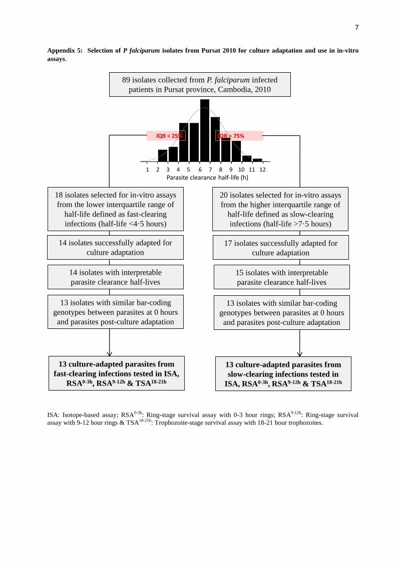
ResultsFrom the 89 patients enrolled, we selected 18 fast-clearing and 20 slow-clearing parasites representing the lower and upper quartiles of the half-life distribution and adapted them to culture as described.20 Assays were ultimately done on parasites from 13 fast-clearing and 13 slow-clearing infections; the other 12 selected parasites were excluded from the study because they did not adapt to culture, did not have a corresponding half-life value that was interpretable, or did not show an identical genotype to the parasite originally obtained from the patient (appendix).
Parasites were collected in Pursat in 2010 (appendix), and used in three stage-specifi c survival assays (fi gure 1). In the RSA for 0–3 h rings (RSA⁰–³ h), the median survival rate of slow-clearing parasites was 47 times greater than that of fast-clearing parasites (table, fi gure 2). By contrast, 9–12 h rings and 18–21 h trophozoites from fast-clearing and slow-clearing infections showed no signifi cant diff erence in survival (table, fi gure 2). The stage-dependent survival patterns diff ered between fast-clearing and slow-clearing parasites (fi gure 2, appendix). Specifi cally, the survival rates of slow-clearing parasites decreased with parasite stage, whereas those of fast-clearing parasites increased with parasite stage (Δ=9·9% [IQR 1·7 to 14·4] vs –0·3% [–1·1 to 0·4]; p=0·007). In an isotope-based sensitivity assay that monitored replication of parasites exposed to drug for 48 h,27 fast-clearing and slow-clearing parasites did not diff er signifi cantly in IC50 and IC90 values for artesunate or dihydroartemisinin (table; appendix).
In patients with falciparum malaria, the age distribution of circulating ring-stage parasites is heterogeneous, ranging from 0 h to 18 h at the time of clinical presentation;28 that is, ring-stage parasites are not necessarily tightly synchronised at the 0–3 h age of development. We therefore sought to assess whether an ex-vivo RSA could distinguish fast-clearing from slow-clearing parasites that have been neither culture-adapted nor experimentally synchronised.
In the prospective 2012 study, 30 (83%) of 36 patients had interpretable half-life values and tri-gas survival rates (appendix), which correlated signifi cantly (fi gure 3). Parasite survival rates did not diff er between the three atmospheres (n=26; p=0·30, Friedman test). The ex-vivo RSA accurately identifi ed artemisinin-resistant infections where they have not been previously described (fi gure 3, appendix). In Preah Vihear, for example, one parasite with a 12·2% survival rate had an 8·17 h half-life, whereas the other six parasites with a median 0·70% survival rate
Fast-clearing parasites (short half-life)
Slow-clearing parasites (long half-life)
p value
RSA⁰�³ h 0·23 (0·14–2·93, 0·01–51·39) 10·88 (4·75–13·91, 0·16–29·14) 0·007
RSA⁹�¹² h 1·07 (0·77–1·70, 0·06–10·00) 2·12 (1·46–3·55, 0·33–8·00) 0·06
TSA¹⁸�²¹ h 0·99 (0·48–2·20, 0·16–4·10) 1·16 (0·78–2·05, 0·38–5·30) 0·54
Dihydroartemisinin IC50 0·71 (0·58–0·94, 0·29–1·20) 0·79 (0·62–0·11, 0·42–1·51) 0·44
Dihydroartemisinin IC90 2·60 (2·28–3·30, 1·54–4·49) 2·46 (1·78–3·02, 1·48–4·40) 0·36
Artesunate IC50 1·00 (0·84–1·47, 0·28–1·71) 1·11 (0·98–1·84, 0·83–2·50) 0·20
Artesunate IC90 3·32 (2·52–3·94, 2·30–5·80) 3·02 (2·38–3·86, 1·99–6·38) 0·70
Data are median (IQR, range). Percentage survival in RSA⁰�³ h, RSA⁹�¹² h, and TSA¹⁸�²¹ h, and IC50 and IC90 values for dihydroartemisinin and artesunate in isotope-based sensitivity assays. p values for signifi cance from Mann-Whitney test. RSA=ring-stage survival assay. TSA=trophozoite-stage survival assay.
Table: Parasite survival in in-vitro assays using culture-adapted isolates
Figure 2: In-vitro survival after exposure to dihydroartemisininResults are expressed as the proportion of viable Plasmodium falciparum parasites after a 6 h exposure of 0–3 h rings (RSA⁰�³ h), 9–12 h rings (RSA⁹�¹² h), and 18–21 h trophozoites (TSA¹⁸�²¹ h) to 700 nM dihydroartemisinin compared with dimethyl sulfoxide. These assays were done on culture-adapted parasite isolates obtained from 13 patients with fast-clearing infections (fi lled circles) and 13 patients with slow-clearing infections (open circles) in Pursat in 2010. The horizontal lines represent the medians and whiskers the IQRs. The solid lines show stage-dependent survival pattern of parasites from slow-clearing infections and the dotted lines the stage-dependent survival pattern of parasites from fast-clearing infections. RSA=ring-stage survival assay. TSA=trophozoite-stage survival assay.
RSA0–3 h RSA9–12 h TSA18–21 h
100
10
1
0·1
0·01
0·001Prop
ortio
n of
via
ble
para
sites
(%; d
ihyd
roar
tem
isini
n-ex
pose
d/no
n-ex
pose
d)

Articles
www.thelancet.com/infection Published online September 11, 2013 http://dx.doi.org/10.1016/S1473-3099(13)70252-4 5
(IQR 0·18–2·0) had a median 2·28 h half-life (1·89–3·52). In Ratanakiri, one parasite with a 38·3% survival rate had a 9·06 h half-life, whereas the other ten parasites had a median 0·40% survival rate (0·26–1·48) and a median 2·28 h half-life (1·90–2·64). Our fi ndings suggest that artemisinin-resistant P falciparum has spread or independently emerged in northern and eastern Cambodia, a possibility that can now be confi rmed with the in-vitro RSA⁰–³ h.
DiscussionP falciparum isolates from slow-clearing and fast-clearing infections in Cambodia respond diff erently to a 6 h, 700 nM exposure to dihydroartemisinin. In the RSA⁰–³ h, rings of slow-clearing parasites had much higher survival rates than those of fast-clearing parasites. In the ex-vivo RSA, survival rates correlated with parasite clearance half-lives. Importantly, the ex-vivo RSA accurately identifi ed slow-clearing infections in Cambodian provinces where they have not yet been described. To our knowledge, these are the fi rst reported in-vitro and ex-vivo dihydro-artemisinin susceptibility data that correlate with in-vivo parasite clearance half-lives (panel). These data qualify the in-vitro RSA⁰–³ h as a new laboratory test for elucidating the mechanism of artemisinin resistance through molecular studies. These studies might include genome-wide association studies,29 associating RSA⁰–³ h survival rates with whole-genome single-nucleotide polymorphism data, phenotypic screening of parasite progeny clones obtained from genetic crosses between artemisinin-sensitive and artemisinin-resistant parental lines, phenotypic characterisation of the diff erent artemisinin-resistant parasite subpopulations circulating in western Cambodia,29 and validation of candidate molecular markers through genetic manipulation of parasites.
Our fi ndings also suggest that the ex-vivo RSA is a feasible, convenient method for detecting the spread and emergence of artemisinin resistance in areas where it has not yet been reported (eg, eastern Cambodia), or the worsening of artemisinin resistance where it is entrenched (eg, western Cambodia). Both types of fi ndings from ex-vivo RSAs might inform national malaria control programmes to expand or intensify containment measures. In a screen and confi rm approach to support such eff orts, we propose that the ex-vivo RSA be used in the fi eld to screen for artemisinin-resistant parasites. Any parasite showing dihydro- artemisinin resistance in this assay can then be adapted to short-term culture in a laboratory, genotyped to ensure its identity to the clinical parasite isolate obtained from a patient, and tested to confi rm dihydroartemisinin resistance with the in-vitro RSA⁰–³ h.
For both artemisinin-sensitive and artemisinin-resistant parasites, we show stage-dependent hetero-geneity of dihydroartemisinin susceptibility in ring forms. In artemisinin-sensitive parasites, 0–3 h rings were more susceptible to dihydroartemisinin than were
9–12 h rings. This fi nding with clinical parasite isolates is consistent with the recent report that 2–4 h rings of artemisinin-sensitive laboratory lines are specifi cally hypersensitive to dihydroartemisinin.18 However, in artemisinin-resistant parasites, 0–3 h rings were less susceptible to dihydroartemisinin than were 9–12 h rings. We tentatively conclude that the susceptibility of Cambodian parasites to dihydroartemisinin is controlled predominantly at the 0–3 h stage of parasite development. This interpretation, and our fi nding that trophozoites are mostly susceptible to dihydroartemisinin irrespective of half-life, is consistent with mathematical modelling predictions30 and transcriptomics data31 from studies of ring-stage parasites.
Half-lives and RSA⁰–³ h survival rates were discordant in four patients (fi gure 2, appendix). Three patients (1007, 1006, and 1009) had fast-clearing infections with parasites showing survival rates of 5·3%, 19·3%, and 51·4%, and a resistant stage-dependent pattern (Δ=1·2%, 17·3%, and 50·2%, respectively). Their patterns diff ered from those of fast clearing-infections (Δ=–0·7% vs 17·3%; p=0·01), being similar to those from slow-clearing-infections (Δ=10·3% vs 17·3%; p=0·56; appendix). To explain this discordance, we postulated that these three parasites had already developed into dihydroartemisinin-susceptible, late ring-stage parasites in the patients’ blood at the time of the fi rst artesunate dose.
To assess this possibility, we reviewed the initial blood smears from these patients and estimated the relative
Figure 3: Correlation of in-vivo parasite clearance half-lives and ex-vivo dihydroartemisinin survival ratesEx-vivo ring-stage survival assays (RSAs) were done on parasite isolates obtained directly from patients with malaria in Pursat, Preah Vihear, and Ratanakiri in 2012. Results from the ex-vivo RSAs are expressed as the proportion of viable parasites after a 6 h exposure to 700 nM dihydroartemisinin compared with dimethyl-sulfoxide-exposed controls. Results from the parasite clearance studies are expressed as the parasite clearance half-life in hours. The proportion of viable parasites in ex-vivo RSAs correlated signifi cantly with the parasite clearance half-life (r=0·74, 95% CI 0·50–0·87; p<0·0001) in Pursat (red), Preah Vihear (blue), and Ratanakiri (green).
100
10
1
0·1
0·01Prop
ortio
n of
via
ble
para
sites
(%; d
ihyd
roar
tem
isini
n-ex
pose
d/no
n-ex
pose
d)
1086420 97531Parasite clearance half-life (h)

Articles
6 www.thelancet.com/infection Published online September 11, 2013 http://dx.doi.org/10.1016/S1473-3099(13)70252-4
age of their ring-stage parasites just before treatment with artesunate (appendix). In thin blood smears made at 0 h, we noted that these three discordant patients indeed had a two-times lower proportion of tiny rings compared with the 12 concordant patients from the slow-clearing group (ie, those having slow-clearing infections with dihydroartemisinin-resistant parasites; 42·4% vs 75·0%; p=0·03). Higher proportions of large, older rings could account for shorter than expected half-lives because these forms are more susceptible to dihydroartemisinin than tiny, young rings. Overall, our fi ndings suggest that the relative abundance of tiny and large rings at the time of the fi rst artemisinin dose aff ects the parasite clearance half-life, and that accurate ex-vivo staging of parasites is crucial for classifying treatment outcome. In one patient (896), having a slow-clearing infection with a parasite showing a survival rate of 0·2%, and a sensitive stage-dependent pattern (Δ=–1·4%; appendix), we cannot rule out an inadequate immune response to infection32 or insuffi cient plasma concentrations of artemisinins.
The RSA⁰–³ h survival rate might be crucially informative in ongoing parasite genetics studies31,33,34 aimed at identifying loci under artemisinin selection, because it is unaff ected by in-vivo variables (eg, pharmacokinetics, haemoglobin type, and acquired immunity) that might aff ect the parasite clearance half-life. Although this phenotype might also be a useful readout in studies to defi ne and validate molecular markers for tracking artemisinin-resistant parasites in the fi eld, the RSA⁰–³ h is a laborious assay. By contrast, the ex-vivo RSA saves weeks of eff ort (results are available in 3 days), and avoids the confounding eff ects of parasite clone elimination and metabolic changes that might accompany the culture adaptation of parasites. In addition to implementing methods that more precisely establish the age of rings, FACS-based or ELISA-based analysis of parasite viability should improve the throughput of dihydroartemisinin-susceptibility studies. Until such methods are developed and validated, we propose the simple ex-vivo RSA as a highly informative surveillance approach for the identifi cation of artemisinin-resistant parasites in areas where slow parasite clearance is suspected. Investigating the relation between RSA survival rates ex vivo and parasite recrudescence rates in vivo might be useful in assessing the clinical eff ect of artemisinin resistance.
ContributorsBW, CA, PL, JMA, SK, SD, CMC, WRJT, OM-P, RMF, and DM
contributed to study design. NK genotyped parasites. BW, CA, and PC
did the in-vitro and ex-vivo drug assays. SSr, SM, CS, BS, and SSu
gathered clinical data. BW, CA, OM-P, RMF, and DM analysed data and
wrote the report.
Confl icts of interestWe declare that we have no confl icts of interest.
AcknowledgmentsWe thank Robert Gwadz, Savuth Koeuth, François Nosten, Eng Ly Pech,
Thomas Wellems, and Chongjun Zhou for their eff orts in support of
this work. This study was funded by the Intramural Research Program,
NIAID, NIH, and by grants from Institut Pasteur du Cambodge (Institut
Pasteur, International Division and Banque Natixis) and Laboratoire
d’excellence IBEID (Agence Nationale de la Recherche, France). BW is
supported by a postdoctoral fellowship from the International Division,
Institut Pasteur, and DM by the French Ministry of Foreign Aff airs.
References1 WHO. Antimalarial drug combination therapy. Geneva: World
Health Organization, 2001.
2 WHO. World malaria report 2012. Geneva: World Health Organization, 2012.
3 Wongsrichanalai C, Pickard AL, Wernsdorfer WH, et al. Epidemiology of drug-resistant malaria. Lancet Infect Dis 2002; 2: 209–18.
4 Noedl H, Se Y, Schaecher K, et al. Evidence of artemisinin-resistant malaria in western Cambodia. N Engl J Med 2008; 359: 2619–20.
5 Dondorp AM, Nosten F, Yi P, et al. Artemisinin resistance in Plasmodium falciparum malaria. N Engl J Med 2009; 361: 455–67.
6 Amaratunga C, Sreng S, Suon S, et al. Artemisinin-resistant Plasmodium falciparum in Pursat province, western Cambodia: a parasite clearance rate study. Lancet Infect Dis 2012; 12: 851–58.
7 Amaratunga C, Mao S, Sreng S, et al. Slow parasite clearance rates in response to artemether in patients with severe malaria. Lancet Infect Dis 2013; 13: 113–14.
8 Phyo AP, Nkhoma S, Stepniewska K, et al. Emergence of artemisinin-resistant malaria on the western border of Thailand: a longitudinal study. Lancet 2012; 379: 1960–66.
Panel: Research in context
Systematic reviewWe searched PubMed with the terms “artemisinin resistant malaria”, limited our search to clinical trials, and used no date or language restrictions. This process produced 56 publications. Any in-vitro or ex-vivo drug assays done in these studies involved the continuous exposure of Plasmodium falciparum parasites to very low concentrations of artemisinins during the entire lifecycle of their blood-stage development. Results of these assays have not consistently correlated with clinical effi cacy.4–6 Only four recent publications describe new assays that were specifi cally designed to measure P falciparum susceptibility to artemisinins.17–20 Klonis and colleagues18 described an assay working with laboratory-adapted parasites that were artemisinin-sensitive. Witkowski and coworkers17 and Teuscher and colleagues19 described assays working with laboratory-adapted parasites and their drug-selected counterparts that became resistant to artemisinins. Witkowski and colleagues20 described a more relevant study using parasites from western and eastern Cambodia, where parasites are commonly resistant and sensitive to artemisinin respectively. None of these studies were designed to test whether in-vitro susceptibility data correlated with in-vivo effi cacy data (ie, parasite clearance rates after artemisinin treatment, the currently accepted clinical phenotype); therefore, these assays could not be clinically validated.
InterpretationWe report for the fi rst time novel in-vitro and ex-vivo ring-stage survival assays (RSAs) that detect artemisinin-resistant, slow-clearing P falciparum infections in patients with malaria. In both assays, early ring-stage parasites are exposed to a pharmacologically relevant pulse of dihydroartemisinin and their survival measured 72 h later. With parasites adapted to culture in the laboratory, the in-vitro RSA can be used to discover the molecular mechanisms of artemisinin resistance, to investigate the mode of action of artemisinins, and to identify artemisinin-resistant parasite strains for testing next-generation antimalarial drugs. The ex-vivo RSA with parasites obtained directly from patients with malaria can be easily implemented in fi eld-based settings to monitor the worsening of artemisinin resistance where it is highly prevalent (eg, western Cambodia), and to map its spread or independent emergence elsewhere in the Greater Mekong Subregion. Also, this simple test might readily be established at sentinel sites in sub-Saharan Africa, where the arrival of artemisinin-resistant P falciparum is expected to be especially devastating. The ex-vivo RSA can thus provide crucial surveillance data to the national malaria control programmes of all countries threatened by artemisinin-resistant malaria.
For the standard operating procedures for ex-vivo and
in-vitro RSA see http://www.wwarn.org/toolkit/procedures/
invitro
Articles
www.thelancet.com/infection Published online September 11, 2013 http://dx.doi.org/10.1016/S1473-3099(13)70252-4 7
9 Kyaw MP, Nyunt MH, Chit K, et al. Reduced susceptibility of Plasmodium falciparum to artesunate in southern Myanmar. PLoS One 2013; 8: e57689.
10 Hien TT, Thuy-Nhien NT, Phu NH, et al. In vivo susceptibility of Plasmodium falciparum to artesunate in Binh Phuoc Province, Vietnam. Malar J 2012; 11: 355.
11 Enserink M. Malaria’s drug miracle in danger. Science 2010; 328: 844–46.
12 Dondorp AM, Fairhurst RM, Slutsker L, et al. The threat of artemisinin-resistant malaria. N Engl J Med 2011; 365: 1073–75.
13 WHO. Emergency response to artemisinin resistance in the Greater Mekong subregion: regional framework for action 2013–2015. Geneva: World Health Organization, 2013.
14 White NJ. The parasite clearance curve. Malar J 2011; 10: 278.
15 Flegg JA, Guerin PJ, White NJ, et al. Standardizing the measurement of parasite clearance in falciparum malaria: the parasite clearance estimator. Malar J 2011; 10: 339.
16 Cui L, Wang Z, Miao J, et al. Mechanisms of in vitro resistance to dihydroartemisinin in Plasmodium falciparum. Mol Microbiol 2012; 86: 111–28.
17 Witkowski B, Lelievre J, Barragan MJ, et al. Increased tolerance to artemisinin in Plasmodium falciparum is mediated by a quiescence mechanism. Antimicrob Agents Chemother 2010; 54: 1872–77.
18 Klonis N, Xie SC, McCaw JM, et al. Altered temporal response of malaria parasites determines diff erential sensitivity to artemisinin. Proc Natl Acad Sci USA 2013; 110: 5157–62.
19 Teuscher F, Chen N, Kyle DE, et al. Phenotypic changes in artemisinin-resistant Plasmodium falciparum lines in vitro: evidence for decreased sensitivity to dormancy and growth inhibition. Antimicrob Agents Chemother 2012; 56: 428–31.
20 Witkowski B, Khim N, Chim P, et al. Reduced artemisinin susceptibility of Plasmodium falciparum ring stages in western Cambodia. Antimicrob Agents Chemother 2012; 57: 914–23.
21 WWARN. Parasite clearance estimator. https://www.wwarn.org/toolkit/data-management/parasite-clearance-estimator (accessed Aug 21, 2013).
22 Moll K, Ljungström I, Perlmann H, et al (eds). Methods in malaria research, 5th edn (version 5.2 revision). http://www.mr4.org/Portals/3/Methods_In_Malaria_Research-5theditionv5-2.pdf (accessed Aug 21, 2013).
23 Le Nagard H, Vincent C, Mentre F, et al. Online analysis of in vitro resistance to antimalarial drugs through nonlinear regression. Comput Methods Programs Biomed 2011; 104: 10–18.
24 Kaddouri H, Nakache S, Houze S, et al. Assessment of the drug susceptibility of Plasmodium falciparum clinical isolates from Africa by using a plasmodium lactate dehydrogenase immunodetection assay and an inhibitory maximum eff ect model for precise measurement of the 50-percent inhibitory concentration. Antimicrob Agents Chemother 2006; 50: 3343–49.
25 WHO. Malaria microscopy quality assurance manual—version 1. Geneva: World Health Organization, 2009.
26 Daniels R, Volkman SK, Milner DA, et al. A general SNP-based molecular barcode for Plasmodium falciparum identifi cation and tracking. Malar J 2008; 7: 223.
27 Desjardins RE, Canfi eld CJ, Haynes JD, et al. Quantitative assessment of antimalarial activity in vitro by a semiautomated microdilution technique. Antimicrob Agents Chemother 1979; 16: 710–18.
28 Silamut K, White NJ. Relation of the stage of parasite development in the peripheral blood to prognosis in severe falciparum malaria. Trans R Soc Trop Med Hyg 1993; 87: 436–43.
29 Miotto O, Almagro-Garcia J, Manske M, et al. Multiple populations of artemisinin-resistant Plasmodium falciparum in Cambodia. Nat Genet 2013; 45: 648–55.
30 Saralamba S, Pan-Ngum W, Maude RJ, et al. Intrahost modeling of artemisinin resistance in Plasmodium falciparum. Proc Natl Acad Sci USA 2010; 108: 397–402.
31 Mok S, Imwong M, Mackinnon MJ, et al. Artemisinin resistance in Plasmodium falciparum is associated with an altered temporal pattern of transcription. BMC Genomics 2011; 12: 391.
32 Lopera-Mesa TM, Doumbia S, Chiang S, et al. Plasmodium falciparum clearance rates in response to artesunate in Malian children with malaria: eff ect of acquired immunity. J Infect Dis 2013; 207: 1655–63.
33 Cheeseman IH, Miller BA, Nair S, et al. A major genome region underlying artemisinin resistance in malaria. Science 2012; 336: 79–82.
34 Takala-Harrison S, Clark TG, Jacob CG, et al. Genetic loci associated with delayed clearance of Plasmodium falciparum following artemisinin treatment in southeast Asia. Proc Natl Acad Sci USA 2012; 110: 240–45.
Supplementary webappendixThis webappendix formed part of the original submission and has been peer reviewed. We post it as supplied by the authors.
Supplement to: Witkowski B, Amaratunga C, Khim N, et al. Novel phenotypic assays for the detection of artemisinin-resistant Plasmodium falciparum malaria in Cambodia: in-vitro and ex-vivo drug-response studies. Lancet Infect Dis 2013; published online Sept 11. http://dx.doi.org/10.1016/S1473-3099(13)70252-4.

1
APPENDIX MATERIAL
Novel phenotypic assays detect artemisinin-resistant Plasmodium falciparum malaria in
Cambodia: in-vitro and ex-vivo drug response studies
Benoit Witkowski, Chanaki Amaratunga, Nimol Khim, Sokunthea Sreng, Pheaktra Chim, Saorin Kim, Pharath Lim,
Sivanna Mao, Chantha Sopha, Baramey Sam, Jennifer M. Anderson, Socheat Duong, Char Meng Chuor, Walter R.
J. Taylor, Seila Suon, Odile Mercereau-Puijalon, Rick M. Fairhurst, Didier Menard
Contents
Appendix 1: Patient information and corresponding data from in-vitro assays performed on P falciparum isolates
from Pursat in 2010.
Appendix 2: Protocols, PCR/nested PCR primer sequences, and LDR probe sequences used to genotype P.
falciparum isolates obtained in Pursat in 2010.
Appendix 3: Patient information and corresponding data from ex-vivo assays performed on P falciparum isolates
from Pursat, Preah Vihear, and Ratanakiri in 2012.
Appendix 4: Grading of asexual P falciparum parasites into two developmental categories: ‘tiny’ (Panel A) and
‘large’ (Panel B) rings.
Appendix 5: Selection of P falciparum isolates from Pursat in 2010 for culture adaptation and use in in-vitro assays.
Appendix 6: Individual stage-dependent patterns in in-vitro survival assays (RSA0-3h
, RSA9-12h
, and TSA18-21h
)
performed on parasite isolates from fast- (Panel A) and slow-clearing (Panel B) infections in Pursat in 2010.

2
Appendix 1: Patient information and corresponding data from in-vitro assays performed on P falciparum
isolates from Pursat in 2010.
Discordant samples are shown in bold.
ID Age
(years) Sex
Parasitemia at
0 hours (/mm3)
Parasite
clearance
half-life
(hours)
Fit of
parasite
clearance
curve - R2
RSA0-3h
survival
rate
(%)
RSA9-12h
survival
rate (%)
TSA18-21h
survival
rate (%)
Artesunate
IC50 (nM)
DHA
IC50
(nM)
% of tiny
rings at
0 hours
906 23 M 33,742 2·20 0.8090 0·15 1·01 0·15 0·28 0·29 42·4
919 37 F 250,000 3·03 0·8847 0·01 0·06 0·17 0·77 0·58 82·9
970 23 M 100,936 3·59 0·8685 0·25 2·1 0·12 0·94 0·88 86·4
189-4 13 M 272,000 3·65 0·9660 0·23 0·46 0·50 1·69 0·97 76·8
915 18 M 50,633 3·69 0·9357 0·35 0·94 0·37 1·34 0·68 65·6
931 29 M 296,666 4·25 0·8884 0·56 1·07 0·52 0·82 0·57 85·4
911 19 M 51,576 4·46 0·8672 0·19 0·60 0·32 0·98 0·76 81·8
918 58 M 351,111 4·54 0·9377 0·14 0·97 0·14 1·52 0·90 77·4
1003 31 M 25,920 4·56 0·9839 0·05 1·16 0·04 1·00 0·60 37·5
1006 48 M 11,882 4·67 0·9680 19·32 6·27 3·08 1·33 0·71 64·8
1007 24 M 16,466 4·71 0·9237 5·30 1·30 4·08 1·71 1·20 42·4
1009 42 M 27,714 4·77 0·9314 51·39 10 5·14 0·87 0·40 27·0
945 10 M 188,500 4·83 0·8714 0·22 1·09 0·20 1·42 1·01 78·1
968 64 M 36,730 7·97 0·9855 8·34 1·71 4·88 0·96 0·68 57·5
818-2 46 M 47,835 7·97 0·9877 13·48 8·00 1·69 1·00 0·81 88·8
976 44 M 65,432 8·21 0·9305 2·18 0·78 2·79 1·14 0·79 65·0
946 17 M 53,626 8·26 0·9458 7·35 1·20 6·13 1·95 1·04 94·4
969 20 M 95,304 8·32 0·9599 6·30 2·12 2·98 2·50 1·51 75·0
950 15 F 79,714 8·54 0·.9495 3·20 3·48 0·92 1·71 1·30 81·8
958 30 M 41,553 8·73 0·9851 29·14 3·62 8·05 1·11 0·71 43·7
896 21 M 82,807 8·75 0·9322 0·16 0·33 0·48 0·83 0·42 76·4
955 48 M 20,242 9·05 0·9326 11·80 2·20 5·36 1·89 1·18 77·4
938 18 M 22,109 9·11 0·9655 14·33 4·00 3·58 0·85 0·49 57·0
990 31 M 18,125 9·45 0·9775 12·60 3·00 4·20 1·09 0·55 75·0
922 26 M 42,240 9·72 0·9589 21·90 2·10 10·43 1·80 0·95 74·5
956 20 M 48,000 10·08 0·9489 10·88 2·01 5·42 1·11 0·69 84·8
3
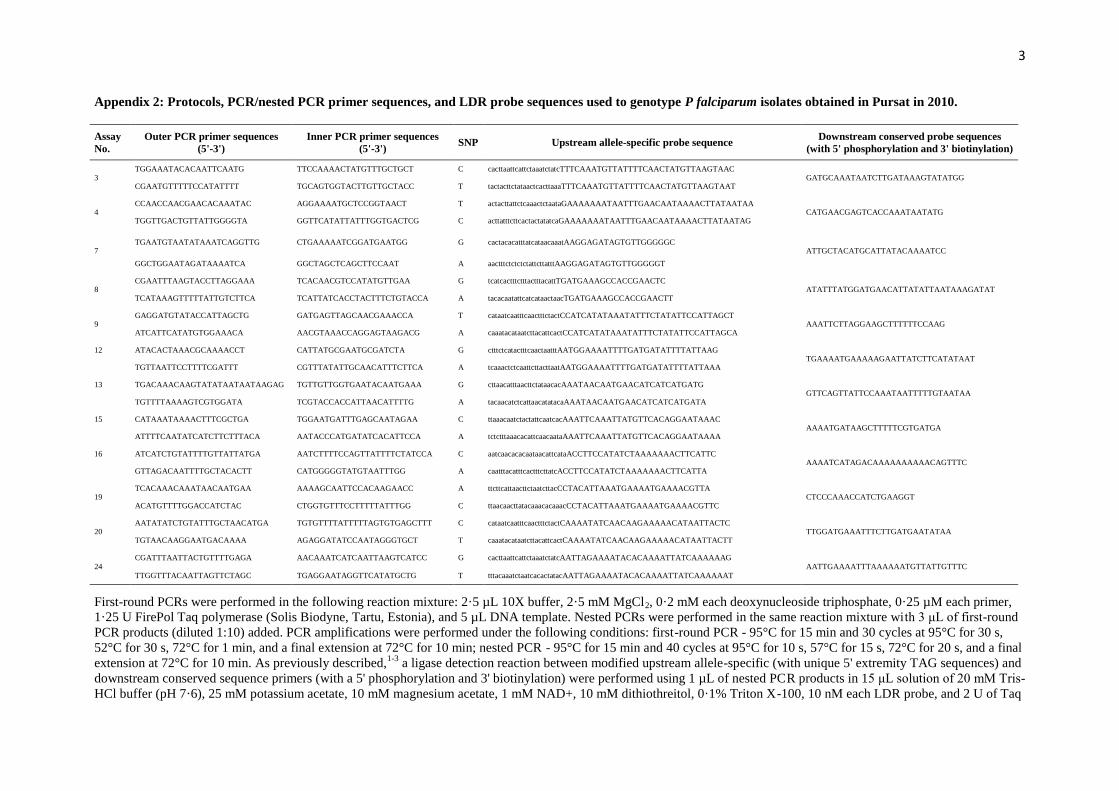
Appendix 2: Protocols, PCR/nested PCR primer sequences, and LDR probe sequences used to genotype P falciparum isolates obtained in Pursat in 2010.
Assay
No.
Outer PCR primer sequences
(5'-3')
Inner PCR primer sequences
(5'-3') SNP Upstream allele-specific probe sequence
Downstream conserved probe sequences
(with 5' phosphorylation and 3' biotinylation)
3 TGGAAATACACAATTCAATG TTCCAAAACTATGTTTGCTGCT C cacttaattcattctaaatctatcTTTCAAATGTTATTTTCAACTATGTTAAGTAAC
GATGCAAATAATCTTGATAAAGTATATGG CGAATGTTTTTCCATATTTT TGCAGTGGTACTTGTTGCTACC T tactacttctataactcacttaaaTTTCAAATGTTATTTTCAACTATGTTAAGTAAT
4 CCAACCAACGAACACAAATAC AGGAAAATGCTCCGGTAACT T actacttattctcaaactctaataGAAAAAAATAATTTGAACAATAAAACTTATAATAA
CATGAACGAGTCACCAAATAATATG TGGTTGACTGTTATTGGGGTA GGTTCATATTATTTGGTGACTCG C acttatttcttcactactatatcaGAAAAAAATAATTTGAACAATAAAACTTATAATAG
7 TGAATGTAATATAAATCAGGTTG CTGAAAAATCGGATGAATGG G cactacacatttatcataacaaatAAGGAGATAGTGTTGGGGGC
ATTGCTACATGCATTATACAAAATCC
GGCTGGAATAGATAAAATCA GGCTAGCTCAGCTTCCAAT A aactttctctctctattcttatttAAGGAGATAGTGTTGGGGGT
8 CGAATTTAAGTACCTTAGGAAA TCACAACGTCCATATGTTGAA G tcatcactttctttactttacattTGATGAAAGCCACCGAACTC
ATATTTATGGATGAACATTATATTAATAAAGATAT TCATAAAGTTTTTATTGTCTTCA TCATTATCACCTACTTTCTGTACCA A tacacaatattcatcataactaacTGATGAAAGCCACCGAACTT
9 GAGGATGTATACCATTAGCTG GATGAGTTAGCAACGAAACCA T cataatcaatttcaactttctactCCATCATATAAATATTTCTATATTCCATTAGCT
AAATTCTTAGGAAGCTTTTTTCCAAG ATCATTCATATGTGGAAACA AACGTAAACCAGGAGTAAGACG A caaatacataatcttacattcactCCATCATATAAATATTTCTATATTCCATTAGCA
12 ATACACTAAACGCAAAACCT CATTATGCGAATGCGATCTA G ctttctcatactttcaactaatttAATGGAAAATTTTGATGATATTTTATTAAG TGAAAATGAAAAAGAATTATCTTCATATAAT
TGTTAATTCCTTTTCGATTT CGTTTATATTGCAACATTTCTTCA A tcaaactctcaattcttacttaatAATGGAAAATTTTGATGATATTTTATTAAA
13 TGACAAACAAGTATATAATAATAAGAG TGTTGTTGGTGAATACAATGAAA G cttaacatttaacttctataacacAAATAACAATGAACATCATCATGATG GTTCAGTTATTCCAAATAATTTTTGTAATAA
TGTTTTAAAAGTCGTGGATA TCGTACCACCATTAACATTTTG A tacaacatctcattaacatatacaAAATAACAATGAACATCATCATGATA
15 CATAAATAAAACTTTCGCTGA TGGAATGATTTGAGCAATAGAA C ttaaacaatctactattcaatcacAAATTCAAATTATGTTCACAGGAATAAAC AAAATGATAAGCTTTTTCGTGATGA
ATTTTCAATATCATCTTCTTTACA AATACCCATGATATCACATTCCA A tctctttaaacacattcaacaataAAATTCAAATTATGTTCACAGGAATAAAA
16 ATCATCTGTATTTTGTTATTATGA AATCTTTTCCAGTTATTTTCTATCCA C aatcaacacacaataacattcataACCTTCCATATCTAAAAAAACTTCATTC AAAATCATAGACAAAAAAAAAACAGTTTC
GTTAGACAATTTTGCTACACTT CATGGGGGTATGTAATTTGG A caatttacatttcactttcttatcACCTTCCATATCTAAAAAAACTTCATTA
19 TCACAAACAAATAACAATGAA AAAAGCAATTCCACAAGAACC A ttcttcattaacttctaatcttacCCTACATTAAATGAAAATGAAAACGTTA
CTCCCAAACCATCTGAAGGT ACATGTTTTGGACCATCTAC CTGGTGTTTCCTTTTTATTTGG C ttaacaacttatacaaacacaaacCCTACATTAAATGAAAATGAAAACGTTC
20 AATATATCTGTATTTGCTAACATGA TGTGTTTTATTTTTAGTGTGAGCTTT C cataatcaatttcaactttctactCAAAATATCAACAAGAAAAACATAATTACTC
TTGGATGAAATTTCTTGATGAATATAA TGTAACAAGGAATGACAAAA AGAGGATATCCAATAGGGTGCT T caaatacataatcttacattcactCAAAATATCAACAAGAAAAACATAATTACTT
24 CGATTTAATTACTGTTTTGAGA AACAAATCATCAATTAAGTCATCC G cacttaattcattctaaatctatcAATTAGAAAATACACAAAATTATCAAAAAAG
AATTGAAAATTTAAAAAATGTTATTGTTTC TTGGTTTACAATTAGTTCTAGC TGAGGAATAGGTTCATATGCTG T tttacaaatctaatcacactatacAATTAGAAAATACACAAAATTATCAAAAAAT
First-round PCRs were performed in the following reaction mixture: 2·5 µL 10X buffer, 2·5 mM MgCl2, 0·2 mM each deoxynucleoside triphosphate, 0·25 µM each primer,
1·25 U FirePol Taq polymerase (Solis Biodyne, Tartu, Estonia), and 5 µL DNA template. Nested PCRs were performed in the same reaction mixture with 3 μL of first-round
PCR products (diluted 1:10) added. PCR amplifications were performed under the following conditions: first-round PCR - 95°C for 15 min and 30 cycles at 95°C for 30 s,
52°C for 30 s, 72°C for 1 min, and a final extension at 72°C for 10 min; nested PCR - 95°C for 15 min and 40 cycles at 95°C for 10 s, 57°C for 15 s, 72°C for 20 s, and a final
extension at 72°C for 10 min. As previously described,1-3
a ligase detection reaction between modified upstream allele-specific (with unique 5' extremity TAG sequences) and
downstream conserved sequence primers (with a 5' phosphorylation and 3' biotinylation) were performed using 1 µL of nested PCR products in 15 μL solution of 20 mM Tris-
HCl buffer (pH 7·6), 25 mM potassium acetate, 10 mM magnesium acetate, 1 mM NAD+, 10 mM dithiothreitol, 0·1% Triton X-100, 10 nM each LDR probe, and 2 U of Taq

4
DNA ligase (New England Biolabs, Beverly, MA, USA). Reaction mixtures were heated to 95°C for 1 min, followed by 32 cycles at 95°C for 15 s and 60°C for 2 min. In a
second step, 5 µL of multiplex LDR products were added to 60 μL of hybridization solution (3 M tetramethylammonium chloride [TMAC], 50 mM Tris-HCl [pH 8·0], 3 mM
EDTA [pH 8·0], 0·10% sodium dodecyl sulfate) containing 2500 MagPlex-TAG Microspheres® (Luminex, Austin, TX, USA) for each allelic set, heated to 95°C for 90 s and
incubated at 37°C for 40 min to allow hybridization between SNP-specific LDR products and microsphere-labelled anti-TAG probes. Following hybridization, 6 μL of
streptavidin-R-phycoerythrin (Molecular Probes, Eugene, OR, USA) in TMAC hybridization solution (20 ng/μL) was added and incubated at 37°C for 40 min in Costar 6511
M polycarbonate 96-well V-bottom plates (Corning Inc., Corning, NY, USA). Detection of SNP-specific products was performed through a MagPix machine (Luminex).
Fluorescence data were managed by xPONENT software (Luminex) and entered into Microsoft Excel software (Microsoft Office 2010). In each run, samples were analyzed
with 3D7, Dd2, and HB3 genomic DNA controls and no template control.
1. Barnadas C, Kent D, Timinao L, et al. A new high-throughput method for simultaneous detection of drug resistance associated mutations in Plasmodium vivax dhfr, dhps
and mdr-1 genes. Malar J 2011;10:282.
2. Carnevale EP, Kouri D, DaRe JT, McNamara DT, Mueller I, Zimmerman PA. A multiplex ligase detection reaction-fluorescent microsphere assay for simultaneous
detection of single nucleotide polymorphisms associated with Plasmodium falciparum drug resistance. J Clin Microbiol 2007;45:752-61.
3. McNamara DT, Thomson JM, Kasehagen LJ, Zimmerman PA. Development of a multiplex PCR-ligase detection reaction assay for diagnosis of infection by the four
parasite species causing malaria in humans. J Clin Microbiol 2004;42:2403-10.
5
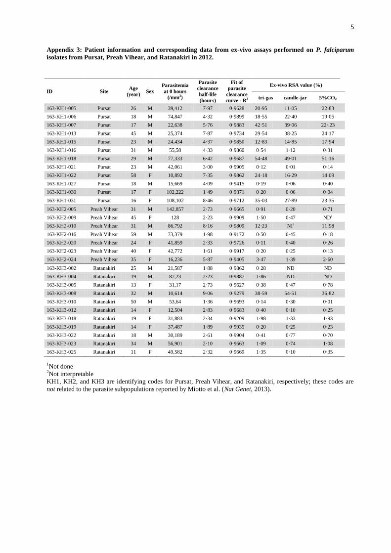
Appendix 3: Patient information and corresponding data from ex-vivo assays performed on P. falciparum
isolates from Pursat, Preah Vihear, and Ratanakiri in 2012.
ID Site Age
(year) Sex
Parasitemia
at 0 hours
(/mm3)
Parasite
clearance
half-life
(hours)
Fit of
parasite
clearance
curve - R2
Ex-vivo RSA value (%)
tri-gas candle-jar 5%CO2
163-KH1-005 Pursat 26 M 39,412 7·97 0·9628 20·95 11·05 22·83
163-KH1-006 Pursat 18 M 74,847 4·32 0·9899 18·55 22·40 19·05
163-KH1-007 Pursat 17 M 22,638 5·76 0·9883 42·51 39·06 22·.23
163-KH1-013 Pursat 45 M 25,374 7·87 0·9734 29·54 38·25 24·17
163-KH1-015 Pursat 23 M 24,434 4·37 0·9850 12·83 14·85 17·94
163-KH1-016 Pursat 31 M 55,58 4·33 0·9860 0·54 1·12 0·31
163-KH1-018 Pursat 29 M 77,333 6·42 0·9687 54·48 49·01 51·16
163-KH1-021 Pursat 23 M 42,061 3·00 0·9905 0·12 0·01 0·14
163-KH1-022 Pursat 58 F 10,892 7·35 0·9862 24·18 16·29 14·09
163-KH1-027 Pursat 18 M 15,669 4·09 0·9415 0·19 0·06 0·40
163-KH1-030 Pursat 17 F 102,222 1·49 0·9871 0·20 0·06 0·04
163-KH1-031 Pursat 16 F 108,102 8·46 0·9712 35·03 27·89 23·35
163-KH2-005 Preah Vihear 31 M 142,857 2·73 0·9665 0·91 0·20 0·71
163-KH2-009 Preah Vihear 45 F 128 2·23 0·9909 1·50 0·47 ND1
163-KH2-010 Preah Vihear 31 M 86,792 8·16 0·9809 12·23 NI2 11·98
163-KH2-016 Preah Vihear 59 M 73,379 1·98 0·9172 0·50 0·45 0·18
163-KH2-020 Preah Vihear 24 F 41,859 2·33 0·9726 0·11 0·40 0·26
163-KH2-023 Preah Vihear 40 F 42,772 1·61 0·9917 0·20 0·25 0·13
163-KH2-024 Preah Vihear 35 F 16,236 5·87 0·9405 3·47 1·39 2·60
163-KH3-002 Ratanakiri 25 M 21,587 1·88 0·9862 0·28 ND ND
163-KH3-004 Ratanakiri 19 M 87,23 2·23 0·9887 1·86 ND ND
163-KH3-005 Ratanakiri 13 F 31,17 2·73 0·9627 0·38 0·47 0·78
163-KH3-008 Ratanakiri 32 M 10,614 9·06 0·9279 38·59 54·51 36·82
163-KH3-010 Ratanakiri 50 M 53,64 1·36 0·9693 0·14 0·30 0·01
163-KH3-012 Ratanakiri 14 F 12,504 2·83 0·9683 0·40 0·10 0·25
163-KH3-018 Ratanakiri 19 F 31,883 2·34 0·9209 1·98 1·33 1·93
163-KH3-019 Ratanakiri 14 F 37,487 1·89 0·9935 0·20 0·25 0·23
163-KH3-022 Ratanakiri 18 M 30,189 2·61 0·9904 0·41 0·77 0·70
163-KH3-023 Ratanakiri 34 M 56,901 2·10 0·9663 1·09 0·74 1·08
163-KH3-025 Ratanakiri 11 F 49,582 2·32 0·9669 1·35 0·10 0·35
1Not done
2Not interpretable
KH1, KH2, and KH3 are identifying codes for Pursat, Preah Vihear, and Ratanakiri, respectively; these codes are
not related to the parasite subpopulations reported by Miotto et al. (Nat Genet, 2013).

6
Appendix 4: Grading of asexual P falciparum parasites into two developmental categories: ‘tiny’ (Panel A)
and ‘large’ (Panel B) rings.
Panel A
Panel B
Representative photomicrographs of P. falciparum isolates collected from patients just prior to receiving a first dose
of artesunate. Giemsa-stained thin blood films are shown. Rings were classified as ‘tiny rings’ when the width of the
cytoplasm band was less than, or equal to, half of the diameter of the nucleus (Panel A) and as ‘large rings’ when the
width of the cytoplasm band was greater than the diameter of the nucleus (Panel B).
7
Appendix 5: Selection of P falciparum isolates from Pursat 2010 for culture adaptation and use in in-vitro
assays.
ISA: Isotope-based assay; RSA0-3h
: Ring-stage survival assay with 0-3 hour rings; RSA9-12h
: Ring-stage survival
assay with 9-12 hour rings & TSA18-21h
: Trophozoite-stage survival assay with 18-21 hour trophozoites.
89 isolates collected from P. falciparum infected
patients in Pursat province, Cambodia, 2010
18 isolates selected for in-vitro assays
from the lower interquartile range of
half-life defined as fast-clearing
infections (half-life <4·5 hours)
14 isolates successfully adapted for
culture adaptation
14 isolates with interpretable
parasite clearance half-lives
20 isolates selected for in-vitro assays
from the higher interquartile range of
half-life defined as slow-clearing
infections (half-life >7·5 hours)
15 isolates with interpretable
parasite clearance half-lives
17 isolates successfully adapted for
culture adaptation
13 isolates with similar bar-coding
genotypes between parasites at 0 hours
and parasites post-culture adaptation
13 isolates with similar bar-coding
genotypes between parasites at 0 hours
and parasites post-culture adaptation
13 culture-adapted parasites from
slow-clearing infections tested in
ISA, RSA0-3h, RSA9-12h & TSA18-21h
0 2 4 6 8 10 12
Parasite clearance half-life (h)1 2 3 4 5 6 7 8 9 10 11 12
IQR > 75%IQR < 25%
13 culture-adapted parasites from
fast-clearing infections tested in ISA,
RSA0-3h, RSA9-12h & TSA18-21h

8
Appendix 6: Individual stage-dependent patterns in in-vitro survival assays (RSA0-3h
, RSA9-12h
, and TSA18-21h
)
performed on parasite isolates from fast- (Panel A) and slow-clearing (Panel B) infections in Pursat in 2010.
Panel A
The dotted red lines represent the stage-dependent survival patterns of parasites that show ‘concordance’ between
half-lives and RSA0-3h
survival rates (∆= -0·7%) and the black solid lines represent the stage-dependent survival
patterns of parasites that show ‘discordance’ between half-lives and RSA0-3h
survival rates (∆= 17·3%, P=0·01,
Mann-Whitney U test).
Panel B
The dotted blue lines represent the stage-dependent survival patterns of parasites that show ‘concordance’ between
half-lives and RSA0-3h
survival rates (∆= 10·3%) and the black solid line shows the stage-dependent survival pattern
of the parasite that showed ‘discordance’ between the half-live and RSA0-3h
survival rate (∆= -1·2%).