Embed Size (px)
Citation preview
Novel ITS1 Fungal Primers forCharacterization of the Mycobiome
Mykhaylo Usyk,a Christine P. Zolnik,a,b Hitesh Patel,c Michael H. Levi,c,d
Robert D. Burka,e
Department of Pediatrics, Albert Einstein College of Medicine, Bronx, New York, USAa; Department of Biology,Long Island University, Brooklyn, New York, USAb; Department of Pathology, Montefiore Medical Center, Bronx,New York, USAc; Department of Pathology, Albert Einstein College of Medicine, Bronx, New York, USAd;Departments of Obstetrics & Gynecology and Women’s Health, Epidemiology and Population Health, andMicrobiology & Immunology, Albert Einstein College of Medicine, Bronx, New York, USAe
ABSTRACT Studies of the human microbiome frequently omit characterization offungal communities (the mycobiome), which limits our ability to investigate howfungal communities influence human health. The internal transcribed spacer 1 (ITS1)region of the eukaryotic ribosomal cluster has features allowing for wide taxonomiccoverage and has been recognized as a suitable barcode region for species-levelidentification of fungal organisms. We developed custom ITS1 primer sets using iter-ative alignment refinement. Primer performance was evaluated using in silico testingand experimental testing of fungal cultures and human samples. Using an expandednovel reference database, SIS (18S-ITS1-5.8S), the newly designed primers showed anaverage in silico taxonomic coverage of 79.9% � 7.1% compared to a coverage of44.6% � 13.2% using previously published primers (P � 0.05). The newly describedprimer sets recovered an average of 21,830 � 225 fungal reads from fungal isolateculture samples, whereas the previously published primers had an average of3,305 � 1,621 reads (P � 0.03). Of note was an increase in the taxonomic coverageof the Candida genus, which went from a mean coverage of 59.5% � 13% to100.0% � 0.0% (P � 0.0015) comparing the previously described primers to the newprimers, respectively. The newly developed ITS1 primer sets significantly improvegeneral taxonomic coverage of fungal communities infecting humans and increasedread depth by an order of magnitude over the best-performing published primer settested. The overall best-performing primer pair in terms of taxonomic coverage andread recovery, ITS1-30F/ITS1-217R, will aid in advancing research in the area of thehuman mycobiome.
IMPORTANCE The mycobiome constitutes all the fungal organisms within an envi-ronment or biological niche. The fungi are eukaryotes, are extremely heterogeneous,and include yeasts and molds that colonize humans as part of the microbiome. Inaddition, fungi can also infect humans and cause disease. Characterization of thebacterial component of the microbiome was revolutionized by 16S rRNA gene frag-ment amplification, next-generation sequencing technologies, and bioinformaticspipelines. Characterization of the mycobiome has often not been included in micro-biome studies because of limitations in amplification systems. This report revisitedthe selection of PCR primers that amplify the fungal ITS1 region. We have identifiedprimers with superior identification of fungi present in the database. We have com-pared the new primer sets against those previously used in the literature and showa significant improvement in read count and taxon identification. These primersshould facilitate the study of fungi in human physiology and disease states.
KEYWORDS ITS1, yeast, fungi, mycobiome, oral, primer design
Received 19 October 2017 Accepted 20November 2017 Published 13 December2017
Citation Usyk M, Zolnik CP, Patel H, Levi MH,Burk RD. 2017. Novel ITS1 fungal primers forcharacterization of the mycobiome. mSphere2:e00488-17. https://doi.org/10.1128/mSphere.00488-17.
Editor Aaron P. Mitchell, Carnegie MellonUniversity
Copyright © 2017 Usyk et al. This is an open-access article distributed under the terms ofthe Creative Commons Attribution 4.0International license.
Address correspondence to Robert D. Burk,[email protected].
A new set of ITS primers is described tocharacterize the mycobiome
RESEARCH ARTICLEClinical Science and Epidemiology
crossm
November/December 2017 Volume 2 Issue 6 e00488-17 msphere.asm.org 1
on July 20, 2018 by guesthttp://m
sphere.asm.org/
Dow
nloaded from
As innovations in the field of next-generation sequencing (NGS) progress andhigh-throughput bioinformatic analyses become more prevalent, the microbiome
has emerged as a field of increasing importance. Microbiome studies have primarilyfocused on the identification of significant bacterial taxa or community states inhuman- and animal-pathogenic conditions because of the ease of community charac-terization by sequencing PCR-amplified fragments of the prokaryotic 16S rRNA gene (1).This gene is ideal for bacterial identification due to its universal presence in pro-karyotes, homologous structure, and evolutionary relatedness allowing taxonomicreconstruction. Although fungi have been shown to be important in human health duein part to their ability to secrete highly active metabolites that are directly involved inpathogenesis (2) and indirectly through modulation of the microbiome (3), the study ofpathogenic and nonpathogenic fungi and fungal communities in general has laggedbehind studies of bacteria (4, 5).
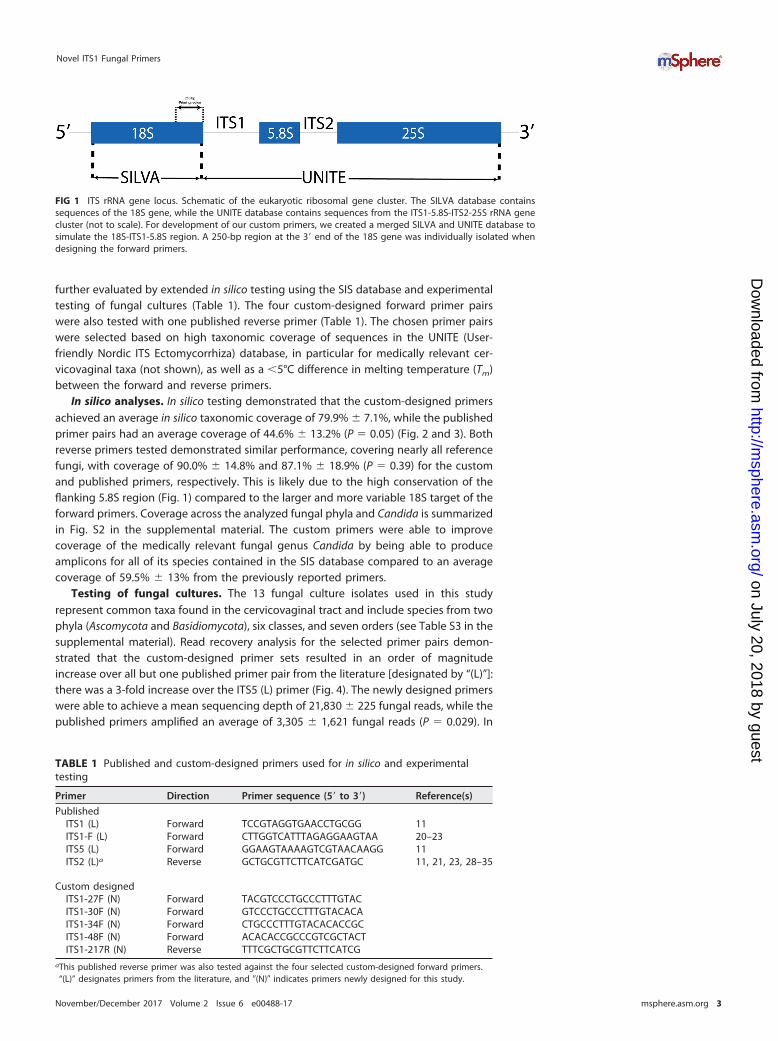
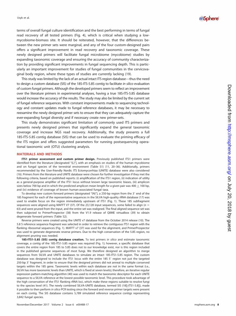
Unlike bacteria, fungal evolution and morphological diversity are not intimatelyassociated with single genetic markers. The morphological system that has primarilybeen used to classify fungi presents a barrier for researchers by including multiplepotential species names for the same organism. For example, morphological differ-ences present throughout the various reproductive stages of fungi (i.e., anamorph andtelemorph) (6) may result in multiple taxonomic classifications for the same organism.In contrast, molecular methods that target specific genetic regions such as cytochromec oxidase subunit 1 (CO1) (7) and the internal transcribed spacers (ITSs) (see Fig. 1) ofthe eukaryotic ribosomal cluster (8), provide a new paradigm for fungal taxonomy.Placement of the same organism into multiple groups due to morphological charac-terization confounds molecular classification, especially when there is significant ge-netic diversity in the organism of interest. Investigators are therefore limited to the useof genetic markers that are informative for species-level identification, but unfortu-nately lack the ability to perform phylogenetic reconstruction given the complexevolutionary history of fungi (4). A currently proposed barcode DNA region for fungalcommunity identification is the ITS1 region, which allows for species-level resolution ina large number of fungi and provides amplicon sizes suitable for current short read NGSplatforms like Illumina (9).
Despite the availability of a suitable DNA region for fungal community analyses,studies of medically relevant fungi use previously developed, “universal” ITS1 primersthat are limited by taxonomic bias as they were developed based on small subset of soilfungi available at the time of their creation and commonly generate a low number ofsequence reads (10–14). The latter is especially important because community-basedanalyses suffer when a sample does not achieve a minimum read threshold necessaryto characterize the community—a threshold readily identifiable through rarefactionanalysis (15). The present study utilizes bioinformatics and an iterative approach toidentify fungal sequences for primer design for broad taxonomic coverage using theITS1 barcode region. This study further expands on the currently available tools bycreating a database of contiguous 18S-ITS1-5.8S (SIS) sequences, which are omittedfrom assembled fungal reference genomes and makes it possible to bioinformaticallyevaluate primer pairs. In addition to in silico evaluation against the SIS database, theprimers were empirically tested with fungal cultures and clinical samples. We providedata that these primers significantly improve fungal taxonomic coverage, fungal readrecovery, and accuracy of characterization of human-associated fungal communities.
RESULTSITS1 primer design. A total of seven (five forward and two reverse) published
primers amplifying the ITS1 region were initially chosen for further analysis based onreported overall taxonomic coverage and amplicon size (see Table S1 in the supple-mental material). Additionally, 85 primers (78 forward and 7 reverse) were customdesigned using the SIS database (see Table S2 in the supplemental material). Threeforward primers and one reverse primer representing the previously published primersand four forward primers and one reverse primer representing the custom primers were
Usyk et al.
November/December 2017 Volume 2 Issue 6 e00488-17 msphere.asm.org 2
on July 20, 2018 by guesthttp://m
sphere.asm.org/
Dow
nloaded from
further evaluated by extended in silico testing using the SIS database and experimentaltesting of fungal cultures (Table 1). The four custom-designed forward primer pairswere also tested with one published reverse primer (Table 1). The chosen primer pairswere selected based on high taxonomic coverage of sequences in the UNITE (User-friendly Nordic ITS Ectomycorrhiza) database, in particular for medically relevant cer-vicovaginal taxa (not shown), as well as a �5°C difference in melting temperature (Tm)between the forward and reverse primers.
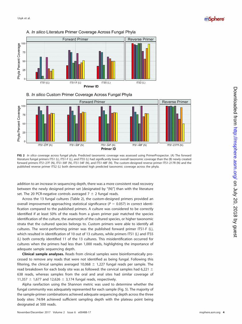
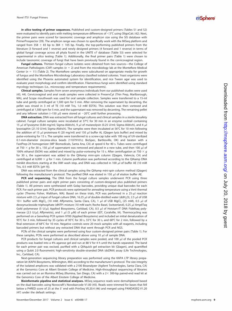
In silico analyses. In silico testing demonstrated that the custom-designed primersachieved an average in silico taxonomic coverage of 79.9% � 7.1%, while the publishedprimer pairs had an average coverage of 44.6% � 13.2% (P � 0.05) (Fig. 2 and 3). Bothreverse primers tested demonstrated similar performance, covering nearly all referencefungi, with coverage of 90.0% � 14.8% and 87.1% � 18.9% (P � 0.39) for the customand published primers, respectively. This is likely due to the high conservation of theflanking 5.8S region (Fig. 1) compared to the larger and more variable 18S target of theforward primers. Coverage across the analyzed fungal phyla and Candida is summarizedin Fig. S2 in the supplemental material. The custom primers were able to improvecoverage of the medically relevant fungal genus Candida by being able to produceamplicons for all of its species contained in the SIS database compared to an averagecoverage of 59.5% � 13% from the previously reported primers.
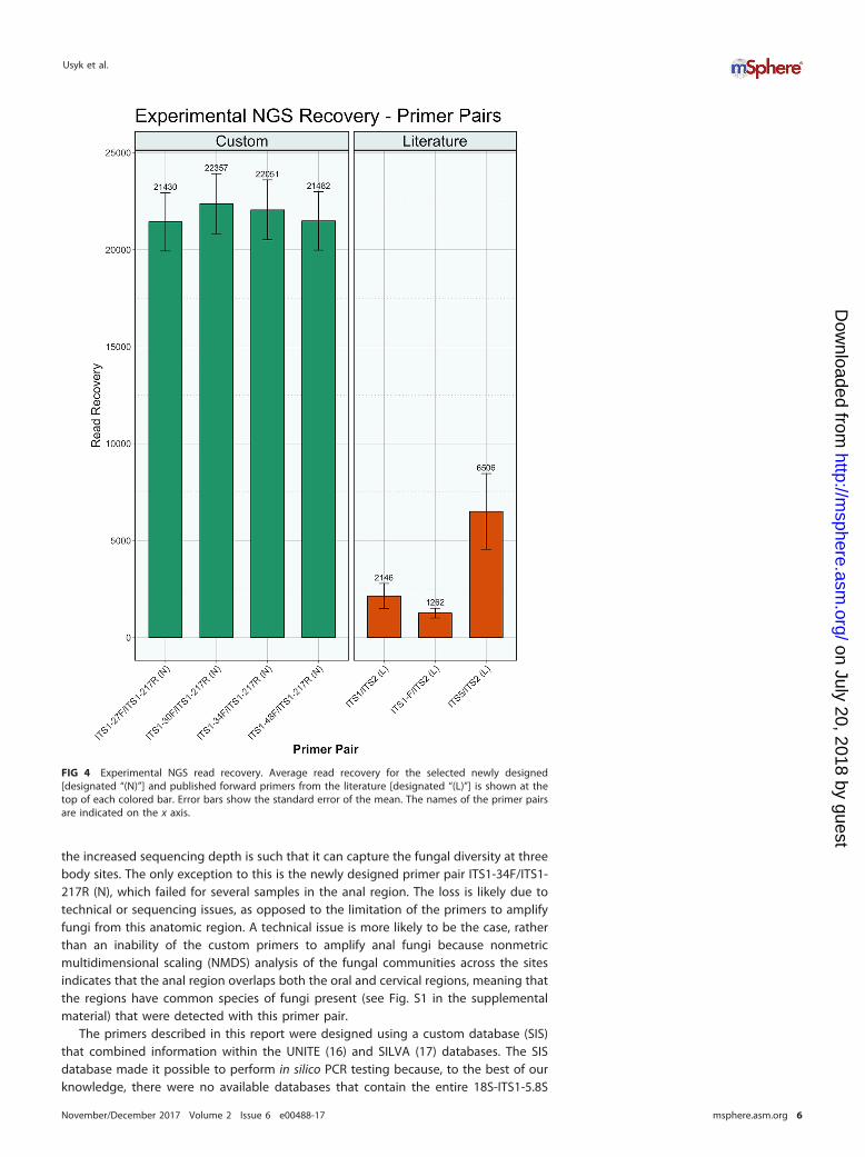
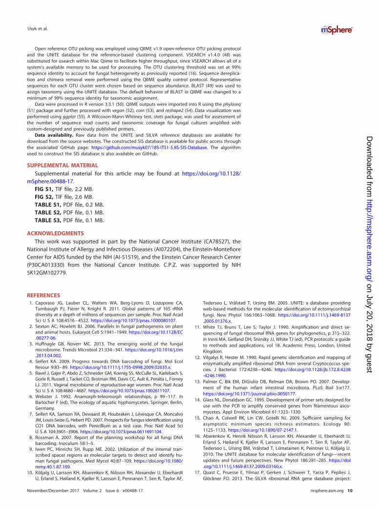
Testing of fungal cultures. The 13 fungal culture isolates used in this studyrepresent common taxa found in the cervicovaginal tract and include species from twophyla (Ascomycota and Basidiomycota), six classes, and seven orders (see Table S3 in thesupplemental material). Read recovery analysis for the selected primer pairs demon-strated that the custom-designed primer sets resulted in an order of magnitudeincrease over all but one published primer pair from the literature [designated by “(L)”]:there was a 3-fold increase over the ITS5 (L) primer (Fig. 4). The newly designed primerswere able to achieve a mean sequencing depth of 21,830 � 225 fungal reads, while thepublished primers amplified an average of 3,305 � 1,621 fungal reads (P � 0.029). In
TABLE 1 Published and custom-designed primers used for in silico and experimentaltesting
Primer Direction Primer sequence (5= to 3=) Reference(s)
PublishedITS1 (L) Forward TCCGTAGGTGAACCTGCGG 11ITS1-F (L) Forward CTTGGTCATTTAGAGGAAGTAA 20–23ITS5 (L) Forward GGAAGTAAAAGTCGTAACAAGG 11ITS2 (L)a Reverse GCTGCGTTCTTCATCGATGC 11, 21, 23, 28–35
Custom designedITS1-27F (N) Forward TACGTCCCTGCCCTTTGTACITS1-30F (N) Forward GTCCCTGCCCTTTGTACACAITS1-34F (N) Forward CTGCCCTTTGTACACACCGCITS1-48F (N) Forward ACACACCGCCCGTCGCTACTITS1-217R (N) Reverse TTTCGCTGCGTTCTTCATCG
aThis published reverse primer was also tested against the four selected custom-designed forward primers.“(L)” designates primers from the literature, and �(N)� indicates primers newly designed for this study.
FIG 1 ITS rRNA gene locus. Schematic of the eukaryotic ribosomal gene cluster. The SILVA database containssequences of the 18S gene, while the UNITE database contains sequences from the ITS1-5.8S-ITS2-25S rRNA genecluster (not to scale). For development of our custom primers, we created a merged SILVA and UNITE database tosimulate the 18S-ITS1-5.8S region. A 250-bp region at the 3= end of the 18S gene was individually isolated whendesigning the forward primers.
Novel ITS1 Fungal Primers
November/December 2017 Volume 2 Issue 6 e00488-17 msphere.asm.org 3
on July 20, 2018 by guesthttp://m
sphere.asm.org/
Dow
nloaded from
addition to an increase in sequencing depth, there was a more consistent read recoverybetween the newly designed primer set [designated by “(N)”] than with the literatureset. The 20 PCR-negative controls averaged 7 � 2 fungal reads.
Across the 13 fungal cultures (Table 2), the custom-designed primers provided anoverall improvement approaching statistical significance (P � 0.057) in correct identi-fication compared to the published primers. A culture was considered to be correctlyidentified if at least 50% of the reads from a given primer pair matched the speciesidentification of the culture, the anamorph of the cultured species, or higher taxonomicstrata that the cultured species belongs to. Custom primers were able to identify allcultures. The worst-performing primer was the published forward primer ITS1-F (L),which resulted in identification of 10 out of 13 cultures, while primers ITS1 (L) and ITS5(L) both correctly identified 11 of the 13 cultures. This misidentification occurred forcultures when the primers had less than 1,000 reads, highlighting the importance ofadequate sample sequencing depth.
Clinical sample analyses. Reads from clinical samples were bioinformatically pro-cessed to remove any reads that were not identified as being fungal. Following thisfiltering, the clinical samples averaged 10,068 � 1,227 fungal reads per sample. Theread breakdown for each body site was as followed: the cervical samples had 6,221 �
638 reads, whereas samples from the oral and anal sites had similar coverage of11,357 � 1,677 and 12,626 � 3,174 fungal reads, respectively.
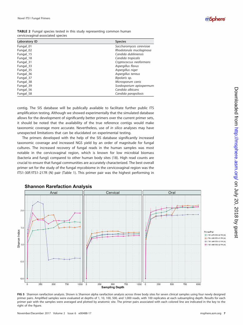
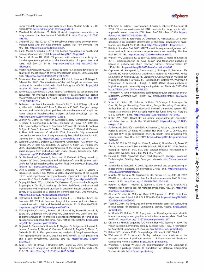
Alpha rarefaction using the Shannon metric was used to determine whether thefungal community was adequately represented for each sample (Fig. 5). The majority ofthe sample-primer combinations achieved adequate sequencing depth across the threebody sites: 74/84 achieved sufficient sampling depth with the plateau point beingdesignated at 500 reads.
FIG 2 In silico coverage across fungal phyla. Predicted taxonomic coverage was assessed using PrimerProspector. (A) The forwardliterature fungal primers ITS1 (L), ITS1-F (L), and ITS5 (L) had significantly lower overall taxonomic coverage than the (B) newly createdforward primers ITS1-27F (N), ITS1-30F (N), ITS1-34F (N), and ITS1-48F (N). The custom-designed reverse primer ITS1-217R (N) and thepublished reverse primer ITS2 (L) both demonstrated high predicted taxonomic coverage across the phyla.
Usyk et al.
November/December 2017 Volume 2 Issue 6 e00488-17 msphere.asm.org 4
on July 20, 2018 by guesthttp://m
sphere.asm.org/
Dow
nloaded from
DISCUSSION
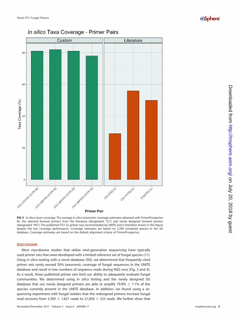
Most mycobiome studies that utilize next-generation sequencing have typicallyused primer sets that were developed with a limited reference set of fungal species (11).Using in silico testing with a novel database (SIS), we determined that frequently citedprimer sets rarely exceed 50% taxonomic coverage of fungal sequences in the UNITEdatabase and result in low numbers of sequence reads during NGS runs (Fig. 3 and 4).As a result, these published primer sets limit our ability to adequately evaluate fungalcommunities. We determined using in silico testing and the newly designed SISdatabase that our newly designed primers are able to amplify 79.9% � 7.1% of thespecies currently present in the UNITE database. In addition, we found using a se-quencing experiment with fungal isolates that the redesigned primers increase fungalread recovery from 3,305 � 1,621 reads to 21,830 � 225 reads. We further show that
FIG 3 In silico taxon coverage. The average in silico taxonomic coverage estimates obtained with PrimerProspectorfor the selected forward primers from the literature [designated “(L)”] and newly designed forward primers[designated “(N)”]. The published ITS1 (L) primer was recommended by UNITE and is therefore shown in this figuredespite the low coverage performance. Coverage estimates are based on 5,789 simulated species in the SISdatabase. Coverage estimates are based on the default alignment criteria of PrimerProspector.
Novel ITS1 Fungal Primers
November/December 2017 Volume 2 Issue 6 e00488-17 msphere.asm.org 5
on July 20, 2018 by guesthttp://m
sphere.asm.org/
Dow
nloaded from
the increased sequencing depth is such that it can capture the fungal diversity at threebody sites. The only exception to this is the newly designed primer pair ITS1-34F/ITS1-217R (N), which failed for several samples in the anal region. The loss is likely due totechnical or sequencing issues, as opposed to the limitation of the primers to amplifyfungi from this anatomic region. A technical issue is more likely to be the case, ratherthan an inability of the custom primers to amplify anal fungi because nonmetricmultidimensional scaling (NMDS) analysis of the fungal communities across the sitesindicates that the anal region overlaps both the oral and cervical regions, meaning thatthe regions have common species of fungi present (see Fig. S1 in the supplementalmaterial) that were detected with this primer pair.
The primers described in this report were designed using a custom database (SIS)that combined information within the UNITE (16) and SILVA (17) databases. The SISdatabase made it possible to perform in silico PCR testing because, to the best of ourknowledge, there were no available databases that contain the entire 18S-ITS1-5.8S
FIG 4 Experimental NGS read recovery. Average read recovery for the selected newly designed[designated “(N)”] and published forward primers from the literature [designated “(L)”] is shown at thetop of each colored bar. Error bars show the standard error of the mean. The names of the primer pairsare indicated on the x axis.
Usyk et al.
November/December 2017 Volume 2 Issue 6 e00488-17 msphere.asm.org 6
on July 20, 2018 by guesthttp://m
sphere.asm.org/
Dow
nloaded from
contig. The SIS database will be publically available to facilitate further public ITSamplification testing. Although we showed experimentally that the simulated databaseallows for the development of significantly better primers over the current primer sets,it should be noted that the availability of the true reference contigs would maketaxonomic coverage more accurate. Nevertheless, use of in silico analyses may haveunexpected limitations that can be elucidated on experimental testing.
The primers developed with the help of the SIS database significantly increasedtaxonomic coverage and increased NGS yield by an order of magnitude for fungalcultures. The increased recovery of fungal reads in the human samples was mostnotable in the cervicovaginal region, which is known for low microbial biomass(bacteria and fungi) compared to other human body sites (18). High read counts arecrucial to ensure that fungal communities are accurately characterized. The best overallprimer set for the study of the fungal mycobiome in the cervicovaginal region was theITS1-30F/ITS1-217R (N) pair (Table 1). This primer pair was the highest performing in
FIG 5 Shannon rarefaction analysis. Shown is Shannon alpha rarefaction analysis across three body sites for seven clinical samples using four newly designedprimer pairs. Amplified samples were evaluated at depths of 1, 10, 100, 500, and 1,000 reads, with 100 replicates at each subsampling depth. Results for eachprimer pair with the samples were averaged and plotted by anatomic site. The primer pairs associated with each colored line are indicated in the key to theright of the figure.
TABLE 2 Fungal species tested in this study representing common humancervicovaginal-associated species
Laboratory ID Species
Fungal_01 Saccharomyces cerevisiaeFungal_02 Rhodotorula mucilaginosaFungal_15 Candida dubliniensisFungal_18 Candida tropicalisFungal_31 Cryptococcus neoformansFungal_33 Aspergillus flavusFungal_35 Aspergillus nigerFungal_36 Aspergillus terreusFungal_37 Bipolaris sp.Fungal_38 Microsporum canisFungal_39 Scedosporium apiospermumFungal_56 Candida albicansFungal_58 Candida parapsilosis
Novel ITS1 Fungal Primers
November/December 2017 Volume 2 Issue 6 e00488-17 msphere.asm.org 7
on July 20, 2018 by guesthttp://m
sphere.asm.org/
Dow
nloaded from
terms of overall fungal culture identification and the best performing in terms of fungalread recovery of all tested primers (Fig. 4), which is critical when studying a low-mycobiome-biomass site. It should be reiterated, however, that the differences be-tween the new primer sets were marginal, and any of the four custom-designed pairsoffers a significant improvement in read recovery and taxonomic coverage. Thesenewly designed primers will facilitate fungal microbiome (mycobiome) studies byexpanding taxonomic coverage and ensuring the accuracy of community characteriza-tion by providing significant improvements in fungal sequencing depth. This is partic-ularly an important improvement for studies of fungal communities in the cervicova-ginal body region, where these types of studies are currently lacking (19).
This study was limited by the lack of an actual intact ITS region database—thus the needto design a custom database (SIS) of the 18S-ITS-5.8S contig to facilitate in silico evaluationof custom fungal primers. Although the developed primers seem to reflect an improvementover the literature primers in experimental analyses, having a true 18S-ITS-5.8S databasewould increase the accuracy of the results. The study may also be limited by the current setof fungal reference sequences. With constant improvements made to sequencing technol-ogy and constant updates made to fungal reference databases, it may be necessary toreexamine the newly designed primer sets to ensure that they can adequately capture theever-expanding fungal diversity and if necessary create new primer-sets.
This study demonstrates significant limitation of commonly used ITS primers andpresents newly designed primers that significantly expand the general taxonomiccoverage and increase NGS read recovery. Additionally, the study presents a full18S-ITS-5.8S contig database (SIS) that can be used to evaluate the priming efficacy ofthe ITS region and offers suggested parameters for running postsequencing opera-tional taxonomic unit (OTU) clustering analysis.
MATERIALS AND METHODSITS1 primer assessment and custom primer design. Previously published ITS1 primers were
identified from the literature [designated “(L)”], with an emphasis on studies of the human mycobiomeand on fungal species of the terrestrial environment (Table S1) (11, 20–36). Additionally, primersrecommended by the User-friendly Nordic ITS Ectomycorrhiza (UNITE) database were also considered(16). Primers from the literature and UNITE database were chosen for further investigation if they met thefollowing criteria, based on published reports: (i) amplification of the ITS1 region, (ii) indication of utilityas a general-purpose amplifier of the ITS1 locus without known large taxonomic biases, (iii) ampliconsizes below 700 bp and in which the predicted amplicon mean length for a given pair was 400 � 100 bp,and (iv) evidence of coverage of known human-associated fungal taxa.
To develop new custom forward primers [designated “(N)”], a 250-bp region from the 3= end of the18S fragment for each of the representative sequences in the SILVA high-quality rRNA database (17) wasused to enable focus on the region immediately upstream of ITS1 (Fig. 1). These 18S subfragmentsequences were aligned using MAFFT V7 (37). Of the 23,128 input sequences, some failed to align (n �63) and were pruned from the input, and the entire set was realigned. The final aligned sequence set wasthen subjected to PrimerProspector (38) from the V1.9 release of QIIME virtualbox (39) to obtaindegenerate forward primers (Table S2).
Reverse primers were created using the UNITE v7 database from the October 2014 release (10). The5.8 S reference sequence fragment was selected in order to retrieve the contiguous ITS1 region with theflanking ribosomal sequences (Fig. 1). MAFFT v7 (37) was used for the alignment, and PrimerProspectorwas used to generate degenerate reverse primers. Due to the high conservation of the 5.8S region, noalignment pruning was needed.
18S-ITS1-5.8S (SIS) contig database creation. To test primers in silico and estimate taxonomiccoverage, a contig of the 18S-ITS1-5.8S region was required (Fig. 1); however, a specific database thatcovers the entire region from 18S to 5.8S does not to our knowledge exist, nor is this region includedin the published genome sequences of most fungi. We therefore designed an algorithm to mergesequences from SILVA and UNITE databases to simulate an intact 18S-ITS1-5.8S region. The customdatabase was designed to include the ITS1 locus with the entire 18S 5= region not just the targeted250-bp 3= fragment, in order to ensure that the designed primers did not anneal to multiple conservedtargets within the 18S gene. Taxonomic levels within each database are not in the same format (i.e.,SILVA has more taxonomic levels than UNITE, which is fixed at seven levels); therefore, an iterative regularexpression pattern-matching algorithm (40) was used to match the taxonomic descriptor for each UNITEsequence to a SILVA reference at the lowest possible taxonomic level. This procedure took advantage ofthe high conservation of the ITS1 flanking rRNA loci, which make these regions suitable to resolve fungito the species level (41). The newly combined SILVA-UNITE database, termed SIS (18S-ITS1-5.8S), madeit possible to then perform in silico PCR testing since the forward and reverse primer targets were presenton each contig. The SIS database contains 5,789 simulated reference sequence contigs representing3,842 fungal species.
Usyk et al.
November/December 2017 Volume 2 Issue 6 e00488-17 msphere.asm.org 8
on July 20, 2018 by guesthttp://m
sphere.asm.org/
Dow
nloaded from
In silico testing of primer sequences. Published and custom-designed primers (Tables S1 and S2)were evaluated to identify pairs with melting temperature differences of �5°C using OligoCalc (42). Next,the primer pairs were scored for taxonomic coverage and amplicon size using the SIS database withPrimerProspector (39). The amplicon range was chosen to specifically work with the MiSeq platform andranged from 358 � 83 bp to 384 � 105 bp. Finally, the top-performing published primers from theliterature (3 forward and 1 reverse) and newly designed primers (4 forward and 1 reverse) in terms ofglobal fungal coverage across all phyla found in the UNITE v7 database (Table S3) were selected forexperimental in silico testing (Table 1). Additionally, the final primer pairs (Table 1) were chosen toinclude taxonomic coverage of fungi that have been previously found in the cervicovaginal region.
Fungal cultures. Thirteen fungal culture isolates were obtained from two sources—the College ofAmerican Pathologists (CAP) samples (n � 2) and from the microbiology lab at the Montefiore MedicalCenter (n � 11) (Table 2). The Montefiore samples were subcultured on appropriate media for growthof fungus and the Montefiore Microbiology Laboratory classified isolated colonies. Yeast organisms wereidentified using the Phoenix automated system for identification, and rice Tween agar was used toevaluate yeast morphology and confirm identification. Filamentous fungi were identified using standardmycology techniques (i.e., microscopy and temperature requirements).
Clinical samples. Samples from seven anonymous individuals from our published studies were used(43, 44). Cervicovaginal and anal swab samples were collected in PreservCyt (Thin Prep, Marlborough,MA), and Scope mouthwash was used for oral sample collection. Samples were transferred to a 15-mltube and gently centrifuged at 1,500 rpm for 5 min. After removing the supernatant by decanting, thepellet was rinsed in 3 ml of TE (10 mM Tris, 1.0 mM EDTA). This solution was then vortexed andcentrifuged at 1,500 rpm for 5 min, and the supernatant was removed by decanting. The remaining pelletand any leftover solution (~150 �l) were stored at �20°C until further processing.
DNA extraction. DNA was extracted from all fungal cultures and clinical samples in a sterile biosafetycabinet. Fungal culture samples were incubated at 37°C for 30 min in an enzyme cocktail containing15 �l of lysozyme (0.84 mg/ml; Sigma-Aldrich), 9 �l of mutanolysin (0.25 U/ml; Sigma-Aldrich), and 4 �llysostaphin (21.10 U/ml; Sigma-Aldrich). The samples were then incubated at 56°C for 10 min followingthe addition of 15 �l proteinase K (20 mg/ml) and 150 �l buffer AL (Qiagen lysis buffer) and mixed bypulse-vortexing for 15 s. The samples were transferred to a screw-cap tube with 100 mg of UV-sterilized0.1-mm-diameter zirconia-silica beads (11079101z; BioSpec, Bartlesville, OK) and beaten with aFastPrep-24 homogenizer (MP Biomedicals, Santa Ana, CA) at speed 6 for 40 s. Tubes were centrifugedat 750 � g for 30 s, 150 �l of supernatant was removed and placed in a new tube, and then 100 �l of100% ethanol (EtOH) was added and mixed by pulse-vortexing for 15 s. After centrifugation at 750 � gfor 30 s, the supernatant was added to the QIAamp mini-spin column (Qiagen, Valencia, CA) andcentrifuged at 6,000 � g for 1 min. Column purification was performed according to the QIAamp DNAminikit directions starting at the AWI wash step, and DNA was collected in 100 �l of buffer AE (10 mMTris, 0.5 mM EDTA [pH 9]).
DNA was extracted from the clinical samples using the QIAamp mini-spin column method (Qiagen)following the manufacturer’s protocol. The purified DNA was eluted in 150 �l of elution buffer AE.
PCR and sequencing. The DNA from the fungal culture samples underwent PCR using threepublished primer pairs and eight primer pairs consisting of custom-designed plus published primers(Table 1). All primers were synthesized with Golay barcodes, providing unique dual barcodes for eachPCR. For each primer pair, PCR protocols were optimized for annealing temperature using a Verti thermalcycler (Thermo Fisher, Waltham, MA). Based on these trials, PCR was performed in a 25-�l reactionmixture with 2.5 �l input of fungal culture DNA, 16.25 �l of double-distilled water (ddH2O), 2.5 �l of USB10� buffer with MgCl2 (10 mM; Affymetrix, Santa Clara, CA), 1 �l of USB MgCl2 (25 mM), 0.5 �l ofdeoxynucleoside triphosphate (dNTP) mixture (10 mM each; Roche Basel, Switzerland), 0.25 �l AmpliTaqGold polymerase (5 U/�l; Applied Biosystems, Carlsbad, CA), 0.5 �l of Hotstart-IT DNA Fidelitaq poly-merase (2.5 U/�l; Affymetrix), and 1 �l (5 �M) of each primer (IDT, Coralville, IA). Thermocycling wasperformed on a GeneAmp PCR system 9700 (Applied Biosystems) and included an initial denaturation of95°C for 3 min, followed by 35 cycles of 95°C for 30 s, 55°C for 30 s, and 68°C for 2 min, followed by afinal extension of 68°C for 10 min. Negative controls were 20 mock samples with all reagents, includingbarcoded primers but without any extracted DNA that went through PCR and NGS.
PCRs of the clinical samples were performed using four custom-designed primer pairs (Table 1). Forthese samples, PCRs were performed as described above using 10 �l of sample DNA.
PCR products for fungal cultures and clinical samples were pooled, and 100 �l of the pooled PCRproducts was loaded into a 4% agarose gel and run at 88 V for 4 h until the bands separated. The bandfor each primer pair was excised, purified with a QIAquick gel extraction kit (Qiagen), and quantifiedusing a Qubit 2.0 fluorometric high-sensitivity double-stranded DNA (dsDNA) assay (Life Technologies,Inc., Carlsbad, CA).
Next-generation sequencing library preparation was performed using the KAPA LTP library prepa-ration kit (KAPA Biosystems, Wilmington, MA) according to the manufacturer’s protocol. The size integrityof the isolated amplicons was validated with a 2100 Bioanalyzer (Agilent Technologies, Santa Clara, CA)at the Genomics Core at Albert Einstein College of Medicine. High-throughput sequencing of librarieswas carried out on an Illumina MiSeq (Illumina, San Diego, CA) with a 2� 300-bp paired-end read kit atthe Genomics Core of the Albert Einstein College of Medicine.
Bioinformatic pipeline and statistical analyses. MiSeq sequence reads were demultiplexed basedon the dual barcodes using Novocraft’s Novobarcode V1.00 (45). Reads were trimmed for bases that fellbelow a PHRED score of 25 at the 3= end with PrinSeq V0.20.4 (46) and merged using PANDASEQ V1.20(47) under the default settings.
Novel ITS1 Fungal Primers
November/December 2017 Volume 2 Issue 6 e00488-17 msphere.asm.org 9
on July 20, 2018 by guesthttp://m
sphere.asm.org/
Dow
nloaded from
Open reference OTU picking was employed using QIIME v1.9 open-reference OTU picking protocoland the UNITE database for the reference-based clustering component. VSEARCH v1.4.0 (48) wassubstituted for usearch within Mac Qiime to facilitate higher throughput, since VSEARCH allows all of asystem’s available memory to be used for processing. The OTU clustering threshold was set at 99%sequence identity to account for fungal heterogeneity as previously reported (16). Sequence dereplica-tion and chimera removal were performed using the QIIME quality control protocol. Representativesequences for each OTU cluster were chosen based on sequence abundance. BLAST (49) was used toassign taxonomy using the UNITE database. The default behavior of BLAST in QIIME was changed to aminimum of 99% sequence identity for taxonomic assignment.
Data were processed in R version 3.3.1 (50). QIIME outputs were imported into R using the phyloseq(51) package and further processed with vegan (52), coin (53), and reshape2 (54). Data visualization wasperformed using ggplot (55). A Wilcoxon-Mann-Whitney test, stats package, was used for assessment ofthe number of sequence read counts and taxonomic coverage for fungal cultures amplified withcustom-designed and previously published primers.
Data availability. Raw data from the UNITE and SILVA reference databases are available fordownload from the source websites. The constructed SIS database is available for public access throughthe associated GitHub page: https://github.com/musyk07/18S-ITS1-5.8S-SIS-Database. The algorithmused to construct the SIS database is also available on GitHub.
SUPPLEMENTAL MATERIALSupplemental material for this article may be found at https://doi.org/10.1128/
mSphere.00488-17.FIG S1, TIF file, 2.2 MB.FIG S2, TIF file, 2.6 MB.TABLE S1, PDF file, 0.2 MB.TABLE S2, PDF file, 0.1 MB.TABLE S3, PDF file, 0.1 MB.
ACKNOWLEDGMENTSThis work was supported in part by the National Cancer Institute (CA78527), the
National Institute of Allergy and Infectious Diseases (AI072204), the Einstein-MontefioreCenter for AIDS funded by the NIH (AI-51519), and the Einstein Cancer Research Center(P30CA013330) from the National Cancer Institute. C.P.Z. was supported by NIH5K12GM102779.
REFERENCES1. Caporaso JG, Lauber CL, Walters WA, Berg-Lyons D, Lozupone CA,
Turnbaugh PJ, Fierer N, Knight R. 2011. Global patterns of 16S rRNAdiversity at a depth of millions of sequences per sample. Proc Natl AcadSci U S A 108:4516 – 4522. https://doi.org/10.1073/pnas.1000080107.
2. Sexton AC, Howlett BJ. 2006. Parallels in fungal pathogenesis on plantand animal hosts. Eukaryot Cell 5:1941–1949. https://doi.org/10.1128/EC.00277-06.
3. Huffnagle GB, Noverr MC. 2013. The emerging world of the fungalmicrobiome. Trends Microbiol 21:334 –341. https://doi.org/10.1016/j.tim.2013.04.002.
4. Seifert KA. 2009. Progress towards DNA barcoding of fungi. Mol EcolResour 9:83– 89. https://doi.org/10.1111/j.1755-0998.2009.02635.x.
5. Ravel J, Gajer P, Abdo Z, Schneider GM, Koenig SS, McCulle SL, Karlebach S,Gorle R, Russell J, Tacket CO, Brotman RM, Davis CC, Ault K, Peralta L, ForneyLJ. 2011. Vaginal microbiome of reproductive-age women. Proc Natl AcadSci U S A 108:4680–4687. https://doi.org/10.1073/pnas.1002611107.
6. Webster J. 1992. Anamorph-teleomorph relationships, p 99 –117. InBarlocher F (ed), The ecology of aquatic hyphomycetes. Springer, Berlin,Germany.
7. Seifert KA, Samson RA, Dewaard JR, Houbraken J, Lévesque CA, MoncalvoJM, Louis-Seize G, Hebert PD. 2007. Prospects for fungus identification usingCO1 DNA barcodes, with Penicillium as a test case. Proc Natl Acad SciU S A 104:3901–3906. https://doi.org/10.1073/pnas.0611691104.
8. Rossman A. 2007. Report of the planning workshop for all fungi DNAbarcoding. Inoculum 58:1–5.
9. Iwen PC, Hinrichs SH, Rupp ME. 2002. Utilization of the internal tran-scribed spacer regions as molecular targets to detect and identify hu-man fungal pathogens. Med Mycol 40:87–109. https://doi.org/10.1080/mmy.40.1.87.109.
10. Kõljalg U, Larsson KH, Abarenkov K, Nilsson RH, Alexander IJ, EberhardtU, Erland S, Høiland K, Kjøller R, Larsson E, Pennanen T, Sen R, Taylor AF,
Tedersoo L, Vrålstad T, Ursing BM. 2005. UNITE: a database providingweb-based methods for the molecular identification of ectomycorrhizalfungi. New Phytol 166:1063–1068. https://doi.org/10.1111/j.1469-8137.2005.01376.x.
11. White TJ, Bruns T, Lee S, Taylor J. 1990. Amplification and direct se-quencing of fungal ribosomal RNA genes for phylogenetics, p 315–322.In Innis MA, Gelfand DH, Sninsky JJ, White TJ (ed), PCR protocols: a guideto methods and applications, vol 18. Academic Press, London, UnitedKingdom.
12. Vilgalys R, Hester M. 1990. Rapid genetic identification and mapping ofenzymatically amplified ribosomal DNA from several Cryptococcus spe-cies. J Bacteriol 172:4238 – 4246. https://doi.org/10.1128/jb.172.8.4238-4246.1990.
13. Palmer C, Bik EM, DiGiulio DB, Relman DA, Brown PO. 2007. Develop-ment of the human infant intestinal microbiota. PLoS Biol 5:e177.https://doi.org/10.1371/journal.pbio.0050177.
14. Glass NL, Donaldson GC. 1995. Development of primer sets designed foruse with the PCR to amplify conserved genes from filamentous asco-mycetes. Appl Environ Microbiol 61:1323–1330.
15. Chao A, Colwell RK, Lin CW, Gotelli NJ. 2009. Sufficient sampling forasymptotic minimum species richness estimators. Ecology 90:1125–1133. https://doi.org/10.1890/07-2147.1.
16. Abarenkov K, Henrik Nilsson R, Larsson KH, Alexander IJ, Eberhardt U,Erland S, Høiland K, Kjøller R, Larsson E, Pennanen T, Sen R, Taylor AF,Tedersoo L, Ursing BM, Vrålstad T, Liimatainen K, Peintner U, Kõljalg U.2010. The UNITE database for molecular identification of fungi—recentupdates and future perspectives. New Phytol 186:281–285. https://doi.org/10.1111/j.1469-8137.2009.03160.x.
17. Quast C, Pruesse E, Yilmaz P, Gerken J, Schweer T, Yarza P, Peplies J,Glöckner FO. 2013. The SILVA ribosomal RNA gene database project:
Usyk et al.
November/December 2017 Volume 2 Issue 6 e00488-17 msphere.asm.org 10
on July 20, 2018 by guesthttp://m
sphere.asm.org/
Dow
nloaded from
improved data processing and web-based tools. Nucleic Acids Res 41:D590 –D596. https://doi.org/10.1093/nar/gks1219.
18. Marsland BJ, Gollwitzer ES. 2014. Host-microorganism interactions inlung diseases. Nat Rev Immunol 14:827– 835. https://doi.org/10.1038/nri3769.
19. Underhill DM, Iliev ID. 2014. The mycobiota: interactions between com-mensal fungi and the host immune system. Nat Rev Immunol 14:405– 416. https://doi.org/10.1038/nri3684.
20. Cui L, Morris A, Ghedin E. 2013. The human mycobiome in health anddisease. Genome Med 5:63. https://doi.org/10.1186/gm467.
21. Gardes M, Bruns TD. 1993. ITS primers with enhanced specificity forbasidiomycetes—application to the identification of mycorrhizae andrusts. Mol Ecol 2:113–118. https://doi.org/10.1111/j.1365-294X.1993.tb00005.x.
22. Martin KJ, Rygiewicz PT. 2005. Fungal-specific PCR primers developed foranalysis of the ITS region of environmental DNA extracts. BMC Microbiol5:28. https://doi.org/10.1186/1471-2180-5-28.
23. Ghannoum MA, Jurevic RJ, Mukherjee PK, Cui F, Sikaroodi M, Naqvi A,Gillevet PM. 2010. Characterization of the oral fungal microbiome (my-cobiome) in healthy individuals. PLoS Pathog 6:e1000713. https://doi.org/10.1371/journal.ppat.1000713.
24. Taylor DL, McCormick MK. 2008. Internal transcribed spacer primers andsequences for improved characterization of basidiomycetous orchidmycorrhizas. New Phytol 177:1020 –1033. https://doi.org/10.1111/j.1469-8137.2007.02320.x.
25. Tedersoo L, Anslan S, Bahram M, Põlme S, Riit T, Liiv I, Kõljalg U, KisandV, Nilsson H, Hildebrand F, Bork P, Abarenkov K. 2015. Shotgun metag-enomes and multiple primer pair-barcode combinations of ampliconsreveal biases in metabarcoding analyses of fungi. MycoKeys 10:1– 43.https://doi.org/10.3897/mycokeys.10.4852.
26. Lennon NJ, Lintner RE, Anderson S, Alvarez P, Barry A, Brockman W, DazaR, Erlich RL, Giannoukos G, Green L, Hollinger A, Hoover CA, Jaffe DB,Juhn F, McCarthy D, Perrin D, Ponchner K, Powers TL, Rizzolo K, RobbinsD, Ryan E, Russ C, Sparrow T, Stalker J, Steelman S, Weiand M, ZimmerA, Henn MR, Nusbaum C, Nicol R. 2010. A scalable, fully automatedprocess for construction of sequence-ready barcoded libraries for 454.Genome Biol 11:R15. https://doi.org/10.1186/gb-2010-11-2-r15.
27. Willger SD, Grim SL, Dolben EL, Shipunova A, Hampton TH, Morrison HG,Filkins LM, O’Toole GA, Moulton LA, Ashare A, Sogin ML, Hogan DA.2014. Characterization and quantification of the fungal microbiome inserial samples from individuals with cystic fibrosis. Microbiome 2:40.https://doi.org/10.1186/2049-2618-2-40.
28. Op De Beeck MO, Lievens B, Busschaert P, Declerck S, Vangronsveld J,Colpaert JV. 2014. Comparison and validation of some ITS primer pairsuseful for fungal metabarcoding studies. PLoS One 9:e97629. https://doi.org/10.1371/journal.pone.0097629.
29. Drell T, Lillsaar T, Tummeleht L, Simm J, Aaspõllu A, Väin E, Saarma I,Salumets A, Donders GG, Metsis M. 2013. Characterization of the vaginalmicro- and mycobiome in asymptomatic reproductive-age Estonianwomen. PLoS One 8:e54379. https://doi.org/10.1371/journal.pone.0054379.
30. Dupuy AK, David MS, Li L, Heider TN, Peterson JD, Montano EA, Dongari-Bagtzoglou A, Diaz PI, Strausbaugh LD. 2014. Redefining the human oralmycobiome with improved practices in amplicon-based taxonomy: dis-covery of Malassezia as a prominent commensal. PLoS One 9:e90899.https://doi.org/10.1371/journal.pone.0090899.
31. Hoffmann C, Dollive S, Grunberg S, Chen J, Li H, Wu GD, Lewis JD,Bushman FD. 2013. Archaea and fungi of the human gut microbiome:correlations with diet and bacterial residents. PLoS One 8:e66019.https://doi.org/10.1371/journal.pone.0066019.
32. Mukherjee PK, Chandra J, Retuerto M, Sikaroodi M, Brown RE, Jurevic R,Salata RA, Lederman MM, Gillevet PM, Ghannoum MA. 2014. Oral my-cobiome analysis of HIV-infected patients: identification of Pichia as anantagonist of opportunistic fungi. PLoS Pathog 10:e1003996. https://doi.org/10.1371/journal.ppat.1003996.
33. Orgiazzi A, Bianciotto V, Bonfante P, Daghino S, Ghignone S, Lazzari A,Lumini E, Mello A, Napoli C, Perotto S, Vizzini A, Bagella S, Murat C,Girlanda M. 2013. 454 pyrosequencing analysis of fungal assemblagesfrom geographically distant, disparate soils reveals spatial patterningand a core mycobiome. Diversity 5:73–98. https://doi.org/10.3390/d5010073.
34. Tang J, Iliev ID, Brown J, Underhill DM, Funari VA. 2015. Mycobiome:approaches to analysis of intestinal fungi. J Immunol Methods 421:112–121. https://doi.org/10.1016/j.jim.2015.04.004.
35. Bellemain E, Carlsen T, Brochmann C, Coissac E, Taberlet P, Kauserud H.2010. ITS as an environmental DNA barcode for fungi: an in silicoapproach reveals potential PCR biases. BMC Microbiol 10:189. https://doi.org/10.1186/1471-2180-10-189.
36. Sapkota R, Knorr K, Jørgensen LN, O’Hanlon KA, Nicolaisen M. 2015. Hostgenotype is an important determinant of the cereal phyllosphere myco-biome. New Phytol 207:1134–1144. https://doi.org/10.1111/nph.13418.
37. Katoh K, Standley DM. 2013. MAFFT multiple sequence alignment soft-ware version 7: improvements in performance and usability. Mol BiolEvol 30:772–780. https://doi.org/10.1093/molbev/mst010.
38. Walters WA, Caporaso JG, Lauber CL, Berg-Lyons D, Fierer N, Knight R.2011. PrimerProspector: de novo design and taxonomic analysis ofbarcoded polymerase chain reaction primers. Bioinformatics 27:1159 –1161. https://doi.org/10.1093/bioinformatics/btr087.
39. Caporaso JG, Kuczynski J, Stombaugh J, Bittinger K, Bushman FD,Costello EK, Fierer N, Peña AG, Goodrich JK, Gordon JI, Huttley GA, KelleyST, Knights D, Koenig JE, Ley RE, Lozupone CA, McDonald D, Muegge BD,Pirrung M, Reeder J, Sevinsky JR, Turnbaugh PJ, Walters WA, Widmann J,Yatsunenko T, Zaneveld J, Knight R. 2010. QIIME allows analysis ofhigh-throughput community sequencing data. Nat Methods 7:335–336.https://doi.org/10.1038/nmeth.f.303.
40. Thompson K. 1968. Programming techniques: regular expression searchalgorithm. Commun ACM 11:419 – 422. https://doi.org/10.1145/363347.363387.
41. Schoch CL, Seifert KA, Huhndorf S, Robert V, Spouge JL, Levesque CA,Chen W, Fungal Barcoding Consortium, Fungal Barcoding ConsortiumAuthor List. 2012. Nuclear ribosomal internal transcribed spacer (ITS)region as a universal DNA barcode marker for fungi. Proc Natl Acad SciU S A 109:6241– 6246. https://doi.org/10.1073/pnas.1117018109.
42. Kibbe WA. 2007. OligoCalc: an online oligonucleotide propertiescalculator. Nucleic Acids Res 35:W43–W46. https://doi.org/10.1093/nar/gkm234.
43. Schlecht NF, Burk RD, Nucci-Sack A, Shankar V, Peake K, Lorde-Rollins E,Porter R, Linares LO, Rojas M, Strickler HD, Diaz A. 2012. Cervical, analand oral HPV in an adolescent inner-city health clinic providing freevaccinations. PLoS One 7:e37419. https://doi.org/10.1371/journal.pone.0037419.
44. Smith BC, Zolnik CP, Usyk M, Chen Z, Kaiser K, Nucci-Sack A, Peake K,Diaz A, Viswanathan S, Strickler HD, Schlecht NF, Burk RD. 2016. Distinctecological niche of anal, oral, and cervical mucosal microbiomes inadolescent women. Yale J Biol Med 89:277–284.
45. Hercus C. 2009. Novocraft short read alignment package. NovocraftTechnologies, Pataling Jaya, Selangor, Malaysia. http://www.novocraft.com.
46. Schmieder R, Edwards R. 2011. Quality control and preprocessing ofmetagenomic datasets. Bioinformatics 27:863– 864. https://doi.org/10.1093/bioinformatics/btr026.
47. Masella AP, Bartram AK, Truszkowski JM, Brown DG, Neufeld JD. 2012.PANDAseq: paired-end assembler for Illumina sequences. BMC Bioinfor-matics 13:31. https://doi.org/10.1186/1471-2105-13-31.
48. Rognes T, Flouri T, Nichols B, Quince C, Mahé F. 2016. VSEARCH: aversatile open source tool for metagenomics. PeerJ 4:e2584. https://doi.org/10.7717/peerj.2584.
49. Altschul SF, Gish W, Miller W, Myers EW, Lipman DJ. 1990. Basic localalignment search tool. J Mol Biol 215:403– 410. https://doi.org/10.1016/S0022-2836(05)80360-2.
50. Team RC. 2014. R: a language and environment for statistical computing.R Foundation for Statistical Computing, Vienna, Austria. https://cran.r-project.org.
51. McMurdie PJ, Holmes S. 2013. phyloseq: an R package for reproducibleinteractive analysis and graphics of microbiome census data. PLoS One8:e61217. https://doi.org/10.1371/journal.pone.0061217.
52. Oksanen J, Kindt R, Legendre P, O’Hara B, Henry M, Stevens H. 2007. Thevegan package. Community ecology package 10:631– 637. R Foundationfor Statistical Computing, Vienna, Austria. https://cran.r-project.org.
53. Batdorf CS. January 1903. Coin-package. US patent US717964 A.54. Wickham H. 2012. reshape2: flexibly reshape data: a reboot of the
reshape package. R package version 1. R Foundation for StatisticalComputing, Vienna, Austria. https://cran.r-project.org.
55. Wickham H, Chang W. 2013. An implementation of the Grammar ofGraphics. R package version. R Foundation for Statistical Computing,Vienna, Austria. https://cran.r-project.org.
Novel ITS1 Fungal Primers
November/December 2017 Volume 2 Issue 6 e00488-17 msphere.asm.org 11
on July 20, 2018 by guesthttp://m
sphere.asm.org/
Dow
nloaded from