Embed Size (px)
Citation preview

RESEARCH Open Access
Novel antibodies to phosphorylated α-synuclein serine 129 and NFL serine 473demonstrate the close molecular homologyof these epitopesNicola J. Rutherford1,2,3, Mieu Brooks1,2 and Benoit I. Giasson1,2,3*
Abstract
Pathological inclusions containing aggregated, highly phosphorylated (at serine129) α-synuclein (αS pSer129) arecharacteristic of a group of neurodegenerative diseases termed synucleinopathies. Antibodies to the pSer129epitope can be highly sensitive in detecting αS inclusions in human tissue and experimental models ofsynucleinopathies. However, the generation of extensively specific pSer129 antibodies has been problematic, insome cases leading to the misinterpretation of αS inclusion pathology. One common issue is cross-reactivity to thelow molecular mass neurofilament subunit (NFL) phosphorylated at Ser473. Here, we generated a series ofmonoclonal antibodies to the pSer129 αS and pSer473 NFL epitopes. We determined the relative abilities of theknown αS kinases, polo-like kinases (PLK) 1, 2 and 3 and casein kinase (CK) II in phosphorylating NFL and αS, whileusing this information to characterize the specificity of the new antibodies. NFL can be phosphorylated by PLK1, 2and 3 at Ser473; however CKII shows the highest phosphorylation efficiency and specificity for this site. Conversely,PLK3 is the most efficient kinase at phosphorylating αS at Ser129, but there is overlay in the ability of these kinasesto phosphorylate both epitopes. Antibody 4F8, generated to the pSer473 NFL epitope, was relatively specific forphosphorylated NFL, however it could uniquely cross-react with pSer129 αS when highly phosphorylated, furthershowing the structural similarity between these phospho-epitopes. All of the new pSer129 antibodies detectedpathological αS inclusions in human brains and mouse and cultured cell experimental models of inducedsynucleinopathies. Several of these pSer129 αS antibodies reacted with the pSer473 NFL epitope, but 2 clones(LS3-2C2 and LS4-2G12) did not. However, LS3-2C2 demonstrated cross-reactivity with other proteins. Our findingsfurther demonstrate the difficulties in generating specific pSer129 αS antibodies, but highlights that the use of multipleantibodies, such as those generated here, can provide a sensitive and accurate assessment of αS pathology.
Keywords: Monoclonal antibodies, α-synuclein, Neurofilament, Parkinson’s disease, Phosphorylation
IntroductionSynucleinopathies are a group of neurodegenerative dis-eases that are characterized by the presence of protein-aceous inclusions containing aggregated α-synuclein (αS)[1–4]. These inclusions can occur within neurons in theform of Lewy bodies (LBs), Lewy neurites and neuronal
cytoplasmic inclusions (NCIs) [1–4], and also within oli-godendrocytes (glial cytoplasmic inclusions; GCIs).Synucleinopathies include Parkinson’s disease (PD), de-mentia with Lewy bodies (DLB) and multiple systematrophy (MSA) [5, 6], and αS positive inclusions alsooccur as a secondary proteinopathy in 50–60 % ofAlzheimer’s disease patients and in many other neurode-generative disorders [7–15]. Within pathological inclusionsαS is aberrantly, highly (~90 %) phosphorylated at serine129 (pSer129), while in its normal native state it is onlyphosphorylated at ~4 %, making this post translationalmodification a useful marker of αS inclusions [16, 17].
* Correspondence: [email protected] of Neuroscience, College of Medicine University of Florida,Gainesville, FL 32610, USA2Center for Translational Research in Neurodegenerative Disease, College ofMedicine University of Florida, Gainesville, FL 32610, USAFull list of author information is available at the end of the article
© 2016 The Author(s). Open Access This article is distributed under the terms of the Creative Commons Attribution 4.0International License (http://creativecommons.org/licenses/by/4.0/), which permits unrestricted use, distribution, andreproduction in any medium, provided you give appropriate credit to the original author(s) and the source, provide a link tothe Creative Commons license, and indicate if changes were made. The Creative Commons Public Domain Dedication waiver(http://creativecommons.org/publicdomain/zero/1.0/) applies to the data made available in this article, unless otherwise stated.
Rutherford et al. Acta Neuropathologica Communications (2016) 4:80 DOI 10.1186/s40478-016-0357-9
Therefore, antibodies recognizing pSer129 αS can be usedas sensitive tools to detect abnormally aggregated αS in hu-man brain tissue, as well as in experimental animal and cellculture studies where αS inclusion formation can be in-duced to assess the pathological outcome of these abnormalaggregated forms of αS.Given the extensive use of pSer129 αS antibodies to as-
sess for the presence of pathological αS inclusions, it is im-portant to generate and validate the specificity of thesereagents. Indeed many pSer129 αS antibodies can cross-react with additional phosphorylated proteins and it hasbeen difficult to generate antibodies that are highly specific[4, 18]. For example, pSer129 αS antibody, 81A [19] thathad been used in many studies to document the formationof pathological αS inclusions in model systems of inducedinclusion formation [20–23], was later shown to lead to theover-representation or misinterpretation of the inclusionformation due to its strong cross-reaction to the low mo-lecular mass neurofilament subunit (NFL) phosphorylatedat serine 473 [18]. This issue underscores the importance ofusing highly specific and well characterized antibodies.Herein, we have generated a series of novel monoclonal
pSer129 antibodies and characterized the relative prefer-ential specificity of casein kinase (CK) II and polo-like ki-nases (PLK) 1, 2 and 3 to phosphorylate Ser129 in αS andSer473 in NFL. Most of these new antibodies demon-strated some variable cross-reactivity with the pSer473NFL epitope. However, we also determined that the previ-ously generated antibody EP1536Y and the new antibodiesLS4-2G12 and LC3-2C2 did not react with phosphory-lated NFL, although EP1536Y and LC3-2C2 had a ten-dency to non-specifically react with cellular nuclei. Wedemonstrated that the new pSer129 αS antibodies couldbe used to detect pathological inclusions in patients withsynucleinopathies, as well as in induced mouse and cellculture models of αS inclusion formation. Collectively,these resources will be highly valuable tools for the field toaccurately assess and monitor αS inclusion formation.
Materials and methodsMouse linesAll procedures were performed according to the NIHGuide for the Care and Use of Experimental Animals andwere approved by the University of Florida InstitutionalAnimal Care and Use Committee. BALB/c mice and αSnull (αS KO) mice [24] were obtained from JacksonLaboratory (Bar Habor, ME). M20 and M83 transgenicmice overexpressing wild-type and A53T human αSrespectively, were previously described [25] and non-transgenic/wild-type (WT) littermates were also used.Stereotaxic [18, 26, 27] and muscle injection procedures[28] were previously described. NFL null (NFL KO) micewere previously described [29] and kindly provided by Dr.Janice Robertson.
AntibodiesMouse anti-NFL antibody NR4 was obtained fromSigma-Aldrich (St. Louis, MO). Anti-pSer129 αS rabbitmonoclonal antibody EP1536Y was obtained fromAbcam (Cambridge, MA). Anti-human αS mouse mono-clonal antibody Syn 204 was previously described [30].pSer129/81A is a mouse monoclonal antibody that onlyreacts with αS when phosphorylated at Ser129 [19], butalso cross-reacts with phosphorylated NFL [18]. Neur-onal specific rabbit anti-βIII tubulin antibody (T2200)was obtained from Sigma-Aldrich (St. Louis, MO).Rabbit polyclonal antibody SNL-4 detects αS residues 2–12 and was previously described [30].
Generation of new mouse monoclonal antibodiesPeptides (Table 1) designed over the region of interestwere synthesized and purified by GenScript USA Inc(Piscataway, NJ). Lyophilized peptides were reconstitutedin phosphate buffered saline (PBS) and conjugated toImject Maleimide-Activated mcKLH (Thermo Scientific,Waltham, MA). Injection solutions were prepared bycombining 100 μg KLH-conjugated peptide in 200 μlPBS with 100 μl of either Freunds complete adjuvant (1st
injection; Sigma Aldrich, St. Louis, MO) or Freundsincomplete adjuvant (subsequent injections; SigmaAldrich, St. Louis, MO) and vortexing for 15 mins untilemulsified. Female BALB/c mice (Jackson Laboratory,Bar Harbor, ME) were injected subcutaneously. A sec-ond subcutaneous injection was administered 3 weekslater. Six weeks following the initial injection, mice wereboosted with an intraperitoneal (IP) injection of 100 μgKLH-conjugated peptide in PBS. Three days later, micewere euthanized and spleens were harvested using asep-tic technique.Mouse myeloma (Sp2/O-Ag14; ATCC, Manassas, VA)
cells were maintained in high glucose (4.5 g/L) Dulbec-co’s Modified Eagle Medium (DMEM) with 10 % NCTC135 Media (Sigma Aldrich, St. Louis, MO), 20 % hybrid-oma grade fetal bovine serum (FBS; Hyclone, Logan,UT), 100 U/ml penicillin, 100 U/ml streptomycin,2 mM L-glutamine, 0.45 mM pyruvate, 1 mM oxaloace-tate, and 0.2 U/ml insulin at 37 °C and 8 % CO2. Spleenswere gently homogenized in 5 % FBS/Hank’s balancedsalt solution (HBSS; Lonza, Walkersville, MD) and cen-trifuged to pellet cells. The cell pellet was resuspended in
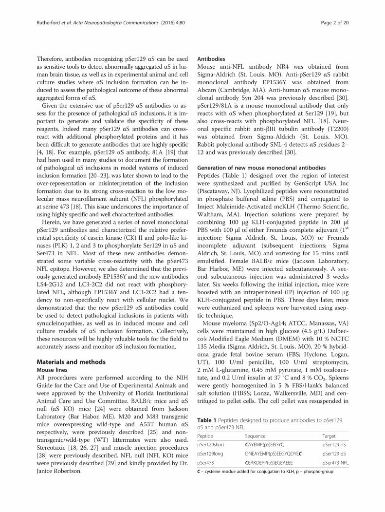
Table 1 Peptides designed to produce antibodies to pSer129αS and pSer473 NFL
Peptide Sequence Target
pSer129short CAYEMP(pS)EEGYQ pSer129 αS
pSer129long DNEAYEMP(pS)EEGYQDYEC pSer129 αS
pSer473 CEAKDEPP(pS)EGEAEEE pSer473 NFL
C – cysteine residue added for conjugation to KLH, p – phospho-group
Rutherford et al. Acta Neuropathologica Communications (2016) 4:80 Page 2 of 20

red blood cell lysis buffer (Sigma Aldrich, St. Louis, MO)and diluted with HBSS after one minute. The cells werethen washed twice by centrifuging at 100 × g for 10 minsand resuspended in HBSS. Sp2/O-Ag14 cells were alsowashed twice with HBSS. Five million Sp2/O-Ag14 cellswere added to 50 million spleen cells and, after centrifu-ging at 100 × g for 10 mins onto a culture dish, fusion wasinduced with 50 % polyethylene glycol 1450 (PEG; SigmaAldrich, St. Louis, MO). After washing with HBSS, cellswere incubated in Sp2/O-Ag14 media at 37 °C with 8 %CO2 overnight. The next day, the cells were gently de-tached from the plate and distributed into 96 well plateswith Sp2/O-Ag14 media/0.5 % hybridoma enhancing sup-plement (Sigma Aldrich, St. Louis, MO)/HAT selectionsupplement (Sigma Aldrich, St. Louis, MO).
Hybridoma screeningAll hybridoma clones were screened for reactivity to theinjected peptide by enzyme-linked immunosorbent assay(ELISA). MaxiSorp plates (Thermo Scientific, Waltham,MA) were coated with 1 μg/ml peptide in PBS andblocked with 5 % FBS/PBS. Media from the hybridomaswere applied to plates, which were then incubated atroom temperature for 3 h. Next, the plates were washedwith PBS, and incubated with goat anti-mouse secondaryantibody conjugated to horse radish peroxidase (HRP;Jackson Immuno Research Labs, West Grove, PA) for 1 hat room temperature. Then, plates were washed and TMBsubstrates (Pierce, Rockford, IL) were applied until colorchanges were observed. Reactions were then quenchedwith 1 M HCl and absorbance was measured at 450 nm.Clones that were positive by ELISA were transferred tolarger culture plates as needed. The positive clones werenext screened by immunohistochemistry (IHC) of a hu-man autopsy case with abundant αS pathology.
Immunohistochemistry analysesParaffin embedded tissue from αS transgenic and WTmice are described in Table 2. Paraffin embedded, for-malin fixed human brain tissue was obtained throughthe University of Florida Neuromedicine Human Brain
Tissue Bank (summarized in Table 3). Sequential tissuesections were deparaffinized with xylenes, and sequentiallyrehydrated with graded ethanol solutions (100-70 %).Antigen retrieval was performed by incubating sections in0.05 % Tween-20 in a steam bath for 30 mins. Endogen-ous peroxidase activity was quenched with 1.5 % hydrogenperoxide/0.005 % Triton X-100/PBS for 20 mins. Sectionswere blocked with 2 % FBS/0.1 M Tris, pH 7.6 then incu-bated with primary antibody overnight at 4 °C. Followingwashing with 0.1 M Tris, pH 7.6, sections were incubatedwith biotinylated horse anti-mouse IgG or biotinylatedhorse anti-rabbit IgG secondary antibody (Vector Labora-tories, Burlingame, CA) diluted in 2 % FBS/0.1 M TrispH 7.6 for 1 h. Next, sections were washed with 0.1 MTris, pH 7.6, then incubated with streptavidin-conjugatedHRP (VECTASTAIN ABC kit; Vector Laboratories,Burlingame, CA) diluted in 2 % FBS/0.1 M Tris pH 7.6 for1 h. Sections were washed with 0.1 M Tris, pH 7.6, andthen developed with 3, 3′diaminobenzidine (DAB kit;KPL, Gaithersburg, MD). Reactions were stopped by im-mersing the slides in 0.1 M Tris, pH7.6, and sections werecounterstained with Mayer’s hematoxylin (Sigma Aldrich,St. Louis, MO). Next, sections were dehydrated with anascending series of ethanol solutions (70–100 %) followedby xylenes, and coverslipped using cytoseal (ThermoScientific, Waltham, MA).
Recombinant αS protein production and purificationRecombinant WT human or mouse αS or human αSwith serine 129 mutated to alanine (S129A) wereexpressed in Escherichia coli (E. coli) BL21 (DE3)/RIL(Agilent Technologies, Santa Clara, CA) using the respect-ive cDNA cloned into the bacterial expression plasmidpRK172, and purified as previously described [31, 32].Protein concentrations were determined by bicinchoninicacid (BCA) assay using bovine serum albumin (BSA;Pierce, Rockford, IL) as a standard.
Recombinant NFL protein production and purificationRecombinant WT mouse NFL or mouse NFL withserine 473 mutated to alanine (S473A) were expressed in
Table 2 Summary of mouse tissue used for immunohistochemical analysis of novel antibodies
Mouse line Treatment Fixative References
M83+/− Intramuscular injection of αS fibrils (motor impaired/terminal) 150 mM NaCl/ 70 % ethanol [28]
M83+/− Intramuscular injection of αS fibrils (motor impaired/terminal) Formalin [28]
M83+/− Cerebral injection of αS fibrils (3 months post-injection) 150 mM NaCl/ 70 % ethanol [26]
M83+/+ Naïve (7 month old; no phenotype) 150 mM NaCl/ 70 % ethanol
M83+/+ Naïve (10–12 month old; motor impaired/terminal) 150 mM NaCl/ 70 % ethanol
M20+/− Cerebral injection of αS fibrils (4 months post-injection) 150 mM NaCl/ 70 % ethanol [27]
M20+/− Cerebral injection of αS fibrils (4 months post-injection) Formalin [27]
WT Naïve 150 mM NaCl/ 70 % ethanol
Rutherford et al. Acta Neuropathologica Communications (2016) 4:80 Page 3 of 20

E. coli BL21 (DE3)/RIL (Agilent Technologies, SantaClara, CA), using the respective cDNA cloned in thebacterial expression plasmid pET-23d and purified aspreviously described [18]. Protein concentrations weredetermined by Bradford assay using BSA as a standard.
Radioactive and non-radioactive kinase reactionsTo determine which kinases could phosphorylate αS andNFL, and to what extent, we performed radioactive kin-ase reactions. NFL and S473A NFL were dialyzed in Trisor 4-(2-hydroxyethyl)-1-piperazineethanesulfonic acid(HEPES) overnight to remove the urea. Recombinantproteins (16.3 pmol; αS, S129A αS, NFL or S473A NFL)were incubated in 25 μl reactions at 30 °C for 8 h with60 ng PLK2 (Life Technologies, Carlsbad, CA) or 20 ngCKII (New England Biolabs, Ipswich, MA), PLK1 orPLK3 (Life Technologies, Carlsbad, CA), 200 μM adeno-sine triphosphate (ATP) with γ-32P ATP (PerkinElmer,Waltham, MA) in manufacturer recommended buffers(CKII buffer: 50 mM Tris-HCl, pH7.5, 10 mM MgCl2,0.1 mM ethylenediamietetraacetic acid (EDTA), 2 mM di-thiothreitol (DTT), 0.01 % Brij 35; PLK1 buffer: 50 mMHEPES, pH7.5, 10 mM MgCl2, 2.5 mM DTT, 0.01 % TritonX-100; PLK2 buffer: 25 mM HEPES, pH7.5, 10 mM MgCl2,5 mM MnCl2, 0.5 mM ethylene glycol-bis(β-aminoethylether)-N,N,N’,N’-tetraacetic acid (EGTA), 0.5 mM Na3VO4,5 mM β-glycerophosphate, 2.5 mM DTT, 0.01 % Triton X-100; PLK3 buffer: 25 mM Tris, pH7.5, 10 mM MgCl2,0.5 mM EGTA, 0.5 mM Na3VO4, 5 mM β-glycerophosphate, 2.5 mM DTT, 0.01 % Triton X-100). Re-actions without protein for each kinase, and without kinasefor each protein, were included as control reactions. Eachreaction was performed in triplicate. Reactions were thenabsorbed onto P-81 phosphocellulose filters (GE Health-care, Buckinghamshire, UK) and immersed in 85 mMphosphoric acid. After extensive washing in 85 mM phos-phoric acid, the filters were rinsed with acetone, air driedand placed in scintillation vials with Econo-Safe LiquidScintillation cocktail (Research Products International,Mount Prospect, IL). Radioactivity (32P) incorporation wasmeasured using an LS5000 TD liquid scintillation counter(Beckman, Brea, CA).CKII and PLK3 reactions for immunoblot antibody
testing were performed as before, but without γ-32PATP. Sodium dodecyl sulfate (SDS) sample buffer was
added to the reactions which were incubated for 10 minsat room temperature. Equimolar amounts of protein wereresolved by SDS-PAGE and analyzed by immunoblot.
Sequential biochemical fractionation of mouse nervoustissueMouse nervous tissues were fractionated as described byEmmer et al. 2011 [33]. Briefly, mice were humanely eu-thanized, and the brain, brainstem and spinal cord wereharvested. The cortex and brainstem/spinal cord weredissected and placed in separate tubes. Tissues were ho-mogenized with 3 volumes per gram of tissue with highsalt (HS) buffer (50 mM Tris, pH7.5, 750 mM NaCl,20 mM NaF, 5 mM EDTA) with a cocktail of proteaseinhibitors (1 mM phenylmethylsulfonyl and 1 mg/mleach of pepstatin, leupeptin, N-tosyl-L-phenylalanylchloromethyl ketone, N-tosyl-lysine chloromethyl ketoneand soybean trypsin inhibitor). The tissue homogenatesthen underwent sedimentation at 100,000 × g for 20mins and the supernatants were removed and kept asthe HS fraction. Pellets were resuspended in 3 volumesper gram of tissue with HS buffer with 1 % Triton X-100(HS/T buffer) and centrifuged at 100,000 × g for 20mins. The supernatants were removed and kept as theHS/T fraction. The pellets were then homogenized in 3volumes per gram of tissue with HS buffer with 1 M su-crose and centrifuged at 100,000 × g for 20 mins to floatthe myelin, which was discarded. Pellets were homoge-nized in 2 volumes per gram of tissue with radioimmu-noprecipitation assay (RIPA) buffer (50 mM Tris,pH 8.0, 150 mM NaCl, 5 mM EDTA, 1 % NP-40, 0.5 %sodium deoxycholate, 0.1 % SDS) plus protease inhibi-tors and centrifuged at 100,000 × g for 20 mins. Super-natants were removed and kept as the RIPA fraction.Pellets were then homogenized in 1 volume per gram oftissue with 2 % SDS/4 M urea by probe sonication andkept as the SDS/U fractions. Protein concentrations ofall fractions were determined by BCA assay using BSA(Pierce, Rockford, IL) as a standard. SDS sample bufferwas added to the fractions which were incubated for 10mins at 100 °C (HS and HS/T fractions) or at roomtemperature (SDS/U fraction only). Equal amounts ofprotein (10 μg for HS and HS/T, and 5 μg for SDS/Ufractions) were resolved by SDS-PAGE and analyzed byimmunoblot.
Preparation of total protein lysate from mouse nervoustissueMice were humanely euthanized and the brain andspinal cord were harvested. The cortex and brainstem/spinal cord were dissected, placed in separate tubes andlysed with 2 % SDS/50 mM Tris, pH 7.5. Samples weresonicated using a probe sonicator until homogenous andincubated for 10 mins at 100 °C. Protein concentrations
Table 3 Summary of human tissue used forimmunohistochemical analysis of novel antibodies
Neuropathologicaldiagnosis
Region(s) examined Fixative Numberof cases
DLB Cingulate cortex,Midbrain
Formalin 2
MSA Pons, Cerebellum Formalin 2
PD Midbrain Formalin 2
Rutherford et al. Acta Neuropathologica Communications (2016) 4:80 Page 4 of 20

were determined by BCA assay using BSA as a standard.SDS sample buffer was added and equal amounts of pro-tein (5 μg) were resolved by SDS-PAGE and analyzed byimmunoblot.
Immunoblotting analysesProtein samples were resolved by electrophoresis on 8,10 or 13 % polyacrylamide gels as indicated, then elec-trophoretically transferred to nitrocellulose membranes.Membranes were blocked with 5 % milk/Tris-bufferedsaline (TBS) then incubated overnight at 4 °C with pri-mary antibodies diluted in 5 % milk/TBS (non-phosphoantibodies) or 5 % BSA/TBS (phospho antibodies). Fol-lowing washing, blots were incubated with HRP conju-gated goat anti-mouse or donkey anti-rabbit secondaryantibodies (Jackson Immuno Research Labs, WestGrove, PA) diluted in 5 % milk/TBS for 1 h. Followingwashing, protein bands were visualized using WesternLightning-Plus ECL reagents (PerkinElmer, Waltham,MA) and images were captured using the GeneG-nome XRQ system and GeneTools software (Syngene,Frederick, MD).
Primary neuronal-glial culturesPrimary cultures (embryonic) were prepared from E16-E18 C3HBL/6 or αS null mouse brains. Cerebral corticeswere dissected from E16-E18 mouse brains and weredissociated in 2 mg/ml papain (Worthington, Lakewood,
NJ) and 50 μg/ml DNAase I (Sigma Aldrich, St. Louis,MO) in sterile HBSS at 37 °C for 20 mins. They werewashed three times in sterile HBSS to inactivate the pa-pain and switched to 1 % FBS in Neurobasal-A growthmedia (Gibco, Waltham, MA), which includes 1 % Glu-taMax Supplement (Life Technologies, Carlsbad, CA), B-27 supplement, 100 U/ml penicillin and 100 μg/mlstreptomycin. The tissue mixture was triturated threetimes using a 5 ml pipette followed by a Pasteur pipette,and strained through a 70 μm nylon cell strainer. Thecell mixture was then centrifuged at 1300 × g for 3 mins,and resuspended in fresh Neurobasal-A media. Theywere then plated on Nunc Lab-Tek II CC2 chamberslides (Life Technologies, Carlsbad, CA) at around100,000-200,000 cells/cm2. Cells were maintained in theNeurobasal-A growth media without FBS at 37 °C in ahumidified 5 % CO2 chamber. The cultures were main-tained for 6 days followed by another 8 days without orwith 20 μg/ml water bath sonicated mouse αS fibrils pre-pared as previously described [34, 35].
Immunofluorescence microscopy analysisFor double immunofluorescence analysis, cells werewashed with PBS and fixed with 4 % paraformaldehyde/PBS. Following PBS washes, cells were blocked with 5 %FBS/PBS/0.1 % Triton X-100 for 30 mins. Cultures werestained with primary antibodies followed by Alexa-fluor488 and 594 conjugated secondary antibodies (Invitrogen,
Fig. 1 In vitro phosphorylation of NFL and αS by CKII and PLK1, 2 and 3. a Amino acid sequences surrounding the target phosphorylation sites(bold) of Ser129 for αS, and Ser473 for NFL. b Quantitative analysis of the stoichiometric phosphorylation of recombinant αS, S129A αS, NFL andS473A NFL in in vitro kinase reactions as described in “Materials and methods” with CKII, PLK1, PLK2 or PLK3. All reactions were performed intriplicate. c Immunoblotting analyses of NFL and S473A NFL used in in vitro kinase reactions with CKII, PLK1, PLK2 or PLK3. Proteins were resolvedonto 8 % polyacrylamide gel and probed with 4F8 (pSer473 NFL) and NR4 (anti-NFL) antibodies. d Immunoblotting analyses of αS and S129A αSused in in vitro kinase reactions with CKII, PLK1, PLK2 or PLK3. Proteins were resolved onto 13 % polyacrylamide gels and probed with 81A(pSer129 αS) and Syn 204 (anti-αS). The mobility of molecular mass markers are shown on the left
Rutherford et al. Acta Neuropathologica Communications (2016) 4:80 Page 5 of 20

Fig. 2 Specificity of novel pSer129 αS antibodies as determined by immunoblotting with NFL and αS proteins phosphorylated in vitro with PLK3or CKII. NFL, S473A NFL, αS and S129A αS were incubated with PLK3 or CKII. The proteins were resolved onto 13 % polyacrylamide gels andanalyzed by immunoblotting with novel αS antibodies (as indicated above each blot) LS7, LS11, LC2-2C2, LS4-1B1, LS4-2C3 and LS4-2G12 andnovel NFL antibody 4F8, to demonstrate their specificity. NR4 (anti-NFL) and Syn 204 (anti-αS) were included to show each protein, respectively.Previously reported pSer129 αS antibodies 81A and EP1536Y were included for comparison. Reactions were performed in duplicate. Antibody 4F8,generated against the pSer473 NFL epitope, can cross react with αS phosphorylated at Ser129. Antibodies 81A, LS11, LS4-1B1 and LS4-2C3 gener-ated against the pSer129 αS epitope can cross react with NFL phosphorylated at Ser473. Antibodies EP1536Y, LS3-2C2 and LS4-2G12 generated againstthe pSer129 αS epitope reacted only with phosphorylated WT αS. Antibody LS7 generated against the pSer129 αS epitope detected both phospho-and non-phospho-αS. The mobility of molecular mass markers are shown on the left
Rutherford et al. Acta Neuropathologica Communications (2016) 4:80 Page 6 of 20

Fig. 3 Characterization of the specificity of novel pSer129 αS antibodies by immunoblotting analyses using biochemically fractionated brainstem/spinal cord mouse tissues. Brainstem/spinal cord from an αS null (αS KO), a WT, a 2 month old non-symptomatic M83 (M83) and a 12 monthold motor impaired M83 (M83-I) mouse were biochemically fractionated into high salt (HS), high salt/Triton X-100 (HS/T) and SDS/urea (SDS/U)fractions as described in “Materials and methods”. 10 μg of HS and HS/T, and 5 μg of SDS/U fractions were resolved onto 13 % polyacrylamidegels and analyzed by immunoblotting with each antibody indicated above. The protein band identified by an arrowhead is αS, while an asteriskdepicts NFL. The mobility of molecular mass markers are shown on the left
Rutherford et al. Acta Neuropathologica Communications (2016) 4:80 Page 7 of 20
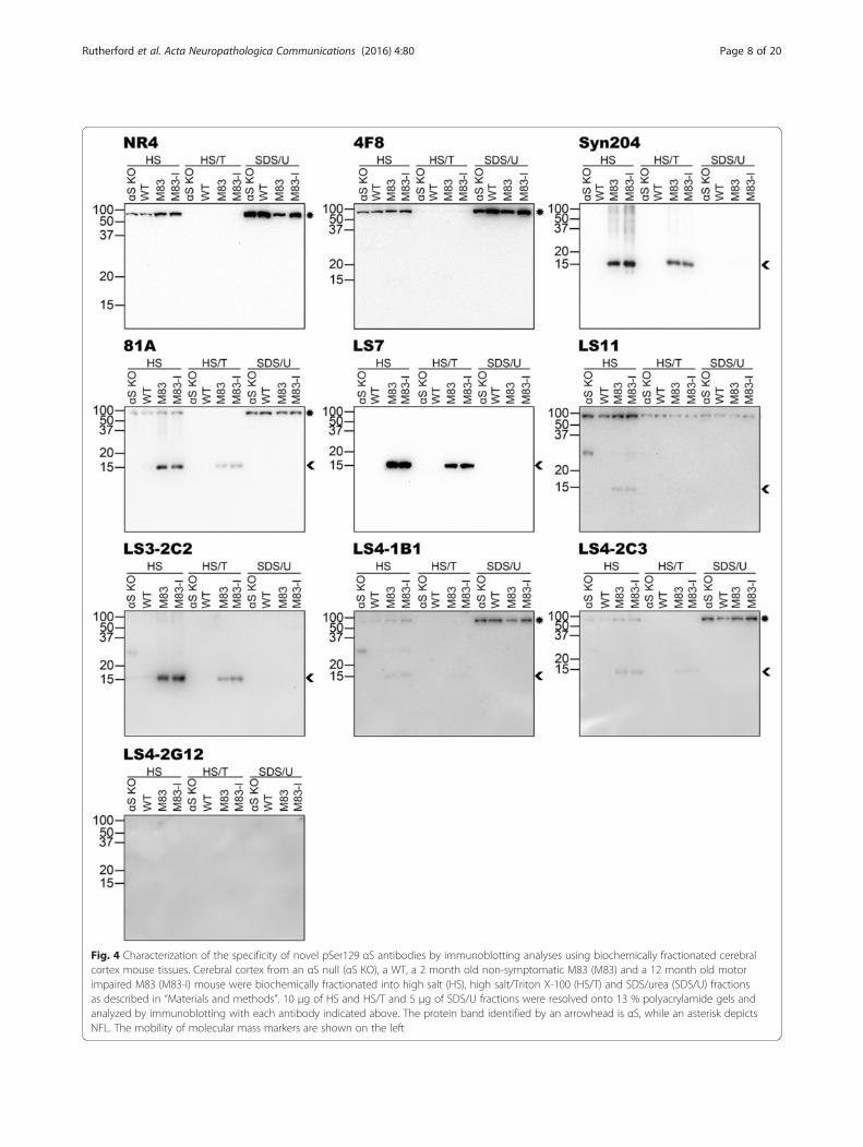
Fig. 4 Characterization of the specificity of novel pSer129 αS antibodies by immunoblotting analyses using biochemically fractionated cerebralcortex mouse tissues. Cerebral cortex from an αS null (αS KO), a WT, a 2 month old non-symptomatic M83 (M83) and a 12 month old motorimpaired M83 (M83-I) mouse were biochemically fractionated into high salt (HS), high salt/Triton X-100 (HS/T) and SDS/urea (SDS/U) fractionsas described in “Materials and methods”. 10 μg of HS and HS/T and 5 μg of SDS/U fractions were resolved onto 13 % polyacrylamide gels andanalyzed by immunoblotting with each antibody indicated above. The protein band identified by an arrowhead is αS, while an asterisk depictsNFL. The mobility of molecular mass markers are shown on the left
Rutherford et al. Acta Neuropathologica Communications (2016) 4:80 Page 8 of 20

Carlsbad, CA). Nuclei were counterstained with 4′,6-dia-midino-2-phenylindole (DAPI; Invitrogen, Carlsbad, CA),and coverslips were mounted using Fluoromount-G(Southern Biotech, Birmingham, AL). Immunofluores-cence images were captured with an Olympus BX51 fluor-escence microscope mounted with a DP71 digital camera(Olympus, Center Valley, PA).
ResultsAssessment of NFL and αS phosphorylation by caseinkinase II and polo-like kinasesCKII was previously reported as a major kinase respon-sible for the phosphorylation of αS, more specifically atSer129 [19, 36, 37], and NFL [38–40]. Characterizationof NFL phosphorylation by CKII was predominantly fo-cused on Ser473, which is the major phosphorylationsite in NFL in vivo [40, 41]. Phosphorylation of αS atSer129 can be mediated by many additional kinases [19,42–47] but some PLKs appear to be quite efficient indriving this modification [42, 43, 47]. To compare therelative abilities of CKII and PLK 1, 2 and 3 to phosphor-ylate αS and NFL, and use this information to generatephosphorylated protein that can be used to assess the spe-cificity of novel antibodies (see below), we assessed therelative but quantitative in vitro phosphorylation of αS
and NFL with CKII and PLK 1, 2 and 3 (Fig. 1b). In theseparallel kinase conditions, PLK3 was the most efficient atphosphorylating αS at a stoichiometric ratio of close to 1mol of phosphate per 1 mol of protein. This phosphoryl-ation was almost completely abolished for S129A αS indi-cating that Ser129 is the major αS phosphorylation site forPLK3. NFL was phosphorylated by PLK1 and PLK3 atmultiple amino residues as shown by the greater stoichio-metric molecular ratio of approximately 4.7 and 6, re-spectively. In addition, the presence of the S473Amutation in NFL only reduced the amount of phosphateincorporation by PLK1 and PLK3 by 0.45 and 0.37 molphosphate/mol protein respectively, indicating that Ser473is not the major site targeted by these kinases. CKII phos-phorylated NFL at a ratio of 1.28 mol phosphate/mol pro-tein, and this was reduced to 0.61 for S473A NFLindicating that, while this is not the only site, it is themajor target site for this kinase.
Generation of novel pSer473 NFL and pSer129 αSantibodiesTo provide more reagents to study the contribution ofaberrant accumulation and/or distributions of αS andNFL in neurodegenerative diseases, we generated novelmonoclonal antibodies against the pSer473 epitope in
Fig. 5 Characterization of the specificity of novel pSer129 αS antibodies by immunoblotting analyses using total lysates of cortex and brainstem/spinal cord tissues from WT and NFL null mice. Cerebral cortex and brainstem/spinal cord (BS/SC) from a WT, an NFL null (NFL KO) and an αS null(αS KO) mouse were dissected and lysed in 2 % SDS/ 50 mM Tris pH 7.5 as described in “Materials and methods”. Equal amounts of proteins (5μg) from each sample was resolved onto 10 % polyacrylamide gels and analyzed by immunoblotting with each antibody indicated above. Theprotein band identified by an asterisk depicts NFL. The mobility of molecular mass markers are shown on the left
Rutherford et al. Acta Neuropathologica Communications (2016) 4:80 Page 9 of 20

Fig. 6 (See legend on next page.)
Rutherford et al. Acta Neuropathologica Communications (2016) 4:80 Page 10 of 20

NFL and the pSer129 epitope in αS. For the pSer473NFL epitope we used a synthetic peptide with phosphor-ylated serine and the adjacent 7 amino acids residues oneither side in the human NFL sequence (Table 1). Weidentified and characterized (see below) one clonetermed 4F8 that was relatively specific for pSer473 inNFL.For the pSer129 αS epitope we performed multiple
attempts to produce αS antibodies that were 1)phospho-specific and 2) relatively specific in detectingLewy pathology. For our most successful approach, themice were first immunized with the pSer129long peptide(αS residues 121–137) then 3 weeks later with thepSer129short peptide (αS residues 124–134; Table 1).The final IP injection used a 1:1 ratio of the 2 peptidesand the initial ELISA antibody screen used the pSer129-long peptide. We immunized several mice using this strat-egy and kept the most promising hybridomas from ourinitial ELISA and IHC screens for further characterization.We have denoted these antibodies LS, meaning longthen short to indicate the peptides that were used forimmunization.
Characterization of novel monoclonal antibodyspecificities using phosphorylated recombinant proteinsTo confirm our findings from the in vitro kinase reac-tions and initially characterize the specificity of the 4F8antibody, we conducted immunoblotting analysis withNFL and S473A NFL individually phosphorylated withCKII and PLK1, 2 and 3 (Fig. 1c). As expected, the signalwas the weakest with PLK2 and the signal was abolishedfor S473A NFL phosphorylated with any of these kinasesindicating that 4F8 is specific for NFL phosphorylated atSer473. Immunoblotting with the previously generatedpSer129 αS antibody 81A [19] was consistent with the invitro radioactivity kinase studies in terms of relativephosphorylation of αS by these kinases (Fig. 1d). Theseresults show that PLK3 strongly phosphorylates bothNFL at Ser473 and αS at Ser129, and that CKII is thepreferred kinase for NFL at this site. Therefore we choseto use these kinase reactions to further screen the speci-ficity of our novel antibodies.We performed immunoblot analyses of CKII and
PLK3 kinase reactions with WT and S473A NFL, andWT and S129A αS (Fig. 2). Immunoblots with anti-NFL
antibody NR4 and anti-αS antibody Syn 204 are includedto demonstrate the respective proteins. In the immuno-blots loaded with CKII reactions, 4F8 appeared specificfor phosphorylated NFL, however the blots with thePLK3 reactions showed that it can also detect αS phos-phorylated at Ser129. As previously published, 81A alsocross reacted with NFL phosphorylated at Ser473 [18],particularly when phosphorylated by CKII. LS3-2C2 andthe commercially available EP1536Y were specific for αSphosphorylated at Ser129. LS7 reacted with αS inde-pendent of its phosphorylation state. Antibodies LS11,LS4-1B1 and LS4-2C3 were specific for αS phosphory-lated at Ser129, however they could also react to someextent with NFL phosphorylated at Ser473. LS4-2G12only showed reactivity with αS phosphorylated at Ser129by PLK3, but it did not react with αS modified with CKIIdue to its low level of phosphorylation, suggesting that itis a weaker antibody.
Assessment of antibody specificity using biochemicallyfractionated mouse nervous tissueTo assess for the more global specificity of the new anti-bodies, we performed immunoblot analyses of sequen-tially fractionated mouse brainstem/spinal cord (Fig. 3)and cerebral cortex (Fig. 4) tissue from αS null (αS KO),WT, 2 month old non-symptomatic M83 (M83) and12 month old motor-impaired M83 (M83-I) mice. TheM83-I mouse contains pathological αS aggregates pre-dominantly in the brainstem/spinal cord [25]. Immuno-blots with anti-NFL antibody NR4 and anti-human αSantibody Syn 204 are included to demonstrate the pres-ence and distribution of these respective proteins. Inthese analyses, 4F8 was quite specific, detecting predom-inantly NFL and only a very faint phospho-αS band inthe brainstem/spinal cord SDS/U fraction of the M83-I mouse (Fig. 3). LS7 showed a similar pattern of reactivityto Syn 204, again indicating that it is not phospho-specific(Figs. 3 and 4). Antibodies LS4-1B1, LS4-2C3 and 81Acould all react with pathological αS that accumulates inthe brainstem/spinal cord SDS/U fraction of M83-I mice(Fig. 3), but they also cross reacted to some extent withphosphorylated NFL present in the brainstem/spinal cordand cortex of all these mice (Figs. 3 and 4). This cross-reactivity to NFL was confirmed by immunoblotting oftotal brainstem/spinal cord and cortex tissues from WT,
(See figure on previous page.)Fig. 6 Comparison of novel antibodies in detecting pathological inclusions in αS transgenic mice injected with αS fibrils in the periphery(intramuscular) or the brain (hippocampus). Representative images of IHC staining of tissue from M83 αS transgenic mice injected in thegastrocnemius muscle, and M83 and M20 αS transgenic mice injected in the hippocampus with recombinant preformed αS fibrils. Images weretaken from the brainstem (muscle injection) and the hippocampus (hippocampal injection). Antibodies EP1536Y and 81A showed robust stainingof induced inclusions (arrows). LS7 stained inclusions weakly with higher general labeling. Novel antibodies raised against the pSer129 αS epitopeLS4-2G12, LS3-2C2 and LS4-2C3 or to the p473 NFL epitope all stained the induced inclusions in these models. Scale bar = 50 μm
Rutherford et al. Acta Neuropathologica Communications (2016) 4:80 Page 11 of 20

Fig. 7 (See legend on next page.)
Rutherford et al. Acta Neuropathologica Communications (2016) 4:80 Page 12 of 20

NFL null and αS null mice (Fig. 5). LS11 detected aggre-gated phosphorylated αS (Fig. 3) but also cross-reactedwith NFL, as shown by the reduction in signal of the~70 kDa band in the total brainstem/spinal cord extractfrom the NFL null mice (Fig. 5). However, it also reactedwith another protein of approximately the same size asNFL prevalent in the cortex, even in NFL and αS nullmice (Fig. 5). This protein also has a different biochemicalfractionation profile compared to NFL in the cortex(Fig. 4). Antibodies LS11, LS3-2C2, LS4-1B1, LS4-2C3and LS4-2G12 also detected a ~30 kDa non-αS protein.LS3-2C2 weakly labeled a few additional protein bands inthe brainstem/spinal cord lysates of all mice indicatingthat these are not αS. Overall, LS4-2G12 appeared to bethe most specific new phospho-αS antibody.
IHC analyses of mouse and human nervous tissue withnew pSer473 NFL and pSer129 αS antibodiesUsing our new and previously generated pSer129 αSantibodies and the new pSer473 NFL antibody 4F8, weperformed IHC analyses on a cohort of mouse tissue de-rived from αS transgenic and WT mice (Table 2) andhuman autopsy cases with a diagnosis of PD, DLB orMSA (Table 3). Our mouse tissue cohort, included pre-viously described [26–28] tissue of αS transgenic miceinjected with αS fibrils either in the gastrocnemiusmuscle (M83 line) or hippocampus that induces the for-mation of αS inclusion pathology (M83 and M20 lines;Fig. 6), and naïve αS transgenic and WT mice (Fig. 7).The tissue in this cohort was fixed with 150 mM NaCl/70 % ethanol. The staining revealed that all of the anti-αS antibodies tested were able to reliably stain αS inclu-sion pathology in this cohort. Likewise, 4F8 stainedmany inclusions. EP1536Y also stained inclusions,however in some sections it had a tendency to non-specifically react with cellular nuclei (see Fig. 7).Phosphorylation-independent antibody, LS7 stained in-clusions, but weaker than the phospho-specific anti-bodies. Interestingly, the general baseline staining of LS7was only evident in the αS transgenic and not in theWT mice (Fig. 7), because it is specific for human αS(Additional file 1: Figure S1). Antibodies that couldcross-react with phospho-NFL such as LS4-2C3, aswell as LS3-2C2 which does not detect phospho-NFL,
also demonstrated staining of axonal processes similarto 4F8 (Fig. 7).For our human autopsy cohort, we performed IHC on
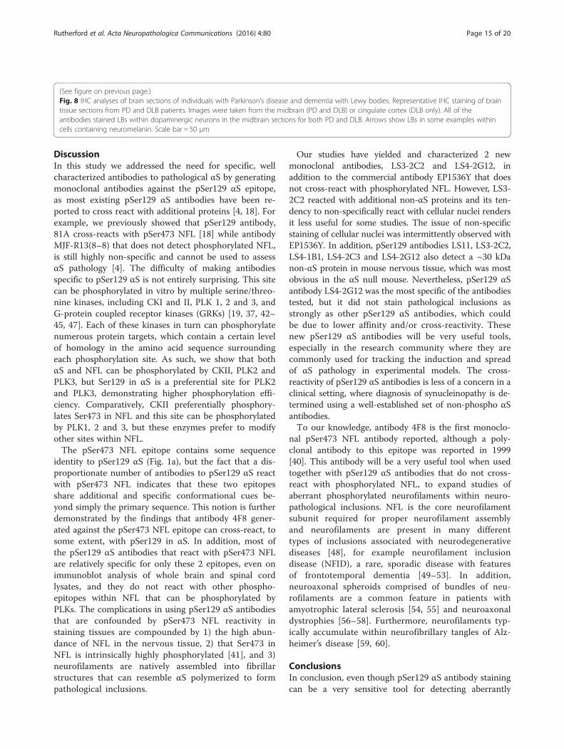
sections of midbrain (PD and DLB), cingulate cortex(DLB; Fig. 8), and pons and cerebellum (MSA; Fig. 9).All of the antibodies tested showed robust reactivity tothe αS inclusion pathology in the PD and DLB cases,apart from LS3-2C2 which displayed weaker stainingparticularly in the DLB cingulate cortex (Fig. 8). AllpSer129 αS antibodies stained GCIs in the MSA tissue;however the strength of the signal differed extensivelybetween the antibodies (Fig. 9). For example, EP1536Yshowed the strongest staining, while LS4-2G12 andLS3-2C2 showed weaker staining, especially in thecerebellum. Although LS4-2C3 labeled αS inclusionswell in the pons, its reactivity with NFL made it diffi-cult to make out many inclusions within the cerebel-lum; the axonal staining being on par with that of4F8. 4F8 itself was able to stain some GCIs, howeverthese were rare, and whether this is really pSer473NFL staining, or just cross-reactivity with pSer129 αSremains in question.Using mouse tissue fixed using formalin or ethanol
(150 mM NaCl/70 % ethanol), we confirmed that all theantibodies could be used to stain tissue preserved withboth types of fixatives (Additional file 2: Figure S2).
Immunofluorescence analyses of αS aggregation inprimary neuronal-glial culturesUsing only the new pSer129 αS antibodies that do notcross-react with phospho-NFL, we determined if thesecould be used to monitor the aggregation of αS in pri-mary neuronal-glial cultures induced by the addition ofexogenous αS preformed fibrils. LS4-2G12 readily de-tected neuronal phosphorylated αS aggregates, andthe paucity of these inclusions in cultures from αSnull mice confirmed that these were comprised of en-dogenous αS (Fig. 10). Antibody LS3-2C2 could alsodetect inclusions comprised of endogenous αS in pri-mary neuronal-glial cultures treated with exogenousαS fibrils, but this antibody was less useful due to thenon-specific cross-reactivity with cellular nuclei (Additionalfile 3: Figure S3).
(See figure on previous page.)Fig. 7 Comparison of pSer129 antibody IHC staining using naïve αS transgenic and WT mice. Representative images of IHC staining of brainstemtissue from a 7 month old non-symptomatic homozygous M83 mouse (M83 unimpaired), a 12 month old motor impaired homozygous M83mouse (M83 motor impaired) and a WT mouse. All antibodies stained perikaryal and neuritic inclusions in the M83 diseased mouse. LS7 showedweaker reactivity to αS pathology than the other antibodies and showed stronger diffused signal in the αS transgenic mice compared to the WTmouse. LS3-2C2 also showed weak staining of pathology. In addition to the pathology in motor impaired M83 mouse, antibodies 4F8, LS3-2C2,LS4-2C3 and 81A also labeled neuronal projections even in WT mice (arrowheads). EP1536Y exhibited some nuclear staining in some mousesections (arrows), as did LS3-2C2, but much weaker than EP1536Y (eg. see arrows in the M83 unimpaired mouse). Scale bar = 50 μm
Rutherford et al. Acta Neuropathologica Communications (2016) 4:80 Page 13 of 20

Fig. 8 (See legend on next page.)
Rutherford et al. Acta Neuropathologica Communications (2016) 4:80 Page 14 of 20
DiscussionIn this study we addressed the need for specific, wellcharacterized antibodies to pathological αS by generatingmonoclonal antibodies against the pSer129 αS epitope,as most existing pSer129 αS antibodies have been re-ported to cross react with additional proteins [4, 18]. Forexample, we previously showed that pSer129 antibody,81A cross-reacts with pSer473 NFL [18] while antibodyMJF-R13(8–8) that does not detect phosphorylated NFL,is still highly non-specific and cannot be used to assessαS pathology [4]. The difficulty of making antibodiesspecific to pSer129 αS is not entirely surprising. This sitecan be phosphorylated in vitro by multiple serine/threo-nine kinases, including CKI and II, PLK 1, 2 and 3, andG-protein coupled receptor kinases (GRKs) [19, 37, 42–45, 47]. Each of these kinases in turn can phosphorylatenumerous protein targets, which contain a certain levelof homology in the amino acid sequence surroundingeach phosphorylation site. As such, we show that bothαS and NFL can be phosphorylated by CKII, PLK2 andPLK3, but Ser129 in αS is a preferential site for PLK2and PLK3, demonstrating higher phosphorylation effi-ciency. Comparatively, CKII preferentially phosphory-lates Ser473 in NFL and this site can be phosphorylatedby PLK1, 2 and 3, but these enzymes prefer to modifyother sites within NFL.The pSer473 NFL epitope contains some sequence
identity to pSer129 αS (Fig. 1a), but the fact that a dis-proportionate number of antibodies to pSer129 αS reactwith pSer473 NFL indicates that these two epitopesshare additional and specific conformational cues be-yond simply the primary sequence. This notion is furtherdemonstrated by the findings that antibody 4F8 gener-ated against the pSer473 NFL epitope can cross-react, tosome extent, with pSer129 in αS. In addition, most ofthe pSer129 αS antibodies that react with pSer473 NFLare relatively specific for only these 2 epitopes, even onimmunoblot analysis of whole brain and spinal cordlysates, and they do not react with other phospho-epitopes within NFL that can be phosphorylated byPLKs. The complications in using pSer129 αS antibodiesthat are confounded by pSer473 NFL reactivity instaining tissues are compounded by 1) the high abun-dance of NFL in the nervous tissue, 2) that Ser473 inNFL is intrinsically highly phosphorylated [41], and 3)neurofilaments are natively assembled into fibrillarstructures that can resemble αS polymerized to formpathological inclusions.
Our studies have yielded and characterized 2 newmonoclonal antibodies, LS3-2C2 and LS4-2G12, inaddition to the commercial antibody EP1536Y that doesnot cross-react with phosphorylated NFL. However, LS3-2C2 reacted with additional non-αS proteins and its ten-dency to non-specifically react with cellular nuclei rendersit less useful for some studies. The issue of non-specificstaining of cellular nuclei was intermittently observed withEP1536Y. In addition, pSer129 antibodies LS11, LS3-2C2,LS4-1B1, LS4-2C3 and LS4-2G12 also detect a ~30 kDanon-αS protein in mouse nervous tissue, which was mostobvious in the αS null mouse. Nevertheless, pSer129 αSantibody LS4-2G12 was the most specific of the antibodiestested, but it did not stain pathological inclusions asstrongly as other pSer129 αS antibodies, which couldbe due to lower affinity and/or cross-reactivity. Thesenew pSer129 αS antibodies will be very useful tools,especially in the research community where they arecommonly used for tracking the induction and spreadof αS pathology in experimental models. The cross-reactivity of pSer129 αS antibodies is less of a concern in aclinical setting, where diagnosis of synucleinopathy is de-termined using a well-established set of non-phospho αSantibodies.To our knowledge, antibody 4F8 is the first monoclo-
nal pSer473 NFL antibody reported, although a poly-clonal antibody to this epitope was reported in 1999[40]. This antibody will be a very useful tool when usedtogether with pSer129 αS antibodies that do not cross-react with phosphorylated NFL, to expand studies ofaberrant phosphorylated neurofilaments within neuro-pathological inclusions. NFL is the core neurofilamentsubunit required for proper neurofilament assemblyand neurofilaments are present in many differenttypes of inclusions associated with neurodegenerativediseases [48], for example neurofilament inclusiondisease (NFID), a rare, sporadic disease with featuresof frontotemporal dementia [49–53]. In addition,neuroaxonal spheroids comprised of bundles of neu-rofilaments are a common feature in patients withamyotrophic lateral sclerosis [54, 55] and neuroaxonaldystrophies [56–58]. Furthermore, neurofilaments typ-ically accumulate within neurofibrillary tangles of Alz-heimer’s disease [59, 60].
ConclusionsIn conclusion, even though pSer129 αS antibody stainingcan be a very sensitive tool for detecting aberrantly
(See figure on previous page.)Fig. 8 IHC analyses of brain sections of individuals with Parkinson’s disease and dementia with Lewy bodies. Representative IHC staining of braintissue sections from PD and DLB patients. Images were taken from the midbrain (PD and DLB) or cingulate cortex (DLB only). All of theantibodies stained LBs within dopaminergic neurons in the midbrain sections for both PD and DLB. Arrows show LBs in some examples withincells containing neuromelanin. Scale bar = 50 μm
Rutherford et al. Acta Neuropathologica Communications (2016) 4:80 Page 15 of 20

Fig. 9 (See legend on next page.)
Rutherford et al. Acta Neuropathologica Communications (2016) 4:80 Page 16 of 20

aggregated αS and pathological inclusions, specificity is acritical issue that needs to be considered for accurateassessments. The intrinsic similarities in primary se-quence surrounding this site to other proteins that arephosphorylated by mutual kinases and the apparentadditional structural homology to the pSer473 NFL epi-tope complicates the generation of highly specific anti-bodies. Nevertheless, from a series of new monoclonal
antibodies, we were able to identify LS4-2G12 that isrelatively specific and useful in detecting αS aggregatesin human brains and experimental models. However,given the varied specificity and properties of pSer129 αSantibodies, as demonstrated here, it should be preferredas a common practice to use a combination of severalpSer129 αS antibodies in addition to an αS antibody toaccurately assess neuropathological changes in αS.
(See figure on previous page.)Fig. 9 IHC analyses of brain sections of individuals with multiple system atrophy. Representative IHC staining of brain tissue sections from MSApatients. Images were taken from the pons or cerebellum. Arrows show GCIs. All of the antibodies stained inclusions, to some extent. EP1536Yshowed the strongest staining. 4F8 stained only rare inclusions, but abundantly labeled axons (arrowheads). Antibodies 81A and LS4-2C3 alsodisplayed strong reactivity to axons in the cerebellum. Antibodies LS4-2G12 and LS3-2C2 displayed weaker staining of inclusions in the cerebellum.Scale bar = 50 μm
Fig. 10 Analysis of the induction of endogenous αS aggregation by treatment with exogenous αS mouse fibrils in primary neuronal-glial culturesusing antibody LS4-2G12. Primary neuronal-glial cultures from WT mice or αS null mice were cultured for 6 days and either maintained withoutother treatment for 8 days (Ct) or treated with mouse αS fibrils (20 μg/ml; αS Fib) for 8 days. Double immunofluorescence analysis with antibodiesLS4-2G12 (red) and specific neuronal marker βIII-tubulin (green) was performed. Cells were also counterstained with DAPI and merged imagesare shown. Higher magnification LS4-2G12 and merged images are shown on the far right. Arrows depict induced labeled αS aggregates. Scalebar = 100 μm and 250 μm for the higher magnification images on the right
Rutherford et al. Acta Neuropathologica Communications (2016) 4:80 Page 17 of 20

Additional files
Additional file 1: Figure S1. Specificity of novel antibody LS7 forhuman αS. (a) Recombinant human and mouse αS (50 ng) were resolvedonto 13 % polyacrylamide gels and analyzed by immunoblotting with LS7and anti-αS antibody SNL-4 (residues 2–12 of αS). The mobility of molecularmass markers are shown on the left. (b) Sequence of the pSer129long peptide(human αS) with the corresponding mouse αS sequence underneath. The lineindicates amino acids that are not shared. (TIF 216 kb)
Additional file 2: Figure S2. IHC analyses showing antibody reactivityin formalin and ethanol fixed tissues. Representative IHC staining of thebrainstem of M83 mice injected in the gastrocnemius muscle with αSfibrils. The tissue of one mouse (left) was fixed in 150 mM NaCl/70 %ethanol and the other (right) was fixed in formalin. All of the antibodieswere able to stain pathology in both formalin and ethanol fixed tissue,however some staining appeared weaker in the formalin fixed tissue(LS4-2G12 and LS3-2C2). Scale bar = 50 μm. (TIF 9583 kb)
Additional file 3: Figure S3. Analysis of the induction of endogenousαS aggregates with exogenous αS mouse fibrils in primary neuronal-glialcultures using antibody LS3-2C2. Primary neuronal-glial cultures from WTmice or αS null mice were cultured for 6 days and either maintainedwithout other treatment for 8 days (Ct) or treated with mouse αS fibrils(20 μg/ml; αS fib) for 8 days. Double immunofluorescence analysis withantibodies LS3-2C2 (red) and specific neuronal marker βIII-tubulin (green)was performed. Cells were also counterstained with DAPI and mergedimages are shown. Higher magnification merged images are shownon the far right. Arrows depict induced labeled αS aggregates andarrowheads depict nuclear staining. Bar = 100 μm and 250 μm for thehigher magnification images on the right. (TIF 7508 kb)
AbbreviationsαS, α-synuclein; αS fib, α-synuclein fibrils; ATP, adenosine triphosphate; BCA,bicinchoninic acid; BSA, bovine serum albumin; BS/SC, brainstem/spinalcord; CK, casein kinase; Ct, control; DAB, 3, 3′diaminobenzidine; DAPI, 4′,6-diamidino-2-phenylindole; DLB, dementia with Lewy bodies; DMEM,Dulbecco’s Modified Eagle Medium; DTT, dithiothreitol; E.coli, Escherichiacoli; EDTA, ethylenediaminetetraacetic acid; EGTA, ethylene glycol-bis(β-aminoethyl ether)-N,N,N’,N’-tetraacetic acid; ELISA, enzyme-linkedimmunosorbent assay; FBS, fetal bovine serum; GCI, glial cytoplasmicinclusion; GRK, G-protein coupled receptor kinase; HBSS, Hank’s balanced saltsolution; HEPES, 4-(2-hydroxyethyl)-1-piperazineethanesulfonic acid; HRP,horse radish peroxidase; HS, high salt; HS/T, high salt/Triton X-100; IHC,immunohistochemistry; IP, intraperitoneal; KO, null (knock-out); LB, Lewybody; M83-I, motor impaired M83 mouse; MSA, multiple system atrophy;NCI, neuronal cytoplasmic inclusion; NFID, neurofilament inclusion disease;NFL, low molecular mass neurofilament subunit; PAGE, polyacrylamide gelelectrophoresis; PBS, phosphate buffered saline; PD, Parkinson’s disease; PEG,polyethylene glycol; PLK, polo-like kinase; RIPA, radioimmunoprecipitationassay; pSer, phosphorylated serine; SDS, sodium dodecyl sulfate; SDS/U,sodim dodecyl sulfate/urea; TBS, Tris-buffered saline; WT, wild-type
AcknowledgementsNot applicable.
FundingThis work was supported by grants from the National Institute ofNeurological Disorders and Stroke (NS089622), the National Institute onAging (AG047266) and the National Parkinson Foundation.
Availability of data and materialsAll data generated or analyzed during this study are included in thispublished article.
Authors’ contributionsNJR generated monoclonal antibodies, performed IHC analyses, produced andpurified recombinant proteins, performed radioactive and non-radioactive kinasereactions, performed immunoblotting analyses and was a major contributorin writing the manuscript. MB maintained the mouse colony and generatedprimary neuronal-glial cultures. BIG generated monoclonal antibodies, harvested
mouse nervous tissue and prepared total protein lysates or performedsequential biochemical fractionation of the tissue, treated primary neuronal-glialcultures with fibrils and performed immunofluorescence analyses of thecultures and was a major contributor in writing the manuscript. All authorsread and approved the final manuscript.
Competing interestsThe authors declare that they have no competing interests.
Ethics approval and consent to participateAll applicable international, national, and/or institutional guidelines for thecare and use of animals were followed.All procedures performed in studies involving animals were in accordancewith the ethical standards of the University of Florida.
Author details1Department of Neuroscience, College of Medicine University of Florida,Gainesville, FL 32610, USA. 2Center for Translational Research inNeurodegenerative Disease, College of Medicine University of Florida,Gainesville, FL 32610, USA. 3McKnight Brain Institute, College of MedicineUniversity of Florida, Gainesville, FL 32610, USA.
Received: 13 July 2016 Accepted: 28 July 2016
References1. Goedert M. Alpha-synuclein and neurodegenerative diseases. Nat Rev
Neurosci. 2001;2:492–501. doi:10.1038/35081564.2. Goedert M, Spillantini MG, Del Tredici K, Braak H. 100 years of Lewy
pathology. Nat Rev Neurol. 2013;9:13–24. doi:10.1038/nrneurol.2012.242.3. Spillantini MG, Schmidt ML, Lee VM, Trojanowski JQ, Jakes R, Goedert M.
Alpha-synuclein in Lewy bodies. Nature. 1997;388:839–40. doi:10.1038/42166.4. Uchihara T, Giasson BI. Propagation of alpha-synuclein pathology:
hypotheses, discoveries, and yet unresolved questions from experimentaland human brain studies. Acta Neuropathol. 2016;131:49–73. doi:10.1007/s00401-015-1485-1.
5. Spillantini MG, Crowther RA, Jakes R, Cairns NJ, Lantos PL, Goedert M.Filamentous alpha-synuclein inclusions link multiple system atrophy withParkinson’s disease and dementia with Lewy bodies. Neurosci Lett. 1998;251:205–8.
6. Tu PH, Galvin JE, Baba M, Giasson B, Tomita T, Leight S, et al. Glialcytoplasmic inclusions in white matter oligodendrocytes of multiple systematrophy brains contain insoluble alpha-synuclein. Ann Neurol. 1998;44:415–22.doi:10.1002/ana.410440324.
7. Hamilton RL. Lewy bodies in Alzheimer’s disease: a neuropathologicalreview of 145 cases using alpha-synuclein immunohistochemistry. BrainPathol. 2000;10:378–84.
8. Hashimoto M, Masliah E. Alpha-synuclein in Lewy body disease andAlzheimer’s disease. Brain Pathol. 1999;9:707–20.
9. Lippa CF, Fujiwara H, Mann DM, Giasson B, Baba M, Schmidt ML, et al. Lewybodies contain altered alpha-synuclein in brains of many familial Alzheimer’sdisease patients with mutations in presenilin and amyloid precursor proteingenes. Am J Pathol. 1998;153:1365–70.
10. Arawaka S, Saito Y, Murayama S, Mori H. Lewy body in neurodegenerationwith brain iron accumulation type 1 is immunoreactive for alpha-synuclein.Neurology. 1998;51:887–9.
11. Wakabayashi K, Yoshimoto M, Fukushima T, Koide R, Horikawa Y, Morita T,et al. Widespread occurrence of alpha-synuclein/NACP-immunoreactiveneuronal inclusions in juvenile and adult-onset Hallervorden-Spatz diseasewith Lewy bodies. Neuropathol Appl Neurobiol. 1999;25:363–8.
12. Newell KL, Boyer P, Gomez-Tortosa E, Hobbs W, Hedley-Whyte ET, VonsattelJP, et al. Alpha-synuclein immunoreactivity is present in axonal swellings inneuroaxonal dystrophy and acute traumatic brain injury. J Neuropathol ExpNeurol. 1999;58:1263–8.
13. Wakabayashi K, Fukushima T, Koide R, Horikawa Y, Hasegawa M, WatanabeY, et al. Juvenile-onset generalized neuroaxonal dystrophy (Hallervorden-Spatz disease) with diffuse neurofibrillary and lewy body pathology. ActaNeuropathol. 2000;99:331–6.
14. Galvin JE, Giasson B, Hurtig HI, Lee VM, Trojanowski JQ. Neurodegenerationwith brain iron accumulation, type 1 is characterized by alpha-, beta-, andgamma-synuclein neuropathology. Am J Pathol. 2000;157:361–8.
Rutherford et al. Acta Neuropathologica Communications (2016) 4:80 Page 18 of 20

15. Neumann M, Adler S, Schlüter O, Kremmer E, Benecke R, Kretzschmar HA.Alpha-synuclein accumulation in a case of neurodegeneration with brainiron accumulation type 1 (NBIA-1, formerly Hallervorden-Spatz syndrome)with widespread cortical and brainstem-type Lewy bodies. ActaNeuropathol. 2000;100:568–74.
16. Anderson JP, Walker DE, Goldstein JM, de Laat R, Banducci K, Caccavello RJ,et al. Phosphorylation of Ser-129 is the dominant pathological modificationof alpha-synuclein in familial and sporadic Lewy body disease. J Biol Chem.2006;281:29739–52. doi:10.1074/jbc.M600933200.
17. Fujiwara H, Hasegawa M, Dohmae N, Kawashima A, Masliah E, Goldberg MS,et al. alpha-Synuclein is phosphorylated in synucleinopathy lesions. Nat CellBiol. 2002;4:160–4. doi:10.1038/ncb748.
18. Sacino AN, Brooks M, Thomas MA, McKinney AB, McGarvey NH, RutherfordNJ, et al. Amyloidogenic α-synuclein seeds do not invariably induce rapid,widespread pathology in mice. Acta Neuropathol. 2014;127:645–65.doi:10.1007/s00401-014-1268-0.
19. Waxman EA, Giasson BI. Specificity and regulation of casein kinase-mediatedphosphorylation of alpha-synuclein. J Neuropathol Exp Neurol. 2008;67:402–16.doi:10.1097/NEN.0b013e31816fc995.
20. Luk KC, Kehm VM, Zhang B, O’Brien P, Trojanowski JQ, Lee VMY. Intracerebralinoculation of pathological α-synuclein initiates a rapidly progressiveneurodegenerative α-synucleinopathy in mice. J Exp Med. 2012;209:975–86.doi:10.1084/jem.20112457.
21. Luk KC, Kehm V, Carroll J, Zhang B, O’Brien P, Trojanowski JQ, et al. Pathologicalα-synuclein transmission initiates Parkinson-like neurodegeneration innontransgenic mice. Science. 2012;338:949–53. doi:10.1126/science.1227157.
22. Volpicelli-Daley LA, Luk KC, Patel TP, Tanik SA, Riddle DM, Stieber A, et al.Exogenous α-synuclein fibrils induce Lewy body pathology leading tosynaptic dysfunction and neuron death. Neuron. 2011;72:57–71. doi:10.1016/j.neuron.2011.08.033.
23. Volpicelli-Daley LA, Gamble KL, Schultheiss CE, Riddle DM, West AB, Lee VM-Y. Formation of α-synuclein Lewy neurite-like aggregates in axons impedesthe transport of distinct endosomes. Mol Biol Cell. 2014;25:4010–23.doi:10.1091/mbc.E14-02-0741.
24. Abeliovich A, Schmitz Y, Fariñas I, Choi-Lundberg D, Ho WH, Castillo PE,et al. Mice lacking alpha-synuclein display functional deficits in thenigrostriatal dopamine system. Neuron. 2000;25:239–52.
25. Giasson BI, Duda JE, Quinn SM, Zhang B, Trojanowski JQ, Lee VM-Y.Neuronal alpha-synucleinopathy with severe movement disorder in miceexpressing A53T human alpha-synuclein. Neuron. 2002;34:521–33.
26. Rutherford NJ, Sacino AN, Brooks M, Ceballos-Diaz C, Ladd TB, Howard JK,et al. Studies of lipopolysaccharide effects on the induction of α-synucleinpathology by exogenous fibrils in transgenic mice. Mol Neurodegener.2015;10:32. doi:10.1186/s13024-015-0029-4.
27. Sacino AN, Brooks M, McKinney AB, Thomas MA, Shaw G, Golde TE, et al.Brain injection of α-synuclein induces multiple proteinopathies, gliosis, anda neuronal injury marker. J Neurosci. 2014;34:12368–78. doi:10.1523/JNEUROSCI.2102-14.2014.
28. Sacino AN, Brooks M, Thomas MA, McKinney AB, Lee S, Regenhardt RW,et al. Intramuscular injection of α-synuclein induces CNS α-synucleinpathology and a rapid-onset motor phenotype in transgenic mice. ProcNatl Acad Sci U S A. 2014;111:10732–7. doi:10.1073/pnas.1321785111.
29. Zhu Q, Couillard-Després S, Julien JP. Delayed maturation ofregenerating myelinated axons in mice lacking neurofilaments. ExpNeurol. 1997;148:299–316. doi:10.1006/exnr.1997.6654.
30. Giasson BI, Jakes R, Goedert M, Duda JE, Leight S, Trojanowski JQ, et al. Apanel of epitope-specific antibodies detects protein domains distributedthroughout human alpha-synuclein in Lewy bodies of Parkinson’s disease.J Neurosci Res. 2000;59:528–33.
31. Giasson BI, Murray IV, Trojanowski JQ, Lee VM. A hydrophobic stretch of 12amino acid residues in the middle of alpha-synuclein is essential forfilament assembly. J Biol Chem. 2001;276:2380–6. doi:10.1074/jbc.M008919200.
32. Greenbaum EA, Graves CL, Mishizen-Eberz AJ, Lupoli MA, Lynch DR,Englander SW, et al. The E46K mutation in alpha-synuclein increasesamyloid fibril formation. J Biol Chem. 2005;280:7800–7. doi:10.1074/jbc.M411638200.
33. Emmer KL, Waxman EA, Covy JP, Giasson BI. E46K human alpha-synucleintransgenic mice develop Lewy-like and tau pathology associated withage-dependent, detrimental motor impairment. J Biol Chem. 2011;286:35104–18. doi:10.1074/jbc.M111.247965.
34. Sacino AN, Thomas MA, Ceballos-Diaz C, Cruz PE, Rosario AM, Lewis J, et al.Conformational templating of α-synuclein aggregates in neuronal-glialcultures. Mol Neurodegener. 2013;8:17. doi:10.1186/1750-1326-8-17.
35. Waxman EA, Giasson BI. A novel, high-efficiency cellular model of fibrillaralpha-synuclein inclusions and the examination of mutations that inhibitamyloid formation. J Neurochem. 2010;113:374–88. doi:10.1111/j.1471-4159.2010.06592.x.
36. Okochi M, Walter J, Koyama A, Nakajo S, Baba M, Iwatsubo T, et al.Constitutive phosphorylation of the Parkinson’s disease associated alpha-synuclein. J Biol Chem. 2000;275:390–7.
37. Ishii A, Nonaka T, Taniguchi S, Saito T, Arai T, Mann D, et al. Casein kinase 2is the major enzyme in brain that phosphorylates Ser129 of human alpha-synuclein: Implication for alpha-synucleinopathies. FEBS Lett. 2007;581:4711–7.doi:10.1016/j.febslet.2007.08.067.
38. Link WT, Grant P, Hidaka H, Pant HC. Casein kinases I and II from squid brainexhibit selective neurofilament phosphorylation. Mol Cell Neurosci. 1992;3:548–58.
39. Link WT, Dosemeci A, Floyd CC, Pant HC. Bovine neurofilament-enrichedpreparations contain kinase activity similar to casein kinase I–neurofilamentphosphorylation by casein kinase I (CKI). Neurosci Lett. 1993;151:89–93.
40. Nakamura Y, Hashimoto R, Kashiwagi Y, Wada Y, Sakoda S, Miyamae Y, et al.Casein kinase II is responsible for phosphorylation of NF-L at Ser-473. FEBSLett. 1999;455:83–6.
41. Xu ZS, Liu WS, Willard M. Identification of serine 473 as a major phosphorylationsite in the neurofilament polypeptide NF-L. J Neurosci. 1990;10:1838–46.
42. Waxman EA, Giasson BI. Characterization of kinases involved in thephosphorylation of aggregated α-synuclein. J Neurosci Res. 2011;89:231–47.doi:10.1002/jnr.22537.
43. Mbefo MK, Paleologou KE, Boucharaba A, Oueslati A, Schell H, Fournier M,et al. Phosphorylation of synucleins by members of the Polo-like kinasefamily. J Biol Chem. 2010;285:2807–22. doi:10.1074/jbc.M109.081950.
44. Pronin AN, Morris AJ, Surguchov A, Benovic JL. Synucleins are a novel classof substrates for G protein-coupled receptor kinases. J Biol Chem. 2000;275:26515–22. doi:10.1074/jbc.M003542200.
45. Hara S, Arawaka S, Sato H, Machiya Y, Cui C, Sasaki A, et al. Serine 129phosphorylation of membrane-associated α-synuclein modulates dopaminetransporter function in a G protein-coupled receptor kinase-dependentmanner. Mol Biol Cell. 2013;24:1649–60. doi:10.1091/mbc.E12-12-0903. S1-3.
46. Zhang S, Xie J, Xia Y, Yu S, Gu Z, Feng R, et al. LK6/Mnk2a is a new kinase ofalpha synuclein phosphorylation mediating neurodegeneration. Sci Rep.2015;5:12564. doi:10.1038/srep12564.
47. Inglis KJ, Chereau D, Brigham EF, Chiou S-S, Schöbel S, Frigon NL, et al. Polo-like kinase 2 (PLK2) phosphorylates alpha-synuclein at serine 129 in centralnervous system. J Biol Chem. 2009;284:2598–602. doi:10.1074/jbc.C800206200.
48. Julien JP, Mushynski WE. Neurofilaments in health and disease. Prog NucleicAcid Res Mol Biol. 1998;61:1–23.
49. Josephs KA, Holton JL, Rossor MN, Braendgaard H, Ozawa T, Fox NC, et al.Neurofilament inclusion body disease: a new proteinopathy? Brain. 2003;126:2291–303. doi:10.1093/brain/awg231.
50. Mackenzie IRA, Feldman H. Neurofilament inclusion body disease with earlyonset frontotemporal dementia and primary lateral sclerosis. ClinNeuropathol. 2004;23:183–93.
51. Cairns NJ, Jaros E, Perry RH, Armstrong RA. Temporal lobe pathology of humanpatients with neurofilament inclusion disease. Neurosci Lett. 2004;354:245–7.
52. Josephs KA, Uchikado H, McComb RD, Bashir R, Wszolek Z, Swanson J, et al.Extending the clinicopathological spectrum of neurofilament inclusiondisease. Acta Neuropathol. 2005;109:427–32. doi:10.1007/s00401-004-0974-4.
53. Uchikado H, Li A, Lin W-L, Dickson DW. Heterogeneous inclusions inneurofilament inclusion disease. Neuropathol. 2006;26:417–21.
54. Corbo M, Hays AP. Peripherin and neurofilament protein coexist in spinalspheroids of motor neuron disease. J Neuropathol Exp Neurol. 1992;51:531–7.
55. Manetto V, Sternberger NH, Perry G, Sternberger LA, Gambetti P.Phosphorylation of neurofilaments is altered in amyotrophic lateral sclerosis.J Neuropathol Exp Neurol. 1988;47:642–53.
56. Nakazato Y, Sasaki A, Hirato J, Ishida Y. Immunohistochemical localization ofneurofilament protein in neuronal degenerations. Acta Neuropathol. 1984;64:30–6.
57. Itoh K, Negishi H, Obayashi C, Hayashi Y, Hanioka K, Imai Y, et al. Infantileneuroaxonal dystrophy–immunohistochemical and ultrastructural studies onthe central and peripheral nervous systems in infantile neuroaxonaldystrophy. Kobe J Med Sci. 1993;39:133–46.
Rutherford et al. Acta Neuropathologica Communications (2016) 4:80 Page 19 of 20

58. Wu E, Dickson DW, Jacobson S, Raine CS. Neuroaxonal dystrophy in HTLV-1-associated myelopathy/tropical spastic paraparesis: neuropathologic andneuroimmunologic correlations. Acta Neuropathol. 1993;86:224–35.
59. Ksiezak-Reding H, Yen SH. Two monoclonal antibodies recognizeAlzheimer’s neurofibrillary tangles, neurofilament, and microtubule-associated proteins. J Neurochem. 1987;48:455–62.
60. Schmidt ML, Lee VM, Trojanowski JQ. Relative abundance of tau andneurofilament epitopes in hippocampal neurofibrillary tangles. Am J Pathol.1990;136:1069–75.
• We accept pre-submission inquiries
• Our selector tool helps you to find the most relevant journal
• We provide round the clock customer support
• Convenient online submission
• Thorough peer review
• Inclusion in PubMed and all major indexing services
• Maximum visibility for your research
Submit your manuscript atwww.biomedcentral.com/submit
Submit your next manuscript to BioMed Central and we will help you at every step:
Rutherford et al. Acta Neuropathologica Communications (2016) 4:80 Page 20 of 20