Embed Size (px)
Citation preview

1
NOTICE OF COPYRIGHT
This manuscript has been authored by UT-Battelle, LLC under Contract No. DE-AC05-
00OR22725 with the U.S. Department of Energy. The United States Government retains and the
publisher, by accepting the article for publication, acknowledges that the United States
Government retains a non-exclusive, paid-up, irrevocable, worldwide license to publish or
reproduce the published form of this manuscript, or allow others to do so, for United States
Government purposes. The Department of Energy will provide public access to these results of
federally sponsored research in accordance with the DOE Public Access Plan
(http://energy.gov/downloads/doe-public-access-plan).
ACKNOWLEDGMENT OF FUNDING (OFFICE OF SCIENCE)
This material is based upon work supported by the U.S. Department of Energy, Office of
Science, Office of Nuclear Physics under contract number DE-AC05-00OR22725 and by U.S.
Department of Energy grants DE-FG02-07ER54912 and DE-SC0018302.

2
Motion of W and He atoms during formation of W fuzz
R.P. Doernera, D. Nishijima
a, S. I. Krasheninnikov
a, T. Schwarz-Selinger
b and M. Zach
c
aCenter for Energy Research, UCSD, La Jolla, CA. 92093-0417, USA.
bMax-Planck Institut für Plasmaphysik, Boltzmannstrasse 2, D-85748, Garching, Germany.
cPhysics Division, Oak Ridge National Laboratory, Oak Ridge, TN, 37830-6329, USA
E-mail address: [email protected]
Abstract
Measurements are conducted to identify the motion of tungsten and helium atoms during the
formation of tungsten fuzz. In a first series of experiments the mobility of helium within the
growing fuzz was measured by adding 3He to the different stages of plasma exposure under
conditions that promoted tungsten fuzz growth. Ion beam analysis was used to quantify the
amount of 3He remaining in the samples following the plasma exposure. The results indicate that
the retention of helium in bubbles within tungsten is a dynamic process with direct implantation
rather than diffusion into the bubbles, best describing the motion of the helium atoms. In the
second experiment, an isotopically enriched layer of tungsten (~92.99% 182
W) is deposited on
the surface of a bulk tungsten sample with the natural abundance of the isotopes. This sample is
then exposed to helium plasma at the conditions necessary to support the formation of tungsten
‘fuzz’. Depth profiles of the concentration of each of the tungsten isotopes are obtained using
secondary ion mass spectrometry (SIMS) before and after the plasma exposure. The depth
profiles clearly show mixing of tungsten atoms from the bulk sample toward the surface of the
fuzz. This supports a physical picture of the dynamic behavior of helium bubbles which, also,
causes an enhanced mixing of tungsten atoms.
I. Introduction
The ability of energetic helium atoms to dramatically alter a variety of metallic surfaces, under
certain circumstances, has been well documented [1]. Perhaps the most well studied system is the
tungsten-helium system, due to the relevance of tungsten as a plasma-facing material for
proposed fusion confinement devices [2]. A recent review documents in detail the changes that
can occur during exposure of W to various plasma conditions, including He [3]. Some of the
most consequential changes that can occur during He plasma exposure are a change in the
surface thermal conductivity making it more difficult to handle the extreme heat flux associated
with controlled fusion confinement devices, embrittlement of the surface due to the presence of
He atoms making transient heat loads more likely to result in surface cracking and morphology
changes to the surface. Any modification to the originally designed surface has the potential to
alter the loss rate of material from the plasma-facing component which could adversely affect the
confinement properties of the core plasma, therefore, it is essential to have an accurate picture of
how the plasma and its surrounding material interact.
Experimental data from high-flux plasma facilities show the following characteristic
morphological features of tungsten samples exposed to the flux of helium ions with energies
significantly below the sputtering threshold: i) at sample temperatures below about 1000 K a

3
layer of helium nano-bubbles with the width ~ 30 nm is formed beneath the front surface, and ii)
at a temperature between 1000 to 2000 K, a dense series of nano-tendrils (i.e. fuzz) covers the
tungsten surface which still contains a bubble-rich layer between the nano-tendrils and the bulk
of the tungsten material [4-6]. Although tungsten fuzz structures have been investigated in a
variety of ways, there is still no theory explaining the fuzz formation mechanism consistent with
all experimental observations. Whereas the formation of nano-bubble layer can be explained by
helium trapping with further trap mutation resulting in the formation of helium clusters and,
eventually, nano-bubbles. However, many issues regarding both the fuzz and nano-bubble layers,
are still conceptually unclear.
In particular: i) once fuzz develops, does it becomes opaque for helium atoms impinging
on the sample, or does helium penetrate through the fuzz structure and still reach the base? ii) do
helium nano-bubbles in both the fuzz tendrils and the base (in particular those located between
the fuzz and the bulk) become “frozen” after they are formed and do not change anymore? iii)
does the fuzz growth and nano-bubble layer formation result in the mixing of tungsten atoms and
thereby impact the distribution and motion of atoms in the sample?
To shed light on these issues, which can provide very useful information on the physics
of both nano-bubble layer formation and fuzz growth, we perform a series of experiments where
we study the dynamics of both tungsten and helium atoms in the course of the fuzz growth by
employing both helium and tungsten isotopes (3He and
182W) and then tagging their location and
concentration with Nuclear Reaction Analysis (NRA) and Secondary Ion Mass Spectrometry
(SIMS), respectively.
The PISCES-A linear plasma device [7] is used to expose tungsten samples to helium
plasma. The He ion flux for these experiments is in the range of 3-5 x 10²²
He ions m-2
s-1
and
provides the heat source to the target to get to the desired target temperature. He ions strike the
target at an energy of ~ 55 eV, due to the application of a negative potential applied to the
tungsten target. This level of energy is chosen to ensure that the incident ions are well below the
sputtering threshold energy of tungsten by 4He which is ~120 eV [8]. The duration of the plasma
exposures will be described subsequently for the case of each experiment.
II. Dynamics of helium nano-bubbles
In the first experiment, the behavior of helium in the W fuzz is examined by utilizing 3He in the
PISCES-A plasma. During the plasma exposure, monitoring the 4He I emission line at 492.193
nm and the 3He I emission line at 492.226 nm with a high-resolution spectrometer provided a
measurement of the 3He content throughout the plasma operation. Figure 1 shows an example of
the integrated He spectra. The area under the peaks provides a measurement of the relative
concentration of each species.
Twenty five mm diameter ITER-grade W disks, supplied by Midwest Tungsten Service,
Inc. via powder metallurgy by press-sintering 99.95% pure tungsten powder, were used for this
experiment. The samples were exposed to a He plasma fluence of approximately 2 x1026
m-2
at
1100 K. The surface of the W sample was visibly observed to turn black, the signature of fuzz
formation, within the first few minutes of the He plasma exposure. The 90 minute exposures
resulted in the formation of roughly 3 m of fuzz on the surface of the sample, as shown in
Figure 2.
Typically NRA is used to profile the D depth distribution in a material using an energetic 3He ion beam as described in [9]. Nuclear reactions between the
3He and D atoms produce
energetic protons and 4He particles and the energy distribution of these reaction products

4
emerging from the sample provides information on the amount and depth of the retained D in the
sample. In our experiments, using 3He allows quantification of the amount of helium residing in
the tungsten after the plasma exposure by NRA utilizing a D ion beam and the 3He(D,p)
4He
reaction. The analysis depth of the deuterium beam is approximately 2 m for the applied energy
in fully dense tungsten. Since the porosity of tungsten fuzz is approximately 95% for the
conditions in this work [10], the probe beam will easily penetrate and sample the extent of the
fuzz and another 1.8 – 1.9 m into the bulk W substrate. The analysis should, therefore, detect
all the 3He in both the fuzz and the interface region between the fuzz and the bulk material. The
depth profile analysis is complicated by the fact that the W density of the fuzzy layer is less than
that of pure W. We can, therefore, only provide some amount of depth analysis of the 3He by
wiping away part of the fuzzy layer and measuring the 3He remaining in the W substrate, the
difference being the total amount of 3He contained in the fuzz.. Any
3He migrating deeper than a
couple of micometers will not be detected in the W sample.
Different tungsten samples were exposed to similar plasma conditions, while the timing
of 3He addition to the plasma was altered for each sample. Two helium supply cylinders were
used in the experiments; one containing pure 4He gas and a second cylinder containing a mixture
of 25% 3He and 75%
4He gas. The flow from each cylinder was independently controlled so that
the composition of the plasma could be varied while the plasma was still operating. After
removal from PISCES-A, the resulting tungsten fuzz layer was wiped off of one half of the
sample surface and then the samples were subsequently analyzed at IPP-Garching using NRA as
described above. In Figures 3-5 we present the NRA data from four different samples
corresponding to different timing of 3He irradiation.
In the first sample, which was exposed to the time-independent mixed-He plasma
containing 25% 3He for one hour, the majority (~80%) of the
3He contained in the sample resides
below the fuzz layer. The 3He distribution is measured at several locations across the face of this
sample (Figure 3) is reproduced here from a previous experimental campaign [11] to facilitate
comparison to the experiments with time-varying 3He flux. However, one particular open
question, that [11] could not provide any information on, is: whether the He residing in the bulk
was incident during the initial period of the plasma exposure (before fuzz tendrils were starting
to form), with subsequent incident He ions populating primarily the growing tendrils, or whether
He ions continue to arrive at the base of the fuzz even once it has formed?
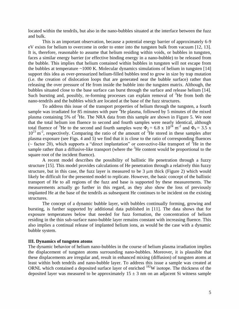
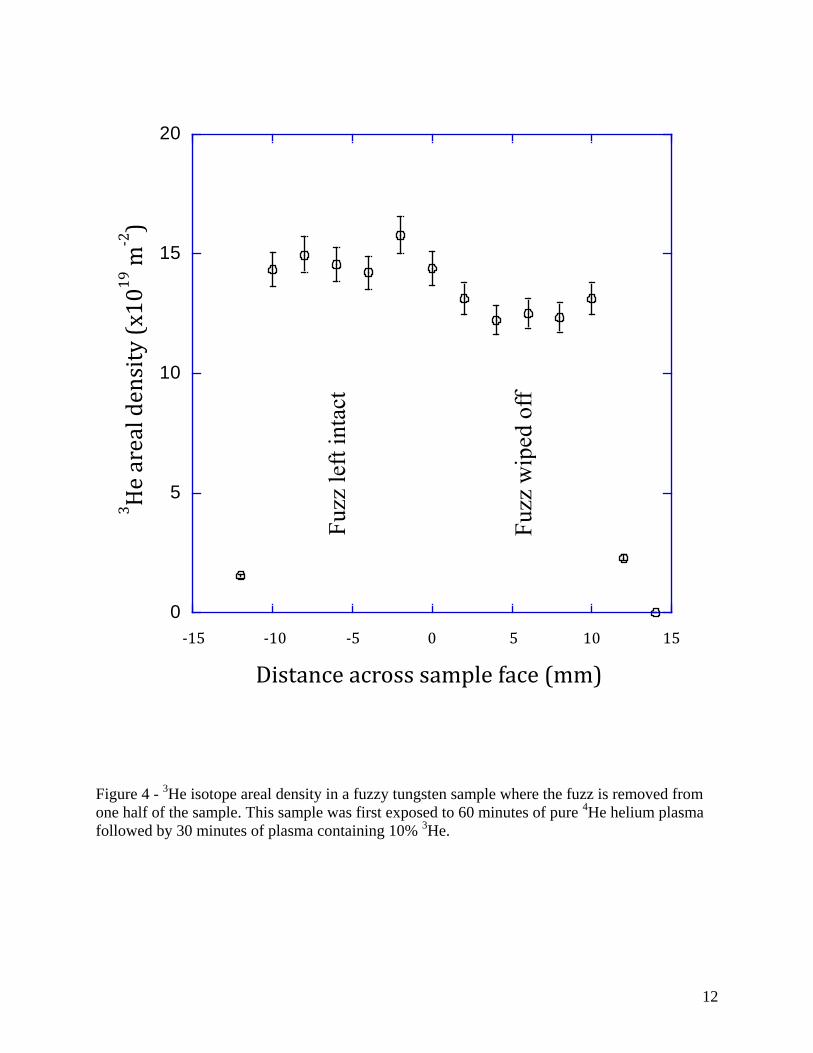
To answer this question, we irradiate the second sample for 60 minutes of pure 4He
plasma followed by 30 minutes of a mixed plasma containing 10% 3He (so that the timing of
3He
flux is incident on a well-developed fuzzy structure) The NRA data from this sample (see Figure
4), show the presence of 3He in both fuzz and the base, undoubtedly demonstrating that fuzz is
not completely opaque for helium ions, which continually penetrate into the base of the sample.
To address the issue of the “frozenness” of nano-bubbles, the third sample was initially
irradiated for 30 minutes using plasma containing a mixture of helium isotopes, specifically 5% 3He. After that, the
3He containing cylinder was valved off and a pure
4He plasma continued
uninterrupted for an additional 60 minutes. The NRA found no measurable 3He (< 5 x 10
17 m
-2)
remaining in this sample after the whole cycle of helium irradiation, neither in the half with the
fuzz intact, nor in the half with the fuzz removed. This clearly demonstrates the dynamic
behavior of the helium nano-bubbles within tungsten, as the 3He that is initially contained within
the fuzz growing for 30 minutes and in the base (assuming a similar distribution to that shown in
Figure 3) is eventually removed from the tungsten during the subsequent 60 minutes of 4He
plasma exposure. Moreover, the dynamic behavior is exhibited not only in the nano-bubbles
5
located within the tendrils, but also in the nano-bubbles situated at the interface between the fuzz
and bulk.
This is an important observation, because a potential energy barrier of approximately 6-9
eV exists for helium to overcome in order to enter into the tungsten bulk from vacuum [12, 13].
It is, therefore, reasonable to assume that helium residing within voids, or bubbles in tungsten,
faces a similar energy barrier (or effective binding energy in a nano-bubble) to be released from
the bubble. This implies that helium contained within bubbles in tungsten will not escape from
the bubbles at temperature ~1000 K. Molecular dynamics simulations of helium in tungsten [14]
support this idea as over-pressurized helium-filled bubbles tend to grow in size by trap mutation
(i.e. the creation of dislocation loops that are generated near the bubble surface) rather than
releasing the over pressure of He from inside the bubble into the tungsten matrix. Although, the
bubbles situated close to the base surface can burst through the surface and release helium [14].
Such bursting and, possibly, re-forming processes can explain removal of 3He from both the
nano-tendrils and the bubbles which are located at the base of the fuzz structures.
To address this issue of the transport properties of helium through the tungsten, a fourth
sample was irradiated for 85 minutes with pure 4He plasma, followed by 5 minutes of the mixed
plasma containing 5% of 3He. The NRA data from this sample are shown in Figure 5. We note
that the total helium ion fluence to second and fourth samples were nearly identical, although
total fluence of 3He to the second and fourth samples were Φ2 = 6.8 x 10
24 m
-2 and Φ4 = 3.5 x
1023
m-2
, respectively. Comparing the ratio of the amount of 3He stored in these samples after
plasma exposure (see Figs. 4 and 5) we find that it is close to the ratio of corresponding fluences
(~ factor 20), which supports a “direct implantation” or convective-like transport of 3He in the
sample rather than a diffusive-like transport (where the 3He content would be proportional to the
square root of the incident fluence).
A recent model describes the possibility of ballistic He penetration through a fuzzy
structure [15]. This model provides calculations of He penetration through a relatively thin fuzzy
structure, but in this case, the fuzz layer is measured to be 3 m thick (Figure 2) which would
likely be difficult for the presented model to replicate. However, the basic concept of the ballistic
transport of He to all regions of the fuzz and base is supported by these measurements. The
measurements actually go further in this regard, as they also show the loss of previously
implanted He at the base of the tendrils as subsequent He continues to be incident on the existing
structures.
The concept of a dynamic bubble layer, with bubbles continually forming, growing and
bursting, is further supported by additional data published in [11]. The data shows that for
exposure temperatures below that needed for fuzz formation, the concentration of helium
residing in the thin sub-surface nano-bubble layer remains constant with increasing fluence. This
also implies a continual release of implanted helium ions, as would be the case with a dynamic
bubble system.
III. Dynamics of tungsten atoms
The dynamic behavior of helium nano-bubbles in the course of helium plasma irradiation implies
the displacement of tungsten atoms surrounding nano-bubbles. Moreover, it is plausible that
these displacements are irregular and, result in enhanced mixing (diffusion) of tungsten atoms at
least within both tendrils and nano-bubble layer. To address this issue a sample was created at
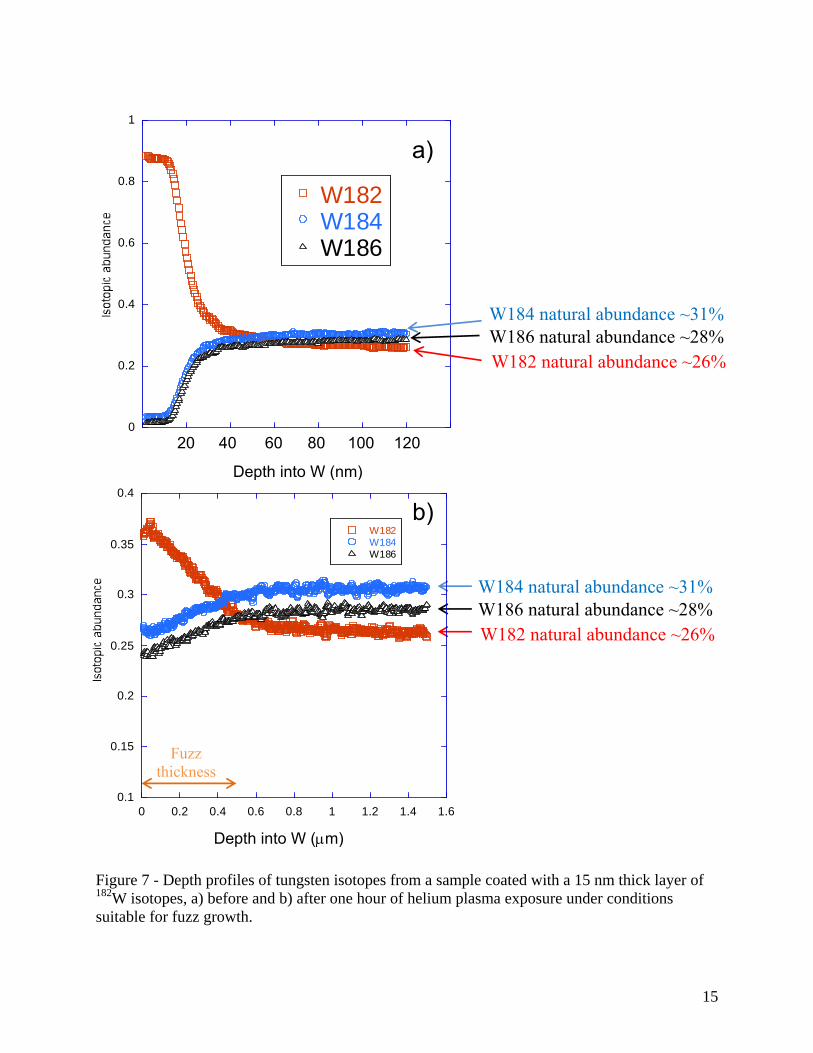
ORNL which contained a deposited surface layer of enriched 182
W isotope. The thickness of the
deposited layer was measured to be approximately 15 ± 3 nm on an adjacent Si witness sample

6
using a KLA Tencor Alphastep 500 Surface Profilometer. The isotopically enriched surface
consisted of ~92.99% 182
W, as can be seen in the inset of Figure 6. For this experiment, the
sample was exposed to helium plasma for one hour to a fluence of 1 x 1026
m-2
at a temperature
of 1150 K. The plasma exposure resulted in the formation of a fuzzy tungsten surface on the
sample. Depth profiles of the various tungsten isotopes were obtained by Secondary Ion Mass
Spectrometry (SIMS) performed by Evans Analytical using a 2 keV oxygen beam, both before
and after the plasma exposure. The depth of the sputtered crater in the W sample was measured
by confocal microscopy following the SIMS analysis. The composition of each isotope is
monitored each second during the 10 minute long sputtering measurement. The fact that the
sputtering yield from the fuzzy surface is less than that of a fully-dense surface is compensated
by the porosity of the fuzz [10]. Therefore, each measurement time is assumed to be an equal
sputtered depth into the profile. The thickness of the fuzzy layer is measured by cross-sectional
SEM imaging (Figure 6) and is superimposed on the SIMS depth profile.
The resultant depth profiles are shown in Figure 6 for the three primary tungsten isotopes
(182
W: 26.50% natural abundance, 184
W: 30.64% natural abundance and 186
W: 28.43% natural
abundance). As can be seen from the figure, the original isotopic enrichment of the surface
(92.99% 182
W, 2.84% 183
W (not shown for clarity), 2.79% 184
W and 1.38% 186
W) is lost when the
tungsten fuzz grows.
A simple integration of the depth profiles obtained from before and after the plasma
exposure can also be made to ensure that the plasma did not erode through the initial 182
W layer.
Assuming the 182
W layer, enriched to 92.99%, had 100% theoretical density, a 15 nm thick layer
would amount to 8.78 x 10²⁰
182
W atoms/m2 initially deposited on the sample surface. Following
the exposure the 182
W depth profile can be integrated to reveal how much of the deposited 182
W
remains in the sample. Assuming the natural abundance of 182
W follows a similar profile shape
to that of 184
W and 186
W as a function of depth into the fuzz as seen in Figure 7, we can integrate
the 182
W curve over the thickness of the fuzz layer. Once the average porosity of the 500 nm
thick fuzz layer (~ 0.3) [11] is taken into account, we obtain 8 x 10²⁰
182
W atoms/m2 atoms still
remaining in the fuzz and hence no erosion occurred under these conditions as anticipated for
this ion energy. This result clearly indicates the tendency for mixing of the tungsten atoms from
beneath the growing tendrils with the material further up toward their tips.
In the absence of the mixing of tungsten atoms caused by the dynamics of helium nano-
bubbles, one would expect that the surface would continue to be enriched with 182
W and regions
deeper in the fuzz structures would contain the isotopic mix of the bulk material. Clearly this is
not observed in our experiment; instead we see a mixture of tungsten isotopes at the surface and
a gradual depth profile approaching the concentrations of the bulk tungsten. On the other hand,
the inter-diffusion of tungsten isotopes is too slow at this temperature [16] and cannot explain the
experimentally observed depth distributions. Therefore, the experimental data presented in both
Sections II, and III, support a physical picture of the dynamic behavior of helium nano-bubbles
which, also, causes an enhanced mixing of tungsten atoms.
A similar result has recently been reported by Fiflis [17], where a thin tungsten coating
was applied to a molybdenum wire and the entire structure was subjected to conditions where a
fuzzy W surface should form. Even though the experiment utilized two different elements, the
result also showed the motion of the substrate material up through the surface layer toward the
tips of the tendrils. It is interesting to note that even in the experiments by Fiflis, using much
thicker W coatings (100 and 200 nm thick) evidence of the Mo substrate could be found at the
surface following fuzz formation.
7
IV. Conclusion
Two different types of experiments on tungsten samples exposure by helium plasma have been
performed. The experiments employed helium and tungsten isotopes, to address the following
conceptual issues: i) does the fuzz become opaque for helium ions impinging on the sample? ii)
are helium nano-bubbles in both the fuzz tendrils, and at their base, “frozen” after they are
formed and no longer undergo changes? iii) does the fuzz growth, and nano-bubble layer
formation, result in the mixing of tungsten atoms and thereby impact the distribution of atoms in
the sample?
In the experiments presented in the Sections II, 3He was included in the initial phase of
the plasma discharge in one sample and toward the end of the plasma discharge in other samples.
No evidence of 3He was observed retained in the tungsten when the
3He was only present in the
plasma initially. On the other hand, 3He was detected in the samples exposed to
3He containing
plasma at the end of the discharge. The amount of 3He in the tungsten sample appears to scale
directly with the fluence to the samples, rather than with a square root of time, which would be
expected for diffusive-like process. An increase proportional to the incident fluence suggests a
direct implantation pathway for He to enter the tungsten substrate. In the Section III, the results
from an experiment are described where an isotopically enriched 182
W surface layer was
deposited on a tungsten substrate having the normal tungsten isotopic distribution. Depth profiles
of the surface before and after fuzz growth revealed the mixing of the tungsten isotopes in the
substrate with the material closer toward the tips of the growing tendrils.
These results demonstrate that: i) fuzz is not completely opaque for helium ions
impinging on the sample and, at least, some incident ions penetrate through to the fuzz base; ii)
helium nano-bubbles exhibit a dynamic behavior which is manifested in nano-bubble bursting
and reformation; iii) as a result of this dynamic process the motion of tungsten atoms in the
region of helium filled bubbles demonstrate enhanced mobility which results in their strong
mixing.
Acknowledgements
This material is based upon work supported by the U.S. Department of Energy, Office of
Science, Office of Nuclear Physics under contract number DE-AC05-00OR22725 and by U.S.
Department of Energy grants DE-FG02-07ER54912 and DE-SC0018302. It was also conducted
as part of the US-EU Bilateral Collaboration on Mixed Materials for ITER.
References
[1] S. Kajita, T. Ishida, N. Ohno et al., Sci. Rep. 6, 30380; doi: 10.1038/srep30380 (2016).
[2] R. A. Pitts, S. Carpentier, F. Escourbiac et al., J. Nucl. Mater. 438 (2013) S48.
[3] G. De Temmerman, T. Hirai and R. A. Pitts, Plasma Phys. Control. Fusion 60(2018)044018.
[4] S. Kajita, W. Sakaguchi, N. Ohno, N. Yoshida and T. Saeki, Nucl. Fusion 49(2009)095005.
[5] M. J. Baldwin and R. P. Doerner, Nucl. Fusion 48 (2008) 035001.
[6] S. Kajita, N. Yoshida, R. Yoshihara, N. Ohno and M. Yamagiwa, J. Nucl. Mat.
418(2011)152.
[7] D. M. Goebel, Y. Hirooka, R. W. Conn et al., J. Nucl. Mater 145-147 (1987)61.
8
[8] W. Eckstein, Sputtering Yields, in: R. Behrisch, W. Eckstein (Eds.): Sputtering by Particle
Bombardment IV, Top. Appl. Phys. 110 (Springer, Berlin, Heidelberg 2007).
[9] M. Mayer, Nucl. Instrum. Methods B 266(2008)1852.
[10] D. Nishijima, M. J. Baldwin, R. P. Doerner and J. H. Yu, J. Nucl. Mater. 415 (2011) S96.
[11] R. P. Doerner, M. J. Baldwin, M. Simmonds et al., Nucl. Mater. and Energy 12 (2017)372.
[12] H. Ullmaier, Nucl. Fusion 24 (1984) 1039.
[13] M. Thompson, A. Deslandes, T. W. Morgan et al, Nucl. Fusion 56 (2016) 104002.
[14] B. D. Wirth, K. D. Hammond, S. I. Krasheninnikov and D. Maroudas, J. Nucl. Mater. 463
(2015) 30.
[15] T. P. C. Klaven, K. Nordlund, T. W. Morgan et al., Nucl. Fusion 56(2016)126015.
[16] J. N. Mundy, S. J. Rothman, N. Q. Lam, H. A. Hoff and L.J. Nowicki, Phys. Rev. B
18(1978)6566.
[17] P. Fiflis, N. Connolly and D. N. Ruzic, J. Nucl. Mater. 482 (2016) 201.

9
Figure 1 – Spectroscopic composition measurement of helium isotope content in the PISCES-A
plasma.
1
0.5
0.75
0.25

10
Figure 2 – Cross section of tungsten fuzz layer following 90 minute He plasma exposure.

11
Figure 3 – 3He isotope areal density measured in a fuzzy tungsten sample after 60 minutes of
plasma containing 25% 3He content. Measurements were made across the sample where half the
fuzz has been wiped off the surface, compared to the other half showing the helium content in
the region with fuzz.
Fuzz
wip
ed o
ff
Fuzz
lef
t in
tact
b)
0
10
20
30
40
50
60
70
80
165 170 175 180 185 190
Distance across sample face (mm)
10
5
15
20
3H
e ar
eal d
ensi
ty (
x10
19 m
-2)
Distance across sample face (mm)
-15 -10 -5 0 5 10 15
12
Figure 4 - 3He isotope areal density in a fuzzy tungsten sample where the fuzz is removed from
one half of the sample. This sample was first exposed to 60 minutes of pure 4He helium plasma
followed by 30 minutes of plasma containing 10% 3He.
0
5
10
15
20
120 125 130 135 140 145 150
Position (mm)
Fu
zz w
iped
off
Fuzz
lef
t in
tact
Distance across sample face (mm)
-15 -10 -5 0 5 10 15
3H
e ar
eal d
ensi
ty (
x10
19 m
-2)

13
Figure 5 – 3He isotope areal density in a fuzzy tungsten sample where the fuzz is removed from
one half of the sample. This sample was first exposed to 85 minutes of pure 4He helium plasma
followed by 5 minutes of plasma containing 5% 3He. (Note y-axis scale difference with figures 3
and 4.)
0
0.5
1
1.5
145 150 155 160 165 170 175
Position (mm)
Fuzz left intact Fuzz wiped off
Distance across sample face (mm)
-15 -10 -5 0 5 10 15
3H
e ar
eal d
ensi
ty (
x10
19 m
-2)

14
Figure 6 - Cross section of tungsten fuzz layer formed on the sample containing the isotopically
enriched surface layer following 60 minute He plasma exposure.
15
Figure 7 - Depth profiles of tungsten isotopes from a sample coated with a 15 nm thick layer of 182
W isotopes, a) before and b) after one hour of helium plasma exposure under conditions
suitable for fuzz growth.
Fuzz
thickness
W182 natural abundance ~26%
W186 natural abundance ~28%
W184 natural abundance ~31%
0.1
0.15
0.2
0.25
0.3
0.35
0.4
0 0.2 0.4 0.6 0.8 1 1.2 1.4 1.6
W182W184W186
Depth into W (microns)
b)
Depth into W (m)
0
0.2
0.4
0.6
0.8
1
W182W184W186
Depth into W (nm)
20 60 80 40 100 120
W182 natural abundance ~26%
W186 natural abundance ~28%
W184 natural abundance ~31%
a)