Embed Size (px)
Citation preview


FALL 1995 VOLUME 31 NO 3 -
T A B L E 0 F C O N T E N T S
EDITORIAL 1995 Challenge Winners
LETTERS
Gem-Quality Grossular-Andradite: A New Garnet from Mali
Mary L. Johnson, Edward Boehm, Hoist Krupp, Joachim W. Zang, and Robert C. Kammerling
Sapphires from Southern Vietnam Christopher P. Smith, Robert C. Kammerling,
Alice S. Keller, Adolf Peretti, Kenneth V. Scarratt, Ngzzyen Dang Khoa, and Saverio Repetto
NOTES AND NEW TECHNIQUES "Ti-Sapphire": Czochralski-Pulled Synthetic Pink Sapphire
from Union Carbide Mary L. Johnson, Meredith E , Mercer, Emmanzzel Fritsch,
Patricia Maddison, and James E , Shigley
REGULAR FEATURES Gem Trade Lab Notes Gem News Book Reviews Gemological Abstracts
ABOUT THE COVER: A n e w find o j w garnets has been made in the Republic o f Mali, western Africa. The lead article in this issue characterizes these garnets, which (ire primarily yellow-green to brown hut 17ls0 (rarely) intense green. They have conipo- sitions between grossular and andradite. The brooch contains Mali garnets, in a range o f colors, and diamonds. The loose stones r u q e in size from the 1.35-ct yellow trinngle ciit to the 18.03-ct oval. The largest intense green stone is 2.90 ct.
All stones and jewelry courtesy o f Giislav Zang, Lapidary, ldar-Oberstein, Germany, jewelry design by Monikii Leyser, o f Kirschweiler, near ldnr-Oberstein. Photo Q Harold s) Erica Van Pelt-Photographers, Los Angeles, C A .
Typesetting [or Gems & Gcmology 1s by Graphix Express, S a m Monicn, C A . Color separations are b y Effective Graphics, Compton, CA . Printing is by Cadmus journal Services, Easton, MD.
0 1995 Gemological Institute of America All rights reserved ISSN 0016-626X

Editor-in-Chief Richard T. Liddicoat
Associate Editors William E. Boyajian Robert C. Kamnierling D. Vincent Manson John Sinlzankas
Technical Editor Carol M. Stockton
Assistant Editor Irv Dierdorff
Editor Alice S. Keller 1660 Stewart St. Santa Monica, C A 90404 (800) 4 2 1-7250 x25 1 e-mail: [email protected]
Subscriptions Jin Lim Cristina Chavira (800) 42 1-7250 x20 1 Fax: (310) 453-4478
Contributing Editor John 1. Koivula
Editors, G e m Trade Lab Notes Robert C . Karnmerling C. W. Fryer
Editors, G e m News Robert C . Kaninierling John 1. Koivula Emmanuel Fritsch
Editors, Book Reviews Susan B. Johnson Jana E. Miyahira
Editor, Gemological Abstracts C. W. Fryer
PRODUCTION Art Director Production Assistant Production Artist
STAFF Christ ine Troianello Gail Young Carol Silver
EDITORIAL G. Robert Crowningshielcl
REVIEW BOARD New York, NY Alan T. Collins London, United Kingdom
Dennis Foltz Santa Monica, CA
Emmanuel Fritsch Santa Monica, CA
C. W. Fryer Santa Monica, CA
Henry A. Hiinni Base], Switzerland
C . S. Hurlbut, Jr. Cambridge, MA
Alan Jobbins Caterharn, United Kingdom
Anthony R. Karnpf Los Angeles, CA
Robert E . Kane Helena, MT
John I . Koivula Santa Monica, CA
A. A. Levinson Calgary, Alberta, Canada
Kurt Nassau P.O. Lebanon, N{ George Rossman Pasadena. CA
Kenneth Scarratt Bangkok, Thailand
Karl Sclimetzer Petershausen. Germany
James E. Shigley Santa Monica. CA
SUBSCRIPTIONS Subscriptions in the U.S.A. arc priced as follows: $59.95 for one year (4 issues), $149.95 for three years (12 issues). Subscriptions sent elsewhere are S70.00 for one year, $180.00 for three years. Special annual subscription rates arc available for all students actively involved in a GIA program: $49.95, U.S.A.; $60.00, elsewhere. Your student number must be listed at the time your subscription is entered. Single issues may be purchased for $15.00 in the U.S.A., $18.00 elsewhere. Discounts arc given for bulk orders of 10 or more of any one issue. A limited number of back issues of G d G are also available for purchase. Please address all inquiries regarding subscriptions and the purchase of single copies or back issues to the Subscriptions Department. To obtain a Japanese translation of Gems eJ Gemology, contact the Association of Japan Gem Trust, Oiuchimachi Cy Bldg., 5-15-14 Ueno, Taito-Icu, Tokyo 110, Japan. Our Canadian goods and service registration number is R126142892.
COPYRIGHT AND REPRINT PERMISSIONS
Gems 01 Gemology welcomes the submission of articles on all aspects of the field. Please see the Suggestions for Authors in the Spring 1995 issue of the journal, or contact the editor for a copy. Letters on articles published in Gems o1 Gemoloey and other relevant matters are also wclcome.
Abstracting is permitted with credit to the source. Libraries are permitted to photocopy beyond the limits of U.S. copyright law for private use of patrons. Instructors are permitted to photocopy isolated articles for noncommercial classroom use without fee. Copying of the photographs by any means other than traditional photocopying tech- niques (Xerox, etc.] is prohibited without the express permission of the photographer (where listed) or author of the article in which the photo appears (where no photographer is listed). For other copying, reprint, or republication per- mission please contact the editor. Gems 01 Gemology is published quarterly by the Gemological Institute of America, a nonprofit educational organi- zation for the jewelry industry, 1660 Stewart Street, Santa Monica, CA 90404. Postmaster: Return undeliverable copies of Gems ed Gemology to 1660 Stewart Street, Santa Monica, CA 90404. Any opinions expressed in signed articles are understood to be the opinions of the authors and not of the publishers.

The Gems & Gemology Challenge has been one of our most popular annual features since its introduction nine years ago. This year was no exception. Hundreds of read- ers from all over the world took this demanding test, which appeared in the Spring 1 995 issue. Our heartfelt congratulations go to all who participated. Those who earned a score of 75% or better received a GIA Continuing Education Certificate rec- ognizing their achievement. Those listed below received a perfect 100% score.
USA: Alabama William A. Lavender, Pelhain + Arizona Kenyon V. Painter, Scottsdale; Norma B. Painter, Scottsdale + California Chris Almquist, Santa Monica; Wendy Beckerson, Santa Monica; Rebecca Ann Bell, Joshua Tree; Kathylee Cook-Roberts, San Jacinto; Eddie Decsi, Santa Monica; Mary Fitzgerald, Santa Monica; Werner R. Hoehne, San Francisco; Claudia Jacobs, Santa Monica; Becka Johnson, Santa Monica; Hui-Ling Kuo, Santa Monica; Sherrie Kysillza, Santa Monica; Douglas R. Mays, Carmel; Catherine McIntyre, Santa Monica; Jana E. Miyahira, Santa Monica; Sina A. Mozafarian, Beverly Hills; Philip A. Owens, Santa Monica; Michael Pace, Elk Grove; Diane H. Saito, Santa Monica; Robert A. Seltzer, San Rafael; Glenn Shaffer, Julian; Judy Shechter-Lankford, Santa Monica; Karen B. Stark, Santa Monica; Judy Steinberg-Briclzer, Santa Monica; Jim Viall, Santa Monica; Teri Von Ende, Santa Monica; Dan Tzu-Tan Wang, Santa Monica; Phil Yurlziewicz, Santa Monica + Colorado Dale Winder;Ft. Collins 4 Connecticut William A. Jeffery, Westport + Florida Mathew Mooney, West Palm Beach; Timothy D. Schuler, Palm Harbor; John E. Williamson, Minis + Illinois Anne Blumer, Bloomington; John Jaeger, Bloomington 4 Massachusetts Mary B. Moses, West Newton 4 North Carolina Jean A. Marr, Kernersville; M. Lewis Nance, Fayetteville; Blair Tredwell, Advance + Nevada Terence E. Terras, Reno + New Jersey John Morlino, Jackson; Lynn Tully, Fanwood + New York Dorothy A. Buzzcll, Siiffern; Clifford H. Stevens, Gansevoort + Ohio Jean Mate, Mentor; Roy J. Pasini, North Olinsted; Jack Schatzley, Toledo + Pennsylvania Peter R, Stadelmeier, Yardley + Texas George L. Blair, Houston; Connie Bradshaw Copeland, Abilene + Virginia Dale S. Summers, Richmond 4 Washington Stephen H. Hall, Kennewiclz + Wisconsin Drew Thiet, Milwaukee; Thomas G. Wendt, Beaver Dam + AUSTRALIA: Malcolm B. Druery, Goonellabah; David A. Keith, Bethania + CANADA: Michael J. P. Cavanagh, Vancouver, British Columbia; Follzert H. De Jong, Victoria, British Columbia; Michael Hyszlza, Coolzstown, Ontario; Diane Kolze, Calgary, Alberta; David R. Lindsay, Bobcaygeon, Ontario; Lina Masse, Montreal, Quebec; Janysz J. Meier, Calgary, Alberta; Ron Plessis, Aldergrove, British Columbia; Jon C. Phillips, Vancouver, British Columbia + FRANCE: Marie-France Chateau, Paris + HONG KONG: Deidre I<. Alejo, Central + INDONESIA: Warli Latumena, Jakarta + ITALY: Mafalda Pasqui, Geneva; Roberto Filippi, Lucca; Gianfranco Zamboni, Agrate + SCOTLAND: James W. M. Heatlie, Edinburgh + SPAIN: Cristina Knorr, Vitoria + SWITZERLAND: Eva Mettler, Zurich + THAILAND: Johanne Cardin- Jack, Khon Kaen
Answers (see pages 74-75 of the Spring 1995 issue for the questions): (1) a, (2) d, (3) a, (4) b, (5) c,
(6) c, (7) b, (8) a, (9) b, (10) a, (1 1) b, (121 c, (13) d/ (14) c, (15) c, (16) bl (17) b, (18) dl (19) a, (201 b, (21) a, (22) dl (23) dl (24) c, (25) b. Our thanks to Warli Latumena of Jakarta for suggesting that we list the winners geographically.
Challenge Winners GEMS & GEMOLOGY Fall 1995 149

S u p b R w s h beds Seem In August 1994, I visited &e ~-~~ r&n around M a l p k a with Dr. M & P W A t h t b q - w d d k W r n d y through three ohidly r e q p d g e t n mpplie~ A 0 Mhing Co,, the joht venture Emwal, d MRU {W- dewskee Rudouprawhie Ud). We toured the f a d - t i w m d m - b e d w & d & m d l w m quantities of mostly smal l faceted samples, including d d w , yellow to brown mddite gamts* d a few demantoid garnets, in addition to emeralds and green beryls. One lot d m . d of appmxhwely 22 gmm ( 110 ct) of top-quality, richly colored rough emeraldsJ dthough most weighed no more than 1-1.5 a.
A few months aftex this visit, a Swiss buinewman with GI- conm&ns in he fama S k e i Uniot~hught sevad lots of U r h emeralds to the GiibeIin Gem-
Fall 1995


~ D W I T E : A NEW GARNET FROM MALI
By Mary L. Joh~~son, Edward Boehm, Horst Krupp, Joachim LV. Zang, and Robert C. Kammerling
A find of new gem garnets has been made arnets have been prized as gems since at least 3200 in contact n~etamorphic deposits in the B.C. (Andrews! 1991). Today! gem garnets are found Republic of Mali, western Africa. These in almost any color. They exhibit broad chemical vari- garnets, primarily yellow-green to brown ability between the lznown end-membersi the most but also (rarely) intense green, have compo- 9 conlinon of which are pyropel a lmandi i~e~ spessartine! sitions between grossular and andradite. grossularl and andradite. (A useful overview of gem gar- Altho~~gll garnets of similar composition are relatively well lznown, this is the first nets was given by Stockton and Manson! 1985; see also documented occzzrrence of gem-qzzality Manson and Stocktonl 1982' and Stockton and Manson! material in commercial quantities. Because 1982/ 1983) for specific color ranges). Garnets are also this gem variety is not represented by any used as ornamental gem materials in their massive of the gem garnet terms previoz~sly in use, forms: Examples include hydrogrossular and grossular. it is described as grossz~l~~r-andradite by the In spring 1994) a new type of gem-quality garnet [fig- GIA Gem Trade Laboratory, It can be dis- ure 1) first appeared on the marlzet in Idar-Oberstein (one tinguished from gross~llar by its absorption of the authors-Dr. Krupp-was first offered rough mate- spectrum and (~lsually) higher refractive rial at this time; see also Frazier and Frazier! 1995a and index; i t can be distinguished from andra- b). This was a transparent! facetable inaterial in the yel- dite by its lower R.1. The staclzed parallel low-to-green-to-brown range! with properties close' but planes of growtll zoning, always visible between crossed polarizers, are diagnostic not idellticall to those of grossular. One version of the
of grossular-andradite. discovery was that the material was found by a West African who had once lived in Idar-Oberstein (Frazier and Frazier' 1995b). In the gem trade' the material is typ- ically called "Mali garnetii or ligrandite garnet."
B. Jdmsmk a M r c h scimtlst at the 43A Among discussions of Mali garnet in the trade press G m r w Lsbmkny, &m?a hhnice,. are reports on their appearance at Intergem in M ~ ~ n i c h in Wk?&. m. mem &.a gmkig&t and w G r a u e F ~ , Gm.o&& Wb&. &&. WIW h w Pak is Memat id , a p@cfst m September i'Impressions of of 1994 Intergem (Frazier '94," and 1994)) Frazier' and 1995a mention and in b;
p&dd dl%-, La Casta, WbM, the 1995 International Colored Gemstone Association a, ~ k v i c a ~ t ~ f ~ ~ n g (ICA) world gemstone mining report (Eliezri and Kremlzowi LapMityh w - m @ n . w e E m & @ the JnSfir~e mrGmtolJe &search, 1994). Four technical reports have been published in i & p a d M o f ~ , W t y d M German (Zang' 1994; Henn et al.! 1994; Lind and Banlz1 ~ , ' ~ y , M. tCmmehgis dcemesi- 1994; Lind et al.' 1995). One stone reportedly from this dent~J%eemi3 h~~ Gem Ttade L d m w m m a . locality was examined in the GIA Gem Trade Laboratory see m w w s a t 'the d oftbrnkk. in September of 1994 (Hurwit e t al.) 1994). Since we
began work on this report in September 1994! a short & & d w y , V d 3T, MI. 3; m. 16&!6
@ 1995 GfmgQkW hsMu&a o f h W c ~ article ill English appeared in the Austrolion Gemmologist (Brightman' 19951.
152 Mali Garnets GEMS & GEMOLOGY Fall 1993

Fig~zre I . One of the most recent additions to the gem
marketplace ore gein garnets from Mali. These gross~zlar-
andradites, froin the first- linown coinmerci~l gem occur- rence o f this material, range in
color from yellow to green to brown. Although mosL of the
stones faceted to date are smaller than 5 ct, some are quite large, as indicated b y
this loose 33.29-ct stone, The stones in the rings are 4.03,
4.57, and 5.53 ct, Co~irtesy of M. Fabrilidnt d Sons, New
York City; photo 0 Harold e9 Erica Van Pelt.
LOCATION AND ACCESS The garnets are being recovered from various local- ities in the "Zone of SangafeIt' near the village of Dialzon (about 100 kin northeast of Bafoulabd! 110 lzrn soutl~west of Nioro) and 130 lzm east of Kayes; air miles in all cases)! in the Kayes Region of Mali (figure 21. The Zone of Sangafk is in the Sahel region (a semiarid area between the Sahara Desert to the north and the savannas to the ~ 0 ~ 1 t h ) . William Daineron) former U.S. Ambassador to Mali! stated that inany garnets come from Sibinndi) "about 20-30 lzln ~ 0 ~ 1 t h of Sandark" (pers. comm.! 1995)! in the vicinity of Diakon. One of our contacts in Mali states that the intense green Mali garnets come from the village of Duvalk.
Access to the mining area is difficult, The closest major townt Kayes! can be reached by train from either Bamako! the capital of Mali, which is about 400 lzm to the so~itheast~ or Dalzarl the capi-
tal of Senegal, which is about 620 km due west) on the Atlantic Coast. From Kayes) one must travel by jeep) or bashe (the local term for bush wagon or taxi]! to cover the remaining 170 lzrn of partially paved roads to the town of Sandark. From there, another 20-30 lzrn of unpaved roads lead to the mining areas around the villages of Sibinndi and Dialzon.
It is possible that some material has been transported out of Mali via Dalzar because of its coastal access. Despite the difficulty in reaching the mining areas) there has been such a rush to the region that extra railroad cars were added to the trains going to Kayes. The rough is carried in flour or rice saclzs and transported by any available vehi- cle to the train station at Sandark.
GEOLOGY AND OCCURRENCE Western Mali is underlain by the Precainbrian West African Craton. The craton has been warped
Mali Garnets CEIMS & GEMOLOGY Fall 1995

into a broad bowl-the Taoudeni Basin-with edges at the Western SaharaIMauritania border and froin Sierra Leone and Guinea to southeast Mali. Cambrian sediments fill the western part of the basin and are overlain by Mesozoic deposits to the east. In the region of interest! the sediments include inagnesian limestones and dolomitic lime- stones (Furon! 1963). These sediments have been intruded in various places by dilzes primarily of diabase (that isl fine-grained gabbroic roclzs mainly consisting of plagioclase and p y r o ~ e n e ) ~ which extend froin the border with Guinea to Ma~lritania (figure 3); these dilzes vary from basalt to albite- quartz pegmatites (Furon) 1963; Bessolesl 1977). Southwest of the town of Nioro, these diabases are Jurassic in age (Cahen et al.! 1984) and form a mas- sif known as the Kaarta) a zone of rugged relief ranging up to 300 m in altitude (Furon, 1963).
According to reports on the regional geology of Mali! garnets formed in contact metan~orphic zones along the boundaries where the widely scattered diabase dilzes intruded into limestones (among other roclzs). Besides garnet! the minerals that formed in the contact zone between diabase and limestone include epidote) (titaniferous) magnetite) prehnite, fluoritel and occasionally chrysoberyl (Hubert! 19141 as reported in Bessolesl 1977; Furon! 1963; Ministere des Mines [no date]). This informa- tion is consistent with statements that have been made elsewhere concerning the geologic occur- rence of gem garnets in Mali. Both Lind and Bank (1994) and Henn et al. (1994) state that the garnets come from coiltact metainorphosed depositsl whichl according to Henn et al. (1994)! consist of clay and feldspar-bearing sandstones and marbles that have been intruded by a diabase. Garnet! epi-
Figure 2. The gross~~lar-andradite garnets st~ldied for this article were all report~dly from deposits near the village of
Diakon, in the Kayes Region of the Republic of Mali, western Africa. (Adapted from: Institut Geographique
National-France, 1993,) 1
Mali Garnets GEMS & GEMOLOGY

Figure 3. This geologic sketch map of western Mali, as well as
parts of Senegal and Mauritania, shows the large diabase intru-
sions. Contact metamorphic bodies, including some that con-
tain gem-quality garnets, are found along the boundaries of the diabase intrusives. Figure
adapted from Furon, 1963.
Precambrian I
UppermosI Precambrian and Cambrian 1 Silurian
1 Diabases and related intrusives
dote, prehnite, and ves~ivianite mineral specimens from Mali were marketed in Tucson, Arizona, in spring 1995 by Dave Bunk Minerals of Wheat Ridge, colorado (with the locality given as "San- dare, Nioro du Sahel, Mali"); also, "bright green" gar- nets as large as 4 cm-some with chalcedony over- growths-have been found growing on some vesu- vianites mined in Mali (W. Dan~eron, pers. conlm., 1995).
In addition to the garnets mined near Sibinndi and Diakon, well-formed opaque brownish red crystals have been found in the village of Bindougou, a few kilometers north of Dialzon. Black garnets have been found a few kilometers east of Diakon, in the village of Trantiinou (W. Dameron, pers. coinm., 1995). Thus far, only a few intense green stones have been found, including two in this study and a recently discovered crystal that pro- duced a 2.9-ct cut stone.
The garnets also occur as waterworn nodules, reportedly from alluvial sources near the contact metamorphic deposits (Eliezri and Kremkow, 1994; Hurwit et al., 1994).
MINING AND PRODUCTION Although garnets from this region have been known since 1914, the recent discoveries represent the first gem-quality stones found in any signifi- cant quantities. We have seen crystals larger than 5.5 kg (Zang, 1994); for the most part, however, the facetable areas in each stone are relatively small.
The largest faceted stone seen to date is a brown 57.5-ct round brilliant (figure 4). Only gemmy cores-generally,nodules~are suitable for faceting (figure 5), although cabochons are being cut from other parts of the rough. Considerable manual labor is required to break up these crystals, and most dealers now insist on purchasing only nodules, so relatively few euhedral crystals are available for mineral collectors and crystallographic study.
The authors calculate, from personal experi- ence, that a parcel of 400-500 crystals, weighing about 200 kg, yields 160 grams of nodules, from which only 60 grams would be facet grade. Of these 60 grams (300 ct) of facet-grade rough, per- haps 150 ct of faceted gems result: a total yield of 0.015%.
Some dealers are believed to have large stoclz- piles of rough Mali garnets in Idar-Oberstein, with substantial quantities also in North America and Australia. We estimate that, as of September 1995, these stockpiles totaled several dozen lzilos of facet-grade material and several hundred lzilos of cabochon-quality rough. According to Joe Freilich of M. Fabrilzant & Sons in New York, their stoclz of faceted Mali garnets exceeds 5,000 ct, primari- ly in sizes up to 5 ct, but with a few larger "col- lector pieces" (again, see figure 1).
MATERIALS AND METHODS For our gemological investigation, we examined 23 faceted stones (some are shown in figure 6), 20
Mali Garnets GEMS & GEMOLOGY Fall 1995
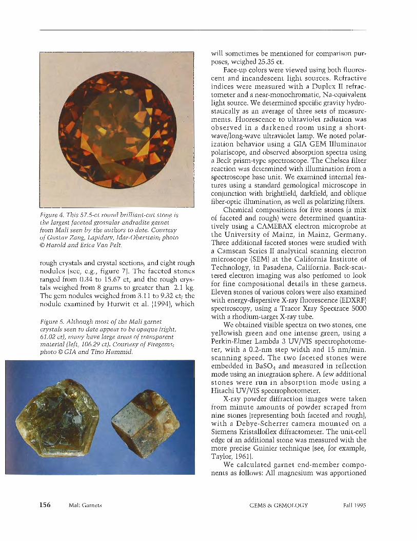
Figure 4. This 57.5-ct round brilliant-cut stone is the largest faceted grossular-andradite garnet from Mali seen by the authors to date. Courtesy of Gustav Zang, Lapidary, Zdar-Oberstein; photo 0 Harold and Erica Van Pelt.
rough crystals and crystal sections, and eight rough nodules (see, e.g., figure 7). The faceted stones ranged from 0.34 to 15.67 ct, and the rough crys- tals weighed from 8 grams to greater than 2.1 kg. The gem nodules weighed from 3.11 to 9.32 ct; the nodule examined by Hurwit et al. (1994), which
Figure 5. Although most of the Mali garnet crystals seen to date appear LO be opaque (right, 61.02 ct), many have large areas of transparent material (left, 106.29 ct). Courtesy of Firegems; photo 0 GZA and Tino Hammid.
will sometimes be mentioned for comparison pur- poses, weighed 25.35 ct.
Face-up colors were viewed using both fluores- cent and incandescent light sources. Refractive indices were measured with a Duplex I1 refrac- tometer and a near-monochromatic, Na-equivalent light source. We determined specific gravity hydro- statically as an average of three sets of measure- ments. Fluorescence to ultraviolet radiation was observed in a darkened room using a short- wave/long-wave ultraviolet lamp. We noted polar- ization behavior using a GIA GEM Illuminator polariscope, and observed absorption spectra using a Beck prism-type spectroscope. The Chelsea filter reaction was determined with illumination from a spectroscope base unit. We examined internal fea- tures using a standard gemological microscope in conjunction with brightfield, darkfield, and oblique fiber-optic illumination, as well as polarizing filters.
Chemical compositions for five stones (a mix of faceted and rough) were determined quantita- tively using a CAMEBAX electron microprobe at the University of Mainz, in Mainz, Germany. Three additional faceted stones were studied with a Camscan Series I1 analytical scanning electron microscope (SEM) at the California Institute of Technology, in Pasadena, California. Back-scat- tered electron imaging was also perfomed to look for fine compositional details in these garnets. Eleven stones of various colors were also examined with energy-dispersive X-ray fluorescence (EDXRF) spectroscopy, using a Tracer Xray Spectrace 5000 with a rhodium-target X-ray tube.
We obtained visible spectra on two stones, one yellowish green and one intense green, using a Perkin-Elmer Lambda 3 UV/VIS spectrophotome- ter, with a 0.2-nm step width and 15 nmlmin. scanning speed. The two faceted stones were embedded in BaS04 and measured in reflection mode using an integration sphere. A few additional stones were run in absorption mode using a Hitachi UV/VIS spectrophotometer.
X-ray powder diffraction images were taken from minute amounts of powder scraped from nine stones (representing both faceted and rough), with a Debye-Scherrer camera mounted on a Siemens Kristalloflex diffractometer. The unit-cell edge of an additional stone was measured with the more precise Guinier technique (see, for example, Taylor, 196 1 ).
We calculated garnet end-member compo- nents as follows: All magnesium was apportioned
Mali Garnets GEMS & GEMOLOGY Fall 1995

to the pyrope component, Py; all manganese to the spessartine component, Sp; all chromium to the uvarovite component, Uv; and all iron (Fe3+) to the andradite component, And. Assignment of all iron as Fe3+ is supported by our spectra, which revealed no evidence of ~ e ~ + (as in almandine). Calcium, aluminum, and silicon were distributed according to stoichiometry; remaining aluminum was apportioned to the grossular component, Gi. Because none of the stones revealed a significant titanium content, Ti was ignored in calculating end members. Components were normalized to 100% (e.g., Grya AndmPylSpl).
CRYSTAL MORPHOLOGY The rough samples we examined (again, see figure 7) occurred in two forms: as crystals, often nearly complete, and as rounded nodules. Most of the
Figure 6. These seven faceted brown, yellow- green, and green grossu- lar-andradite garnets from Mali (ranging from 0.85 t o 2.39 ct) are part o f the s t udy sample. Stones courtesy of Pala Properties International and Thomas M. Schneider; photo 0 G I A and Tino Hammid.
crystals showed dodecahedral d (1 101 faces; some also showed minor trapezoheclral n (21 1) faces; and, in at least one case, the trapezohedron was the major form shown. Most crystals showed regu- lar shapes, but a few were distorted; distorted crys- tals are flattened in a variety of directions.
All the crystals appeared to have dodecahedral core zones, and some very large crystals showed only the dodecahedral habit (that is, not all crystals have trapezohedral faces). In some cases, gemmy yellowish green or green cores were found in crys- tals with dull, nearly opaque, brown surface "skins" (again, see figure 5). The surface regions were less transparent than the cores. At least one crystal was so dark brown as to appear nearly black, and one showed a bright green surface region (figure 8).
Most crystal faces on the predominantly dodec- ahedral crystals were sharply reflective, although
Mali Garnets GEMS & GEMOLOGY Fall 1995

Figure 7. Also in the study sample were these seven rough crystals and eight nodules of Mali garnet.
From top left down: approximately 126-gram dodecahedron, 24-gram trapezol~edron/dodecahe- dron, 16-and 8-gram dodecahedra. From top right down: approximately 780 gram (94 x 71 x 66 cm)
dodecahedron, and 22- and 13-gram distorted dodecahedra/trapezohedra. The eight nodules in
the center weigh from 3.11 to 9.32 ct. Courtesy o f Pala Properties International, Firegems, and the
authors; photo by Shane F. McClure.
some had rough surfaces (figure 91, which may be due to intergrowth with other minerals. Some dodecahedra1 internal surfaces (and all faces on those crystals that were predominantly trapezohe- dral) had a rough or stepped appearance. This appearance may be due to the dissolution of inter- grown minerals, especially calcite, during either weathering or processing of the mined material.
In contrast to these crystals, the "nodules" we examined-actually cobbed rough material, rather than alluvial nodules-had rounded surfaces and no crystal faces. The curved surfaces were proba- bly caused by fracturing along curved fluid-filled
inclusions (see "Appearance with Magnification and in Polarized Light," below].
PHYSICAL APPEARANCE AND GEMOLOGICAL PROPERTIES Gemological properties of the 23 faceted stones examined are given in table 1 and described below, with additional reference to the results for the eight gem nodules examined, as appropriate.
Color. The 23 faceted stones ranged from slightly greenish yellow to dark orangy brown and from greenish yellow to yellowish green and intense green (again, see figure 6). The most common col- ors appear to be greenish yellow to yellow-green (figure 10). The brownish colors include brownish greenish yellow, brown-orange, and dark orangy brown. The colors of two green samples resembled those typically associated with tsavorite garnet.
Yellow-green Mali garnets typically are of medi- um tone, with a stronger yellow component than tsavorites from East Africa and transparent green chromium-bearing grossulars from Quebec (see, for
Figure 8. The bright green surface on the top of this 28.0 x26.3 x 25.4 m m bicolored crystal from Mali was found to have a composition slightly different from the yellowish green regions. Courtesy of William Dameron; photo by Maha DeMaggio.
Mali Garnets GEMS & GEMOLOGY Fall 1995

TABLE 1. Gemological properties of yellowish green, green, and orange-to-brown grossular-andradite garnet from Mali.
Property
Color
Color distribution Refractive index Optic character Color lilter reaction Absorption spectrum
Fluorescence to long- and short-wave UV
Specific gravity Growth zoning
Inclusions
Yellowish green (18)"
Slightly greenish yellow to light yellowish green Even 1.752-1.769 SR, often ADR Negative (appears green) Weak 415,440 band, sometimes faint 465,495 lines
Inert
3.64-3.68 Dodecahedral
Fingerprints, sometimes small crystals
Green (2) Orange to brown (3)
Green
Even 1.762-1.764 Moderate ADR Red ( I ) , negative (1) Weak 415 band or 440 cutoff, moderate 445 band, and 600 line Inert
3.65-3.67 Dodecahedral
Fingerprints, sometimes small crystals
Brown-orange to dark orangy brown Even to uneven 1.773-1.779 Moderate lo strong ADR Negative (appears green) 440 cutoff and/or 445 band
Inert
3.67-3.68 Dodecahedral or trapezohedral Fingerprints, sometimes small crystals
a Numbers in parentheses represent number olsamples studied. All data are from this study. b~~ = Singly relractive: ADR = anomalously doubly retractive.
instance, Anderson, 1966; Dunn, 1978; Schmetzer and Bank, 1982). Intense green and orangy brown stones from Mali cannot be distinguished from other types of garnets on the basis of color.
Refractive Indices. We recorded R.I. ranges of 1.752-1.769 for the yellowish green and green stones we examined; and 1.773-1.779 for the orangy brown stones. These are consistent with R.I. values of 1.755-1.782 reported in the literature for Mali gar- nets (Zang, 1994; Henn et al., 1994; Lind and Bank, 1994; Brightman, 1995). Grossular is usually report- ed as having a refractive index between 1.73 and 1.76, and the R.I. of andradite is in the 1.880- 1.895 range (Stockton and Manson, 1985).
Polariscope Reaction. Seventeen of the 23 faceted stones showed moderate-to-strong anomalous double refraction (ADR) when viewed in a polar- iscope. All showed anomalous birefringent colors when examined microscopically between crossed polarizing filters. Strong ADR was also observed in all of the rough nodules that were sufficiently transparent to be so examined.
Optical Absorption Spectroscopy. All 31 nodules and faceted stones that we examined with the handheld spectroscope showed a band centered about 440 nm (with the center ranging from 435 to 450 nm). We also noted a 415-nm line in the 17 faceted stones and nodules with the lightest colors. We saw two faint lines in the 460-470
Mali Garnets
nm and 495-500 nm regions in six of these 17 stones. In the stone with the most intense green color, we also saw a line at 600 nm (which we ten- tatively attributed to chromium). The spectra for the darkest toned (orangy brown and green) stones had a cutoff at about 440 nm. In general, the strongest feature-the cutoff or 440-nm band- was consistent with the typical spectrum for andra- dite (figure 11; see also Payne, 1981).
Figure 9. This surface on a 13-gram garnet crys- tal from Mali shows evidence that additional minerals, possibly blades of epidote, had grown adjacent to it. Courtesy of Firegems. Photo- micrograph by John 1. Koivula; magnified 2x.
GEMS & GEMOLOGY Fall 1995

Figure 10. Most of the Mali garnets seen to date have been in the greenish yellow to yellow-green range. These six faceted yellow-green grossular- andradite garnets (1.76 to 2.55 ct) from Mali are part of the test sample. Most stones courtesy of
Pala Properties International; photo 0 GIA and Tino Hammid.
Specific Gravity. Values determined for our sam- ples are consistent with ranges reported by Zang (1994), Lind and Bank (1994), and Brightman (1995): 3.63 to 3.70. Henn et al. (1994) report the much larger range of 3.58 to 4.19; the highest value is anomalous. Our faceted stones showed a general increase in specific gravity with increase in refrac- tive index, although there was considerable vari- ability from one stone to the next.
Appearance with Magnification and in Polarized Light, For the cleanest stones we examined, inclu- sions were very rare or nonexistent. However, all stones showed pronounced dodecahedra1 growth
Mali Garnets
zoning (sometimes only visible as parallel layers). Although these layers were frequently visible (as color zoning) with standard illumination (figure 12, left), they were especially distinct when the stones were examined between crossed polarizers, showing up as dull-to-bright-gray birefringent lay- ers (anomalous birefringence; figure 12, right). Such growth zoning took on an unusual appear- ance in the darkest brown stone: It consisted of darker orange trapezoids, which we suspect are trapezohedral{211) growth zones (figure 13).
The most common inclusions were curved, partially healed fractures, or "fingerprint" inclu- sions (figure 14), seen in nine faceted stones and near the surfaces of all the nodules (figure 15). In fact, the rounded surfaces of the (cobbed) nodules could be seen to be made up of material etched or broken along the curved fingerprints. Two-phase fluid inclusions were seen in a healed fracture in one stone.
Five of the faceted stones contained tiny included crystals, which were too small and too far from the surface to identify. Although the stone described by Hurwit et al. (1994) showed a very fine, wispy, "horsetail" (visible at 50x magnifica- tion), we did not detect this feature in any of the sample nodules or faceted stones.
CHEMICAL COMPOSITION Electron Microprobe Analysis. Results of quantita- tive chemical analyses for five stones are given in table 2. Two yellowish green faceted stones, analy- ses MA01 and MA02 (previously described by one of the authors: Zang, 1994), are typical in color and gemological properties of most grossular-andra- dites coming from Mali, with compositions of approximately GrgoAndi An 1 1 -point traverse (figure 16) was made across a portion of sample MA19, which had a green core (average composi-
Figure 11. The absorption spectrum seen with a handheld spectroscope in most grossular-andra- dite garnets from Mali resembles this spectrum for typical yellowish green demantoid andradite garnet. Figure 0 GIA.
GEMS & GEMOLOGY Fall 1995

Figure 12. Straight growth zoning parallel to two dodecaliedral faces was commonly seen in the test sample of grossular-andradite garnet from Mali (left). This feature appears particularly sharp when viewed between crossed polarizers (right). Photomicrographs by John I . Koivula; magnified 20x.
tion Gr73And25Py2) and a 0.5-mm-thick brown rim (average composition Gr3g5And65Pyi5]. Sample MA93, an intense green stone, contained chromium (0.63 wt.% Cr203), but no vanadium was detected (approximate composition GrT9 And16Py3Uv2]. Sample MA23, a brown rhombohe- dral crystal about 1.5 cm in diameter, was found to have a relatively homogeneous composition (about Gr30And68Py2] that included 0.04 wt.% V2o3.
Scanning Electron Microscopy. With the scanning electron microscope, analyses can be made of indi- vidual submicron spots, and of rectangular areas up to the size of the (magnified) field of view. Both techniques were used in this study. The results are also given in table 2; two faceted stones had com- positions similar to those seen in the microprobe analyses of Mali garnets. (Unfortunately, we were not able to run both electron microprobe and SEM analyses on the same stones.) A third faceted greenish yellow stone, R-2596, contained signifi- cantly more magnesium (about 1 wt.% MgO; pyrope content about 4 mole '36). This stone also had a significantly lower R.I. (1.752) than the other greenish yellow stones in our sample.
These faceted samples were imaged in baclz- scattered electron (BSE) mode. In two stones, no compositional detail was seen using BSE imaging. However, R-2596 showed brighter stripes against an evenly toned background (figure 17). A bright region in BSE imaging corresponds to a higher average atomic weight [see, e.g., Newbury, 1975), and these stripes represent linear regions of lower magnesium content. For this stone, we successful- ly obtained analyses from one point on a BSE- bright stripe and two spots (plus a regional average)
Mali Garnets
in BSE-darker regions. There was no appreciable difference in iron (calculated as andradite) content between light and dark regions, but there was sig- nificantly more magnesium (pyrope component] in the darker regions than in the bright stripe.
EDXRF analyses of 11 additional samples gave results that were consistent with those reported above.
Composition Determined by Indirect Methods, Properties such as refractive index, specific gravity, and unit-cell-edge length vary linearly between end-member compositions in the garnet mineral group (Deer et al., 1982). For some garnets in our study with measured compositions, we checked whether these properties can be used to predict garnet compositions. Our samples fit chemically
Figure 13. The growth zoning seen here in a 15.67-ct dark orangy brown garnet from Mali appears to parallel trapezohedral growth faces. Photomicrograph by John I . Koivula; magnified 20x.
GEMS & GEMOLOGY Fall 1995

Figure 14. Partially healed fractures were among the most common internal features seen in the
Mali garnets examined. Photomicrograph by John I. Koivzila; magnified 30x.
between the end members grossular and andradite, with minor deviations caused by small amounts of other end members such as pyrope, spessartine, uvarovite and, possibly, hydrogrossular. A good estimate of a given stone's composition can be made by measuring its refractive index and inter- polating between 1.734 (pure grossular) and 1.887 (pure andradite); the other indirect measurements, especially specific gravity, have lower certainties and are less useful for this determination.
Spectrophotometry and Causes of Color. The two faceted stones for which optical absorption spectra were studied in detail-yellowish green sample MA02 and intense green sample MA93-were very similar in chemical composition (as determined by electron microprobe analysis), about GrolAnd15,5. Intense green sample MA93 contained 0.63 wt.% Cr203, but no chromium was detected in sample MA02.
The spectrum of yellowish green sample MA02 shows a small peak at 408 nin, a broad band centered at 432 nm, and a weaker, still broader band centered at 585 nm (figure 18). The color- inducing element in this sample is trivalent (ferric) iron, which has absorption bands with maxima at about 432 and 585 nm (see Moore and White, 1972; Amthauer, 1976; Rossman, 1988; Burns, 1993). The weak manganese peak at 408 nm has
162 Mali Garnets
no appreciable influence on the color (Moore and White, 1972; Duffy, 1990).
The absorption spectrum of intense green sam- ple MA93 shows a maximum at 478 nin, followed by a minimum near 526 nin (i.e., maximum trans- mission in the green part of the spectrum) and a strong band in the 550-650 nm range that has a maximum intensity at about 608 nm. Two sharp lines are evident at at about 696 nin and 700 nm (again, see figure 18). In this spectrum, chromium is the most important color-inducing element, as indicated by the two wide bands with maxima at about 478 and 608 nm; the sharp lines are also due to chromium (Moore and White, 1972; Amthauer, 1972; Burns, 1993). The color is intensified by the influence of ferric iron, similar to those bands seen in sample MA02: The 435-nm band can be easily seen in the spectrum, and the Fe3+ peak around 585 nm-although less prominent-is also observed.
For some orangy brown stones on which spectra were also run, the 435-11111 band was visible as a shoulder on a strong background absorption that increased toward the blue and violet region of the spectrum. In lighter-toned orangy brown stones, the 585-nm band could also be seen; however, in general the strong absorption edge dominated the spectrum.
DISCUSSION Classification of Garnet Type. For the yellow- green, intense green, and orangy brown garnets from
Figure 15. The curved surfaces of the nodules appear to be broken along healed fractures. This fracture is barely under the surface of a 3.11-ct yellowish green garnet nodule from Mali. Photomicrograph by John I. Koivula; magnified 15x.
GEMS & GEMOLOGY Fall 1995

TABLE 2. Quantitative chemical analyses of eight Mali garnetsa
Results
Microprobe analyses, by sample number Analytic SEM analyses, by sample number
MA01 MA02 MA93 MA23 MA19 R-2597 R-2594 R-2596
Yellowish Yellowish Intense Brown Green Brown Greenish Yellow- Greenish Greenish green green green core rim (3) yellow green yellow yellow
BSE dark ( 3 )b BSE light (1)b
I . % oxides
MgO A1203 Si02 CaO Ti02
Cr203 V2° MnO
Fe203 Sum
Mole % garnet end-members
PY SP Uv And Gr
Unit-cell edge R.I. S.G.
a~icruprobe analyses were run on a CAMEBAX election microprobe. and analytical SEM analyses wre run on a Camscan Series I1 SEM. Elemental concentrations below the detection limits of the microprobe are listedas 'nd:" vanadium was not looked lor with the SEM (as indicated by a dash). Consult the authors lor furlher experimental details. "SSE = ~xliscallered electron imaging.
Mali, which have more than 50% grossular compo- nent, the proper mineralogic name is grossular gar- net or, since the iron is in the Fe3+ (ferric) oxidation state, ferrian grossular garnet. As a gem variety, we are referring to this material as grossular-andradite garnet, since i t lies between the two pure end- members. This is analogous to Stockton and Man- son's (1985) use of the terms pyrope-almandine, pyrope-spesscirtine, and almandine-spesscirtine to describe intermediate garnets in the pyralspite sub- group. For any (probably orange-to-brown) garnets with compositions of 50% (or more) andradite, we propose also using the grossular-andradite designa- tion, although the mineralogic name for such gar- nets would be (aluminian) andradite. Some gem garnet names currently in use by the GIA Gem Trade Laboratory are given in table 3.
Intermediate grossular-andradite garnets, son~etimes called grandite garnets in the petro- logic and mineralogic literature, are common as
Mali Garnets
Figure 16. These calculated garnet end-member components were derived from a microprobe tra- verse across part o f a Mali garnet section (speci- men MA19; see table 2) that had a green core and a brown rim. Manganese (spessartine) values are exaggerated by 1 OOx for clarity. Pyrope contents do not vary significantly in this stone.
1 GROSSULAR I 1 B ANDRADITE 1
SPESSARTINE (xlOO)
80.00 .
1 . m I .'
70.00 .
Â¥ . 60.00 '
S? 50.00
? 40.00 I . Â
30.00 . 0 ^ 8 . Â
20.00 . I l l a ; 10.00 GREEN < > BROWN
0.00 1 1 1 1 1 I I I 1 r 6.0 6.2 6.4 6.6 6.8 7.0 7.2 7.4 7.6
DISTANCE FROM CORE (mm)
GEMS & GEMOLOGY Fall 1995

semi-translucent to opaque dull-colored garnets in contact-metamorphic (slzarn) deposits, but they have not been previously documented as facetable gem materials. However, another gem- quality intermediate garnet in the uvarovite-grossu- lar-andradite series was described by Burns (1973): "lime" green crystals from a drill core at Marvel Loch, Western Australia, that were found to have a composition of Gr55Andi3Uv32. The largest was reportedly about 5 m m in diameter. The Mali grossular-andradites are extraordinary among the contact-metamorphic ugrandite garnets for their combination of size and transparency.
Possible Causes of Growth Zoning and Anomalous Birefringence. The most pronounced visual feature of the grossular-andradite garnets from Mali is lin- ear growth zoning, which we saw when the stones were magnified, sometimes with standard darlz- field i l lumination, bu t always when viewed between crossed polarizers. This feature is typical- ly found in grandite garnets and has been exten- sively studied in the mineralogical literature. The possible causes include: variation and ordering between Fe3+ and A1 (Lessing and Standish, 1973; Alzizulzi, 1984; Alzizulzi et al., 1984; Hirai and
Figure 17. Compositional variation in a 1.86-ct greenish yellow grossular-andradite garnet from Mali (R-2596) can be seen as variations in brightness in this back-scattered electron inicige taken with a scanning electron microscope. Bright bands are lower in pyrope than the dark bands. (The dark band to the left is the edge of the table facet; the dark spots are dust particles on the stone's surface.) Electronmicrograph by Mary L. lohnson; magnified 88x.
Mali Garnets
I , , I , !
400 500 600 700 WAVELENGTH (nm)
Figure 18. Ultraviolet-visible absorption spectra for yellowish green (MA02) and intense green (MA93) grossular-andradite garnets from Mali show peaks associated with trivalent iron (FeJi), the principal cause of color in sample MA02, and chromium (Cr), another important color- inducing element in sample MA93.
Nalzazawa, 1986; Sobs et al., 1991; Jamtveit, 1991); s t ra in (Foord and Mills, 1978; McAloon and Hofmeister, 1993); orientational variation of OH groups (Rossman and Aines, 1986; Allen and Buseclz, 1988); and twinning within individual lay- ers (Hirai and Nalzazawa, 1982). Another possible cause for the zoning may be immiscibility unmix- ing between components in the grandite and pyral- spite subgroups (Boseniclz et al., 1995).
TABLE 3. Chemical compositions and refractive index ranges for gem garnetsa
Gem garnet Chemical composition Refractive indices
Pyrope Pyrope-almandine Almandine Almandine-spessartine
Spessartine Pyrope-spessartine Grossular
Andradite
a Derived from Slochlon and Manson (fg851 and piesen1 study
CONCLUSIONS Gem Nomenclature and the Distinguishing Features. Mali garnets examined to date were greenish yel-
GEMS & GEMOLOGY Fall 1995

low to green or greenish yellow to orangy brown, with an R.I. range between 1.752 and 1.782, and S.G.'s between 3.63 and 3.70 (disregarding the sin- gle 4.19 value of Henn et al., 1994). The absorption spectra and chemistry of these garnets agreed with those of garnets colored primarily by trivalent iron (with the exception of the intense green stones, which also contained chromium and possibly vanadium). The most common internal features seen were parallel growth planes, somet imes showing dodecahedra1 or trapezohedral angular boundaries (again, see figures 12 and 13) that are related to the anomalous birefringence of the material. These growth features are ubiquitous in, and may be considered diagnostic for, grossular- andiadiie garnets. "Fingerprint" inclusions, small white and dark anhedral crystals, and (one) very wispy horsetail inclusion have also been seen in stones from Mali. All these properties are consis- tent with the material from Mali being an inter- mediate garnet in the grossular-andradite series of the ugrandite (uvarovite-grossular-andradite) sub- group, with less than 10% pyralspite (pyrope plus [possibly] almandite plus spessartine) components.
At the moment, the gem trade seems to be calling this material "Mali garnet," or "Mali grossular garnet." It is also being sold as "grandite garnet" a short-hand term used by petrologists but no t a n a m e accepted by t h e In t e rna t iona l Mineralogical Association (IMA). The GIA Gem Trade Laboratory calls this material g r o s s ~ ~ l ~ ~ r - andiadite (as opposed to simply giossular) because its properties lie between the two gem species. To wit: (1 ) the range of refractive indices extends beyond, and barely overlaps, that which is tradi- tionally considered to be grossular, and does not reach the andradite range; (2) the yellow-green, green, and orangy brown colors and absorption spectra are consistent with andradite (pure grossu- lar is colorless); and (3) the absorption spectrum (440 band) is consistent w i th andradite (pure grossular has no absorption spectrum). The com- position extends from grossular toward andradite, far beyond the most Fe-rich transparent grossulars reported by Manson and Stoclzton (1982). Trans- parent s tones on both sides of the 50150 split between grossular and andradite should have gem properties consistent with this classification.
Effect on the Gem Market. Most gem-quality grossular-andradite garnets from Mali seen thus far
are reminiscent of pale greenish yellow to yellow- green grossulars from Tanzania, and are similar in appearance to faceted chrysoberyl from Sri Lanlza and Brazil. Some rare stones resemble peridot. The re are also numerous brown to brownish orange stones which can be quite brilliant, due to their (andradite-derived) higher R.1.l~ and disper- sions. These may serve as inexpensive substitutes for "cognac" diamonds. Although only a few intense green stones have been found so far, Mali is a promising source for these garnets. In light of the current low supply of tsavorite from Kenya and Tanzania, and the extreme rarity of fine deman- toids, this potential new source would be a wel- come alternative.
This new gem variety of garnet will undoubted- ly reach mass-marketing channels soon. Stockpiles of rough, rumored to total several tons, await pro- cessing in Idar-Oberstein, Australia, and America. Also ready for cutting as of early September 1995 were several dozen kilograms of facet-grade nodules and several hundred kilos of cabochon-grade materi- al. Already, thousands of carats of fashioned gems are poised to enter the marketplace.
ACKNOWL,EDGMENTS: Dino DeGhionno and Philip Owens, staff gemologists in the GIA Gem Trade Laboratory (GIA GTL), Santa Monica, characterized the stones gemologically; laboratory technicians Dijon Do~~phner (GIA GTL) and Sam Muhlmeister (GIA Research, Santa Monica), as well as color researcher Yon Liu (GIA Research, Santa Monica), provided results of EDXRF and UV-visible analyses. Dr. John Armstrong and Paul A. Carpenter of the Analytical Facility of the Division of Geological and Planetary Sciences, California Institute of Technology, helped set up the SEM analyses. Warde da Fonseca, of the Institute for Gemstone Research, Department of Gensciences, University of Mainz, Germany, provided assistance with the X-ray powder diffraction analysis done in Germany.
Additional study samples and locality and production information were kindly provided by Bill Larson, Pala Properties International, Fallbrook, California; William Dameron, former U.S. Ambassador to Mali, currently in Washington, DC; Joe Freilich, M. Fabrilzant o) Sons, New York City; and Thomas M. Schneider, San Diego, California. We also thank John I . Koivula for his photomicrography. MLJ thanks Mark Parisi for his constant support.
Mali Garnets GEMS & GEMOLOGY Fall 1995 165

REFERENCES Akizuki M. (1984) Origin of optical variations in grossular-
iindradite garnet. American Mineralogist, Vol. 69, No. 314, pp. 328438.
Akizuki M., Nakai H., Suzuki T. (1984) Origin of iridescence in grandite garnet. American Mineralogist, Vol. 69, No. 9/10, pp. 896-901.
Allen P.M., Buseck P.R. (1988) XRD, FTIR, and TEM studies of optically anisotropic grossular garnets. American Mineralogist, Vol. 73, No. 516, pp. 568-584.
Amthauer G. (1976) Kristallchemie und Farbe chromhaltiger Granate: Neues Jahrbuch fur Minerulogie Abhandlungen, Vol. 126, NO. 2, pp. 158-186.
Anderson B.W. (1966) Transparent green grossular-a new gem variety; together with observations on translucent grossular and idocrase. lournal of Gemmology, Vol. 10, NO. 4, pp. 113-1 19.
Andrews C. (1991) Ancient Egyptian Jewelry. Harry N. Abrams, New York.
Bessoles B. (1977) Ciologie de 1'Afrique: Le Craton Ouest Africuin. Bureau de Recherches Geologiques et Minikres, Memoire No. 88, Paris, France.
Bosenick A,, Geiger C. A,, Schaller T,, Sebald A. (1995) A ^Si MAS NMR and IR spectroscopic investigation of synthet- ic pyrope-grossular garnet solid solutions. American Mineralogist, Vol. 80, No. 314, pp. 691-704.
Brightman R. (1995) A new variety of grossular garnet with extended gemmological cons tan t s . Aus t ra l i an Gemmologist, Vol. 19, No. 1, pp. 19-22.
Burns R.G. (1993) Mineralogical Application of Crystal Field Theory, 2nd ed. Cambridge University Press, Cambridge, England, pp. 108-159.
Burns R.L. (1973) A member of the ngrandite garnet series found in Western Australia. Australian Gemmologist, Vol. 1 1, No. 12, pp. 19-20.
Ccihen L., Snelling N.J., Delhal J., Vail J.R., Bonhomme M., Ledent D. (1984) Geochronology and Evolution of Africa. Clarendon Press, Oxford, England.
Deer W.A., Howie R.A., Zussman J. (1982) Rock Forming Minerals, Volume 1A. Orthosilicates, 2nd ed. Longman Group, London, pp. 484-497 and 617-636.
Duffy J.A. (1990) Bonding, Energy Levels and Bands in Inorganic Solids. Longman Group, London, pp. 44-55.
Dunn P.J. (1978) On the composition of some Canadian green garnets. Canadian Mineralogist, Vol. 16, pp. 205-206.
Eliezri I.Z., Kremkow C. (1994) The 1995 1CA world gemstone mining report. ICA Gazette, December, pp. 1, 12-19,
Foord E.E., Mills B.A. (1978) Biaxiality in "isometric" and "dimetric" crystals. American Mineralogist, Vol. 63, No. 314, pp. 316-325.
Frazier S., Frazier A. (1995a) Just in from Intergem. Lapidary lournal, Vol. 48, No. 10, pp. 32-36.
Frazier S., Frazier A. (1995b) New innovations reign at 10th Intergem. Colored Stone, Vol. 8, No. 1, pp. 473-474.
Furon R. (1963) Geology of Africa, 2nd ed. Transl. by A. Hallam and L. A. Stevens, Hafner Publishing Co., New York, NY.
Henn U., Bank H., Milisenda C.C. (1994) Gen~mologische Kurzinformationen: Grossular-Andraclit Mischl<ristalle aus Mali. Zeitscrift der Deutschen Gemmologischen Gesellschaft, Vol. 43, No. 314, p. 105.
Hirai H., Nakazawa H. (1982) Origin of iridescence in garnet: An optical interference study. Physics and Chemistry of Minerals, Vol. 8, No. 1, pp. 25-28.
Hirai H., Nakazawa H. (19861 Grandite garnet from Nevada: Conf i rmat ion of or igin of i r idescence by e lec t ron microscopy and interpretation of a moire-like texture. American Mineralogist, Vol. 71, No. 112, pp. 123-126.
Hubert H. (1914) Les coulees diabasiques de I'Afrique occi-
dental francaise. Con~ptes Rendus de 1'Acudemie Science Franpise, Vol. 159, pp. 1007-1009.
Hurwit K., Johnson M. L., Fritsch E. (1994) Gem trade lab notes: Grossular-andrkidite garnet from Mali. Gems is) Gemology, Vol. 30, No. 4, pp. 265-266.
Impressions of Intergem '94 (1994). Bangkok Gems a)
Jewellery, Vol. 8, No. 5, pp. 64-65. Institut Gkographique National-France (1993) Mali, Carte
Gdndrale au 1:2 000 000 (map]. Paris, France. Jamtveit B. (19911 Oscillatory zoning patterns in hydrother-
mal grossular-andraclite garnet: Nonlinear dynamics in regions of immiscibility. American Mineralogist, Vol. 76, No. 718, pp. 1319-1327.
Lessing P., Standish R.P. (1973) Zoned garnet from Crested Butte, Colorado. American Mineralogist, Vol. 58, No. 9/10, pp. 840-842.
Lind Th., Bank H. (1994) Neues Vorkomrnen von transpar- enten grunen, gelben und braunen Granaten (Grossularen) in Mali. Schweitzerische Uhrmacher und Goldschmiede Zeitung, No. 12, p. 122.
Lind Th., Bank H., Henn U. (1995) Schleifwiirdige Granate (Grossulare) von einem neuen Vorkommen in Mali. Zeitscrift der Deutschen Gemnzologischen Gesellschaft, Vol. 44, No. I, pp, 17-24.
Manson D.V., Stockton C.M. (1982) Gem-quality grossular garnets. Gains a) Gemology, Vol. 18, No. 4, pp. 204-213.
McAloon B.P., Hofmeister A.M. (1993) Single-crystal absorp- tion and reflection infrared spectra of birefringent grossu- lar-andradite garnets. American Mineralogist, Vol. 78, No. 9/10, pp. 957-967.
Ministere des Mines, de 1'Industrie e t de llHydraulique, Republique du Mali (no date) Preliminary note about the "pierres precieuses de Sangafe Simbiti" (precious stones of Sangafe Simbiti), 2 pp.
Moore R.K., White W.B. (1972) Electronic spectra of transi- t ion meta l ions in s i l icate garnets . C a n a d i a n Mineralogist, Vol. 11, pp. 791-81 1.
Newbury D.E. (1975) Image formation in the scanning elec- tron microscope. In J. Goldstein and H. Yakowitz, Eds., Practical Scanning Electron Microscopy, Plenum Press, New York, pp. 95-148.
Payne T . (1981) T h e andradi tes of San Benito County, California. Gems a) Gemology, Vol. 17, No. 3, pp. 157-160.
Rossman G.R. (1988) Optical spectroscopy. Reviews in Mineralogy, Vol. 18, Chapter 7, pp. 207-254.
Rossman G.R., Aines R.D. (1986) Spectroscopy of a birefrin- gent grossular from Asbestos, Quebec, Canada. American Mineralogist, Vol. 71, No. 516, pp. 779-780.
Schmetzer K., Bank H. (1982) Gelbgruner Grossular aus Ostafrika. Zeitschrift der Deutschen Gemmologischen Gesellschaft, Vol. 3 1, No. 112, pp. 8 1-84.
Soos M., Jhnosi M., Dodony I., Lovas G. (1991) Anomalous grandite garnet from Recsk, Mhtra Mts. (N-Hungary). Part 1. Chemical composition, optical and diffraction proper- ties. Neues {ahrbuch fur Mineralogie Monatshefte, Vol. 199 1, No. 2, pp. 76-86.
Stockton C.M., Manson D.V. (1982) Gem garnets: The orange to red-orange color range. In Proceedings of the 1982 International Gemological Symposium, Gemological Institute of America, Santa Monica, CA, pp. 327-338.
Stockton CM. , Manson D.V. (1983) Gem anclraclite garnets. Gems a) Gemology, Vol. 19, No, 4, pp. 202-208.
Stockton C.M., Manson D.V. (1985) A proposed new classifi- cation for gem-quality garnets. Gems a) Gemology, Vol. 21, NO. 4, pp. 205-218.
Taylor A. (1961) X-ray Metallography, John Wiley &. Sons, New York, pp. 762-763.
Zang J. (1994) Neue Grossulare aus Dionboko/Mali. Lapis, Vol. 19, No. 10, pp. 45-46,
166 Mali Garnets GEMS & GEMOLOGY Fall 1995

UNDER THE SUN
GIA TUCSON '96 CLASSES AND SEMINARS
CLASS TOPICS INCLUDE:
Diamond Grading
Gem Identification
Day with Colored Stones
Green Gem Alternatives
Introduction to Counter Sketching
Detecting Treated Emerald, Ruby & Sapphire
Identifying Challenging Synthetics
Blue Gem Alternatives
Channel Setting
Bench Tricks
Advanced Fabrication
4 Mold Making
Wax Carving Techniques
Jewelry Repair & Setting I Jewelry Repair & Setting I1 Advanced Stone Setting - Forging
Advanced Wax Carving Techniques
. A Finishing Techniaues - Basic Prong Setting
Soldering
Finishing
Plus, find out all that's new in GIA GEM Instruments,
Gems& Gemology, the GIA Bookstore, GIA Education, CIA
Arms, and GIA Alumni Associates - at the CIA booths
at the AGTA Show in the Convention Center Galleria and
the GLDA Show in the Holiday Inn Broadway Lobby
spaces 6 & 7.
For more information about CIA classes and
seminars in Tucson,
Call Today Toll-Free: (800) 42 1-7250, ext. 269 Outside the U.S. Call: ( 3 10) 829-2991 ext. 269
or FAX: ( 3 10) 453 -7674

By Christopher P. Smith, Robert C. Kammeding, Alice S. Keller, Adolf Peretti, Kenneth V, Scarratt, Nguyen Dang Khoa, and Saverio Repetto
Gem-quality blue to bluish green sap- phires from basaltic terrains in southern provinces of Vietnam have been entering the gemstone market since the late 1980s. Visits to two of the sapphire-producing areas-Phan Thiet and Di Linh-con- firmed that the sapphires occur in associa- tion with alkali basalts. Mining was done by hand at the Phan Thiet localities and with relatively sophisticated equipment at Di Linh. Investigation of 250 faceted sam- ples revealed gemological, spec~roscopic, and chemical properties similar to those of other basaltic sapphires. Notable internal features are prominent growth structures, distinctive color zoning, various cross- hatch to lath-like "cloud" patterns, and several mineral inclusions.
Mr. Smith is manager of laboratory services &I
(he GObelin Gemmotoofca/ Laboratory, Lucerne, Switzerland;Mr.Kammertingisvicepresidentof Research & Development, GIA Gem Trade Lafaorstory, Santa Monica, Ca bmo; Ms. KeSer is &or of Genre & Gemology; Or, Perm is an Independentgemotogtoatconsultant h
Swteerland; Mr. scarrat is direotor of the Asian Insiitute of Gemotoofca/ Scimes , Bangkok, Ttetend; Mr. Nguyen is elector of Tanmh Resources Ply; PensIwst, New SouWi Wctes, Australia; Mr. Repetto is gawat directw offfie Gemotogtod Institute of Vietnam, Hanoi.
Seethe attheendofthearticle. Qms & Genvtow, Vol. 31. Na 3, pp. 168-186
Gemotogical Institute of Amaica
1 n recent years, considerable attention has been focused on the deposits of ruby and fancy-colored sapphires in
northern Vietnam (see, e.g., Kane et al., 1991; Kammerling et al., 1994). Yet, significant corundum deposits have also been found in southern Vietnam. Since the late 1980s, blue to bluish green gem-quality sapphires (figure 1) have been recovered from secondary deposits in basaltic terrains in the provinces of Binh Thuan (formerly Thuanh Hai), Lam Dong, Dong Nai, and Dac Lac (figure 2). With the start of both organized and wide-scale independent mining in approximately 1990, thousands of carats of these sapphires began to enter the gem market through Thailand and else- where. To date, mining activity has been erratic, although periods of intense activity marked the first half of the 1990s. In fact, a spring 1995 report stated that more than 10,000 people had gone to central "Darlac" [Dac Lac] Province in search of sapphires ("Sapphire Fever Strikes . . . 1995). Because the blue sapphire deposits of southern Vietnam appear to cover a fairly wide area, and they are eas- ily mined from secondary gravels, future prospects appear promising.
In late 1992 and early 1993, several of the authors (RCK, SR, AP, KVS, ASK) visited the deposits at Phan Thiet (Binh Thuan Province) and Di Linh (Lam Dong Province) and col- lected firsthand information and samples. Additional samples were obtained through various marketing channels within Vietnam. At the time of our visits, there was no formal min- ing in Dong Nai Province, although we were told that sap- phire had been found in the area of Gia Kiem, in central Dong Nai. Nor was there organized activity at Dac Lac. This report focuses on the geologic occurrence of these blue to bluish green sapphires, the mining methods used to extract them, and their gemological properties (especially their dis- tinctive internal structure and other internal features).
Vietnam Sapphire GEMS & GEMOLOGY Fall 1995

Figure 1. Commercial quantities of blue sap-
phire have been mined from various regions in
southern Vietnam. These Vietnamese sapphires
range from 0.50 to 1.58 ct. Jewelry courtesy of The Gold Rush, Northridge,
California; photo b y Shane F. McClure.
GEOLOGY AND OCCURRENCE In northern Vietnam, the gem corundums (primarily rubies and fancy sapphires, although some blue sap- phires have been reported) occur in metamorphic roclzs, specifically marbles (see, e.g., Kane et al., 1991). In contrast, the blue to bluish green gem corundums in southern Vietnam are found in allzali basalts (volcanic igneous roclzs). Like the first gem corundums discovered in northern Vietnam, howev- er, those in the south are recovered from secondary deposits, mainly alluvials (placers).
The geologic setting of these southern Vietnam sapphires is similar to that of alkali basalts else- where in Southeast Asia and China, such as Mingxi in Fujian Province (Keller and Keller, 1986), Penglai on Hainan Island (Wang, 1988), and Changle in Shandong Province, China (Guo et al., 1992b); Chanthaburi-Trat and Kanchanaburi, Thailand (Vichit et al., 1978); Pailin, Cambodia (Jobbins and Berrange, 1981); and Monghlzak, Myanmar (Hlaing, 1993). At these localities, the alluvial and eluvial deposits result from concentration of the gem corun- dums by weathering and mechanical processes; zir- con, spinel, and garnet may also be recovered.
Vietnam Sapphire
The volcanic activity in Southeast Asia that brought the corundum-bearing allzali basalts to the surface began at least 13 million years (My) ago and has continued to the present (see, e.g., Barr and Macdonald, 1981). During Middle and Late Ceno- zoic time (since about 37 My ago), Southeast Asia experienced block faulting as a result of regional ten- sion, with the subsequent formation of grabens (long, narrow troughs, such as those in East Africa and the Rhine Valley, bounded by normal faults). Thus, topographically, this part of southern Viet- nam may be viewed as consisting of numerous part- ly eroded plateaus separated by trough-like valleys. Volcanic activity along the rifts resulted in a broad basalt cover over much of the area. The depressions were filled not only with basalts but also with sedi- ments, including corundum, derived from the weath- ering of rock formations at higher elevations (i.e., the upraised fault blocks; Barr and Macdonald, 1981).
Basalt is a general term for fine-grained, darlz- colored, mafic (high Fe and Mg) igneous rocks coin- posed chiefly of calcium-rich plagioclase and calci- um-rich clinopyroxene (mainly augite). There are two major types of basalt: tholeiite and allzali. In
GEMS & GEMOLOGY Fall 1995

CAMBODIA.
Figure 2. To date, the most important sapphire-producing areas of ; southern Vietnam have been associated with the alkali basalts of Binh
Thuan, Lam Dong, Dons, Nai, and Dac Lac provinces. The deposits at Da Ban and Ma Lam in Binh Thuan Province and Dl Linh in Lam Dong
Province were visited by the authors.
areas where more than one type of basalt occurs, as in southern Vietnam and elsewhere in Southeast Asia, usually the older flows are tholeiites and the younger flows alkali basalts. Corundum is found only in association with allzali basalts, which con-
Figure 3. At Da Ban, most of the mining is done with simple hand tools, like the pick used by these miners to remove the potentially gem- bearing gravels. Photo by R. C. Kammerling.
Vietnam Sapphire
stitute a small percentage of all basalts; they are characterized by higher allzali (>3-5 wt.% Na20 + K20] and lower silica (40-50 wt.% SO2) contents compared to the more common tholeiitic basalts (see, e.g., Levinson and Coolz, 1994).
Basaltic magmas form within the upper mantle (that part of the Earth below the crust) by a process known as partial melting (i.e., melting of selective components-about 5-10%-0f mantle peridotite). Of the two major types of basalts, allzali basalts typically form at greater depths (about 60-120 lzm) than tholeiites (about 20-80 km). In addition, allzali basalts frequently contain abundant xenoliths and xenocrysts (rocks and crystals, respectively, includ- ed in a magma but not formed from the magma itself), which were picked up from the mantle as the magmas rose to the surface. Among these xenocrysts are sapphire and other varieties of corundum (see, e.g., Levinson and Coolz, 1994).
To date, sapphires have been found in basaltic alluvials in four provinces in southern Vietnam: Binh Thuan, Lam Dong, Dong Nai, and Dac Lac. The authors gained access to mining operations in Binh Thuan (the Phan Thiet District] and Lam Dong (the Di Linh District).
GEMS & GEMOLOGY Fall 1995

THE PHAN THIET DISTRICT The Phan Thiet sapphire-mining district in Binh Thuan Province lies near the coastal city of Phan Thiet, about 200 km (124 miles) due east of Ho Chi Minh City (again, see figure 21. At the time of our visits, travel into the mining areas by lion-Vietnamese nationals required special permits, which were checked at police posts throughout the region. The authors were accompanied by local government officials when they went into the Phan Thiet min- ing district, which included two large areas: Da Ban and Ma Lam.
Da Ban Mining Area. Access. Da Ban is located near Hong Son Village, about 28 km (17 miles] northeast of Phan Thiet City. We reached the min- ing area by driving a combination of paved and winding dirt roads and then walking the last 1 km northwest through a shallow valley. Unlike the lush tropical vegetation we had encountered in the ruby-mining areas in northern Vietnam, the vegeta- tion in this hot, dry region was mostly scrub.
Mining. The mining area was a field of small, irreg- ularly shaped pits typically about 1-2 in deep; local miners told us that the sapphires were found rela- tively close to the surface. We could not discern a distinct gravel layer in any of the pits. Although most were very small and shallow, barely holes in the ground, some of the pits were as large as 3 x 4 m (as shown in Koivula et al., 19921.
In November 1992, the area was being worked by several small groups of local villagers. They used agricultural hand tools-hoes and shovels~first to remove the vegetation and topsoil, and then to dig down to and through the gem-bearing zone (figure 31. They shoveled the gravels into wood-framed sieves and then washed them using water that had accumulated in their or another pit. The miners examined the gravels and removed any gem materi- al by hand on site.
Local villagers told the authors that a t the very height of activity only a few weeks earlier, 2,000-3,000 people had worked about a 7-lzm2 of this mining district. They said that mining activity had decreased recently because (1) local govern- ment officials had made concerted efforts to curb illegal digging, and (2) much of the material was relatively low quality and so brought low prices.
At one house in the area, miners showed us a small lot (about 100 ct] of rough stones that ranged from light to very dark blue to greenish blue. They
Figure 4. This miner throws gravel into a simple n~e ta l sieve for washing a1 Ma Lam. As at Da Ban, many miners work in the hot sun, shoveling dirt and gravels, and then carrying the heavy sieves to nearby ponds. Photo by R. C. Kammerling.
said that this material, much of which did not appear to be cuttable, was recovered over four days by seven people. This seemed to confirm the assessment that generally only poor-quality material was being found at that time.
Ma Lam Mining Area. Access. We reached Ma Lam from Phan Thiet City by driving a circuitous route north-northwest about 20 kin (12 miles), through Sara and Ham Chinh villages. The final kilometer or so was on a poorly delineated dirt road.
Mining. This field of mining pits extended over a larger area-several square kilometers-than that at Da Ban. Here, the pits ranged from about 0.5 to 2 m deep. Miners used shovels to excavate the sandy soil until they reached the gravel-bearing zone. They threw the gravels (figure 4) into crude metal
Vietnam Sapphire GEMS & GEMOLOGY Fall 1995

sieves that they took to nearby flooded, previously excavated pits for washing (figure 5). As at Da Ban, there were no distinct boundaries to the gravel layer.
We encountered several dozen men, women, and children mining in the area, singly and in groups of up to about 10. We were told that, in November 1992, about 400 people were mining in the region, compared to about 1,000 only a few months previously. The miners reported an average total yield for Ma Lam of about 100 stones a day.
Marketing. Some of the sapphires recovered by local miners in Binh Thuan Province were marlzet- ed in Phan Thiet City through a government sell- ing office, ac tua l ly a branch of t he Vie tnam National Gold, Silver, and Precious Stones Corpor- ation (figure 6). Here, we saw several lots (about 2,000
Vietnam Sapphire
carats total) of rough sapphires. The best of these were nearly euhedral to slightly rounded subhedral crystals displaying strong green and blue dichro- ism. Most of the stones were small-less than 3 ct-although a few were over 10 ct. The larger pieces were nearly opaque. In many of the stones, we noted distinct color banding as well as what appeared to be epigenetic iron-based staining.
Only about 5% of the material we saw was of cutting quality. However, Donato Cremaschi, an experienced gem cutter working in Ho Chi Minh City at the time, told us that about 10% of the sap- phires brought into his offices were cut table. Because of the fees charged by the government sell- ing office, and the desire to control distribution themselves, miners and dealers typically sold as much as 95% of their production through channels other than the official government market.
DI LINH DISTRICT Location and Access. The Di Linh District, encoin- passing the Di Linh and Binh Dien mining areas, is located in Lam Dong Province, adjacent to and north of Bin11 Thuan Province. Although the min- ing areas can be reached by driving from Ho Chi Minh City to Phan Thiet and then traveling almost due north, a more direct route is to drive northeast from Ho Chi Minh City to Da Lat City, which can be used as a base from which to visit Di Linh City. From Ho Chi Minh City, which is near sea level, to
Figure 6. Lots of rough sapphire were brought to the Phan Thiet branch of the Vietnam National Gold, Silver, and Precious Stones Corporation by local miners and dealers at the time of the authors' visit. Photo by R. C. Kammerling
GEMS & GEMOLOGY Fall 1995

Da Lat, the road climbs to an elevation of about 1,500 m; it passes through rubber tree and eucalyp- tus plantations, and then tea and coffee plantations closer to Da Lat. The Summer Palace of the former imperial families is located in the resort city of Da ~ a i . Locals regularly pan for gold in the river that runs through the city.
Two of the authors (AP, KS) visited the mining operation of the Vietnam National Gems Company (Vinagemco) at Di Linh in October 1992. (Vinagemco had a joint venture with the Thai firm BH Mining to mine rubies and sapphires in Luc Yen.) They reached the mining area by traveling about 40 lzm (24 miles] southwest from Da Lat, then leaving the main road and traveling southeast about 2 km through hilly country on a rough, winding trail. During the rainy season (approxi- Figure 7. At Di Linh, i n Lam Dong Province, the m a t e l ~ august)^ a large joint VenL~re vinagemco uses a backhoe to wheel-drive vehicle can negotiate the road. The remove the overburden and reach the gem gravels, topography of the mine area is typical of that at A dump truck then carries the gravels to a nearby many other basaltic regions: undulating, with washing plant. Photo b y I<. V. Scarratt. numerous flat-topped hills.
Mining. Unlike the rudimentary independent min- ing seen in the Phan Thiet district, Vinageinco had mechanized much of their sapphire-recovery opera- tion at Di Linh (figure 7). A backhoe removed the overburden and then the sapphire-bearing gravels. A dump truck took the gravels to a processing plant about 100 m from the mining site for wash- ing. The gravels were sent through a wooden hop- per, into a rotating trommel, and then onto a wood- en jig. At the time of our visit, however, this process had produced a potentially deadly environ- ment in and around the washing plant: Large areas of quicksand, some 3 m deep, had formed from the run-off water.
There was also considerable independent min- ing in the Di Linh area. Miners worked the hills sur- rounding the Vinagemco plant, and then brought the excavated gravels to nearby streams for washing.
GEMOLOGICAL CHARACTERISTICS OF SOUTHERN VIETNAM SAPPHIRES Materials and Methods. The test sample for this study consisted of more than 250 faceted, non-heat- treated gem-quality sapphires ranging from 0.14 to 3.44 ct, and six rough crystals ranging from 3.54 to 7.32 ct. All were obtained from various marketing channels in Vietnam and represented to be from southern Vietnam. This entire collection was exain-
Vietnam Sapphire
ined to document the range of color, diaphaneity, reaction to ultraviolet luminescence, and micro- scopic features.
For more detailed analyses, a subset of roughly 100 of the faceted stones was selected to represent the full range of colors, including tone and satura- tion as well as hue variations, of the complete test sample. The gemstones in this group were exam- ined by the senior author (CPS) to produce the broader scope of data presented in this article. Of this subset, 50 samples were also examined to record their refractive indices, birefringence, optic character, pleochroism, and optical absorption spectra (desk-model spectroscope), all with stan- dard gemological instrumentation. Specific gravity was determined on 25 faceted stones and five of the rough crystals by the hydrostatic weighing method.
Approximately 50 stones were selected from the above 100 samples for spectroscopic and chemi- cal analysis, as well as forexamination of growth structure. A Perkin Elmer Lambda 9 spectrometer was used to perform polarized ultraviolet-visible through near-infrared spectroscopy (slit opening 0.5 nm, interval 0.2, response 1.0, and scanning speed 120 nm/min.) on two groups of stones: 49 in the region 200-900 nin, with a beam condenser and polarizing filters; and six in the region 300-2000 nm, with a calcite polarizer and beam condenser. We performed infared analyses on 39 samples with
GEMS & GEMOLOGY Fall 1995

a Pye-Unicam FTIR 9624 spectrometer, using a dif- fused reflectance unit for sample measurement. Semi-quantitative chemical analyses were conduct- ed on 53 specimens by means of energy-dispersive X-ray fluorescence (EDXRF) analysis. This was per- formed on a Tracer Northern Spectrace 5000 sys- tem, using a program specially developed by Prof. W. B. Stem, Institute of Mineralogy and Petrogra- phy, University of Basel.
The internal growth structures of 80 faceted stones were analyzed with the use of a horizontal microscope, a specially designed stoneholder, and a mini-goniometer attached to one of the oculars on the microscope, employing the methods described by Schmetzer (1986a and b ) and Kiefert and Schmetzer (1991a). Inclusion analysis was per- formed on almost 40 mineral inclusions in 32 stones. Some were analyzed with a scanning elec- tron microscope-energy dispersive spectrometer (SEM-EDS), others with X-ray diffraction analysis, and some with both techniques. Some of the stones were polished to expose specific inclusions.
Visual Appearance. Most of the crystals studied appeared rounded or tumbled, with no indication of the original crystal form. Some, however, showed less evidence of weathering and revealed overall forms (i.e., crystal habits) that were either oblong or bloclzy. Clearly displayed on the surfaces of these crystals were severely etched or dissolved features typical of rough sapphires from basaltic deposits (figure 8); these result from contact with the highly
Figure 8. The rough crystal surfaces of the south- ern Vietnam sapphires displayed the heavy disso- lution effects typical of corundums that have been brought to the surface by alkali basalts. Overhead illumination.
174 Vietnam Sapphire
corrosive alkaline basalt magmas (Tombs, 1991; Coenraads, 1992a and b). Consequently, a t best there was only an approximate indication of the original crystal habit. We also noted chipped or bro- ken areas, and parting along basal planes. See the "Growth Characteristics" section for specific details about crystal habit.
Face up, the faceted samples were predomi- nantly blue to greenish blue to bluish green, with tones from medium-light to very dark (figure 9). Many were so dark that they appeared black in standard daylight. A common trait was strong, eye- visible color zoning (again, see "Growth Character- istics" below).
In diaphaneity, the faceted samples ranged from transparent to translucent, depending on the nature and number of inclusions as well as on color satura- tion and tone. Much of the sample material was essentially devoid of eye-visible inclusions, although most displayed an overall "sleepy" appearance.
Gemological Properties, The standard gemological properties for the sample stones are listed in table 1. They were found to be consistent with corun- dum in general (see, e.g., Webster, 1983; Liddicoat, 1989) and, in particular, with blue to bluish green sapphires from various other basaltic sources (see, e.g., Keller, 1982; Coldham, 1985; Wang, 1988; Hughes, 1990; Hurlbut and Kammerling, 199 1).
Pleochroism. All samples exhibited a distinct to strong dichroism when viewed with a calcite dichroscope. Hues ranged from blue to violetish blue parallel to the c-axis, and blue-green to yellow- green perpendicular to the c-axis. It is important to note that all of the blue to bluish green sapphires examined for this study displayed pleochroic colors in these hue ranges. The variations in face-up appearance, with the exception of color zoning, were related to differences in the orientation of the faceted stones with respect to their optic axis.
Growth Characteristics. As mentioned previously, the external condition of the rough crystals pre- vented identification of their growth morphology by standard techniques. We were able to accom- plish this only by documenting the internal growth structures and color zoning, which represent a his- tory of the original sapphire crystal as it grew below the earth's surface. Classification of the crys- tal habits and growth characteristics can help sepa- rate natural corundum from its synthetic counter-
GEMS & GEMOLOGY Fall 1995

parts, as well as help distinguish between corun- d u m ~ from various sources (see, e.g., Peretti et al., 1995).
Growth Structures. Prominent growth structures were observed in essentially all of the faceted sap- phires examined. These structures consisted of straight and angular sequences of growth planes composed of the basal pinacoid c (0001), positive rhombohedra1 r (1011) faces, and different dipyra- midal faces, such as n (2243), z (2241), v (4483)) and co (14 14 28 3) or v (4481). Limitations of the inter- pretation method do not allow for a separation of these last two dipyramidal faces, which hereafter are called co/v. Most of the faceted stones examined contained enough internal crystallographic infor- mation for us to extrapolate their original crystal habit (figure 10). By far, two dipyramidal crystal habits, some of which have been modified to barrel
Vietnam Sapphire
Figure 9. The sapphires from southern Vietnam typically range in color from medium-ligh t to very dark blue to bluish green. Less frequently seen are green to yellow- ish green sapphires, or stones with a violet color. These Vietnamese sapphires range from 0.42 to 3.44 ct . Photo by Shane F. McClure.
shapes, dominated in the southern Vietnam sap- phires. These were composed of c, r, z and c, r, n, and z faces (as shown in figure 11). In both cases, the basal pinacoid c was small and subordinate, while the dipyramid z was the largest and most dominant crys- tal face. In rare instances, we observed a change between these two crystallographic habits during the growth of a single sapphire crystal.
To a much lesser degree, we were able to recon- struct other crystal habits, including thick tabular habits with c, r, and z faces, or c, r, and co/v, both of which possessed dominant c and r crystal faces. In addition, in rare instances, we also observed these tabular crystal habits with very small and subordi- nate n crystal faces present sporadically. A barrel- shaped dipyramidal habit of c, r, n, and v faces was also observed (again, see figure 11). Some faceted stones only displayed growth structures of c, r, and n faces; however, such a habit has not been docu-
GEMS & GEMOLOGY Fall 1995

mented in sapphires from basaltic sources, and these stones may have been cut from fragments of a larger crystal.
Color Zoning, Obvious color zoning was present in most of the stones examined (figure 12). This con- sisted of straight and angular color banding that was parallel to the previously described crystal planes
TABLE 1. Gemological characteristics of basaltic sapphires from southern Vietnam.
No, of Properties samples Observations
Color Weak to highly saturated colors ranging from blue to bluish green, with tones from lighl to extremely dark
Clarity Very clean to heavily included, most in the range of slightly to moderately included
Figure 10. The internal growth structures seen in many of the sapphires provided enough information to enable the senior author to extrapolate the origi- nal crystal habit. This 0.44-ct Vietnamese sapphire shows a combination of crystal planes, composed of: basal pinacoid c (0001), the roughly horizontal growth planes; connected to a more subordinate series of dipyramid n (2243) crystal planes, creating an angle of 118.8O with the basal growth planes; and dominant &pyramid z (2241) crystal planes, the nearly vertical growth planes, creating an angle of 100.4' with the basal growth planes. Rotating this stone about the vertical axis through 30° a combination of c (0001) and r (10i l) crystal planes were visible, creating an angle of 122.4'. This infor- mation permits a crystal habit of c, r, n, and z to be assumed for the original crystal (also refer to figure 11 b). Immersion, view perpendicular to the c-axis, transmitted illumination, magnified 1 5 ~ .
Refractive index
Birefringence 50
Optic character 50 Uniaxial negative
Specific gravity 25 faceted 5 crystals
Pleochroism Slrong dichroism: blue to violetish blue paral- lel to the c-axis and rnoslly green-blue to yel- low-green perpependicular to the c-axis
Fluorescence to 250 UV radiation
Inert to long- and short-wave
Optical absorption 45 spectrum (nm)
450 (strong, broad) 460 (moderate to strong, broad) 469 (weak to moderate, broad)
Internal featuresa 250 Strong color zoning, prominent growth slruc- lures, laminated twinning, very fine grained clouds, several types of cloud patterns, various needle-like inclusions, "fingerprints," and neg- ative crystals and crystalline inclusions of : Plagioclase feldspar (Na, Al, Si I K, Ca) Uranpyrochlore (Nb, U, Ca, Ti, Ta, Na 1 Fe) Columbite (Nb, Fe, Mn 1 Ta, Ti)b Zircon (Zr, Si I HI) llmenite (Fe, Ti I Mn[?l) Pyrrhotile (Fe, S) Magnetite-hercynite (spinel),
(Fe, A l l Si, Ti, Mn Zn) Chromite-hercynite (spinel) (Fe, Cr, Al) Goethite (XRD) Kaolinite (IR speclroscopy)
and growth structures. Alternation in color banding between consecutive growth layers was common. This occurred most frequently in two basic forms: (1) strong, sharply bordered blue bands alternating with narrow, colorless bands (figure 13); and (2) strong blue bands alternating with yellowish to brownish bands (figure 14). These two forms of color banding typically occurred parallel to r and/or z. Parallel to the basal pinacoid c crystal planes, zones with very little or no color were observed, or in some instances color banding alternated between light blue and colorless.
a Major/minor elements determined tor those crystalline inclusions ide111itied by SEM-EDS are given in parentheses alter the inclusions.
Also identified by X-ray dillraction analysis (XRD).
Vietnam Sapphire GEMS & GEMOLOGY Fall 1995

One of the more intriguing types of color zon- ing consisted of a colorless "core" through the cen- ter of the original crystal. These "cores" represent crystal growth by one of two distinct methods: (1) a colorless sapphire crystal (core) formed during ini- tial crystal growth and was subsequently over- grown by a sapphire with a strong blue color; or (2) the sapphire grew with a crystallographically pre- ferred color orientation, where the basal planes are
Figure 1 1. The sapphires from southern Vietnam revealed several different crystal habits. O f these, two oblong to barrel-shaped forms were statistically abundant, com- posed of: (A) dominant dipyramid z (2241) faces, with subordinate basal pinacoid c (0001) and posi- tive rhombohedron r (1 011) faces; (B) don~immt z faces, in addition to more subordinate dipyramid n (2243), c, and r faces. Addition- ally, tabular crystal habits were also indicated, composed of: (C) dominant c and r faces, with inter- mediate to subordinate z faces; (D) dominant c and r faces, with inter- mediate dipyramid co (14 14 28 3) or v (4481) faces. TO a lesser extent, another barrel-shaped crystal form was identified, composed of: (E) dominant dipyramid v (4483) faces, i n addition to subordinate n, c, and r faces.
Inclusions. The most prominent inclusion feature consisted of whitish, very fine-grained "clouds" of minute inclusions confined to growth planes; at
Figure 12. Distinct color zoning is readily seen in the southern Vietnam sapphires when they are immersed in methylene iodide. Also note the remainders of the colorless "cores," Immersion, photo b y Shone F. McClure.
colorless while the positive rhombohedra and dipyramid faces are strong blue. Often these color- less "cores" had interesting geometric forms (figure 1.51, with blue outlines created by a combination of positive rhombohedra and dipyramid forms. In faceted stones, these "cores" commonly looked like colorless "holes" in otherwise blue stones, or they were merely colorless zones of various shapes and sizes (again, see figure 12).
Twinning. Twinning parallel to the r faces was seen in several of the stones examined (figure 16). They usually had only one direction of laminated
- I. twinning, parallel to a single positive rhombohe- dron r crystal face; occasionally, however, there were two or three twinning systems, parallel to 1 additional positive rhombohedra1 planes.
Vietnam Sapphire GEMS & GEMOLOGY Fall 1995

Figure 13. Strong, sharply bordered blue color bands alternating with narrow colorless bands was one type of distinctive color zoning noted in the sapphires from southern Vietnam. Photomicrograph by Dr. Ed~iard J. Gii belin; magnified 50X.
lower magnification, they look like whitish zonal bands (figure 17). EDXRF analysis indicated that generally the particles in these clouds were not related to TiO; (i.e., rutile] content. In a couple of instances, however, we recorded a higher level of Ti02 in areas where these clouds were present (up to 0.24 wt.%; also refer to table 2) as compared to other, more transparent areas in the stone.
Less commonly, concentrations of white parti- cles were seen to occur in other forms, such as fine, narrow, planar clusters grouped together to create cross-hatch or lath-like patterns (figure 18). The individual particles resembled thin blades or very
Figure 15. Colorless cores were frequently seen in the southern Vietnam sapphires, oriented parallel to the c-axis of the original rough sapphire crystal. This triangular image results from strongly colored r planes surrounding colorless c planes. Immersion, transmitted illumination, magnified 1 5 ~ .
Figure 14. Additional distinctive color zoning, alternating between strong blue and light brown to yellow, tended to concentrate parallel to r (vertical) and/or z (diagonal) planes. Immersion, transmitted illumination, magnified 16X.
fine, acicular crystals that are crystallographically oriented. Also noteworthy were dust-like clusters reminiscent of snowflakes (figure 19). Stringers of pinpoint inclusions were often seen in curved wisps following no particular crystallographic ori- entation, or extending in a straight line perpendicu- lar to growth planes.
Needle-like inclusions were also noted in a small number of samples, along the intersection of two or three laminated twin planes. Typically
Figure 16. Laminated twinning (shown here as bright vertical planes) was relatively common in the sample sapphires, generally following only one direction, parallel to a single r (10i l ) face. However, occasionally two or three sys- tems were observed, parallel to additional rhombohedra1 faces. Immersion, transmitted illumination between crossed Polaroids; pho- tomicrograph by Karl Schmetzer.
178 Vietnam Sapphire GEMS & GEMOLOGY Fall 1995

Figure 17. The most commonly observed inclu- sion feature in the sapphires from southern Vietnam consisted of clouds of fine-grained par- ticles following internal growth planes, and thus appearing as straight and angular whitish bands. Shown here parallel to two dipyramidal z planes. Oblique fiber-optic illumination, mag- nified 2 5 ~ .
these needles extended in only one direction (figure 20), but they extended in two or three directions in a small number of stones. Occasionally, these inclusions were orange-brown when penetrated by late-forming minerals that have resulted from weathering, such as lzaolinite (refer to the "Infrared Spectrometer" section below).
While not generally found in basaltic sapphires, very fine needles (presumably of rutile or ilmenite) have been documented in corundum from other basaltic sources, such as Thailand (Schlussel, 1991). We observed similar-appearing inclusions in a small number of the sapphires from southern Vietnam, but we were not able to analyze them to determine their true identity.
TABLE 2. Semi-quantitative chemical analyses of trace elements in the blue to bluish green basaltic sapphires from southern Vietnam
Oxide Content Measured in (wt. %) >80% of samples
Fe203 0.43 -1.82 0.55-1.60
Ti02 0.003-0.240 0.01-0.09
Ga203 0.021 -0.052 0.02-0.04
"2O5 0.001 -0.01 7 0.00-0.01
MnO 0.000-0.01 7 0.00-0.01
Cr203 0.000-0.009 0.00-0.01
aAna/yses conducted on 53 specimens by EDXRE See "Materials and Methods" section for details.
Relatively few crystalline inclusions were observed in the sapphires examined. To date, we have identified a total of nine different mineral inclusions by SEM-EDS and/or X-ray diffraction analysis (again, see table 1). Most common were transparent colorlkss crystals of plagioclase feldspar (figure 21), which appeared in a variety of forms, including flat and tabular, oblong and prismatic, as well as more equidiinensional shapes. Curiously, some of these transparent, colorless crystals had their own inclusions (again, refer to figure 21). Prismatic crystals of zircon were accompanied by small stress fractures, and "comet tail" inclusions- caused by growth disturbances within the host sap- phire-trailed some crystals. In several sapphires, we identified octahedral crystals of uranpyrochlore, many of which were surrounded by radiation-damage llhalos" oriented parallel to the basal crystal planes (fig-
Figure 18. Some of the more distinctive inclusion features consisted of unique cross-hatch (left) to lath-like (right) cloud patterns, composed of individual whitish particles which resemble thin "blades" or very fine acicular crystals. Left: Darlzfield ill~~mination, magnified f i x ; photomicro- graph by Dr. Eduard 1. Gubelin. Right: Oblique fiber-optic illumination, magnified 25x.
Vietnam Sapphire GEMS & GEMOLOGY Fall 1995

Figure 19. Snowflalze-like inclusions were observed in a few of the sapphires from southern Vietnam. Oblique fiber-optic illumination, magnified 30x.
ure 22). Some of these crystals appeared bright orange to orange-red, whereas others were essentially black; some were well-formed octahedra, whereas others were slightly rounded or even broken fragments.
Opaque, black-appearing, euhedral-to-subhedral (massive, nondistinct) crystals were commonly observed. These were most frequently identified as
Figure 20. Needle-like inclusions extend in a single direction along the intersection of two systems of twin lamellae. Occasionally these fine tubules appeared yellow to brown when penetrated by lzaolinite. Oblique fiber-optic illumination, magnified 2 5 ~ .
columbite (figure 23) and ilmenite (figure 24), but some were pyrrhotite. Severely corroded crystals of magnetite-hercynite (spinel) had a "brassy" color and strong iridescence when illuminated with an intense fiber-optic light source. Smaller opaque, black, octahedral crystals were identified as chromite-hercynite (spinel). Table 1 gives the major and minor elements found in some of the mineral inclusions identified by SEM-EDS.
A wide variety of negative crystals were also observed. Some looked like translucent yellowish orange to orange-brown solid inclusions, having been completely filled-via associated fractures reaching the surface-by late-forming minerals such as goethite (figure 25). Healed fractures were particularly abundant (figure 26); often they, too, were yellowish orange to orange-brown as a result of epigenetic staining by iron-hydroxides.
Absorption Spectra. Desk-model Spectroscope. Within the visible range, a general absorption to approximately 425 nm was apparent, together with a series of strong absorption bands centered at about 450, 460, and 469 nm. The first two bands commonly overlapped and merged to form a single broad band. Such strong absorption characteristics
Figure 21. Transparent colorless crystals of pla- gioclase feldspar were the most frequently observed sizable mineral inclusion. These crys- ~ a l s were present in several shapes, including flat and tabular to more equidimensional forms. Some even had their own inclusions. Oblique fiber-optic illumina lion, magnified 50x.
180 Vietnam Sapphire GEMS & GEMOLOGY Fall 1995

A*' .&- ..- Figure 22. U ~ ~ ~ i p y r o c h l o r e crystals, common in sapphires from other basaltic occurrences, were identified in several of the sapphires from south- ern Vietnam. Typically, they appeared bright orange to orange-red and black. They were often associated with radiation-damage halos, which extended parallel to the basal growth planes. Oblique fiber-optic illumination, magnified 3 5 ~ .
are typical of blue to bluish green sapphires with high i r o ~ contents.
UV-Vis-NIR Spectrometer. Because corundum is dichroic, the face-up color appearance of these faceted sapphires is essentially a consequence of the crystallographic orientation during cutting. Therefore, we will address all of the samples mea-
Figure 23. The southern Vietnam sapphires also contained groups of black-appearing acicular columbite crystals. The thinner "rods" dis- played a rich red color along the edges when viewed with a pinpoint fiber-optic light source. Magnified W X .
sured (blue to bluish green) as a single group when comparing their polarized spectra.
The spectra recorded had features typical of those seen in blue sapphire, with variations in the relative intensities of the absorption bands associ- ated with the color-causing mechanisms (Fe3+, Fe^++Ti^, Fe2+wFe3+) observed (figure 27 a-cj; however, the spectrum illustrated in figure 27c was by far the most common (seen in almost 80% of the stones examined). All spectra displayed promi- nent peaks in the ultraviolet region at 375 nnl and 387 nm, as well as in the visible region at 450, 460, and 469 nm, which are all related to pairs of triva- lent iron ( ~ e ~ + ; see, e.g., Emmett and Douthit, 1993). Typically, no distinct variation was observed in these bands in the spectra parallel and perpendic- ular to c, although a weak "shoulder" was occa- sionally seen at the base of the 375-nm peak (at about 380 nm) perpendicular to c.
Blue coloration in sapphire is essentially caused by intervalence charge-transfer processes (IVCT) of the ion pairs Fe^@Ti^ and FeZ+@Fe3+ (see, e.g., Fritsch and Rossman, 1987, 1988a and b; Fritsch and Mercer, 1993). Because these absorption bands are highly pleochroic, they cause a shift in the posi- tion of the absorption maxima perpendicular and parallel to the c-axis (see, e.g., Schmetzer and Bank, 1980; Kiefert, 1987; Schmetzer, 1987).
Infrared Spectrometer. In addition to showing the dominant absorption characteristics of corundum
Figure 24. Opaque, black crystals of ilmenite- observed in different euhedral to subhedral forms-were another addition to the diverse n~ineral inclusions found i n the southern Vietnam sapphires. Oblique fiber-optic illumi-
Vietnam Sapphire GEMS & GEMOLOGY Fall 1995

Figure 25. Staining was commonly observed in the fracture systems, as was the filling of nega- tive crystals-a result of the epigenetic penetra- tion by late-forming minerals such as goethite. Oblique fiber-optic illumination, magnified 30x.
between approximately 300 and 1000 cm-1 (peak positions at about 760, 642, 602, and 450 cm-1; Wefers and Bell, 19721, these (non-heat-treated] sap- phires revealed a series of absorption pealzs in the 2500400 cm-1 region. The strongest was centered at about 3309 cm-1, typically with a shoulder at about 3293 cm-1. Additional pealzs were seen at about 3232 and 3189 cm-1, with very minor peaks at about 3367 and 3269 cm-1 (figure 28). Generally, only the 3309 and 3232 cm-1 pealzs were present. This region of the spectrum identifies structural OH groups bound to various trace elements (i.e., vanadium, titanium, iron, or magnesium) in the corundum structure (Volynets et al., 1972; Beran, 1991). Additionally, the mineral kaolinite was indi- cated in fracture systems (again, see figure 20) by the presence of four OH absorption peaks at approximately 3697, 3669, 3652, and 3620 cm-1 (Hager and Greiff, in preparation).
Chemical Analysis. EDXRF analysis revealed alu- minum (All as the sole major element, which is consistent with the chemical formula of corundum (A1203). Oxygen (O), with an atomic number of 8 (too low to be detected by this method), is assumed to be stoichiometric. Typical of basaltic sapphires, iron (Fe) was the most significant minor element, present as an impurity. Necessary to the color-
Figure 26. Healed fractures were common in the sapphires from southern Vietnam. Oblique fiber-optic illumination, magnified 3 5 ~ .
causing mechanism in blue sapphires (Fe2+++Ti4+), titanium was the next most important trace ele- ment recorded, followed by measurable concentra- tions of gallium (Ga). Other trace elements were sometimes present in negligible amounts, includ- ing V, Cr, and Mn. The complete chemical data are given in table 2,
DISCUSSION In general, the blue to bluish green sapphires from southern Vietnam have properties and characteris- tics similar to those of sapphires from other alkali basalt environments, including Australia (Coldham, 19851, Cambodia (Jobbins and Berrange, 1981)) China (Keller and Keller, 1986; Wang, 1988; Guo et al., 1992b), Nigeria (Kiefert and Schmetzer, 1987), and Thailand (Vichit et al., 1978; Schlussel, 1991).
Prominent growth structures and color zon- ing-in parallel, straight, or angular to geometric patterns-established the crystal morphology. The internal growth structures of the southern Vietnam sapphires are similar to those of sapphires from other basaltic deposits around the world (Kiefert and Schmetzer, 1987; Kiefert, 1987; Kiefert and Schmetzer, 1991b). Although a "stepped" occur- rence of c and r faces, as described by Kiefert and Schmetzer (1987) for basaltic sapphires from Nigeria, was also occasionally present in the sap- phire from Vietnam, it was not as dominant as that illustrated for Nigerian material. Color zoning in the southern Vietnam sapphires was even more distinctive than the growth structures: strong,
182 Vietnam Sapphire GEMS &. GEMOLOGY Fall 1995

sharply bordered blue color banding that alternated with narrow, colorless bands or yellowish to brownish bands parallel to the crystal planes r and z. I11 addition, many of the sapphires revealed a col- orless "core."
500 600 700 WAVELENGTH (nm)
I I I I I I 300 400 500 600 700 801
WAVELENGTH (nm)
I I I I I I 300 400 500 600 700 801
WAVELENGTH (nm)
Various types of "clouds" were the most com- mon inclusions observed. Although the very fine- grained whitish clouds may be seen in sapphires from other sources (e.g. Kashmir, Australia, Cambodia, or Sri Lanlza), the cross-hatch or lath- like patterns have not been seen in other sapphires. The dust-like "snowflalze" inclusions encountered occasionally appeared similar to those documented for sapphires from Kashmir (Schwieger, 1990).
The very fine, whitish needles (presumably rutile or ilmenite) seen in our samples lacked the well-formed, strongly iridescent appearance of the rutile needles and platelets observed in sapphires from metamorphic environments such as Sri Lanka and Burma (see, e.g., Gubelin and Koivula, 1986).
All of the mineral inclusions identified-pla- gioclase feldspar, zircon, uranpyrochlore, coluinbite, ilmenite, pyrrhotite, chroinite-hercynite and mag- netite-hercynite (solid-solution-series spinels), as well as goethite-have been observed and identified in sapphires from other basaltic sources (see, e.g., Gubelin and I<oivula, 1986; Kiefert, 1987; Kiefert and Schmetzer, 1987; Guo et al., 1992a; Guo, 1993).
The UV-Vis-NIR absorption spectra obtained for our samples are typical of sapphires in general and, more specifically, of basaltic sapphires, partic- ularly in relation to the absorption bands toward the near-infrared region resulting from Fe2+^Fe3+ IVCT. This particular color-causing mechanism is absent in sapphires of metamorphic origin (such as
Figure 27. These three sets of UV-visible spectra are representative for the blue to b111isl1 green sapphires from southern Vietnam. Typical of sapphires from basaltic sources, they are domi- nated b y three essential absorption mechanisms (Fe^, Fc*+<->Ti^, and Fe^^Fe3+). Sharp bands in the ultraviolet at 375 and 387 n m , as well as in the visible region at 450 n m , with bands at 460 and 469 n m , are the result of trivalent iron (Fe3+) absorption. Charge-trans fer absorption by Fe2+<->Ti4+ is responsible for the absorption n ~ a x i m u m between 550 and 600 n m in the ordi- nary spectrum (vibrational direction perpendic- ular to the c-axis) and approximately 680 to 750 nin in the extraordinary spectrum (parallel to the c-axis). (Fe2+<->Fe3+ charge transfers account for maxima in the region between approximate- l y 870 and 890 n m , seen in these spectra as a general trend of increasing absorption toward the near-infrared.)
Vietnam Sapphire GEMS & GEMOLOGY Fall 1995

3500 3000 2500 WAVENUMBER (crn.' )
Figz~re 28, The infrared spectra of the (non-heat- treated) basaltic Vietnamese sapphires typically revealed a series of distinct absorption peaks. These absorption features are related to structural OH groups within the sapphire. Spectra recorded on a Pye-Unicam 9624 FTZR spectrophotometer.
those from Sri Lanka, Myanmar, or Kashmir), although it can be produced to a minor degree in metamorphic sapphires through heat treatment (Schmetzer 1987). It has been suggested that the trends of certain spectral curves in the region 280-880 nm might aid in identifying sapphires from specific basaltic deposits around the world (see, e.g., Poirot, 1992). The great variability in spectra recorded in this sample, however, is in agreement with Schmetzer (1987,) and strongly suggests the need for care when attempting to use these properties to identify a basaltic sapphire's possible country of origin.
The structural OH groups recorded in the mid- infrared region of the spectrum of the southern Vietnam sapphires are the same as those common- ly recorded in sapphires of basaltic origin (e.g., Moon and Philips, 1994). However, these absorp- tion peaks have also been documented in non- basaltic corundums as well as in Verneuil and hydrothermally grown synthetic corundum (for fur- ther reference, see Smith, 1995, pp. 326-328).
The EDXRF results are consistent with those chemical analyses reported for sapphires from other basaltic sources (Pearson, 1982; Tombs, 1991; Guo et al., 1992b; Guo, 1993).
At present, many researchers believe that sap- phires found in association with alkali basalts did not form from the basalts themselves (e.g., Coenraads et al., 1990; Coenraads, 1992b; Guo et al., 1992a, 1994; Levinson and Cook, 1994). A com-
Vietnam Sapphire
parison of the internal growth structures and the condition of the external surface features in our sample supports this thesis: that is, that the sap- phires did not form in those (highly aggressive) magmas; rather, the basalts merely acted as the transport mechanism by which the sapphires were brought to the surface.
Some blue sapphires are also known to come from the predominantly ruby-producing areas of Luc Yen and Quy Chau in northern and central Vietnam (respectively). However, both of these sources are metamorphic, so the properties of these sapphires are undoubtedly more similar to those from metamorphic sources.
CONCLUSION Since their initial discovery in the late 1980s, thou- sands of carats of blue to bluish green sapphires from southern Vietnam have entered the world gem market. The secondary deposits from alkali basalts in Binh Thuan and Lam Dong provinces were at the height of their production in 1991 and 1992, when virtually thousands of Vietnamese flocked to these areas to mine for the corundums. Largely because miners were not able to realize the same financial returns as in the ruby-producing sources in central and northern Vietnam (Quy Chau and Luc Yen), but also because of govern- ment efforts to control the gem-producing regions, most current efforts are by groups of independent miners, usually small scale. The mixing of synthet- ic gem materials with natural ones has plagued the sapphire-producing regions in Vietnam just as it has the ruby-producing localities: Three of the authors were offered flame-fusion synthetic blue sapphires as natural while at the mining sites.
The combination of absorption spectrum and internal characteristics determined for the south- em Vietnam sapphires should provide a clear and ready means of separating them from their synthet- ic counterparts. This collection includes the vari- ous "cloud" patterns and mineral inclusions observed, as well as the internal growth structures and color zoning. In most cases, these sapphires can be readily separated from sapphires of meta- morphic (marble-type) sources such as Sri Lanka, Kashmir, and Burma, because of the comparatively higher iron contents present in basaltic sapphires and the properties that high Fe influences (i.e., min- eral inclusions, spectra, and chemistry). Separating these sapphires from those of other basaltic sources is more difficult. Nevertheless, there were certain
GEMS & GEMOLOGY Fall 1995

inclusion features found in our samples-cross- hatch or lath-like "cloud" patterns, the distinctive color zoning, and the colorless "coreu-that we have not seen in sapphires from other basaltic sources. These may prove useful for such separa- tions in the future.
Aclz~~owledgments: The authors are grateful to Dr. A. A. Levinson, University of Calgary, Alberta, Canada, and Dr. Steve Bergman, Arco Exploration and Production Technology, Piano, Texas, for helping with the discussion of geology and occurrence. Dr. Karl Schmetzer, Petershausen, Germany, kindly pro- vided critical review and discussion of the paper. Gustave P. Calderon, formerly of the GIA Gem Trade Laboratory, Santa Monica, performed some X-ray diffraction analyses. Scanning electron microscopy was performed by Dr. Dietmar Schwarz, of the
Giibelin Gemmological Laboratory; Ms. B. Beclzer, of the Institute for Gemstone Research, fohannes Gutenberg University, Mainz, Germany; and Prof. Guggenheim, Mr. Mathys, and Mr. Duggelin, of the Institute of Mineralogy and Petrography, University of Basel, Switzerland. Prof. S. Graeser, of the Institute o f Mineralogy and Petrography, University of Basel, also performed X-ray diffraction analyses. Prof. W . B. Stern, of the Institute of Mineralogy and Petrography, University o f Basel, and Nicole Surdez, of the Giibelin Gemmological Laboratory, provided the EDXRF analyses. Nicolas Francfort, of Banter S.A., Versoix, Switzerland, polished some of the samples for inclusion analysis. George Bosshart, managing director of the Gijbelin Gemmological Laboratory, provided useful comments and s~~ggestions; and Ira Kaplan supplied the jewelry shown in figure 1 . All photomicrographs are by the senior author except where otherwise noted.
REFERENCES Ban- S.M., Macdonald A.S. (1981) Geochemistry and geochronol-
ogy of the late Cenozoic basalts of Southeast Asia: Summary. Geological Society of America Bulletin, Vol. 92, Part 1, No. 8, pp. 588-512.
Beran A. (1991) Trace hydrogen in Verneuil-grown corundum and i ts color varieties-An IR spectroscopic s tudy. European Journal of Mineralogy, Vol. 3, pp. 971-975.
Coenraads R.R., Sutherland F.L., Kinny P.D. (1990) The ori- gin of sapphires: U-Pb dating of zircon inclusions sheds new light. Mineralogical Magazine, Vol. 54, Part 1, pp. 113-122.
Coenraads R.R. (1992a) Surface features on natural rubies and sapphires derived from volcanic provinces. Journal of Gemmology, Vol. 23, No. 3, pp. 151-160.
Coenraads R.R. (1992b) Sapphires and rubies associated with volcanic provinces: Inclusions and surface features shed light on their origin. Australian Gemmologist, Vol. 18, No. 3, pp 70-78, 90.
C o l ~ l h a m T. (1985) Sapphires from Australia. Gems a)
Gemology, Vol. 21, No. 3, pp. 130-146. Emmett J.L., Douthit T.R. (1993) Heat treating the sapphires
of Rock Creek, Montana. Gems 0) Geniology, Vol. 29, No. 4, pp. 250-272.
Fritsch E., Rossman G.R. (1987, 198th and b) An update on color in gems. Gems a) Gemology. Part 1: Introduction and colors caused by dispersed metal ions, Vol. 23, No. 3, pp. 126-139. Part 2: Colors involving multiple atoms and color centers, Vol. 24, No. 1, pp. 3-15. Part 3: Colors caused by band gaps and physical phenomena, Vol. 24, NO. 2, pp. 81-102.
Fritsch E., Mercer M. (1993) Letter: Blue color in sapphire caused by Fe^/Fe3* intervalence charge transfer. Gems Q)
Gemology, Vol. 29, No. 3, pp. 151, 226. Gubelin E.J., Koivula J.I. (19861 Photoatlas of Inclusions in
Gemstones. ABC Edition, Zurich. Guo J.F., O'Reilly S.Y., Griffin W.L. (1992a) Origin of sap-
phire in eastern Australian basalts: Inferred from inclu- sion studies. Geological Society of Australia Abstracts No. 32, Geological Society of Australia, Sydney, pp. 219-220.
Guo J., Wang F., Yakoumelos G. (1992b) Sapphires from
Changle in Shanclong Province, C h i n a . G e m s ed Gemology, Vol. 28, No. 4, pp. 255-260.
Guo J.F. (19931 Corundum from Basaltic Terrains. Ph.D. thesis, Macquarie.University, Sydney, Australia.
Guo J.F., Griffin W.L., O'Reilly S.Y. (1994) A cobalt-rich spinel inclusion in a sapphire from Bo Ploi, Thailand. Mineralogical Magazine, Vol. 58, No. 2, pp. 247-258.
Higer T.C., Greiff S.K. (19951 A study of hydrous components in corundum, in preparation.
Hlaing U.T. (1993) A note on a new Shan State's sapphire deposit. Australian Gemmologist, Vol. 18, No. 5, p. 164.
Hughcs R.W. (1990) Cor~111d~1n1, 1st ed. Butterworths Gem Books, Butterworth-Heinemann, London.
Hurlbut C.S., Kammerling R.C. (1991) Gemology, 2nd ed. Wiley Interscience, New York.
Jobbins E.A., BerrangiS J.1'. (1981) The Pailin ruby and sap- phire gemfield, Cambodia. Journal of Gemmology, Vol. 17, No. 8, pp. 555-567.
Kammerling R.C., Keller A.S., Scarratt K.V., Repetto S. (1994) Update on mining rubies and fancy sapphires in northern Vietnam. Gems ei) Gemology, Vol. 30, No. 2, pp. 109-1 14.
Kane R.E., McClure S.F., Kainn~erling R.C., Khoa N.D., Mora C., Repetto S., Khai N.D., Koivula J.I. (1991) Rubies and fancy sapphires from Vietnam. Gems el Gemology, Vol. 27, NO. 3, pp. 136-155.
Keller P.C. (1982) The Chanthaburi-Trat gem field, Thailand. Gems a) Gemology, Vol. 18, No. 4, pp. 186-196.
Keller A.S., Keller P.C. (1986) The sapphires of Mingxi, Fujian Province, China. Gems s) Gendogy, Vol. 22, No. 1, pp. 41-45.
Kiefert L. (1987) Mineralogische Untersuchungen zur Charak- terisierung und Unterscheidung naturlicher und synthetisch- er Saphire. Diplomarbeit, Universitiu Heidelberg, 203 pp.
Kiefert L., Schmetzer I<. (1987) Blue and yellow sapphire from Kaduna Province, Nigeria. Journal of Gemmology, Vol. 20, NO. 7-8, pp. 427-442.
Kiefert L., Schmetzer K, (199la) The microscopic determination of structural properties for the characterization of optical uni- axial natural and synthetic gemstones. Part 1: General consid- erations and description of the methods. Journal of Gemmology, Vol. 22, No. 6, pp. 344-354.
Kiefert L., Schmetzer I<. (1991b) The microscopic determination of structural properties for the characterization of optical uni-
Vietnam Sapphire GEMS & GEMOLOGY Fall 1995

axial natural and synthetic gemstones. Part 3: Examples for the applicability of structural features for the distinction of natural and synthetic sapphire, ruby, amethyst and citrine. Journal of Gemmology, Vol. 22, No. 7, pp. 471-482.
Koivula J.I., Kan~merling R.C., Fritsch E (1992). Gem news: Update on Vietnam gems. Gems a) Gemology, Vol. 28, -. . No, 4, pp. 274-275, '-
Levinson A.A.. Cook F.A. (19941 Gem corundum in alkali basalt: origin and occurrence'. Gems a) Gemology, Vol. 30, NO. 4, pp. 253-262.
Liddicoat R.T. (1989) Handbook of Gem Identification, 12th ed. Gemological Institute of America, Santa Monica, CA.
Moon A.R., Phillips M.R. (1994) Defect clustering and colour in Fe, Ti: AliOi. Journal of American Ceramics Society, Vol. 77, No. 2, pp. 356-367,
Pearson G. (1982) The approximate iron content of some sap- phires. Australian Gemmologist, Vol. 14, No. 12, pp. 347-349.
Peretti A., Schmetzer I<., Bernhardt H.-J., Mouawad F. (1995) Rubies from Mong Hsu. Gems is) Gemology, Vol. 31, No. 1, pp. 2-26.
Poirot J-P. (1992) Spectrornetrie et fluorescence X, des aides pour la determination de types de gisement de saphirs. Revue de Gemmologie A.P.G., No. 110, pp. 7-9.
Sapphire fever strikes in central Vietnam (1995) JewelSiam, Vol. 6, No. 2, April-May 1995, p. 18.
Schliissel R. (19911 Die Saphir-Lagerstatte von Bo Ploi in der Provinz Kanschanaburi, Thai land. Zei tschrif t der Deutschen Gemtnolo~schen Gesellschaft, Vol. 40, No. 4, - . . pp. 165-180.
Schmetzer K., Bank H. (19801 Explanations of the absorption spectra of natural and synthetic Fe- and Ti-containing cbrundums. Neues lahrbuch f u r ~ i n e r a l o ~ i c Abhandlungen, Vol. 139, No. 2, pp. 216-225.
Schmetzer K. (1986a) An improved sample holder and its use in the distinction of natural and synthetic ruby as well as natu- ral and synthetic amethyst. [ovrnal of Gemmology, Vol. 20, No. 1, pp. 2043.
Schmetzer K. (1986b) Naturliche und synthetische Rubine- Eigenschaften und Bestimmung. Schwieizerbart, Stuttgart, Germany.
Schmetzer K. (1987) Zur Deutung der Farbursache blauer Saphire~eine Diskussion. Neues Jahrbuch fur Mineralogie Abhandlungen, Vol. 142, No. 8, pp. 337343.
Schwieger R. (19901 Diagnostic features and heat treatment of Kashmir sapphires. Gems a) Gemology, Vol. 26, No. 4, pp. 267-280.
Smith C.P. (1995) Contribution to the nature of the infrared spectrum for Mong Hsu rubies. Journal of Gemmology, Vol. 24, No. 5, pp. 321-335.
Tombs G . (1991) Some comparisons between Kenyan, Austral ian and Sri Lankan sapphires. Aus t ra l ian Gemmologist, Vol. 17, No. 11, pp. 446-449.
Vichit P., Vudhichativanich S., Hansawek R. (1978) The dis- tribution and some characteristics of corundum-bearing basalts in Thailand. journal of the Geological Society of Thailand, pp. M4-1 to M4-27.
Volynets F.K., Sidrova E.A., Stsepuro N.A. (1972) OH-groups in corundum crystals which were grown with t h e Verneuil technique. Journal of Applied Spectroscopy, Vol. 17, pp. 1088-1091.
Wang F, (1988) The sapphires of Penglai, Hainan Island, China. Gems a) Genwlogy, Vol. 24, No. 3, pp. 155-160.
Webster R. (1983) Gems: Their sources, descriptions and identification, 4th ed. Butterworths, London.
Wefers I<,, Bell G.M. (1972) Oxides a n d Hydroxides of Alumina, Alcoa Research Laboratories Technical Paper No. 19, Alcoa Research Laboratories, St. Louis, Missouri.
+ Learn to grade colored stones using the GIA Colored Stone Grading System
+ Gain the skills and product knowledge you need to sell more colored stones
+ Learn how to test and identify gems + Practice with stones GIA lends you + Earn the GIA Colored Stones, Gem Identification
and Colored Stone Grading Certificates
FOR MORE INFORMATION OR TO ENROLL TODAY
Call Toll-Free (800) 421-7250, Ext. 292 Outside the U.S. (310) 829-2991, Ext. 292
GGcs5
Vietnam Sapphire GEMS & GEMOLOGY Fall 1995
B A C K I S S U E S O F Spring 1987 Modern" Jewelry: Retro to Abstract Infrared Spectroscopy in Gem Identification A Study of the General Electric Synthetic Jadeite Iridescent Orthoamphibole from Greenland Summer 1987 Gemstone Durability: Design to Display Vdessels Mine Sugilite Three Notable Fancy-Color Diamonds The Separation of Natural from Synthetic
Emeralds by Infrared Spectroscopy The Rutilated Topaz Misnomer Fall 1987 An Update on Color in Gems. Part I The Lennix Synthetic Emerald Kyocera Corp. Products that Show Play-of-Color Man-Made Jewelry Malachite Inamori Synthetic Cat's-Eye Alexandrite Winter 1987 The De Beers Gem-Quality Synthetic Diamonds Queen Conch "Pearls" The Seven Types of Yellow Sapphire and Their
Stability to Light Summer 1988 The Diamond Deposits of Kalimantan, Borneo An Update on Color in Gems. Part 3
Fall 1991 Rubies and Fancy Sapphires from Vietnam New Rubies from Morogoro, Tanzania Bohemian Garnet-Today Winter 1991 Marine Mining of Diamonds off Southern Africa Sunstone Labradorite from the Ponderosa Mine Nontraditional Gemstone Cuttina Nontransparent ' C Z from Russia
limited quantities ~fthese issues are stil l available snr,ã, lggz Gem-Quality Green Zoisite Kilbourne Hole Peridot Fluid Inclusion Study of Q u e r t o o Opal Natural-Color Nonconductive Gray-to-Blue Diamonds Peridot as an Interplanetary Gemstone Summer 1992 Gem Wealth of Tanzania Gamma-Ray Spectroscopy and Radioactivity Dved Natural Corundum as a Rubv Imitation
Fall 1988 An Economic Review of Diamonds The Sapphires of Penglai, Hainan Island, China Iridescent Orthoamphibole from Wyoming Detection of Treatment in Two Green Diamonds Winter 1988 Gemstone Irradiation and Radioactivity Amethyst from Brazil Opal from Opal Butte, Oregon Kyocera's Synthetic Star Ruby Spring 1989 The Sinkankas Library The Gujar Killi Emerald Deposit Beryl Gem Nodules from the Bananal Mine Opalite:" Plastic Imitation Opal Summer 1989 Filled Diamonds Synthetic Diamond Thin Films Grading the Hope Diamond Diamonds with Color-Zoned Pavilions
Fall 1989 Polynesian Black Pearls The Capoeirana Emerald Deposit Brazil-Twinned Synthetic Quartz Thermal Alteration of Inclusions in Rutilated Topaz Chicken-Blood Stone from China
Winter 1989
Spring 1990 Gem Localities of the 1980s Gemstone Enhancement and Its Detection Synthetic Gem Materials of the 1980s New Technologies of the 80s Winter 1990 The Dresden Green Diamond Identification of Kashmir Sapphires A Suite of Black Diamond Jewelry Emeraldolite Spring 1991 Age, Origin, and Emplacement of Diamonds Emeralds of Panjshir Valley, Afghanistan Summer 1991 Fracture Filling of Emeralds: Opticon and "Oils" Emeralds from the Ural Mountains, USSR Treated Andamooka Matrix Opal
A; Update on Sumitomo Synthetic Diamonds Fall 1992 Ruby and Sapphire Mining in Mogok leached and polymer-impregnated Jadeite Radiation-Induced Yellow-Green in Garnet Winter 1992 Determining the Gold Content of Jewelry Metals Diamond Sources and Production Sapphires from Changle, China Spring 1993 Quecnsland Boulder Opal Update on Diffusion-Treated Corundum:
Red and Other Colors A New Gem Beryl Locality: Luumaki, Finland De Beers Near Colorless-to-Blue Experimental
Complete your back issues of 1 Gems & Gemology NOW!
I Single Issues" $ 9.95 ea U S. $ 14 00 ea. elsewhere Complete Volumes: ' 1986,1987,1991, 1992,1993,1994 $ 36.00 ea. vol. U.S. $ 45.00 ea. vol elsewhere
I Three-year set $ 99.00 U.S. $125.00 elsewhere Five-year set $160 00 U.S. $200.00 elsewhere 1
1 I d i s c o lor GlAAnnual Fund donors a1 lhe BoosIeis Cucle le~lel and above. 1 T O ORDER: Call toll free (BOO) 421 -7250, ext. 202 or
(310) 829-2991, ext. 202 FAX (310) 453-4478 OR WRITE: GEG Subscriptions GtA
1660 Stewart Street, Santa Monica, CA 90404 USA I
Gem-Quality Synthetic Diamonds Summer 1993 Flux-Grown Synthetic Red and Blue Spinels
from Russia Emeralds and Green Beryls of Upper Egypt Reactor-Irradiated Green Topaz Fall 1993 Jewels of the Edwardians A Guide Map to the Gem Deposits of Sri Lanka Two Treated-Color Synthetic Red Diamonds Two Near-Colorless General Electric Type lla
Synthetic Diamond Crystals Winter 1993 Russian Gem-Quality Synthetic Yellow Diamonds Heat Treating Rock Creek (Montana) Sapphires Garnets from Allay, China
Spring 1994 The Anahi Ametrine Mine, Bolivia Indaia Sapphire Deposits of Minas Gerais, Brazil Flux-Induced Fingerprints in Synthetic Ruby Summer 1994 Synthetic Rubies by Douros Emeralds from the Mananjary Region,
Madagascar: Internal Features Synthetic Forsterile and Synthetic Peridot Update on Mining Rubies and Fancy Sapphires in
Northern Vietnam Fall 1994 Filled Diamonds: Identification and Durability Inclusions of Native Copper and Tenorite in
Cuprian-Elbaite Tourmaline, Parafba, Brazil Winter 1994 Color Grading of Colored Diamonds in the GIA
Gem Trade Laboratory Ruby and Sapphire from the Ural Mountains, Russia Gem Corundum in Alkali Basalt
Some issues from 1984-1986 are also available Please call the Subscriptions Office for details. Supplies of all issues are limited.
O R D E R N O W !

NOTES AND NEWTECHNIQUES
By Mary L. Johnson, Meredith E. Mercer, Emmanuel Fritsch, Patricia Maddison, and James E. Shigley
One of the most interesting of the commercially available Czochralski- pulled synthetic corundums is pink ccTi-sapphire. JJ This material owes its pleasant, somewhat orangy pink color t o trivalent titanium, a com- mon trace element in natural sapphires and rubies that occurs here in an unusual oxidation state. Although most of thegemological features are. typical of Czochralski-pulled corundum (gas bubbles or small "pinpointJJ inclusions, color zoning), these synthetic sapphires have dis- tinctive fluorescence and optical spectra. These properties are also due to the chromophore ~ i ^ .
Czochralski-pulled synthetic sapphire was origi- nally grown for technological applications, which require pure crystals with good clarity and even color distribution. Consequently, materials origi- nally intended for such applications can be excel- lent candidates for use as gems. Czochralski- pulled synthetic rubies have been known for a number of years (see, for instance, Nassau and Crowningshield, 1969, and references cited by
ABOUT THE AUTHORS
Dr. Johnson is a research scientist, and Ms. Maddison Is a former senior staff gemologist, in the GIA Gem Trade Laboratory, Santa Monica, California; Ms. Mercer is a former research associate, Dr. Fritsch is former manager, and Dr. Shigley is director of GIA Research, Santa Monica.
See acknowledgments at end of article.
Gems & Gemology, Vol. 31, No. 3, pp. 188-195. 01 995 Gemological Institute of America
Notes & New Techniques
Schmetzer, 1986, pp. 14-15). Major producers of Czochralslzi-pulled corundum for the gem mar- ket include Kyocera (see, e.g., Schmetzer, 1986; Koivula and Kammerling, 1988, 1989); Union Carbide Corporation of Washougal, Washington (Koivula et a]., 1992; this article); and Russian sources (Peretti and Smith, 1994). Recently, Czo- chralslzi-pulled synthetic ruby and pink sapphire have been in the center of a controversy regard- ing promotional claims about so-called "recrys- tallized rubies" (Catalano, 1995a and b; Kammer- ling et al., 1995b).
Union Carbide is currently using the Czo- chralslzi technique to manufacture synthetic sap- phires in a variety of colors, specifically for gem applications (figure 1). Although this material has been on the market for a few years (see, e.g., Koivula et al., 19921, we felt that a complete char- acterization of the various colors of Czochralslzi- pulled material was desirable, as none has been published to date. We began our study with the
GEMS & GEMOLOGY Fall 1995

relatively new pink synthetic Ti-sapphires (figure 2), because-since they are colored by titanium (Ti) rather than chromium [Cr)-identification of this material is more straightforward thanfor many ot h er colors of Czochralski-pulledcorundum. We have adopted the term (synthetic)Ti-sapphire, because that is the name by whichit is known in the laser field.
Although the material in this study comes fromUnion Carbide, synthetic Ti-sapphire from othersources may be on the market, including materialfrom China ("Pink synthetic sapphire, " 1991).
BACKGROUNDThe Czochralski crystal-growth technique, alsoknown as pulling from the melt, consists oftouching a starting-material "seed" to the surface of m olten crystal constituents within thehot zone of a furnace, and then withdrawing theseed crystal very slowly, so that only the meltthat wets the seed crystal solidifies. The newlysolidified material is still in contact with theremaining melt, which continues to solidify asthe seed-is gradually pulled farther away. Thisresults in a single-crystal rod of high-quality material. Czochralski pulling was originally used tomake samples of pure metals (e.g., bismuth); today,it is also used for the production of low-strain, lowdefect crystals for technical applications, and forpurposes where the growth direction of the resultant boul e is important (as in the case of synthetic alexandrite, see, e.g ., Kane, 1987) . A generalintroduction to Czochralski pulling is given byNassau (1980), and more detail can be found inbooks by Brice (1986), Sloan and McGhie (1988),and Arend and Hulliger (1989)j the original publication on this process is by Czochralski (1918).Morita et al. (1993) describe Czochralski growthconditions for synthetic sapph ire, especially withregard to the formation of included bubbles inthe growing crystal.
Pink synthetic Ti-sapphire is especially useful for las er applications, because it s electronicand optical properties allow it to produce highenergy laser light over a wide range of frequencies in and near the visible range (Fletcher andBischel, 1990). Although it is grown primarily bythe Czochralski process, it has also been grownby both the flame-fusion and flux methods (see,e.g., Moncorge et al ., 1988). The coloring agent istrivalent titanium (Ti3+), and the quality of thismaterial for laser purposes is determined in part
Figure 1. Unio n Carbide Corporation hasproduced many colors of syn thetic sapphire bythe Czo chralski -pulling technique. Som e exam ples include (from lower left]: annealed pink.(1.44 ct), as-grown pink (0.8 7 ct), green (2.28 ct),ye llow (1.66 ci), brown (4.92 ct}, viole tish blu e(2.20 ct}, red (1.18 ct}, near colorless (0.89 ct}.Photo © CIA and Tin o Hominid.
by oxidized Ti4+ (the common oxidation state oftitanium in terrestrial minerals): the lower theTi 4+ concentration, the higher the laser efficiency. Because of this, synthetic Ti-sapphire is usually annealed under reducing conditions (see, e.g.,Moulton, 1986)/ which converts residual Ti 4 +
impurity to Ti3+.
MATERIALS AND METHODSWe examined nine facet ed samples and five samples of rough material in two general colors : slightly orangy pink and purplish pink. The more satu-
Notes & New Techniques GEMS & GEMOLOGY Fall 1995 189

rated, slightly orangy pink samples (including material described by Union Carbide as having been annealed in a reducing atmosphere subse- quent to crystal growth) are labeled "annealed" throughout this article. The paler, more purplish pink faceted sample resembles in all particulars the sample of as-grown rough material provided by Union Carbide, and these two samples are labeled "as grown" below.
Gemological examination of the faceted sam- ples was performed as follows. Face-up colors were described by trained observers using both daylight-equivalent fluorescent and incandescent light sources; in no case (faceted or "rough") was any significant change in hue observed between the two light sources. These colors were described in terms of predominant and modifier hues (such as "purplish pink"). The refractive index (R.I.) of each was measured with a Duplex I1 refractome- ter and a filtered, near-monochromatic, Na-equiv- alent light source. Specific gravity was determined by the hydrostatic method. Fluorescence was observed by trained gemologists in a darkened room using a standard short-wave/long-wave ultraviolet lamp. Polarization behavior was noted
using a GIA GEM Illuminator polariscope, and absorption spectra were observed using a Beck handheld spectroscope on a GIA GEM Instru- ments base. To examine the internal features, we used a standard gemological microscope with vari- ous lighting techniques. For part of this examina- tion, the samples were immersed in methylene iodide (diiodomethane). We tested for visible lumi- nescence (often called transmission luminescence) in two ways: (1) with the loose stone on the spec- troscope base; and (2) through the microscope, using a fiber-optic illuminator with a halogen light bulb as illumination source.
Trace-element chemistry was determined by energy-dispersive X-ray fluorescence (EDXRF) spectroscopy, using a Tracer Xray (now Spectrace Instruments) Spectrace 5000 with a rhodium-tar- get X-ray tube. We performed quantitative analy- ses using the Fundamental Parameters method of Criss and Birlzs (see, e.g., Jenkins, 1980) with two sets of operating conditions: first, with an X-ray tube voltage of 15 kV, a 15-mA tube current, no filter, and a live time of 200 seconds, for determi- nation of low-atomic-weight elements; and sec- ond, with an X-ray tube voltage of 25 lzV, 25 mA
TABLE 1. Gemological properties of synthetic Ti-sapphires and other natural and synthetic pink sapphires.
Property "Annealed synthetic Ti-sapphire
"As-grown" Natural Cr-doped Czochralski synthetic Ti-sapphire pink sapphire synthetic pink sapphire
Color Pink to orangy pink Color distribution Even to uneven Clarity Transparent
Pleochroism (e)
Pleochroism (0)
Optic character Color filter reaction Transmission
luminescence R.I. (n,.) R.I. (no) Birefringence Specific gravity Luminescence
to long-wave UV Luminescence
to short-wave UV Spectroscope
spectrum (nm) Inclusions Immersion
Yellow to orangy pink Light pink to pinkish red Uniaxial negative Usually red (1 none, 1 orangy pink)
None to moderate red 1.759-1.760 1.767-1.769 0.008-0.009 3.99 - 4.01 Inert to lain! red
Weak to moderate blue, slightly to very chalky 410 cutoff. weak 470,500 band, 570 band, 670-690 cutolt Usually pinpoints; gas bubbles in one case Color banding has gradual borders, if visible
Light pink Pink to purple pink Pink Even (Variable) Even Transparent Transparent to opaque Transparent
Near colorless Light purplish pink Uniaxial negative Greenish yellow
Slight scattering 1.760 1.768 0.008 4.00 inert
Strong blue, slightly chalky No sharp bands
Pinpoints (Too faint to see)
Orangy pink to orange-pink Purplish pink to purple-pink Uniaxial negative Moderate pink to dark red
Usually none, seldom red 1.757-1.771 1.765-1.779 0.008 3.95 -4.10 Strong orange-red to strong red
Faint to weak orange to red
468.5, 475, 476.5, 659.2, 668,692.8,694.2 lines (Variable) (Variable)
Orangy pink Purplish pink Uniaxial negative Red
None 1.760 1.768 0.008 3.99-4.01 Moderate-to-strong red or pinkish red, not chalky Moderate red, not chalky; moderate pink, chalky 468.5, 475, 476.5, 659.2, 668, 692.8,694.2 lines None seen No features seen
Source of data 8 laceted samples, this study 1 laceted sample, this study Webster (1994), GIA (1988) 6 laceled samples, new dataa
a Testing perlowed by GIA Gem Trade Laboratory using same methods as lor the Union Carbide synthetic Ti-sapphire. Samples loaned by Jerry Manning Opal Co., New York City.
190 Notes & New Techniques GEMS & GEMOLOGY Fall 1995

tube current, aluminum filter, and live time of 200 seconds, for determination of intermediate- atomic-weight elements. Standards included tsa- vorite (for Al, Si), garnet (for Al, Si, Mn, Fe), and synthetic corundum (for Al); yields for Ca, Ti, V, Cr, and Ga were extrapolated from those for other elements. The standards were calibrated against microprobe compositions determined by Paul Car- penter at the California Institute of Technology. Ti is reported as weight percent titanium dioxide (TiOZj that is, as Ti4+), although it primarily occurs as Ti203 (Ti^) in synthetic Ti-sapphire.
Two polished samples of annealed and as-grown "rough" (288B and 297B) were also analyzed using a five-crystal JEOL Model 733 electron microprobe operating a t a beam accelerating potential of 15 lzV, a current of 35 nA, and a spot size between 10 and 25 urn. Standards include: A1203 for All anorthite for Ca, T i 0 2 for Ti, Cr203 for Cr, and synthetic fayalite for Fe. Total iron was calculated as FeO, and total titanium as Ti02. Ga was not detected in these samples. The data were corrected using the program CITZAF (Arm- strong, 1988). Each specimen was analyzed a t three randomly selected locations.
Chatham flux synthetic Kashan Ilux synthetic Flame-fusion synthetic pink sapphire pink sapphire pink sapphire
Purplish pink Purplish pink Pink to purplish pink (Not reported) Even Usually uneven (Not reported) Transparent Transparent
Orangy pink Orangy pink Orangy pink to orange-pink Purplish pink Purplish pink Purplish pink to purple-pink Uniaxial negative Uniaxial negative Uniaxial negative Strong red Moderate red Moderate pink to dark red
(Not reported) (Not reported) (Not reported) 1.759 1.760 1.762 1.768 1.769 1.770 0.009 0.009 0.008 3.99-4.00 4.00 About 4.00 Strong orangy red, Moderate to strong red Moderate to strong red weakly to moderately chalky Strong orangy red, Moderate red Weak orangy red weakly to moderately chalky 468.5, 475,476.5, 659.2, 468.5, 475. 476.5, 659.2 468.5, 475, 476.5, 659.2, 668,692.8, 694.2 lines 668, 692.8, 694.2 lines 668,692.8,694.2 lines Flux, platinum metal, fingerprints Wispy veils, rain, stringers Gas bubbles, etc. (Not reported) (Not reported) Color banding
(usually pronounced), etc. Kammerling el al. (1994) Henn and Schrader (1985), Webster (1994), GIA (1988)
Kammerling el al (l995a)
Notes & New Techniques
Figure 2. These two annealed synthetic Ti-sap- phires (2.97 ct and 1.44 ct) were cut from materi- al similar LO the 343.33-ct section of a boule on which they are seated. Courtesy of Union Carbide Corp.; photo by Robert Weldon.
We recorded UV-visible spectra on a Hitachi U-4001 spectrophotometer, using a resolution of 1 nm. To better understand the causes of color, we prepared crystallographically oriented pieces of some of our specimens so that we could obtain polarized absorption spectra. For this purpose, we selected three pieces of "rough" (two annealed, and one as-grown). From each specimen, we cut and polished a flat, parallel-windowed section containing the optic axis direction, as deter- mined by the X-ray precession method. Polarized spectra were obtained using calcite polarizers.
RESULTS Physical and Optical Properties. For the nine faceted samples, table 1 summarizes the gemologi- cal properties and compares them to properties of natural pink sapphires, Czochralslzi-pulled synthetic pink sapphires colored by chromium, Chatham flux-grown synthetic pink sapphires, Kashan flux-grown synthetic sapphires, and com- mercially available flame-fusion synthetic pink sapphires.
A number of these properties distinguish synthetic Ti-sapphire from natural pink sapphires and from synthetic pink sapphires colored by chromium. These include its unusual lack of flu- orescence to long-wave UV radiation, and espe- cially its chalky blue fluorescence to short-wave
GEMS & GEMOLOGY Fall 1995

Figtire 3. Extremely small gas bubbles can be seen in this 2.97-ct Czochralski-pulled Ti-sap- phire at 50x magnification. In the other samples we examined, these "pinpoints" were too small to be resolved as gas bubbles. Photomicrograph by John I . Koivula.
UV. The spectra of synthetic Ti sapphire as seen with a handheld spectroscope also appear unusu- ally weak, wi th no chromium absorption or emission bands seen. These properties do not distinguish synthetic Ti-sapphires produced by Czochralslzi pulling from Ti-sapphires synthe- sized by other means; however, there are no known pink natural gem sapphires that owe their color to titanium alone.
Magnification. Inclusions. With the fiber-optic light, we observed small "pinpoint" inclusions (high-relief bubbles) in all of the faceted annealed samples examined. In one sample (figure 3), these 'pinpoints" were large enough to be resolved as gas bubbles. The as-grown faceted sample also contained numerous pinpoint inclusions.
Features Seen with Immersion. We observed very subtle color banding in two annealed samples,
with the regions of different colors having diffuse boundaries (seen with immersion in methylene iodide) or color zoning visible only in polarized light. We did not discern any color zoning in the other annealed samples, or in the as-grown syn- thetic Ti-sapphire. Although Nassau as well as Crowningshield (1969) and Schmetzer (1986) mention finely striated curved color banding in Czochralslzi-pulled synthetic sapphires, no such bands were observed in these samples. We looked for twinning, but did not see any.
Trace-Element Chemistry. T h e quanti tat ive measurements made by EDXRF analysis for cal- cium (Ca), Ti, vanadium (V), chromium (Cr), and iron (Fe) are reported in table 2. These elements are all commonly found in natural and synthetic corundums. We looked for gallium in all sam- ples, but found no evidence of it. We also exam- ined two of the samples (annealed 288B and as- grown 297B) wi th the electron microprobe, which yielded the same compositions as did EDXRF. Both analytical techniques are precise to  0.01 wt.%, based on counting statistics.
All of the annealed synthetic Ti-sapphires were found to contain titanium (ranging from 0.06 to 0.25 wt.% TiOo) and iron (in one case, as much as 0.04 wt.% FeO). The two as-grown synthetic Ti-sapphires revealed less titanium on average (0.04 to 0.08 wt.% titanium as TiOo) than the annealed samples, but they also have less saturat- ed colors.
UV-Visible Spectroscopy. Pairs of polarized absorption spectra in the E t c (that is, incident light polarized perpendicular to the optic axis) and Ellc
TABLE 2. Par t i a l c h e m i c a l a n a l y s e s of 1 1 s y n t h e t i c T i -sapphi res . '
Annealed (slightly orangy pink) As-grown (purplish pink)
Trace e l e m e n t s b o . 0078 No. 0079 No. 288~' No. 288C No. 288D (1.44 ct) (2.97 ct) (11.41 ct) (8.40 ct) (11.34 ct)
- - - -- - - -- - -
CaO tr I r I r tr 0 01 Ti02 0.18 0.16 0.10 0.12 0.11 vz03 nd tr tr nd nd 0203 nd nd tr nd tr FeO tr tr tr tr tr
No. 1530 (207.30 ct)
I r 0.13 nd nd 0.01
No. 1531 (522.57 ct) -- . -
No. 1532 No. 1637 (542.34 ct) (1 07 ct) - . . - - - - -
I r 0.02 0.16 0.25 nd tr nd tr nd 0.04
No. 2976' No. 1636 (10.37 cl) (0.87~1)
lr 0.01 0.08 0.04 nd tr nd nd tr tr
a Average ot three spot analyses by EOXRF, in 'fit.% oxides. ' Abbrevialions lor analyses' nd = no1 detected in, Ir = trace (delecled at an amount lower than 0.01 wl. 7 6 ) Electron micropiobe analyses of three poinls on Ihese samples oave the same average resull as EDXRF analyses.
192 Notes & New Techniques GEMS & GEMOLOGY Fall 1995

I 4 0 0 5 0 0 6 1 1 0 7 0 0 8 0 0 9 0 0 1 0 0 0
WAVELENGTH (nm)
Figure 4. The polarized absorption spectra from the UV-visible spectrophotometer for annealed (slightly orangy pink) synthetic Ti-sapphire reveal important features at 485, 560, and 390 n m (sample 1532), The peaks at 485 and 560 n m provide evidence of the presence of Ti3+ and have not been found to date in natural sapphires.
(that is, incident light polarized parallel to the optic axis) directions are shown in figures 4 and 5 for annealed (slightly orangy) and as-grown (pur- plish) pink sapphire, respectively.
Four pieces of slightly orangy pink sapphire "rough" and four faceted annealed synthetic Ti- sapphires showed very similar [unpolarized) UV- visible spectra, with a prominent peak centered at approximately 485 nm, a strong shoulder a t roughly 560 nm, and a smaller peak centered at about 390 nm.
The spectrum of the as-grown piece of syn- thetic pink sapphire, which looks purplish rather than orangy, superimposes the 4851560 pair on a weak but very broad band with its maximum at about 840 nm (figure 5). This feature causes the 560 shoulder to appear to be almost equal in size to the 485 peak in the E-LC direction.
DISCUSSION Separation from Natural Sapphire. In general, Czochralslzi-pulled corundum may be separated from very fine, inclusion-free natural examples with great difficulty, or only by advanced testing
methods. In many stones, inclusions are not visi- ble, or they are so small as to be ambiguous in interpretation-and one cannot conclude that a stone is synthetic simply because it is "too good." In the case of synthetic Ti-sapphire, however, some of the gemological properties are different from those of natural pink sapphires because syn- thetic Ti-sapphire contains Ti3+, which has not been found to date in pink natural sapphires, and has little or no Cr3+. These distinctive properties include: no luminescence to long-wave UV radia- tion; chalky blue luminescence to short-wave W radiation (due to octahedral Ti4+06 impurities: see below); and a faint but different spectrum as seen with a spectroscope (especially with regard to the lack of chromium emission lines in the red]. Also, the samples in our study contained "pinpointJ1 inclusions, which were sometimes resolvable as gas bubbles. In the final analysis, when there are not enough gemological clues to identify an un- known stone unambiguo~isly, a synthetic Ti-sap- phire can be reliably separated from a pink sap- phire colored by chron~ium by means of its chem- istry or W-visible absorption spectrum.
Causes of the Colors. Natural pink sapphires, like rubies, owe their color to chromium [Nassau, 1980; Fritsch and Rossinan, 1988). Broad chromium
Figure 5. The polarized absorption spectra for as-grown (purplish pink) synthetic Ti-sapphire (a separate slice of sample 297B) superimpose the 4851560 pair on a weak but broad band with a maximum at about 840 nm. This latter peak is consistent with Fe-Ti charge transfer.
400 501 603 71x1 to3 sm I ts9
WAVELENGTH (nm)
Notes & New Techniques GEMS & GEMOLOGY Fall 1995

pealzs in natural pinlz sapphires and rubies occur at roughly 410 nm and 550 nm, whereas the pealzs in synthetic Ti-sapphires are seen at approx- imately 390, 485, and 560 nin. The natural orangy pinlz sapphires that we have seen, and that have been reported in the literature (Schmetzer et al., 1983; Fritsch and Rossman, 1988), contain chromi- um and iron (sometimes determined to be Fe3+) or chromium and magnesium (Mg2+). Therefore, they show chromium spectra in addition to other features.
Annealed Pink. The coloration of the slightly orangy pink (annealed) synthetic sapphires we examined is due to trivalent titanium: The pealzs seen in the UV-visible spectra at 485 and 560 nm are typical in position and polarization for Ti3+ in corundum (McClure, 1962; Townsend, 1968; Eigenmann et al., 1972; Moslzvin et al., 1980; Moulton, 1986; again, see figures 4 and 5.) The small peak at 390 nm is not seen in most materi- als colored by Ti3+, and has been attributed to defects (Bessonova et al., 1976) or included &Ti05 (Winkler et al., 1966).
As-Grown Pink. An additional broad feature is found in the spectrum of as-grown synthetic Ti- sapphire at about 840 nm (figure 5); this peak is broader at lower wavelengths in the E-LC direction. The position and polarization behavior of this peak is consistent with Fe-Ti charge transfer, as seen in Verneuil synthetic sapphires doped with both Fe and Ti (see, for instance, Eigenmann et al., 1972). (However, Moulton [I9861 has described this peak as due to charge transfer between Ti3+ and Ti4+.) Only trace amounts of iron-such as those present in our samples, according to the EDXRF results in table 2-would be necessary to account for this broad feature, which would disappear with reduction-annealing (as in the Eigenmann et al. study, and as seen in our samples).
Luminescence Behavior. The blue fluorescence to short-wave UV radiation observed in our sam- ples is virtually identical to that described by Blasse and Verweij (1990) for Ti-doped synthetic sapphires. These authors showed that this emis- sion is due to charge-transfer transitions in a titanate group, that is, Ti4+ in octahedral coordi- nation in a site in the corundum structure that is normally empty. (This group is usually designat-
ed Ti^Og.) We therefore believe that the fluores- cence behavior of our samples, both annealed and as-grown, is due to these titanate groups, which are present as impurities because of the titanium doping. The fluorescence is more pronounced in the as-grown samples, which have greater amounts of Ti in oxidized species.
The red transmission luminescence ( i t . , lumi- nescence excited by transmitted visible light) observed in our annealed samples is due to Ti3+. Although some rubies and pink sapphires colored by Cr3+ also luminesce, and can be used for laser materials ("ruby" lasers), details of the emission spectra for the two chromophores are quite different.
SUMMARY AND CONCLUSION Czochralslzi-pulled synthetic corundum is not as common in the marketplace as flame-fusion syn- thetic corundum, but it can be far more difficult to separate from clean natural material. Identification is easiest for a Czochralslzi-pnlled material for which the cause of color is different from that in the corresponding natural stone. In the case of (possibly orangy) pink synthetic sapphires col- ored by Ti3+, the faint absorption spectrum laclz- ing "chrome lines" and the unusual fluorescence behavior are highly suspicious. Ti3+ is responsi- ble for the absorption spectrum (and red trans- mission luminescence), and an oxidized intersti- tial titanium impurity causes the short-wave UV fluorescence. When an unknown stone is sus- pected of being synthetic Ti-sapphire, a cautious gemologist may wish to confirm this by using high magnification to search for included gas bub- bles; failing that, advanced testing may be in order.
Acknowledgments: The authors thank Milan Kokta of Union Carbide Corporation, Washougol, Washington, and Gerry Manning of Manning Opal Company, New York City, for providing samples and information; Paul Carpenter and Larry Henling of the California Institute of Technology, Pasadena, for microprobe analyses and X-ray diffraction precession photogra- phy, respectively; and, from GIA, Sam Muhlmeister for additional EDXRF analyses and help with the UV- visible spectrophotomeiry, Scott Hemphill for pro- cessing UV-visible spectra, and Martin Chung and foe Garcia for faceting some of the samples used in this study. Dino DeGhionno and Philip Owens, of the GIA Gem Trade Laboratory, kindly provided addi- tional gemological characterization of the samples.
194 Notes &. New Techniques GEMS & GEMOLOGY Fall 1995

REFERENCES
Arend H., Hulliger J. (1989) Crystal Growth in Science and Technology. Plenum Press, New York.
Armstrong J.T. (19881 Quantitative analysis of silicate and oxide materials: Comparison of Monte Carlo, ZAF, and (bfrz) procedures. In D. E. Newbury, Ed., Microbearn Analysis-1988, San Francisco Press, San Francisco, CA, pp. 239-246.
Bessonova T.S., Stanislavskii M.P., Khaimov-Malkov V.Ya. (19761 Effect of heat treatment and irradiation on absorp- tion spectra of Ti and Si corundum. Optical Spectros- copy, Vol. 41, No. 1, pp. 87-88.
Blasse C., Verweij J.W.M. (1990) The luminescence of titani- urn as sapphire laser material. Materials Chemistry and Physics, Vol. 26, pp. 131-1 73.
Brice J.C. (1986) Crystal Growth Processes. Blackie and Son, Glasgow.
Catalano D.A. (1995a) New created gem irks veteran growers. National Jeweler, Vol. 39, No. 3, February 1, pp. 1, 146.
Ca ta lano D.A. (1995b) Gem recrysta l l izat ion t o fore. National Jeweler, Vol. 39, No. 6, March 16, 1995, pp. 3, 95.
Czocliralski J. (1918) Ein neues Verfahren zur Messung der Kristallisationsgeschwindigl~eit der Metalle. Zeitschri/t flir physilzalische Chemie, Vol. 92, pp. 21 9-22 1.
Eigenmann I<., Kurtz I<., Ciinthard Hs.H. (19721 Solid state reactions and defects in doped Verneuil sapphire. 111. Systems a-AI2O3:Fe, a-A1203:Ti, and a-A1203:(Fe,Ti). Helvetica Physica A d a , Vol. 45, pp. 452-480.
Fletcher P.W., Bischel W.K. (1990) Ti tanium-sapphire: Today's hot laser. R d D Magazine, Vol. 32, No. 5, pp. 124-128.
Fritsch E., Rossman C.R. (1988) An update on color in gems. Part 3: Colors caused by band gaps and physical phe- nomena. Gems a) Gemology, Vol. 24, No. 2, pp. 81-102.
Gemological I n s t i t u t e of America [CIA] (1988) Gem Reference Guide. Gemological Institute of America, Santa Monica, CA.
Henn U., Schrader H.-W. (1985) Some aspects of identifica- tion of Kashan synthetic rubies. Journal of Gemmology, Vol. 19, No. 6, pp. 469-478.
Jenkins R. (1980) An Introduction to X-ray Spectrometry. Heyclen and Sons, Philadelphia, PA.
Kammerling I<.C., Koivula J.I., Fritsch E. (1994) An examina- tion of Chatham flux-grown synthetic pink sapphires. Journal of Gemmology, Vol. 24, No. 3, pp. 149-154.
Ktunmerling R.C., Koivula J.I., Fritsch E., Johnson M.L., DeChionno D.C. (1995a) Gem news: Faceted Kashan synthetic rubies and sapphires. Gems a) Gemology, Vol. 31, No. 1, p. 70.
Kammerling R.C., Koivula J.I., Fritsch E., Johnson M.L., DeChionno D.G. (1995b) Gem news: Update on "recrys- tallized" corundum. Gems ei) Gemology, Vol. 31, No. 2, pp. 135-136.
Kane R.E. (1987) Inamori synthetic cat's-eye alexandrite. Gems &> Gemology, Vol. 23, No. 3, pp. 158-161.
Koivula J.I., Kammerling R.C. (1988) A gemological look at Kyocera's new synthetic star ruby. Gems &> Gemology, Vol. 24, No. 4, pp. 237-240.
Koivula J.I., Kamnierling R.C. (1989) The gemmology of Kyocera's new synthet ic s tar ruby. South African Gemmologist, Vol. 3, No. 2, pp. 5-14.
Koivula J.I., Kammerling R.C., Fritsch E. (1992) Gem news- Titanium-doped synthet ic pink sapphire. Gems &> Gemology, Vol. 28, No. 1, p. 66.
McClure D.S. (1962) Optical spectra of transition metal ions in corundum. Journal of Chemical Physics, Vol. 36, No. 3, pp. 2757-2779.
Moncorgk R., Boulon G., Vivien D., Lejus A. M., Collongues R., Djevahirdjian V., Djevahirdjian K., Cagnard R. (1988) Optical properties and tunable laser action of Verneuil- grown single crystals of A ~ ~ o ~ : T ~ ~ + . IEEE Journal of Quantum Electronics, Vol. 24, No. 6, pp. 1049-1051.
Morita S., Sekiwa H., Toshima H., Miyazawa Y. (1993) The growth of alumina single crystals by the Czocliralski method. Journal of the Ceramic Society of Japan, Vol. 101, pp. 108-1 12.
Moskvin N.A., Sandulenko V.A., Sidorova E.A. (1980) Color centers and luminescence in corundum crystals con- taining titanium. Journal of Applied Spectroscopy, Vol. 32, NO. 6, pp. 592-596.
Moulton P.F. (1986) Spectroscopic and laser characteristics of Ti:A1203. Journal of the Optical Society of America, Series B, Vol. 3, No. 1, pp. 125-133.
Nassau K . (1980) Gems Made by Man. Chilton Book Co., Radnor, PA.
Nassau K., Crowningshield R. (1969) The synthesis of ruby. Part three: The mystery of "reconstructed" ruby solved. Lapidary Journal, Vol. 23, No. 3, pp. 440-444, 446.
Peretti A,, Smith C.P. (1994) Letters to the editor. Journal of Gemmology, Vol. 24, No. 1, pp. 61-63.
"Pink synthetic sapphire" (1991) Jewellery News Asia, No. 84, August 1991, p. 116.
Schmetzer K. (1986) Naturlich und synthetische Rubine: Eigenschaften und Bestimmung. E. Schweitzerbart'sclie Verlagsbuchlandlung (Nagele 11. Obermiller), Stuttgart.
Schmetzer K., Bosshart G., Hiinni H.A. (1983) Naturally-col- ored and treated yellow and orange-brown sapphires. Journal of Gemmology, Vol. 18, pp. 607-622.
Sloan G.J. , McGhie A.A. (19881 Techn iques of Melt Crystallization. Techniques of Chemistry, Vol. 19, Ed. by A. Weissberger, John Wiley, New York.
Townsend M.C. (1968) Visible charge transfer in blue sap- phire. Solid State Communications, Vol. 6, pp. 81-83.
Webster R. (1994) Gems-Their Sources, Descriptions and Identifications, 5th ed. Ed. by P. G. Read, Butterworths, London.
Winkler E.R., Sarver J.F., Cutler I.B. (1966) Solid solution of t i tanium dioxide in aluminum oxide. Journal of the American Ceramic Society, Vol. 49, No. 12, pp. 634-63 7.
Notes & New Techniques GEMS & GEMOLOGY Fall 1995

Editors Robert C. Kammerling and C. W. Fryer GIA Gem Trade Laboratory, West Coast
1 Contributing Editors GIA Gem Trade Laboratory, East Coast G. Robert Crowningshield Thomas Moses LAB NOTES Ilene Reinitz * Karin Hurwit
GIA Gem Trade laboratory, West Coast Mary L. Johnson * Shane F. McClure Cheryl Y. Wentzell
ALEXANDRITE
Fracture Filled, with High R.I. Values
Recently, the West Coast GIA Gem Trade Laboratory was asked to identi- f y a 0.45-ct semitransparent pear- shaped modified brilliant that exhib- ited a color change from dark bluish green in daylight-equivalent fluores- cent lighting to dark reddish purple in incandescent light. Although stan- dard gemological testing identified the stone as natural alexandrite, it had atypically high refractive-index values: 1.753-1.76 1. Energy-disper- sive X-ray fluorescence (EDXRF) chemical analysis, performed by GLA Research, revealed unusually high concentrations of chromium, titani- um, and iron-to one or more of which we tentatively attribute the high R.I. values.
This stone was also noteworthy because of several surface-reaching fractures that contained a transpar- ent, colorless substance. This filler flowed within the fractures when the heated tip of a thermal reaction tester was brought near t h e surface. Therefore, the stone was identified as natura l a l exandr i t e w i t h a note accompanying the conclusion of the identification report that evidence of clarity enhancement was present.
RCK and SFM
SYNTHETIC ALEXANDRITE
Flux Grown, without Diagnostic Inclusions In almost all cases where classic gem-testing methods are sufficient to separate natural gems from their syn-
thetic counterparts, characteristic inclusions provide the key informa- tion. In our experience, this is typi- cally true for flux-grown synthetic alexandrite, which usually contains inclusions of residual flux. By con- trast, although we can sometimes identify Czochralski-pulled synthetic alexandrite by its inclusions-curved growth features or gas bubbles-we typically must rely on infrared spec- troscopy to separate this material from natural alexandrite.
In April 1995, the West Coast lab received for identification a 1.08- ct transparent oval brilliant tha t appeared green under daylight-equiv- alent fluorescent lighting and pinkish purple under incandescent light. This color change-combined with refrac- tive indices of 1.746-1.75 1, a biaxial ~-
optical interference figure, and the diagnostic absorption spectrum- identified it as the alexandrite variety of chrysoberyl, but not whether it was natural or synthetic. The weak red fluorescence to both long- and short-wave ultraviolet radiation was similarly ambiguous, as reactions of these strengths are consistent with both natural alexandrite and the flux- grown synthetic.
Magnification revealed straight and angular growth that we felt was indicative, but not proof, of a flux synthetic. What did prove the speci- men to be synthetic was infrared spectroscopy, especially the absence of the water-related absorptions around 3000 cm-1 that are typical of natural alexandrite (see, e.g., C. M. Stockton and R. E. Kane, "The Dis- tinction of Natural from Synthetic
Alexandrite by Infrared Spectro- scopy," Gems &> Gemology, Spring 1988, pp. 44-46). RCIZ
CORUNDUM
Diffusion Treated Three transparent purplish red oval mixed-cut s tones (I-1% c t ) were received in the East Coast lab for an identification report. They were simi- lar in color to those rubies that are commonly mined in Thailand (figure 1). The gemological properties, pri- marily the optical ones, identified them as corundum. Although the spectra of the t w o larger s tones lacked distinct absorption features, the spectrum of the smallest one had a weak absorption band at 450 nin. No other absorption feature typical of either ruby or sapphire was seen in the spectra of any of the stones.
With l o x magnification, we noted in t h e smal les t s tone an opaque, light grayish blue silky zone w i t h a hexagonal ou t l ine tha t appeared to be the core of the stone- and thus provided proof of natural ori- gin. This feature probably accounted for the weak 450-nm absorption band in that stone. There was also a small "fingerprint" inclusion and a crystal that had altered to look like a "cotton ball." The other two stones had inclu- sions of fine fingerprints and some
-- -- - Editor's note: The initials at the end of each item identity the contributing editor who provided that item.
Gems & Gemology, Vol. 31, No. 3, pp. 196-203 0 1995 Gemological Institute of America
196 Gem Trade Lab Notes GEMS & GEMOLOGY Fall 1995

Figure 1. These diffusion-treated red stones (1-1 'A ct) resemble those rubies that are typically seen from Thailand.
Figure 2. With immersion, the stones in figure 1 show concentrations of color at the facet junc- tions-typical of diffusion-treated stones.
transparent crystals. However, the quise seen in the East Coast laborato- facets on all three stones lacked the ry last fall. The diamond contained a smooth, flat look of a well-cut gem. row of included crystals, most of Instead, the surfaces were uneven, which appeared to be diamond (figure with some areas that were slightly rippled and had many minute cracks, giving the illlpres~ion of a surface Figure 3, This crystal in a row of coating. included crystals is green. It
When we examined die middle- imparts 0 green component to sized stone in diffused light, through the face.up appearance of this the crown, we noted that all of the otherwise near.colorless 3.36-,-t pavilion facet junctions seemed to be marqI~ise diamond, more strongly colored. When the Magnified 1 2 6 ~ . stones were immersed in methylene iodide (figure 2)) this concentration of color along the facet junctions was easily seen, proving diffusion treat- ment. On the basis of these observa- tions, we identified the stones simply as diffusion-treated corundum. IZH
DIAMOND
Inclusions Affect Body Color Sometimes inclusions are the main cause of color in a diamond. In the most common example of this phe- nomenon, dark inclusions in a near- colorless diamond contribute to an appearance considerably darker than the intrinsic body color and may lead to a lower color grade for the stone.
P I
More rarely, a bright colored inclu- sion contributes to the color of a col- ored diamond, which was the case with a 3.36-ct very light green mar- -
3). However, at least one of the crys- tals was prismatic and yellow-green, an appearance that suggested either pale diopside or enstatite.
Testing indicates this inclusion is the sole source of green color in the diamond. A visible spectrum showed no absorption at 741 nm, the primary line of the GR1 center, which proved that the stone had not been emosed to ionizing radiation. Small dark - spots around some of the diamond- like included crystals looked at first like radiation stains, but at higher magnification we determined that they were graphite.
In other cases, colored inclusions do not contribute to the color of a diamond. In figure 4, for example, an eye-visible brownish orange inclu- sion breaks the table surface of a 5.10-ct Fancy yellow cut-cornered rectangular modified brilliant-cut dia- mond. Color and morphology suggest that the inclusion is garnet. EDXRF analysis of the inclusion showed the presence of iron and manganese, which supports this identification and suggests almandine-spessartine. Although this eclogitic garnet was centered in the table and substantial- ly lowered the diamond's clarity grade, i t made a striking contrast against the yellow body color of the host diamond but did not seem to contribute to the overall color of the stone. TM and 1R
Gem Trade Lab Notes GEMS & GEMOLOGY Fall 1995
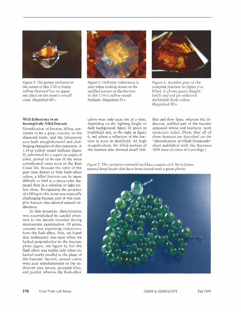
Figure 4. The garnet inclusion in Figure 5. Ordinary iridescence is the center of this 5.10-ct Fancy seen when looking down on the yellow diamond has no w a r - unfilled portion of the fracture ent effect on the stone's overall in this 1.19-ct yellow round color. Magnified 40x. brilliant. Magnified 15x.
Figure 6, Another part of the complex fracture in figure 5 is filled; i t shows green (bright- field) and red (in reflected darkfield) flash colors. Magnified 20 x.
With Iridescence in an Incompletely Filled Fracture Identification of fracture filling con- tinues to be a great concern in the diamond trade, and the laboratory sees both straightforward and chal- lenging examples of this treatment. A 1.19-ct yellow round brilliant (figure 5), submitted for a report on origin of color, proved to be one of the more complicated cases seen in the East Coast lab. Because the color of the gem may distort or hide flash-effect colors, a filled fracture can be more difficult to find in a fancy-color dia- mond than in a colorless to light-yel- low stone. Recognizing the presence of a filling in this stone was especially challenging because part of this com- plex fracture also showed natural iri- descence.
In this situation, identification was accomplished by careful atten- tion to the details revealed during microscopic examination. Of prime concern was separating iridescence from the flash effect. First, we found that iridescence was seen when we loolzed perpendicular to the fracture plane (again, see figure 5), but the flash effect was visible only when we loolzed nearly parallel to the plane of the fracture. Second, several colors were seen simultaneously in the iri- descent area (green, greenish blue, and purple], whereas the flash-effect
Gem Trade Lab Notes
colors were only seen one at a time, depending on the lighting (bright or dark background; figure 6): green in brightfield and, at the right in figure 6, red where a reflection of the frac- ture is seen in darlzfield. At high magnification, the filled portion of the fracture also showed small bub-
bles and flow lines, whereas the iri- descent, unfilled part of the fracture appeared white and feathery, with moderate relief. (Note that all of these features are described on the 'Identification of Filled Diamonds" chart published with the Summer 1995 issue of Gems &> Gemology.)
Figure 7. This imitation emerald necklace consists of 5.10-16.0 m m natural beryl beads that have been coated with a green plastic.
GEMS & GEMOLOGY Fall 1995

In keeping with GIA GTL poli- cy, we did not determine either color origin or a color grade for this stone. Instead, we issued an identification report only, with a comment in the conclusion that disclosed that the diamond had a foreign material in surface-reaching fractures. IR
Imitation EMERALD Plastic-Coated Beryl
When staff members in the East Coast lab saw the broken necklace of variegated translucent-to-transparent green beads shown in figure 7, our first inlpression was that we were looking at an emerald necklace with unusually symnletrical beads. The results of our first test-exposing the necklace to long-wave UV radia- tion-did not in~mediately arouse suspicion, as the chalky yellow reac- tion would be expected of heavily oiled emeralds. The dull reddish appearance through the Chelsea color filter also was not out of the ordinary. However, thorough examination of the beads with magnification gave cause for alarm: Around each of the drill holes, we noted a dark green ring that contained flattened gas bubbles. Figure 8 shows these features in a bead that our client kindly allowed us to remove from the strand for fur- ther testing. Examination of the loose bead readily revealed a transparent
Figure 8. With 10 x magni- fication, a green coating with flattened gas bubbles can be seen on one bead that was removed from the strand shown in figure 7.
Gem Trade Lab Notes
green coating. When some of the coating was removed and tested sepa- rately, it appeared red under the color filter and fluoresced chalky yellow when exposed to long-wave UV radi- ation. Its elastic nature and reaction to the "hot point" identified the coat- ing as a plastic. With the coating completely removed, the bead appeared light green in color, and the optical and physical properties matched those of natural beryl. Therefore, the necklace was identi- fied as green plastic-coated natural beryl. GRC
Figure 9. A small chip on the jadeite portion of this assemblage reveals how very thin the jadeite shell is. Magnified 25x.
JADEITE JADE
Assemblages From time to time, the laboratory receives for identification items that are later identified as assemblages. Among the less-common assembled stones we have reported on in this section are a spinel-synthetic ruby doublet (Winter, 1984, p. 2311, a star sapphire-sapphire doublet (Fall 1985, p, 171)) a ruby doublet (Spring 1987, pp. 47-48), and assembled cultured blister pearls (Summer 1991, p. 11 1).
On rare occasions, we encounter assembled stones that have been fab- ricated from jadeite (see, e.g., the jadeite doublets described in the Spring 1958 and Fall 1986 Lab Notes sections, pp. 134 and 172, respective- ly]. We have also seen, in an unassembled state, examples of a jadeite assemblage that consisted of three pieces of translucent, near-col- orless jadeite: (1) a cabochon that was inserted into (2) a hollow dome and cemented to (3) a flat base. The thin space between the hollow dome and the cabochon is filled with a green, jelly-like substance that gives the assembled stone the color of fine green jadeite. Such stones are diffi- cult to identify if they are mounted in such a way that the contact zone between the hollow cabochon and the flat back cannot be examined directly.
In spring 1995, the West Coast lab received for identification a translucent mottled-green carving
that measured about 33.33 mm long x 22.03 mm wide. The dent11 could not be determined-and the back could not be examined-because the carving was mounted in a closed- back yellow-metal pendant. A spot R.I. reading gave a value of 1.66, and examination with a desk-model
' prism spectroscope revealed a series of "chrome lines" in the red end of the spectrum. These properties are consistent with natural-color jadeite. However, n~agnification revealed that the carving was actually com- posed of a thin, hollow shell of jadeite that had been filled with a transparent, colorless substance. The filler contained some small gas bub- bles, a few of which could be seen through the top of the carving. We also noted remnants of the filler on the surface of the carving, at various points around the circun~ference, and it was exposed at one place-where the thin shell had a small chip (figure 9). When probed with a sharp metal pin, the filler was easily indented. FTIR spectroscopy of the filler revealed strong absorptions similar to those of some nolvmers known to
A ,
have been used in the impregnation of jadeite. Using a millimeter gauge, we estimated the thickness of the jadeite shell at the edge of the carving to be only 0.05 to 0.10 mm. On the basis of this examination, we identi- fied the item as an assemblage con- sisting of a thin, hollow, natural- color jadcite jade carving filled with colorless plastic.
GEMS & GEMOLOGY

Figure 11. The thin edge of the Figure 12, When examined from hollowed-out carving shown in above using magnification and figure 10 was visible between bright fiber-optic illumination, the mounting and the bottom of gas bubbles can be seen in the the stone. The plastic filler filler where it contacts the (which is actually colorless, but jadeite shell. Magnified 16 x. appears green in this photo because of the jadeite shell) can be seen under the edge of the jadeite, following the contours
Figure 10. This 25.05 x 18-72 mm of the carving. Magnified 13x. mounted carving proved to be an assemblage of jadeite jade and plastic. -
Soon after identifying this assem- blage, the West Coast lab received a similar-appearing mounted carving (25.05 x 18.72 mm; see figure 10) from another client. Gemological testing proved that this was also an assem- blage that consisted of a natural-color jadeite jade shell filled with colorless plastic (figure 11). This jadeite shell was estimated to be even thinner, with many gas bubbles in the filler. Several of these were seen at the con- tact of the jadeite and filler when the piece was examined from above using intense fiber-optic illumination (figure 12). Small openings in the back of the mounting enabled us to see through the plastic filler to the back of the hollowed-out carving. We were surprised to see that the back of the carving conformed exactly to the contours of the top. We do not know how these assemblages were manu- factured. Considering the extremely thin shell (so thin that i t actually fractured a t one prong), i t would seem to be a very exacting and diffi- cult process.
200 Gem Trade Lab Notes
These were the first such items we had encountered in the GIA Gem Trade Laboratory (an apparently simi- lar item-a carving of Guan Im-was recently examined by the AIGS labo- ratory in Bangkok, as reported in {ewelSiam, Vol. 6, No. 4, 1995, p. 110). However, we have previously
examined thin, dark-colored jadeite mounted in a closed-back setting that was highly polished on the inside to act like a mirror. This improved the apparent transparency and lightened the color to a pleasing tone. It is likely that the assemblages described in the preceding two paragraphs were carved from similar-perhaps even darker green-pieces of jadeite. The thin shell was then filled with plastic for added strength. Such dark green jadeite is often called Yunan jade. It
Figure 13. At first glance, this 62,26-ct glass imitation (50.10 x 26.30 x m) appearsA- L- -----
GEMS & GEMOLOGY Fall 1995

may be cut into thin pieces and used, for example, in drop earrings, where i t displays an attractive color in transmitted light. RCK and SFM
Finely Detailed Glass Imitation Over the years, both East and West Coast labs have examined a number of glass imitat ions of jade. These have included hololith bracelets and "carved" stones set in jewelry (see, Figure 15. These dyed feldspar e.g., Gem Trade Lab Notes, Fall 1982, scarabs, measuring 14.0 x 10.0 x pp. 171-1 72, Fall 1983, pp. 173-174). 5.3 m m each, were misrepre- In addition to these items, we have sented in the trade as lapis seen many other glass imitations of lazuli. carved and engraved jadeite at trade shows.
In spring 1995, the West Coast lab received for identification what at tile past we cllaracterized by fairly first appeared to be a 62.26-ct piece of coarse ,wai ls in rather low relief, carved jadeite (figure 13). Gemological This item, however, exhibited very testing, however, revealed properties fine (jeta il in high relief (figure 14). inconsistent with jadeite, including a The intricacy of the apparent carving 1.56 spot R.1.; a weak/ mottled green- contributed greatly to the effective- ish blue fluorescence to short-wave ness of ^ imitation, RCK UV radiation; and a spectrum show- ing general absorption from 400 to 480 n m and from 650 to 700 nm. Magnification revealed a number of Imitation LAPIS LAZULI
keyfeatures, including gas bubbles, flow lines, mold marks, fine surface cracks resembling crazing, and minute conchoidal fractures with a vitreous luster. On the basis of this examination, we identified the item as glass, a manufactured product.
All the glass imitations of carved jadeite that we had encountered in
The pair of blue scarabs shown in fig- ure 15 were submitted to the East Coast lab in October 1994. They had been offered in t h e trade a s lapis lazuli, a gem mater ia l t h a t they superficially resembled. With lox magnification (figure 161, the material looked more like sodalite, except that the color distribution was "wrongI1' with concentrations of blue color in
Figure 14. At 45x magnification, the unusually fine detail of the item in figure 13 is evident-as is the fact that i t was molded, not carved.
the fractures that proved that the color was produced by dye. More test- ing, including X-ray diffraction analy- sis, proved that the material was dyed feldspar. I t remains to be seen whether this represents an attempt to develop a commercial product, or it was simply a "one-shot" experiment.
GRC
Figure 16. Magnification of one of the scarabs in figure 15 reveals dye concentrations in the fractures, which are respon- sible for the lapis-like color. Magnified 1 Ox.
gem materials. Among the better known and more convincing of these
+ are the plastic imitation that shows a true play-of-color and the glass imita- tion known as "Slocum stone."
However, phenomenal opal is not the only opal used as a gem inate- rial. Other types include a translu- cent-to-opaque blue material from Peru that can closely resemble chal- cedony colored by chrysocolla (see, for exanlple, the Summer 1991 Gem News section, pp. 120-121) and vari- ous transparent to semi-transparent types. The latter include colorless 'water" opal and the yellow, red, or
Figure 17. The 3.55-ct oval orange modified brilliant (about 12.01 x 9.79 x 6.39 m m ) on the left is actually glass. The 2.35-ct triangular step cut is a natural opal from Mexico.
OPAL
Glass Imitation "Mexican"
So-called "precious" opal-opal dis- playing play-of-color-has been imi- tated by a number of manufactured
Gem Trade Lab Notes GEMS & GEMOLOGY Fall 1995

orange-to-brown material that is known in the trade as "fire," "cher- ry," or "Mexican" opal.
In the summer of 1995, the West Coast laboratory was asked to exam- ine two faceted stones, a 3.55-ct transparent orange oval modified bril- liant (figure 17) and a 2.21-ct trans- parent red emerald cut. These report- edly came from a parcel of stones that our client had purchased as opal in Mexico. However, subsequent gemological testing of samples by the client revealed properties that were outside the reported ranges for opal. GIA GTL determined the following properties on the two samples we tested (those for the red sample are given first): R.1,-1.522, 1,480; S.G. (determined hydrostatically)-2.64, 2.39; polariscope reaction-singly refractive, anomalously doubly refractive with snake-like bands noted; UV fluorescence-both were inert to long-wave UV radiation and fluoresced a faint, chalky yellow to short-wave UV. Magnification revealed small gas bubbles in both stones.
While the R.I. and S.G. values of the red specimen were well above the
general values reported in the gemo- logical literature for opal, those of the orange stone-which had the more convincing appearance, with minute inclusions that could easily have been overlooked without careful microscopic examination-were only slightly higher than those reported for the upper ranges of opal. However, the type of opal from Mexico that these two stones resembled typically has below-average values, that is, an R.I. of 1.44 or lower and an S.G. of about 2.00-2.10 (see R. T. Liddicoat, Handbook of Gem Identification, 12th ed., 1987, p. 280). On the basis of the information gathered, we identi- fied both stones as glass, a manufac- tured product.
For research purposes, we exam- ined these specimens further using Fourier-Transform infrared (FTIR) spectroscopy and EDXRF analysis. For comparison, we also tested a ref- erence sample of opal from Mexico that had a very similar appearance (again, see figure 17). The natural opal revealed many features in its FTlR spectrum, including: a sharp peak at 5253 cin-1; a broader peak centered at 5100 cm-1; a less intense
Figure 18, These approximately 9.4-mm beads make convincing replicas of black pearls.
pair of peaks at 45 15 cm-1 (sharp) and 4415 cm-1 (broad), which are not found in all opals; a shoulder at about 3950 cm-1; and strong absorption from 3735 cm-1 to 600 cm-1. In con- trast, the spectra of the two glass samples were very similar to each other, but very different from the nat- ural stone: They showed a very small, broad absorption peak at 4490 cm-1, a broad plateau with an absorp- tion edge at 3558 cni-1 and an absorp- tion peak a t 2775 cm-1, and very strong absorption from about 2250 cm-1 to 600 cm-1. (Synthetic opals have FTIR spectra different from either of these.)
The EDXRF data were also quite interesting. The reference natural Mexican opal contained Si, I<, Ca, Mn, and Fe. [It is believed that the color of these opals is due to disseini- nated iron oxide/hydroxicle particles; see, e.g., E. Fritsch and G. R. Rossman, "An Update on Color in Gems. Part 3," Gems o) Gemology, Summer 1988, pp. 81-102). In the redder of the two glass samples, we found additional elements, notably Se. Subsequent exan~ination with high-energy X-rays of additional orange and red glass samples with similar gemological properties showed that Cd is present in such glasses as well. Many red and orange glasses owe their color to the pres- ence of cadmium selenide or cadmi- um sulfo-selenide; these glasses are con~monly referred to as "selenium glass" (see, for instance, C . A. Hampel, Ed., Encyclopedia of the Chemical Elements, Reinhold, New York, 1968, p. 632). These glasses can mimic the colors and diaphaneity of "cherry" and "fire" opals wi th remarkable success.
ML/, RCK, and SFM
^ Black PEARL Imitations
F wearls (averaeine about 9.4 n1n1 in r
- - - 9 - - - - diameter; figure 18) arrived a t the
Gem Trade Lab Notes GEMS &. GEMOLOGY Fall 1995
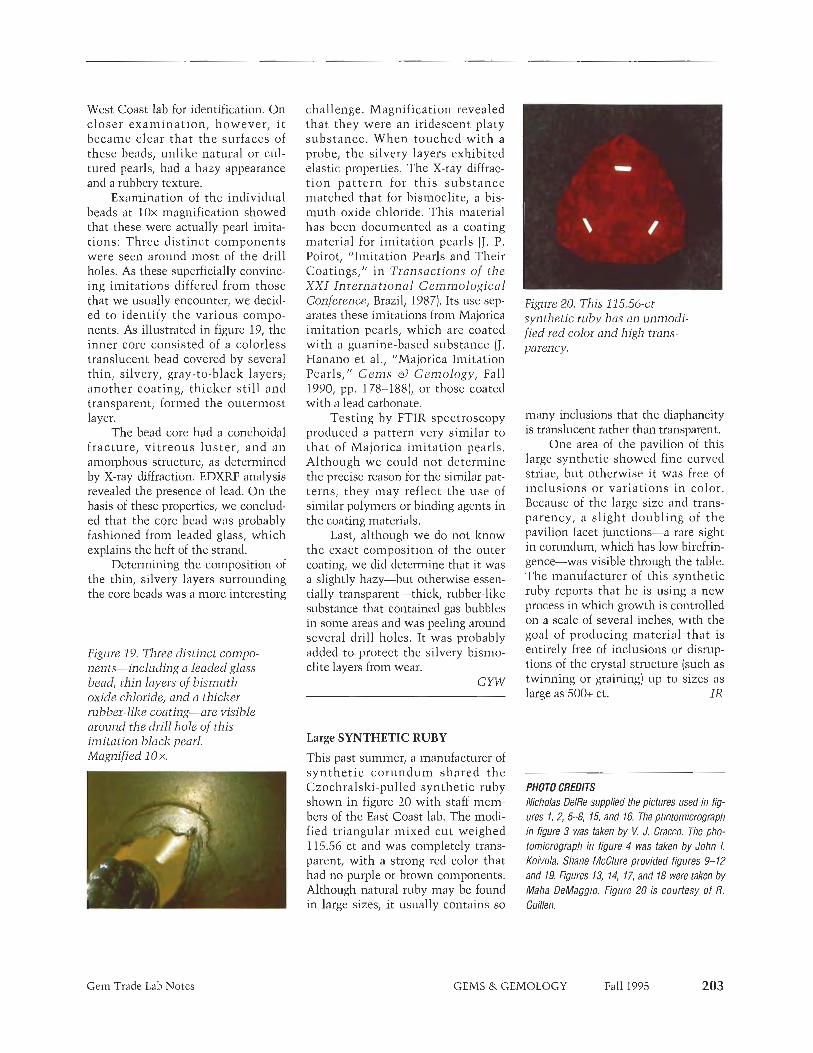
West Coast lab for identification. On closer examinat ion, however, i t became clear that the surfaces of these beads, unlike natural or cul- tured pearls, had a hazy appearance and a rubbery texture.
Examination of the individual beads at lox magnification showed that these were actually pearl imita- tions: Three distinct components were seen around most of the drill holes. As these superficially convinc- ing imitations differed from those that we usually encounter, we decid- ed to identify the various compo- nents. As illustrated in figure 19, the inner core consisted of a colorless translucent bead covered by several thin, silvery, gray-to-black layers; another coating, thicker still and transparent, formed the outermost layer.
The bead core had a conchoidal fracture, v i t reous luster , and an amorphous structure, as determined by X-ray diffraction. EDXRE analysis revealed the presence of lead. On the basis of these properties, we conclud- ed that the core bead was probably fashioned from leaded glass, which explains the heft of the strand.
Determining the composition of the thin, silvery layers surrounding the core beads was a more interesting
Figure 19. Three distinct compo nents-including a leaded glass bead, thin layers of bismuth oxide chloride, and a thicker rubber-like coating-are visible around the drill hole of this imitation black pearl. Magnified 10 x.
Gem Trade Lab Notes
challenge. Magnification revealed that they were an iridescent platy substance. When touched wi th a probe, the silvery layers exhibited elastic properties. The X-ray diffrac- t ion pat tern for th i s substance matched that for bismoclite, a bis- muth oxide chloride. This material has been documented as a coating material for imitation pearls (J . P. Poirot, "Imitation Pearls and Their Coatings," in Transactions of the XXI International Gemmological Conference, Brazil, 1987). Its use sep- ardtes these imitations from Majorica imitation pearls, which are coated with a guanine-based substance IJ. Hanano e t al., "Majorica Imitation Pearls," G e m s o) Gemology , Fall 1990, pp. 178-188), or those coated with :I lead carbonate.
Testing by FTIR spectroscopy produced a pattern very similar to that of Majorica imitation pearls. Although we could not determine - the precise reason for the similar pat- terns, they may reflect the use of similar polymers or binding agents in the coating materials. -
Last, although we do not know the exact composition of the outer coating, we did determine that it was a slightly hazy-but otherwise essen- tially transparent-thick, rubber-like substance that contained gas bubbles in some areas and was peeling around several drill holes. It was probably added to protect the silvery bismo- elite layers from wear.
CYW
Large SYNTHETIC RUBY
This past summer, a manufacturer of syn the t i c co rundum shared the Czochralski-pulled synthetic ruby shown in figure 20 with staff mem- bers of the East Coast lab. The modi- fied triangular mixed cut weighed 115.56 ct and was completely trans- parent, with a strong red color that had no purple or brown components. Although natural ruby may be found in large sizes, it usually contains so
Figure 20. This 115.56-ct synthetic nzby has an unmodi fied red color and high trans- parency.
many inclusions that the diaphaneity is translucent rather than transparent,
One area of the pavilion of this large synthetic showed fine curved striae, but otherwise i t was free of inclusions or variations i n color. Because of the large size and trans- parency, a slight doubling of the pavilion facet junctions-a rare sight in corundum, which has low birefrin- gence-was visible through the table. The manufacturer of this synthetic ruby reports that he is using a new process in which growth is controlled on a scale of several inches, with the goal of producing material that is entirely free of inclusions or disrup- tions of the crystal structure (such as twinning or graining) up to sizes as large as 500+ ct. IR
-
PHOTO CREDITS Nicholas DeIRe supplied the pictures used i11 fig- ores 1, 2, 5-8, 15, and 16. The photomicrograph in figure 3 was taken by V. J. Cracco. The pho- tomicrograph in figure 4 was taken by John I. Koivula. Shane McClure provided figures 9-12 and 19. Figures 13, 14, 17, and 18 were taken by Maha DeMaggio. Figure 20 is courtesy of R. Guillen.
G E M S & GEMOLOGY Fall 1995

GEM Robert C. Kammerling, John 1. Koivula, Mary L, Johnson, and Emmanuel Fritsch
Contributing Editors Dino DeGhionno, GIA GTL, Santa Monica, CA Henry A. Hanni, SSEF, Basel, Switzerland Karl Schmetzer, Petershausen, Germany
Diamond with mobile diamond inclusion. Some of the most interesting gemstone inclusions are those that con- tain one or more mobile components. Among these natu- ral kinetic works of art are inclusions that change appear- ance when heat is applied. An example is a two-phase, carbon dioxide fluid inclusion in quartz that homogenizes to a single phase when a thermal reaction tester is brought near. Sometimes, just the heat from a micro- scope's incandescent light is enough to cause this change. Other inclusions change their appearance when a stone is merely repositioned in the tweezers or stoneholder, as with three-phase inclusions in emeralds.
Inclusions with visibly mobile phases are rare in dia- mond. So two of the editors (RCK and JIK] were delighted at the opportunity to briefly examine a flat, twinned dia- mond crystal (with one polished octahedral face) that con- tained a movable diamond inclusion [figure 1). We learned from the owner that the diamond was previously exam- ined in detail by one of the contributing editors (HAH); the results of his investigation follow.
The slightly brownish gray diamond weighed 1.09 ct and measured 7.23 mm long x 3.03 mm deep. It was transparent, except in the area of the cavity containing the movable inclusion, where it was semi-transparent.
The diamond crystal was twinned after the spinel law (i.e., it was a made), and the large cavity roughly duplicat- ed the external contours of the host. The mobile crystal inclusion within that cavity was triangular (almost equi- lateral) and indented along its horizontal plane of symme- try (the twin plane); trigons were observed on its surface, and it was less transparent than the host but of a similar color. UV-visible absorption spectrometry revealed that the crystal inclusion is a diamond with the same type-la characteristics as its host (although the inclusion's absorption was about 2.5 times stronger).
On the basis of this examination, we feel that the mobile diamond inclusion resulted from a growth stage earli- er than that of the hollow host. Although we could not iden- tify the material that originally filled the cavity, it probably was a gas that escaped through a fissure during cutting of the octahedral face. The height of the cavity, about 1.7 rnm, did not permit the diamond inclusion to tumble freely, although it did move easily in two dimensions.
Update on Russian diamond mining. While in Russia to attend the Sixth International Kimberlite Conference, in Novosibirsk, GIA Directors James E. Shigley and D. Vincent Manson visited lumberlite pipes in the Siberian
204 Gem News
Figure 1. Note the move- ment of this triangular diamond inclusion in a large cavity when its host 1.09-ct made is turned 180'. Photos by John I. Koivula.
GEMS & GEMOLOGY Fall 1995

Figure 2. Visitors are dwarfed by machinery inside the Udachna ya diamond-processing plant, in the Siberian Sakha (Yakutia) Republic of the Russian Federation. Photo by James E. Shgley.
Sakha (Yakutia] Republic, the source of 95% of Russia's diamond wealth. They provided the following report on the current status of diamond mining in this region.
There was almost no activity at two of the sites. Flooding had forced the closure o f the International pit. The quarry-like Zarnitza prospect (site of the first Siberian diamond find in 1954) was also inactive.
The other pipes visited-the Mir, Udachnaya, Aikhal, and Jubilee-were active to some degree. All were open-pit operations.
The Mir pit, also plagued by flooding, showed lim- ited evidence of recent mining. However, ore was being trucked from nearby stockpiles, presumably headed for processing.
Udachnaya seemed to be the most productive of the sites. The large, six-story processing plant there appeared to be in full operation (figure 2). It serviced not only that giant pit, but also some ore trucked in from the Aikhal and Jubilee sites, about 80 lzm (50 miles) away.
No information was provided on production fig- ures at any of the sites, nor were visitors given any but the vaguest details on day-to-day operations. However, Udachnaya officials did show visitors about 20 color- less, gem-quality diamonds, which ranged from approximately one-half to 20 carats.
During the GIA team's visit to this remote region near the Arctic Circle, daylight lasted from 3 a.m. until midnight; the daytime high temperature was a balmy 80° (26OC), a far cry from the endless nights and harsh weather that miners must endure during winter months'. It was interesting to learn that winter mining at Udachnaya ceases when temperatures drop below about -50° (-4S°C] not because miners cannot cope with the cold, but because an inversion layer forms at that temperature, trapping potentially deadly methane gas in the deep pit.
Spy ship looks for diamonds. Namco, the Namibian Minerals Corporation, has chartered a 2,200-ton vessel- formerly a Soviet spy ship-for use in its offshore dia- mond operations, according to the July 3, 1995, issue of Diamond Intelligence Briefs. The ship will be used for evaluations and bulk samplings in Hottentots Bay and in the mouth of the Olifants River.
COLORED STONES
Cat's-eye apatites from Madagascar. A Spring 1995 Gem News entry (p. 60) described large faceted blue-green apatites from Madagascar, which are sometimes called 'neon" or "Paraiba" apatites. This year, we also saw a Figure 3. Note the pronounced eye and unusual few large cat's-eye apatites from this same locality. One color of this 11.45-ct blue-green apatite cabochon, such stone was an 11.45-ct cabochon (figure 3), loaned to reportedly from Madagascar. Short, barely visible GIA by Thomas M. Schneider, of San Diego, California. white needles cause the chatoyancy (inset). Photo Magnification revealed the cause of the chatoyancy-a by Shane F. McChue; photomicrograph by John. I. multitude of tiny, white-appearing, reflecting, short acicu- Koivulu, magnified 50x.
Gem News GEMS & GEMOLOGY Fall 1995

Figure 4. Often sold as Eilat stone from Israel (left, 5.40 ct) is this similar-appearing material from Peru (right, 5.41 ct). Photo by Maha DeMaggio.
lar "fibers" (figure 3 [inset]). Many longer, needle-like black inclusions (oriented in the same direction) were also present, but they seemed too sparse to significantly con- tribute to the chatoyancy. Although cat's-eye apatite is not uncommon, stones of this color are very unusual.
"Eilat stone" from Israel and Peru. The trade name Eilat stone is given to a blue-to-green ornamental rock with a mottled appearance that contains variable amounts of copper minerals. According to Webster's Gems (5th ed., Butterworth-Heinemann, 1994), typical constituents are chrysocolla, turquoise, and pseudomalachite (a copper phosphate mineral). The material is named for its original source, reportedly King Solomon's copper mines, not far from the Israeli Red Sea port of Eilat.
This gem material has been seen in the trade for many years, and was readily available at the February Tucson shows for at least the past several years. However, one knowledgeable dealer said that much of the material being sold as Eilat stone is not from Israel. Instead, simi- lar-appearing material, recovered as a by-product of copper mining in Peru, is being misrepresented as Eilat stone, that is, as coming from the Israeli source. An Israeli gem dealer confirmed this, adding that material was being exported from South America to Israel and then re-export- ed as Eilat stone. Two representative pieces-one from Israel and one from Peru-are shown in figure 4.
Because it is often necessary to characterize the min- erals in ornamental gem materials such as these-which are essentially inhomogeneous rocks~we frequently need advanced testing for their identification. It would be diffi- cult, if not impossible, for most gemologists to separate the two materials using standard gem-testing methods.
Six-rayed star emerald. Recently, a 10.03-ct round, cabo- chon-cut emerald was submitted by M. P. Steinbach, of Gindorf, Germany, to one of the contributing editors (KS) for examination. The emerald had a fairly well-defined
206 Gem News
star (figure 5) and was said to originate from a Brazilian emerald mine.
The stone had strong pleochroismf with an intense blue-green color parallel, and a light yellowish green color perpendicular, to the c-axis. This pleochroism was due to a distinct "aquamarine" (that is, iron-bearing) component, which was found in the absorption spectrum of the gem- stone in addition to the well-known chromium absorp- tion bands of emerald.
Microscopic examination showed channel-like inclu- sions that were oriented parallel to the c-axis. However, the star was actually caused by three series of as-yet- unidentified small, elongated particles with an orienta- tion perpendicular to the c-axis.
Although cat's-eye emeralds from different localities are occasionally found in the trade, star emeralds are exceptionally rare. The Spring 1995 Gem News section contains an entry on cat's-eye and star emeralds (pp. 60-61), including a 5.93-ct stone reportedly from Brazil's Santa Terezinha de Goias mining area. Because the aqua- marine component in the absorption spectrum of the 10.03-ct stone is typical of emeralds from Santa Terezinha, this stone may also come from that locality.
Faceted genthelvite: A gemological first. The editors were recently provided for examination-by C. D. (Dee) Parsons, of Santa Paula, California-a small faceted gem-
Figure 5. This unusual 10.03-ct star emerald revealed features consistent with emeralds from Santa Terezinha, Brazil. Photo 0 GIA and Tino Hammid.
GEMS & GEMOLOGY Fall 1995

stone that proved to be most unusual. The transparent purplish red-brown stone (figure 6) weighed just 0.33 ct and measured 4.38 x 3.57 x 2.19 nim. The only internal features in this modified rectangular step-cut were a series of mirror-like fractures, most of which were con- centrated along one edge. It had been sold to Mr. Parsons as "danalite" by a Brazilian dealer.
We first subjected the stone to the standard battery of gemological tests, with the following results: R.I. of 1.742; singly refractive; S.G. (by the hydrostatic method) of 3.68-3.70: inert to both long- and short-wave UV radia- tion. A most unusual feature was that it showed a typical almandine-rliodolite visible-light absorption spectrum (see, e.g., R. T. Lidthcoat, Handbook of Gem Identifica- tion, 12th ed., 19871.
A search of the gemological literature uncovered nothing on the mineral danalite, and the properties attributed to danalite in mineralogical texts did not match those that had been obtained from the faceted stone. Since danalite, Fe4Be3(Si04)3S, forms a solid solu- tion with helvite, Mn4Be3(Si04)3S, and genthelvite, Zn4Be3(Si04)3S, we decided to compare the properties of this stone witli those reported for other end-members of the helvite mineral group. A virtually perfect match was obtained for genthelvite.
To confirm the identity, we tested the stone with both X-ray'powder diffraction analysis and energy disper- sive X-ray fluorescence (EDXRF) spectrometry. X-ray diffraction analysis showed that the stone was in the helvite group, while EDXRF revealed a significant amount of zinc present, in addition to the expected sili- con and sulfur. Lesser amounts of manganese and iron were also detected. The greater amount of zinc, coupled with the R.I. and S.G. values, identified this stone as gen-
Figure 6. This 0.33-ct genthelvite is the first faceted example of this mineral [hat the editors have encountered. Photo by Maha DeMaggio,
Gem News
Figure 7. The 71.02-ct (39.33 x 16.88 x8.04 m m ) l~az~sn~annite cabochon (right) was cutfrom material similar to the accompanying 37.87-ct tetragonal hausmannite crystal. Photo @ GIA and Tino Hammid..
thelvite. (Beryllium [Be] and oxygen [O] cannot be detect- ed by EDXRF. ]
Because all the gemological properties shown by this stone fell within the range reported for pyrope-almandine mixtures, it would be in~possible for a gemologist, armed witli only the traditional series of gem-testing instru- merits, to identify this stone as genthelvite. X-ray diffrac- tion and chemical analyses would also be needed.
'Drusy" hausmannite. In recent years, we have noted the trade's increased interest in gems whose surfaces are not polished smooth, but are left naturally crystalline. When the size of these crystals is measured in hundredths to tenths of millimeters, die surface sparkles. This effect is seen in the so-called drusy gem materials, including uvarovite matrix, cobaltian calcite and dolomite, and quartz druses over chrysocolla and psilomelane (see, for instance, the Spring 1991 and 1994 Gem News sections, pp. 49 and 53, respectively).
Gary Abbott, of Abbott Gems, Roswell, Georgia, has been cutting "drusy" cabochons of larger-grained materi- als, including the black opaque manganese oxides haus- mannite (figure 7) and manganite. His hausmamite cabo- chons can take on an imposing, architectural appearance. One editor (MLJ) acquired a 71.02-ct drusy cabochon from Mr. Abbott for examination. Because of the nature of the material, we were able to obtain only a few gemological properties, as follows: R.1.-over the limits of a standard refractometer (greater than 1.81); no absorption features noted with a desk-model spectroscope; no magnetism.
GEMS & GEMOLOGY Fall 1995

Specific gravity was not determined due to the roughness of the surface (too many air bubbles remained trapped between crystals to permit a meaningful determination). We saw no pleochroism with polarized reflected light, although in reflected light the edges of polished crystal faces looked brownish black whereas the rectangular cores looked darker and "purer" black. An X-ray powder diffraction pattern showed only lines for hausmannite.
Hausmannite from the Kalahari manganese field in South Africa occurs as crusts of tetragonal (pseudo-octa- hedral) crystals growing from the matrix in near-parallel orientation. Cut one way (as in figure 7), the crystals show square cross-sections; cut another way, they form parallel triangles. As was the case with our sample, Mr. Abbott usually backs the pieces with a lighter material to provide rigidity.
Costa Rican jade museum. On a trip to Costa Rica late last year, one of the editors (RCK) visited the Marco Fidel Tristin Castro Jade Museum in the capital, San Jose. Located on the top floor of the Instituto Nacional de Seguros (National Insurance Institute), this small, but very well-organized, facility displays hundreds of pre- Columbian jadeite carvings. A few are back-lit to show their translucency and richness of color.
In pre-Columbian times, the area encompassed by what is now the Republic of Costa Rica was home to a major lapidary industry, according to displays within the museum and the museum guidebook (C. Balser, Ed., Jade Piecolombiano de Costa Rica, 3rd ed., Instituto Nacional de Seguros, San Jose, Costa Rica, 1993). Jade first became a regional commodity around 800 B.C., when groups of Olmecs arrived from the north in search of the prized "blue jade," described in the museum catalog as a blue- green diopside jadeite. (However, no source of jadeite has been found in Costa Rica; most is believed to have come from nearby Guatemala.) Many Olmec lapidary tech- niques and motifs~especially anthropomorphic axe-gods (typically about 10-12 cm high)-are seen in the carvings discovered in Costa Rica. Regional lapidary activity peaked in the period from aroimd 100 B.C. to 500 A.D.
Other jade-like materials were also worked by the native lapidaries. Some (including green-gray talc, ste- atite, serpentine, and green jasper) are also displayed in the museum.
Green onal from Serbia. In addition to the well-known play-of-color and relatively transparent "jelly" opals, we occasionally see nonphenomenal opals of lower diaphane- ity that are valued for their unusual body colors. Such materials include green opal from East Africa (see, e.g., the article by J. I. Koivula and C. W. Fryer in Gems o) Gemology, Winter 1984, pp. 226-227) and pink opal from Peru (Winter 1991 Gem News, pp. 259-260).
At a major gem show earlier this year, the editors came across some rough material labeled "crysopal." According to the vendor, it came from southern Serbia, near the
Gem News
Figure 8. This 21.08-ct cabochon and the 64-gram piece of rough opal from which i t was cut are reportedly from Serbia. Photo by Maha DeMaggio,
Macedonian border, and first entered the market by way of Sweden in 1992. By early 1995, however, it was no longer coming out of Serbia because of the armed con- flicts in that area.
Anders Karlsson, of Rox Arcana, Carmel, California, subsequently donated a rough sample to GIA, from which was cut a 21.08-ct pear-shaped cabochon (figure 8). The translucent cabochon was a pale, slightly bluish green (Munsell 7.5G 8/51, with a lightly mottled color distribu- tion, an aggregate optic character, and a negative color-fil- ter reaction (that is, it appeared green through the Chelsea filter). The spot R.I. was 1.46; S.G., measured hydrostati- cally, was 2.10, but measurement was complicated by the fact that the stone was porous, soaking up water as weights were taken. The cabochon was inert to short- wave W radiation, and fluoresced an even, faint chalky green to long-wave UV. The spectrum seen through a handheld spectroscope showed cut-off edges at 450 and 620 nm. Magnification revealed some healed fractures and veining. Qualitative EDXRF spectrometry showed major Si and minor Ni (nickel, probably the cause of color), K, Ca, V, and Zn.
The gemological properties recorded for this Serbian material are comparable to those of Tanzanian green opal, described in the previously mentioned article by Koivula and Fryer. Green nickel-bearing opal has also been found in Poland, Australia, and Peru.
"Deep" quartz from Africa. Just when we thought that we had heard every name for quartz, one of the editors saw a new one-"deep" quartz. Specimens of some otherwise ordinary loolung rock crystal quartz were being offered as 'deep" quartz by one enterprising dealer at the February Tucson shows. His explanation for the name: The speci-
GEMS & GEMOLOGY Fall 1995

Figure 9. The Eastern Desert of Egypt was given as the source of this 27.94-ct green quartz scarab. Photo by Maha DcMaggio.
rnens came from "deep" in die Earth-one to one-and-a- half miles below its surface, to be exact, in the Vaal Reefs mine at Orkney, South Africa.
Egyptian green quartz. During a visit to Egypt, one of the editors (RCK) purchased in the town of Luxor a number of gem materials from Egyptian localities. Although the ven- dor, a local jeweler and avid gem and mineral collector, could identify most of the materials in question and their geographic origins, he had little information about some of them.
One such ornamental gem was an opaque, medium- dark green material that the vendor had purchased from Bedouin tribespeople. They reportedly collected it some-
Figure 10. This 18.07-ct tablet (18.25 x 16.35 x 6.25 mm) is quartz with inclusions of hematite or a similar material. Photo by Maha DeMaggio.
where in the Eastern Desert. The vendor indicated that, while it superficially resembled some feldspars and ser- pentines from the same general area, this green stone was harder to fashion, requiring more time to cut. A 27.94-ct mottled green scarab fashioned from this material was purchased for examination (figure 91.
Standard gen~ological testing produced: a 1.54 spot R.I.; a faint yellowish green luminescence to long-wave UV radiation, and no reaction to short-wave UV; a vague absorption band at 670 nm, as noted through a desk- model prism spectroscope; and a 2.64 S.G., determined hydrostatically. Magnification revealed a pseudo-fibrous structure. X-ray powder diffraction analysis produced a match with a standard reference pattern for quartz.
EDXRF analysis revealed, in addition to the expected silicon, a number of other elements. Among these was a fairly large concentration of chromium, which was the most likely cause of the green color. Chromium, howev- er, is virtually unknown as a trace-element chromophore in quartz. This fact, combined with the abundance of other elements, indicated that finely disseminated, chromium-colored mineral inclusions produced the over- all green color of this gem.
Quartz with "hematite" inclusions. In the Spring 1995 Gem News section, we reported on "strawberry" quartz colored by inclusions of goethite and lepidocrocite (pp. 63-64). In July 1995, Michael Randall, of Gem Reflections, San Anseln~o, California provided two sam- ples of a similar material for our examination. This "span- gled" quartz (see, e.g., figure lo), reportedly from Madagascar, was colored by bright orangy red flakes of an iron-bearing material, probably thin plates of hematite (figure 11).
The most obvious features seen with magnification were curving transparent orangy red platy inclusions. Randomly oriented throughout the cabochon, they were
Figure 11. The hematite-like features in the quartz tablet shown in figure 10 appeal as thin, curving orangy red plates when viewed with l o x magnift- cation. Photomicrograph by John I . IZoivt11~1,
Gem News GEMS i3 GEMOLOGY Fall 1995

Figure 12. This 10-strand necklace (about 100 grams total weight) is made up ofapproximately 3,800 beads of resin that are said to come from western Africa. Although commonly sold as "myrrh," it probably is not the resin of the relatively rare Commiphora myrrha. Photo by Maha DeMaggio.
concentrated in one layer parallel to a rhombohedra1 face in a rough sample that we also examined. The morpholo- gy of these plates was unusual. Each grew outward from one corner in an elongated fashion (as the host quartz crystal grew), sometimes pinching and re-swelling to form an "hourglass" or "cello" shape (one such plate is visible in figure 11). Also seen were radial crystal aggregates that appeared white to pale green.
Aromatic resin necklace: A form of myrrh? Some natural plant materials are recognized as gems. The most notable example of plant-produced gem materials is the fossil resin amber. Modem resins such as copal are also used for jewelry purposes. Another resin sometimes used as an ornamental material is the yellow-brown material sold as "myrrh."
One of the editors (RCK) recently acquired a multi- strand necklace of this material (figure 12) in order to characterize its gemological properties. The 100-gram necklace was composed of ten 36-inch (90 c111) strands with about 380 button-shaped beads, 2.5 to 5 m m in diameter, per strand. Gemological testing of a few beads separated from one strand revealed the following proper- ties: color-yellowish brown to brown; diaphaneity- translucent; color distribution-even; aggregate reaction in the polariscope ; R.1.-1.41 (spot); S.G.-1.27 (measured hydrostatically); fluorescence-even chalky moderate yel- low to long-wave UV, even chalky weak yellow to short- wave UV; phosphorescence-none; absorption spec- trumÑ430-n cutoff (that is, a typical "brown" spec-
Gem News
trum]. With magnification, individual beads showed a mottled appearance, with brown speckles. This material melts easily when approached with a thermal reaction tester ("hot point"), scratches with a fingernail (Mohs hardness below 2.51, and has a granular fracture and a characteristic sweet, spicy odor. The hardness is so low that "myrrh" beads can be polished by rubbing them against a sheet of paper.
Fourier-Transform infrared (FTIR) spectroscopy of one bead revealed broad absorption peaks at 5180, 4778, and 4000 cm-1, and two sharp peaks at 4339 and 4264 cm-1; this is distinct from the spectrum of the harder resin amber (see, e.g., "Amber Simulant: Natural Resin in Plastic," in the Summer 1995 Gem News, pp. 134-136).
Myrrh has a long and illustrious history. The actual substance called myrrh is a gum resin produced by Commiphora (Balsamodendron) species, especially Commiphora myrrha. In 1770, Dr. John Cook wrote The Natural History of Lac, Amber, and Myrrh (etc.), which detailed the medicinal properties of this substance (quot- ed by H. Fraquet, Amber, Butterworths, London, 1987). L. S. Dubin, in The History of Beads, from 30,000 B.C. to the Present (Abrams, New York, 1987, pp. 90-911, inen- tions the late medieval practice of wearing myrrh-filled pomanders as rosary beads, in the hopes of warding off the plague. Possibly the most famous use of myrrh was, with gold and frankincense, as a gift from the three Wisemen to the newborn Jesus (Matthew 2:ll). As an aside, the "murrhina" or "m~irrhine" that Pliny mentioned in his Natural History was not myrrh, but probably banded flu- orite or chalcedony (see, e.g., J. Sinlzanlzas, Gemology: An Annoted Bibliography, Scarecrow Press, Matuchen, NJ, 1993, entries 952, 1461, 5004, 5615, 5747, 6834).
Other "organic" materials made into scented beads include: sandalwood, cloves, "California Flower Beads" of the 1910's to 1920's (probably dyed and scented plaster), and beads made from a paste of aloe wood, cloves, musk, ambergris, flower petals, and aromatic leaves (P. Francis, Beads of the World, Schdfer Publishing, Atglen, PA, 1994, p. 37). Francis (op. cit.) notes that the material commonly called myrrh-in our opinion, probably including this necklace-is the resin of a tree or shrub different from the somewhat rare C. myrrh. He adds that it is "shaped into beads, colored and given various forms in Western Africa."
Update on Mong Hsu ruby. Smaller rubies-under 1 ct- from Mong Hsu, Myanmar [Burma), were plentiful at this year's Tucson shows, but there seemed to be fewer stones above 1 ct than the previous year. Echoing this observa- tion was a dealer with Gemstone International, Seattle, Washington. He had difficulty, he said, in locating good Mong Hsu stones in the 2-ct range on a trip to Bangkok in late 1994, although such stones were fairly easy to find the year before.
However, one feature of heat-treated Mong Hsu rubies is threatening their distribution altogether. The
GEMS & GEMOLOGY Fall 1995

Fig-lie 13, These three rubies (0.83-1.18 ct) are from the Cowee Valley, North Carolina. Photo by Maha DeMaggio,
rubies are typically packed in a flux, such as boron, for heat treatment; some of this flux evidently reacts during the high-temperature treatment to form a colorless glassy solid that fills some surface-reaching fractures in the rubies. (Peretti et al. speculated that this material might be an aluminum borate; see "Rubies from Mong Hsu," Gems ed Gemology, Spring 1995, pp. 2-26.) Stones with such fillings are not acceptable to some ruby customers: Buyers in Japan and Europe have returned them to their suppliers. Thailand's gem traders and exporters recently agreed to halt sales of treated gems from Chanthaburi, which is where the Mong Hsu rubies are heated (see "Thailand Halts the Export of All Treated Gemstones," Diamond Intelligence Briefs, March 19, 1995, p. 1227).
Rubies and sapphires from North Carolina. GIA researchers recently examined a suite of rough and faceted rubies and sapphires from the Cowee Valley, Macon County, North Carolina (figure 13). These samples were loaned to GIA by Robin Dinnes, of Cowee Valley Lapidary, Franklin, North Carolina. One of the better- known occurrences of gem corundum in the United States (for a description, see Gemstones of North America, by J. Sinkanlzas, Van Nostrand Reuihold, 1975, pp. 50-53), this locality has been mined on a small scale for gem material and mineral specimens for the past 65 years. The gem corundum occurs as crystals embedded in either blue or red clays, along with other alluvial minerals such as rhodolite and pyrope garnet, rutile, and silliinanite.
The suite that we examined consisted of 10 faceted stones, 11 cabochons, and some rough material. Two thirds were sapphires, and the remainder were rubies. The largest faceted sapphire was 2.65 ct, and the largest faceted ruby was 1.35 ct.
Most of the faceted rubies were semi-transparent (even cloudy) because of numerous fractures. Most of these fractures were partially healed, and appeared as veils
Gem News
Figwe 14. Inclusions of brownish red mills are evi- dent in this ruby from the Cowee Valley, North Carolina. Photomicrograph by John I. Koivula; magnified 25x.
or "fingerprints." Rubies found in the red clays were usu- ally stained yellow-brown (presumably by iron oxides) on their surfaces and along fractures, In addition, the samples contained prominent grains of brownish red rutile inclu- sions (figure 14), as well as tiny, intersecting needles. All of the rubies were color zoned, with irregular-shaped areas that appeared either more purplish red or more orangy red (possibly due to the previously mentioned staining). The gemological properties of this material were consistent with those of rubies in general. EDXRF analysis of three samples revealed the presence of Cr, Fe, Ti, Ca, and Ga- all elements often found in natural rubies.
The faceted sapphires were similar to the rubies in their rutile and needle-like inclusions, and in their semi- transparent or translucent appearance. In some cases, cabochon-cut sapphires exhibited weak asterism.
This was one of the first opportunites we have had to examine material known to be from this locality.
Purple scapolite from Tajikistan. The importance of Russia and other republics of the former Soviet Union as sources of gemstones, both natural and synthetic, contin- ues to be reinforced at major gem shows. In addition to those materials mentioned in the Spring and Summer Gem News columns, we have recently seen faceted pur- ple scapolite, which is being marketed by the Russian Colored Stone Company, of Golden, Colorado. The scapo- l i te is reportedly a new find in Tajikistan's Pamir Mountains, at about 5,000 n~ elevation-so high that miners must wear oxygen masks while they work, according to company representative Alexey Eremin.
Mr. Eremin loaned us a 1.79-ct (7.40-7.50 x 6.83 mm) round modified brilliant (figure 15) for examination. Gemological properties determined were: color-purple; diaphaneity-transparent; pleochroism-strong, purple and near colorless; optic character-uniaxial; R.1.-1.539 and 1.550; birefringenceÑ0.011 specific gravity (deter-
GEMS & GEMOLOGY Fall 1995

Figure 15. Tajikistan's Pamir Mountains are the source of this 1.79-ct scapolite. Photo by Maha DeMaggio.
mined hydrostatically)-2.61; fluorescence-inert to long- wave UV and a very weak pinkish orange reaction to short-wave UV; absorption spectrum-very faint absorption line at 600 nin. With magnification, a few needle-like crystals (one broken into a stringer] were visible in the stone.
Gem scapolite is a mineral series between the two end-members marialite, Na4(A13SioOT4)Cl, and meionite, Ca4(AI&i6OT4)CO3. Using refractive indices of the speci-
men stone, and comparison data in Deer, Howie and Zussman's Introduction to Rock Forming Minerals (Longman Group, London, 1974, p. 387)) we determined its con~position-assuming that it contained only these two end-members-to be about 81% marialite and 19% meioni te.
Large spinel from Tajikistan. Spinel is another gem material being recovered from this former Soviet republic in Central Asia. We know of one particularly exceptional stone, a 532-ct piece of rough spinel, that was fashioned into a number of gems. The largest of these gems weighed 146.43 ct and was named the "Katherina" by the purchas- er of the rough, the Netherlands-based ACT International (see "Spectacular Spinel," by P. Bancroft, Lapidary loiunal, February 1990, p. 25).
The Katherina was subsequently recut to 123.14 ct (E. Caplan, pers. comm., 1995) and the refashioned stone loaned to GIA for examination. Although recut, it retained its basic step-cut cushion shape (figure 16). The stone exhibited properties consistent with natural spinel from other localities. Described as "virtually flawless" in the Lapidary lournal article, the stone revealed a small plane of what appeared to be octahedral negative crystals near the culet when examined with magnification. These sub- stantiate the stone's natural origin. EDXRF chemical anal- ysis by GIA Research showed only a tiny amount of zinc (Znl, which is consistent with Zn concentrations that . ,,
have been documented in other spinels from this locality.
Color-change zircons. Many color-change gemstones have been described in the gemological literature, but not until Scottish gemologist Alan Hodgkinson informed us of a
Figure 16. This 123.14-ct spinel was cut from a 532- ct piece of rough that came from Tajikistan's Pamir Mountains. Courtesy of Evan Caplan, Los Angeles; photo by Shane F. McClure.
212 Gem News GEMS & GEMOLOGY Fall 1995

Figure 17. Opticon, or a similar synthetic resin, was used to impregnate this 17.19-ct fibrous- appearing malachite. Photo by Maha DeMaggjo.
2.25-ct stone that turned blue in day [or fluorescent) light and green in incandescent light had we ever heard of a color-change zircon. Mr. Hodglzinson forwarded the fol- lowing report of his examination of this stone, which was loaned to him by Keith Mitchell .
The refractive indices were 1.923 and 1.985, with a birefringence of 0.062. The specific gravity was 4.68. When the stone was examined in incandescent light, the color of the ordinary ray was "olive" green and that of the extraordinary ray, greenish blue. Through the Chelsea filter, a grayish pink color was evident. There was no reaction to long- or short-wave UV: A Geiger counter detected slight radioactivity [with 1,3 counts per second above baclzground), which is typical for green zir- cons. In the handheld spectroscope, the stone displayed a strong uranium spectrum. EDXRF analysis showed zir- conium and silicon to be the two major components. Also present was hafnium-which commonly substi- tutes for zirconium in nature-and uranium. The urani- um concentration was estimated to be 1.55 wt.% U02.
ENHANCEMENTS
Impregnated malachite. Although polymer-impregnated jadeite has received the most attention in the trade and technical press lately, other gem materials are also simi- larly treated. These include porous turquoise and a chalky-appearing opal from Piaui, Brazil.
At a major gem show this year, the editors found some unusual-appearing malachite. The vendor inain- tained that i t came from Morenci, Arizona, an area known as a source of turquoise and other copper-miner- a1 gems. What first caught our attention was the materi- al's orientation: It had been cut to show its fibrous tex- ture along the length of the cabochons [figure 17). The material also exhibited both unusually high luster and depth of color.
Gem News
A sample cabochon was purchased for gemological investigation. R.I. testing on the flat base revealed a 1.58 reading with no birefringence blink. Spot R.I. testing on different areas of the top produced either vague readings in the high 1.50s or a very weak birefrigence blink in the range expected for malachite (about 1.66 to over-the-liin- its). Specific gravity, determined by hydrostatic weighing, gave a value of 3.76; although low for compact malachite, it was within the range reported in the gemological litera- ture for this gem species. The stone was inert to short- wave UV. Long-wave UV caused the top of the stone to fluoresce a very weak, mottled chalky blue; a similar but stronger reaction was seen from the base of the stone. With magnification, we noted a transparent, colorless sub- stance that formed a layer on the base and partially filled a large cavity. This substance was most apparent in the areas that reacted to long-wave UV. Infrared spectroscopy revealed absorption features similar to those that we have documented for the epoxy resin Opticon.
Another vendor selling similar-appearing materi- al-Joe Jehlks, of Horizon Mineral Company, Houston, Texas-said that he used Opticon resin to treat Morenci malachite and described how he did it. The rough material was first heated in a toaster oven to roughly 150°-260 for a few hours. It was then removed from the oven, coated with Opticon, and returned to the oven for more heating [typically overnight). The material was then slabbed and the pro- cess repeated on the individual slabs: heating, coating with Opticon, then heating again. This sequence was repeated after preforming and through the pre-polish- ing stage. By the time the material was ready for its final polish, it was thoroughly impregnated, so that further Opticon treatment was not necessary. Mr. Jehlks maintained that the chemical catalyst-that is, the polymerizing agent-provided with Opticon was not necessary in this treatment process. The multiple low-temperature heatings alone were sufficient to harden the synthetic resin.
SYNTHETICS AND SIMULANTS - Unusual color-zoned synthetic amethyst. While visit- ing the firm of Pinky Trading in Bangkok, one of the editors (RCK) saw a synthetic amethyst crystal from Russia with exceptionally uneven-"speckled" or "leopard spotu-color distribution. Although a dis- tinct, patchy color distribution is typical for natural amethyst, zoning in synthetic amethyst is usually very subtle (see, for example, figure 12 in "A Simple Procedure t o Separate Natural from Synthet ic Amethyst on the Basis of Twinning," by R . Crowningshield et al., Gems es) Gemology, Fall 1986, pp. 130-139).
The Russian crystal (figure 18) was obtained for examination. The typical crystal morphology of quartz explains its unusual appearance. Quartz is a mineral that
GEMS & GEMOLOGY Fall 1995

tain entries on Czochralslzi-pulled synthetic sapphires seen at the 1993 and 1994 Tucson shows. All the colors
Figure 18. Color zoning is very apparent in this 167-gram piece of synthetic amethyst manufac- tured in Russia. Photo by Maha DeMaggio.
appears to have hexagonal (six-sided] symmetry, but it really is trigonal, or symmetric to three-fold rotations. Crystals typically have six pyramidal faces coming to a common apex. These are called rhombohedra1 faces, which appear alike at first glance, but actually occur in two alternating sets: positive (r) and negative (z) faces. In natural and synthetic quartz, color-causing impurities concentrate selectively in different sectors, or regions where certain faces grow; in particular, the darker purple color in natural amethyst-and the purple zones in ametrine-usually develop in sectors under the positive r faces (see, e.g., F. Hassan, "Amethyst," Mineralogical Record, Vol. 3, No. 5, 1972, pp. 221-225; P. M. Vascon- celos et al., "The Anahi Arnetrine Mine, Bolivia," Gems &> Gemology, Spring 1994, pp. 4-23).
Because the Russian specimen grew on a seed plane cut perpendicular to the c-axis, it looks very different from natural crystals in shape. Instead of one set each of r and z faces, the Russian synthetic has multiple parallel sets of these faces, in three orientations each. Microscopic observation of the color zoning in the Russian specimen demonstrated that the darker-color regions (r faces) grew as expanding rhombic pyramids from points inside the crystal (figure 19).
The editors are not aware of any similar "speckled" or "leopard spot" color zoning in natural amethyst from any documented locality. Therefore, the presence of such zoning should be considered indicative of synthetic origin.
More on Czochraski "pulled" synthetic sapphires. The Spring 1993 and Summer 1994 Gem News sections con-
214 Gem News
mentioned in those entries-includmg blue-were again seen at the 1995 shows. According to one vendor of this material, Gerry Manning of Manning International, New York, the pulled synthetic blue sapphire crystals are unevenly colored. The first-grown portions of the rod- shaped crystals are lightest in color; the rods become increasingly darker toward the opposite end (the last part to crystallize). The color zoning described by Mr. Manning differs from that seen in split boules of flame- fusion synthetic blue sapphire. There, the color is typical- ly lightest in the center and progressively darker toward the periphery.
Mr. Manning provided two faceted blue specimens (figure 20) for gemological characterization. Properties determined were: R.1.-np = 1.760; no = 1.768; birefrin- genceÑ0.008 S.G. (determined hydrostatically)Ñ4.0 to 4.01. Both specimens were inert to long-wave UV and fluoresced a weak, chalky bluish green to short-wave UV. All these properties are similar to those of flame-fusion synthetic blue sapphires. We also noted weak red fluores- cence to intense transmitted light from a fiber-optic light source, and the samples appeared faintly pink through the Chelsea color filter. These latter two reactions have been noted in some violetish blue flame-fusion synthetic sao- phires that contain traces of chromium, as well as in some natural blue-to-violet sapphires from Sri Lanka that also contain traces of chromium.
Magnification revealed minute gas bubbles in both samples. Unlike flame-fusion synthetic sapphires, howev- er, although the color of our samples did not appear totally uniform we could not resolve curved color banding using
Figure 19. Magnification reveals that the color of the synthetic amethyst in figure 18 formed as expanding rhombohedra1 wedges in the crystal. This color zoning is unlike anything we have seen in natural amethyst. Photomicrograph by John I. Koivula; magnified 4x.
GEMS & GEMOLOGY Fall 1995

Figure 20. The Czochralski method was used to produce these two synthetic sapphires (1.64 and 1.76 ct). Photo by Maha DeMaggio.
diffused transmitted light and magnification. Only when this lighting technique was combined with immersion of the samples in methylene iodide could we see wide, slight- ly curved blue banding, which is proof of synthesis.
Tanzanite-colored synthetic sapphire. As tanza~ute con- tinues to be one of the more commercially important col- ored stones, materials that resemble tanzanite are increas- ingly in demand. In the Spring 1995 Gem News section (p. 62)) we reported on iolite (the mineral cordicrite), which has been promoted as a less-expensive alternative to tanzanite. Another recently promoted alternative is violet-blue synthetic sapphire, marketed under such trademarked names as "Cortanite" and "Coranite." International Colored Gemstone Association (ICA) Lab Alert No. 83, by Mrs. Shyamala Femandes, stated that this material has been sold in India as tanzanite.
Recently, we examined five samples of this synthetic sapphire, ranging from 0.97 to 4.07 ct (see, e.g., figure 21).
Four were obtained from Gemstones International, of Sayreville, New Jersey; the fifth was marketed by Lannyte Corp., of Houston, Texas.
Gemological properties were determined as follows: color-violetish blue to violet; diaphaneity-transparent; pleochroisn~~rnoderate, violet (ordinary ray) to light blue, light grayish blue, or light greenish blue [extraordi- nary ray]; optic character-uniaxial negative; Chelsea fil- ter reaction-none to weak red; R.1.-nE = 1.760 to 1.761 and no = 1.769; birefringenceÑ0.00 to 0.009; S .G.4 .00 to 4.02; fluorescence-inert to long-wave UV, even, chalky weak-to-moderate bluish green to blue-green to short-wave UV; very faint to weak red transmission; absorption spectrum-none (four samples), diffuse 560-620 band and faint 690 line (darkest sample). All five samples had obvious curved color banding (figure 22) and pinpoint inclusions that are most likely gas bubbles (typi-
Figure 22. Curved color banding is obvious in this synthetic sapphire being marketed as a tanzanite substitute. The material is also strongly color zoned, with darker crowns and lighter pavilions, Photomicrograph by John I. Koivula; immersion, magnified 3 x.
Figure 21. These two synthetic sapphires (2.00 ct and 4.07 ct) are typical of material being sold as a ~anzanite simulant. Photo by Maha DeMaggio. cal of Vemeuil synthetic sapphires); the curved banding
was also obvious when viewed with magnification and short-wave UV as a radiation source.
EDXRF spectroscopy revealed, in addition to major aluminum, trace amounts of iron, titanium, and-in the darkest-colored stone (from Lannyte)-0.003 wt.% Cr as CroOi.
There were at least two possible explanations for the atypically violet hues of these synthetics: the orientation of the faceted samples with regard to the optic axis of the material, and their chromium content. All five samples were strongly color zoned, with darker crowns and near- colorless culets (again, see figure 22). The optic axes of the five were oriented at 45O, 50-60° 55-60 80-85O, and 90° respectively, to the table (that is, the optic axis ori-
Gem News GEMS & GEMOLOGY Fall 1995
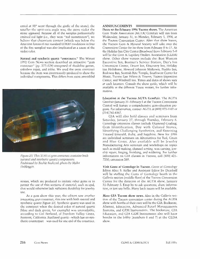
ented at 90Â went through the girdle of the stone); the smaller the optic-axis angle was, the more violet the stone appeared. Because all of the samples preferentially emitted red light (i.e., they were "red transmitters"), we believe that chromium content (which was below the detection limits of our standard EDXRF conditions in four of the five samples) was also implicated as a cause of the violet color.
Natural and synthetic quartz "constructs." The Winter 1992 Gem News section described an attractive "gem construct" (pp. 277-278) con~posed of rhodolite garnet, colorless topaz, and iolite. We used the term construct because the item was intentionally produced to show the individual components. This differs from most assembled
Figure 23. This 33.83-ct gem construct contains both natural and synthetic quartz components. Fashioned by Bashir Beelaad; photo by Moho DeMaggio.
stones, which are produced to imitate other gems or to permit the use of thin sections of material, such as opal, that would otherwise lack sufficient durability for jewelry use.
At a gem show this year, the editors saw another interesting gem construct, this one with both natural and synthetic quartz (figure 23). Synthetic quartz was used in the construct when the desired color of natural quartz (blue and dark green, for example) was unavailable, according to Gul Beelzzad, of Freedom Valley Gems, Fren~ont, California. Rutilated quartz-which has no syn- thetic counterpart-was used for one end of the construct.
216 Gem News
ANNOUNCEMENTS Dates set for February 1996 Tucson shows. The American Gem Trade Association (AGTA) GemFair will run from Wednesday, January 31, to Monday, February 5, 1996, at the Tucson Convention Center. After that show leaves, the Tucson Gem & Mineral Society will take over the Convention Center for its show from February 8 to 11. At the Holiday Inn City Center (Broadway) from February 2-9 will be die Gem & Lapidary Dealers Association (GLDA) show. Other show venues include the: Best Western Executive Inn, Boatner's Service Station, Day's Inn Convention Center, Desert Inn, Discovery Inn, Holiday Inn Holidome, Howard Johnson Midtown, Pueblo Inn, Rodeway Inn, Scottish Rite Temple, Southwest Center for Music, Tucson East Hilton & Towers, Tucson Exposition Center, and Windmill Inn. Times and dates of shows vary at each location. Consult the show guide, which will be available at the different Tuson venues, for further d o r - ma tion.
Education at the Tucson AGTA GemFair. The AGTA GemFair (January 3 1-February 5 at the Tucson Convention Center) will feature a comprehensive gem-education pro- gram. For information, contact AGTA at (800) 972-1 162 or (214) 742-4367.
CIA will also hold classes and seminars from Saturday, January 27, through Tuesday, February 6. Gemology extension classes include Diamond Grading, G e m Identif ication, Day wi th Colored Stones, Identifying Challenging Synthetics, and Detecting Treated Emerald, Ruby, and Sapphire. New for 1996 are individual seminars on Alternatives for Red, Green and Blue Gems . Also available wi l l be Jewelry Manufacturing Arts seminars and workshops on topics such as mold making, channel setting, wax carving, jew- elry repair, forging, finishing, and soldering. For further information on GIA classes in Tucson, call (800) 421- 7250, extension 269.
Visit Gems &> Gemology in Tucson. Gems &> Gemology Editor Alice S. Keller and Assistant Editor Irv Dierdorff will be staffing the Gems a) Gemology booth in the Galleria section (middle floor) of the Tucson Convention Center for the duration of the AGTA show, January 31-February 5. Drop by to ask questions, share informa- tion, or just say hello. Many back issues will be available.
More GIA Tucson show news. Also in the Galleria sec- tion of the Tucson convention center during the AGTA show with booths of their own will be the GIA: Bookstore, Alumni, Education, Advanced Retail Managements Systems, and GEM Instrunlents. The bookstore, GIA Education, and GIA GEM Instruments also will have booths in the lobby (numbers 6 and 7) at the GLDA show.
GEMS & GEMOLOGY Fall 1995

STANDARD CATALOG OF GEM VALUES, 2nd Edition By Anna M. Miller and John Sinlzankas, 271 pp., illus., publ. by Geoscience Press, Tucson, AZ, 1994. US$24. OO*
This update of Dr. Sinkanlzas' 1968 Van Nostrand's Standard Catalog of Gems carries through most of the same basic information in simple terms that nude the original a classic. It is an excellent introduction to the characteristics of value in gem rough, cut stones, engraved gems, gem carv- ings, and pearls. One of the first price guides ever published, it still covers areas that none of its current competi- tors have reached, especially in gem rough and uncommon stones.
Unfortunately, the editing and reproduction of the second edition are not of the same quality as the first. The frequent typographical errors, misspellings, partial sentences, and misdirected references-not seen in the first edition-detract from the book's readability. The same 15 blaclz- and-white photos were sharper 25 years ago, In addition, the 1994 vol- ume continues factual errors found in the original edition (e.g., De Beers rough diamond sales are still called "sightings") as well as some out-of- date information and locality names (e.g., Rhodesia, Peking). The updated section on faceted diamonds is espe- cially weak, both in facts and prices.
Prices quoted are identified as '1993 U.S. average retail prices," but the authors do not explain what con- stitutes retail markets for gem rough, loose stones, and the like. As with any price list, the usefulness of this one is limited by how broadly the material being valued is categorized. Vague descriptions with wide price ranges are almost never wrong, but they are of limited benefit to buyers, sellers, and appraisers. On the positive side, the price listings do give indications of the extremes of the markets for each variety, as described.
In this second edition, the strong content outweighs the disappointing publication quality. I look forward to a more polished third edition.
CHARLES I. CARMONA Guild Laboratories Inc. Los Angela, California
SUSAN B. JOHNSON AND JANA E, MIYAHIRA, EDITORS
OTHER BOOKS RECEIVED Diamond Exploration into the 21st Century, edited by W. L. Griffin, 367 pp., illus., publ. a s Vol. 53 of the Journal of Geochemical Exploration, Elsevier Science, Amsterdam, 1995, US$250. Diamond exploration is being feverishly conducted on all con- tinents except Antarctica; currently about US$400 million is spent annu- ally. This collection of 18 invited papers by authors from eight coun- tries gives an overview of modem &a- mond exploration research, tech- niques, and results. Four papers from Russia are particularly interesting, given the Russians' success in dia- mond exploration and the fact that their approaches developed differently because of their long isolation.
The volume is divided into four parts: (1) Backgro~u~d (5 papers) covers the formation of diamonds and the nature, abundance, location, and selected economic aspects of primary (pipe) deposits; (2) Area Selection (3 papers) discusses geologic controls and prediction of favorable areas for primary deposits; [3) Finding the Target ( 6 papers) considers proper methods for collecting indicator min- eral san~ples, laboratory processing, and geophysical exploration methods, as well as the basic principles of allu- vial diamond exploration; and (4) Priori t isat ion a n d Evaluation ( 4 papers) includes interpretation of the chemical compositions of indicator minerals, petrographic studies, and sta- tistical evaluation of several parameters (e.g., microdiamond counts) used to determine those pipes that are likely to be economic-recognizing that fewer than 1 % of all pipes discovered will be economic.
This book contains much newly released information on diamond exploration. Knowledgeable gemolo- gists will find the Background part particularly valuable.
A. A. LEVINSON University of Calgary
Calgary, Alberta, Canada
The New Alchemists: Breaking Through the Barriers of High Pressure, by Robert M. Hazen, xvi, 287 pp., illus., publ. by Smithsonian Press, Washington, DC, 1993. US$23.00. * Although the title may sound more like a chemical treatise than anything to do with gems, the "high pressure" is actually that needed to convert car- bon into diamond. In fact, this book is a valuable history of the long search for successful diamond synthesis.
The author provides an interest- ing, readable text, n~ostly written in the style of National Geographic magazine. It is based on his long expe- rience in high-pressure research in the
laboratory of the Carnegie Institute, Washington, DC. In the course of his work, he learned much about early experiments in high-pressure research -as by Percy W. Bridgman (1 882 -1961), Loring Coes Jr. (1915-19781, and o the r s~which ultimately led to the success of General Electric and ASEA, the Swedish equivalent of GE (ASEA actually produced synthetic diamond before GE, but, for some still unex- plained reason, according to Hazen, kept this fact secret). Hazen relates the official and personal struggles of these investigators in a very entertain- ing fashion, with the result that much anecdote leavens what otherwise might have been a dull text.
This treatise is the best baclz- ground source for the modem history of diamond synthesis. It is whole- heartedly recommended to all stu- dents of diamond history.
JOHN SINIWNKAS Perilithon Books
San Diego, California
This book is available for purchase through the GIA Bookstore, 1660 Stewart Street, Santa Monica, CA 90404. Telephone (800) 421 -7250, ext. 282; outside the U.S. (310) 829-299 1, exl. 282. Fax: (3 10) 449- 1 16 1.
Book Reviews GEMS & GEMOLOGY
A B S T R A C T S L
C. W. FRYER, EDITOR
REVIEW BOARD
Charles E. Ashbaugh Ill Isotope Products Laboratories Burbank, California
Andrew Christie GIA, Santa Monica
Jo Ellen Cole GIA, Santa Monica
Maha DeMaggio GIA, Santa Monica
Emmanuel Fritsch University of Nantes, France
Juli Cook-Golden Royal Oak, Michigan
Michael Gray Missoula, Montana
Patricia A. S. Gray Missoula, Montana
Professor R, A, Howie Royal Hollo way University of London United Kingdom
Mary L. Johnson GIA, Santa Monica
Robert C. Kammerling GIA Gem Trade Lab, Santa Monica
A. A. Levinson University of Calgary Calgary, Alberta, Canada
Loretta B. Loeb Visalia, California
Elise B. Misiorowski GIA, Santa Monica
Gary A. Roskin European Gemological Laboratory Los Angeles, California
Jana E. Miyahira GIA, Santa Monica
James E. Shigley GIA, Santa Monica
Christopher P. Smith Gubelin Gemmological Laboratory Lucerne, Switzerland
Carol M. Stockton Los Angeles, California
Rolf Tatje Duisburg University Duisburg, Germany
COLORED STONES AND ORGANIC MATERIALS Lapis. Vol. 19, No. 11, 1994, 72 pp. This jubilee (200th) issue-entirely in German-will sat- isfy all who crave to learn about the lore and adventure surrounding gems. W. Nesenzew describes the discovery of the Siberian diamond deposits in "Die russischen Diamanten," from his book Im Reich der Steine (In the Realm of Stones, Th~in/Frankf~~rt am Main, 1986). An extract from F. 1UeinJs book Sn~aragde unter dem Urwald (Emeralds Under the Jungle, Berlin, 1941) explores the risks of the Colombian emerald industry early in this cen- tury, the rediscovery of the Chivor mine, and the discov- ery of the 630-ct Patricia emerald (now in the collection of the American Museum of Natural History, New York). P. Voillot takes the reader on a "wild trip" to find the aquamarines of Hunza Valley ("Der wilde Weg zu den Aquamarinen"), just in time to see the opening of a rich pegmatite pocket. A. Weerth's "never-ending story" ["Lapis-Lazuli, die unendliche Geschichte") covers the entire history of lapis lazuli, surveys deposits worldwide, and describes the condition of Afghanistan's mines today. Also included are a comprehensive, up-to-date report on diamond mineralogy ("Steckbrief Diamant") and a description of an expedition that retraced part of Alexander von Humboldt's 1829 travels to Siberia. Many photos, both historical and contemporary, illustrate the issue. RT
Microsurfaces, a photo essay dedicated to Bill Hicks. J. I. Koiv~ila, Australian Gemmologist, Vol. 18, No. 10, May 1994, pp. 323-325.
A world-renowned photographer of inclusions, John I. Koivula offers 12 color photomicrographs as tribute to the late Bill Hicks, former editor in chief of Australian Gemmologist. These photomicrographs show spectacu- lar gemstone surface growth and etch features that often reflect internal-growth conditions. Mr. Koivula chal- lenges the view of those who believe that only mirror-flat surfaces on crystal faces represent perfection. Masterful lighting emphasizes the geometric three-dimensional beauty of these surface features. The photos and accom- panying article are a fitting tribute to a dedicated editor.
IEC
This section is designed to provide as complete a record as prac- tical of the recent literature on gems and gemology Articles are selected for abstracting solely at the discretion of the section edi- tor and his reviewers, and space limitations may require that we include only those articles that we feel will be of greatest interest to our readership.
Inquiries for reprints of articles abstracted must be addressed to the author or publisher of the original material.
The reviewer of each article is identified by his or her initials at the end of each abstract. Guest reviewers are identified by their full names. Opinions expressed in an abstract belong to the abstrac- ter and in no way reflect the position of Gems & Gemology or GIA.
0 1995 Gemological Institute of America
Gemological Abstracts GEMS & GEMOLOGY Fall 1995

Mineral industry surveys: Gemstones. U.S. Departn~ent of the Interior, Bureau of Mines, Annual Review 1994, 16 pp.
Updated annually, this survey summarizes world gem production and consumption statistics. A two-page sum- mary describes die nature of the study, with most of the information contained in the tables. These include: a four-page guide to the gemological properties and other information on selected gem materials, synthetic gein- stone production methods, value of domestic gemstone production (by gemstone), diamond prices, colored stone prices, diamond exports and re-exports (i.e., of imported diamonds faceted in the United States) to various coun- tries, U.S. imports for consumption of diamonds (by kind, weight, and country), U.S. imports for consumption of colored stones (by kind and country), U.S. imports of syn- thetic and imitation gemstones, summary table of U.S. imports (by variety), and world production statistics for diamonds (1990-1994). New this year is the inclusion of shell (as nuclei for cultured pearls) in the "Value of U.S. gen~stone production" statistics.
Interesting facts gleaned from these tables include: 1994 U.S. (natural) gemstone production rose to $22.2 million (M) from $19.5M in 1993 (excluding $33M in shell bead nuclei-about the same as 1993). The most importantgems in 1994 were corundum ($2.81M in 1994, up from $0.313M in 19931, gem feldspar ($2.62M in 1994, up from $0.701M in 1993), turquoise ($1.71M in 1994, down from $3.04M in 1993)) opal ($1.12M in 1994, up from $0.639M in 19931, and garnet ($0.78M in 1994, up from S0.233M in 1993). Production of some materials declined, notably tourmaline ($9.53M in 1993, $14,000 in 19941, peridot ($1.52M in 1993, $0.635M in 1994)) and agate ($1.41M in 1993, $0.234M in 19941.
In 1994, the U.S. imported more rough diamonds from the United Kingdom (482,000 ct, for $224.8M) than from any other country; more carats of unset faceted dia- monds < 0.5 ct each from India than from any other coun- try (6.15 Mct, for $1125.7M), and more carats of unset faceted diamonds over 0.5 ct each fro111 Israel (1.35 Mct, for $1570.3M). The U.S. imported the most carats of emeralds (2.31M) from India, but the greatest value of emeralds ($89.1M) from Colombia. The U.S. imported the most corundum gems-rubies and sapphires~from Thailand. The U.S. imported about $10M of synthetic stones each from Germany and Thailand, and $48.1M of imitation gemstones from Austria (home of Swarovsky). Total imports in 1994 were $5.73 billion in unset (rough and cut] diamonds, $227M in emeralds, $197M in corun- dum gems, nearly $29M in pearls, $4.63M in coral, $143.7M in other natural gemstones, $42.66M in syn- thetics, and $60.9M in imitation gems.
A few warnings: The gemstone guide contains some terms that the CIA Gem Trade Laboratory (for instance) does not use, including bixbite for red beryl, chrysolite for yellow or green or brown cluysoberyl (sometimes peridot is also called chrysolite), cairngorm for smoky quartz, and hiddenite for yellow to green spodumene. Also, 'Gemological Institute of America (GIA) color grades"
are NOT "rare white" for E color, or "traces of color" for G, H, or I (as was stated in the footnote to table 4 of this monograph). ML1
Notes from the Gem and Pearl Testing Laboratory Bahrain-4. A. Bubshait and N. B. Sturman, Journal of Gemmology, Vol. 24, No. 6, 1995, pp. 401-404.
The concentric structure of natural pearls (testing pearls is 95% of the lab's work) seen with X-radiography varies considerably, from pronounced to barely visible. The lat- ter occurs in pearls that probably formed over long peri- ods of time with stable environmental conditions, while pearls that grew in less stable conditions variously pro- duced more calcium carbonate or more conchiolin (the dark rings in X-radiographs).
The lab recently encountered an unusual green syn- thetic corundum that looked like an emerald and was ini- tially purported to be glass; its identification was straight- forward. A filled crackled Verneuil synthetic ruby exhib- ited white lines on an X-radiograph. The filling material contained numerous flattened bubbles and a surface lus- ter distinctly different from that of the host synthetic ruby. The source of these treated synthetic rubies is still unknown. Five "emeralds" offered at a suspiciously low price proved to be synthetic on the basis of their low R.I.'s, low S.G.'s, and characteristic inclusions. On the basis of these properties, four appeared to be Biron or Russian hydrothermal synthetics and one a Seiko syn- thetic emerald. CMS
Photogenic inclusions in moldavite. A. de Goutikre, Journal of Gemmology, Vol. 24, No. 6, 1995, pp. 415-419.
Tektites, including moldavite, are natural glasses that formed when meteorites struck the Earth. The samples illustrated here are from Czechoslovakia and Thailand, but tektites have been found in many localities around the world. Three types of inclusions in inoldavite- lechatelierite (pure silica glass), gas bubbles, and "Schlieren" (elongatecl, "treacly" inclusions)-are superbly illustrated in these photomicrographs. When all three are found together, they are indicative (although not conclusive) of southern Bohemia or southern Moravia as the locality of origin. CMS
Trace elements as colouring agents in jadeites. H. Harder, Journal of Gemmology, Vol. 24, No. 7, 1995, pp. 508-5 1 1.
The broad range of colors in which jadeite occurs- including white, lilac, brown, orange, blue, black, cind green-is caused by trace elements, the effects of which are summarized in this review article. Most common is iron. Its oxidation states are responsible for the range white-green-black. Chromium is responsible for the finest green colors, while nickel also may contribute to variations in green. Lilac, mauve, and lavender are due to varying proportions of manganese, cobalt, chromium, and iron. C M S
Gemological Abstracts GEMS & GEMOLOGY Fall 1995 219
1995 production keeps pace. National Jeweler, August 1, 1995, p. 24.
Despite expected decreases because of the long 1994 win- ter, this year's production of freshwater cultured pearls from the Tennessee River was as strong as the year before, according to the American Pearl Company. A production increase is expected for 1996 because pearl-farming oper- ations started early, in February 1995 (instead of late March), because of a mild winter.
The American Pearl Company has been culturing fancy-shaped pearls on the Kentucky Lake of the Tennessee River for about 20 years, during which demand for fancy shapes has steadily increased. Production of semi-round-to-round cultured freshwater pearls is limited but is expected to increase in the next three to four years. Natural pearl production, however, is down. Zebra inus- sels, brought from Europe on the hulls of ships, are suffo- cating those Tennessee River mussels that produce pearls and are used for nuclei production, Although the American Pearl Company no longer produces shell nuclei, "Not even the Japanese have been able to develop an alternative," according to American Pearl Company president Gina Latcndressc. The company is working with government agencies to reproduce endangered species on the Tennessee River. MD
DIAMONDS Argyle diamond output flat. Diamond Industry Week,
July 31, 1995, p. 6. The Argyle (AK1) open-pit diamond mine produced 0.5% fewer diamonds in the first half of 1995 than in the first half of 1994, despite processing more ore. (The figures are 19.044 million carats [Met] from 6.676 million metric tons of ore for the first six months of 1995, versus 19.144 Mct from 6.017 million metric tons of ore for the same period in 1994.) Diamonds are recovered from two types of ore, hard rock and alluvium, with 1995 yields of 4.15 ct per metric ton of hard ore recovered (down from 4.77 ct per metric ton in 1994), and 0.575 ct per metric ton of alluvial ore recovered (about the same as 1994).
The figures are "in line," according to the article, with expectations that AK1 surface-mine reserves will be exhausted by 2004. Mining may continue underground, however, and Argyle venture partners, CRA Ltd. and Ashton Mining Ltd., are expected to announce their plans in this regard by late 1995. M u
Diamonds. W. D. Hausel, International California Mining Journal, Vol. 64, No. 12, August 1995, pp. 28-3 1.
This article reviews a few properties of natural diamonds, especially those from diamond pipes on the Colorado- Wyoming border. Natural diamond crystals are typically octahedral, dodecahedral, or hexoctahedral in habit. Some inclusions in diamond can be used to date the dia- mond; "host rocks" are substantially younger than the diamonds themselves. Mineral inclusions in diamonds are of two petrologic types-peridotitic (P-type) or
eclogitic (E-type). P-types contain inclusions of olivine, orthopyroxene, clinopyroxene, pyrope garnet, chromite, diamond, graphite, and/or sulfides; E-type diamonds may contain sodium-rich clinopyroxene, almandite-pyrope garnet, rutile, lzyanite, corundum, coesite, graphite, and/or diamond.
Over 100 lzimberlite pipes have been found along the Colorado-Wyoming border by searching for eclogitic and peridotitic garnets. Eclogitic pyropes that occur with clia- monds are classified as "Group I" garnets. They typically contain high percentages of sodium and calcium but arc low in chromium. The Sloan 1 lzimberlite in Colorado is an eclogitic lzimberlite pipe. M u
Diamond fever dwindling in Australia. J. Rice, Mazal U'Bracha, Vol. 12, No. 67, May 1995, pp. 82-83.
Australian diamond exploration efforts are suffering eco- nomic setbacks as once-optimistic investors pull out of start-up mining and exploration companies. The firms were darlings of the international stock market a few years ago, when inexperienced investors poured in cash, hoping for lucrative discovcries. In 1995, even as the first promising exploration reports came out, fears of oversup- ply and unrestricted Russian production fueled the exo- dus from Australian diamond-mining stocks. The market also realized that small companies are too undercapital- ized to engage in serious diamond exploration.
Analyst David Russell, of Saw James Capel Ltd. (no city or country given), believes that investor fears of over- production are largely groundless and that problems of oversupply are temporary. AC
Diamond interest in Argentina. Mining Journal, London, July 21, 1995, p. 44.
One of the earliest recognized pieces of evidence in sup- port of plate tectonics (known as "continental drift" in early years of the theory's formation) was how the Atlantic Ocean coastlines of South America and Africa seemed to fit together like gigantic puzzle pieces. Part of those once interlocked pieces, now separated by the Atlantic, are South Africa and Argentina/Uruguay. Perhaps using this reasoning, an Australian company (Challenger Mining International Ltd.) and two Canadian companies (La Plata Gold and Rome Resources Corp.) have separately requested permission to explore for gold and diamonds in 50,000-hectare regions of Argentina's Buenos Aires Province. This province is thought to be geologically similar to mining regions of South Africa, according to provincial officials. ML1
Diamond sales for DFR. Mining Journal, London, July 21, 1995, p. 46.
Diamond Fields Resources (DFR) has sold at tender in Antwerp 6,636 carats of diamonds for C$1.1 million. These diamonds come from DFR's holdings in South Africa and Namibia. In South Africa, the Loxton Dal mine has recently been upgraded, with a new separation plant to improve diamond recovery. In Namibia, DFR
220 Gemological Abstracts GEMS & GEMOLOGY Fall 1995

marine geologists have confirmed a resource of over one million carats (with an average price of US$165 per carat), in certain parts of the concession at water depths between 30 and 80 nl. MLI
Exploration: BHP-DiaMet Update. Mining Journal, London, February 24, 1995, p. 144.
One of the still-promising Canadian diamond areas is the Northwest Territories Lac de Gras region. Diamond valua- tions have been completed for three pipes controlled by BHP-DiaMet. The Fox pipe was given a value of US$126 per carat (based on 2,060 carats at an ore grade of 0.27 carats per ton); the Panda pipe was valued at $132 per carat (based on 2,778 carats at a grade of 0.93 carats per ton); and the Misery pipe had a value of $44 per carat (based on 438 carats at a grade of 3.3 carats per ton). Draft environmen- tal impact statement guidelines have been published, and BHP-DiaMet is preparing a response. MLJ
Exploration in permafrost. J. Chadwick, Mining Magazine, Vol. 325, No. 8336, February 1995, pp. 103, 105.
Although this article is about a lead-zinc mine (the Polaris mine, on Little Cornwallis Island, Northwest Territories) in the Canadian Arctic, one suspects that pro- posed diamond-mining ventures in die region face simi- lar problems. Miners must cope with wind-chill factors pushing temperatures to -lOO° and very little precipita- tion (about the same as the Sahara Desert). Temperatures in the mine must be kept below freezing because the mine is completely contained in pernirifrost: Walls would "melt" if allowed to warm. Other problems include polar bears; the corrosive and costly drilling fluid (CaCl-bearing salt water); access to supplies (Europe is closer than southern Canada); and surveying problems, in a place where magnetic instrumentation does not work. In deep- er permafrost, gas emergencies are also possible. MLJ
In search of Russia's motherlode. S. Miller, Mazal U'Brcicha, Vol. 9, No. 64, January 1995, pp. 68-69.
Archangel Diamond Corporation (ADC) has accomplished what other mining companies only dream of: It obtained a license to explore the virtually untapped former Soviet Union's emerging Archangel diamond region, believed to be the second richest in the world, in ;i joint venture with the Russian Archangel State Geological Enterprise. ADC's 25-year license permits the exploration of major properties in northwest Russia near the border with Finland. Five lzimberlite pipes at one site (Lomonosov;i) are expected to yield 250 million carats, with a projected worth of about US$37 billion. Another site (Verldotina) has eight pipes, and as many as 10 more may be in the area. All diamonds recovered so far have been of "good quality." Geologists have drilled 24 holes hi a third property (Windy Ridge) and recovered diamonds fro111 all.
ADC Chief Executive Officer Andrew Malim down- played reports that diamond mining in the area is prohib- itively costly and difficult, citing comparatively easy site access by road, air, and-eventually-a railroad that is
now under construction. Because slurry mii~ing is used, kimberlite ore can be processed for as little as $7 a ton, compared to $15 a ton worldwide. AC
Limpopo diamond search. Minim Journal, London, August 11, 1995, p. 96.
South Africa has granted a prospecting permit to Duo Corp., allowing them to search for alluvial diamonds in Limpopo River gravels, in the South Africa/Zimbabwe border region. However, this 9,000-hectare area near Kruger National Park contains one of the last pristine forests of baobab trees, ;is well as sycamore figs, fever trees, and a rich arcl~eological site. Consequently, envi- ronmental groups oppose strip mining. MLJ
Majhgawan diamondiferous pipe, Madhya Pradesh, India-A review. A. K. Chatterjee and I<. S. Rao, Journal Geological Society of India, Vol. 45, No. 2, 1995, pp. 175-1 89.
The Majhgawan pipe, which is 15 lzm from the city of Panna in central India, was discovered in 1827, about 40 years before the South African pipes. Thus, it is the first diamond-bearing pipe (primary deposit) ever discovered. However, like other diamond deposits in India, it was considered alluvial (secondary) until 1930, when its pri- mary nature was recognized and it was reclassified as a kiinberlite.
Studies since 1988 have reclassified the pipe as a lainproite. This paper reviews the history of the Majhgawan pipe, primarily from a geologic perspective, with the objective of determining parameters useful in finding other diamond-bearing pipes in die region.
After detailed evaluation of the mineralogy, petrolo- gy, and chemistry of the rocks that comprise the pipe, the authors conclude that it has characteristics intermediate between those of kiinberlite and lamproite. In addition to geophysical surveys, heavy-mineral indicators could be used to find this type of pipe, including high-chromium garnet (G-9 type), high-magnesia chromite and spinel, and the absence of ilmenite.
This pipe has been India's main diamond producer in recent years. Current annual production averages about 18,000 carats; at this rate, reserves are sufficient to the middle of the next century. The ore grade is about 0.1 carat per metric ton, and diamonds average 0.45 ct in size. Gem diamonds (42% of production) arc typically of high quality and presently worth about US$200 per carat.
AAL
The origin, formation and emplacement of diamonds. M. Strather, Australian Gemmologist, Vol. 18, No. 11, 1994, pp. 342-345.
Intended for a nontechnical audience, this short review article addresses where diamonds come from, how they form, and how they arrive at the surface of the Earth. For the most part, it is a good summary of current knowledge on these subjects; however, some statements are not quite correct.
Gemological Abstracts GEMS & GEMOLOGY Fall 1995 221
There is a pressure-teinperat~~re regime~called the "Diamond Stability ZoneM-in the Earth's upper mantle in which diamonds nucleate and form. The carbon in dia- monds comes from two sources: homogenized "primor- dial" carbon that has been present since Earth's formation, and "recycled" carbon (from limestones, etc.) that has been subducted back into the Earth. As indicated by inclusions, there are two diamond types: (1) those formed in ultramaf- ic roclzs (peridotites, i.e., igneous roclzs of extremely silica- depleted composition, related to oceanic basalts); and (2) those formed in high-pressure metamorphic roclzs (eclog- ites). Peridotitic (ultramafic) diamonds contain primordial carbon; eclogitic diamonds contain recycled carbon.
Diamonds are brought to the Earth's surface by columns ("plumes") of magma rising from depths of about 130-180 km. There are two types of diamond-bearing igneous bodies: (1) lh~berlites~carbon-dioxide-rich mag- mas related to carbonatites, whch form "champagne flutet'-shaped bodies and may contain both peridotitic and eclogitic diamonds (as in, e.g., South Africa); and (2) lam- proites-allzali-rich magmas, which form "inargarita glass1'-shaped bodies and contain primarily eclogitic dia- monds (as in, e.g., Australia). Diamonds are generally older than their transporting magmas. Kimberlites rise suddenly: The most dramatic estimates of their emplacement ascent times have them climbing 135 I a n in four to 15 hours!
As for "less-than-correct" statements: The Earth is most probably about 4.5 billion years old (F. Press and R. Siever, Earth, 2nd ed., W. H. Freeman and Co., San Francisco, CA, 19781 not 4.70 billion, and the oldest "base- , . ment" roclzs known are 3.9-billion-year-old metamorphic roclzs in Greenland. It is somewhat misleading to say that "all the present continents (except Europe) have more than one craton." Cratons under both North America and Europe are composites, according to the 1992 Levinson et al. Gems &> Gemology article listed as one of Ms. Strather's references. If a craton must be 1.5 billion years old before a lzimberlite forms, this does not mean that most lzimberlites are older than 2.5 billion years. In addi- tion, magma pipes, as plumes, probably punch their way up through weaker regions in the crust and do not require pre-existing fractures extending down through the crust. The most serious oversight however, is the failure to ref- erence statements as they occur in the text.
The field of high-pressure petrology is large, lively, and constantly evolving. New chapters in the life-stories of diamonds are likely to surface at any geologic confer- ence. Despite the few inaccuracies, Ms. Strather is to be lauded for attempting to distill a large amount of raw data into such a compact article. MLl
Russian sales bring down De Beers earnings. International California Mining Journal, Vol. 64, No. 10, June 1995, p. 63.
De Beers's 1994 earnings were 7% lower than 1993 ($555 million versus $595 million). De Beers Chairman Julian Ogilvie Thompson said that Russian sales of polished and rough diamonds reduced sales by the Central Selling
Organisation (CSO) by 3%. De Beers and the CSO were trying to persuade Russian authorities to adhere to quotas.
MLI
Russian update. Diamond Industry Week, June 12, 1995, pp. 5-6.
Russia produced 19.72 million carats of diamonds in 1994, and sold 26.36 million carats of rough and cut stones, worth US$2.28 billion. (The discrepancy in these numbers is not as serious as it first appears; Russia exports diamonds to Israel and Belgium for processing, which are then imported back into Russia.) The Ydachnyi [Udachnaya] mine in Yalzutia, operated by Alinazy Rossii- Sakha (ARS), accounted for 18 million carats of 1994 Russian production; ARS was responsible for 98% of all diamonds produced in Russia.
As for sales, Russia wants its current quota of 26% of all CSO sales upped to 33%. However, De Beers claims that Russia already exported diamonds worth US$700- 800 million outside of the CSO agreement. Russia seeks to improve its market position by increasing domestic diamond polishing, but this also requires negotiations with the CSO on pricing policies. ML/
Sibeka anniversary. Mining Journal, London, June 16, 1995, p. 450.
For 50 years, the world's top diamond producer (by vol- ume) was Brussels-based Sibelza, which operated the mines of Mbujimaya and Lulua in Zaire. This short report profiles Sibelza, which celebrated its 75th anniversary in 1994. Currently, the company is active in four diamond- industry sectors: mining in Zaire (partial ownership of the Zaire mining company Miba); diamond research in the United States (Exmin) and China (partial ownership of Hunan Sibeka); synthetic diamond production (partial ownership of Megapode Holdings); and the manufacture of super-abrasive tools (Diamant Boart).
MibaJs diamond production (in Zaire) rose from 4.53 million carats (Mct) in 1993 to 4.88 Mct in 1994. Ten years ago, however, diamond exports from Zaire were around 20 Mct annually. Ore grades for alluvial deposits are falling, to 3.16 ct/m3; however, lzimberlite ore grades have risen to 2.49 ct/m3. Supply restrictions and "clan- destine operators" continue to cause problems.
The consumption of industrial diamonds has increased 13% per year for the last 20 years, and the world supply is around 700 Mct per year. Of this, only 60 Mct annually comes from "industrial grade" natural dia- monds, so more than 90% of industrial diamonds are syn- thetic. Consequently, Megapode reached record sales vol- umes last year, but prices for industrial diamonds have dropped due to excess supply. MLI
'Stealth' bucket excavates diamonds. Mining Magazine, Vol. 173, No. 1, July 1995, p. 6,
Diamond mining is rough on machinery. Extensive research into "aggressive" wear conditions at the Orapa and Letlhalzcine open-pit diamond mines in Botswana was used to lengthen the lifespan of wheel-loader buckets
222 Gemological Abstracts GEMS & GEMOLOGY Fall 1995

fivefold. Computer-assisted strain gauges and design led to modifications that include single-piece casting and relocated welds. MLJ
Striker finds kimberlite evidence. Mining Journal, London, July 14, 1995, p. 26.
Striker Resources NL has found evidence of more kiln- berlites at its Beta Creek project in the far north of Western Australia. Also, Striker is purchasing the adja- cent Forrest River tenements, which contain five &a- mond-bearing kimberlites. MLJ
United Reef diamond recovery proceeding on schedule. Diamond Industry Week, August 7, 1995, pp. 5-6.
On June 9, 1995, United Reef Ltd. opened a new diamond recovery plant on its Bamingui River diamond property in the Central African Republic. In June and July, 1,202 dia- monds (518.31 carats) were recovered from the river grav- els, including three "gem quality" stones of 6.24, 7.32, and 9.70 ct. The first sale of diamonds from the property is scheduled for September. MLJ
GEM LOCALITIES Angola's minerals. Miner attraction. The Economist, July
1, 1995, pp. 56, 58. After nearly three decades of civil war, Angola now appears to have a fragile peace. This presents great oppor- tunities for mineral exploration in general, and for dia- mond production in particular. At present, artisanal min- ing (in this case, illegal digging and smuggling) of good- quality alluvial diamonds is again prevalent; about US$20 million worth are now "exported" each week (an annual rate of approximately $1 billion).
New laws favorable to foreign investment (the state's monopoly on mineral rights has been abolished) are attracting the attention of diamond companies from sev- eral countries. These companies are more interested in the potential of the 600-odd known kimberlite pipes, as production from these is easier to protect and control than that from the more spread-out alluvial deposits.
A Brazilian-Russian-Angolan consortium is particu- larly interested in the Catoca pipe in Lunda Norte Province. When mining begins there (possibly by the end of the century), it is expected to produce 940,000 carats annually for the first nine years and up to 5 n~illion carats per year for the next 30 years after that. Angola is also rich in gold, copper, platinum, phosphates, quartz, man- ganese, oil, marble, and industrial minerals. AA L
Chinese ruby and sapphire-a brief history. 0 . Balibert and R. W. Hughes, Journal of Gemmology, Vol. 24, No, 7, 1995, pp. 467473,
Information on the history of gem mining in China is vir- tually nonexistent, and the few known sources remain untranslated from the original Chinese. As a result, this article focuses primarily on the development of corun- dum mining since the late 1970s. The most important localities are Penglai (Hainan Island), and deposits in
Fujian, Shandong, and Yunnan Provinces. Other, less well-documented localities are also cited. Material from Shandong Province is routinely heat treated, but experi- ments on Penglai corundum have met with mixed suc- cess. Only Yunnan, to date, is producing significant quan- tities of ruby. CMS
Emerald mine to become nature preserve. International California Mining Journal, Vol. 64, No. 11, July 1995, p. 30.
Most of the Rist and Ellis Emerald Mines property in Hiddenite, North Carolina, has been purchased by a former Hiddenite resident, who plans to turn the 190 acres into a state park, with a museum and a nature preserve. Eileen Shcirpe, 87, bought 19 parcels at public auction for $648,850. Ms. Sharpe spent her childhood days hunting for treasure. And she found i t ~ q u a r t z , emeralds, and aquama- rine. She wanted to make sure that future generations of children had the same chance. North Carolina would have lost one of its most unique natural wonders if the land had been broken up, she said. Gemstones were first discovered in Hiddenite in 1875, and for many years the Rist family operated the property as a public mining site. ML/
Ontario-Amethyst County. Die Amethystminen an der Thunder Bay, Ontario, Kanada (The Amethyst Mines of Thunder Bay, Ontario, Canada). J. Zenz, Lapis, Vol. 20, No. 2, 1995, pp. 3540 , 58.
This article (in German) gives comprehensive informa- tion on amethyst deposits in the Thunder Bay, Ontario, region: Their genesis and occurrence in veins of different magmatic and sedimentary formations and basaltic lavas; their discovery and exploitation; and a description of five major amethyst mines. Some of the amethyst is used in jewelry, but the mines are mainly of interest to tourists and, of course, mineral collectors. Numerous photos show the mines, the range of amethyst found there, and other quartz varieties also recovered. RT
The Roraima. E. B. Heylmiui, International California Muung Journal, Vol. 64, No. 5, January 1995, pp. 31-33.
The Roraima Group of Precambrian rocks is at least 1.8 billion years old and forms highlands throughout Venezuela, Guyana, Surinam, and northern Brazil. Rock types include conglomerates, sandstones (conglomeritic and cross-beclcled), quartzite, chert, jasper, and red shales; the most important alluvial minerals found in stream beds from the Roraima are gold and diamonds. Roraima roclzs form some of the world's highest cliffs (up to 2,600 feet-800 m-tall) and distinctive flat-topped mountains called "tep~iys," which have their own isolated ecosys- tems. Similar roclzs in Sierra Leone and Liberia, in west- ern Africa, are probably the same rock formations, but they split from the Roraima when the Atlantic Ocean opened up.
Diamonds are found in paleochannels far down- stream from the Roraima roclzs. Most are colorless or yel- low, but some are brown or black. The author labels as
Gemological Abstracts GEMS & GEMOLOGY Fall 1995 223

"probably a gross exaggeration" tales that prospectors climbing isolated outcrops of the Roraima Group have scooped up diamonds by the handful from some ledges. Even so, diamonds appear to be found throughout the group, including atop 9,219-foot (2,810-in) Mt. Roraima.
The Roraima Group is probably the source for allu- vial diamonds in the Rio Caroni and other rivers in Venezuela. The Amazonas basin in southern Venezuela, and adjoining regions in Brazil and Colombia, are good prospects for further diamond exploration. The jungle environment makes the three-to-four-month dry season the best time to explore. MLT
Trove of Maya jade recovered in Belize shaft. National Geographic, Vol. 18, No. 19, May 1995, p. 19.
hi the summer of 1994, archaeologist Pamela Weiss discov- ered 900 jade artifacts in a shaft in the n i n s of ci Maya city near the village of Blue Creek in Belize. It is believed that the jade pieces~beads, earspools, and pendants, some of which bear the likenesses of gods associated with Maya kingshipare related to a royal funeral that took place about 1,500 years ago. Many of the pieces were ritually "killed" before burial. This trove is second in size only to the 1984 discovery of Maya jade at Calalunul, Mexico. MLf
INSTRUMENTS AND TECHNIQUES The declinometer for refractonieters: The latest develop-
ments. A. Molink i Sala, fo~irnal of Gemmology, Vol. 24, No. 6, 1995, pp. 405409.
The improved version of this aid to refracton~ctry is described in detail and its use discussed. (A declinometer enables a gemologist to better manipulate a gemstone on the refractometer's hemicylinder, so that more accurate data can be obtained.) For coinplcte information on the use and application of this instrument, interested readers should also look up the 1985 Journal of Gemmology arti- cle by the same author. CMS
The infrared microscope and rapid identification of gem- stones. G. Yan, L. Jingszhi, and Z. Beili, Journal of Gemmology, Vol. 24, No. 6, 1995, pp. 411414.
The difficulties that are sometimes encountered in obtaining good infrared spectra from faceted gemstones usually can be overcome by the use of an infrared micro- scope like the one described in this article. First use the microscope's visible-light optics to focus on the sample, and then use its infrared system to collect the data. Samples of cat's-eye diopside, lzornerupine, and sillin~an- ite illustrate the method's effectiveness. Infrared spectra (illustrated) in the range of 500 to 1500 cm-1 clearly sepa- rate these gem materials, which can be difficult to distin- giiish by traditional gemological methods. CMS
More on the classical chemical testing of minerals. D. M. Morong, Mineral News, Vol. 11, No, 3, March 1995, pp. 8-9.
Many field collectors and "hands-on" gemologists would like to identify unknown mineral samples by themselves.
Before die sophisticated modern era of analytical chem- istry, with its microprobes and mass spectrometers, chemists had successfully identified mineral species with little more than a workbench, some glassware, a few chem- icals, and a contact goniometer. Modern mineralogical do- it-yourselfers can duplicate many of these early determina- tive tests, but they probably prefer to do so under less haz- ardous conditions than their 19th-century predecessors. This article describes some equipment required to build a home analytical chemistry laboratory, and recommends some simple precautions to safely work there. One main concern of the author is that tests should yield useful results; to that end, it is suggested that known samples be tested at the same time as unknown ones, and that the work environment be kept scrupulously clean.
Chemistry is, in some ways, an innately hazardous activity, and many necessary chemicals-acids, ammo- nia, even minerals containing arsenic, sulfur, or mer- c u r y ~ c a n n o t be made harmless. Nevertheless, home chemistry experimentation builds self-reliance, and the detective work involved can be very enjoyable. This arti- cle is a follow-up to a previous article in Mineral News by the same author. ML,J
The pulse catliodoluminescence of corundunis (in Russian with English abstract]. V. I. Solomonov, S. G. Mikhailov, V. V. Osipov, V. N. Avdonin, 1M. F. Vasilevslzaya, and V. I. Yakshin, Proceedings of the Russian Mineralogical Society, Vol. 123, No. 6, 1994, pp. 39-5 1.
This proposed pulse cat1~odol~1minscence (PCL) method uses nanosecond high-current, pulse-repeated electron beams as an alternative to the usual continuous low-cur- rent electron streams. Measurements are taken in the 340-800 nm range, and do not involve destruction or pre- heating of the sample. Spectra of natural and synthetic corunduins (except Vietnamese sapphires) have a strong red band with dominant chromium lines at 694.3 and 692.9 nm; the maxin~um intensity was shown by syn- thetic rubies and the minimum intensity, by gray-blue natural corundum. There are two new broad bands seen in the PCL spectrum of corundums: in the blue (482 nmJ and green (555 nm). They dominate the PCL spectra of Vietnamese sapphires. Some PCL spectra of spinels are also presented. RA H
JEWELRY HISTORY Around the mall and beyond. M. Kernan, Smithsonian,
Vol. 25, No. 11, February 1995, pp. 16-19. Over the years, historians with the Smithsonicin Archives have interviewed several long-time curators of the muse- urn's various departments. This article summarizes part of one such interview, a fascinating oral history given by the late Edward P. Henderson, a former curator in the National Museum of Natural History's Department of Mineral Sciences. In the interview, Mr. Henderson remi- nisced about the circuitous journey of the Hope Diamond after the 1947 death of owner Evalyn Walsh McLean until
224 Gemological Abstracts GEMS & GEMOLOGY Fall 1995

it was donated to the Sinithsonian by famed New York jeweler Harry Winston (who mailed it there) several years later. Mr. Henderson also relates his work with mete- orites (having one cut in half at a local Navy yard); the task of cleaning and appraising gems recovered from the rubble of post-war Japan; and some of the quirky person- alities he dealt with during his museum tenure, which started in 1929 and extended until 1988 (the last 23 years as an honorary research associate, following his formal retirement in 1965). AC
The Cheapside Hoard confusion. J. G. Gosling, Journal of Gemmology, Vol. 24, No. 6, 1995, pp. 395400.
In 1912, a "treasure" of 16th-century jewelry was unearthed near the corner of Friday Street and Cheapside in London. Perhaps more fascinating than the jewelry itself are the many stories about how the treasure was found. Common to all versions is that workmen unearthed the jewelry, and a London "antiquaryu- George Lawrence-purchased it from them. Items from the Cheapside Hoard are displayed in various British museums (mostly at the Museum of London), but the author makes a plea for the entire collection to be reunit- ed and a new catalog published. Four items are pictured and described, including a watch set in a Colombian emerald crystal and pendants of amethyst and emerald fashioned to look like bunches of grapes. CMS
Materials used in manufacturing cylinder seals. D. Liberty, Celalor, Vol. 9, No. 4, April 1995, p. 22.
Ancient Mesopotamia, like modern China, used seals to stamp signatures. However, Mesopotamian seals were cylinder-shaped, and were rolled across unfired clay "docu- ments" to sign them. Ideally, hard, decorative stones- such as lapis lazuli-were used for these cylinder seals. However, since all stones were rare in the Tigris-Euphrates Valley, less valuable materials (and imitations) were also used. Materials included: limestone and rock crystal (4th millennium B.C.); lapis, fire-hardened steatite (soapstone), bitumen covered with gold foil, and glass and "glassy paste" imitations of lapis (3rd millennium B.C.); lower- quality lapis, serpentine, and chlorite (Akkadian period, late 3rd millennium B.C.); hematite and magnetite rock (2nd millennium B.C.); and varieties of quartz and chal- cedony as well as faience (2nd millennium B.C. and more recently). Cylinder-seal production in the Middle East con- tinued until the end of the Achaemenid period (330 B.C.).
This short review ends by recommending D. CoUonls book, First Impressions (University of Chicago Press, 19881, for more information about cylinder seals. MLJ
JEWELRY RETAILING Rare Gems Fetch Record Highs in Geneva. National
Jeweler, July 16, 1995, p. 26. The Antiquorium's May gem and jewelry auction in Geneva, Switzerland, took in over US$1 million. Buyers focused on colored stones and period pieces, including an
Art Deco tutti-frutti necklace set with rubies, sapphires, pearls, and a carved 150-ct emerald. It sold for $54,000. Rare and Exotic Gemstones, a new auction section, debuted with four of six lots selling. A yellow-gold, plat- inum, and pearl chain-from which was suspended a 12.50-ct conch pearl-went for more than $27,000. The "world's largest red emerald" [red beryl], which weighed 8.03 ct and had been estimated at $150,000 to $160,000, did not sell. MD
Sales down at Myanma emporium. National Jeweler, Vol. 39, No. 10, May 16, 1995, p. 32.
According to auction organizer Myanma Gems Enter- prise, more than US$7 million was brought in from the sale of 380 lots at the 32nd Myanina Gems, Jade & Pearl Emporium held in Yangon, Myanmar, in March. Sales were down 15% from last year's $8.5 million. Most lots contained jadeite; a few contained low-quality South Sea pearls. Jadeite sales at the auction totaled almost $5.4 million, up 10.5% over the $4.9 million sold in 1994. However, the number of lots sold decreased by 17%, from 280 in 1994 to 232 this year. A total of 277 companies from 15 countries attended the sale. MD
PRECIOUS METALS 'Fingerprinting' gold samples aids theft detection. Mining
Magazine, Vol. 172, No. 2, February 1995, pp. 122-123.
A new technique has been developed for identifying gold-and other precious metals-in bullion, artifacts, or jewelry samples. Called LA-ICP-MS, for Laser Ablation- Inductively Coupled Plasma-Mass Spectrometry, the technique measures the element and isotope composi- tion and the relative ratio of trace elements in material ablated (i.e., removed or vaporized) by laser from a sam- ple. Each source (mining district, mine, or historic period) may potentially have a unique "fingerprint," and a library of standard sample "fingerprints" from various sources is being produced. Unfortunately, this short report does not say how much sample is ablated to produce the "finger- printu-that is, whether the testing could be considered destructive. ML1
SYNTHETICS AND SIMULANTS Russian hydrothermal synthetic emeralds: Character-
izatioii of the inclusions. F. Sosso and B. Piacenza, Journal of Gemmology, Vol. 24, No. 7, 1995, pp. 501-507,
The seven faceted synthetic emeralds and one "rough" sample examined for this article came from a single 39- gram specimen, which originally had a colorless beryl seed plate in its center, as well as a portion of the metal wire on which the seed had been suspended during growth. SEM-EDS qualitative chemical analyses were obtained on the inclusions. The synthetic emerald was found to have traces of the same elements-Fe, Cr, and Ni-as composed this wire. Inclusions with a metallic
Gemological Abstracts GEMS & GEMOLOGY Fall 1995 225

luster consisted of Fe, Cr, and Ti oxides; this is the first documented occurrence of Ti in inclusions in hydrother- mal synthetic emerald. Various other microscopic fea- tures are also described and illustrated in this informative article, which sheds further light on the complex area of emerald identification. CMS
TREATMENTS Identification of bleached wax- and polymer-impregnated
jadeite by X-ray photoelectron spectroscopy. T. L. Tan, T S. Tay, F, C. Loh, K. L. Tan, and S. M. Tang, foivnd of Gemmology, Vol. 24, No. 7, 1995, pp. 475-483.
X-ray photoelectron spectroscopy (XPS) has been found effective in nondestructively identifying this problematic jadeite treatment. The results of XPS tests on 16 samples of treated and untreated jadeite agreed with testing by tra- ditional gemological and infrared spectroscopic methods. The authors describe the technical details of this tech- nique (new to gemology) in great depth. Sample XPS spec- tra clearly illustrate the differences among natural, wax- polished, bleached wax-impregnated, and bleached poly- mer-impregnated jadeite. The authors warn that this technique may not detect other impregnation materials, so a negative test does not necessarily mean that a par- ticular sample is untreated. CMS
MISCELLANEOUS Artisanal mining comes of age. Mining Journal, London,
June 16, 1995, pp. 446-447. Artisanal mining is mining without mechanical assis- tance. These small-scale operations, which "have a history as long as mankind," have undergone a resurgence over the past 25 years. For instance, in sub-Saharan Africa, it is esti- mated that 1.5 million workers are involved in artisanal mining of gold alone, annually producing over 40 tons.
Artisanal mining is problematic because it is gener- ally "manifestly illegal, environmentally unsound, and socially disruptive." As a result, beginning in the 1970s, several conferences have attempted to define and regulate "small-scale mining." A breakthrough occurred at a 1993 conference in Harare, sponsored by the United Nations. A round-table discussion in May 1995, hosted by the World Bank, followed up on the Harare precepts.
Some attendees at these meetings have recommend- ed shifting regulatory focus from enforcing prohibition of artisanal mining to transforming it into formal, regulated mining: They advocate making artisanal mining main- stream rather than marginal. In fact, it was suggested that anyone who makes ;I mineral find should be able to reg- ister it in his or her own name as a property right (with- out the need for capitalization or.working of the claim].
MLI
If You're Looking for the Definitive Text on Gemstones . . . This Is It!
GEMS: Their Sources, Descriptions and Identification Fifth Edition, by Robert Webster, edited by Peter G. Read 1
Since it was First published in 1962, this book has become universally recognized as the most comprehensive and cilithorita- tive treatise on gem materials in the English language. In this fifth edition, the 40 chapters, appendices and tables have been revised by 15 gemologists chosen for their expertise in the relevant sections.
This edition includes additional gem materials, new gem sources, new synthetic materials, and the most recently introduced gem testing equipment and techniques. Many new illustrations are also provided.
Primarily aimed at professional jewelers, gemologists, and gemology students, it also appeals to gem collectors and the lay person who is interested in gemstones.
0201 169 $120.00 plus shipping Plus sales MX for CA, NY & Canada, Price subject to pnhlhher'.~ change without notice.
'Ib Order Call the GXA Bookstore IbO-Free (800)421-7250 exk 702 Outside the U.S. Call (310) 829-2991 ext. 702 FAX (3 10) 449- 1161