Embed Size (px)
Citation preview
NORMAL PRESSURE HYDROCEPHALUS (NPH)Walking Problems • Urinary Incontinence • Mild Dementia
ii
Contributing Editor:
Claire S. Houston, R.N., M.S.
�
The purpose of this booklet is to help patients and their families gain a better understanding of Normal Pressure Hydrocephalus (NPH) and the available treatment. Although no ideal treatment
currently exists, numerous advances have contributed to better patient outcomes over the past few decades. We at Medtronic are committed to the continued development of the highest quality medical products used in the treat-ment of NPH, and are striving to further improve patient outcomes in the years to come.
Contents
Anatomy and Physiology ..............................................................4Hydrocephalus ..................................................................................6Normal Pressure Hydrocephalus (NPH) ..................................8Signs and Symptoms of NPH ....................................................10Diagnosing NPH ............................................................................. 1�Treatment ........................................................................................ 18Surgical Procedure ....................................................................... 2�Follow-Up Care .............................................................................. 25Complications ................................................................................ 27Glossary ............................................................................................ �1Questions ........................................................................................ �5
4
Anatomy and Physiology
To understand Normal Pressure Hydrocephalus (NPH), a basic knowledge of the anatomy and physiology of the brain is helpful.
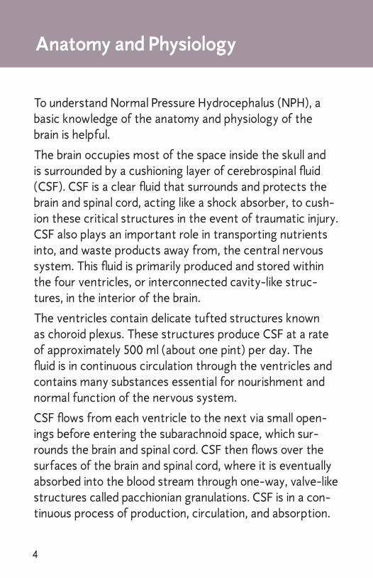
The brain occupies most of the space inside the skull and is surrounded by a cushioning layer of cerebrospinal fluid (CSF). CSF is a clear fluid that surrounds and protects the brain and spinal cord, acting like a shock absorber, to cush-ion these critical structures in the event of traumatic injury. CSF also plays an important role in transporting nutrients into, and waste products away from, the central nervous system. This fluid is primarily produced and stored within the four ventricles, or interconnected cavity-like struc-tures, in the interior of the brain.
The ventricles contain delicate tufted structures known as choroid plexus. These structures produce CSF at a rate of approximately 500 ml (about one pint) per day. The fluid is in continuous circulation through the ventricles and contains many substances essential for nourishment and normal function of the nervous system.
CSF flows from each ventricle to the next via small open-ings before entering the subarachnoid space, which sur-rounds the brain and spinal cord. CSF then flows over the surfaces of the brain and spinal cord, where it is eventually absorbed into the blood stream through one-way, valve-like structures called pacchionian granulations. CSF is in a con-tinuous process of production, circulation, and absorption.
5
Choroid Plexus
Lateral Ventricles
Third Ventricle
Aqueduct of Sylvius
Fourth Ventricle
Subarachnoid Space
Sagittal Sinus
Arachnoid Space
Brain
Skull
Figure 1. Cerebrospinal Fluid (CSF) Circulatory Pathway.
View of the center of the brain with the ventricles and surrounding structures. The arrows show the major pathway of CSF flow.
Cerebrospinal Fluid (CSF) Circulatory Pathway
There is a delicate balance associated with this process, which keeps the total volume of CSF at a constant level.
6
Hydrocephalus
In some specific medical conditions, there is an excess of cerebrospinal fluid (CSF) in the ventricles of the brain. This is called hydrocephalus, a term derived from two Greek words: hydro for water and kephale for head.
Hydrocephalus develops when the volume of CSF in the ventricles is higher than normal. This occurs when the flow of CSF is blocked within the ventricular system, or when absorption into the blood stream cannot keep up with the volume being produced. In other words, there is more CSF being produced than being absorbed.
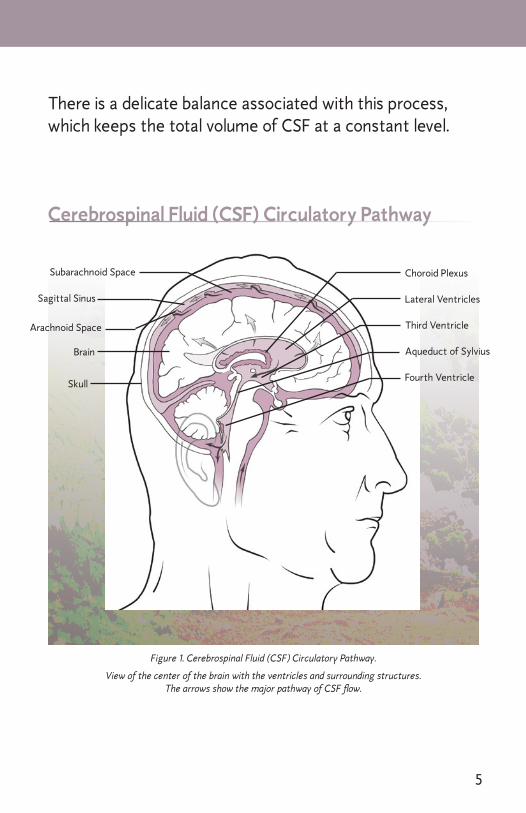
Normal vs. Hydrocephalic Ventricles
Figure 2. Normal (left) and enlarged hydrocephalic ventricles (right)

7
Hydrocephalus can be congenital, which simply means that the condition existed before birth. It is also possible to have acquired hydrocephalus, which develops after birth due to a variety of reasons, such as trauma, brain tumor, scar tissue, or meningitis. In both, the excessive amount of CSF, can be caused by a blockage in the ventricular system of the brain preventing the normal flow of CSF, or as the result of a problem with CSF absorption. Additionally, there is a condi-tion known as normal pressure hydrocephalus (NPH), which occurs later in life and is the result of an imbalance in CSF production and absorption.
Condition existed before birth
Develops after birth as a result of trauma
or disease
Affects the elderly
Cause not always known
Types of Hydrocephalus
Congenital Acquired NPH
8
Normal Pressure Hydrocephalus (NPH)
Normal Pressure Hydrocephalus is a condition that affects individuals typically in their sixties or seventies, and can rob them of their golden years. It is characterized by the gradual onset of a triad of symptoms, which most com-monly include difficulty walking, urinary incontinence, and mild dementia. NPH can be difficult to diagnose, since not all of the symptoms may come about at the same time. Furthermore, these symptoms are often associated with other conditions that are common in an aging population, e.g. Parkinson’s disease, osteoarthritis, and Alzheimer’s dis-ease, to name a few. Left untreated NPH will progress and significantly impair one’s quality of life.
Coping with the symptoms of NPH can be taxing for both patients and family members as this affects many aspects of life, including relationships, social activities, physical health, and mental health. Patients can become depressed and sulk, or frustrated and angry, all of which tend to make the situation worse.
Cause and Incidence Like other types of hydrocephalus, the defining charac-teristic of NPH is the enlargement of the ventricles in the brain. It is not entirely known why this enlargement pro-duces the symptoms associated with NPH, but most evi-dence seems to suggest a disturbance of the frontal lobe region of the brain. The expanded ventricles seem to dis-tort the nerve pathways between the brain and the spinal
�
cord, thus causing the symptoms. The blood flow to the brain has been seen to decrease in some cases as well.
For over 50% of patients with NPH, the cause of the hy-drocephalus cannot be determined. This is called idiopathic NPH. In the other cases, there is a history of brain hemor-rhage (such as from an aneurysm rupture or brain trauma) or meningitis. However, it is not clear why or how these conditions lead to NPH.
According to the National Council on Aging (NCOA) the incidence of NPH is not entirely clear. While some experts say that approximately �75,000 people have NPH, esti-mates have ranged from about 200,000 to 750,000 cases. It is said that about 5% of patients with dementia are likely to have NPH, which unlike Alzheimer’s disease, can be a reversible disorder. There is not predilection related to sex or race.
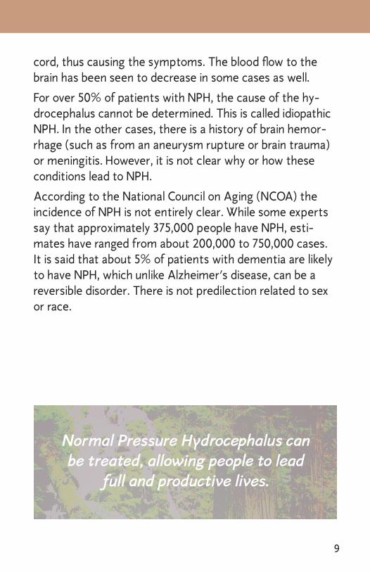
Normal Pressure Hydrocephalus can be treated, allowing people to lead
full and productive lives.
10
Signs and Symptoms of NPH
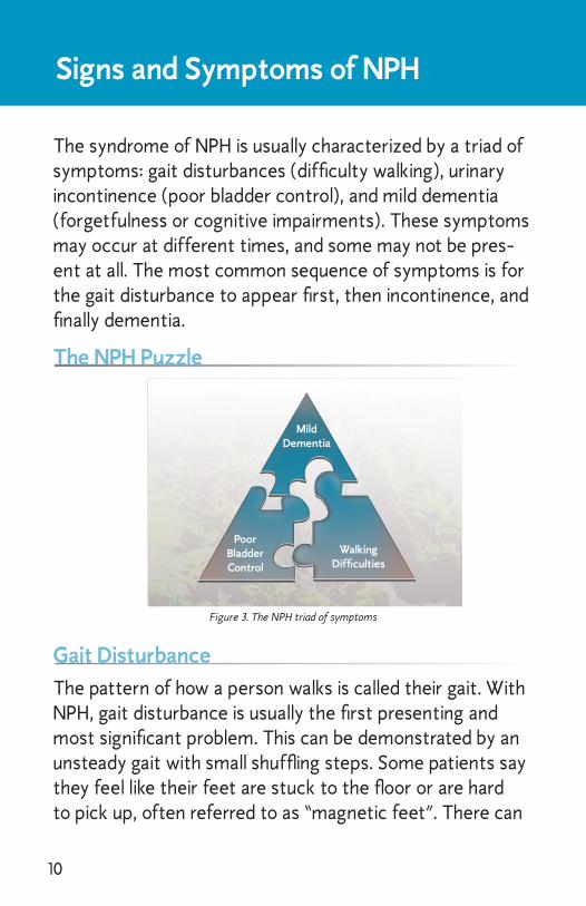
The syndrome of NPH is usually characterized by a triad of symptoms: gait disturbances (difficulty walking), urinary incontinence (poor bladder control), and mild dementia (forgetfulness or cognitive impairments). These symptoms may occur at different times, and some may not be pres-ent at all. The most common sequence of symptoms is for the gait disturbance to appear first, then incontinence, and finally dementia.
Figure 3. The NPH triad of symptoms
The NPH Puzzle
Gait Disturbance
The pattern of how a person walks is called their gait. With NPH, gait disturbance is usually the first presenting and most significant problem. This can be demonstrated by an unsteady gait with small shuffling steps. Some patients say they feel like their feet are stuck to the floor or are hard to pick up, often referred to as “magnetic feet”. There can
MildDementia
PoorBladderControl
WalkingDifficulties
11
also be some difficulty negotiating steps and stairs, and falls are not uncommon. Frequently, patients and families seek medical attention initially due to a fall.
Urinary IncontinenceIn most cases urinary incontinence (impaired bladder con-trol) shows up later in the progression of NPH, and in some situations, is never a problem. For those who are affected, symptoms can vary significantly and may include:
Frequency – the need to go to the bathroom or urinate often.
Urgency – the sudden and compelling need to pass urine, accompanied by discomfort. If ignored this may produce incontinence.
Frank Incontinence – the loss of control to hold back urine.
Although this is not life threatening, urinary incontinence can affect activities of daily living, cause embarrassing so-cial moments, and produce psychological pain.
Mild DementiaThere may be some forgetfulness or short term memory loss (e.g. not recalling where the keys are or what one had for breakfast) associated with NPH. Some patients may lose interest in things they have previously enjoyed (e.g. playing with grandchildren, playing cards, doing crossword puzzles). In some instances there can be mood changes, manifested by withdrawal or having a flat affect, meaning to act without animation or noticeable excitement. Unlike other dementias, the dementia caused by NPH can some-times be reversed or at least stabilized.
12
Because all three of these symptoms can be associated with the aging process, and the majority of the NPH popu-lation is older than 60 years, people often assume that they must live with these problems. This attitude can lead to delays in seeking medical attention which further de-lays the diagnosis and treatment of patients. Since NPH is a degenerative disease with a variable rate of progress, it is suggested that the earlier the diagnosis, the better the chance for successful treatment. However, some who have experienced symptoms for several years have seen im-provement with treatment. Consequently, it makes sense to have a medical evaluation when NPH is being considered as a potential diagnosis.
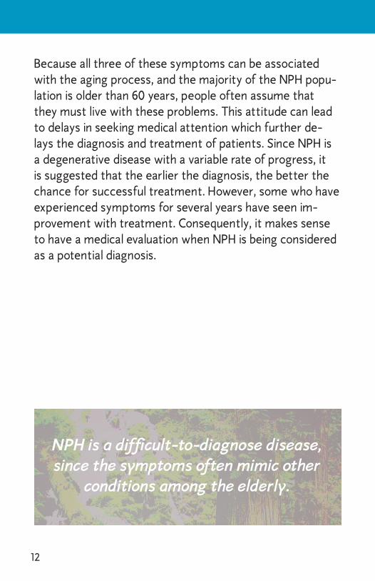
NPH is a difficult-to-diagnose disease, since the symptoms often mimic other
conditions among the elderly.
1�
Diagnosing NPH
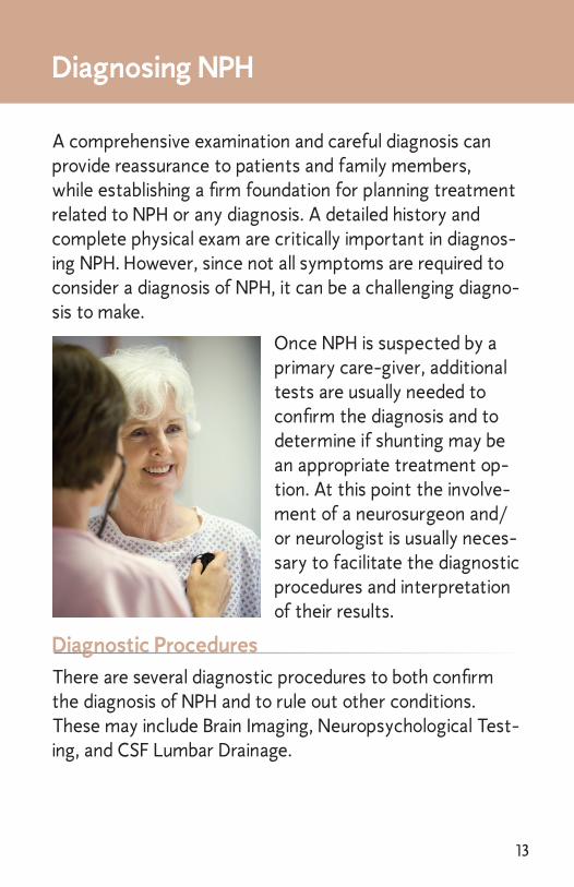
A comprehensive examination and careful diagnosis can provide reassurance to patients and family members, while establishing a firm foundation for planning treatment related to NPH or any diagnosis. A detailed history and complete physical exam are critically important in diagnos-ing NPH. However, since not all symptoms are required to consider a diagnosis of NPH, it can be a challenging diagno-sis to make.
Once NPH is suspected by a primary care-giver, additional tests are usually needed to confirm the diagnosis and to determine if shunting may be an appropriate treatment op-tion. At this point the involve-ment of a neurosurgeon and/or neurologist is usually neces-sary to facilitate the diagnostic procedures and interpretation of their results.
Diagnostic ProceduresThere are several diagnostic procedures to both confirm the diagnosis of NPH and to rule out other conditions. These may include Brain Imaging, Neuropsychological Test-ing, and CSF Lumbar Drainage.
14
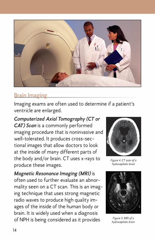
Brain ImagingImaging exams are often used to determine if a patient’s ventricle are enlarged.
Computerized Axial Tomography (CT or CAT) Scan is a commonly performed imaging procedure that is noninvasive and well-tolerated. It produces cross-sec-tional images that allow doctors to look at the inside of many different parts of the body and/or brain. CT uses x-rays to produce these images.
Magnetic Resonance Imaging (MRI) is often used to further evaluate an abnor-mality seen on a CT scan. This is an imag-ing technique that uses strong magnetic radio waves to produce high quality im-ages of the inside of the human body or brain. It is widely used when a diagnosis of NPH is being considered as it provides
Figure 4. CT scan of a hydrocephalic brain
Figure 5. MRI of a hydrocephalic brain
15
important detail of the ventricular system and can help to rule out other disorders, e.g. subdural hematoma, tumor, infection, or a structural abnormality.
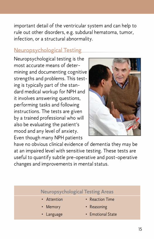
• Attention • Reaction Time
• Memory • Reasoning
• Language • Emotional State
Neuropsychological Testing Areas
Neuropsychological Testing
Neuropsychological testing is the most accurate means of deter-mining and documenting cognitive strengths and problems. This test-ing is typically part of the stan-dard medical workup for NPH and it involves answering questions, performing tasks and following instructions. The tests are given by a trained professional who will also be evaluating the patient’s mood and any level of anxiety. Even though many NPH patients have no obvious clinical evidence of dementia they may be at an impaired level with sensitive testing. These tests are useful to quantify subtle pre-operative and post-operative changes and improvements in mental status.
16
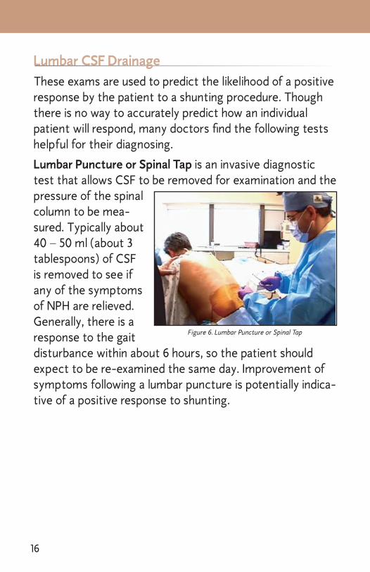
Lumbar CSF DrainageThese exams are used to predict the likelihood of a positive response by the patient to a shunting procedure. Though there is no way to accurately predict how an individual patient will respond, many doctors find the following tests helpful for their diagnosing.
Lumbar Puncture or Spinal Tap is an invasive diagnostic test that allows CSF to be removed for examination and the pressure of the spinal column to be mea-sured. Typically about 40 – 50 ml (about � tablespoons) of CSF is removed to see if any of the symptoms of NPH are relieved. Generally, there is a response to the gait disturbance within about 6 hours, so the patient should expect to be re-examined the same day. Improvement of symptoms following a lumbar puncture is potentially indica-tive of a positive response to shunting.
Figure 6. Lumbar Puncture or Spinal Tap
17
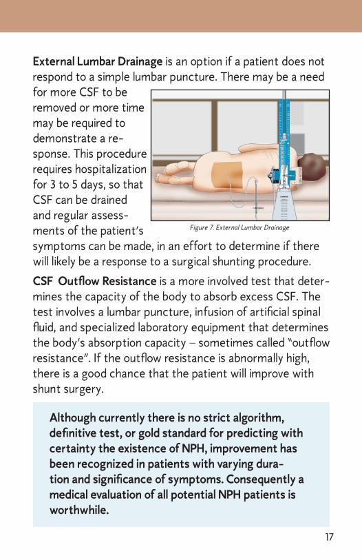
External Lumbar Drainage is an option if a patient does not respond to a simple lumbar puncture. There may be a need for more CSF to be removed or more time may be required to demonstrate a re-sponse. This procedure requires hospitalization for � to 5 days, so that CSF can be drained and regular assess-ments of the patient’s symptoms can be made, in an effort to determine if there will likely be a response to a surgical shunting procedure.
CSF Outflow Resistance is a more involved test that deter-mines the capacity of the body to absorb excess CSF. The test involves a lumbar puncture, infusion of artificial spinal fluid, and specialized laboratory equipment that determines the body’s absorption capacity – sometimes called “outflow resistance”. If the outflow resistance is abnormally high, there is a good chance that the patient will improve with shunt surgery.
Figure 7. External Lumbar Drainage
Although currently there is no strict algorithm, definitive test, or gold standard for predicting with certainty the existence of NPH, improvement has been recognized in patients with varying dura-tion and significance of symptoms. Consequently a medical evaluation of all potential NPH patients is worthwhile.
18
Treatment
The only effective treatment of NPH involves the surgical implantation of a shunt system. A shunt is a device that reroutes CSF from the nervous system to another area of the body. In order to divert CSF, the surgeon will insert a device, made of silicone and polypropylene plastic, under the skin. There are no parts on the outside of the body.
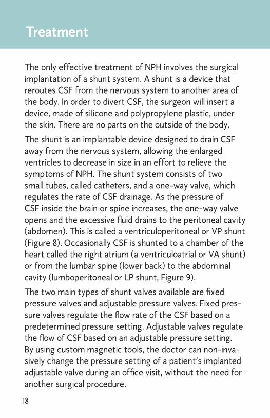
The shunt is an implantable device designed to drain CSF away from the nervous system, allowing the enlarged ventricles to decrease in size in an effort to relieve the symptoms of NPH. The shunt system consists of two small tubes, called catheters, and a one-way valve, which regulates the rate of CSF drainage. As the pressure of CSF inside the brain or spine increases, the one-way valve opens and the excessive fluid drains to the peritoneal cavity (abdomen). This is called a ventriculoperitoneal or VP shunt (Figure 8). Occasionally CSF is shunted to a chamber of the heart called the right atrium (a ventriculoatrial or VA shunt) or from the lumbar spine (lower back) to the abdominal cavity (lumboperitoneal or LP shunt, Figure �).
The two main types of shunt valves available are fixed pressure valves and adjustable pressure valves. Fixed pres-sure valves regulate the flow rate of the CSF based on a predetermined pressure setting. Adjustable valves regulate the flow of CSF based on an adjustable pressure setting. By using custom magnetic tools, the doctor can non-inva-sively change the pressure setting of a patient’s implanted adjustable valve during an office visit, without the need for another surgical procedure.
1�
PeritonealCavity
VentricularCatheterValveVentricles
Cardiac/PeritonalCatheter
Figure 8. Ventriculoperitoneal (VP) or ventriculoatrial (VA) shunt placement
Figure 9. Lumboperitoneal (LP) shunt placement
Both of these types of valves can include overdrainage pro-tection in the form of a siphon control device. The purpose of a siphon control device is to minimize excessive drainage due to gravity, which can cause more CSF to drain when the patient is in the upright position.
20
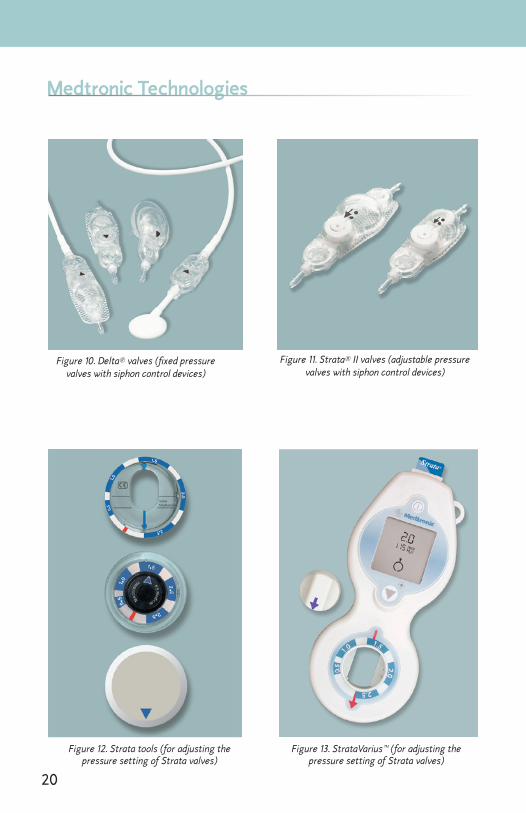
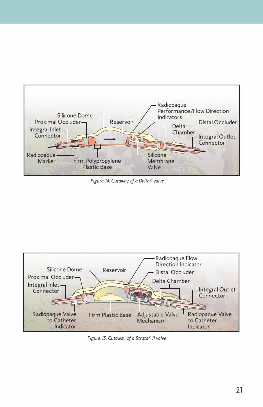
Figure 10. Delta® valves (fixed pressure valves with siphon control devices)
Figure 11. Strata® II valves (adjustable pressure valves with siphon control devices)
Figure 13. StrataVarius™ (for adjusting the pressure setting of Strata valves)
Medtronic Technologies
Figure 12. Strata tools (for adjusting the pressure setting of Strata valves)
21
Integral InletConnector
Proximal OccluderSilicone Dome
Radiopaque FlowDirection Indicator
Reservoir
Delta ChamberIntegral OutletConnector
Radiopaque Valveto CatheterIndicator
Adjustable ValveMechanism
Firm Plastic BaseRadiopaque Valveto Catheter
Indicator
Distal Occluder
Figure 15. Cutaway of a Strata® II valve
Integral InletConnector
Proximal OccluderSilicone Dome
Reservoir
RadiopaquePerformance/Flow Direction Indicators
DeltaChamber
Distal Occluder
Integral OutletConnector
RadiopaqueMarker Firm Polypropylene
Plastic Base
Silicone MembraneValve
Figure 14. Cutaway of a Delta® valve
22
The pressure setting of an implanted adjustable valve can be non-invasively
changed by a doctor
PrecautionSince adjustable valves include magnets, special precau-tions must be observed around magnetic sources. If a device is known to contain a magnet, keep it away from the immediate valve location, i.e. the skin next to the valve. For patients that are undergoing an MRI procedure, it will be necessary for the doctor to check the valve pressure setting afterwards and readjust the valve if necessary. Ask your doctor for specifics on the sensitivity to com-mon environmental levels of electromagnetic radiation for particular valves.
2�
Surgical Procedure
Each type of shunt has advantages and disadvantages. Because each patient is unique, the neurosurgeon will de-cide which type of shunt is used, based on their own expe-rience and the individual needs of the patient.
The surgical procedure is carried out under general anes-thesia with sterile conditions in the operating room. Although the operation is relatively short (often the sur-gery can be done in under an hour), careful preparation for the surgery adds extra time. In order to help prevent infec-tion, some of the hair may need to be shaved. The head and body are washed with a special antibacterial soap. Sterile linen is used to cover the patient and to maintain the sterile environment throughout the surgical procedure.
Ventriculoperitoneal (VP) ShuntFor VP shunts, a few small incisions (cuts) are required to do the procedure (See Figure 8). One incision is made in the scalp (the skin covering the head). A small hole is then made in the skull and a tiny opening is made in the dura, a protective covering of the brain. These openings are made to accommodate the ventricular (proximal) catheter being placed into the lateral ventricle. The neurosurgeon usually places the shunt valve above or behind one ear and at-taches it to the two catheters. The distal catheter is then tunneled under the skin to the abdomen where an ad-ditional small incision, about 0.5” is made. Finally, the end of the catheter is carefully placed in the peritoneal cavity. Following the operation, small sterile bandages are applied to each incision.
24
Lumboperitoneal (LP) ShuntFor lumboperitoneal shunts, an incision is made near the base of the spine, and a small catheter is inserted into the subarachnoid space of the lower spine (See Figure �). This is the same location where a lumbar puncture takes place. The catheter is attached to the valve, which is usually placed around the hip bone, and then the distal catheter is tunneled under the skin to the abdomen, where it is insert-ed in the peritoneal cavity.
Post-OperationImmediately after surgery, the patient will go to the post-anesthesia care unit. Patients generally remain there for about an hour for close observation and then go to their assigned hospital room. The length of hospitalization varies from patient to patient. Most patients leave the hospital within two to seven days, depending on their clinical prog-ress. Although this is the usual procedure when a shunt is placed, each patient may have a slightly different experi-ence based on their neurosurgeon, the hospital protocol, and the need to individualize patient care.
Regular visits to your neurosurgeon or neurologist will be necessary.
25
Follow-Up Care
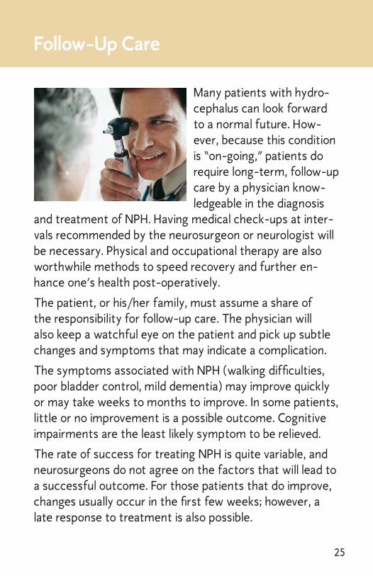
Many patients with hydro-cephalus can look forward to a normal future. How-ever, because this condition is “on-going,” patients do require long-term, follow-up care by a physician know-ledgeable in the diagnosis
and treatment of NPH. Having medical check-ups at inter-vals recommended by the neurosurgeon or neurologist will be necessary. Physical and occupational therapy are also worthwhile methods to speed recovery and further en-hance one’s health post-operatively.
The patient, or his/her family, must assume a share of the responsibility for follow-up care. The physician will also keep a watchful eye on the patient and pick up subtle changes and symptoms that may indicate a complication.
The symptoms associated with NPH (walking difficulties, poor bladder control, mild dementia) may improve quickly or may take weeks to months to improve. In some patients, little or no improvement is a possible outcome. Cognitive impairments are the least likely symptom to be relieved.
The rate of success for treating NPH is quite variable, and neurosurgeons do not agree on the factors that will lead to a successful outcome. For those patients that do improve, changes usually occur in the first few weeks; however, a late response to treatment is also possible.
26
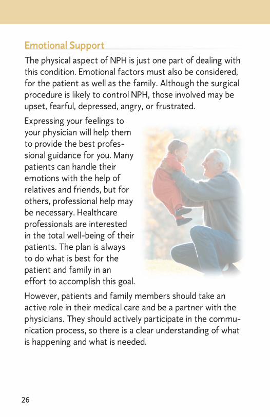
Emotional Support The physical aspect of NPH is just one part of dealing with this condition. Emotional factors must also be considered, for the patient as well as the family. Although the surgical procedure is likely to control NPH, those involved may be upset, fearful, depressed, angry, or frustrated.
Expressing your feelings to your physician will help them to provide the best profes-sional guidance for you. Many patients can handle their emotions with the help of relatives and friends, but for others, professional help may be necessary. Healthcare professionals are interested in the total well-being of their patients. The plan is always to do what is best for the patient and family in an effort to accomplish this goal.
However, patients and family members should take an active role in their medical care and be a partner with the physicians. They should actively participate in the commu-nication process, so there is a clear understanding of what is happening and what is needed.
27
The potential complications associated with shunt surgery must be viewed not only as those associated with the ac-tual surgical procedure but also those that may occur days, weeks, or even years later. Unlike most surgical procedures, where the risks are highest during the operation itself, most of the common problems associated with shunting can and do occur at a later time.
The major complications of shunting are obstruction, infection, overdrainage of CSF, and mechanical break-age. Patients and families should be alert to the signs and symptoms of shunt complications and recognize that shunt revisions may be necessary to replace a component that is not working.
Complications
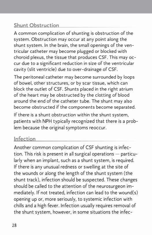
Symptoms of Shunt Complications
• Loss of coordination or balance
• Personality change or confusion
• Loss of bladder control
• Fever
• Redness or swelling along the shunt tract
• Headache
• Difficulty in waking up or staying awake
• Seizures
• Paralysis
28
Shunt Obstruction A common complication of shunting is obstruction of the system. Obstruction may occur at any point along the shunt system. In the brain, the small openings of the ven-tricular catheter may become plugged or blocked with choroid plexus, the tissue that produces CSF. This may oc-cur due to a significant reduction in size of the ventricular cavity (slit ventricle) due to over-drainage of CSF.
The peritoneal catheter may become surrounded by loops of bowel, other structures, or by scar tissue, which can block the outlet of CSF. Shunts placed in the right atrium of the heart may be obstructed by the clotting of blood around the end of the catheter tube. The shunt may also become obstructed if the components become separated.
If there is a shunt obstruction within the shunt system, patients with NPH typically recognized that there is a prob-lem because the original symptoms reoccur.
InfectionAnother common complication of CSF shunting is infec-tion. This risk is present in all surgical operations — particu-larly when an implant, such as a shunt system, is required. If there is any unusual redness or swelling at the site of the wounds or along the length of the shunt system (the shunt track), infection should be suspected. These changes should be called to the attention of the neurosurgeon im-mediately. If not treated, infection can lead to the wound(s) opening up or, more seriously, to systemic infection with chills and a high fever. Infection usually requires removal of the shunt system, however, in some situations the infec-

2�
tion may be controlled with intensive antibiotic therapy, without the removal of the shunt.
Since the shunt system is an implant or a “foreign body,” a patient may develop an allergic or inflammatory reaction to it at any time. Although this is uncommon it is important to report any inflammation or open sores over any part of the implanted system to the attention of the neurosurgeon immediately.
CSF OverdrainageOverdrainage is generally caused when too much CSF drains out of the ventricles too quickly. Overdrainage may produce a variety of signs and symptoms. Patients gener-ally will experience a headache that is worse when upright and reduced by lying down.
Other complications can also occur in the days and months following the surgery. The most serious complication is a subdural hematoma (blood clot) on the surface of the brain. This can result from draining too much fluid out of the brain too quickly, causing the brain tissue to shift away from the skull and blood vessels on the surface of the brain to tear and bleed.
A shunt-related hematoma commonly occurs as a result of a fall, even if there are no apparent injuries. Symptoms associated with a subdural hematoma vary from headaches, paralysis, and may be as severe as coma or death. The risk of an NPH shunt patient developing a subdural hematoma is approximately 5 to 10%.
�0
Mechanical BreakageShunt systems include several components linked in se-quence, providing an open pathway for CSF to flow from a ventricle in the brain or the subarachnoid space of the spine, to the peritoneal cavity or right atrium of the heart. Any disconnection between the elements of the shunt sys-tem may result in improper drainage of CSF.
It is important for patients with NPH and their families to be aware of the potential complications that can occur following shunt surgery. It is essen-tial for patients, family members, and caregivers to have knowledge of the signs and symptoms of these complications and the necessity for emergency medical evaluation in these instances.
�1
Glossary
Abdominal Cavity: The area of the body between the chest and pelvis containing the liver, intestines, kidneys and other organs. (Synonymous with the peritoneal cavity)
Antibiotic: Any substance (such as Penicillin) which destroys or inhibits the growth of bacteria
Arachnoid: A thin membrane, internal to the dura
Atrium: One of the two upper chambers of the heart
Cerebrospinal Fluid (CSF): The fluid filling the ventricles of the brain and surrounding the brain and spinal cord
CAT or CT scan: Abbreviation for computerized axial tomogra-phy, a special x-ray technique which outlines the ventricles and other structures of the brain in cross section
Choroid Plexus: Delicate structures in the ventricles of the brain that produce CSF
Coma: A state in which the patient doesn’t respond to stimulation
Congenital: A condition present since birth
CSF: Cerebrospinal fluid
Diagnosis: Determination of a patient’s problem
Distal Catheter: Shunt catheter that is farthest from the ven-tricles, usually in the peritoneal cavity
Dura: The outer most, fibrous membrane that surrounds the brain and spinal cord (also dura mater)
Dementia: Refers to a group of symptoms involving progressive impairment of brain function. This may include forgetfulness, apathy, and/or deterioration in ones level of functioning on a day-to-day basis
�2
Gait Disturbance: An unusual and uncontrollable problem with walking, which may include weakness of the legs, unsteady walking, and small shuffling steps
Foreign Body: An object, such as an implant, introduced into a liv-ing body from the outside
Hydrocephalus: Excessive build-up of CSF in the ventricles of the brain, causing compression on other surrounding structures of the brain
Hydrocephalus, Acquired: Hydrocephalus developed after birth
Hydrocephalus, Communicating: Hydrocephalus in which there is no obstruction between ventricles and subarachnoid space
Hydrocephalus, Congenital: Hydrocephalus existing before or at birth
Hydrocephalus, Non-Communicating or Obstructive: Hy-drocephalus in which there is obstruction of CSF flow between ventricles
Hydrocephalus, Normal Pressure: A syndrome characterized by enlargement of the ventricles along with a triad of symptoms that include gait disturbance, dementia, and impaired bladder control
Incontinence: The inability to control the passage of urine. This can range from occasional leakage of urine, to a complete inability to hold any urine
Intracranial Pressure (ICP): The pressure within the skull
Isotope: A radioactive material used for determining spinal fluid flow and shunt function
Lumbar Puncture: A procedure to obtain a specimen of CSF by positioning a small needle or catheter into the subarachnoid space, at the level of the lumbar spine, in order to withdraw CSF for appropriate testing. Also known as a Spinal Tap
��
Lumbar Spine: The area of the spine in the small of the back
Meninges: The coverings of the brain and spinal cord made up of the dura mater, arachnoid, and pia mater
Meningitis: Inflammation or infection of the meninges
MRI: Abbreviation for Magnetic Resonance Imaging. By means of magnetic energy, images are taken, showing the ventricles and other structures within the brain
Neurological: Pertaining to the nervous system (including the brain and spinal cord)
Normal Pressure Hydrocephalus: A syndrome characterized by enlargement of the ventricles within the brain along with a triad of symptoms that may include gait disturbance, dementia, and impaired bladder control
Peritoneum: Lining of the abdominal cavity
Peritoneal Cavity: The abdominal cavity
Pia mater: A very delicate, highly vascular, layer of tissue that is adherent to the brain and spinal cord, following all the contours
Polypropylene: Plastic used in the manufacture of shunt systems
Proximal Catheter: Shunt catheter in the ventricle
Shunt (noun): A system of tubing used to drain CSF from the ven-tricles or subarachnoid space into another area of the body
Shunt (verb): Surgical procedure during which a shunt system is implanted
Silicone: A polymer characterized by inertness in the body tissues and used in the manufacture of shunt systems and other medi-cal devices
Skull: The boney structure that surrounds and protects the brain
Slit Ventricle: Excessive narrowing of the lateral ventricle due to over-drainage of CSF
�4
Spinal Cord: An elongated structure that is a component of the central nervous system. It is the main pathway for information connecting the brain and peripheral nervous system. The hu-man spinal cord is protected by the bony spinal column.
Stuporous: A semi-conscious condition, in which the patient is very sleepy
Subarachnoid Space: A place where cerebrospinal fluid circulates
Subdural Hematoma: A collection of blood between the dura, outer covering of the brain, and the surface of the brain
Urinary Incontinence: The inability to control the passage of urine. This can range from occasional leakage of urine, to a complete inability to hold urine
Valve: A one-way device used to control drainage of CSF from the ventricles of the brain or the lumbar area of the spine
Ventricles: The four cavities (two lateral, one third, and one fourth) lying within the brain

�5
Questions
For more information on NPH and the available treatments, talk to your physician, neurosurgeon, or neurologist.
You can also visit Medtronic’s website at www.medtronic.com
or the Hydrocephalus Association at www.hydroassoc.org
�6
NPH cannot be “cured,” but it can be effectively treated,
allowing the patient to lead a full and productive life.
Medtronic Neurologic Technologies 125 Cremona Drive • Goleta, California ��117-5500 USA(800) 468-�710 USA/Canada(�01) �44-0645 International (800) 468-�71� FAX(�01) ��6-26�8 FAX International
For more information, please contact your neurosurgeon or neurologist, or refer to www.medtronic.com.
Caution: Federal (USA) law restricts these devices to sale by or on the order of a physician. Refer to product package insert for instructions, warnings, precautions and complications.
LIT5��66 Rev. B �.2007©Medtronic, Inc. 2007All Rights ReservedPrinted in USA
Strata® and Delta® are registered trademarks of Medtronic, Inc. StrataVarius™ is a trademark of Medtronic, Inc.
This therapy is not for everyone. Please consult your physician. A prescription is required. For more information, please contact us at 800-510-67�5 or refer to our website at www.medtronic.com.