Embed Size (px)
Citation preview

8/12/2019 Normal CT Chest
http://slidepdf.com/reader/full/normal-ct-chest 1/81
CHESTNormal CT ANATOMY
BYMAMDOUH MAHFOUZ MD
Cairo university

8/12/2019 Normal CT Chest
http://slidepdf.com/reader/full/normal-ct-chest 2/81
IndicationsIndications
Patient preparationPatient preparation Fasting 4Fasting 4--6 hours6 hours Patient positionPatient position SupineSupine
ScanogramScanogram FrontalFrontal
• To assess equivocal plain X-ray findings
• Staging of lung neoplasms
• Metastatic workup of extrathoracic malignancies
• Diagnosis of diffuse lung disease with HRCT
• Assessment of bronchiectasis
• Assessment of suspected post-traumatic vascular injury

8/12/2019 Normal CT Chest
http://slidepdf.com/reader/full/normal-ct-chest 3/81
IndicationsIndications
Patient preparationPatient preparation Fasting 4Fasting 4--6 hours6 hours
Patient positionPatient position
SupineSupine
ScanogramScanogram FrontalFrontal
No required preparation unless the patient is going to be sedated
or injected with contrast material
FASTING FOR 4FASTING FOR 4 -- 6 HOURS6 HOURS

8/12/2019 Normal CT Chest
http://slidepdf.com/reader/full/normal-ct-chest 4/81
Contrast injection50-100ml of water soluble contrast material [urographine, isovist,…] bolus injection
Not indicated when Evaluating diffuse lung disease.
Evaluating bronchiectasis
Screening for lung deposits
Some cases of trauma.

8/12/2019 Normal CT Chest
http://slidepdf.com/reader/full/normal-ct-chest 5/81
10mm sections from lung apex to the C/P angles
Mediastinal window, lung window, bone window?!
Reconstructed images

8/12/2019 Normal CT Chest
http://slidepdf.com/reader/full/normal-ct-chest 6/81
Scanning techniquesScanning techniquesStandard Examination
High resolution [HRCT]

8/12/2019 Normal CT Chest
http://slidepdf.com/reader/full/normal-ct-chest 7/81
Standard CT High Resolution CT,HRCTHRCT

8/12/2019 Normal CT Chest
http://slidepdf.com/reader/full/normal-ct-chest 8/81
Scanning techniquesScanning techniques
Spiral, Helical, volumetric CT
Multi-Detector, Multi-Slice CT

8/12/2019 Normal CT Chest
http://slidepdf.com/reader/full/normal-ct-chest 9/81
Normal pulmonary vascularity

8/12/2019 Normal CT Chest
http://slidepdf.com/reader/full/normal-ct-chest 10/81
Normal pulmonary vascularity

8/12/2019 Normal CT Chest
http://slidepdf.com/reader/full/normal-ct-chest 11/81

8/12/2019 Normal CT Chest
http://slidepdf.com/reader/full/normal-ct-chest 12/81

8/12/2019 Normal CT Chest
http://slidepdf.com/reader/full/normal-ct-chest 13/81
LI
M
A

8/12/2019 Normal CT Chest
http://slidepdf.com/reader/full/normal-ct-chest 14/81

8/12/2019 Normal CT Chest
http://slidepdf.com/reader/full/normal-ct-chest 15/81
CT AngiographyCT Angiography

8/12/2019 Normal CT Chest
http://slidepdf.com/reader/full/normal-ct-chest 16/81

8/12/2019 Normal CT Chest
http://slidepdf.com/reader/full/normal-ct-chest 17/81
3DCTA Angiography demonstrates a filling defect of the right Iliac artery

8/12/2019 Normal CT Chest
http://slidepdf.com/reader/full/normal-ct-chest 18/81
Detailed examination of the Superior Mesenteric Artery and Celiac Artery.Scan time = 9.4 seconds. 1mm slice thickness

8/12/2019 Normal CT Chest
http://slidepdf.com/reader/full/normal-ct-chest 19/81
F 35Y

8/12/2019 Normal CT Chest
http://slidepdf.com/reader/full/normal-ct-chest 20/81
Mediastinal anatomy

8/12/2019 Normal CT Chest
http://slidepdf.com/reader/full/normal-ct-chest 21/81

8/12/2019 Normal CT Chest
http://slidepdf.com/reader/full/normal-ct-chest 22/81

8/12/2019 Normal CT Chest
http://slidepdf.com/reader/full/normal-ct-chest 23/81

8/12/2019 Normal CT Chest
http://slidepdf.com/reader/full/normal-ct-chest 24/81

8/12/2019 Normal CT Chest
http://slidepdf.com/reader/full/normal-ct-chest 25/81

8/12/2019 Normal CT Chest
http://slidepdf.com/reader/full/normal-ct-chest 26/81

8/12/2019 Normal CT Chest
http://slidepdf.com/reader/full/normal-ct-chest 27/81
App
An An
P
Ap
8/12/2019 Normal CT Chest
http://slidepdf.com/reader/full/normal-ct-chest 28/81
An
App
An
Ap
P

8/12/2019 Normal CT Chest
http://slidepdf.com/reader/full/normal-ct-chest 29/81

8/12/2019 Normal CT Chest
http://slidepdf.com/reader/full/normal-ct-chest 30/81

8/12/2019 Normal CT Chest
http://slidepdf.com/reader/full/normal-ct-chest 31/81
An App
An
Ap
P App
AnL
P
App
PL An
M

8/12/2019 Normal CT Chest
http://slidepdf.com/reader/full/normal-ct-chest 32/81

8/12/2019 Normal CT Chest
http://slidepdf.com/reader/full/normal-ct-chest 33/81
An
App
An Ap
P
App App
An
L
P
PL
An
M
SS IILL
MM

8/12/2019 Normal CT Chest
http://slidepdf.com/reader/full/normal-ct-chest 34/81

8/12/2019 Normal CT Chest
http://slidepdf.com/reader/full/normal-ct-chest 35/81

8/12/2019 Normal CT Chest
http://slidepdf.com/reader/full/normal-ct-chest 36/81

8/12/2019 Normal CT Chest
http://slidepdf.com/reader/full/normal-ct-chest 37/81

8/12/2019 Normal CT Chest
http://slidepdf.com/reader/full/normal-ct-chest 38/81
7

8/12/2019 Normal CT Chest
http://slidepdf.com/reader/full/normal-ct-chest 39/81
1
4
3
7
6
5 2

8/12/2019 Normal CT Chest
http://slidepdf.com/reader/full/normal-ct-chest 40/81
M 44Y with malignant liver
F 45Y with post irradiation
changes after radical mastectomy

8/12/2019 Normal CT Chest
http://slidepdf.com/reader/full/normal-ct-chest 41/81
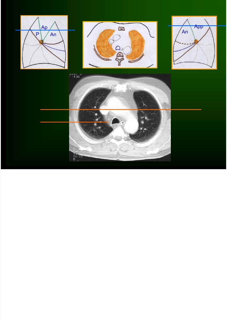
35Y male with fever and expectoration

8/12/2019 Normal CT Chest
http://slidepdf.com/reader/full/normal-ct-chest 42/81
45Y male with chest pain and hemoptysis

8/12/2019 Normal CT Chest
http://slidepdf.com/reader/full/normal-ct-chest 43/81
43Y male with acute chest
pain and hemoptysis

8/12/2019 Normal CT Chest
http://slidepdf.com/reader/full/normal-ct-chest 44/81

8/12/2019 Normal CT Chest
http://slidepdf.com/reader/full/normal-ct-chest 45/81
Thank

8/12/2019 Normal CT Chest
http://slidepdf.com/reader/full/normal-ct-chest 46/81
سبح نك للهم و بحمدك نشهد ن ال له ال نت نستغفرك و نتوب ليك
Thank you

8/12/2019 Normal CT Chest
http://slidepdf.com/reader/full/normal-ct-chest 47/81
THANK
YOU
نتوب ليك
و
نستغفرك
ال له ال نت
نشهد ن
بحمدك
و
سبح نك للهم

8/12/2019 Normal CT Chest
http://slidepdf.com/reader/full/normal-ct-chest 48/81
Thank

8/12/2019 Normal CT Chest
http://slidepdf.com/reader/full/normal-ct-chest 49/81
Thank you

8/12/2019 Normal CT Chest
http://slidepdf.com/reader/full/normal-ct-chest 50/81

8/12/2019 Normal CT Chest
http://slidepdf.com/reader/full/normal-ct-chest 51/81

8/12/2019 Normal CT Chest
http://slidepdf.com/reader/full/normal-ct-chest 52/81
Figures 7A, B & C Small branches arising from the left
pulmonary artery are seen on the CT scan. The relationship
of theazygos, aorta and esophagus isshown.

8/12/2019 Normal CT Chest
http://slidepdf.com/reader/full/normal-ct-chest 53/81
Figures 8A, B & CThe azygos arch is seen enter-ing
the superior vena cava.Note the small lymph nodes lying
within fat anterior to the trachea. This space is readily
accessible to the mediastino-scope.

8/12/2019 Normal CT Chest
http://slidepdf.com/reader/full/normal-ct-chest 54/81
Figures 9A, B & C The superior vena cava is lateral to the
aortic arch.

8/12/2019 Normal CT Chest
http://slidepdf.com/reader/full/normal-ct-chest 55/81
Figures 1OA, B & C Five vessels are seen cut in cross-
section.

8/12/2019 Normal CT Chest
http://slidepdf.com/reader/full/normal-ct-chest 56/81

8/12/2019 Normal CT Chest
http://slidepdf.com/reader/full/normal-ct-chest 57/81

8/12/2019 Normal CT Chest
http://slidepdf.com/reader/full/normal-ct-chest 58/81

8/12/2019 Normal CT Chest
http://slidepdf.com/reader/full/normal-ct-chest 59/81
•Cavitating neoplasm with pul. deposits

8/12/2019 Normal CT Chest
http://slidepdf.com/reader/full/normal-ct-chest 60/81

8/12/2019 Normal CT Chest
http://slidepdf.com/reader/full/normal-ct-chest 61/81
•Lat. normal
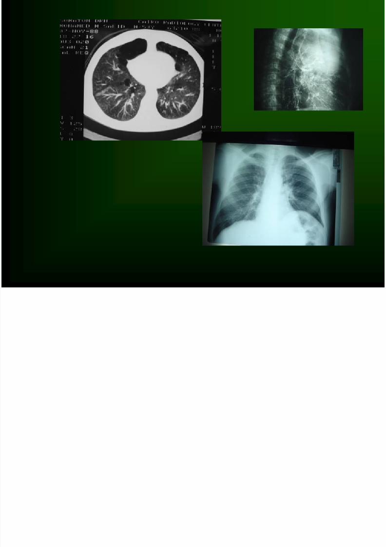
M 45Y PANCOAST’S TUMOR

8/12/2019 Normal CT Chest
http://slidepdf.com/reader/full/normal-ct-chest 62/81
M 45Y PANCOAST’S TUMOR

8/12/2019 Normal CT Chest
http://slidepdf.com/reader/full/normal-ct-chest 63/81

8/12/2019 Normal CT Chest
http://slidepdf.com/reader/full/normal-ct-chest 64/81

8/12/2019 Normal CT Chest
http://slidepdf.com/reader/full/normal-ct-chest 65/81
8/12/2019 Normal CT Chest
http://slidepdf.com/reader/full/normal-ct-chest 66/81

8/12/2019 Normal CT Chest
http://slidepdf.com/reader/full/normal-ct-chest 67/81

8/12/2019 Normal CT Chest
http://slidepdf.com/reader/full/normal-ct-chest 68/81

8/12/2019 Normal CT Chest
http://slidepdf.com/reader/full/normal-ct-chest 69/81

8/12/2019 Normal CT Chest
http://slidepdf.com/reader/full/normal-ct-chest 70/81
8/12/2019 Normal CT Chest
http://slidepdf.com/reader/full/normal-ct-chest 71/81

8/12/2019 Normal CT Chest
http://slidepdf.com/reader/full/normal-ct-chest 72/81

8/12/2019 Normal CT Chest
http://slidepdf.com/reader/full/normal-ct-chest 73/81

8/12/2019 Normal CT Chest
http://slidepdf.com/reader/full/normal-ct-chest 74/81

8/12/2019 Normal CT Chest
http://slidepdf.com/reader/full/normal-ct-chest 75/81

8/12/2019 Normal CT Chest
http://slidepdf.com/reader/full/normal-ct-chest 76/81

8/12/2019 Normal CT Chest
http://slidepdf.com/reader/full/normal-ct-chest 77/81

8/12/2019 Normal CT Chest
http://slidepdf.com/reader/full/normal-ct-chest 78/81

8/12/2019 Normal CT Chest
http://slidepdf.com/reader/full/normal-ct-chest 79/81
66 year ’ s old patient with multiple TB abscesses

8/12/2019 Normal CT Chest
http://slidepdf.com/reader/full/normal-ct-chest 80/81
63Y male with multiple hydatid cysts
8/12/2019 Normal CT Chest
http://slidepdf.com/reader/full/normal-ct-chest 81/81
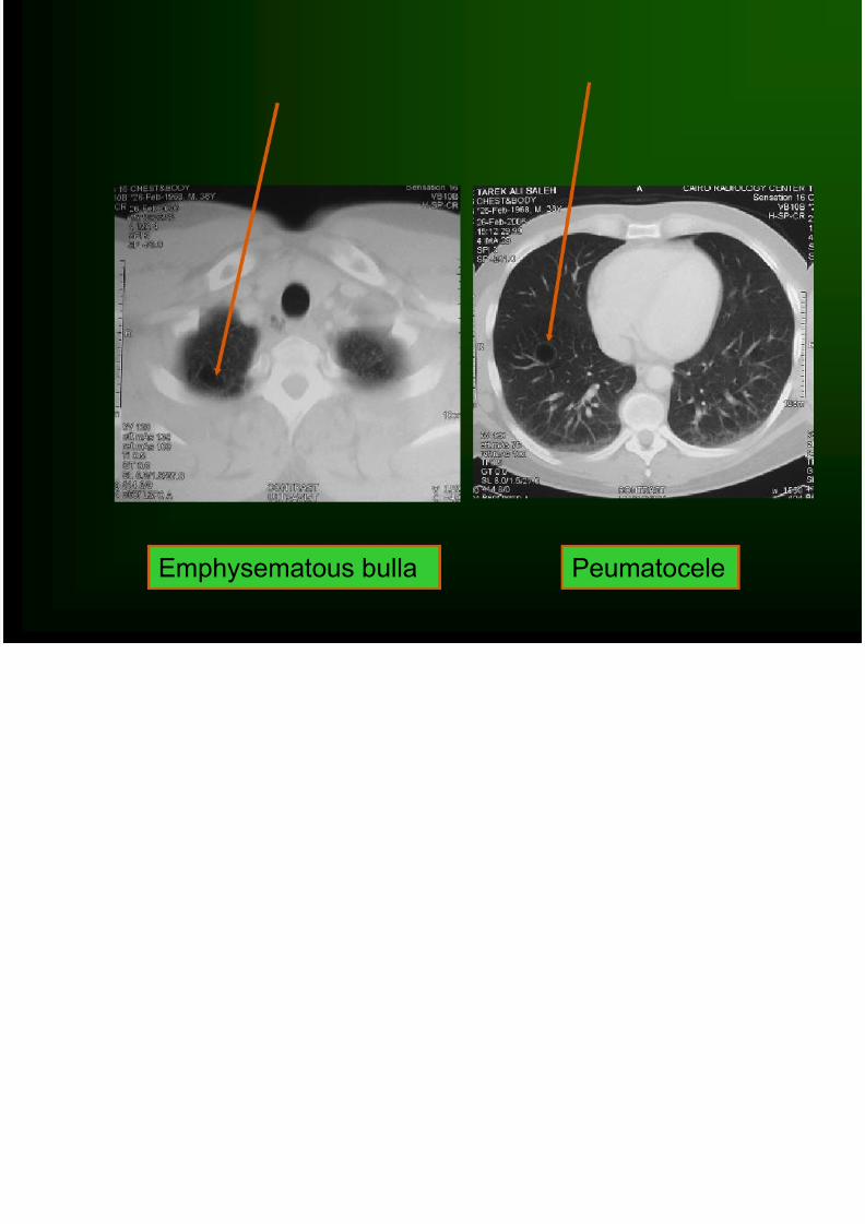
Emphysematous bulla Peumatocele