Embed Size (px)
Citation preview

Proc. Natl. Acad. Sci. USAVol. 91, pp. 8175-8179, August 1994Immunology
Nonpeptide ligands for human yH T cellsYOSHIMASA TANAKA*t, SHIGETOSHI SANO*t, EDWARD NIEVES*, GENNARO DE LIBERO§, DOMENICO ROSA§,ROBERT L. MODLIN¶, MICHAEL B. BRENNERI, BARRY R. BLOOM*t, AND CRAIG T. MORITAII*Howard Hughes Medical Institute and tDepartments of Microbiology and Immunology and *Biochemistry, Albert Einstein College of Medicine, Bronx, NY10461; §Experimental Immunology, Department of Research, University Hospital, Hebelstrasse 20, CH-4031 Basel, Switzerland; fUniversity of California atLos Angeles School of Medicine, Division of Dermatology and Department of Microbiology and Immunology, Los Angeles, CA 90024; and IILymphocyteBiology Section, Department of Rheumatology and Immunology, Brigham and Women's Hospital and Harvard Medical School, Boston, MA 02115
Contributed by Barry R. Bloom, April 1, 1994
ABSTRACT y8 T cells respond to a variety of microbialpathogens and transormed cells. Their limited receptor rep-ertoire and activation by mycobacterial antigens resistant toproteases suggest that they may recognize nonpeptide antigens.We have tested a variety of nonpeptide molecules for stimula-tion of human y8 T cells. Synthetic alkyl phosphates, partic-ularly monoethyl phosphate (MEP), selectively activated yS Tcells and stimulated their proliferation in vitro. All y6T cellsstimulated by MEP expressed Vy2/V82 receptors. The puri-fied natural ligand of mycobacteria is chemically similar to,though distinct from, MEP and contains a phosphate residuethat is critical for biological activity. Recognition and expan-sion of a specific T-cell receptor-bearing population to non-peptide ligands is unprecedented among T cells. We suggestthat MEP mimics small natural ligands capable of expandingone subset of yS T cells and that this recognition of nonpeptideantigens may play an important role in human immunity topathogens.
T cells expressing y8 receptors represent a distinct popula-tion of T cells differing from a(3 T cells in cell surfacephenotype, in limited combinatorial diversity of their T-cellantigen receptors (TCRs), and in selective anatomical local-ization (1-3). It has been suggested that yS T cells may playa unique role in the defense against pathogens (4, 5). Recentexperimental evidence supporting a role for y8 T cells hasbeen reported for Listeria infections in mice (6).
In humans, evidence is also accumulating for a role for 'y6T cells in the immunity to microorganisms. y6T cells bearingVy2/V82 receptors undergo an extrathymic peripheral ex-pansion in infancy that is not linked to major histocompati-bility complex (MHC) or non-MHC genes but, instead, maybe due to antigenic exposure (7). Furthermore, these sameV'y2/V82-bearing T cells expand acutely with certain myco-bacterial, bacterial, and parasitic infections (8-11) and re-spond to soluble mycobacterial and bacterial extracts in vitro(9, 12-15). Although the ligands recognized by Vy2/V82-bearing T cells have not been identified, the resistance of themycobacterial ligands to proteases suggests that they may notbe peptides (16). Widespread recognition of nonpeptide li-gands by a T-cell subset would be unprecedented, given ourpresent knowledge of antigens for af3 T cells. In the presentstudy, we have identified monoalkyl phosphates, particularlymonoethyl phosphate (MEP), as nonpeptide ligands for yS Tcells. These synthetic compounds have chemical similaritiesto the natural mycobacterial ligand. Recognition ofmonoalkyl phosphates is restricted to Vy2/VS2-bearing Tcells and appears to involve directly the y6 TCR.
MATERIALS AND METHODSPreparation of Monoalkyl Phosphates. Alkyl phosphates
were prepared by allowing phosphorus pentoxide (Fluka) toreact with the indicated alcohol (17). To obtain pure MEP, thecrude mixture was purified by anion-exchange HPLC (Di-onex), followed by gel-filtration chromatography (Bio-GelP2). To verify the identity of the compound, fast atombombardment mass spectral analyses were performed on aFinnigran MAT-90 mass spectrometer in the negative ionmode and gave the expected exact mass of MEP [124.999 g(M - H-)] to within 8 ppm of its theoretical value. Diethylphosphate and triethyl phosphate were purchased from Ko-dak and Aldrich, respectively. (3-Hydroxyethyl phosphatewas prepared from ethylene oxide by the action of aqueousdisodium hydrogen phosphate (17) and purified by anion-exchange chromatography (QAE Sephadex A-25; Pharma-cia). Phosphoglycolic acid was purchased from Sigma.
Derivation and Maintenance of T-Cell Clones. All T-cellclones were derived by limiting dilution. DG clones werederived from the synovial fluid of a rheumatoid arthritispatient by stimulation with Mycobacterium tuberculosis. The12G12 'y8 T-cell clone was derived from M. tuberculosisH37Ra-stimulated Ficoll/Hypaque-purified peripheral bloodmononuclear cells (PBMC) of a patient with tuberculoidleprosy. 'y6 clones SD9, 10E12, 4H1, SE3, S58, PS7, 4B6,24B1, 5C, and JF1 and af3 T-cell clones were derived from M.tuberculosis-stimulated PBMC of a normal donor. 'yS Tclones HF.2, HD.109, CP.2B2, HF.15, and HF.41 werederived from PBMC with phytohemagglutinin (18,19), and vyT clones T7A5, T7C6, T7C7, T5B9, MD16, MD21, MD22,and MD26 were similarly derived (20).
Proliferation and Cytotoxicity Assays. Assays were per-formed in triplicate using 4-10 x 104 cloned v8 or a(3 T cellsplus 5-10 x 104 irradiated allogeneic PBMC per flat or roundbottom well of a 96-well plate. M. tuberculosis antigen wasprepared by suspending 100 mg of heat-killed H37Ra (Difco)with 10 ml of distilled water followed by sonication. For someexperiments, crude reaction mixtures of alkyl phosphateswere diluted 1:200 for use. After 24-42 hr, the cells werepulsed with [3H]thymidine (1 uCi/well; 1 Ci = 37 GBq),harvested at 48 hr, and assayed by liquid scintillation. Forcytotoxicity assays, primary lymphocytes expanded for 7days with MEP (30 uM) were added to Daudi or K562 cells(2 x 10³) labeled for 90 min with 100 ACi of 51Cr, andchromium release was measured at 6 hr.MEP-Induced Expansion and Flow Cytometric Analysis of
yST Cells. PBMC from healthy donors were cultured at 1.2x 10⁶ per ml in Yssel's medium together with 30 AM MEP orM. tuberculosis lysate (1:30 dilution). On day 7, viable cells
Abbreviations: MEP, monoethyl phosphate; PPD, purified proteinderivative of tuberculin; PBMC, Ficoll/Hypaque-purifled peripheralblood mononuclear cells; TCR, T-cell antigen receptor; MHC, majorhistocompatibility complex; FACS, fluorescence-activated cell sort-ing.
8175
The publication costs of this article were defrayed in part by page chargepayment. This article must therefore be hereby marked "advertisement"in accordance with 18 U.S.C. §1734 solely to indicate this fact.

Proc. Natl. Acad. Sci. USA 91 (1994)
BE 12500 -
ft0.Qco 10000-00
o 7500-0.
0 5000-*_
e 2500-q-
0.0. 00.1
Alkyl Phosphate (dilution)1 10
Ethyl Phosphate (AM)
C
0c 100-00.0 75
EE 50-
2 25-
0-
0.1 1 10 100 1000 10000
MEP Derivatives (jiM)
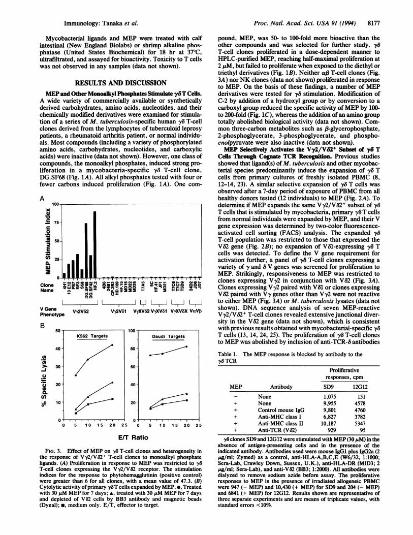
FIG. . y8T cells respond to monoalkyl phosphates. (A) The y8T-cell clone DG.SF68, which expresses Vy2/V2, proliferated when culturedwith crude reaction mixtures of alkyl phosphates. *, Methyl phosphate; e, MEP; *, n-propyl phosphate; *, n-butyl phosphate; E, amylphosphate; o, isopropyl phosphate; A, isoamyl phosphate; O, sec-butyl phosphate. (B) Selective stimulation of the y8 T-cell clone SD9 by MEP.*, MEP; *, diethyl phosphate (DEP); A, triethyl phosphate (TEP). (C) Stimulation ofthe y8T-cell clone SD9 by MEP derivatives. The percentageof the maximal proliferation response is shown. *, MEP (shown for comparison from B); o, hydroxyl derivative (,f3hydroxyethyl phosphate);A, carboxyl derivative (phosphoglycolic acid).
were separated by Ficoll/Hypaque and then stained withantibodies and analyzed. Antibodies used included phyco-erythrin-conjugated anti-TCR y/&-l, fluorescein isothiocy-anate-conjugated anti-CD3 (Becton Dickinson), anti-V82chain (BB3; a generous gift of L. Moretta, Natl. Cancer Res.Inst., Genoa, Italy), anti-Vy2 chain (TiyA; a generous gift ofF. Triebel, Inst. Gustave-Roussy, Villejuif, France), andfluorescein isothiocyanate-conjugated anti-mouse IgG anti-body (Ortho Diagnostic Systems Laboratories, Webster,TX). Stained cells were analyzed on a FACScan analyzer(Becton Dickinson). Controls with isotype-matched antibod-ies established the quadrants in such a way that >99% of thecells are in the double-negative region.
Partial Purification of the Major Natural Ligand fromMycobacteria. The major natural ligand was partially purifiedfrom Mycobacteriumfortuitum or Mycobacterium smegmat-is culture supernatant by ultrafiltration (Ultrasette 1K; Fil-
B
tron), activated charcoal/Celite column (1:2, wt/wt; 2.5 x1.5 cm) (21), barium precipitation, and reversed-phase andanion-exchange chromatography.Enzymatic Treatment of MEP and the Natural Mycobacte-
rial Ligand. For protease treatments, mycobacterial ligandsand purified protein derivative of tuberculin (PPD; 500 ug/ml; Statens Serumsinstitut, Copenhagen) were treated withthe endoprotease, pronase E (Sigma), subtilisin (Sigma), orproteinase K (Boehringer Mannheim) (all at 4 pg/400 pd). At30 hr, the samples were heated at 650C for 3 min and treatedwith either a second aliquot of endoprotease or an aliquot ofthe exopeptidase carboxypeptidase Y (Boehringer Mann-heim) at 40 tug/400 ,l. After an additional 20 hr, the sampleswere heated at 650C for 3 min and used in proliferation assayswith either the DG.SF68 clone for the samples or the myco-bacteria-reactive CD4+ a/B T-cell line, DG.1 (22), for the PPDsamples.
Donor;
Cultured with; SS
Medium |
0
MEP I-3C- , T
y6T cells (% CD31 cells) Anti-V82 (log green fluorescence)
FIG. 2. Effect of MEP on primary PBMC and the specificity in the response of 'y8 T cells to monoalkyl phosphate ligands. (A) Expansionby MEP of y8 T cells in primary cultures of PBMC as determined by two-color FACS analysis. The increase of -y8 T cells by MEP is comparableto that stimulated by M. tuberculosis (M.tb) lysates. (B) Two-color FACS analysis showing that yS T cells expanded in primary culture by MEPexclusively expressed V82. The percentages in the quadrants of interest are shown.
A
-U- MEP
-a- DEP
-A-- TEP
Ass -
YT
PS
L.0
0
BB
VM
P R
PS
PR
80
8176 Immunology: Tanaka et al.
Proc. Natl. Acad. Sci. USA 91 (1994) 8177
Mycobacterial ligands and MEP were treated with calfintestinal (New England Biolabs) or shrimp alkaline phos-phatase (United States Biochemical) for 18 hr at 370C,ultrafiltrated, and assayed for bioactivity. Toxicity to T cellswas not observed in any samples (data not shown).
RESULTS AND DISCUSSIONMEP and Other Monoalkyl Phosphates Stimulate y6T Cells.
A wide variety of commercially available or syntheticallyderived carbohydrates, amino acids, nucleotides, and theirchemically modified derivatives were examined for stimula-tion of a series of M. tuberculosis-specific human )y6 T-cellclones derived from the lymphocytes of tuberculoid leprosypatients, a rheumatoid arthritis patient, or normal individu-als. Most compounds (including a variety of phosphorylatedamino acids, carbohydrates, nucleotides, and carboxylicacids) were inactive (data not shown). However, one class ofcompounds, the monoalkyl phosphates, induced strong pro-liferation in a mycobacteria-specific yv T-cell clone,DG.SF68 (Fig. 1A). All alkyl phosphates tested with four orfewer carbons induced proliferation (Fig. 1A). One com-
A100-
x0
C75-
so.C: 50-E* 25-
IL'U
0*
CloneName
V GenePhenotyp
B
._a
0
a.CD)._-
A IILICNX)N @_1t@D ___ @T 9SN
V-cm cs-CM 4D-q wf V -VCl LC , 14 .111QN LX
26(A(A VV A-01IL
Vy2V82 Vy2V81 VyXV82 VyXV81 VyXV8X VaVf3
0 5 10 15 20 25 0 5 10 15 20 25
E/T Ratio
FIG. 3. Effect of MEP on y8 T-cell clones and heterogeneity inthe response of Vy2/V62+ T-cell clones to monoalkyl phosphateligands. (A) Proliferation in response to MEP was restricted to y8
T-cell clones expressing the Vy2/VS2 receptor. The stimulationindices for the response to phytohemagglutinin (positive control)were greater than 6 for all clones, with a mean value of 47.3. (B)Cytolytic activity ofprimary yy8T cells expanded by MEP. *, Treatedwith 30 puM MEP for 7 days; *, treated with 30 ALM MEP for 7 daysand depleted of V$2 cells by BB3 antibody and magnetic beads(Dynal); *, medium only. E/T, effector to target.
pound, MEP, was 50- to 100-fold more bioactive than theother compounds and was selected for further study., y5T-cell clones proliferated in a dose-dependent manner toHPLC-purified MEP, reaching half-maximal proliferation at2 ,uM, but failed to proliferate when exposed to the diethyl ortriethyl derivatives (Fig. 1B). Neither af3 T-cell clones (Fig.3A) norNK clones (data not shown) proliferated in responseto MEP. On the basis of these findings, a number of MEPderivatives were tested for y8 stimulation. Modification ofC-2 by addition of a hydroxyl group or by conversion to acarboxyl group reduced the specific activity ofMEP by 100-to 200-fold (Fig. 1C), whereas the addition ofan amino grouptotally abolished biological activity (data not shown). Com-mon three-carbon metabolites such as &3-glycerophosphate,2-phosphoglycerate, 3-phosphoglycerate, and phospho-enolpyruvate were also inactive (data not shown).MEP Selectively Activates the Vy2/VS2+ Subset of y8T
Cells Through Cognate TCR Recognition. Previous studiesshowed that ligand(s) ofM. tuberculosis and other mycobac-terial species predominantly induce the expansion of yv Tcells from primary cultures of freshly isolated PBMC (8,12-14, 23). A similar selective expansion of 'y6 T cells wasobserved after a 7-day period of exposure ofPBMC from allhealthy donors tested (12 individuals) to MEP (Fig. 2A). Todetermine if MEP expands the same V'y2/V82+ subset of y8T cells that is stimulated by mycobacteria, primary y8T cellsfrom normal individuals were expanded by MEP, and their Vgene expression was determined by two-color fluorescence-activated cell sorting (FACS) analysis. The expanded yvT-cell population was restricted to those that expressed theV82 gene (Fig. 2B); no expansion of V81-expressing y8 Tcells was detected. To define the V gene requirement foractivation further, a panel of y8 T-cell clones expressing avariety of y and 8 V genes was screened for proliferation toMEP. Strikingly, responsiveness to MEP was restricted toclones expressing V'y2 in conjunction with Va2 (Fig. 3A).Clones expressing Vy2 paired with V81 or clones expressingV62 paired with Vy genes other than Vy2 were not reactiveto either MEP (Fig. 3A) or M. tuberculosis lysates (data notshown). DNA sequence analysis of seven MEP-reactiveVy2/V52+ T-cell clones revealed extensive junctional diver-sity in the Va2 gene (data not shown), which is consistentwith previous results obtained with mycobacterial-specific 'y8T cells (13, 14, 24, 25). The proliferation of vy8 T-cell clonesto MEP was abolished by inclusion of anti-TCR-8 antibodies
Table 1. The MEP response is blocked by antibody to theyS TCR
Proliferativeresponses, cpm
MEP Antibody SD9 12G12
- None 1,075 151+ None 9,955 4578+ Control mouse IgG 9,801 4760+ Anti-MHC class I 6,827 3782+ Anti-MHC class II 10,187 5347+ Anti-TCR (V62) 929 95
y8 clones SD9 and 12G12 were stimulated with MEP (30 EM) in theabsence of antigen-presenting cells and in the presence of theindicated antibody. Antibodies used were mouse IgG1 plus IgG2a (2pg/ml; Zymed) as a control, anti-HLA-A,B,C,E (W6/32, 1:1000;Sera-Lab, Crawley Down, Sussex, U.K.), anti-HLA-DR (MID3; 2Pg/ml; Sera-Lab), and anti-V82 (BB3; 1:2000). All antibodies weredialyzed to remove sodium azide before assay. The proliferativeresponses to MEP in the presence of irradiated allogeneic PBMCwere 947 (- MEP) and 10,430 (+ MEP) for SD9 and 204 (- MEP)and 6841 (+ MEP) for 12G12. Results shown are representative ofthree separate experiments and are means of triplicate values, withstandard errors <10%.
Daudi Targets
80-
60 -
40 -
20
I
Immunology: Tanaka et al.
.e

Proc. Natl. Acad. Sci. USA 91 (1994)
(Table 1), which, together with the V gene specificity,suggests that the vy TCR is directly involved in the recog-nition of MEP. To further confirm TCR involvement in therecognition of nonpeptide ligands, transfection of Vy2 andV52 genes from a mycobacteria-specific y8 T-cell clone intothe TCR- Jurkat mutant, J.RT3-T3.5, conferred responsive-ness to MEP and the natural mycobacterial ligand (J.Bukowski and C.T.M., unpublished results). Finally, MEP-stimulated Vy2/V62-bearing T cells have the capacity to lysehuman transformed hematopoietic cells, illustrated in Fig. 3B(for Daudi and K562 cells). Thus, monoalkyl phosphatecompounds appear to act as antigens or superantigens thatstimulate Vy2/V82-bearing cells through their TCR in amanner similar to that of mycobacterial ligands.MEP Shares Major Chemical Properties with the Natural
Mycobacterial Ligand. To determine if the natural mycobac-terial ligand for 'y8 T cells is chemically similar to MEP, themajor natural ligand was partially purified from two repre-sentative mycobacterial species, M. fortuitum and M. smeg-matis. These partially purified ligands had similar chromato-
A100
to,1la
5-0
Absorbance Biological(260 nm) Activity
C
Q.0
0
o0
000
000Le-.o1mL-
B
E0.0
00
o000
._0
-
0
a-
graphic properties and were able to expand primary Vy2/V82T cells in a manner identical to crude mycobacterial prepa-rations. On the basis ofour purification protocols, the naturalligand has a molecular mass of <1 kDa, lacks significanthydrophobicity, and is anionic at pH 8. It is unlikely that themajor ligand is a nucleotide or an oligosaccharide because anactivated charcoal column failed to absorb the ligand (69-94% recovery), whereas almost all ribo- and deoxyribonu-cleotide compounds, as evidenced by the 0.15% recovery ofthe starting absorbance at 260 nm (Fig. 4A and refs. 26 and27), and oligosaccharides (21) were bound. Underscoring thenonpeptide nature of the mycobacterial ligand, proteasedigestion with various endoproteases or a combination of anendoprotease followed by carboxypeptidase Y (which digestsmost peptides including those with blocked amino termini)(28) did not affect the bioactivity ofthe natural mycobacterialligand, whereas the bioactivity ofthe PPD preparation for themycobacteria-specific CD4+ a,8 T-cell line, DG.1, was com-pletely abolished (Fig. 4B). In contrast to protease treatment,treatment with alkaline phosphatase completely abolished
M. fortuitum ligand (dilution) PPD (ug/ml)
0.0001 0.001 0.01
30000-
20000-
10000-
0.1 0.01 0.1
M. fortuitum ligand (dilution) MEP (dilution) M. smegmatis ligand (dilution)
FIG. 4. Purification and enzymatic treatment of the natural mycobacterial ligand and MEP. (A) Selective binding, by an activated charcoalcolumn, of nucleotides (which constitute the major compounds with absorbance at 260 nm) and oligosaccharides (21) but not the mycobacterialligand from M. smegmatis. Open bar, before activated charcoal; solid bar, after activated charcoal. (B) The natural mycobacterial ligand wasresistant to digestion with endo- and exoproteases, whereas the antigen for the mycobacteria-specific CD4+ a,3 T-cell line DG. 1 was completelydestroyed. Proliferative responses are shown for the DG.SF68 clone for the M. fortuitum ligand and the DG.1 line for PPD. Partially purifiedM. fortuitum ligand and PPD were untreated (o), mock-treated (o), treated with pronase E (A), treated with subtilisin (*), treated with proteinaseK (A), or treated with a combination of carboxypeptidase Y and either pronase E (v), subtilisin (O), or proteinase K (v). (C) The purifiedmycobacterial ligand and MEP were resistant to digestion with carboxypeptidase Y but were totally inactivated by treatment with alkalinephosphatase. Proliferative responses are shown for the DG.SF68 clone for M. fortuitum and MEP (Left and Center) and the SD9 clone for M.smegmatis (Right). Partially purified M. fortuitum ligand and crude MEP were untreated (o), mock-treated with the addition to 1 (0) or 10 (A)mM MgCl2, treated with calf intestinal alkaline phosphatase at 4 units/400 IlI or 40 units/400 ,ul (o), treated with shrimp alkaline phosphataseat 20 units/400 ,ul (o), or treated with carboxypeptidase Y at 40 gg/400 ,l (A). (Right) Partially purified M. smegmatis ligand was untreated(-), treated with heat-inactivated shrimp alkaline phosphatase (o), or treated with native shrimp alkaline phosphatase at 50 units/500 ,ul (o).
8178 Immunology: Tanaka et al.
7S-
25-

Proc. Natl. Acad. Sci. USA 91 (1994) 8179
the biological activity of the purified natural ligand and MEP,indicating that the compounds contain a critical phosphateresidue (Fig. 4C). The natural ligand appears to be signifi-cantly more bioactive on a molar basis than MEP (perhaps inthe picomolar range), as it is bioactive at levels below ourability to detect phosphate groups. We conclude that inaddition to their similar biological activities, MEP and thenatural mycobacterial ligand are chemically similar; they aresmall, hydrophilic, anionic molecules with critical phosphateresidues required for biological activity. Although similar,the natural ligands and MEP had distinct chromatographicproperties on anion-exchange and thin-layer chromatography(data not shown) and are, therefore, distinct compounds.Apart from the monoalkyl phosphates and the mycobac-
terial ligand, Vy2/V82-bearing T cells have been stimulatedin vitro by other antigens including Gram-positive and Gram-negative bacteria (9, 14, 15), the malarial parasite (Plasmo-dium vivax; refs. 29 and 30), and the tumor cell lines Daudi(31) and RPMI 8226 (32). This same Vy2/V82+ subset of y6cells expands in vivo in response to environmental stimuli,from a minor subpopulation in neonatal cord blood to dom-inate the y8population in adult peripheral blood (7). A similaracute in vivo expansion of V'y2/V82-bearing T cells (up to40%o of CD3Y peripheral blood T cells) is noted with certainbacterial infection (9-11) and in leprosy skin lesions (8, 33).We propose that the monoalkyl phosphates mimic naturalligands (such as those present in mycobacteria) that areresponsible for these in vivo expansions. As such, themonoalkyl phosphates may prove useful as immunothera-peutic agents to stimulate the Vy2/V82 subset of y6T cells.This subset of yv T cells may have been selected to respondto antigens entirely distinct from the peptide antigens towhich a,8 T-cells respond. These antigens are low molecularmass, phosphorylated compounds produced by a variety ofpathogens. By recognizing these nonpeptide ligands, 'y8 Tcells probably play a unique role in the human immunity toa wide spectrum of pathogenic bacteria and parasites.
Note Added in Proof. A recent report (34) suggests that the naturalcompound of mycobacteria able to stimulate vyS T cells may be aderivative of thymidine triphosphate, although the structure of theactive compound was not determined. The natural compound char-acterized in our report represents the major ligand in fast-growingmycobacteria (>990% of total activity) and is essentially devoid ofOD2,0 absorbing nucleotides.
The first two authors contributed equally to this work. We thankDrs. E. Hehre, P. Stanley, E. Beckman, J. Bukowski, and S.Porcelli for helpful discussion, V. Warren and C. Rapelje for helpwith the FACS analysis, the Carbohydrate Fractionation Facilityat A.E.C.O.M. for the use of their Dionex HPLC, and K. Uyemurafor V region sequencing. This research was supported by researchgrants from the Howard Hughes Medical Foundation (B.R.B. andY.T.), the National Institutes of Health (M.B.B. and R.L.M.), theLeukemia Society Scholar Award (M.B.B.), the Swiss NationalFund (G.D.L.), and the Arthritis Foundation (C.T.M.).
1. Brenner, M. B., Strominger, J. L. & Krangel, M. S. (1988)Adv. Immunol. 43, 133-192.
2. Porcelli, S., Brenner, M. B. & Band, H. (1991) Immunol. Rev.120, 137-183.
3. Raulet, D. H., Spencer, D. M., Hsiang, Y. H., Goldman, J. P.,Bix, M., Liao, N. S., Zijstra, M., Jaenisch, R. & Correa, I.(1991) Immunol. Rev. 120, 185-204.
4. Janeway, C. A., Jr. (1988) Nature (London) 333, 804-806.5. Haas, W., Pereira, P. & Tonegawa, S. (1993) Annu. Rev.
Immunol. 11, 637-685.6. Mombaerts, P., Arnoldi, J., Russ, F., Tonegawa, S. &
Kaufmann, S. H. E. (1993) Nature (London) 365, 53-56.7. Parker, C. M., Groh, V., Band, H., Porcelli, S. A., Morita, C.,
Fabbi, M., Glass, D., Strominger, J. L. & Brenner, M. B.(1990) J. Exp. Med. 171, 1597-1612.
8. Modlin, R. L., Pirmez, C., Hofman, F. M., Torigian, V.,Uyemura, K., Rea, T. H., Bloom, B. R. & Brenner, M. B.(1989) Nature (London) 339, 544-548.
9. Hara, T., Mizuno, Y., Takaki, K., Takada, H., Akeda, H.,Aoki, T., Nagata, M., Ueda, K., Matsuzaki, G., Yoshikai, Y.& Nomoto, K. (1992) J. Clin. Invest. 90, 204-210.
10. Bertotto, A., Gerli, R., Spinozzi, F., Muscat, C., Scalise, F.,Castellucci, G., Sposito, M., Candio, F. & Vaccaro, R. (1993)Eur. J. Immunol. 23, 1177-1180.
11. Sumida, T., Maeda, T., Takahashi, H., Yoshida, S., Yonaha,F., Sakamoto, A., Tomioka, H., Koike, T. & Yoshida, S. (1992)Infect. Immun. 60, 2554-2558.
12. Kabelitz, D., Bender, A., Schondelmaier, S., Schoel, B. &Kaufmann, S. H. E. (1990) J. Exp. Med. 171, 667-679.
13. Panchamoorthy, G., McLean, J., Modlin, R. L., Morita, C. T.,Ishikawa, S., Brenner, M. B. & Band, H. (1991) J. Immunol.147, 3360-3369.
14. De Libero, G., Casorati, G., Giachino, C., Carbonara, C.,Migone, N., Matzinger, P. & Lanzavecchia, A. (1991) J. Exp.Med. 173, 1311-1322.
15. Bender, A. & Kabelitz, D. (1993) Clin. Exp. Rheumatol. 11,295-299.
16. Pfeffer, K., Schoel, B., Guile, H., Kaufmann, S. H. E. &Wagner, H. (1990) Eur. J. Immunol. 20, 1175-1179.
17. Kosolapoff, G. M. (1950) Organophosphorous Compounds(Wiley, New York), pp. 220-227.
18. Morita, C. T., Verma, S., Aparicio, P., Martinez, C., Spits, H.& Brenner, M. B. (1991) Eur. J. Immunol. 21, 2999-3007.
19. Spits, H., Paliard, X., Vandekerckhove, Y., van Vlasselaer, P.& de Vries, J. (1991) J. Immunol. 147, 1180-1188.
20. Casorati, G., De Libero, G., Lanzavecchia, A. & Migone, N.(1989) J. Exp. Med. 170, 1521-1535.
21. Putman, E. W. (1957) Methods Enzymol. 3, 57-61.22. Porcelli, S., Morita, C. T. & Brenner, M. B. (1992) Nature
(London) 360, 593-597.23. Hacker, G., Kromer, S., Heeg, K., Ivanyi, J., Wagner, H. &
Pfeffer, K. (1992) Infect. Immun. 60, 2753-2757.24. Ohmen, J. D., Barnes, P. F., Uyemura, K., Lu, S. Z., Grisso,
C. L. & Modlin, R. L. (1991) J. Immunol. 147, 3353-3359.25. Davodeau, F., Peyrat, M. A., Hallet, M. M., Gaschet, J.,
Houde, I., Vivien, R., Vie, H. & Bonneville, M. (1993) J.Immunol. 151, 1214-1223.
26. Meek, J. L. (1986) Proc. Nati. Acad. Sci. USA 83, 4162-4166.27. Hurlbert, R. B. (1957) Methods Enzymol. 3, 785-805.28. Pamer, E. G., Wang, C. R., Flaherty, L., Lindahl, K. F. &
Bevan, M. J. (1992) Cell 70, 215-223.29. Goodier, M., Fey, P., Eichmann, K. & Langhorne, J. (1992)
Int. Immunol. 4, 33-41.30. Behr, C. & Dubois, P. (1992) Int. Immunol. 4, 361-366.31. Fisch, P., Malkovsky, M., Kovats, S., Sturm, E., Baakman,
E., Klein, B. S., Voss, S. D., Morrissey, L. W., DeMars, R.,Welch, W. J., Bolhuis, R. L. H. & Sondel, P. M. (1990) Sci-ence 250, 1269-1273.
32. Selin, L. K., Stewart, S., Shen, C., Mao, H. Q. & Wilkins,J. A. (1992) Scand. J. Immunol. 36, 107-117.
33. Uyemura, K., Deans, R. J., Band, H., Ohmen, J., Pancha-moorthy, G., Morita, C. T., Rea, T. H. & Modlin, R. L. (1991)J. Exp. Med. 174, 683-692.
34. Constant, P., Davodeau, F., Peyrat, M.-A., Poquet, Y., Puzo,G., Bonneville, M. & Fournie, J.-J. (1994) Science 264, 267-270.
Immunology: Tanaka et al.
![WJGP World Journal of Gastrointestinal Pathophysiology...51Cr-EDTA Whole intestine Urine Bjarnason et al[14], 1983 Lactulose/mannitol1 Small intestine Plasma/urine Rao et al[11], 2011](https://img.dokumen.tips/doc/110x75/60e0a2015420b723937de93c/wjgp-world-journal-of-gastrointestinal-pathophysiology-51cr-edta-whole-intestine.jpg)


![Status of Sterile Neutrinos Carlo Giunti...71Ge) = 11.43 ±0.03days [Hampel, Remsberg, PRC 31 (1985) 666] σG.S.(51Cr) = 55.3 ×10−46 cm2 (1 ±0.004) 3σ σ(51Cr) = σG.S.(51Cr)](https://img.dokumen.tips/doc/110x75/6055ba62f5dad3213c6c6690/status-of-sterile-neutrinos-carlo-giunti-71ge-1143-003days-hampel-remsberg.jpg)