Embed Size (px)
Citation preview
NCGC
ATccbleowcTTbaiborparccaTpw3
KBtn
ooC
Eg0
Ed
Review Article
3
onvital Tooth Bleaching: A Review of the Literature andlinical Proceduresianluca Plotino, DDS,* Laura Buono, DDS,* Nicola M. Grande, DDS,*ornelis H. Pameijer, DMD, DSc, PhD,† and Francesco Somma, MD, DDS*
mcsh(rAipa
abee
bda
p(ttip
tb
E
sb
I
cop
r
bstractooth discoloration varies in etiology, appearance, lo-alization, severity, and adhesion to tooth structure. Itan be defined as being extrinsic or intrinsic on theasis of localization and etiology. In this review of the
iterature, various causes of tooth discoloration, differ-nt bleaching materials, and their applications to end-dontically treated teeth have been described. In thealking bleach technique the root filling should be
ompleted first, and a cervical seal must be established.he bleaching agent should be changed every 3–7 days.he thermocatalytic technique involves placement of aleaching agent in the pulp chamber followed by heatpplication. At the end of each visit the bleaching agent
s left in the tooth so that it can function as a walkingleach until the next visit. External bleaching of end-dontically treated teeth with an in-office techniqueequires a high concentration gel. It might be a sup-lement to the walking bleach technique, if the resultsre not satisfactory after 3– 4 visits. These treatmentsequire a bonded temporary filling or a bonded resinomposite to seal the access cavity. There is a defi-iency of evidence-based science in the literature thatddresses the prognosis of bleached nonvital teeth.herefore, it is important to always be aware of theossible complications and risks that are associatedith the different bleaching techniques. (J Endod 2008;4:394 – 407)
ey Wordsleaching, discoloration, in-office bleaching, root filledeeth, thermocatalytic technique, walking bleach tech-ique
From the *Department of Endodontics, Catholic Universityf Sacred Heart, Rome, Italy; and †Professor Emeritus, Schoolf Dental Medicine, University of Connecticut, Farmington,onnecticut.
Address requests for reprints to Gianluca Plotino, DDS, Vialeonora Duse, 22, 00197 Rome, Italy. E-mail address:[email protected]/$0 - see front matter
Copyright © 2008 by the American Association ofndodontists.oi:10.1016/j.joen.2007.12.020
r
94 Plotino et al.
History of Bleaching TeethReports on bleaching discolored nonvital teeth were first described during the
iddle of the 19th century (1), advocating different chemical agents (2). Initially,hlorinated lime was recommended (3), followed later by oxalic acid (4, 5) and agentsuch as chlorine compounds and solutions (6 – 8), sodium peroxide (9), sodiumypochlorite (10), or mixtures consisting of 25% hydrogen peroxide in 75% etherpyrozone) (11, 12). An early description (1884) of the use of hydrogen peroxide waseported by Harlan (13). Superoxol (30% hydrogen peroxide) had been mentioned bybbot (14) in 1918. Prinz (15) in 1924 recommended using heated solutions consist-ng of sodium perborate and Superoxol for cleaning the pulp cavity. Some authorsroposed using light (16, 17), heat (16, 18, 19 –22), or electric currents (23, 24) toccelerate the bleaching reaction by activating the bleaching agent.
Sodium perborate in the form of monohydrate, trihydrate, or tetrahydrate is useds a hydrogen peroxide–releasing agent. It has been used since 1907 as an oxidizer andleaching agent, especially in washing powder and other detergents. The whiteningfficacy of sodium perborate monohydrate, trihydrate, or tetrahydrate mixtures withither water or hydrogen peroxide is not different (25).
Causes of Tooth DiscolorationThe correct diagnosis of the cause of discoloration of teeth is of great importance
ecause it has a profound effect on the treatment outcome. It is therefore necessary thatental practitioners have an understanding of the etiology of tooth discoloration torrive at a correct diagnosis leading to an appropriate treatment plan (26).
Tooth color is determined by a combination of phenomena associated with opticalroperties and light (27). Essentially, tooth color is determined by the color of dentin28) and by intrinsic and extrinsic colorations (26). Intrinsic color is determined byhe optical properties of enamel and dentin and their interaction with light (28). Ex-rinsic color depends on material absorption on the enamel surface (29). Any changen enamel, dentin, or coronal pulp structure can cause a change of the light-transmittingroperties of the tooth (30).
Tooth discoloration varies in etiology, appearance, location, severity, and affinityo tooth structure (31). It can be classified as intrinsic, extrinsic, or a combination ofoth, according to its location and etiology (32).
xtrinsic CausesThe principal causes are chromogens derived from habitual intake of dietary
ources, such as wine, coffee, tea, carrots, oranges, licorice, chocolate, or from to-acco, mouth rinses, or plaque on the tooth surface (26, 32).
ntrinsic CausesSystemic causes are 1) drug-related (tetracycline); 2) metabolic: dystrophic cal-
ification, fluorosis; and 3) genetic: congenital erythropoietic porphyria, cystic fibrosisf the pancreas, hyperbilirubinemia, amelogenesis imperfecta, and dentinogenesis im-erfecta.
Local causes are 1) pulp necrosis, 2) intrapulpal hemorrhage, 3) pulp tissueemnants after endodontic therapy, 4) endodontic materials, 5) coronal filling mate-
ials, 6) root resorption, and 7) aging.JOE — Volume 34, Number 4, April 2008
bpaafbtaass
mcsfcd
aa(rotd
deeepiff3cmtrmramsp
opcdtlo
P
rp
ohic
tntcmpccpt
tiifcrcdf
crs2
P
cagdcc
ca
E
5bolsftiti
pd
Review Article
J
Extrinsic stain is found on the tooth surface and for this reason cane removed with relative ease. Affinity of materials to the tooth surfacelays a critical role in the deposition of extrinsic stain. The types ofttractive forces include long-range interactions such as electrostaticnd van der Waals forces and short-range interactions such as hydrationorces, hydrophobic interactions, dipole-dipole forces, and hydrogenonds (33). These interactive forces allow the chromogens to get closeo the tooth surface and determine whether adhesion will occur. Theffinity of chromogen adhesion varies with the material, and the mech-nisms that determine the strength of adhesion are not clearly under-tood (34). For example, clinical evidence has shown that tea and coffeetains become more difficult to remove with aging (34).
In the past, extrinsic tooth discoloration has been classified asetallic or nonmetallic stain (35). There are several problems with this
lassification. First, it does not explain the mechanism of discoloration;econd, the extent of staining is variable and might depend on multipleactors; third, all metals do not cause extrinsic discoloration. To over-ome these problems, a new classification based on the chemistry ofental discoloration has been proposed (34).
The most commonly used procedure to remove discoloration fromtooth surface is by using abrasives (such as prophylactic pastes) (31) orcombination of abrasive and surface active agents such as toothpastes
36). These methods prevent stain accumulation and to a certain extentemove extrinsic stains (34); however, satisfactory stain removal dependsn the type of discoloration (36). Unfortunately, the chemical interactionshat determine the affinity of different types of materials that cause extrinsicental stains are not well-understood (34).
Unlike extrinsic discolorations that occur on the surface, intrinsiciscoloration is due to the presence of chromogenic material withinnamel or dentin, incorporated either during odontogenesis or afterruption (31). This type of stain can be divided into 2 groups, pre-ruptive and posteruptive (31, 34). The most common type ofre-eruptive staining is endemic fluorosis caused by excessive fluoridengestion during tooth development. A method for removal of dentalluorosis stains, on the basis of the structural characteristics of theluorotic tooth, has been reported to be effective (37). It encompassed
principal stages: enamel etching with 12% hydrochloric acid, appli-ation of pure sodium hypochlorite, and filling the chemically openedicroporosities with a light-cured dental adhesive. Staining caused by
etracycline administration also occurs during odontogenesis and is aesult of antibiotic interactions with hydroxyapatite crystals during theineralization phase. Malformation of dental tissues occurring as a
esult of inherited conditions such as dentinogenesis imperfecta andmelogenesis imperfecta can also cause pre-eruptive dental stain. He-atologic disorders such as erythroblastosis fetalis, thalassemia, and
ickle cell anemia might also cause pre-eruptive stain because of theresence of coagulated blood within the dentinal tubules.
Traumatic events that influence tooth formation can represent an-ther cause of pre-eruptive discoloration (31). Teeth might acquireosteruptive intrinsic stains in a similar way. Normal aging can alsoause posteruptive dental stain caused by the deposition of secondaryentin, tertiary dentin, and pulp stones (38). It has also been reported
hat dental procedures might lead to tooth discoloration, owing to re-ease of metals from, for instance, amalgam restorations or incompletebturation of the pulp chamber after endodontic treatment (34).
Causes of Intrinsic Local Discolorationulp Necrosis
Bacterial, mechanical, or chemical irritation of the pulp mightesult in tissue necrosis, causing release of noxious by-products that can
enetrate tubules and discolor the surrounding dentin (39). The degree fOE — Volume 34, Number 4, April 2008
f discoloration is directly related to the duration of time that the pulpas been necrotic. The longer the discoloration compounds are present
n the pulp chamber, the greater the discoloration. This discolorationan usually be bleached intracoronally (40).
Intrapulpal HemorrhagePulp extirpation or severe tooth trauma can cause hemorrhage in
he pulp chamber caused by rupture of blood vessels. Blood compo-ents subsequently flow into the dentinal tubules, causing a discolora-
ion of the surrounding dentin (41, 42). Initially, a temporary colorhange of the crown to pink can be observed. This is followed by he-olysis of red blood cells. The released heme then combines with the
utrefying pulpal tissue to form iron (26, 43). The iron in turn can beonverted by hydrogen sulfates that are produced by bacteria to dark-olored iron sulfates, which discolor the tooth grey. These products canenetrate deep into the dentinal tubules and can cause discoloration of
he entire tooth.Grossman (44) asserted in 1943 that the depth of dentinal pene-
ration determines the degree of discoloration, although there was little,f any, scientific evidence to support this hypothesis (26). In vitro stud-es have recently shown that the major cause of discoloration of nonin-ected traumatized teeth is the accumulation of the hemoglobin mole-ule or other hematin molecules (45). In the absence of infection, theelease of iron from the protoporphyrin ring is an unlikely event, be-ause there are no reactions caused by bacterial by-products. An un-erstanding of the nature of tooth staining after trauma is of importance
or the manufacturing of bleaching agents with specific activity.If trauma is the cause of pulpal necrosis, the discoloration be-
omes more severe over time. However, if not, the discoloration can beeversed, and the tooth can regain its original shade (26). It has beenhown that the pinkish hue seen initially after trauma might disappear in–3 months if the tooth becomes revascularized (18).
ulp Tissue Remnants after Endodontic TreatmentThe same events that characterize intrapulpal hemorrhage can be
aused by trauma inflicted during pulp extirpation and failure to removell pulp remnants. Tissues remaining in the pulp chamber disintegrateradually, and blood components can flow into the tubules, causingiscoloration. If the access cavity is inadequate, some pulp remnantsan remain inside the pulp chamber, in particular in the pulp horns,ausing coronal discoloration (46, 47).
Removal of all tissues and correct intracoronal bleaching in theseases are usually successful, but they are unnecessary and can bevoided if all pulpal remnants are removed from the access cavity.
ndodontic MaterialIncomplete removal of filling materials and sealer remnants (48 –
2) or medicaments containing tetracycline (53) from the pulp cham-er can cause endodontically treated teeth to discolor. This is a frequentccurrence but can easily be prevented by removing all materials to a
evel just below the bone. Filling material and intracanal medicamentsealed in the pulp chamber are in direct contact with dentin, sometimesor long periods, allowing penetration into dentinal tubules. These ma-erials need to be in direct contact with dentin for a period of time beforet is possible to appreciate any visible coronal shade alteration. Althoughhere is no penetration in the enamel, it is still possible to see a changen tooth color (38).
Although intracoronal bleaching is the treatment of choice, therognosis, however, depends on the type of sealer and duration ofiscoloration (49). For example, stains made by metallic ions are dif-
icult to remove with bleaching treatment. Therefore, the need to re-
Nonvital Tooth Bleaching 395
mb
C
dgdrrcrdosr
bFa
R
ejt
A
ssso
os
bar
sogp
dgrcrwbi
e(cm
tudrcthas
sbcc
C
ioebaeqst3teta
ciaih
wacsp
ct21
mtmco
Review Article
3
ove all remnants using burs to clean the chamber walls before startingleaching procedures cannot be emphasized enough.
oronal Filling MaterialsMicroleakage of old resin composite restorations might cause
ark discoloration of margins and, over time, of dental tissues. Amal-am used as filling material after endodontic therapy can turn the dentinark grey, owing to dark-colored metallic components. When used toestore lingual access preparation or a developmental groove in ante-ior teeth, as well as in premolar teeth, amalgam might discolor therown. This type of discoloration is difficult to bleach and tends toeappear over time because of the tenacity of the oxidizing products toental tissues. Sometimes the dark appearance of the crown is causednly by the amalgam restoration that can be seen through the toothtructure. In such cases, replacing the amalgam with an esthetic resto-ation usually corrects the problem (40).
Metal posts used to construct a core might cause discolorationecause of transparency of enamel or because of released metallic ions.requently, dark-pigmented dentin can be seen when these restorationsre removed (40).
oot ResorptionRoot resorption, while clinically asymptomatic, might occasionally
xhibit an initial pink appearance (pink spot) at the cementoenamelunction (CEJ) that can serve in the differential diagnosis of the origin ofhe discoloration (26).
ging (Dystrophic Calcification)During the natural aging process the physiologic deposition of
econdary dentin affects the light-transmitting properties of teeth, re-ulting in a gradual darkening (26) based on a narrowing of the pulppace, resulting in an increase in tooth structure that in turn affectspacity. In addition, the chemical structure of teeth changes over time.
Bleaching Agents for Whitening of Root-filled TeethThe bleaching agents that are most commonly used for whitening
f root-filled teeth are hydrogen peroxide, carbamide peroxide, andodium perborate.
Hydrogen peroxide is the active ingredient in currently used toothleaching materials. It might be applied directly or can be produced bychemical reaction from carbamide peroxide (54) or sodium perbo-
ate (55).Peroxides can be classified as organic and inorganic. They are
trong oxidizers and can be considered as products of hydrogen per-xide (H2O2) when substitution of hydrogen atoms with metals (inor-anic peroxides) or with organic radicals (organic peroxides) takeslace (40).
Hydrogen peroxide is used in dentistry as a whitening material atifferent concentrations from 5%–35%. At a high concentration hydro-en peroxide is caustic, burns tissues on contact, and can release freeadicals. High-concentration solutions must be handled with care be-ause they are thermodynamically unstable and might explode unlessefrigerated and kept in a dark container. Because of its low moleculareight, this substance can penetrate dentin and can release oxygen thatreaks the double bonds of the organic and inorganic compoundsnside the dentinal tubules (56, 57).
The breakdown of hydrogen peroxide into active oxygen is accel-rated by application of heat, the addition of sodium hydroxide, or light58, 59). Hydrogen peroxide–releasing bleaching agents are thereforehemically unstable. Only fresh preparations should be used, which
ust be stored in a dark cool place. o96 Plotino et al.
Carbamide peroxide [CO(NH2)2H2O2] is an organic white crys-alline compound and is formed by urea and hydrogen peroxide andsed in different concentrations. In a hydrophilic environment it breaksown into approximately 3% hydrogen peroxide and 7% urea. Cur-ently, the most popular commercial bleaching preparations containingarbamide peroxide usually also include glycerin at different concen-rations because this makes it more chemically stable compared withydrogen peroxide. Ten percent carbamide peroxide bleaching agentslso demonstrated higher antibacterial effect than a 0.2% chlorhexidineolution (60).
Sodium perborate is an oxidizing agent available as a powder. It istable when dry; however, in the presence of acid, warm air, or water, itreaks down to form sodium metaborate, hydrogen peroxide, and nas-ent oxygen. Sodium perborate is easier to control and safer than con-entrated hydrogen peroxide solutions.
omparison StudiesSeveral studies have reported bleaching effectiveness by compar-
ng mixtures of sodium perborate with distilled water or hydrogen per-xide in different concentrations. Rotstein et al (61, 62) and Weigert al (63) did not report any significant difference in the effectivenessetween sodium perborate mixed with 3%–30% hydrogen peroxidend the sodium perborate– distilled water mixture. However, the whit-ning effect of the second mixture can take longer, so that more fre-uent changes of the bleaching agent might be necessary. The shadetability of teeth treated by a mixture of perborate and water is as high ashe shade stability of teeth in which a mixture of sodium perborate with% or 30% hydrogen peroxide was used (25, 62). Other surveys found
hat mixing sodium perborate with 30% hydrogen peroxide was moreffective than mixing with water (64, 65). Freccia et al (66) showed thathe walking bleach technique with a mixture of 30% hydrogen peroxidend sodium perborate was as effective as the thermocatalytic technique.
Other hydrogen peroxide–separating agents such as sodium per-arbonate can be used to bleach discolored teeth. Suspensions consist-ng of sodium percarbonate and water or 30% hydrogen peroxide hadgood bleaching effect on teeth that were artificially stained in vitro by
ron sulfide (67). However, clinical studies with sodium percarbonateave not been reported.
Aldecoa and Mayordomo (68) described good clinical successhen using a mixture consisting of sodium perborate and 10% carb-mide peroxide gel. This suspension was used as a temporary intra-oronal filling after application of a regular walking bleach paste withodium perborate and hydrogen peroxide. The authors claimed that thisrocedure led to long-term stability of the tooth whitening therapy.
An in vitro study reported that sodium perborate and Superoxolombinations were more effective than sodium perborate alone. Al-hough the difference was not statistically significant, this study reported0% difference in the effectiveness between freshly refrigerated and-year-old agents (64).
Clinical Bleaching Techniques for EndodonticallyTreated Teeth
Bleaching of endodontically treated teeth that present with chro-atic alterations is a conservative alternative to a more invasive esthetic
reatment such as placement of crowns or veneers. Furthermore, whenetal-free restorations are planned, bleaching of the prosthetic core
an be useful in improving the final esthetic results. These materials notnly depend on light transmission characteristics but also on the color
f the teeth that are being restored.JOE — Volume 34, Number 4, April 2008
E
ap
I
p
smenu
pd
f
P
tdhbtgvarts
oprTid
ltmbsitcrb
ipc
W
acdc
bNimtt
uopocctrta
P
asmrccArfrtcttb
Fitwei(
Review Article
J
xtrinsic Stain TreatmentThe most commonly used procedure to treat extrinsic stains is by
professional hygiene treatment and by polishing tooth surfaces withrophylactic cups and more or less aggressive abrasive pastes.
ntrinsic Stain TreatmentIntrinsic stains from systemic causes are difficult to treat without a
rosthetic rehabilitation.The microabrasion technique can be successfully used to treat
uperficial discolored enamel defects (69). At the present time, enamelicroabrasion is considered a conservative technique to improve the
sthetic appearance of teeth with surface defects. Because this tech-ique removes superficial external enamel, a correct diagnosis is oftmost importance (70).
For intrinsic stains to be removed, chemicals such as hydrogeneroxide are required because they penetrate enamel and dentin andecolorize or solubilize the chromogens (31).
Internal discoloration of teeth represents the primary indicationor whitening of root-filled teeth (41).
reliminary TreatmentIt is important to determine the cause of tooth discoloration. First,
he surface of the tooth should be cleaned thoroughly to determine theegree of external discoloration. For that reason it is very important toave a preliminary professional hygiene treatment before starting theleaching. The patient should be informed that the results of bleachingherapy are not predictable, and that complete recovery of color is notuaranteed in all cases (71). Furthermore, information should be pro-ided concerning the different treatment stages, possible complications,nd the fact that application of the bleaching agent often needs to beepeated to obtain optimum results (39). It is very helpful to take pre-reatment and post-treatment photographs to show the patient the re-ults obtained at the end of the treatment.
Before treatment a radiograph should be made to check the qualityf the root filling. The filling should not only prevent coronal-apicalassage of microorganisms but also prevent bleaching agents fromeaching the apical tissues, potentially having detrimental effects (71).herefore, an inadequate root filling should be replaced before bleach-ng, and the new filling material should be allowed to set for at least 7ays after obturation before starting the bleaching procedures (39).
Deficient restorations should be identified and replaced; cariousesions should be restored. If restorations do not match the shade of theooth, they should be replaced at the end of the treatment with materials
atching the whitened tooth. The final shade of the tooth as a result ofleaching cannot be reliably predicted, and this makes it difficult toelect the correct shade of filling material before bleaching. Therefore,t is advisable to restore carious lesions or replace deficient fillings withemporary materials before treatment or to replace restorations afterompletion of bleaching. It should be emphasized that the tooth beestored with high quality fillings to ensure the effectiveness of theleaching agent and to avoid leakage of the agent into the oral cavity.
Furthermore, it is of great importance to apply rubber dam tosolate the treated tooth, to prevent reinfection of the root canal, and torotect the adjacent structures from the bleaching agent. For difficultases it is possible to use a liquid dam.
alking Bleach TechniqueThe first description of the walking bleach technique (Fig. 1) with
mixture of sodium perborate and distilled water was mentioned in aongress report by Marsh and published by Salvas (72). In this proce-ure, the mixture was left in the pulp cavity for a few days, and the access
avity was sealed with provisional cement. The mixture of sodium per- iOE — Volume 34, Number 4, April 2008
orate and water was reconsidered by Spasser (73) and modified byutting and Poe (74), who advocated the use of 30% hydrogen peroxide
nstead of water to improve the bleaching effectiveness of the mixture. Aixture of sodium perborate and water or hydrogen peroxide continues
o be used today and has been described many times as a successfulechnique for intracoronal bleaching (62, 75–77).
There are numerous studies that have reported on the successfulse of the walking bleach technique for correction of severely discol-red teeth caused by incorporation of tetracycline (68, 78 – 84). Thisrocedure starts with intentional devitalization and root canal treatmentf the tooth to enable application of the bleaching agent into the pulphamber. Because the methods of intentional devitalization and rootanal treatment have risks, the advantages and disadvantages of thisherapy should be assessed. Restorative treatment options such as ce-amic veneers should be considered as an alternative procedure. Fur-hermore, there is now evidence that prolonged bleaching with carb-mide peroxide can also reach the desired results (38, 85– 88).
reparation of the Pulp CavityBefore preparation of the access cavity, rubber dam should be
pplied to protect the adjacent structures. The access cavity should behaped in such a way that remnants of restorative materials, root-fillingaterials, and necrotic pulp tissue are completely removed. For this
eason, in particular for maxillary incisors, it is very important to in-lude the mesial and distal pulp horns in the access cavity, because theyan contain necrotic pulpal remnants, which can cause discoloration.dditional cleaning of the pulp cavity with sodium hypochlorite is alsoecommended (39). In some reports, conditioning of the dentin sur-ace of the access cavity with 37% orthophosphoric acid is suggested toemove the smear layer and to open the dentinal tubules. This promoteshe penetration of the bleaching agent deep into the tubules and in-reases its effectiveness (89). It has also been recommended to cleanhe pulpal cavity with alcohol before application of the bleaching agento dehydrate dentin and reduce surface tension; consequently theleaching agent will penetrate with greater ease into the dentin, achiev-
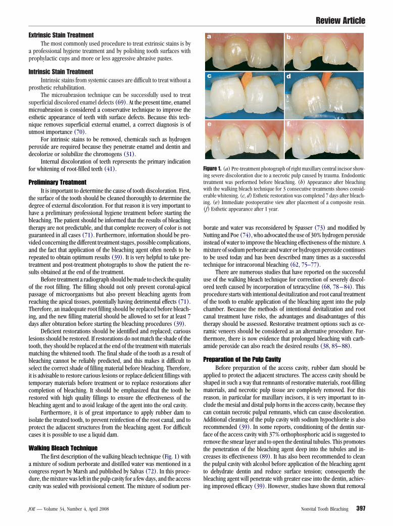
igure 1. (a) Pre-treatment photograph of right maxillary central incisor show-ng severe discoloration due to a necrotic pulp caused by trauma. Endodonticreatment was performed before bleaching. (b) Appearance after bleachingith the walking bleach technique for 3 consecutive treatments shows consid-rable whitening. (c, d) Esthetic restoration was completed 7 days after bleach-ng. (e) Immediate postoperative view after placement of a composite resin.f) Esthetic appearance after 1 year.
ng improved efficacy (39). However, studies have shown that removal
Nonvital Tooth Bleaching 397
obtoald
C
bwrbrnb
asrfadvaatzpFttam
tspctc
mtialTeTctsbtoCtlm
aHt
A
rd(pbdca
T
tpqcifrrad
Ft
Review Article
3
f the smear layer with 37% orthophosphoric acid does not improve theleaching effectiveness of either sodium perborate or a high concen-ration of hydrogen peroxide (90, 91). Furthermore, the pre-treatmentf dentin with acid might lead to an increased diffusion of bleachinggents into the periodontium (92). Therefore, removal of the smearayer from the dentin of the pulp chamber before the bleaching proce-ures is still a controversial issue.
ervical SealThe root filling should be reduced 1–2 mm below the CEJ. This can
e determined by using a periodontal probe placed in the pulp cavity,hile reproducing the corresponding external probing to the CEJ. To
emove filling material up to this level, Gates-Glidden or Largo burs cane used. It is important to clean the cavity surfaces from debris andemnants of endodontic materials, because the presence of contami-ants on the surfaces might negatively influence the efficacy of theleaching agent.
A root filling does not adequately prevent diffusion of bleachinggents from the pulpal chamber to the apical foramen (93, 94). Han-en-Bayless and Davis (95) indicated that a base is required to preventadicular penetration of bleaching agents. Therefore, sealing the rootilling with a base is essential, for which a variety of dental materials suchs glass-ionomer cements, intermediate restorative material (IRM), hy-raulic filling materials (Cavit, Coltosol), resin composites, photo-acti-ated temporary resin materials (Fermit), zinc oxide– eugenol cement,nd zinc phosphate cement have been suggested as an interim sealinggent during bleaching techniques. McInerney and Zillich (96) foundhat Cavit and IRM provided better internal sealing of the dentin than didinc phosphate cement, whereas Hansen-Bayless and Davis (97) re-orted that Cavit was a more effective barrier to leakage than IRM.urthermore, hydraulic filling materials (Cavit and Coltosol) providedhe most favorable cavosurface seal when they were firmly packed intohe cavity space to prevent microleakage, when compared with a photo-ctivated temporary resin material (Fermit), zinc oxide– eugenol ce-ent, and a zinc oxide phosphate cement (98).
Temporary sealing materials need to be removed before providinghe final restoration of the access cavity. Rotstein et al (99) demon-trated that a 2-mm layer of glass-ionomer cement was effective inreventing penetration of 30% hydrogen peroxide solution into the rootanal. Thus, the use of this material as a base during bleaching presentshe additional advantage that it can be left in place after bleaching andan serve as a base for the final restoration.
The sealing material should reach the level of the epithelial attach-ent or the CEJ, respectively, to avoid leakage of bleaching agents into
he periodontium (100). The shape of the cervical seal should be sim-lar to the external anatomic landmarks, thus reproducing CEJ positionnd interproximal bone level. A flat barrier, level with the labial CEJ,eaves a large portion of the proximal dentinal tubules unprotected.herefore, the barrier should be determined by probing the level of thepithelial attachment at the mesial, distal, and labial aspects of the tooth.he intracoronal level of the barrier is placed 1 mm incisal to theorresponding external probing of the attachment. With this methodhe coronal outline of the attachment defines an internal pattern of thehape and location of the barrier (100). However, the impact of theleaching agents on the discolored dentin should not be hampered byhe cervical seal. Dentin tubules at the coronal third of the root run in anblique direction from the apex to the crown, so that the tubules at theEJ are originating more apically inside the root canal. If bleaching ofhe cervical region of the tooth is required, a stepwise reduction of theabial part of the seal and use of a mild bleaching agent are recom-
ended for the final dressings (99). t
98 Plotino et al.
The placement of a piece of rubber dam has been suggested to acts a further barrier to isolate filling material from the bleaching agent.owever, Hosoya et al (101) reported no significant differences be-
ween the groups with and without the placement of this barrier.
pplication of the Bleaching AgentSodium perborate (tetrahydrate) mixed with distilled water in a
atio of 2:1 (g/mL) is a suitable bleaching agent (102). In case of severeiscoloration, 3% hydrogen peroxide can be applied in lieu of water74). The bleaching agent can be applied with an amalgam carrier orlugger and should be changed every 3–7 days. Successful bleachingecomes apparent after 2– 4 visits, depending on the severity of theiscoloration. The patients should be instructed to evaluate the tootholor on a daily basis and return when the bleaching is acceptable tovoid “over-bleaching” (39) (Fig. 2).
emporary FillingBefore application of the bleaching agent, the enamel margins of
he cavity should be etched with 37% orthophosphoric acid to accom-lish an adhesive temporary filling. The walking bleach technique re-uires a sound seal around the access cavity with a resin composite orompomer to ensure its effectiveness and to avoid leakage of the bleach-ng agent into the oral cavity. This cannot be guaranteed if temporaryilling materials are being used (103). In addition, a good seal preventsecontamination of the dentin with microorganisms and reduces theisk of renewed staining. It is often difficult to place filling materials onsoft bleaching agent. A small sterile cotton pellet impregnated with aentin bonding agent, placed on the bleaching agent and then light-
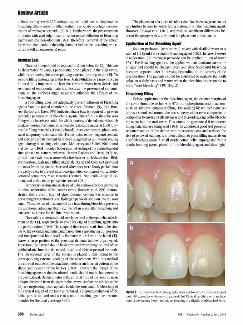
igure 2. (a) Pre-treatment photograph shows a yellow-brown discoloration ofooth #8 caused by endodontic treatment. (b) Clinical results after 3 applica-
ions of the walking bleach technique, resulting in a slightly overbleached tooth.JOE — Volume 34, Number 4, April 2008
cfDbts
RR
ceiticcgroichubmcbaaddretDaaaptpFv
r
T
bboo
fctpscpo
Teet
tt
I
no(awtb(btadtgr
bc
bsib
Fracnia
Review Article
J
ured, simplifies the placement of a filling material. The temporaryilling should only be attached to the enamel margins of the access cavity.uring this phase of the treatment, the pulp chamber is filled with theleaching agent and not with an adhesively attached restorative material, so
hat no internal stabilization of the tooth is provided. Therefore, the patienthould be informed about an increased risk of fracture (71).
estoration of the Access Cavity and Postoperativeadiograph
After bleaching, the access cavity should be restored with a resinomposite, which is bonded by means of the acid-etch technique tonamel and dentin. This avoids recontamination with bacteria and stain-ng substances and improves the stability of the tooth. A sound restora-ion with sealed dentinal tubules is a prerequisite to successful bleach-ng therapy (71, 80). Some authors (80, 104) recommend using resinomposites with lighter shades to compensate for bleaching that was notompletely successful. The adhesive strength of resin composites andlass-ionomer cements to bleached enamel and dentin is temporarilyeduced (105–112). It has been established that remnants of peroxider oxygen inhibit the polymerization of resin composites (109, 113). Its less likely that changes in the enamel structure might influence resinomposite adhesion (114, 115). Nevertheless, the appearance of theybrid layer in bleached enamel is less regular and distinct than innbleached enamel (116). This might explain why access cavities ofleached teeth that are restored with resin composite occasionally showarginal leakage (117). The negative influence of hydrogen peroxide–
ontaining bleaching agents on adhesion can be reduced by moderateevelling of the cavity before acid etching (118). The same can bechieved by pre-treatment of enamel with dehydrating agents such aslcohol and the use of acetone-containing adhesives (119, 120). Toissolve remnants of peroxide, the cavity can also be cleaned with so-ium hypochlorite (62). A contact time of at least 7 days with water isecommended to avoid the reduction of adhesion of composites tonamel (115, 121, 122). Optimal bonding to bleached dental hardissue can be achieved after a period of about 3 weeks (123, 124).uring this period, the color of the bleached tooth should be stable andcalcium hydroxide dressing placed in the pulp cavity for buffering thecid pH that can occur on cervical root surfaces after intracoronalpplication of bleaching agents (71, 125). The calcium hydroxide sus-ension temporarily placed into the pulp chamber after completion ofhe bleaching procedure does not interfere with the adhesion of com-osite materials used for final restoration of the access cavity (126).urthermore, compromised bonding to bleached enamel can be re-ersed with sodium ascorbate, an antioxidant (127).
A postoperative radiograph after bleaching and regular follow-upadiographs are recommended (128).
hermocatalytic TechniqueThis technique (Fig. 3) has been proposed for many years as the
est technique to bleach nonvital teeth because of the strong interactionetween hydrogen peroxide and heat (44, 45, 77, 129 –133). More-ver, a common clinical technique is to use 30%–35% hydrogen per-xide placed in the pulp chamber between appointments (134 –136).
Preparation of the access cavity consists of cleaning, removal ofilling materials, and all the preparation procedures described for dis-olored teeth when using the walking bleach technique. However, thisechnique involves placement of 30%–35% hydrogen peroxide in theulp chamber followed by heat application by electric heating devices orpecially designed lamps. It has been observed that heat applicationauses a reaction that increases bleaching properties of the hydrogeneroxide (2). Heat might be applied by using a heated metal instrument
r other commercial heat applicators (Touch’n Heat, System B; Analytic 1OE — Volume 34, Number 4, April 2008
echnology, Orange, CA). Heat application is repeated 3 or 4 times atvery appointment, changing the pellet with “fresh” bleaching agent atach visit. When heat is applied, a reaction produces foam and releaseshe oxygen present in the preparation.
At the end of each appointment the bleaching agent is sealed intohe pulp chamber for additional bleaching between appointments as inhe walking bleach technique.
n-Office TechniqueSome authors have described the successful clinical use of exter-
al bleaching of nonvital root-filled teeth (Fig. 4) with carbamide per-xide gels or hydrogen peroxide at high concentrations (15%–35%)137–139). The whitening gel is applied by means of a bleaching traynd is placed directly on the tooth, which is isolated with rubber dam orith other techniques (Fig. 4b), without an access opening. Other au-
hors recommended that the pulp chamber should be accessible duringleaching to enable the penetration of the gel into the discolored tooth140, 141). In this case, the whitening gel is applied by means of aleaching tray to bleach both buccal surface and pulp chamber through
he access opening. There are certain risks with this technique, in thatn unsealed access opening enables bacteria and stains to penetrate intoentin. Even with a good root filling, the passage of bacteria through the
ooth can be observed (142). Therefore, a restorative material such aslass-ionomer cement or resin composite should be used to seal theoot filling at the orifice.
The in-office procedures can also be used when the walkingleach technique does not produce satisfactory results after 3– 4 appli-ations (71).
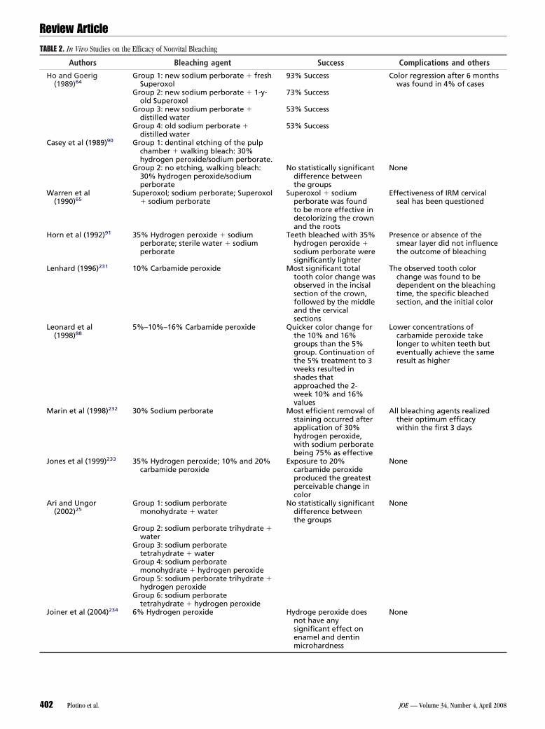
Prognosis of Nonvital Bleached TeethDespite many clinical reports, there are few scientific evidence-
ased studies on tooth whitening (143). Some in vivo and in vitrotudies are summarized in Tables 1 and 2. Most reports present optimalnitial results after bleaching, with complete color matching of theleached tooth (teeth) with the adjacent one(s) (2, 45, 68, 80, 84, 102,
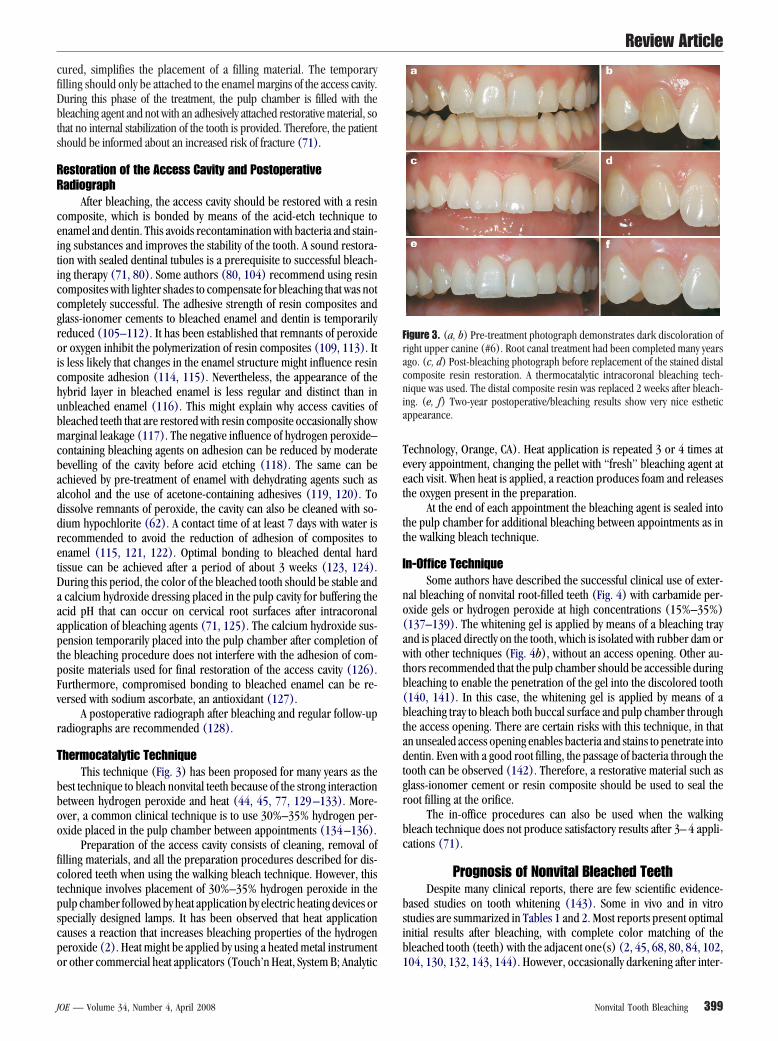
igure 3. (a, b) Pre-treatment photograph demonstrates dark discoloration ofight upper canine (#6). Root canal treatment had been completed many yearsgo. (c, d) Post-bleaching photograph before replacement of the stained distalomposite resin restoration. A thermocatalytic intracoronal bleaching tech-ique was used. The distal composite resin was replaced 2 weeks after bleach-
ng. (e, f) Two-year postoperative/bleaching results show very nice estheticppearance.
04, 130, 132, 143, 144). However, occasionally darkening after inter-
Nonvital Tooth Bleaching 399
ndmtmrcn
fsti1nepfa(r(p7ts1mhbopttrr
(omnt
enamp(nm1atccfds
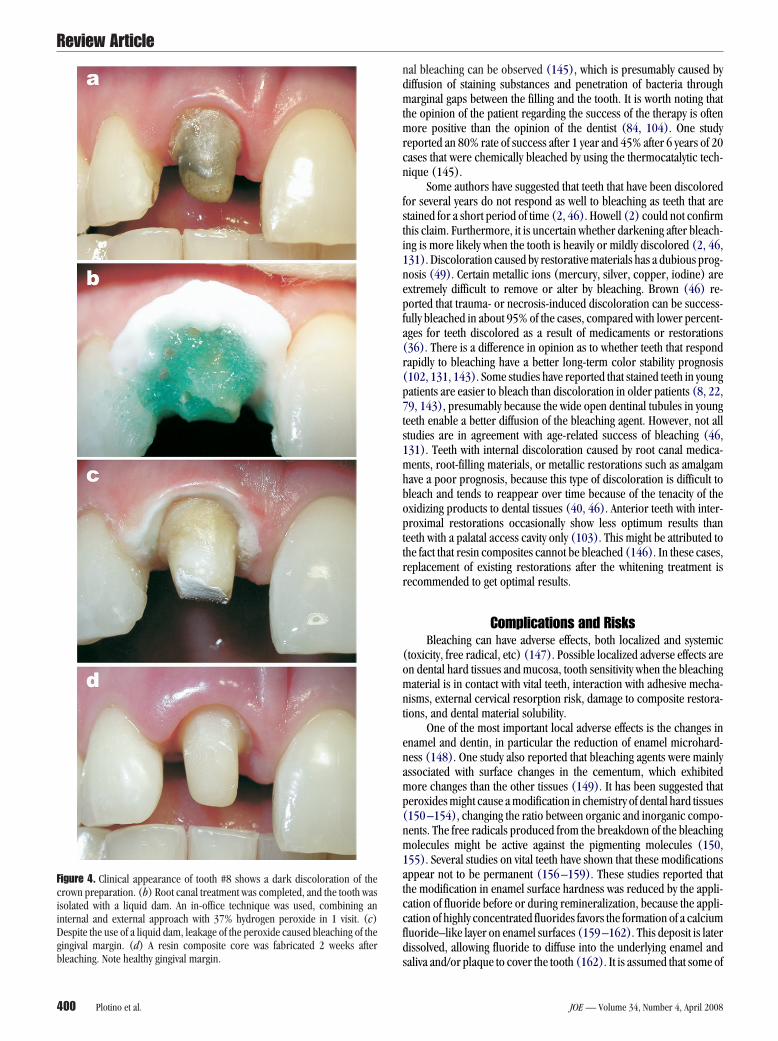
FciiDgbleaching. Note healthy gingival margin.
Review Article
400 Plotino et al.
al bleaching can be observed (145), which is presumably caused byiffusion of staining substances and penetration of bacteria througharginal gaps between the filling and the tooth. It is worth noting that
he opinion of the patient regarding the success of the therapy is oftenore positive than the opinion of the dentist (84, 104). One study
eported an 80% rate of success after 1 year and 45% after 6 years of 20ases that were chemically bleached by using the thermocatalytic tech-ique (145).
Some authors have suggested that teeth that have been discoloredor several years do not respond as well to bleaching as teeth that aretained for a short period of time (2, 46). Howell (2) could not confirmhis claim. Furthermore, it is uncertain whether darkening after bleach-ng is more likely when the tooth is heavily or mildly discolored (2, 46,31). Discoloration caused by restorative materials has a dubious prog-osis (49). Certain metallic ions (mercury, silver, copper, iodine) arextremely difficult to remove or alter by bleaching. Brown (46) re-orted that trauma- or necrosis-induced discoloration can be success-
ully bleached in about 95% of the cases, compared with lower percent-ges for teeth discolored as a result of medicaments or restorations36). There is a difference in opinion as to whether teeth that respondapidly to bleaching have a better long-term color stability prognosis102, 131, 143). Some studies have reported that stained teeth in youngatients are easier to bleach than discoloration in older patients (8, 22,9, 143), presumably because the wide open dentinal tubules in young
eeth enable a better diffusion of the bleaching agent. However, not alltudies are in agreement with age-related success of bleaching (46,31). Teeth with internal discoloration caused by root canal medica-ents, root-filling materials, or metallic restorations such as amalgam
ave a poor prognosis, because this type of discoloration is difficult toleach and tends to reappear over time because of the tenacity of thexidizing products to dental tissues (40, 46). Anterior teeth with inter-roximal restorations occasionally show less optimum results than
eeth with a palatal access cavity only (103). This might be attributed tohe fact that resin composites cannot be bleached (146). In these cases,eplacement of existing restorations after the whitening treatment isecommended to get optimal results.
Complications and RisksBleaching can have adverse effects, both localized and systemic
toxicity, free radical, etc) (147). Possible localized adverse effects aren dental hard tissues and mucosa, tooth sensitivity when the bleachingaterial is in contact with vital teeth, interaction with adhesive mecha-
isms, external cervical resorption risk, damage to composite restora-ions, and dental material solubility.
One of the most important local adverse effects is the changes innamel and dentin, in particular the reduction of enamel microhard-ess (148). One study also reported that bleaching agents were mainlyssociated with surface changes in the cementum, which exhibitedore changes than the other tissues (149). It has been suggested that
eroxides might cause a modification in chemistry of dental hard tissues150 –154), changing the ratio between organic and inorganic compo-ents. The free radicals produced from the breakdown of the bleachingolecules might be active against the pigmenting molecules (150,
55). Several studies on vital teeth have shown that these modificationsppear not to be permanent (156 –159). These studies reported thathe modification in enamel surface hardness was reduced by the appli-ation of fluoride before or during remineralization, because the appli-ation of highly concentrated fluorides favors the formation of a calciumluoride–like layer on enamel surfaces (159 –162). This deposit is laterissolved, allowing fluoride to diffuse into the underlying enamel and
igure 4. Clinical appearance of tooth #8 shows a dark discoloration of therown preparation. (b) Root canal treatment was completed, and the tooth wassolated with a liquid dam. An in-office technique was used, combining annternal and external approach with 37% hydrogen peroxide in 1 visit. (c)espite the use of a liquid dam, leakage of the peroxide caused bleaching of theingival margin. (d) A resin composite core was fabricated 2 weeks after
aliva and/or plaque to cover the tooth (162). It is assumed that some of
JOE — Volume 34, Number 4, April 2008

tii4fae1
tpCg
t
tairtSae
3wdoT
T
Review Article
J
he fluoride is supporting the remineralization of enamel (160). Chem-cal irritations of the oral mucosa are due to the bleaching agent’s activengredients (Fig. 4c). This irritation is usually mild and transient (Fig.d). Curtis et al (163) reported no adverse effects on oral soft tissues
rom a 10% carbamide peroxide bleaching material. Bleaching agentsffect enamel and dentin bond strengths because they cause a change ofnamel and dentin, resulting in free radical–induced damage (110,64).
Occurrence of external cervical resorption is a serious complica-ion after internal bleaching procedures (145, 165). The first 4 re-orted cases were published by Harrington and Natkin (166) in 1979.ervical resorption is an external resorption with an inflammatory ori-in caused by trauma or intracoronal bleaching (144).
Heithersay (167) analyzed cervical resorption cases and reported
ABLE 1. In Vivo Studies on the Success Rate of Nonvital Bleaching
Authors Bleaching agent
Brown (1965)46 Thermocatalytic: 30% hydroperoxide, followed bywalking bleach: 30%hydrogen peroxide
Tewari and Chawla (1972)129 Thermocatalytic: 30% hydroperoxide
Howell (1981)131 Thermocatalytic: 30% hydroperoxide, followed bywalking bleach: 30%hydrogen peroxide
Feiglin (1987)145 Thermocatalytic: 130 volumehydrogen peroxide followwalking bleach: sodiumperborate � mixture of 3/4water and 1/4 130 volhydrogen peroxide
Friedman et al (1988)144 Three different techniques: (thermocatalytic: 30%hydrogen peroxide; (2)walking bleach: 30%hydrogen peroxide; (3)thermocatalytic � walkingbleach: 30% hydrogenperoxide
Holmstrup et al (1988)102 Walking bleach: sodiumperborate � water
Anitua et al (1990)84 Walking bleach: sodiumperborate � 110 volhydrogen peroxide
Waterhouse and Nunn (1996)228 30% Hydrogen peroxidesolution mixed with sodiumperborate granules
Abou-Rass et al (1998)80 Walking bleach: sodiumperborate � 30% hydrogeperoxide
Bizhang et al (2003)229 Extracoronal (10% carbamidperoxide); walking bleach(sodium perborate � 3%hydrogen peroxide);extracoronal � walkingbleach (10% carbamideperoxide)
Amato et al (2006)230 Mixture of sodium perborateand 120 vol hydrogenperoxide
hat 24.1% were caused by orthodontic treatment, 15.1% by dental v
OE — Volume 34, Number 4, April 2008
rauma, 5.1% by surgery (eg, transplantation or periodontal surgery),nd 3.9% by intracoronal bleaching. A combination of internal bleach-ng with one of the other causes is responsible for 13.6% of cervicalesorption cases. The combination of bleaching and history of trauma ishe most important predisposing factor for cervical resorption (168).everal studies reporting on long-term follow-up evaluations show anssociation between external resorption and bleaching of nonvital teeth,ven many years after bleaching (68, 80, 84, 102, 169 –171).
Animal studies have shown histologic evidence of resorption aftermonths of bleaching (172, 173). However, after 1 month no changesere detected. Cervical resorption is mostly asymptomatic and is usuallyetected only through routine radiographs (174). Sometimes swellingf the papilla or percussion sensitivity can be observed (169, 171).eeth that were root-filled as a result of trauma more often show cer-
Success Complications and others
75% Success (39% complete,46% partial); 25% failure(17.5% no Improvement,7.5% refractorydiscoloration)
None
95% Success; 5% failure The only failure wassuccessfully bleachedagain
97% Success (53% complete,44% partial); 3% failure
The only failure wasassociated with aninsufficient filling
45% Success; 55% failure Teeth of younger patientsshowed better successrates
50% Success; 29% acceptable;21% failure
Highest percentage offailure occurred between2–8 years after whiteningtreatment
90% Success (63% good, 27%acceptable); 10% failure
3 Teeth with translent pain
Assessment of the dentist:100% success (98%complete, 2% partialsuccess). Assessment of thepatient: 99.4% success;0.4% failure
None
83% Success None
93% Success; 7% failure Failure because of internalcervical deposit thatcould be successfullybleached
Whitening effect ofextracoronal bleaching �walking bleach (10%carbamide peroxide) was aseffective as walking bleach(sodium perborate � 3%hydrogen peroxide)
Extracoronal bleaching �walking bleach reducedthe treatment time incomparison to thewalking bleach by 50%
69.9% Success; 37.1% failures 22.2% of the failuresshowed fistula, pain, andperiradicular and/orlateroradicular bone lysis
gen
gen
gen
ing
1)
n
e
ical resorption (2, 46, 68, 80, 84, 102, 104, 129, 131, 143, 144).
Nonvital Tooth Bleaching 401
T
Review Article
4
ABLE 2. In Vivo Studies on the Efficacy of Nonvital Bleaching
Authors Bleaching agent Success Complications and others
Ho and Goerig(1989)64
Group 1: new sodium perborate � freshSuperoxol
93% Success Color regression after 6 monthswas found in 4% of cases
Group 2: new sodium perborate � 1-y-old Superoxol
73% Success
Group 3: new sodium perborate �distilled water
53% Success
Group 4: old sodium perborate �distilled water
53% Success
Casey et al (1989)90 Group 1: dentinal etching of the pulpchamber � walking bleach: 30%hydrogen peroxide/sodium perborate.
Group 2: no etching, walking bleach:30% hydrogen peroxide/sodiumperborate
No statistically significantdifference betweenthe groups
None
Warren et al(1990)65
Superoxol; sodium perborate; Superoxol� sodium perborate
Superoxol � sodiumperborate was foundto be more effective indecolorizing the crownand the roots
Effectiveness of IRM cervicalseal has been questioned
Horn et al (1992)91 35% Hydrogen peroxide � sodiumperborate; sterile water � sodiumperborate
Teeth bleached with 35%hydrogen peroxide �sodium perborate weresignificantly lighter
Presence or absence of thesmear layer did not influencethe outcome of bleaching
Lenhard (1996)231 10% Carbamide peroxide Most significant totaltooth color change wasobserved in the incisalsection of the crown,followed by the middleand the cervicalsections
The observed tooth colorchange was found to bedependent on the bleachingtime, the specific bleachedsection, and the initial color
Leonard et al(1998)88
5%–10%–16% Carbamide peroxide Quicker color change forthe 10% and 16%groups than the 5%group. Continuation ofthe 5% treatment to 3weeks resulted inshades thatapproached the 2-week 10% and 16%values
Lower concentrations ofcarbamide peroxide takelonger to whiten teeth buteventually achieve the sameresult as higher
Marin et al (1998)232 30% Sodium perborate Most efficient removal ofstaining occurred afterapplication of 30%hydrogen peroxide,with sodium perboratebeing 75% as effective
All bleaching agents realizedtheir optimum efficacywithin the first 3 days
Jones et al (1999)233 35% Hydrogen peroxide; 10% and 20%carbamide peroxide
Exposure to 20%carbamide peroxideproduced the greatestperceivable change incolor
None
Ari and Ungor(2002)25
Group 1: sodium perboratemonohydrate � water
No statistically significantdifference betweenthe groups
None
Group 2: sodium perborate trihydrate �water
Group 3: sodium perboratetetrahydrate � water
Group 4: sodium perboratemonohydrate � hydrogen peroxide
Group 5: sodium perborate trihydrate �hydrogen peroxide
Group 6: sodium perboratetetrahydrate � hydrogen peroxide
Joiner et al (2004)234 6% Hydrogen peroxide Hydroge peroxide doesnot have anysignificant effect onenamel and dentin
None
microhardness
02 Plotino et al. JOE — Volume 34, Number 4, April 2008
npokmrHtwsr
tddcgbtfrpmdpb
sdtasidtpt5
dt1eppIdoapwrac
bwncst
blseplcaNnc
uohid3i
abatalpdcdwomTapsonfoHicee
hp
at
ti
scCm
Review Article
J
The mechanism responsible for resorption in bleached teeth hasot yet been adequately explained. There is speculation that hydrogeneroxide can diffuse through the dentinal tubules, cement, and peri-dontal ligament and can reach bone (175, 176). Harrington and Nat-in (166) postulated that hydrogen peroxide directly induces an inflam-atory resorption process. On its own, hydrogen peroxide is not very
eactive, and the body has mechanisms in place to deal with it (177).owever, in the presence of inflammation, proinflammatory agents ac-
ivate reduced nicotinamide adenine dinucleotide phosphate oxidase,hich produces superoxides that can react with hydrogen peroxide. It is
peculated that the resultant hypochlorous acid, N-chloroamines, andeactive hydroxyl ions might initiate some disease processes (178).
Lado et al (171) suggested that application of bleaching agents ledo denaturation of dentin proteins by the oxidizing agents, which in-uces a foreign body reaction (64). It has been postulated that thisenaturation can be caused by heat (66, 169, 179) or by pH variationaused by bleaching agents (126, 180, 181). Price et al (182) investi-ated the pH of some bleaching agents and found that the in-officeleaching products were acidic. The low pH value of highly concen-rated hydrogen peroxide can be considered a tissue damage–inducingactor because an acidic environment is optimal for osteoclastic activityesulting in bone resorption (144, 183). Lee et al (184) found that it isossible to specifically relate the changes in pH of the extraradicularedium to the quantity of hydrogen peroxide detected in the extrara-
icular environment. Furthermore, in vitro studies demonstrated thatH in extraradicular medium surrounding the root increased withleaching time (184 –186).
Lee et al (184) reported that it is unlikely that cervical root re-orption is the result of an acidic extraradicular pH environment pro-uced by the bleaching agent. Bleaching agents cause superficial struc-ural changes to dentin (187), and the acid pH probably produces ancid-etch effect on dentin, opening up the smear layer that covers the cuturface of dentinal tubules, thus increasing its permeability (188). Thisn turn permits greater diffusion of hydrogen peroxide through theentinal tubules. Perhaps if the level of hydrogen peroxide goes beyondhe critical level, then destructive cervical root resorption can takelace. According to Halliwell et al (189), levels of hydrogen peroxidehat are less than 20 �mol/L should be safe; however, when it exceeds0 �mol/L, it is cytotoxic to most living cells.
Diffusion of hydrogen peroxide to cervical tissues is increased byifferent predisposing factors. Studies and case reports indicate thathese factors are related to the occurrence of cervical resorption (71,43, 144). Patients who had bleaching therapy at a young age often havexternal resorption (68, 80, 84, 102, 144, 169 –171). A possible ex-lanation is that hydrogen peroxide can more easily penetrate into theeriodontium because of wide open dentinal tubules in young teeth.ncreased permeability of dentin is associated with both decreasingentin thickness and high surrounding temperature (190). Applicationf heat (thermocatalytic method) leads to widening of dentinal tubulesnd facilitates diffusion of molecules into the dentin (191). This ex-lains the increasing dissemination of hydrogen peroxide into dentinith an increase in temperature (192). Moreover, application of heat
esulted in generation of hydroxyl radicals from hydrogen peroxide thatre extremely reactive and have been shown to degrade components ofonnective tissue (193).
As a consequence, today the thermocatalytic technique is used lessecause of the high risk of external root resorption that is associatedith heat application (173–179). In contrast, the walking bleach tech-ique with a sodium perborate– hydrogen peroxide solution did notause cervical resorption, even 1 year after bleaching (179). This ob-ervation might be explained by the fact that sodium perborate inhibits
he function of macrophages; macrophages stimulate both osteoclastic fOE — Volume 34, Number 4, April 2008
one resorption and destruction of dentin and cementum induced byytic processes in periodontal tissue (194). The results of an in vitrotudy reported that even a sodium perborate solution, which is consid-red to be a relatively safe bleaching agent, proved to be toxic to theeriodontal ligament cells (195). Furthermore, it has also been estab-
ished that formulations with either 30% hydrogen peroxide alone or inombination with sodium perborate are more toxic to periodontal lig-ment cells as compared with a perborate-water suspension (195).evertheless, correlation of the present data to the clinical situation isot possible and cannot be used as a factor to argue in support of riskingervical resorption.
Carbamide peroxide has been more recently recommended forse in intracoronal bleaching (196). Thirty-five percent carbamide per-xide showed the lowest levels of extraradicular diffusion, whereas 35%ydrogen peroxide showed the highest, with sodium perborate having
ntermediate values (184). Considering the low levels of extraradiculariffusion and its effectiveness as an intracoronal bleaching agent (197),5% carbamide peroxide might be regarded as the intracoronal bleach-
ng agent of choice.Clinically, a natural anatomic defect at the CEJ can be found in
bout 10% of all teeth, thus causing dentin to be exposed (198). Whenleaching teeth with exposed dentinal tubules at the CEJ, as a result ofnatomic defects or cervical erosion, the use of bleaching agents con-aining a high percentage of active oxidizing compounds should bevoided. In addition, the time of action should be reduced becauseaboratory studies have demonstrated that the penetration of hydrogeneroxide into the cervical region can be facilitated by cemental rootefects or particular morphologic patterns at the CEJ (199 –201). Ac-ording to Rotstein et al (185), lack of root cementum resulted iniffusion of up to 82% of hydrogen peroxide (30% concentration),hich had been applied in the pulp chamber. However, disseminationf hydrogen peroxide into dentin cannot be totally prevented by usingixtures of sodium perborate with 30% hydrogen peroxide or water.
he amount of hydrogen peroxide diffusion is significantly lower whenmixture of sodium perborate–tetrahydrate and water is used, com-
ared with application of 30% hydrogen peroxide mixed with differentodium perborates (202). Although in these cases there is less diffusionf peroxide into the surrounding tissues, a valid cervical seal is stillecessary. The lack of a cervical seal represents another predisposing
actor for an enhanced diffusion of hydrogen peroxide into the peri-dontal tissues. This corroborates the conclusion by MacIsaac andoen (165) in their extensive literature review of intracoronal bleach-
ng that the common thread through all reported cases of externalervical root resorption was the lack of an intermediate base. This is anffective means of reducing the diffusion of hydrogen peroxide into thextraradicular environment (99).
It has also been reported that there is an increased diffusion ofydrogen peroxide to cervical tissues after pre-treatment of dentin in theulp chamber with 5% NaOCl (203).
A follow-up radiograph of the bleached tooth within the first yearfter treatment is recommended to diagnose possible cervical resorp-ion as early as possible.
Treatment prognosis depends mainly on the extent of the resorp-ion process. The extent of resorption serves as a guide for the cliniciann selecting the correct treatment (204).
Extraction is often inevitable in cases of severe external root re-orption and when the lesion cannot be controlled (205, 206). In theseases an implant-supported restoration is an acceptable treatment.areful space evaluation of the implant site must be performed withodel-based planning (207).
Inflammatory osteolytic lesions have a low pH value that is optimal
or hard tissue resorption (208). Root canal dressings consisting ofNonvital Tooth Bleaching 403
criptwredbg
aos(o(t
oostt
rtase
slf(lwmma(m
pftc
ac
dlpgbp2a
a
tits
Review Article
4
alcium hydroxide are able to induce a higher pH in dentin (209). Ifesorption occurs, Friedman et al (144) suggested that calcium hydrox-de recalcification treatment should be attempted. Tronstad et al (209)ostulated that reparative hard tissue formation would be stimulated byhis treatment. Clinical cases have shown that an intracoronal dressingith calcium hydroxide can sometimes prevent progression of external
esorption (180, 181). However, on a radiograph only osseous regen-ration of the defect and no hard dental tissue regeneration could beetected. Recalcification treatment will fail if there is communicationetween the resorption and the oral cavity (183). In these cases, sur-ical repair should be considered.
Cervical resorption can also be treated with a direct restorationfter gaining surgical access to the defect (210 –212). This does notffer the advantage to induce biologic changes in the periodontal tis-ues, effectively allowing continued resorption after surgical repair.181). It has been speculated that this might also happen when the areaf resorption is not completely instrumented during surgical treatment183). Another disadvantage is the significant compromise in periodon-al health and esthetics that often results after surgical repair (181).
If recalcification or surgical repair is not feasible, or if the resultsf surgical repair are esthetically or periodontally unsatisfactory, theption of surgical crown lengthening combined with appropriate re-torative treatment is still available (181). On the negative side, one haso accept an increase in crown-root ratio and removal of supportingissues from adjacent teeth.
If a lesion can be easily accessed, a limited labial flap might beaised to permit cleaning of the lesion, thus exposing sound tooth struc-ure. Then, it is recommended to place 90% trichloroacetic acid on theffected margins to necrotize granulation tissue. The area of resorptionhould be restored with an appropriate material, dictated mostly bysthetic demands (213).
In severe cases in which the lesion is not only subgingival butubcrestal as well, a labial flap must be raised to permit cleaning of theesion, exposing sound tooth structure and sealing with a temporaryilling material. After this initial treatment, it is recommended to userapid) orthodontic tooth extrusion combined with fiberotomy fol-owed by definitive restorative treatment (214 –216). This treatmentill result in a shorter root, potentially leading to an increase in toothobility. Loss of marginal attachment as result of crown lengthening isore detrimental than loss of an equivalent amount of root length by
pical resorption or orthodontic extrusion. According to Kalkwarf et al217), 3 mm of apical root resorption is equivalent to approximately 1m crestal bone loss.
A case has been reported in which a novel combined endodontic/eriodontal treatment was performed (218). Reconstruction of the de-ect was achieved by using resin composite to restore the coronal por-ion of the defect up to the CEJ followed by mineral trioxide aggregateement below to the bone.
The interdisciplinary treatments described above offer a system-tic approach to invasive cervical resorption, which continues to be alinical challenge because it is difficult to detect during its early stages.
It has been reported that bleaching can increase resin solubility,ecrease enamel bond strength, and consequently increase marginaleakage (219 –221). Ten percent carbamide peroxide has been re-orted to adversely effect the physical properties of zinc phosphate andlass-ionomer luting agents (222). Finally, bleaching techniques do notleach synthetic materials; thus, existing restorations might need re-lacement to improve color matching after successful bleaching (223–27). One study has documented an increase in mercury release frommalgam restorations that were exposed to carbamide peroxide (31).
It can be concluded from this review that bleaching is an important
nd valuable adjunct in endodontic treatment. Proper diagnosis, selec-04 Plotino et al.
ion of bleaching materials, placement techniques, and an understand-ng of the biologic interaction with soft and hard tissues are all factorshat determine not only immediate success but also long-term success,afety, and patient satisfaction as well.
References1. Truman J. Bleaching of non-vital discoloured anterior teeth. Dent Times
1864;1:69 –72.2. Howell RA. Bleaching discoloured root-filled teeth. Br Dent J 1980;148:159 – 62.3. Dwinelle WW. Ninth annual meeting of American Society of Dental Surgeons: article
X. Am J Dent Sc 1850;1:57– 61.4. Atkinson CB. Bleaching teeth, when discolored from loss of vitality: means for
preventing their discoloration and ulceration. Dental Cosmos 1862;3:74 –7.5. Bogue EA. Bleaching teeth. Dental Cosmos 1872;141–3.6. Taft J. Bleaching teeth. Am J Dent Sci 1878/1879;12:364.7. Atkinson CB. Hints and queries. Dental Cosmos 1879;21:471.8. Harlan AW. The dental pulp, its destruction, and methods of treatment of teeth
discolored by its retention in the pulp chamber or canals. Dental Cosmos1891;33:137– 41.
9. Kirk EC. Hints, queries, and comments: sodium peroxide. Dental Cosmos1893;35:1265–7.
10. Messing JJ. Bleaching. J Br Endod Soc 1971;5:84 –5.11. Atkinson CB. Fancies and some facts. Dental Cosmos 1892;34:968 –72.12. Dietz VH. The bleaching of discolored teeth. Dent Clin North Am 1957;1:897–902.13. Harlan AW. The removal of stains from teeth caused by administration of medical
agents and the bleaching of pulpless tooth. Am J Dent Sci 1884/1885;18:521.14. Abbot CH. Bleaching discoloured teeth by means of 30 per cent perhydrol and the
electric light rays. J Allied Dent Society 1918;13:259.15. Prinz H. Recent improvements in tooth bleaching. A clinical syllabus. Dental Cos-
mos 1924;66:558 – 60.16. Rosenthal P. The combined use of ultra-violet rays and hydrogen dioxide for bleach-
ing teeth. Dental Cosmos 1911;53:246 –7.17. Brininstool CL. Vapor bleaching. Dental Cosmos 1913;55:532.18. Andreasen FM. Transient apical breakdown and its relation to colour and sensibility
changes after luxation injuries to teeth. Endod Dent Traumatol 1986;2:9 –19.19. Merrell JH. Bleaching of discoloured pulpless tooth. J Can Dent Assoc 1954;20:380.20. Stewart GG. Bleaching discoloured pulpless teeth. J Am Dent Assoc 1965;70:325– 8.21. Caldwell CB. Heat source for bleaching discolored teeth. Ariz Dent J 1967;13:18 –9.22. Hodosh M, Mirman M, Shklar G, Povar M. A newmethod of bleaching discolored
teeth by the use of a solid state direct heating device. Dent Dig 1970;76:344 – 6.23. Kirk EC. The chemical bleaching of teeth. Dental Cosmos 1889;31:273– 83.24. Westlake A. Bleaching teeth by electricity. Am J Dent Sci 1895;29:101.25. Ari H, Ungor M. In vitro comparison of different types of sodium perborate used for
intracoronal bleaching of discolored teeth. Int Endod J 2002;35:433– 6.26. Watts A, Addy M. Tooth discoloration and staining: a review of the literature. Br Dent
J 2001;190:309 –16.27. Jahangiri L, Reinhardt SB, Mehra RV, Matheson PB. Relationship between tooth
shade value and skin color: an observational study. J Prosthet Dent 2002;87:149 –52.
28. Ten Bosch JJ, Coops JC. Tooth color and reflectance as related to light scattering andenamel hardness. J Dent Res 1995;74:374 – 80.
29. Joiner A, Jones NM, Raven SJ. Investigation of factors influencing stain formationutilizing an in situ model. Adv Dent Res 1995;9:471– 6.
30. Joiner A. Tooth colour: a review of the literature. J Dent 2004;32:3–12.31. Dahl JE, Pallesen U. Tooth bleaching-a critical review of the biological aspects. Crit
Rev Oral Biol Med 2003;14:292–304.32. Hattab FN, Qudeimat MA, al-Rimawi HS. Dental discoloration: an overview. J Esthet
Dent 1999;11:291–310.33. Scannapieco FA, Levine MJ. Saliva and dental pellicles. In: Genco RJ, Goldman HM,
Cohen WD, eds. Contemporary periodontics. St Louis: Mosby, 1990.34. Nathoo SA. The chemistry and mechanism of extrinsic and intrinsic discoloration.
J Am Dent Assoc 1997;128:6S–10S.35. Gorlin RJ, Goldman HM. Enviromental pathology of the teeth. In: Thoma’s oral
pathology. 6th ed. St Louis: Mosby, 1970.36. Nathoo SA, Gaffar A. Studies on dental stain induced by antibacterial agents and
rationale approaches for bleaching dental staine. Adv Dent Res 1994;9:462–70.37. Belkhir MS, Douki N. An new concept for removal of dental fluorosis stains. J Endod
1991;17:288 –92.38. Vogel RI. Intrinsic and extrinsic discoloration of the dentition. J Oral Med
1975;30:99 –104.39. Attin T, Paque F, Ajam F, Lennon AM. Review of the current status of tooth whitening
with the walking bleach technique. Int Endod J 2003;36:313–29.40. Rostein I. Tooth discoloration and bleaching. In: Ingle JI, Bakland LK, eds. End-
odontics. 5th ed. Hamilton, Ontario, Canada: BC Decker Inc, 2002:845– 60.
JOE — Volume 34, Number 4, April 2008
1
1
1
1
1
1
1
1
1
1
1
1
1
Review Article
J
41. Arens D. The role of bleaching in esthetics. Dent Clin North Am 1989;33:319 –36.42. Goldstein RE, Garber DA. Complete dental bleaching. Berlin: Quintessence, 1995.43. Guldener PHA, Langeland K. Endodontologie. 3rd ed. Stuttgart, New York: Georg
ThiemeVerlag, 1993.44. Grossman L. Root canal therapy. Philadelphia: Lea and Febiger, 1943.45. Marin PD, Bartold PM, Heithersay GS. Tooth discoloration by blood: an in vitro
histochemical study. Endod Dent Traumatol 1997;13:132– 8.46. Brown G. Factors influencing successful bleaching of the discolored root-filled
tooth. Oral Surg Oral Med Oral Pathol 1965;20:238 – 44.47. Faunce F. Management of discolored teeth. Dent Clin North Am 1983;27:657–70.48. van der Burgt TP, Plaesschaert AJM. Tooth discoloration induced by dental mate-
rials. Oral Surg Oral Med Oral Pathol 1985;60:666 –9.49. van der Burgt TP, Plaesschaert AJM. Bleaching of tooth discoloration caused by
endodontic sealers. J Endod 1986;12:231– 4.50. van der Burgt TP, Eronat C, Plaesschaert AJM. Staining patterns in teeth discolored
by endodontic sealers. J Endod 1986;12:187–91.51. van der Burgt TP, Mullaney TP, Plaesschaert AJM. Tooth discoloration induced by
endodontic sealers. Oral Surg Oral Med Oral Pathol 1986;61:84 –9.52. Davis MC, Walton RE, Rivera EM. Sealer distribution in coronal dentin. J Endod
2002;28:464 – 6.53. Kim ST, Abbot PV, McGinley P. The effects of Ledermix paste on discolouration of
mature teeth. Int Endod J 2000;33:227–32.54. Budavari S, O’Neil MJ, Smith A, Heckelman PE (). The Merck index: an encyclope-
dia of chemicals, drugs, and biologicals. Rahway, NJ: Merck and Co Inc, 1989.55. Hägg G. General and inorganic chemistry. Stockholm: Almqvist and Wiksell Förlag
AB, 1969.56. Seghi RR, Denry I. Effect of external bleaching on indentation and abrasion char-
acteristics of human enamel in vitro. J Dent Res 1992;7:1340 – 4.57. Zappalà C, Caprioglio D. Discromie dentali: sistemi di sbiancamento alla poltrona
e domiciliari. Dent Cadmos 1993;15:13– 43.58. Hardman PK, Moore DL, Petteway GH. Stability of hydrogen peroxide as a bleaching
agent. Gen Dent 1985;33:121–2.59. Chen JH, Xu JW, Shing CX. Decomposition rate of hydrogen peroxide bleaching
agents under various chemical and physical conditions. J Prosthet Dent1993;69:46 – 8.
60. Gurgan, S, Bolay S, Alacam R. Antibacterial activity of 10% carbamide peroxidebleaching agents. J Endod 1996;22:356 –7.
61. Rotstein I, Zalkind M, Mor C, Tarabeah A, Friedman S. In vitro efficacy of sodi-umperborate preperations used for intracoronal bleaching of discolored non-vitalteeth. Endod Dent Traumatol 1991;7:177– 80.
62. Rotstein I, Mor C, Friedman S. Prognosis of intracoronal bleaching with sodiumperborate preparations in vitro: 1-year study. J Endod 1993;19:10 –2.
63. Weiger R, Kuhn A, Lost C. In vitro comparison of various types of sodium perborateused for intracoronal bleaching of discolored teeth. J Endod 1994;20:338 – 41.
64. Ho S, Goerig AC. An in vitro comparison of different bleaching agents in the discol-ored tooth. J Endod 1989;15:106 –11.
65. Warren MA, Wong M, Ingram TA III. An in vitro comparison of bleaching agents onthe crowns and roots of discolored teeth. J Endod 1990;16:463–7.
66. Freccia WF, Peters D, Lorton L, Bernier W. An in vitro comparison of non-vitalbleaching techniques in the discolored tooth. J Endod 1982;8:70 –7.
67. Kaneko J, Inoue S, Kawakami S, Sano H. Bleaching effect of sodium percarbonate ondiscolored pulpless teeth in vitro. J Endod 2000;26:25– 8.
68. Aldecoa EA, Mayordomo FG. Modified internal bleaching of severe tetracyclinediscolorations: a 6-year clinical evaluation. Quintessence Int 1992;23:83–9.
69. Croll TP. Enamel microabrasion: observations after 10 years. J Am Dent Assoc1997;128:45S–50S.
70. Croll TP. Enamel microabrasion for removal of superficial dysmineralization anddecalcification defects. J Am Dent Assoc 1990;120:411–5.
71. Baratieri LN, Ritter AV, Monteiro S Jr, Caldeira deAndrada MA, Cardoso Vieira LC.Non-vital tooth bleaching: guidelines for the clinician. Quintessence Int 1995;26:597– 8.
72. Salvas CJ. Perborate as a bleaching agent. J Am Dent Assoc 1938;25:324.73. Spasser HF. A simple bleaching technique using sodium perborate. N Y State Dentl
J 1961;27:332– 4.74. Nutting EB, Poe GS. A new combination for bleaching teeth. J South Californian Dent
Assoc 1963;31:289.75. Nutting EB, Poe GS. Chemical bleaching of discolored endodontically treated teeth.
Dent Clin North Am 1967;11:655– 62.76. Serene TP, Snyder DE. Bleaching technique (pulpless anterior teeth). J South Cal-
ifornian Dent Assoc 1973;41:30 –2.77. Boksman L, Jordan RE, Skinner DH. Non-vital bleaching internal and external. Aust
Dent J 1983;28:149 –52.78. Hayashi K, Takamizu M, Momoi V, Furuya K, Kusunoki M, Kono A. Bleaching teeth
discolored by tetracycline therapy. Dent Surv 1980;56:17–25.
OE — Volume 34, Number 4, April 2008
79. Abou-Rass M. The elimination of tetracycline discoloration by intentional endodon-tics and internal bleaching. J Endod 1982;8:101– 6.
80. Abou-Rass M. Long-term prognosis of intentional endodontics and internal bleach-ing of tetracycline-stained teeth. Comp Contin Educ Dent 1998;19:1034 –50.
81. Fields JP. Intracoronal bleaching of tetracycline-stained teeth. J Endod 1982;8:512–3.
82. Walton RE, O’Dell NL, Lake FT, Shimp RG. Internal bleaching of tetracycline-stainedteeth in dogs. J Endod 1983;9:416 –20.
83. Lake FT, O’Dell N, Walton RE. The effect of internal bleaching on tetracycline indentin. J Endod 1985;11:415–20.
84. Anitua E, Zabalegui B, Gil J, Gascon F. Internal bleaching of severe tetracyclinediscolorations: four-year clinical evaluation. Quintessence Int 1990;21:783– 8.
85. Nathoo S, Stewart B, Petrone ME, et al. Comparative clinical investigation of thetooth whitening efficacy of two tooth whitening gels. J Clin Dent 2003;14:64 –9.
86. Teixeira EC, Hara AT, Serra MC. Use of 37% carbamide peroxide in the walkingbleach technique: a case report. Quintessence Int 2004;35:97–102.
87. Kihn PW, Barnes DM, Romberg E, Peterson K. A clinical evaluation of 10 percent vs15 percent carbamide peroxide tooth-whitening agents. J Am Dent Assoc 2000;131:1478 – 84.
88. Leonard RH, Sharma A, Haywood VB. Use of different concentrations of carbamideperoxide for bleaching teeth: an in vitro study. Quintessence Int 1998;29:503–7.
89. Hulsmann M. Endodontie. Stuttgart, New York: Georg ThiemeVerlag, 1993.90. Casey LJ, Schindler WG, Murata SM, Burgess JO. The use of dentinal etching with
endodontic bleaching procedures. J Endod 1989;15:535– 8.91. Horn DJ, Hicks L, Bulan-Brady J. Effect of smear layer removal on bleaching of
human teeth in vitro. J Endod 1998;24:791–5.92. Fuss Z, Szajkis S, Tagger M. Tubular permeability to calcium hydroxide and to
bleaching agents. J Endod 1989;15:362– 4.93. Costas FL, Wong M. Intracoronal isolating barriers: effect of location on root canal
leakage and effectiveness of bleaching agents. J Endod 1991;17:365– 8.94. Smith JJ, Cunningham CJ, Montgomery S. Cervical canal leakage after internal
bleaching procedures. J Endod 1992;18:476 – 81.95. Hansen-Bayless J, Davis R Sealing ability of two intermediate restorative materials in
bleached teeth. Am J Dent 1992;5:151– 4.96. McInerney ST, Zillich R. Evaluation of internal sealing ability of three materials. J
Endod 1992;18:376 – 8.97. Hansen-Bayless J, Davis R. Sealing ability of two intermediate restorative materials
in bleached teeth. Am J Dent 1992;5:151– 4.98. Hosoya N, Cox CF, Arai T, Nakamura J. The walking bleach procedure: an in vitro
study to measure microleakage of five temporary sealing agents. J Endod2000;26:716 – 8.
99. Rotstein I, Zyskind D, Lewinstein I, Bamberger N. Effect of different protective basematerials on hydrogen peroxide leakage during intracoronal bleaching in vitro. JEndod 1992;18:114 –7.
00. Steiner DR, West JD. A method to determine the location and shape of an intracoro-nal bleach barrier. J Endod 1994;20:304 – 6.
01. Hosoya N, Cox CF, Arai T, Nakamura J. The walking bleach procedure: an in vitrostudy to measure microleakage of five temporary sealing agents. J Endod2000;26:716 – 8.
02. Holmstrup G, Palm AM, Lambjerg-Hansen H. Bleaching of discoloured root-filledteeth. Endod Dent Traumatol 1988;4:197–201.
03. Waite RM, Carnes DL, Walker WA III. Microleakage of TERM used with sodiumperborate/water and sodiumperborate/superoxol in the ‘walking bleach’ tech-nique. J Endod 1998;24:648 –50.
04. Glockner K, Hulla H, Ebeleseder K, Stadler P. Five-year folllow-up of internal bleach-ing. Braz Dent J 1999;10:105–10.
05. Titley KC, Torneck CD, Smith DC, Adibfar A. Adhesion of composite resin to bleachedand unbleached bovine enamel. J Dent Res 1988;67:1523– 8.
06. Titley KC, Torneck CD, Ruse ND. The effect of carbamide peroxide gel on the shearbond strength of a microfil resin to bovine enamel. J Dent Res 1992;71:20 – 4.
07. Murchison DF, Charlton DG, Moore BK. Carbamide peroxide bleaching: effects onenamel hardness and bonding. Oper Dent 1992;17:181–5.
08. Garcia-Godoy F, Dodge WW, Donohue M, O’Quinn JA. Composite resin bondstrength after enamel bleaching. Oper Dent 1993;18:144 –7.
09. Dishman MV, Covey DA, Baughan LW. The e¡ects of peroxide bleaching on compos-ite to enamel bond strength. Dent Mat 1994;9:33– 6.
10. Josey AL, Meyers IA, Romaniuk K, Symons AL. The effect of avital bleaching tech-nique on enamel surface morphology and the bonding of composite resin toenamel. J Oral Rehabil 1996;23:244 –50.
11. Swift EJ Jr, Perdigao J. Effects of bleaching on teeth and restorations. CompendContin Educ Dent 1998;19:815–20.
12. Titley KC, Torneck CD, Smith DC, Applebaum NB. Adhesion of a glass ionomercement to bleached and unbleached bovine dentin. Endod Dent Traumatol
1989;5:132– 8.Nonvital Tooth Bleaching 405
1
1
1
1
1
1
1
1
1
1
1
1
1
1
1
1
1
1
111
1
1
1
11
1
1
1
1
1
1
1
1
1
1
1
1
1
1
1
1
1
1
1
1
1
1
1
1
1
1
1
1
1
11
1
1
1
1
11
1
1
1
1
Review Article
4
13. Torneck CD, Titley KC, Smith DC, Adibfar A. The influence of time of hydrogenperoxide exposure on the adhesion of composite resin to bleached bovine enamel.J Endod 1990;16:1123– 8.
14. Ruse ND, Smith DC, Torneck CD, Titley KC. Preliminary surface analysis of etched,bleached, and normal bovine enamel. J Dent Res 1990;69:1610 –3.
15. Torneck CD, Titley KC, Smith DC, Adibfar A. Effect of water leaching on the adhesionof composite resin to bleached and unbleached bovine enamel. J Endod1991;17:156 – 60.
16. Titley KC, Torneck CD, Smith DC, Chernecky R, Adibfar A. Scanning electron mi-croscopy observations on the penetration and structure of resin tags in bleachedand unbleached bovine enamel. J Endod 1991;17:72–5.
17. Barkhordar RA, Kempler D, Plesh O. Effect of non-vital tooth bleaching on mic-roleakage of resin composite restorations. Quintessence Int 1997;28:341– 4.
18. Cvitko E, Denehy GE, Swift EJ Jr, Pires JA. Bond strength of composite resin toenamel bleached with carbamide peroxide. J Esthet Dent 1991;3:100 –2.
19. Kalili KT, Caputo AA, Yoshida K. Effect of alcohol of composite bond strength tobleached enamel (abstract 1440). J Dent Res 1993;72(spec iss):285.
20. Barghi N, Godwin JM. Reducing the adverse effect of bleaching on composite-enamel bond. J Esthet Dent 1994;6:157– 61.
21. Adibfar A, Steele A, Torneck CD, Titley KC, Ruse D. Leaching of hydrogen peroxidefrom bleached bovine enamel. J Endod 1992;18:488 –91.
22. Titley KC, Torneck CD, Ruse ND, Krmec D. Adhesion of a resin composite tobleached and unbleached human enamel. J Endod 1993;19:112–5.
23. Cavalli V, Reis AF, Giannini M, Ambrosano GM. The effect of elapsed time followingbleaching on enamel bond strength of resin composite. Oper Dent 2001;26:597– 602.
24. Shinohara MS, Rodrigues A, Pimenta AF. In vitro microleakage of composite resto-rations after non-vital bleaching. Quintessence Int 2001;32:413–7.
25. Kehoe JC. pH reversal following in vitro bleaching of pulpless teeth. J Endod1987;13:6 –9.
26. Demarco FF, Freitas JM, Siva MP, Justino LM. Microleakage in endodonticallytreated teeth; in£uence of calcium hydroxide dressing following bleaching. IntEndod J 2001;34:495–500.
27. Lai SCN, Tay FR, Cheung GSP, et al. Reversal of compromised bonding in bleachedenamel. J Dent Res 2002;81:477– 81.
28. European Society of Endodontology. Consensus report of the European Society ofEndodontologyon quality guidelines for endodontic treatment. Int Endod J1994;27:115–24.
29. Tewari A, Chawla HS. Bleaching of non-vital discoloured anterior teeth. J IndianDent Assoc 1972;44:130 –3.
30. Kopp RS. A safe, simplified bleaching technique for pulpless teeth. Dent Surv1973;49:42– 4.
31. Howell RA. The prognosis of bleached root-filled teeth. Int Endod J 1981;14:22– 6.32. Weine FS. Endodontic therapy. 3rd ed. St Louis: Mosby, 1982.33. Boksman L, Jordan RE, Skinner DH. A conservative bleaching treatment for the
non-vital discolored tooth. Compend Contin Educ Dent 1984;5:471–5.34. Weisman MI. Efficient bleaching procedure for the pulpless tooth. Dent Dig
1963;69:347–52.35. Lowney JJ. Simplified technique for bleaching a discoloured tooth. Dent Dig
1964;70:446.36. Cohen S. A simplified method for bleaching discolored teeth. Dent Dig
1968;74:301–3.37. Putter H, Jordan RE. The‘walking’ bleach technique. J Esthet Dent 1989;1:191–3.38. Swift EJ Jr. Treatment of a discolored, endodontically treated toothwith home
bleaching and composite resin. Pract Periodontics Aesthet Dent 1992;4:19 –21.39. Frazier KB. Nightguard bleaching to lighten a restored, non-vital discolored tooth.
Compend Contin Educ Dent 1998;19:810 –3.40. Liebenberg WH. Intracoronal lightening of discolored pulpless teeth: a modified
walking bleach technique. Quintessence Int 1997;28:771–7.41. Carillo A, Trevino MVA, Haywood VB. Simultaneous bleaching of vital and an open-
chamber non-vital tooth with 10% carbamide peroxide. Quintessence Int1998;29:643– 8.
42. Barthel CR, Strobach A, Briedigkeit H, Gobe UB, Roulet JF. Leakage in roots coro-nally sealed with di¡erent temporary fillings. J Endod 1999;25:731– 4.
43. Niederman R, Ferguson M, Urdaneta R. Evidence based esthetic dentistry. J EsthetDent 1998;10:229 –34.
44. Friedman S, Rotstein I, Libfeld H, Stabholz A, Heling I. Incidence of external rootresorption and esthetic results in 58 bleached pulpless teeth. Endod Dent Traumatol1988;4:23– 6.
45. Feiglin B. A 6-year recall study of clinically chemically bleached teeth. Oral Surg OralMed Oral Pathol 1987;63:610 –3.
46. Monaghan P, Lim E, Lautenschlager E. Effects of home bleaching preparations on
composite resin color. J Prosthet Dent 1992;68:575– 8.06 Plotino et al.
47. Anderson DG, Chiego DJ, Glickman JR, McCauley LK. A clinical assessment of theeffect of 10% carbamide peroxide gel on human pulp tissue. J Endod 1999;25:247–50.
48. Attin T, Kielbassa AM, Shwanenbergg M, Helklwig E. Effect of fluoride treatment onremineralization of bleached enamel. J Oral Rehabil 1997;24:282– 6.
49. Zalkind M, Arwaz JR, Goldman A, Rotstein I. Surface morphology changes in humanenamel, dentin and cementum following bleaching: a scanning electron microscopystudy. Endod Dent Traumatol 1996;12:82– 8.
50. Haywood VB. History, safety and effectiveness of current bleaching techniques andapplications of the nightguard vital bleaching techniques. Quintessence Int1992;23:471– 88.
51. Heling I, Parson A, Rotstein I. Effect of bleaching agents on dentin permeability toStreptococcus faecalis. J Endod 1995;21:540 –2.
52. Lewinstein I, Hirschfeld Z, Stabholz A, Rotstein I. Effect of hydrogen peroxide andsodium perborate on the microhardness of human enamel and dentin. J Endod1994;20:61–3.
53. Powell LV, Bales DJ. Tooth bleaching: its effect on oral tissues. J Am Dent Assoc1991;122:50 – 4.
54. Tong LS, Pang MK, Mok NY, King NM, Wei SH. The effects of etching, micro-abra-sion, and bleaching on surface enamel. J Dent Res 1993;72:67–71.
55. Goldstain RE. The changing esthetic dental practice. J Am Dent Assoc 1994;125:1447–57.
56. Ten Cate JM, Arens J. Remineralization of artificial enamel lesions in vitro. CariesRes 1977;11:277– 86.
57. Featherstone JDB, Cutress TW, Rodgers BE, Dennison PJ. Remineralization of arti-ficial caries like lesions in vivo by a self administered mouthrinse or paste. CariesRes 1982;16:235– 42.
58. Arends J, Gelhard TBFM. In vivo remineralization of human enamel (1983). In:Demineralization and remineralization of the teeth. Leach SA, Edgar WM, eds.Oxford: IRL Press, 1983:1–16.
59. Attin T, Hartmann O, Hilgers RD, Hellwig E. Fluoride retention of incipient enamellesions after treatment with a calcium fluoride varnish in vivo. Arch Oral Biol1995;40:169 –74.
60. LeGeros RZ. Chemical and crystallographic events in the caries process. J Dent Res1990;69(spec iss):567–74.
61. Bruun C, Givskov H. Formation of CaF2 on sound enamel and in caries-like lesionsafter different forms of fluoride applications in vivo. Caries Res 1991;25:96 –100.
62. Rolla G, Saxegaard E. Critical evaluation of the composition and use of topicalfluorides, with emphasis on the role of calcium fluoride in caries inhibition. J DentRes 1990;69(spec iss):780 –5.
63. Curtis JW, Dickinson GL, Downey MC, et al. Assessing the effects of 10 percentcarbamide peroxide on oral soft tissues. J Am Dent Assoc 1996;127:1218 –23.
64. Perdigao J, Franci C, Swift EJ Jr, Ambrose WW, Lopes M. Ultra-morphological studyof the interaction of dental adhesives with carbamide peroxide-bleached enamel.Am J Dent 1998;11:291–301.
65. MacIsaac AM, Hoen MM. Intracoronal bleaching: concerns and considerations.J Can Dent Assoc 1994;60:57– 64.
66. Harrington GW, Natkin E. External resorption associated with bleaching of pulplessteeth. J Endod 1979;5:344 – 8.
67. Heithersay GS. Invasive cervical resorption: analysis of potential predisposing fac-tors. Quintessence Int 1999;30:83–95.
68. Heithersay GS. Invasive cervical resorption. Endod Topics 2004;7:73–92.69. Harrington GW, Natkin E. External resorption associated with bleaching of pulpless
teeth. J Endod 1979;5:344 – 8.70. Heithersay GS, Dahlstrom SW, Marin PD. Incidence of invasive cervical resorption
inbleached root-filled teeth. Aust Dent J 1994;39:82–7.71. Lado EA, Stanley HR, Weismann MI. Cervical resorption in bleached teeth. Oral Surg
Oral Med Oral Pathol 1983;55:78 – 80.72. Heller D, Skriber J, Lin LM. Effect of intracoronal bleaching on external cervical root
resorption. J Endod 1992;18:145– 8.73. Rotstein I. In vitro determination and quantification of 30% hydrogen peroxide
penetration through dentin and cementum during bleaching. Oral Surg Oral MedOral Pathol 1991;72:602– 6.
74. Trope M. Cervical root resorption. J Am Dent Assoc 1997;128(spec iss):56 –9.75. Pashley DH, Livingston MJ. Effect of molecular size on permeability coefficients in
human dentine. Arch Oral Biol 1978;23:391–5.76. Wang J-D, Hume WR. Diffusion of hydrogen ion and hydroxyl ion from various
sources through dentine. Int Endod J 1988;21:17–26.77. Halliwell B, Clement MV, Ramalingam J, Long LH. Hydrogen peroxide. Ubiquitous in
cell culture and in vivo? Life 2000;50:251–7.78. Grisham MB. Oxidants and free radicals in inflammatory bowel disease. Lancet
1994;344:859 – 61.79. Madison S, Walton R. Cervical root resorption following bleaching of endodontically
treated teeth. J Endod 1990;16:570 – 4.
JOE — Volume 34, Number 4, April 2008

1
1
1
1
1
1
1
1
1
1
1
1
1
1
1
1
1
1
1
1
2
2
2
2
2
2
22
2
2
2
2
2
2
2
2
2
2
2
2
2
2
2
2
2
2
2
2
2
2
2
2
2
2
2
Review Article
J
80. Montgomery S. External cervical resorption after bleaching a pulpless tooth. OralSurg Oral Med Oral Pathol 1984;57:203– 6.
81. Gimlin DR, Schindler WG. The management of post bleaching cervical resorption.J Endod 1990;16:292–7.
82. Price RBT, Sedarous M, Hiltz GS. The pH of tooth whitening products. J Can DentAssoc 2000;66:421– 6.
83. Vaes A. On the mechanisms of bone resorption: the action of parathyroid hormoneon the excretion and synthesis of lysosomal enzymes and on extracellular release odacid by bone cells. J Cell Biol 1968;39:676 –97.
84. Lee GP, Lee MY, Lum SOY, Poh RSC, Lim K-C. Extraradicular diffusion of hydrogenperoxide and pH changes associated with intracoronal bleaching of discolouredteeth using different bleaching agents. Int Endod J 2004;37:500 – 6.
85. Rotstein I, Torek Y, Misgav R. Effect of cementum defects on radicular penetrationof 30% H2O2 during intracoronal bleaching. J Endod 1991;17:230 –3.
86. Weiger R, Kuhn A, Lost C. Radicular penetration of hydrogen peroxide duringintra-coronal bleaching with various forms of sodium perborate. Int Endod J1994;27:313–7.
87. Rotstein I, Dankner E, Goldman A, Heling I, Stabholz A, Zalkind M. Histochemicalanalysis of dental hard tissues following bleaching. J Endod 1996;22:23– 6.
88. Carrasco LD, Froner IC, Corona SA, Pecora JD. Effect of internal bleaching agents ondentinal permeability of nonvital teeth: quantitative assessment. Dent Traumatol2003;19:85–9.
89. Halliwell B, Clement MV, Ramalingam J, Long LH. Hydrogen peroxide: ubiquitous incell culture and in vivo? Life 2000;50:251–7.
90. Outhwaite WC, Livingston MJ, Pashley DH. Effects of changes in surface area, thick-ness, temperature and post extraction time on human dentine permeability. ArchOral Biol 1976;21:599 – 603.
91. Pashley DH, Thompson SM, Stewart FP. Dentin permeability: effects of temperatureon hydraulic conductance. J Dent Res 1983;62:956 –9.
92. Rotstein I, Torek Y, Lewinstein I. Effect of bleaching time and temperature on theradicular penetration of hydrogen peroxide. Endod Dent Traumatol1991;7:196 – 8.
93. Dahlstrom SW, Heithersay GS, Bridges TE. Hydroxyl radical activity inthermo-cata-lytically bleached root-filled teeth. Endod Dent Traumatol 1997;13:119 –25.
94. Jimenez-Rubio A, Segura JJ. The e¡ect of the bleaching agent sodium perborate onmacrophage adhesion in vitro: implications in external cervical root resorption.J Endod 1998;24:229 –32.
95. Kinomoto Y, Carnes DL, Ebisu S. Cytotoxicity of intracanal bleaching agents onperiodontal ligament cells in vitro. J Endod 2001;27:574 –7.
96. Vachon C, Vanek P, Friedman S. Internal bleaching with 10% carbamide peroxidein vitro. Pract Period Aesthet Dent 1998;10:1145– 8.
97. Lim MY, Lum SO, Poh RS, Lee GP, Lim KC. An in vitro comparison of the bleachingefficacy of 35% carbamide peroxide with established intracoronal bleaching agents.Int Endod J 2004;37:483– 8.
98. Ten Cate AR. Oral histology: development, structure, and function. 2nd ed. St Louis,MO: Mosby, 1985.
99. Rotstein I, Torek Y, Misgav R. Effect of cementum defects on radicular penetrationof 30% H2O2 during intracoronal bleaching. J Endod 1991;17:230 –3.
00. Koulaouzidou E, Lambrianidis T, Beltes P, Lyroudia K, Papadopoulos C. Role ofcemento enamel junction on the radicular penetration of 30% hydrogen peroxideduring intracoronal bleaching in vitro. Endod Dent Traumatol 1996;12:146 –50.
01. Neuvald L, Consolaro A. Cemento-enamel junction: microscopic analysis and exter-nal cervical resorption. J Endod 2000;24:74 –7.
02. Weiger R, Kuhn A, Lost C. Radicular penetration of hydrogen peroxide duringintra-coronal bleaching with various forms of sodium perborate. Int Endod J1994;27:313–7.
03. Barbosa SV, Safavi KE, Spangberg SW. Influence of sodium hypochlorite on thepermeability and structure of cervical human dentine. Int Endod J 1994;27:309 –12.
04. Heithersay GS. Treatment of invasive cervical resorption: an analysis of results usingtopical application of trichloracetic acid, curettage, and restoration. QuintessenceInt 1999;30:96 –110.
05. Goon WWY, Cohen S, Borer RF. External cervical root resorption following bleach-
ing. J Endod 1986;12:414 – 8.OE — Volume 34, Number 4, April 2008
06. Latcham NL. Postbleaching cervical resorption. J Endod 1986;12:262– 4.07. Smidt A, Nuni E, Keinan D. Invesive cervical root resorption: treatment rationale
with an interdisciplinary approach. J Endod 2007;33:1383–7.08. McCormick JE, Weine FS, Maggio JD. Tissue pH of developing periapical lesions in
dogs. J Endod 1983;9:47–51.09. Tronstad L, Andreasen JO, Hasselgren G, Kristerson L, Riis I. pH changes in dental
tissues after root canal filling with calcium hydroxide. J Endod 1981;7:17–21.10. Meister F, Haasch GC, Gerstein H. Treatment of external resorption by a combined
endodontic-periodontic procedure. J Endod 1986;12:542–5.11. Friedman S. Surgical-restorative treatment of bleaching related external root re-
sorption. Endod Dent Traumatol 1989;5:63–7.12. Al-Nazhan S. External root resorption after bleaching: a case report. Oral Surg Oral
Med Oral Pathol 1991;72:607–9.13. Heithersay GS. Treatment of invasive cervical resorption: an analysis of results using
topical application of trichloracetic acid, curettage, and restoration. QuintessenceInt 1999;30:96 –110.
14. Latcham NL. Management of a patient with severe post bleaching cervical resorp-tion. A clinical report. J Prosthet Dent 1991;65:603–5.
15. Emery C. External cervical resorption: a case study using orthodontic extrusion.Dent Up 1996;23:325– 8.
16. Heithersay GS. Combined endodontic-orthodontic treatment of transverse root frac-tures in the region of the alveolar crest. Oral Surg Oral Med Oral Pathol 1973;36:404 –15.
17. Kalkwarf KL, Krejci RF, Pao YC. Effect of apical root resorption on periodontalsupport. J Prosthet Dent 1986;56:317–9.
18. Gonzales JR, Rodekirchen H. Endodontic and periodontal treatment of an externalcervical resorption. Oral Surg Oral Med Oral Pathol Oral Radiol Endod 2007;104:e70 –7.
19. Cavalli V, de Carvalho RM, Giannini M. Influence of carbamide peroxide-basedbleaching agents on the bond strength of resin-enamel/dentin interfaces. PesquiOdontol Bras 2005;19:23–9.
20. Loretto SC, Braz R, Lyra AM, Lopes LM. Influence of photopolymerization lightsource on enamel shear bond strength after bleaching. Braz Dent J 2004;15:133–7.
21. Josey AL, Meyers IA, Romaniuk K, Symons AL. The effect of a vital bleaching tech-nique on enamel surface morphology and the bonding of composite resin toenamel. J Oral Rehabil 1996;23:244 –50.
22. Jefferson KL, Zena RB, Giammara B. Effects of carbamide peroxide on dental lutingagents. J Endod 1992;18:128 –32.
23. Turker SB, Biskin T. Effect of three bleaching agents on the surface properties ofthree different esthetic restorative materials. J Prosthet Dent 2003;89:466 –73.
24. Wattanapayungkul P, Yap AU. Effects of in-office bleaching products on surfacefinish of tooth-colored restorations. Oper Dent 2003;28:15–9.
25. Yap AU, Wattanapayungkul P. Effects of in-office tooth whiteners on hardness oftooth-colored restoratives. Oper Dent 2002;27:137– 41.
26. Robertello FJ, Meares WA, Gunsolley JC, Baughan LW. Effect of peroxide bleaches onfluoride release of dental materials. Am J Dent 1997;10:264 –7.
27. Miles PG, Pontier JP, Bahiraei D, Close J. The effect of carbamide peroxide bleachon the tensile bond strength of ceramic brackets: an in vitro study. Am J OrthodDentofacial Orthop 1994;106:371–5.
28. Waterhouse PJ, Nunn JH. Intracoronal bleaching of nonvital teeth in children andadolescents: Interim results. Quintessence Int 1996;27:447–53.
29. Bizhang M, Heiden A, Blunck U, Zimmer S, Seemann R, Roulet JF. Intracoronalbleaching of discoloured non-vital teeth. Oper Dent 2003;28:334 – 40.
30. Amato M, Scaravilli MS, Farella M, Riccitiello F. Bleaching teeth treated endodon-tically: long-term evaluation of a case series. J Endod 2006;32:376 – 8.
31. Lenhard M. Assessing tooth color change after repeated bleaching in vitro with a 10percent carbamide peroxide gel. J Am Dent Assoc 1996;127:1618 –24.
32. Marin PD, Heithersay GS, Bridges TE. A qualitative comparison of traditional andnon-peroxide bleaching agents. Endod Dent Traumatol 1998;14:64 –7.
33. Jones AH, Diaz-Arnold AM, Vargas MA, Cobb DS. Colorimetric assessment of laserand home bleaching techniques. J Esthet Dent 1999;11:87–94.
34. Joiner A, Thakker G, Cooper Y. Evaluation of a 6% hydrogen peroxide tooth whit-
ening gel on enamel and dentine microhardness in vitro. J Dent 2004;32:27–34.Nonvital Tooth Bleaching 407