Embed Size (px)
Citation preview

1
Non-transgenic genome modification in plant cells
Ira Marton1,2, Zuker Amir1, Shklarman Elena2, Zeevi Vardit3, Tovkach Andrey3, Roffe
Suzy1,2, Ovadis Marianna2, Tzfira Tzvi *3, and Vainstein Alexander2
1 Danziger Innovations Ltd., Mishmar Hashiva Village, P.O. Box 24, Beit Dagan 50297, Israel
2 Institute of Plant Sciences and Genetics in Agriculture, The Robert H. Smith Faculty of
Agriculture, Food and Environment, The Hebrew University of Jerusalem, P.O. Box 12,
Rehovot 76100, Israel
3 Department of Molecular, Cellular and Developmental Biology, The University of Michigan,
Ann Arbor, MI 48109
Running title: Genome modification in plants
Corresponding authors:
T. Tzfira, [email protected]
The author responsible for distribution of materials integral to the findings presented in this
article in accordance with the Journal policy described in the Instructions for Authors
(http://www.plantphysiol.org) is: A. Zuker ([email protected]).
Plant Physiology Preview. Published on October 19, 2010, as DOI:10.1104/pp.110.164806
Copyright 2010 by the American Society of Plant Biologists
www.plantphysiol.orgon August 5, 2018 - Published by Downloaded from Copyright © 2010 American Society of Plant Biologists. All rights reserved.

2
Zinc finger nucleases (ZFNs) are a powerful tool for genome editing in eukaryotic cells.
ZFNs have been used for targeted mutagenesis in model and crop species. In animal and
human cells, transient ZFN expression is often achieved by direct gene transfer into the
target cells. Stable transformation, however, is the preferred method for gene expression
in plant species, and ZFN-expressing transgenic plants have been used for recovery of
mutants that are likely to be classified as transgenic due to the use of direct gene-
transfer methods into the target cells. Here we present an alternative, non-transgenic
approach for ZFN delivery and production of mutant plants using a novel Tobacco rattle
virus (TRV)-based expression system for indirect transient delivery of ZFNs into a variety
of tissues and cells of intact plants. TRV systemically infected its hosts and virus ZFN-
mediated targeted mutagenesis could be clearly observed in newly developed infected
tissues as measured by activation of a mutated reporter transgene in tobacco and
petunia plants. The ability of TRV to move to developing buds and regenerating tissues
enabled recovery of mutated tobacco and petunia plants. Sequence analysis and
transmission of the mutations to the next generation confirmed the stability of the ZFN-
induced genetic changes. Because TRV is an RNA virus that can infect a wide range of
plant species, it provides a viable alternative to the production of ZFN-mediated mutants
while avoiding the use of direct plant-transformation methods.
Methods for genome editing in plant cells have fallen behind the remarkable progress made in
whole-genome sequencing projects. The availability of reliable and efficient methods for
www.plantphysiol.orgon August 5, 2018 - Published by Downloaded from Copyright © 2010 American Society of Plant Biologists. All rights reserved.

3
genome editing would foster gene discovery and functional gene analyses in model plants and
the introduction of novel traits in agriculturally important species (Puchta, 2002; Hanin and
Paszkowski, 2003; Reiss, 2003; Porteus, 2009). Genome editing in various species is typically
achieved by integrating foreign DNA molecules into the target genome by homologous
recombination (HR). Genome editing by HR is routine in yeast cells (Scherer and Davis, 1979)
and has been adapted for other species, including Drosophila, human cell lines, various fungal
species and mouse embryonic stem cells (Baribault and Kemler, 1989; Venken and Bellen,
2005; Porteus, 2007; Hall et al., 2009; Laible and Alonso-Gonzalez, 2009; Tenzen et al., 2009).
In plants, however, foreign DNA molecules, which are typically delivered by direct gene-transfer
methods (e.g. Agrobacterium and microbombardment of plasmid DNA), often integrate into the
target cell genome via non-homologous end joining (NHEJ) and not HR (Ray and Langer, 2002;
Britt and May, 2003).
Various methods have been developed to indentify and select for rare site-specific foreign
DNA integration events or to enhance the rate of HR-mediated DNA integration in plant cells.
Novel T-DNA molecules designed to support strong positive- and negative-selection schemes
(e.g. Thykjaer et al., 1997; Terada et al., 2002), altering the plant DNA-repair machinery by
expressing yeast chromatin remodeling protein (Shaked et al., 2005), and PCR screening of
large numbers of transgenic plants (Kempin et al., 1997; Hanin et al., 2001) are just a few of the
experimental approaches used to achieve HR-mediated gene targeting in plant species. While
successful, these approaches, and others, have resulted in only a limited number of reports
describing the successful implementation of HR-mediated gene targeting of native and
transgenic sequences in plant cells (reviewed by Puchta, 2002; Hanin and Paszkowski, 2003;
Reiss, 2003; Porteus, 2009; Weinthal et al., 2010).
HR-mediated gene targeting can potentially be enhanced by the induction of genomic
double-strand breaks (DSBs). In their pioneering studies, Puchta et al. (Puchta et al., 1993,
1996) showed that DSB induction by the naturally occurring rare-cutting restriction enzyme I-
www.plantphysiol.orgon August 5, 2018 - Published by Downloaded from Copyright © 2010 American Society of Plant Biologists. All rights reserved.
4
SceI leads to enhanced HR-mediated gene targeting in plants. Expression of I-SceI and another
rare-cutting restriction enzyme (I-CeuI) also led to efficient NHEJ-mediated site-specific
mutagenesis and integration of foreign DNA molecules in plants (Salomon and Puchta, 1998;
Chilton and Que, 2003; Tzfira et al., 2003). Naturally occurring rare-cutting restriction enzymes
thus hold great promise as a tool for genome editing in plant cells (Carroll, 2004; Paques and
Duchateau, 2007). However, their wide application is hindered by the tedious and next to
impossible re-engineering of such enzymes for novel DNA-target specificities (Paques and
Duchateau, 2007).
A viable alternative to the use of rare-cutting restriction enzymes is the zinc finger nucleases
(ZFNs), which have been used for genome editing in a wide range of eukaryotic species,
including plants (e.g. Bibikova et al., 2001; Porteus and Baltimore, 2003; Lloyd et al., 2005;
Urnov et al., 2005; Wright et al., 2005; Beumer et al., 2006; Moehle et al., 2007; Santiago et al.,
2008; Shukla et al., 2009; Tovkach et al., 2009; Townsend et al., 2009; Osakabe et al., 2010;
Petolino et al., 2010; Zhang et al., 2010). Here too, ZFNs have been used to enhance DNA
integration via HR (e.g.\ Shukla, 2009 #3894; Townsend, 2009 #3892} and as an efficient tool
for the induction of site-specific mutagenesis (e.g. \Zhang, 2010 #4086; Lloyd, 2005 #3087) in
plant species. The latter is more efficient and simpler to implement in plants as it does not
require co-delivery of both ZFN-expressing and donor DNA molecules and it relies on NHEJ—
the dominant DNA-repair machinery in most plant species (Ray and Langer, 2002; Britt and
May, 2003).
ZFNs are artificial restriction enzymes composed of a fusion between an artificial Cys2His2
zinc-finger protein DNA-binding domain and the cleavage domain of the FokI endonuclease.
The DNA-binding domain of ZFNs can be engineered to recognize a variety of DNA sequences
(for recent reviews see Durai et al., 2005; Porteus and Carroll, 2005; Carroll et al., 2006). The
FokI endonuclease domain functions as a dimer, and digestion of the target DNA requires
proper alignment of two ZFN monomers at the target site (Durai et al., 2005; Porteus and
www.plantphysiol.orgon August 5, 2018 - Published by Downloaded from Copyright © 2010 American Society of Plant Biologists. All rights reserved.

5
Carroll, 2005; Carroll et al., 2006). Efficient and coordinated expression of both monomers is
thus required for the production of DSBs in living cells. Transient ZFN expression, by direct
gene delivery, is the method of choice for targeted mutagenesis in human and animal cells (e.g.
Urnov et al., 2005; Beumer et al., 2006; Meng et al., 2008). Among the different methods used
for high and efficient transient ZFN delivery in animal and human cell lines are plasmid injection
(Morton et al., 2006; Foley et al., 2009), direct plasmid transfer (Urnov et al., 2005), the use of
integrase-defective lentiviral vectors (Lombardo et al., 2007) and mRNA injection (Takasu et al.,
2010).
In plant species, however, efficient and strong gene expression is often achieved by stable
gene transformation. Both transient and stable ZFN expression have been used in gene-
targeting experiments in plants (Lloyd et al., 2005; Wright et al., 2005; Maeder et al., 2008; Cai
et al., 2009; de Pater et al., 2009; Shukla et al., 2009; Tovkach et al., 2009; Townsend et al.,
2009; Osakabe et al., 2010; Petolino et al., 2010; Zhang et al., 2010). In all cases, direct gene-
transformation methods, using PEG, silicon carbide whiskers or Agrobacterium, were deployed.
Thus, while mutant plants and tissues could be recovered, potentially without any detectable
traces of foreign DNA, such plants were generated using a transgenic approach and are
therefore still likely to be classified as transgenic. Furthermore, the recovery of mutants in many
cases is also dependent on the ability to regenerate plants from protoplasts, a procedure that
has only been successfully applied in a limited number of plant species. Therefore, while ZFN
technology is a powerful tool for site-specific mutagenesis, its wider implementation for plant
improvement may be somewhat limited, both by its restriction to certain plant species and by
legislative restrictions imposed on transgenic plants.
Here we describe an alternative to direct gene transfer for ZFN delivery and for the
production of mutated plants. Our approach is based on the use of a novel Tobacco rattle virus
(TRV)-based expression system, which is capable of systemically infecting its host and
spreading into a variety of tissues and cells of intact plants, including developing buds and
www.plantphysiol.orgon August 5, 2018 - Published by Downloaded from Copyright © 2010 American Society of Plant Biologists. All rights reserved.

6
regenerating tissues. We traced the indirect ZFN delivery in infected plants by activation of a
mutated reporter gene and we demonstrate that this approach can be used to recover mutated
plants.
Results
TRV can be used for indirect foreign gene expression in plants. The efficient use of viral-
based vectors for transient expression of foreign genes in plant cells (Gleba et al., 2007)
prompted us to explore whether viral vectors can be used as an alternative, non-direct gene-
transfer method for the expression of ZFNs in plant cells. We selected the positive-strand RNA
virus TRV because of its use in a wide range of plant species (Ratcliff et al., 2001; Liu et al.,
2002). Viral vectors are more often used for induced gene silencing (Carrillo-Tripp et al., 2006)
than for overexpression of foreign genes (Lico et al., 2008). We thus first tested whether TRV
can mediate the overexpression of a reporter gene in various target plant tissues, and in
particular in the growing and newly developing tissues that are often used for regeneration and
development of new plants. To this end, we cloned the fluorescent reporter gene DsRed2 under
the control of the double subgenomic promoter (sg-P) to produce pTRV2-Δ2b-sgP::DsRed2
(based on the generic structure of pTRV2-Δ2b-sgP::GOI, Figure 1A), transformed the vector
into Agrobacterium cells and used the agroinfiltration method to inoculate Nicotiana
benthamiana, Nicotiana tabacum cv. Samsung and Petunia hybrida cv. Burgundy Dream plants.
Strong and uniform expression of the DsRed2 reporter gene in cells of leaves that developed
after the infection was clearly observed 7 days post-inoculation (Figure S1). Continued and
strong expression (even several months post-inoculation) was also clearly observed in various
newly developed plant tissues and organs, including roots (Figure 2). DsRed2 expression was
not observed in non-infected, control plants (data not shown). It is important to note that newly
developed virus-infected tissues which have not been exposed to Agrobacterium cells, and
plants regenerating from such tissues, are not considered transgenics as they do not carry
www.plantphysiol.orgon August 5, 2018 - Published by Downloaded from Copyright © 2010 American Society of Plant Biologists. All rights reserved.

7
foreign DNA sequences. We nevertheless tested whether plants can also be mechanically
inoculated by recombinant pTRV. We used sap from pTRV2-Δ2b-sgP::DsRed2-infected plants
to infect the stems and leaves of healthy N. benthamiana and P. hybrida plants. Here again,
strong and uniform DsRed2 expression was observed in plant tissues and organs developed on
infected plants (e.g. Figure S2). Thus, while agroinfiltration is simpler to perform than
mechanical inoculation, it is possible to avoid using Agrobacterium altogether during infection of
the target plants.
TRV can be used for dual foreign gene expression in plants. ZFN-mediated gene targeting
typically requires the expression of two different ZFN monomers in a single cell. Co-delivery of
two ZFNs into a single cell often poses an obstacle to the use of this technology for genome
editing in eukaryotic cells. This technical challenge has been addressed, for example, by using
a lentiviral vector system (Lombardo et al., 2007) or dual-expression cassettes (Cai et al., 2009;
de Pater et al., 2009). We thus tested whether TRV can drive the simultaneous expression of
two foreign genes in infected plant tissues. We first tested whether co-infection with two pTRVs,
each carrying a different reporter gene, would result in the co-expression of two genes in a
single cell. We cloned the Rssu-EGFP (chloroplast-targeted EGFP fused to Rubisco transit
peptide) reporter gene under the control of the sg-P promoter to produce pTRV2-Δ2b-
sgP::Rssu-EGFP and used this construct in co-inoculation experiments with pTRV2-Δ2b-
sgP::DsRed2 (both vectors are based on the structure of the generic vector pTRV2-Δ2b-
sgP::GOI, Figure 1A) . Figure S3 shows that both reporter proteins (i.e. DsRed2 and Rssu-
tagged EGFP) were co-expressed in infected N. tabacum mesophyll cells developed from
agroinfiltrated plants. Co-expression of both DsRed2 and EGFP in infected leaves of N.
benthamiana plants was also observed following their mechanical inoculation (data not shown),
whereas expression of GFP and DsRed2 was not observed in non-infected, control plants (data
not shown). Note, however, that co-expression of the reporter genes was much less efficient
www.plantphysiol.orgon August 5, 2018 - Published by Downloaded from Copyright © 2010 American Society of Plant Biologists. All rights reserved.

8
than single-gene expression; in many instances, mosaic-like expression was observed in
infected tissues and organs. This can potentially be attributed to uneven spread of, and/or
competition between the two similar viruses in the infected cells.
We next tested whether the cloning of two reporter genes on a single pTRV2 construct can
lead to their more uniform and consistent expression in infected cells. The genes were either
separated by the Thosea asigna virus (TaV) sequence T2A (Figure 1B) or driven by separate
double subgenomic promoters (Figure 1C). Figure 3A shows uniform distribution of both EGFP
and DsRed2 in cells of various infected plant species when the pTRV2-Δ2b-sgP::DsRed2-T2A-
EGFP vector, in which the reporter genes were separated by the T2A sequence, was used for
infection. Similarly, co-expression of EGFP and DsRed2 could also be observed in plant cells
infected by pTRV2-Δ2b-[sgP::EGFP][sgP::DsRed2], in which the expression of each reporter
gene is driven by its own subgenomic promoter, as shown in Figure 3B for infected N. tabacum
plants. A similar expression pattern was observed in N. benthamiana and P. hybrida plants,
infected by either agroinfiltration or direct infection methods, with continuous and strong
expression in various plant tissues and organs of the infected plants, throughout their growth
and development (data not shown). Neither GFP nor DsRed2 expression was observed in non-
infected, control plants (data not shown).
pTRV-mediated ZFN expression leads to site-specific mutagenesis. Our data indicate that
pTRV2 vectors can be used for the simultaneous efficient delivery of two foreign genes into cells
and growing and developing organs of plants. We next tested whether pTRV2-mediated
expression of a ZFN protein can lead to genomic modifications in infected plant cells. We used
a visual transgenic repair assay (Tovkach et al., 2009; Tovkach et al., 2010) in which ZFN
activity is measured by restoration of a mutated GUS-encoding gene (mGUS) (Figure 4A). In
this assay, a uidA gene is engineered to carry a stop codon within the 6-bp spacer of the ZFN
target site, leading to premature termination of uidA translation in plant cells. Digestion of the
www.plantphysiol.orgon August 5, 2018 - Published by Downloaded from Copyright © 2010 American Society of Plant Biologists. All rights reserved.

9
uidA sequence and misrepair of the DSB site may lead to activation of GUS expression. We
produced transgenic N. tabacum and P. hybrida plants using a mGUS construct which was
engineered to carry the QQR ZFN target site (Tovkach et al., 2009; Tovkach et al., 2010). We
also produced pTRV2-Δ2b-sgP::QQR vector (based on the generic vector pTRV2-Δ2b-
sgP::GOI, Figure 1A), which expresses the QQR ZFN under the control of sgP. Transgenic
plants were then infected by agroinfiltration (or by direct delivery of viral virions) and targeting of
the mGUS-coding sequence was detected by histochemical GUS staining of infected tissues
which developed post-inoculation. pTRV2-mediated ZFN expression led to site-specific
mutagenesis in a variety of tissues and organs which developed after the inoculation, as
determined by GUS expression (Figure 4). Thus, for example, GUS expression was clearly
visible in newly developed leaves of N. tabacum (Figure 4B) and P. hybrida (Figure 4C) plants.
More importantly, ZFN-mediated targeting could also be detected in newly developed buds
(Figure 4D and E), developing primordia (e.g. Figure 4F) and even flowers and reproductive
tissues of infected plants (e.g. Figure 4G and H). GUS expression was not observed in the
tissue of non-infected plants (data not shown).
We further investigated the molecular outcome of virus-mediated ZFN expression in infected
plants by randomly analyzing several TRV-infected P. hybrida and N. tabacum lines/tissues by
PCR amplification and DNA sequencing. We observed a wide variety of changes in the mGUS-
targeted region. Figure 5 shows the sequencing data of some of the detected mutation events,
which revealed the presence of small deletions and/or insertions at the target site, some of
which could explain the reconstruction of a functional uidA gene.
Recovery of mutated N. tabacum and P. hybrida plants. To demonstrate the feasibility of our
approach for the generation of fully developed mutant plants, we sampled several petioles and
lower leaf parts from infected N. tabacum and P. hybrida plants: tissues were placed into
regeneration medium, primordia were subcultured and buds were set to root and further develop
www.plantphysiol.orgon August 5, 2018 - Published by Downloaded from Copyright © 2010 American Society of Plant Biologists. All rights reserved.

10
into mature healthy plants. Analyses of GUS in primordia, buds and mature plants (e.g. Figure
6A) revealed strong uniform expression. We next investigated the stability and heredity of the
changes in those plants by harvesting seeds from flowering N. tabacum and P. hybrida plants
(e.g. Figure 6B) and analyzing uidA-transgenic plantlets for GUS activity. As expected, GUS
expression could clearly be detected in both P. hybrida (Figure 6C) N. tabacum (Figure 6D)
seedlings, demonstrating that the virally expressed ZFN-induced mutations were stably
inherited. Sequencing analyses of several P. hybrida seedlings (e.g. Figure 7A) confirmed the
nature of the ZFN-induced mutation and showed, as expected, a (single-type) mutation that
could explain the reconstruction of a functional uidA gene in all siblings derived from a given
plant. Similarly, sequencing analysis of N. tabacum seedlings further confirmed the nature of the
ZFN-induced mutation in this species (Figure 7A). RT-PCR analysis also revealed that, as
expected from a non-seed-transmissible virus, the newly developed seedlings were free of viral
particles (Figure 7B). Our findings thus show that viral vectors can be successfully used to
induce permanent and heritable mutations in plants by indirect transfer of ZFNs into the target
tissues, and that mutated plants can be recovered from existing organs, producing virus- and
ZFN-free mutated offspring.
www.plantphysiol.orgon August 5, 2018 - Published by Downloaded from Copyright © 2010 American Society of Plant Biologists. All rights reserved.

11
Discussion
ZFN-mediated site-specific mutagenesis relies on inaccurate DSB repair by NHEJ and has been
used to target various transgene and native sequences in Arabidopsis and tobacco plants (e.g.
Lloyd et al., 2005; Maeder et al., 2008; de Pater et al., 2009; Tovkach et al., 2009; Osakabe et
al., 2010; Zhang et al., 2010). ZFNs have also been used for transgene removal in tobacco
plants (Petolino et al., 2010), leading to NHEJ-mediated truncated repair of the targeted sites. In
addition, ZFNs (when delivered with donor DNA molecules) have been used to stimulate site-
specific HR-mediated integration of donor DNA molecules into the genomes of tobacco and
corn plants (Shukla et al., 2009; Townsend et al., 2009). However, their expression in tobacco
and corn target cells also led to site-specific mutagenesis. Thus, ZFNs hold great potential as
site-specific mutagens and further development of this technology is expected to accelerate
gene discovery and lead to the development of novel crop plants.
Key to the implementation of ZFNs for site-specific mutagenesis is their efficient expression
in regenerating cells or tissues, from which mutated plants can potentially arise. Transgenic
approaches have been used in Arabidopsis for efficient expression of ZFNs in L2 cells of the
shoot apical meristem from which mutated seeds will eventually develop. Both induction (e.g.
heat shock- or estrogen-inducible stable expression systems (Lloyd et al., 2005; Tovkach et al.,
2009; Zhang et al., 2010)) and overexpression (i.e. a constitutively expressed ZFN (de Pater et
al., 2009)) have been used to drive the expression of ZFNs in transgenic Arabidopsis plants. In
a similar approach, ZFN-overexpressing tobacco lines have been crossed with target tobacco
lines, allowing the ZFNs to function and remove their target sequence from the target tobacco
plant genomes (Petolino et al., 2010). While in Arabidopsis, tobacco and other species that can
be propagated by seeds, the ZFN transgene can potentially be eliminated in successive
generations, the mutated offspring are likely to be classified as transgenic, due to stable
incorporation of the ZFN expression cassette in the parental lines.
www.plantphysiol.orgon August 5, 2018 - Published by Downloaded from Copyright © 2010 American Society of Plant Biologists. All rights reserved.

12
Transient ZFN expression can potentially be used as an alternative to ZFN-expressing
transgenic plants. Indeed, direct plasmid transfer and Agrobacterium-mediated gene-transfer
methods have been the methods of choice for ZFN delivery into tobacco and corn target cells,
respectively (Shukla et al., 2009; Townsend et al., 2009). Nevertheless, while proven useful for
generating ZFN-free mutated plants (as determined by molecular analysis), the use of direct,
albeit transient DNA-transfer methods for the delivery of ZFN-expression constructs into target
cells may still lead to unwanted and hard to detect traces of foreign DNA in the mutated lines.
Thus, even when using transient ZFN expression, crop plants can potentially be classified as
transgenic or be subjected to extensive investigation to confirm that they do not possess any
traces of foreign DNA within their genome.
The recovery of mutants from transient ZFN expression experiments depends on the ability
to regenerate plants from single cells without direct selection, a procedure that has only been
successfully applied to a limited number of plant species (e.g. tobacco protoplasts). Therefore,
while ZFN technology is a powerful tool for site-specific mutagenesis, its wider implementation
for plant improvement may be somewhat limited, by both its restriction to certain plant species
and legislative restrictions imposed on transgenic plants. An infection system which can lead to
high levels of ZFN expression in a wide variety of plant species, as well as organs and tissues,
and which will allow regeneration of mutated and ZFN-free plants is thus needed.
The strong and uniform expression pattern observed in pTRV-infected plants (Figures 2
and 3), which was attributed to the virus's systemic movement through the infected plant's cells
and organs, led us to suggest TRV as an efficient vector for the delivery of ZFNs into growing
and developing plant tissues. Furthermore, since expression of ZFNs can lead to site-specific
mutagenesis, we suggested that the mutated plants could potentially be regenerated or even
directly developed from the infected tissues without the need for direct DNA transfer into them.
Indeed, we demonstrate that virus-mediated ZFNs can lead to site-specific mutagenesis in
newly developed tissues (Figures 4 and 5) and that mutant plants can be recovered from the
www.plantphysiol.orgon August 5, 2018 - Published by Downloaded from Copyright © 2010 American Society of Plant Biologists. All rights reserved.

13
infected tissues (Figure 6). The ability of pTRV to move from cell to cell and to target different
tissues could potentially allow the recovery of mutated plants from various other cells and
tissues. We should emphasize that newly developed virus-infected tissues which have not been
directly transformed, and plants regenerating from such tissues, are not transgenic.
Furthermore, we demonstrated that plants can also be mechanically inoculated by recombinant
pTRV from the sap of infected plants. It is thus possible to avoid using direct gene-transfer
methods during infection of the target plants.
ZFN-induced genomic DSBs can be repaired by NHEJ. This error-prone DNA-repair
mechanism often leads to small deletions, insertions and/or substitutions at the ZFN-cleavage
site (Le Provost et al., 2010; Weinthal et al., 2010). As expected, and in line with previous
studies demonstrating that ZFN-mediated site-specific mutagenesis can lead to a variety of
molecular changes at the break sites (Lloyd et al., 2005; Wright et al., 2005; Maeder et al.,
2008; Cai et al., 2009; de Pater et al., 2009; Shukla et al., 2009; Tovkach et al., 2009;
Townsend et al., 2009; Osakabe et al., 2010; Petolino et al., 2010; Zhang et al., 2010), we
observed a wide variety of changes in the mGUS-targeted region (Figure 5). Sequencing
analysis of several N. tabacum and P. hybrida seedlings (Figure 7) confirmed the heritable
nature of the ZFN-induced mutation in these species and RT-PCR analysis revealed that, as
expected from a non-seed-transmissible virus, the offspring of N. tabacum and petunia
seedlings were virus-free.
There are thus several advantages to using RNA viral vectors over direct transformation
methods: viruses do not integrate into the genome, they often lead to high gene expression in a
variety of target tissues (Marillonnet et al., 2005; Gleba et al., 2007; Lindbo, 2007; Lico et al.,
2008), they move from cell to cell (Ratcliff et al., 2001), and they are not transmissible through
seeds (Ratcliff et al., 2001). The ability of TRV, for example, to target more than 400 species,
including a wide variety of commercially important plant species
(http://www.dpvweb.net/dpv/showdpv.php?dpvno=398), suggests that it may be useful for
www.plantphysiol.orgon August 5, 2018 - Published by Downloaded from Copyright © 2010 American Society of Plant Biologists. All rights reserved.

14
targeting experiments and transgene removal in model and economically important plants.
Furthermore, our strategy could potentially be adopted for use with other plant viruses capable
of infecting plant species which may not be susceptible to TRV.
To conclude, our data show that ZFN-expressing viral particles can travel to various plant
tissues and organs and that ZFN expression can lead to genomic changes in newly developed
plant tissues and organs. We also show that fully developed and healthy mutated plants can be
recovered from the virus-infected tissues. Moreover, our report extends the use of ZFN
technology to P. hybrida, a commercially important plant species, and suggests that the pTRV
vectors we developed can be used to extend the use of ZFN-targeting technology beyond
model plants.
Materials and Methods
DNA constructs. pTRV2-Δ2b-sgP generic plasmid was constructed by removing nt 1342 to
1647 from pTRV2 (accession no. AF406991) and adding the Pea early browning virus coat
protein (CP) subgenomic promoter (sgP, nucleotides 323 to 509, accession no. X78455) as a
XhoI/SmaI fragment. pTRV2-Δ2b-sgP::DsRed2 was constructed by PCR-amplifying the DsRed2
coding sequence and cloning it into the HpaI/SmaI sites of pTRV2-Δ2b-sgP. pTRV2-Δ2b-
sgP::Rssu-EGFP was constructed by fusing the EGFP coding sequence with the transit peptide
of Pea ribulose-1,5-bisphosphate carboxylase small subunit (Rssu) (nucleotides 1086-1259,
accession no. X00806) and cloning this into the HpaI/SmaI sites of pTRV2-Δ2b-sgP. pTRV2-
Δ2b-[sgP::EGFP] was generated by PCR-amplifying the EGFP coding sequence and cloning it
into the HpaI/SmaI sites of pTRV2-Δ2b-sgP. pTRV2-Δ2b-[sgP::EGFP][sgP::DsRed2] was
constructed by PCR amplification of the sgP-DsRed2 sequence from pTRV2- Δ2b-sgP::DsRed
and cloning the product into the SmaI site of pTRV2-Δ2b-[sgP::EGFP]. To generate pTRV2-
Δ2b-sgP::DsRed2-T2A-EGFP, the T2A sequence was first cloned into pBluescript SK, to which
the EGFP and DsRed2 sequences were added, and the resultant DsRed-T2A-EGFP fragment
www.plantphysiol.orgon August 5, 2018 - Published by Downloaded from Copyright © 2010 American Society of Plant Biologists. All rights reserved.

15
was cloned as a KpnI-SacI fragment into pTRV2-Δ2b. pTRV2-Δ2b-sgP::QEQ-ZFN was
constructed by PCR amplification of the QEQ-ZFN coding sequence from pSAT4.hspP.QQR
(Tovkach et al., 2009) and cloning as a XhoI/SmaI fragment into pTRV2-Δ2b-sgP-NLS.
Transgenic plants. The pRCS2.[KAN][QQR-TS*mGUS] binary vector (Tovkach et al., 2009),
carrying a mutated GUS reporter gene with the QQR ZFN (Lloyd et al., 2005) recognition site
and a functional plant kanamycin resistance gene, was used for the transformation of Nicotiana
benthamiana, N. tabacum cv. Samsung and Petunia hybrida using the standard leaf disc
transformation method (Guterman et al., 2006).
Viral infection and production of target plantlets. For agroinoculation, pTRV1 and
recombinant pTRV2 vectors were mobilized into Agrobacterium strain EHA105 as previously
described (Liu et al., 2002). Agrobacterium cultures were grown overnight at 28°C in LB medium
supplemented with 50 mg L-1 kanamycin and 100 µM acetosyringone. Cells were harvested by
centrifugation and resuspended to an OD600 of 5 in MS buffer supplemented with 10 mM MgSO4
and 100 µM acetosyringone. Following an additional 3 h of incubation at 28°C, pTRV1 bacterial
suspension was mixed with pTRV2 suspension at a 1:1 ratio. The mixed culture was then
diluted 10-fold with inoculation buffer and infiltrated into the abaxial side of a leaf using a 2 ml
syringe. For mechanical infection, sap from 2- to 3-week-old infected plants was extracted by
grinding infected plant tissues with a mortar and pestle in 20 mM phosphate buffer pH 6.8. The
extract was centrifuged to remove cells debris, mixed with carborundum and rubbed on young
leaves of healthy plants. To generate mutant plants from primordia, petioles or basal parts of
leaves of infected plants, they were cultured on MS with or without 1.5 µg ml-1 cytokinin (BA).
The plant organs were transferred to fresh medium once every 2 weeks. The cytokinin
concentration was reduced according to the level of regeneration success.
www.plantphysiol.orgon August 5, 2018 - Published by Downloaded from Copyright © 2010 American Society of Plant Biologists. All rights reserved.

16
Imaging. A confocal laser-scanning microscope (CLSM510, Zeiss, Jena, Germany) was used
for tissue and subcellular imaging. EGFP and DsRed2 were excited at 488 and 545 nm, and
images were collected at 505-530 nm and 585-615 nm, respectively. Chlorophyll
autofluorescence was excited at 488 nm and imaging was collected at 650-670 nm. For whole-
plant and organ imaging, stereoscopic fluorescent microscope MZFLIII equipped with a
DC300FX camera was used (Leica Microsystems Ltd. Wetzlar, Germany).
Analysis of gene-targeting events. Gene-targeting events were detected by GUS staining of
virus-infected plants and their offspring as previously described (Tovkach et al., 2009). For
molecular analysis of targeting events, total DNA was isolated from virus-infected plant tissues
and seedlings of putatively targeted plants according to Bernatzky and Tanksley (Bernatzky and
Tanksley, 1986), and was subjected to PCR and sequence analysis as described previously
(Tovkach et al., 2009). For detection of viral sequences, total RNA was purified from plant
tissues using the Plant RNA/DNA Purification Kit (Norgen Biotek) and viral sequences were
amplified using Access RT-PCR (Promega).
Acknowledgments This work was funded by Danziger Innovations Ltd., Danziger “Dan” Flower
Farm and the Israel Science Foundation (grant no. 269/09). We thank Danziger “Dan” Flower
Farm for providing the plant material. A.V. is an incumbent of the Wolfson Chair in Floriculture.
References
Baribault H, Kemler R (1989) Embryonic stem cell culture and gene targeting in transgenic mice. Mol Biol Med 6: 481-492
www.plantphysiol.orgon August 5, 2018 - Published by Downloaded from Copyright © 2010 American Society of Plant Biologists. All rights reserved.

17
Bernatzky R, Tanksley SD (1986) Genetics of actin-related sequences in tomato. Theor Appl Genet 72: 314-321
Beumer K, Bhattacharyya G, Bibikova M, Trautman JK, Carroll D (2006) Efficient gene targeting in Drosophila with zinc-finger nucleases. Genetics 172: 2391-2403
Bibikova M, Carroll D, Segal DJ, Trautman JK, Smith J, Kim YG, Chandrasegaran S (2001) Stimulation of homologous recombination through targeted cleavage by chimeric nucleases. Mol Cell Biol 21: 289-297
Britt AB, May GD (2003) Re-engineering plant gene targeting. Trends Plant Sci 8: 90-95 Cai CQ, Doyon Y, Ainley WM, Miller JC, Dekelver RC, Moehle EA, Rock JM, Lee YL,
Garrison R, Schulenberg L, Blue R, Worden A, Baker L, Faraji F, Zhang L, Holmes MC, Rebar EJ, Collingwood TN, Rubin-Wilson B, Gregory PD, Urnov FD, Petolino JF (2009) Targeted transgene integration in plant cells using designed zinc finger nucleases. Plant Mol Biol 69: 699-709
Carrillo-Tripp J, Shimada-Beltran H, Rivera-Bustamante R (2006) Use of geminiviral vectors for functional genomics. Curr Opin Plant Biol 9: 209-215
Carroll D (2004) Using nucleases to stimulate homologous recombination. Methods Mol Biol 262: 195-207
Carroll D, Morton JJ, Beumer KJ, Segal DJ (2006) Design, construction and in vitro testing of zinc finger nucleases. Nature Protocols 1: 1329-1341
Chilton M-D, Que Q (2003) Targeted integration of T-DNA into the tobacco genome at double-strand breaks: new insights on the mechanism of T-DNA integration. Plant Physiol. 133: 956-965
de Pater S, Neuteboom LW, Pinas JE, Hooykaas PJ, van der Zaal BJ (2009) ZFN-induced mutagenesis and gene-targeting in Arabidopsis through Agrobacterium-mediated floral dip transformation. Plant Biotechnol J 7: 821-835
Durai S, Mani M, Kandavelou K, Wu J, Porteus MH, Chandrasegaran S (2005) Zinc finger nucleases: custom-designed molecular scissors for genome engineering of plant and mammalian cells. Nucleic Acids Res 33: 5978-5990
Foley JE, Yeh JR, Maeder ML, Reyon D, Sander JD, Peterson RT, Joung JK (2009) Rapid mutation of endogenous zebrafish genes using zinc finger nucleases made by Oligomerized Pool ENgineering (OPEN). PLoS ONE 4: e4348
Gleba Y, Klimyuk V, Marillonnet S (2007) Viral vectors for the expression of proteins in plants. Curr Opin Biotechnol 18: 134-141
Guterman I, Masci T, Chen X, Negre F, Pichersky E, Dudareva N, Weiss D, Vainstein A (2006) Generation of phenylpropanoid pathway-derived volatiles in transgenic plants: rose alcohol acetyltransferase produces phenylethyl acetate and benzyl acetate in petunia flowers. Plant Mol Biol 60: 555-563
Hall B, Limaye A, Kulkarni AB (2009) Overview: generation of gene knockout mice. Curr Protoc Cell Biol Chapter 19: Unit 19 12 19 12 11-17
Hanin M, Paszkowski J (2003) Plant genome modification by homologous recombination. Curr Opin Plant Biol 6: 157-162
Hanin M, Volrath S, Bogucki A, Briker M, Ward E, Paszkowski J (2001) Gene targeting in Arabidopsis. Plant J 28: 671-677
Kempin SA, Liljegren SJ, Block LM, Rounsley SD, Yanofsky MF, Lam E (1997) Targeted disruption in Arabidopsis. Nature 389: 802-803
Laible G, Alonso-Gonzalez L (2009) Gene targeting from laboratory to livestock: current status and emerging concepts. Biotechnol J 4: 1278-1292
Le Provost F, Lillico S, Passet B, Young R, Whitelaw B, Vilotte JL (2010) Zinc finger nuclease technology heralds a new era in mammalian transgenesis. Trends Biotechnol 28: 134-141
www.plantphysiol.orgon August 5, 2018 - Published by Downloaded from Copyright © 2010 American Society of Plant Biologists. All rights reserved.

18
Lico C, Chen Q, Santi L (2008) Viral vectors for production of recombinant proteins in plants. J Cell Physiol
Lindbo JA (2007) TRBO: a high-efficiency tobacco mosaic virus RNA-based overexpression vector. Plant Physiol 145: 1232-1240
Liu Y, Schiff M, Dinesh-Kumar SP (2002) Virus-induced gene silencing in tomato. Plant J. 31: 777-786
Liu Y, Schiff M, Marathe R, Dinesh-Kumar SP (2002) Tobacco Rar1, EDS1 and NPR1/NIM1 like genes are required for N-mediated resistance to tobacco mosaic virus. Plant J. 30: 415-429
Lloyd A, Plaisier CL, Carroll D, Drews GN (2005) Targeted mutagenesis using zinc-finger nucleases in Arabidopsis. Proc Natl Acad Sci USA 102: 2232-2237
Lombardo A, Genovese P, Beausejour CM, Colleoni S, Lee YL, Kim KA, Ando D, Urnov FD, Galli C, Gregory PD, Holmes MC, Naldini L (2007) Gene editing in human stem cells using zinc finger nucleases and integrase-defective lentiviral vector delivery. Nat Biotechnol 25: 1298-1306
Maeder ML, Thibodeau-Beganny S, Osiak A, Wright DA, Anthony RM, Eichtinger M, Jiang T, Foley JE, Winfrey RJ, Townsend JA, Unger-Wallace E, Sander JD, Muller-Lerch F, Fu F, Pearlberg J, Gobel C, Dassie JP, Pruett-Miller SM, Porteus MH, Sgroi DC, Iafrate AJ, Dobbs D, McCray PB, Jr., Cathomen T, Voytas DF, Joung JK (2008) Rapid "open-source" engineering of customized zinc-finger nucleases for highly efficient gene modification. Mol Cell 31: 294-301
Marillonnet S, Thoeringer C, Kandzia R, Klimyuk V, Gleba Y (2005) Systemic Agrobacterium tumefaciens-mediated transfection of viral replicons for efficient transient expression in plants. Nat Biotechnol 23: 718-723
Meng X, Noyes MB, Zhu LJ, Lawson ND, Wolfe SA (2008) Targeted gene inactivation in zebrafish using engineered zinc-finger nucleases. Nat Biotechnol 26: 695-701
Moehle EA, Rock JM, Lee YL, Jouvenot Y, Dekelver RC, Gregory PD, Urnov FD, Holmes MC (2007) Targeted gene addition into a specified location in the human genome using designed zinc finger nucleases. Proc Natl Acad Sci USA 104: 3055-3060
Morton J, Davis MW, Jorgensen EM, Carroll D (2006) Induction and repair of zinc-finger nuclease-targeted double-strand breaks in Caenorhabditis elegans somatic cells. Proc Natl Acad Sci U S A 103: 16370-16375
Osakabe K, Osakabe Y, Toki S (2010) Site-directed mutagenesis in Arabidopsis using custom-designed zinc finger nucleases. Proc Natl Acad Sci U S A 107: 12034-12039
Paques F, Duchateau P (2007) Meganucleases and DNA double-strand break-induced recombination: perspectives for gene therapy. Curr Gene Ther 7: 49-66
Petolino JF, Worden A, Curlee K, Connell J, Strange Moynahan TL, Larsen C, Russell S (2010) Zinc finger nuclease-mediated transgene deletion. Plant Mol Biol 73: 617-628
Porteus M (2007) Using homologous recombination to manipulate the genome of human somatic cells. Biotechnol Genet Eng Rev 24: 195-212
Porteus MH (2009) Plant biotechnology: Zinc fingers on target. Nature 459: 337-338 Porteus MH, Baltimore D (2003) Chimeric nucleases stimulate gene targeting in human cells.
Science 300: 763 Porteus MH, Carroll D (2005) Gene targeting using zinc finger nucleases. Nat Biotechnol 23:
967-973 Puchta H (2002) Gene replacement by homologous recombination in plants. Plant Mol Biol 48:
173-182 Puchta H, Dujon B, Hohn B (1993) Homologous recombination in plant cells is enhanced by in
vivo induction of double strand breaks into DNA by a site-specific endonuclease. Nucleic Acids Res 21: 5034-5040
www.plantphysiol.orgon August 5, 2018 - Published by Downloaded from Copyright © 2010 American Society of Plant Biologists. All rights reserved.

19
Puchta H, Dujon B, Hohn B (1996) Two different but related mechanisms are used in plants for the repair of genomic double-strand breaks by homologous recombination. Proc Natl Acad Sci USA 93: 5055-5060
Ratcliff F, Martin-Hernandez AM, Baulcombe DC (2001) Technical Advance. Tobacco rattle virus as a vector for analysis of gene function by silencing. Plant J 25: 237-245
Ray A, Langer M (2002) Homologous recombination: ends as the means. Trends Plant Sci 7: 435-440
Reiss B (2003) Homologous recombination and gene targeting in plant cells. Int Rev Cytol 228: 85-139
Salomon S, Puchta H (1998) Capture of genomic and T-DNA sequences during double-strand break repair in somatic plant cells. EMBO J. 17: 6086-6095
Santiago Y, Chan E, Liu PQ, Orlando S, Zhang L, Urnov FD, Holmes MC, Guschin D, Waite A, Miller JC, Rebar EJ, Gregory PD, Klug A, Collingwood TN (2008) Targeted gene knockout in mammalian cells by using engineered zinc-finger nucleases. Proc Natl Acad Sci U S A 105: 5809-5814
Scherer S, Davis RW (1979) Replacement of chromosome segments with altered DNA sequences constructed in vitro. Proc Natl Acad Sci U S A 76: 4951-4955
Shaked H, Melamed-Bessudo C, Levy AA (2005) High frequency gene targeting in Arabidopsis plants expressing the yeast RAD54 gene. Proc Natl Acad Sci USA 102: 12265-12269
Shukla VK, Doyon Y, Miller JC, Dekelver RC, Moehle EA, Worden SE, Mitchell JC, Arnold NL, Gopalan S, Meng X, Choi VM, Rock JM, Wu YY, Katibah GE, Zhifang G, McCaskill D, Simpson MA, Blakeslee B, Greenwalt SA, Butler HJ, Hinkley SJ, Zhang L, Rebar EJ, Gregory PD, Urnov FD (2009) Precise genome modification in the crop species Zea mays using zinc-finger nucleases. Nature 459: 437-441
Takasu Y, Kobayashi I, Beumer K, Uchino K, Sezutsu H, Sajwan S, Carroll D, Tamura T, Zurovec M (2010) Targeted mutagenesis in the silkworm Bombyx mori using zinc finger nuclease mRNA injection. Insect Biochem Mol Biol
Tenzen T, Zembowicz F, Cowan CA (2009) Genome modification in human embryonic stem cells. J Cell Physiol
Terada R, Urawa H, Inagaki Y, Tsugane K, Iida S (2002) Efficient gene targeting by homologous recombination in rice. Nat Biotechnol 20: 1030-1034
Thykjaer T, Finnemann J, Schauser L, Christensen L, Poulsen C, Stougaard J (1997) Gene targeting approaches using positive-negative selection and large flanking regions. Plant Mol Biol 35: 523-530
Tovkach A, Zeevi V, Tzfira T (2009) A toolbox and procedural notes for characterizing novel zinc finger nucleases for genome editing in plant cells. Plant J 57: 747-757
Tovkach A, Zeevi V, Tzfira T (2010) Validation and expression of ZFNs and in plant cells. Methods in Molecular and Cellular Biology
Townsend JA, Wright DA, Winfrey RJ, Fu F, Maeder ML, Joung JK, Voytas DF (2009) High-frequency modification of plant genes using engineered zinc-finger nucleases. Nature 459: 442-445
Tzfira T, Frankmen L, Vaidya M, Citovsky V (2003) Site-specific integration of Agrobacterium tumefaciens T-DNA via double-stranded intermediates. Plant Physiol. 133: 1011-1023
Urnov FD, Miller JC, Lee YL, Beausejour CM, Rock JM, Augustus S, Jamieson AC, Porteus MH, Gregory PD, Holmes MC (2005) Highly efficient endogenous human gene correction using designed zinc-finger nucleases. Nature 435: 646-651
Venken KJ, Bellen HJ (2005) Emerging technologies for gene manipulation in Drosophila melanogaster. Nat Rev Genet 6: 167-178
Weinthal D, Tovkach A, Zeevi V, Tzfira T (2010) Genome editing in plant cells by zinc finger nucleases. Trends Plant Sci 15: 308-321
www.plantphysiol.orgon August 5, 2018 - Published by Downloaded from Copyright © 2010 American Society of Plant Biologists. All rights reserved.

20
Wright DA, Townsend JA, Winfrey RJ, Jr., Irwin PA, Rajagopal J, Lonosky PM, Hall BD, Jondle MD, Voytas DF (2005) High-frequency homologous recombination in plants mediated by zinc-finger nucleases. Plant J 44: 693-705
Zhang F, Maeder ML, Unger-Wallace E, Hoshaw JP, Reyon D, Christian M, Li X, Pierick CJ, Dobbs D, Peterson T, Joung JK, Voytas DF (2010) High frequency targeted mutagenesis in Arabidopsis thaliana using zinc finger nucleases. Proc Natl Acad Sci USA 107: 12028-12033
www.plantphysiol.orgon August 5, 2018 - Published by Downloaded from Copyright © 2010 American Society of Plant Biologists. All rights reserved.

21
Figure legends
Figure 1 Structure and key features of pTRV-based expression vectors. (A) pTRV-Δ2b-
sgP::GOI designed to drive the expression a single gene of interest (GOI) under the control of
the sgP constitutive promoter. (B) pTRV-Δ2b-sgP::GOI1-T2A-GOI2 designed to drive the co-
expression of two genes as a single transcript in which the coding sequences of the two genes
(GOI1 and GOI2) are separated by a T2A sequence. (C) pTRV-Δ2b-[sgP::GOI1][sgP::GOI2]
designed to drive the co-expression of two genes from two independent subgenomic
promoters. The T-DNA region of each vector is presented. Also shown are (i) the constitutive
35S promoter (35sP) and the nopaline synthase terminator (nosT), needed for the production of
primary viral transcript following agroinfiltration, and (ii) the 5’ and 3’ untranslated regions of the
TRV2 needed for viral replication and transcription. The multiple cloning site (MCS) includes
EcoRI, XbaI, KpnI, SacI and XhoI.
Figure 2 pTRV-mediated expression of a single reporter gene (DsRed2) in newly developed
tissues and organs. Plants were infected by pTRV-Δ2b-sgP::DsRed2. Images were taken by
fluorescence stereoscope. DsRed2 expression is shown in red.
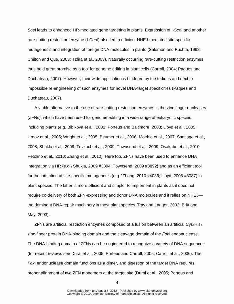
Figure 3 pTRV-mediated co-expression of two reporter genes in newly developed plant cells.
(A) Co-expression of fluorescent reporter genes DsRed2 and EGFP in newly developed leaves
of pTRV-Δ2b-sgP::DsRed2-T2A-EGFP-infected plants. DsRed2 and EGFP fluorescence are in
orange and green, respectively, and plastid autofluorescence is in dark red. (B) Co-expression
of DsRed2 and EGFP in newly developed leaves of pTRV-Δ2b-[sgP::DsRed2][sgP::EGFP]-
infected N. tabacum plant. DsRed2 and EGFP fluorescence are in red and green, respectively,
www.plantphysiol.orgon August 5, 2018 - Published by Downloaded from Copyright © 2010 American Society of Plant Biologists. All rights reserved.

22
and plastid autofluorescence is in dark red. Images in panels A and B are single confocal
sections.
Figure 4 pTRV-mediated ZFN expression leads to site-specific mutagenesis in newly developed
tissues of infected plants. (A) Outline of the mutated GUS (mGUS) reporter gene-repair assay
designed to monitor ZFN-mediated mutagenesis in transgenic plants. The mGUS-encoding
gene is disrupted by a stop codon (in red) within the 6-bp spacer of the ZFN target site.
Reconstruction of active GUS gene occurs by putative deletion of a CTG sequence. The ZFN
binding sequences are shown in green and the GUS initiation codon in blue. (B-H) Detection of
site-specific mutagenesis events in newly developed tissues of pTRV-Δ2b-sgP::QQR infected
plants by X-Gluc staining. GUS expression was detected in newly developed N. tabacum cv.
Samsung (B) and P. hybrida (C) leaves, 13 and 22 days post-inoculation, respectively, and in
newly developed P. hybrida (D) and N. tabacum cv. Samsung (E) buds, 11 and 50 days post-
inoculation, respectively. GUS expression was also detected in P. hybrida developing primordia
(F) and P. hybrida flower and reproductive tissues (G and H) of the GUS-positive mature plant.
Figure 5 Molecular analysis of ZFN-mediated mutagenesis events in P. hybrida (P) and N.
tabacum (T) plants. The initiation codon and the ZFN-binding sites on the upper strand of the
mGUS sequences are in blue and purple, respectively. The stop codon sequence is in red. The
predicted outcome of positive (+) or negative (-) GUS expression is indicated on the right.
Figure 6 Mutant plants can develop directly, without a regeneration step, from virus-infected
plants and can stably pass the mutation on to their offspring. Uniformly GUS-stained plantlets,
exemplified here with infected P. hybrida (A), which were regenerated from infected plants, and
allowed to root, mature (B) and set seed. Also shown are GUS-stained P. hybrida (C) and N.
tabacum (D) seedlings obtained from mature, virus-infected plants.
www.plantphysiol.orgon August 5, 2018 - Published by Downloaded from Copyright © 2010 American Society of Plant Biologists. All rights reserved.

23
Figure 7 Molecular analysis of randomly selected GUS-positive P. hybrida and N. tabacum
seedlings. (A) Mutagenesis events in P. hybrida (P) and N. tabacum (T) offspring. The initiation
codon and the ZFN-binding sites on the upper strand of the mGUS sequences are in blue and
purple, respectively. Stop codon is in red. (B) Upper panel: RT-PCR analysis of pTRV genomes
in infected, healthy and mutated seedlings of P. hybrida and N. tabacum. Lower panel: RT-PCR
analysis of housekeeping gene (the plastid 23S RNA gene). pTRV genomes (identified by RT-
PCR amplification of TRV2 sequence coat protein[CP]) were detected in infected, but not
healthy or mutated seedlings (M, DNA marker ladder).
www.plantphysiol.orgon August 5, 2018 - Published by Downloaded from Copyright © 2010 American Society of Plant Biologists. All rights reserved.

24
Supplemental figure legends
Figure S1 Expression of fluorescent reporter gene DsRed2 in cells of newly developed leaves
of plants which were agroinfiltrated with pTRV-Δ2b-sgP::DsRed2. DsRed2 and plastid
autofluorescence are shown in orange and dark red, respectively.
Figure S2 Expression of fluorescent reporter gene DsRed2 in tissues and organs of plants
which were mechanically infected with pTRV-Δ2b-sgP::DsRed2. DsRed2 expression is shown
in red.
Figure S3 Expression of chloroplast-targeted Rssu-EGFP and DsRed2 fluorescent reporter
genes in newly developed tissues and organs of plants which were co-infected with pTRV2-
Δ2b-sgP::Rssu-EGFP and pTRV2-Δ2b-sgP:: DsRed2. DsRed2 expression is shown in orange,
EGFP expression in green and plastid autofluorescence in purple.
www.plantphysiol.orgon August 5, 2018 - Published by Downloaded from Copyright © 2010 American Society of Plant Biologists. All rights reserved.

Fig 1
35SP nosTCP sgP GOI
35SP nosTCP sgP GOI1 GOI2
35SP nosTCP sgP GOI1 GOI2sgP
T2A
A
B
C
MCS
MCS
MCS SmaI
SmaI
SmaI5’
5’
5’ 3’
3’
3’
Figure 1 Structure and key features of pTRV-based expression vectors. (A) pTRV-Δ2b-sgP::GOI designed to drive the expression a single gene of interest (GOI) under the control of the sgP constitutive promoter. (B) pTRV-Δ2b-sgP::GOI1-T2A-GOI2 designed to drive the co-expression of two genes as a single transcript in which the coding sequences of the two genes (GOI1 and GOI2) are separated by a T2A sequence. (C) pTRV-Δ2b-[sgP::GOI1][sgP::GOI2] designed to drive the co-expression of two genes from two independent subgenomic promoters. The T-DNA region of each vector is presented. Also shown are (i) the constitutive 35S promoter (35sP) and the nopaline synthase terminator (nosT), needed for the production of primary viral transcript following agroinfiltration, and (ii) the 5’ and 3’ untranslated regions of the TRV2 needed for viral replication and transcription. The multiple cloning site (MCS) includes EcoRI, XbaI, KpnI, SacI and XhoI.
www.plantphysiol.orgon August 5, 2018 - Published by Downloaded from Copyright © 2010 American Society of Plant Biologists. All rights reserved.

N. benthamiana P. hybridaN. tabacum
DsRed2
Bright
field
Fig 2
Figure 2 pTRV-mediated expression of a single reporter gene (DsRed2) in newly developed tissues and organs. Plants were infected by pTRV-Δ2b-sgP::DsRed2. Images were taken by fluorescence stereoscope. DsRed2 expression is shown in red.
www.plantphysiol.orgon August 5, 2018 - Published by Downloaded from Copyright © 2010 American Society of Plant Biologists. All rights reserved.
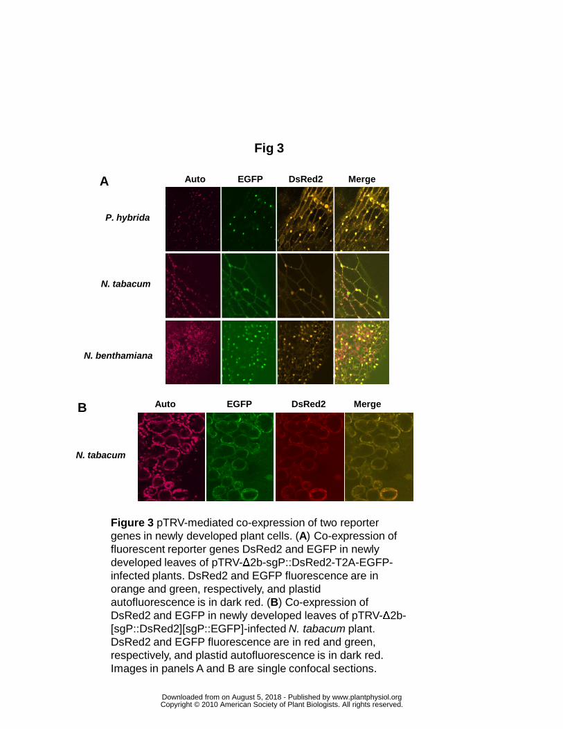
Fig 3
N. benthamiana
P. hybrida
N. tabacum
DsRed2EGFPAuto Merge
DsRed2EGFPAuto Merge
N. tabacum
A
B
Figure 3 pTRV-mediated co-expression of two reporter genes in newly developed plant cells. (A) Co-expression of fluorescent reporter genes DsRed2 and EGFP in newly developed leaves of pTRV-Δ2b-sgP::DsRed2-T2A-EGFP-infected plants. DsRed2 and EGFP fluorescence are in orange and green, respectively, and plastid autofluorescence is in dark red. (B) Co-expression of DsRed2 and EGFP in newly developed leaves of pTRV-Δ2b-[sgP::DsRed2][sgP::EGFP]-infected N. tabacum plant. DsRed2 and EGFP fluorescence are in red and green, respectively, and plastid autofluorescence is in dark red. Images in panels A and B are single confocal sections.
www.plantphysiol.orgon August 5, 2018 - Published by Downloaded from Copyright © 2010 American Society of Plant Biologists. All rights reserved.
Fig 4
B
AATGTTCTTCCCCTCCTGAGGGGAAGAATTA ...TACAAGAAGGGGAGGACTCCCCTTCTTAAT ...
M S T N S * V G A L
ATGTTCTTCCCCTCAGGGGAAGAATTA ... TACAAGAAGGGGAGTCCCCTTCTTAAT ...
M S T N S V G A L
digestion and repair by NHEJ
C
D E
F G H
Figure 4 pTRV-mediated ZFN expression leads to site-specific mutagenesis in newly developed tissues of infected plants. (A) Outline of the mutated GUS (mGUS) reporter gene-repair assay designed to monitor ZFN-mediated mutagenesis in transgenic plants. The mGUS-encoding gene is disrupted by a stop codon (in red) within the 6-bp spacer of the ZFN target site. Reconstruction of active GUS gene occurs by putative deletion of a CTG sequence. The ZFN binding sequences are shown in green and the GUS initiation codon in blue. (B-H) Detection of site-specific mutagenesis events in newly developed tissues of pTRV-Δ2b-sgP::QQR infected plants by X-Gluc staining. GUS expression was detected in newly developed N. tabacum cv. Samsung (B) and P. hybrida (C) leaves, 13 and 22 days post-inoculation, respectively, and in newly developed P. hybrida (D) and N. tabacum cv. Samsung (E) buds, 11 and 50 days post-inoculation, respectively. GUS expression was also detected in P. hybrida developing primordia (F) and P. hybrida flower and reproductive tissues (G and H) of the GUS-positive mature plant.
www.plantphysiol.orgon August 5, 2018 - Published by Downloaded from Copyright © 2010 American Society of Plant Biologists. All rights reserved.

mGUS GACGGTACCATGTTCTTCCCCTCCTG--AGGGGAAGAATTACGTCCTGTAGAAACCCCA -T7 GACGGTACCATGTTCTTCCCCTCCTG-GAGGGGAAGAATTACGTCCTGTAGAAACCCCA -P47 GACGGTACCATGTTCTTCCCCTCCTG-AAGGGGAAGAATTACGTCCTGTAGAAACCCCA -P41 GACGGTACCATGTTCTTCCCCTCCTGTGAGGGGAAGAATTACGTCCTGTAGAAACCCCA -P30 GACGGTACCATGTTCTTCCCCACCCG--AGGGGAAGAATTACGTCCTGTAGAAACCCCA + P29 GACGGTACCATGTTCTTCCCCTCTTG--AGGGGAAGAATTACGTCCTGTAGAAACCCCA -T115 GACGGTACCATGTTCTTCCCCTC--G--AGGGGAAGAATTACGTCCTGTAGAAACCCCA -T11 GACGGTACCATGTTCTTCCCCT--TG--AGGGGAAGAATTACGTCCTGTAGAAACCCCA –T6 GACGGTACCATGTTCTTCCCCT------AGGGGAAGAATTACGTCCTGTAGAAACCCCA +P62 GACGGTACCATGTTCTTCCCTT---G--AGGGGAAGAATTACGTCCTGTAGAAACCCCA –T4 GACGGTACCATGTTCTTCCCCTC-------GGGAAGAATTACGTCCTGTAGAAACCCCA –T114 GACGGTACCATGTTCTTCCCCT-------GGGGAAGAATTACGTCCTGTAGAAACCCCA –T101 GACGGTACCATGTTCTTCCCCA-------GGGGAAGAATTACGTCCTGTAGAAACCCCA -P26 GACGGTACCATGTTCTTCC-----------GGGAAGAATTACGTCCTGTAGAAACCCCA -P49 GACGGTACCATGTTCTTCCCC-------------------TCGTCCTGTAGAAACCCCA -P48 GACGGTA---AG------------------GGGAAGAATTACGTCCTGTAGAAACCCCA –T104 GACGGTACCATGT-------------------------TTACGTCCTGTAGAAACCCCA -P51 GACGGTA----------------------------GAATTACGTCCTGTAGAAACCCCA –T111 GACGGTACCATG------------------------------TTCCTGTAGAAACCCCA +P34 GACGGTACCATG------------------------------TTCTT-------CCCCA -P25 GAC---------------------------------------------------CCCCA -
Fig 5
Figure 5 Molecular analysis of ZFN-mediated mutagenesis events in P. hybrida (P) and N. tabacum (T) plants. The initiation codon and the ZFN-binding sites on the upper strand of the mGUS sequences are in blue and purple, respectively. The stop codon sequence is in red. The predicted outcome of positive (+) or negative (-) GUS expression is indicated on the right.
www.plantphysiol.orgon August 5, 2018 - Published by Downloaded from Copyright © 2010 American Society of Plant Biologists. All rights reserved.

Fig 6
A B
C D
Figure 6 Mutant plants can develop directly, without a regeneration step, from virus-infected plants and can stably pass the mutation on to their offspring. Uniformly GUS-stained plantlets, exemplified here with infected P. hybrida (A), which were regenerated from infected plants, and allowed to root, mature (B) and set seed. Also shown are GUS-stained P. hybrida (C) and N. tabacum (D) seedlings obtained from mature, virus-infected plants.
www.plantphysiol.orgon August 5, 2018 - Published by Downloaded from Copyright © 2010 American Society of Plant Biologists. All rights reserved.

A mGUS ATGTTCTTCCCCTCCTGAGGGGAAGAATTAT-1-13 ATGTTCTTCCCCT------GGGAAGAATTAT-2-11 ATGTTCTTCCCCT------GGGAAGAATTAT-2-8 ATGTTCTTCCCCT------GGGAAGAATTAP-1-3 ATGTTCTTCCCCTC---AGGGGAAGAATTAP-1-4 ATGTTCTTCCCCTC---AGGGGAAGAATTAP-1-5 ATGTTCTTCCCCTC---AGGGGAAGAATTAP-II-3 ATGTTCTTCCCCTC---AGGGGAAGAATTAP-II-4 ATGTTCTTCCCCTC---AGGGGAAGAATTA
B tobaccoseedlings
petunia seedlings
CP
23S
M M
Fig 7
Figure 7 Molecular analysis of randomly selected GUS-positive P. hybrida and N. tabacum seedlings. (A) Mutagenesis events in P. hybrida (P) and N. tabacum (T) offspring. The initiation codon and the ZFN-binding sites on the upper strand of the mGUSsequences are in blue and purple, respectively. Stop codon is in red. (B) Upper panel: RT-PCR analysis of pTRV genomes in infected, healthy and mutated seedlings of P. hybrida and N. tabacum. Lower panel: RT-PCR analysis of housekeeping gene (the plastid 23S RNA gene). pTRV genomes (identified by RT-PCR amplification of TRV2 sequence coat protein [CP]) were detected in infected, but not healthy or mutated seedlings (M, DNA marker ladder).
www.plantphysiol.orgon August 5, 2018 - Published by Downloaded from Copyright © 2010 American Society of Plant Biologists. All rights reserved.