Embed Size (px)
Citation preview

Nocturnal monitoring of home non-invasiveventilation: the contribution of simple tools such aspulse oximetry, capnography, built-in ventilatorsoftware and autonomic markers ofsleep fragmentation
Jean-Paul Janssens,1 Jean-Christian Borel,2,3 Jean-Louis Pepin,2 on behalf of theSomnoNIV Group
ABSTRACTComplex respiratory events, which may havea detrimental effect on both quality of sleep and controlof nocturnal hypoventilation, occur during sleep inpatients treated with non-invasive ventilation (NIV).Among these events are patient-ventilator asynchrony,increases in upper airway resistance (with or withoutincreased respiratory drive) and leaks. Detection of theseevents is important in order to select the mostappropriate ventilator settings and interface. Simple toolscan provide important information when monitoring NIV.Pulse oximetry is important to ensure that adequateoxygen saturation is provided and to detect eitherprolonged or short and recurrent desaturations.However, the specificity of pulse oximetry tracingsduring NIV is low. Transcutaneous capnography helpsdiscriminate between hypoxaemia related to ventilation/perfusion mismatch and hypoventilation, documentscorrection of nocturnal hypoventilation and may detectventilator-induced hyperventilation, a possible cause forcentral apnoea/hypopnoea and glottic closure. Dataprovided by ventilator software help the clinician byestimating ventilation, tidal volume, leaks and the rate ofinspiratory or expiratory triggering by the patient,although further validation of these signals byindependent studies is indicated. Finally, autonomicmarkers of sympathetic tone using signals such as pulsewave amplitude of the pulse oximetry signal can providereliable information of sleep fragmentation.
INTRODUCTIONHome non-invasive ventilation (NIV) aims tocorrect daytime and nocturnal hypoventilation andassociated symptoms and to ensure adequatenocturnal oxygen saturation measured by pulseoximetry (SpO2). During sleep, specific respiratoryevents may occur resulting both from sleep-relatedphysiological changes and use of NIV.1 Among theseevents, described in another article in this series,2 arerepetitive leaks, upper airway instability and residualobstructive events, recurrent decreases in ventilatorycommand with or without glottic closure, orpatient-ventilator asynchrony. An appropriatestrategy for monitoring these respiratory events isnecessary. Monitoring tools can be limited to therecognition of their consequences such as oxygendesaturation or increases in transcutaneous carbon
dioxide tension (PtcCO2). The latest generation ofhome ventilators3 are often equipped with sophis-ticated built-in software capable of recording a widerange of parameters over several months, and thusoffering information to the clinician on items such ascompliance and leaks, among many other respira-tory parameters.This review describes the contributions, limits
and caveats of non-invasive assessment of NIVduring sleep through simple techniques such aspulse oximetry, combined capnography and pulseoximetry, built-in ventilator software and auto-nomic markers of sleep fragmentation. Morecomplex and complete evaluations can beperformed by polygraphic or polysomnographicrecordings which will be discussed in a later articlein this series.2
PULSE OXIMETRY AND NON-INVASIVEVENTILATIONNocturnal oxygen desaturation is considered as oneof the major determinants of adverse neuro-cognitive and cardiovascular consequences occur-ring during chronic respiratory failure and sleepapnoea syndrome.4e6 Pulse oximetry has theadvantages of simplicity, short set-up time andshort time response (seconds); disadvantages aremotion artefacts and sensitivity to perfusion.Information provided by pulse oximetry alsodepends on the device used and its settings,particularly signal averaging time; the magnitude ofdesaturations decreases when averaging timeincreases and this influences the sensitivity of thetechnique. In the field of sleep apnoea it has beenshown that the number of hypopnoeas associatedwith desaturations of $4% could change signifi-cantly according to the device used and this couldaffect clinical decisions.7 Sampling frequency (onaverage 25 times/s) and signal averaging time mayvary considerably between devices (reported range2e21/s). New generation pulse oximeters usuallyaverage SpO2 signals over <10 s; high speed signalaveraging also improves detection of motion arte-facts, which can be quite frequent in subjects withsleep-disordered breathing.8 9 The accuracy of SpO2
measurements reported in sleep studies is 2e6%compared with arterial blood-derived determina-tions of haemoglobin saturation which are in the
1Division of Pulmonary Diseases,Geneva University Hospital,Geneva, Switzerland2Pole Reeducation etPhysiologie et Laboratoire HP2,INSERM ERI 0017, UniversiteJoseph Fourier, Grenoble,France3Association medico-techniqueAGIR a dom, Meylan, France
Correspondence toJean Paul Janssens, CentreAntituberculeux, Division ofPulmonary Diseases, GenevaUniversity Hospital, RueGabrielle Perret-Gentil 4, 1211Geneva 14, Switzerland;[email protected]
Received 1 April 2010Accepted 19 August 2010
Janssens J-P, Borel J-C, Pepin J-L, et al. Thorax (2010). doi:10.1136/thx.2010.139782 1 of 8
Review series Thorax Online First, published on October 22, 2010 as 10.1136/thx.2010.139782
Copyright Article author (or their employer) 2010. Produced by BMJ Publishing Group Ltd (& BTS) under licence.
on 6 August 2018 by guest. P
rotected by copyright.http://thorax.bm
j.com/
Thorax: first published as 10.1136/thx.2010.139782 on 22 O
ctober 2010. Dow
nloaded from

range 75e100%. When >90%, the accuracy of SpO2 readings is62%; at values <80%, however, the accuracy can decreaseconsiderably with a trend towards under-reading.
SpO2 is obviously an important item to monitor in homeventilated patients. During spontaneous breathing it has beensuggested that nocturnal oximetry could be specific for identi-fying sleep-related breathing disturbances,10 but that themorphological pattern of SpO2 desaturations could not accu-rately separate central from obstructive events.11 In patientsusing NIV at night, variations in SpO2 should be interpretedmore cautiously. Indeed, pulse oximetry tracings during NIVmay show a wide variety of events. The major limitation of thetechnique is the absence of specificity of the SpO2 variations.Recurrent desaturations may reflect, for instance, upper airwayinstability and residual obstructive events, decreases in ventila-tory command with or without glottic closure, or repetitiveleaks interrupted by microarousals. Central events such asdecreases in ventilatory command with or without glotticclosure may be residual events insufficiently corrected by NIV insubjects with central sleep apnoea syndromes or induced byventilation per se (figure 1A).12 13 During spontaneousbreathing, prolonged desaturations may reflect ventilation/perfusion (V/Q) mismatch in severe obstructive or restrictivedisorders, position-dependent V/Q mismatch in severe obesityor persistence of alveolar hypoventilation. Prolonged desatura-tions (10e30 min) with concurrent acceleration of heartfrequency occurring approximately every 90e120 min duringthe night are also typical of REM sleep hypoventilation. Inventilated patients, however, the same aspect can also resultfrom prolonged leaks or insufficient pressure support irrespectiveof sleep stage (figure 1B). Furthermore, in subjects with oxygensupplementation, pulse oximetry is unreliable for the detectionof hypoventilation.14 In spite of these limitations, pulseoximetry is a valuable screening tool for patients established onhome ventilation who do not appear to have any problemsrelated to NIV and who are not receiving supplemental oxygen.Among the advantages of the technique are its low cost, ease ofbasic interpretation, possible use in telemedicine programmesand transmission of devices with recordings by mail.15
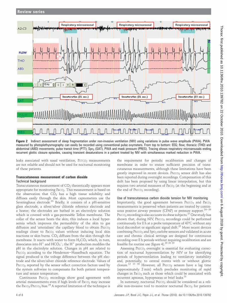
Microarousals are associated with momentary increases insympathetic activity.16 A wide range of physiological cardiovas-cular parameters such as heart rate, blood pressure, pulse transittime and pulse wave amplitude are related to sympathetic toneand have been implemented as indirect methods for detectingsleep fragmentation. Pulse wave amplitude (PWA) measured byphotoplethysmography can easily be recorded using conventionalpulse oximeters (figure 2). Peripheral vasoconstrictor responsesassociated with microarousals are visually identifiable by markedreductions in PWA (figure 2).17 Thus, a simple oximetric recordingduring NIV can provide information not only on residual oxygendesaturations but also on sleep fragmentation.
Interestingly, there are no precise guidelines as to what shouldbe considered as satisfactory in terms of nocturnal oxygenationduring home NIV; however, maintaining an adequate nocturnalSpO2 most probably decreases secondary pulmonary hyperten-sion, improves respiratory muscle function and, in patients withdiurnal hypoxaemia, survival. As stated by Langevin et al,18 SpO2
is a key feature of nasal ventilation monitoring; without at leastadequate control of oxygenation, ventilator settings cannot beaccepted as correct. A reasonable goal is to adjust ventilatorsettings to obtain a mean nocturnal SpO2 $90%, with <10% ofthe total recording time <90% after correction of leaks.19
Oxygen supplementation should be provided only in case of V/Q mismatch incompletely treated by NIV.
Although nocturnal pulse oximetry combined with daytimearterial blood gases are often used as the only evaluation of homeNIV, daytime arterial blood gases may reflect nocturnal values ofarterial carbon dioxide tension (PaCO2) poorly,20 21 and anacceptable average SpO2 may be associated with importantnocturnal respiratory events or poor control of nocturnal hypo-ventilation.22 Thus, visual inspection of oximetry traces isimportant for the detection of nocturnal respiratory events.However, the only way of determining whether decreased averageSpO2 values or prolonged desaturations are related to V/Qmismatch or hypoventilation is through measurement ofnocturnal carbon dioxide. To summarise, pulse oximetry(including visual inspection of traces) should be used as ascreening tool in stable home-ventilated patients withoutsupplemental oxygen to identify and exclude patients who do notrequire more detailed, expensive and time-consuming investiga-tions (figure 3). Further studies, however, are needed to determinethe most cost-effective procedure between systematic polygraphyor polysomnography or use of oximetry as a screening tool toavoid more expensive procedures in patients with normal arterialoxygen saturation (SaO2) tracings.
NON-INVASIVE ASSESSMENT OF NOCTURNAL ARTERIALCARBON DIOXIDE TENSIONAssessment of PaCO2 is essential for evaluating the adequacy ofalveolar ventilation when monitoring patients treated by NIV.To date, repeated sampling of arterial blood remains the ‘goldstandard’ for estimating the adequacy of ventilatory support.However, repeated sampling of arterial blood does not reliablyassess control of nocturnal hypoventilation because the patientwill usually wake. Arterial catheterisation needs costly equip-ment, specially trained personnel and, in most cases, the envi-ronment of an intensive care unit. Furthermore, repeated arterialpunctures or arterial catheterisation are additional sources ofdiscomfort for the patient and are not appropriate for routineassessment in stable patients. The simplest approach is tomeasure PaCO2 by arterial puncture at the end of the night todocument night-to-morning increases in PaCO2. However, bloodis most often sampled after arousal and thus after a short periodof appropriate ventilation. In this condition, a normal morningPaCO2 does not actually reflect the abnormal time course of PaCO2
during the night.20 21 Non-invasive assessment of PaCO2 can beperformed by measuring transcutaneous carbon dioxide tension(PtcCO2) or peak expired carbon dioxide (PETCO2).
Peak expired carbon dioxideSimple portable devices are available for measuring PETCO2.However, PETCO2 is a poor predictor of PaCO2. The relationshipbetween PaCO2 and PETCO2 depends on the physiological deadspace (VD) and the patients’ ventilatory mode (tidal volume,VT): PETCO2 ¼ PaCO2 (1 � VD/VT). The relationship betweenPaCO2 and PETCO2 therefore depends on the type and extent ofthe underlying parenchymal disorder. Since VT decreases physi-ologically during sleep, the [PaCO2 � PETCO2] gradient increasesduring sleep studies because of a higher VD/VT ratio. Also, inmost patients with NIV ventilation is not homogeneous andexpired CO2 does not reach an adequate plateau. PETCO2 istherefore unreliable in patients with chronic respiratory failure,particularly in chronic obstructive pulmonary disease (COPD); itis technically difficult to measure with the continuous flowthrough the mask related to bilevel pressure support.In summary, because of limitations related to underlying
diseases and additional problems induced by non-intentional
2 of 8 Janssens J-P, Borel J-C, Pepin J-L, et al. Thorax (2010). doi:10.1136/thx.2010.139782
Review series
on 6 August 2018 by guest. P
rotected by copyright.http://thorax.bm
j.com/
Thorax: first published as 10.1136/thx.2010.139782 on 22 O
ctober 2010. Dow
nloaded from

Figure 1 Interpretation of oxygen saturation measured by pulse oximetry (SpO2) patterns in patients using non-invasive ventilation (NIV). (A) Recurrentoscillations of SpO2 in a patient treated with NIV. This pattern is sensitive but not specific and can be associated with (i) central events resulting fromrespiratory instability under NIV, (ii) persistent obstructive events occurring in upper airways, (iii) intermittent non-intentional leaks. (B) Sustained SpO2desaturations during NIV (>10 min). This non-specific pattern can result from residual hypoventilation or prolonged non intentional leaks.
Janssens J-P, Borel J-C, Pepin J-L, et al. Thorax (2010). doi:10.1136/thx.2010.139782 3 of 8
Review series
on 6 August 2018 by guest. P
rotected by copyright.http://thorax.bm
j.com/
Thorax: first published as 10.1136/thx.2010.139782 on 22 O
ctober 2010. Dow
nloaded from
leaks associated with nasal ventilation, PETCO2 measurementsare not reliable and should not be used for nocturnal monitoringof these patients.
Transcutaneous measurement of carbon dioxideTechnical backgroundTranscutaneous measurement of CO2 theoretically appears moreappropriate for monitoring PaCO2. This measurement is based onthe observation that CO2 has a high tissue solubility anddiffuses easily through the skin. Most capnometers use theSeveringhaus electrode.23 Briefly, it consists of a pH-sensitiveglass electrode, a silver/silver chloride reference electrode anda heater; the electrodes are bathed in an electrolyte solutionwhich is covered with a gas-permeable Teflon membrane. Thecollar of the sensor heats the skin; this induces a local hyper-aemia which improves the permeability of the skin to gasdiffusion and ‘arterialises’ the capillary blood to obtain PtcCO2
readings closer to PaCO2 values without inducing local skinreactions or skin burns. CO2 diffuses from the skin through themembrane. It reacts with water to form H2CO3 which, in turn,dissociates into H+ and HCO3
�; the H+ production modifies thepH in the electrolyte solution. Changes in pH are related toPtcCO2 according to the HendersoneHasselbach equation. Thesignal produced is the voltage difference between the pH elec-trode and the silver/silver chloride reference electrode. Values ofPtcCO2 reported by the sensor reflect correction factors used bythe system software to compensate for both patient tempera-ture and sensor temperature.
Continuous PtcCO2 recordings show good agreement witharterial measurements even if high levels of PaCO2 may increasethe PaCO2/PtcCO2 bias.24 A reported limitation of the technique is
the requirement for periodic recalibration and changes ofmembrane in order to ensure sufficient precision of trans-cutaneous measurements, although these limitations have beengreatly improved in recent devices. PtcCO2 sensor drift has alsobeen reported during overnight recordings. Compensation of thisdrift has been proposed by using linear interpolation, but thisrequires two arterial measures of PaCO2 (at the beginning and atthe end of PtcCO2 recording).
Use of transcutaneous carbon dioxide tension for NIV monitoringImportantly, the good agreement between PtcCO2 and PaCO2
measurements is preserved when patients are treated by contin-uous positive airway pressure (CPAP) or pressure support.24e26
PtcCO2 recording is also accurate in obese subjects.27One studyhasshown that, during NIV, PtcCO2 recordings could be performedcontinuously for 8 h at a probe temperature of 438Cwithout anylocal discomfort or significant signal drift.25 More recent devicescombining PtcCO2 and SpO2 earlobe sensors and validated in acutecare and chronic clinical settings are designed for continuousrecording over 8 h periods without requiring recalibration and arefeasible for routine use (figure 4).26 28 29
Assessing PtcCO2 overnight is essential for evaluating correc-tion of nocturnal hypoventilation by NIV or for identifyingperiods of hyperventilation leading to ventilatory instabilityand, potentially, to central events with or without glotticclosure.12 13 30 However, all PtcCO2 sensors have a lag time(approximately 2 min) which precludes monitoring of rapidchanges in PaCO2 such as those which could be associated withrecurrent apnoeas, hypopnoeas or brief leaks.31
In summary, nocturnal PtcCO2 should be considered as a reli-able non-invasive tool to monitor nocturnal PaCO2 for patients
Figure 2 Indirect assessment of sleep fragmentation under non-invasive ventilation (NIV) using variations in pulse wave amplitude (PWA). PWAmeasured by photoplethysmography can easily be recorded using conventional pulse oxymeters. From top to bottom: EEG; flow; thoracic (THO) andabdominal (ABD) movements; pulse transit time (PTT); SpO2 (SAT), PWA and mask pressure (PRES). Tracing shows respiratory microarousals endingrecurrent glottic closure episodes, causing transient desaturations in a patient treated by NIV with simultaneous marked reduction in PWA.
4 of 8 Janssens J-P, Borel J-C, Pepin J-L, et al. Thorax (2010). doi:10.1136/thx.2010.139782
Review series
on 6 August 2018 by guest. P
rotected by copyright.http://thorax.bm
j.com/
Thorax: first published as 10.1136/thx.2010.139782 on 22 O
ctober 2010. Dow
nloaded from
treated with NIV on a long-term basis. Limitations of thetechnique are the cost of the devices, the increase in biasbetween arterial and transcutaneous values at high PaCO2
values24 and the occasional occurrence of unexplained errantvalues. Recent devices are, however, easy to use with user-friendly software and can be connected to polysomnographysoftware. Finally, nocturnal PtcCO2 reveals the occurrence ofepisodes of hypoventilation but provides no information as totheir cause (eg, inappropriate settings, leaks).
Data available from NIV machinesMost NIV device designers and manufacturers have incorporatedflow and pressure sensors and the possibility of storing raw dataof these parameters on a long-term basis. Specific software
allows home care providers or clinicians to download these dataonto a personal computer.Downloaded data can be separated into three categories. The
first is a synthesis report (ie, trend of each parameter recordedduring a given period). Depending on the manufacturers and themachines (table 1), compliance, settings, estimations (notabsolute values) of leaks, tidal volume, respiratory frequency,minute ventilation and respiratory cycles triggered by thepatient are provided. The second category is a detailed dataanalysis in which raw data of a given parameter can be analysedcycle by cycle, and the third category is a polygraphic dataanalysis. In this situation, by adding an external moduleconnected to the machine, physiological parameters such asoxygen saturation, heart rate and respiratory effort can be
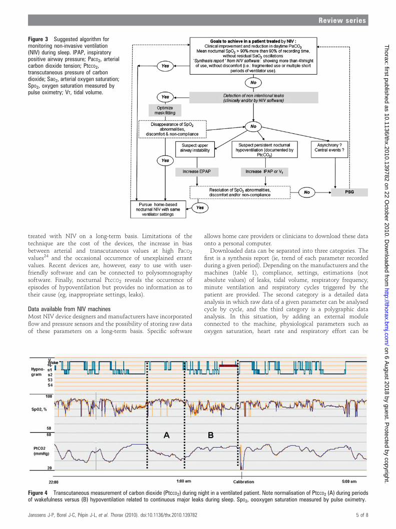
Figure 3 Suggested algorithm formonitoring non-invasive ventilation(NIV) during sleep. IPAP, inspiratorypositive airway pressure; PaCO2, arterialcarbon dioxide tension; PtcCO2,transcutaneous pressure of carbondioxide; SaO2, arterial oxygen saturation;SpO2, oxygen saturation measured bypulse oximetry; VT, tidal volume.
Figure 4 Transcutaneous measurement of carbon dioxide (PtcCO2) during night in a ventilated patient. Note normalisation of PtcCO2 (A) during periodsof wakefulness versus (B) hypoventilation related to continuous major leaks during sleep. SpO2, oooxygen saturation measured by pulse oximetry.
Janssens J-P, Borel J-C, Pepin J-L, et al. Thorax (2010). doi:10.1136/thx.2010.139782 5 of 8
Review series
on 6 August 2018 by guest. P
rotected by copyright.http://thorax.bm
j.com/
Thorax: first published as 10.1136/thx.2010.139782 on 22 O
ctober 2010. Dow
nloaded from

recorded and displayed in addition to the signals already storedby the device.
There are large discrepancies in parameters provided by thedifferent software. This reflects the fact that relevant parametersfor monitoring NIV have not yet been clearly defined by clini-cians and that recommendations in this field should be proposedby scientific societies. Second, the validity of several parametersestimated by the NIV devices (minute ventilation, VT, apnoea-hypopnoea index) must be further validated by independentclinical and/or bench test studies. In the absence of such vali-dation, information provided should only be considered asindicators of trends without any guarantee as to linearity of theestimations provided.
Compliance and pattern of daily use of ventilatorMedical history is often unreliable when assessing compliancewith NIV and recorded data in the ventilator are therefore veryhelpful for assessing average daily use of the ventilator and alsofor documenting special patterns of ventilator use which maysuggest inappropriate settings and patient discomfort (eg, frag-mented nocturnal use, multiple short periods of ventilator use).
Tidal volumeVTas reported by ventilator software is an indicator of effectiveventilation although subject to variability in accuracyaccording to leaks. On average, the target for VT is z10 ml/kg,taking into account ideal body weight.32 A high variability ofVT may suggest leaks or periodic breathing, spontaneous orventilator-induced.
Respiratory rate and percentage of spontaneous versus imposedrespiratory cyclesRespiratory rate (RR) analysis allows an estimation of thedifference between spontaneous RR and the preset back-up RR.Indeed, ventilators detect breaths either triggered by the patientor by the ventilator (unrewarded inspiratory efforts are notreported although some may be detected through the analysis ofdetailed flow curves if provided). Whether a high or low back-upRR is preferable is subject to debate. Restrick et al33 found nodifference in terms of efficacy of ventilation (assessed by SpO2
and PtcCO2) between a spontaneous mode and a back-up rate of10/min. Conversely, Parreira et al34 found that a controlled mode(back-up RR at 17 or 25/min) was more effective than the Smode. In many centres back-up RR is usually set at approxi-mately two breaths below spontaneous RR; this can be easilyadjusted using RR monitoring by ventilator. However, forpatients with neuromuscular diseases, ‘capturing’ the patient’sspontaneous RR is also common practice. The percentage ofrespiratory cycles triggered by the patient is also an estimationwhich should be interpreted with caution; a low percentage oftriggered (spontaneous) cycles can be interpreted either as ‘thepatient’s RR is captured by the ventilator ’ or ‘the ventilator doesnot detect the patient’s inspiratory efforts’.
Leaks: average value and 95% percentileThe major impact of leaks on efficacy of ventilation and qualityof sleep and the frequency with which major leaks occur duringnasal ventilation in stable patients has been well described.35e38
Thus, measurement of leaks is potentially a very useful adjunct
Table 1 Summary of data available from built-in software in home ventilators.
Device Software Synthesis data Detailed data Polygraphic data
VPAPIII-ST (A)VPAPIV-ST
ReScan 3.10 Compliance, ventilator settings,VE, VT, RR, leaks, residual AHI
VE, RR, leaks, residual AHIand flow cycle by cycle forVPAP IV
Simultaneous recording of SpO2and heart rate (available withadditional module: RESLINK)ResMed
HARMONY SYNCHRONY Encore Pro 2 Compliance, ventilator settings,VE, VT, RR, leaks, flow, total numberof apnoeas, alarms
VE, VT, RR, leaks, flow (cycleby cycle display possible withadditional Alice PDX module)
Simultaneous recording of SpO2,heart rate and respiratory effort(available with additional Alice PDXmodule)
Philips Respironics
Trilogy Direct view Compliance, ventilator settings,VE, VT, RR, leaks, flow, totalnumber of apnoeas, alarms
VE, VT, RR, leaks, pressure,flow (display cycle by cycle)and alarms
Philips Respironics
VENTIMOTION Ventisupport Compliance, ventilator settings,VE, VT, RR, leaks, flow,inspiratory time/total time, alarms
Pressure, flow, level of leaks Pressure, flow and leaks can betransferred to any polygraph withthe ‘Analog box-Weinmann’
Weinman (Hamburg, Germany)
GK 425 Silverlining 3 Compliance, ventilator settings,RR, pressure
NA
Tycohealthcare(Pleasanton, CA, USA)
SMARTAIR, SMARTAIR+LEGENDAIR, SUPPORTAIR
Airox Com 3.5.1 Compliance, ventilator settings,technical alarms and ventilationalarms
VT, RR, inspiratory time/total time,leaks, pressure/time and flow/timecurves (online or differed cycle bycycle display require connection toa PC while patient is under NIV)
Simultaneous recording of SpO2available with SUPPORTAIRmonitoring of SpO2Airox-Covidien (Pau, France)
Monnal T30 AirLiquide(Antony, France)
Bora Soft V.6 Compliance, ventilator settings,VE, VT, RR, leaks
NA
VS III, VS INTEGRA Easydiag 2 Compliance, ventilator settings,VE, VT, RR, leaks
VE, VT, RR, leaks (online or differedcycle by cycle display are possible)ResMed
ELYSEE 150 Easyview 150 (2.11) Compliance, ventilator settings,VE, VT, RR, leaks
VE, VT, RR, leaks (online cycle by cycledisplay is possible while patient isunder NIV)
ResMed
VIVO 30e40 Vivo PS software 3 Compliance, ventilator settings,VE, VT, RR, leaks, flow, alarms
Pressure, flow, VT, leaks (online ordiffered cycle by cycle display arepossible)
Breas (Molnlycke, Sweden)
VIVO 50 VIVO 50 PC software Compliance, ventilator settings,VE, VT, RR, leaks, flow, alarms
Pressure, flow, VT, leaks (online ordiffered cycle by cycle display arepossible)
Breas (Molnlycke, Sweden)
AHI, estimation of apnoea-hypopnoea index under NIV (residual AHI); NA, not available; NIV, non-invasive ventilation; PC, portable computer; RR, respiratory rate measured by ventilator; VE,estimation of minute ventilation; VT, estimation of tidal volume.
6 of 8 Janssens J-P, Borel J-C, Pepin J-L, et al. Thorax (2010). doi:10.1136/thx.2010.139782
Review series
on 6 August 2018 by guest. P
rotected by copyright.http://thorax.bm
j.com/
Thorax: first published as 10.1136/thx.2010.139782 on 22 O
ctober 2010. Dow
nloaded from

to information retrieved by the ventilator software, althoughresults have yet to be independently validated for most marketeddevices. Values are reported either as a total estimation of leaks(from which intentional leaks through the mask must besubtracted) or include intentional leaks by recording in the devicethe type of mask used. Threshold values considered acceptable bymanufacturers thus vary according to whether or not intentionalleaks are taken into account. Adjustment of interface andventilator settings aims to maintain the 95th percentile of leakvalues under a predetermined threshold value. Rabec et al recentlycompared data from a commercially available system with datarecorded on a bench test with a pneumotachograph and inducedleaks; the results were highly significantly correlated with a lowbias and limits of agreement.39
Apnoea-hypopnoea indexAlthough reported by ventilator software of several differentdevices, the effective meaning and reliability of the apnoea andapnoea-hypopnoea indices has yet to be determined. In mostcases, manufacturers use the same algorithms for defining orrecognising events that they developed for their auto-CPAPdevices. This is inappropriate and probably largely inaccurate inpatients during NIV.
Integrated multichannel modulesCertain bilevel ventilators can be coupled to multichannelmodules that provide trend data on minute ventilation, respi-ratory rate, air leaks, SpO2 and heart rate. Such modules obtainthis information from signals received through the respiratorycircuit and require additional connections to a pulse oximeterwith or without a detector of respiratory movements. Data canbe stored on a memory card and transferred to a computer forsimultaneous on-screen viewing. Rabec et al39 used a commer-cially available multichannel module to evaluate whether or notventilation was efficient in clinically stable home-ventilatedpatients. Their criteria to define ineffective ventilation were: (1)leakage >24 l/min for >20% of the trace duration; (2) prolongeddesaturations (>30% of the tracing with an SpO2 <90%),whether or not accompanied by a simultaneous reduction ofminute ventilation (>10% reduction compared with baseline) inthe absence of significant leaks; and (3) cumulative desaturationdips (>3%) for >10% of the trace duration. In patients receivinglong-term NIV, 53% of traces were abnormal; the most frequentfinding was leaks. Interestingly, neither pH nor PaCO2 werepredictive of abnormal recordings in these patients.
CONCLUSIONSSimple tools are available for monitoring the efficacy of long-term NIV. Pulse oximetry is the simplest approach. It is a roughindicator of major problems related to NIV but loses sensitivityin subjects with oxygen supplementation. Abnormalities onpulse oximetry tracings are non-specific and do not allowa distinction to be made between, for instance, V/Q mismatchversus alveolar hypoventilation. However, pulse oximetry isa very useful screening tool in stable patients receiving homeventilation. By using pulse wave amplitude analysis, oximetricrecordings can also provide indirect assessment of sleep frag-mentation when using NIV. Transcutaneous capnography hasbecome a routine approach in many centres: it is the only non-invasive method allowing assessment of correction of nocturnalhypoventilation. Drawbacks are the price of the equipment,occasional unexplained errant values and a lag time of severalminutes which renders analysis of very short respiratory eventsimpossible. Built-in software of home ventilators supplies very
valuable data (leaks, ventilatory mode, percentage of sponta-neous inspirations and expirations, residual apnoea and hypo-pnoea indices), although signals are estimations and must beindependently validated; furthermore, the relevance of somesignals has not yet been determined. Combined approaches suchas integrated modules allowing recording of multiple parametersoffer a very useful approach for detecting frequent problemssuch as leaks and their impact on SpO2.
Competing interests None.
Provenance and peer review Not commissioned; externally peer reviewed.
REFERENCES1. Elliott MW. Non-invasive ventilation during sleep: time to define new tools in the
systematic evaluation of the technique. Thorax. Published Online First 20 August2010. doi:10.1136/thx.2010.142117.
2. Gonzalez-Bermejo J, Perrin C, Janssens JP, et al. Proposal for a systematicanalysis of polygraphy or polysomnography for identifying and scoring abnormalevents occurring during non-invasive ventilation. Thorax 2010 doi:10.1136/thx.2010.142653.
3. Rabec C, Rodenstein D, Leger P, et al. Ventilator modes and settings during noninvasive ventilation: effects on respiratory events and implications for theiridentification. Thorax Published Online First: 14 October 2010. doi:10.1136/thx.2010.142661.
4. Carroll N, Bain RJ, Smith PE, et al. Domiciliary investigation of sleep-relatedhypoxaemia in Duchenne muscular dystrophy. Eur Respir J 1991;4:434e40.
5. Punjabi NM, Newman AB, Young TB, et al. Sleep-disordered breathing andcardiovascular disease: an outcome-based definition of hypopneas. Am J Respir CritCare Med 2008;177:1150e5.
6. Santos C, Braghiroli A, Mazzini L, et al. Sleep-related breathing disorders inamyotrophic lateral sclerosis. Monaldi Arch Chest Dis 2003;59:160e5.
7. Zafar S, Ayappa I, Norman RG, et al. Choice of oximeter affects apnea-hypopneaindex. Chest 2005;127:80e8.
8. Giuliano KK, Higgins TL. New-generation pulse oximetry in the care of critically illpatients. Am J Crit Care 2005;14:26e37; quiz 38e9.
9. McMorrow RC, Mythen MG. Pulse oximetry. Curr Opin Crit Care 2006;12:269e71.10. Pepin JL, Levy P, Lepaulle B, et al. Does oximetry contribute to the detection of
apneic events? Mathematical processing of the SaO2 signal. Chest 1991;99:1151e7.11. Series F, Kimoff RJ, Morrison D, et al. Prospective evaluation of nocturnal oximetry
for detection of sleep-related breathing disturbances in patients with chronic heartfailure. Chest 2005;127:1507e14.
12. Parreira VF, Delguste P, Jounieaux V, et al. Glottic aperture and effective minuteventilation during nasal two-level positive pressure ventilation in spontaneous mode.Am J Respir Crit Care Med 1996;154:1857e63.
13. Parreira VF, Jounieaux V, Aubert G, et al. Nasal two-level positive-pressureventilation in normal subjects. Effects of the glottis and ventilation. Am J Respir CritCare Med 1996;153:1616e23.
14. Fu ES, Downs JB, Schweiger JW, et al. Supplemental oxygen impairs detection ofhypoventilation by pulse oximetry. Chest 2004;126:1552e8.
15. Vitacca M, Bianchi L, Guerra A, et al. Tele-assistance in chronic respiratory failurepatients: a randomised clinical trial. Eur Respir J 2009;33:411e18.
16. Pepin JL, Tamisier R, Borel JC, et al. A critical review of peripheral arterial tone andpulse transit time as indirect diagnostic methods for detecting sleep disorderedbreathing and characterizing sleep structure. Curr Opin Pulm Med 2009;15:550e8.
17. Haba-Rubio J, Darbellay G, Herrmann FR, et al. Obstructive sleep apnea syndrome:effect of respiratory events and arousal on pulse wave amplitude measured byphotoplethysmography in NREM sleep. Sleep Breath 2005;9:73e81.
18. Langevin B, Leger P, Gerard M, et al. Monitoring nasal ventilation. Eur Respir Rev1993;3:260e5.
19. Gonzalez C, Ferris G, Diaz J, et al. Kyphoscoliotic ventilatory insufficiency: effects oflong-term intermittent positive-pressure ventilation. Chest 2003;124:857e62.
20. Janssens JP, Heritier-Praz A, Staneczek O, et al. Agreement between daytimemeasurement of arterial blood gases (ABG), nocturnal pulse-oximetry (NPO) andtranscutaneous capnography in home mechanical ventilation. Eur Respir J2002;20:155S.
21. Paiva R, Krivec U, Aubertin G, et al. Carbon dioxide monitoring during long-termnoninvasive respiratory support in children. Intensive Care Med 2009;35:1068e74.
22. Guo YF, Sforza E, Janssens JP. Respiratory patterns during sleep in obesity-hypoventilation patients treated with nocturnal pressure support: a preliminary report.Chest 2007;131:1090e9.
23. Severinghaus J. Transcutaneous blood gas analysis. Respir Care 1982;27:152e9.24. Cuvelier A, Grigoriu B, Molano LC, et al. Limitations of transcutaneous carbon
dioxide measurements for assessing long-term mechanical ventilation. Chest2005;127:1744e8.
25. Janssens JP, Perrin E, Bennani I, et al. Is continuous transcutaneous monitoring ofPCO2 (TcPCO2) over 8 h reliable in adults? Respir Med 2001;95:331e5.
26. Senn O, Clarenbach CF, Kaplan V, et al. Monitoring carbon dioxide tension andarterial oxygen saturation by a single earlobe sensor in patients with critical illness orsleep apnea. Chest 2005;128:1291e6.
Janssens J-P, Borel J-C, Pepin J-L, et al. Thorax (2010). doi:10.1136/thx.2010.139782 7 of 8
Review series
on 6 August 2018 by guest. P
rotected by copyright.http://thorax.bm
j.com/
Thorax: first published as 10.1136/thx.2010.139782 on 22 O
ctober 2010. Dow
nloaded from

27. Maniscalco M, Zedda A, Faraone S, et al. Evaluation of a transcutaneous carbondioxide monitor in severe obesity. Intensive Care Med 2008;34:1340e4.
28. Bendjelid K, Schutz N, Stotz M, et al. Transcutaneous PCO2 monitoring in criticallyill adults: clinical evaluation of a new sensor. Crit Care Med 2005;33:2203e6.
29. Parker SM, Gibson GJ. Evaluation of a transcutaneous carbon dioxide monitor(“TOSCA”) in adult patients in routine respiratory practice. Respir Med 2007;101:261e4.
30. Jounieaux V, Aubert G, Dury M, et al. Effects of nasal positive-pressure hyperventilationon the glottis in normal awake subjects. J Appl Physiol 1995;79:176e85.
31. Janssens JP, Howarth-Frey C, Chevrolet JC, et al. Transcutaneous PCO2 to monitornoninvasive mechanical ventilation in adults: assessment of a new transcutaneousPCO2 device. Chest 1998;113:768e73.
32. Janssens JP, Derivaz S, Breitenstein E, et al. Changing patterns in long-termnoninvasive ventilation: a 7-year prospective study in the Geneva Lake area. Chest2003;123:67e79.
33. Restrick LJ, Fox NC, Braid G, et al. Comparison of nasal pressure support ventilationwith nasal intermittent positive pressure ventilation in patients with nocturnalhypoventilation. Eur Respir J 1993;6:364e70.
34. Parreira VF, Delguste P, Jounieaux V, et al. Effectiveness of controlled andspontaneous modes in nasal two-level positive pressure ventilation in awake andasleep normal subjects. Chest 1997;112:1267e77.
35. Bach JR, Robert D, Leger P, et al. Sleep fragmentation in kyphoscolioticindividuals with alveolar hypoventilation treated by NIPPV. Chest1995;107:1552e8.
36. Meyer TJ, Pressman MR, Benditt J, et al. Air leaking through themouth during nocturnal nasal ventilation: effect on sleep quality. Sleep1997;20:561e9.
37. Rabec CA, Reybet-Degat O, Bonniaud P, et al. [Leak monitoring in noninvasiveventilation] (in Spanish). Arch Bronconeumol 2004;40:508e17.
38. Teschler H, Stampa J, Ragette R, et al. Effect of mouth leak on effectiveness ofnasal bilevel ventilatory assistance and sleep architecture. Eur Respir J1999;14:1251e7.
39. Rabec C, Georges M, Kabeya NK, et al. Evaluating noninvasive ventilation usinga monitoring system coupled to a ventilator: a bench-to-bedside study. Eur Respir J2009;34:902e13.
8 of 8 Janssens J-P, Borel J-C, Pepin J-L, et al. Thorax (2010). doi:10.1136/thx.2010.139782
Review series
on 6 August 2018 by guest. P
rotected by copyright.http://thorax.bm
j.com/
Thorax: first published as 10.1136/thx.2010.139782 on 22 O
ctober 2010. Dow
nloaded from