Embed Size (px)
Citation preview

NB Review
Yang Chai,
DDS, PhD


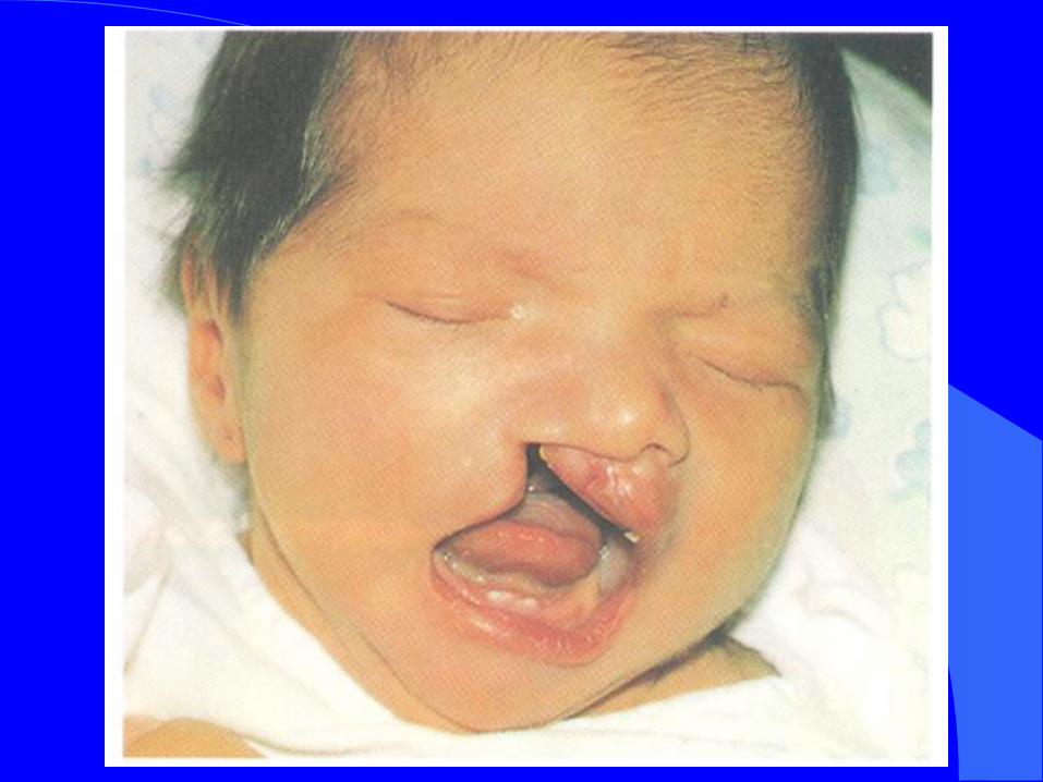
Treacher Collins
Syndrome


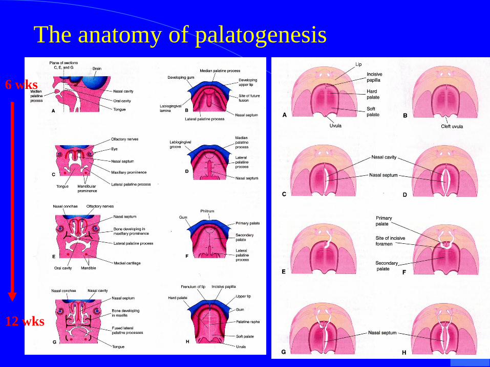
The anatomy of palatogenesis
6 wks
12 wks


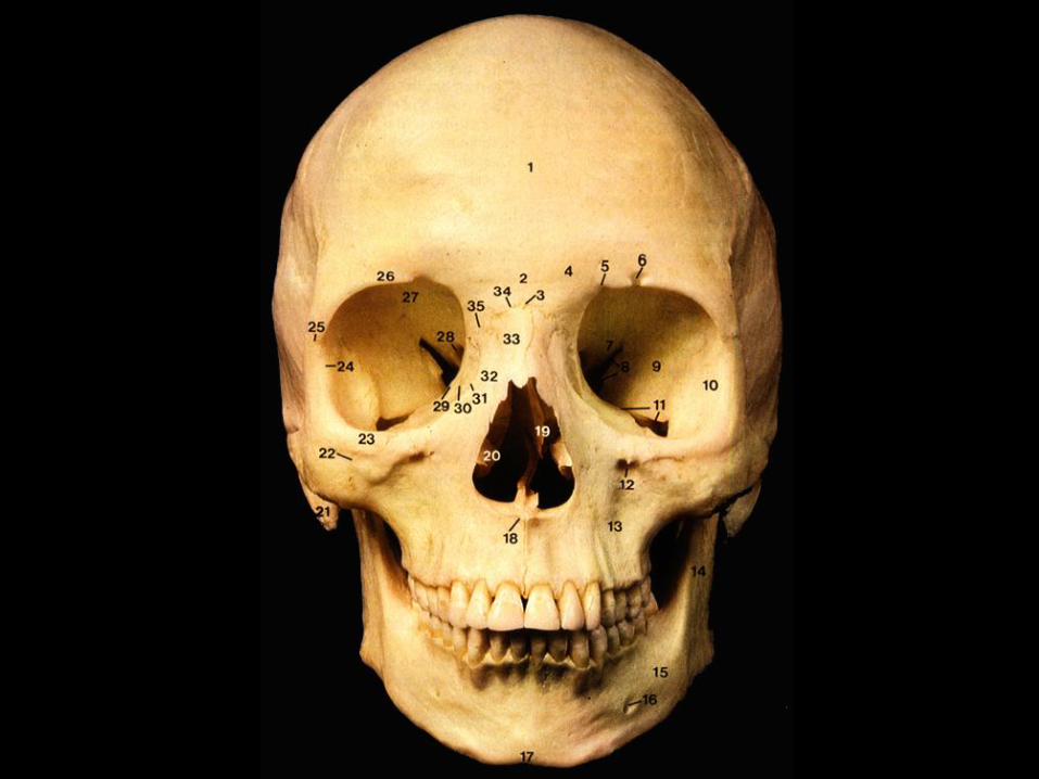
Cranium
1. Calvaria
2. Base of Skull
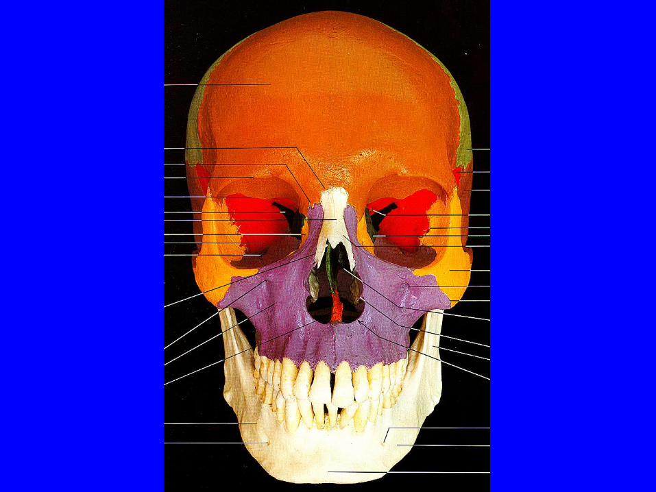


Internal Aspect of the Skull
Anterior Cranial Fossa
Boundaries:
Ant. Frontal Bone Upward Sweep
Post. Lesser Wing of Sphenoid
and Tuberculum Sellae
Contents:
Frontal Lobes of the Cerebrum

Middle Cranial Fossa
Boundaries:
Ant. Lesser Wing of Sphenoid and
Tuberculum Sellae
Post. Laterally Two Oblique Petrous of
Temporal Bone and Medially the
Dorsum Sellae
Contents: Hypophysis Cerebri in the Middle
Hypophyseal Fossa and Temporal
Lobes of Brain Laterally

Posterior Cranial Fossa
Boundaries:
Ant. Laterally Two Oblique Petrous of
Temporal Bone and Medially the
Dorsum Sealle
Post. Occipital Bone Upward Sweep
Contents: Cerebellum, Occipital Lobes of
Cerebrum and Brain Stem


Base of Skull 1. Anterior Portion
Extends to Hard Palate
2. Middle Portion
Tangent Line at Anterior most Point
of the Foramen Magnum
3. Posterior Portion
The Rest of Base Skull
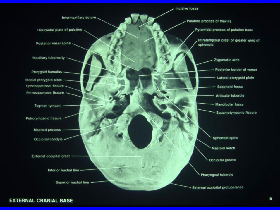


Temporal Fossa Boundaries
Anterior: Zygoma & Zygomatic Process of
Frontal Bone
Superior: Temporal Line
Posterior: Temporal Line
Inferior: Zygomatic Arch, Infratemporal
Crest of the Greater Wing of the Sphenoid
Lateral: Zygomatic Arch
Medial: Bone Structure of Skull

Infratemporal Fossa Contents: Muscles of Mastication and their
Vascular and Nerve Supply
Boundaries:
Ø Ant. Infratemporal Surface of the Maxilla
Ø Med. Lateral Surface of Lateral Pterygoid Plate of Sphenoid
and Pterygomaxillary Fissure
Ø Sup. Infratemporal Crest of Sphenoid and Infratemporal
Surface of the Greater Wing of the Sphenoid Post.
Anterior Limits of the Mandibula Fossa (glenoid fossa)
Ø Inf. Open
Ø Lat. Ramus of Mandible


Branches of External
Carotid Artery 1. Superior thyroid AA.
a. infrahyoid
b. sternocleidomastoid
c. superior laryngeal
d. cricothyroid
e. terminal branches to thyroid gland
2. Ascending pharyngeal AA.
a. pharyngeal
b. meningeal
c. inferior tympanic (to tympanic cavity)


continued
3. Lingual AA.
travels deep to hypoglossal nerve (CN XII) to the muscles of tongue.
4. Facial AA.
5. Ascending palatine AA.
supplies muscles in the
a. superior pharynx (superior constrictor) b. soft palate c. tonsils d. auditory tube
6. Occipital AA.
7. Posterior auricular AA.
8. Maxillary AA.
9. Superficial temporal AA.


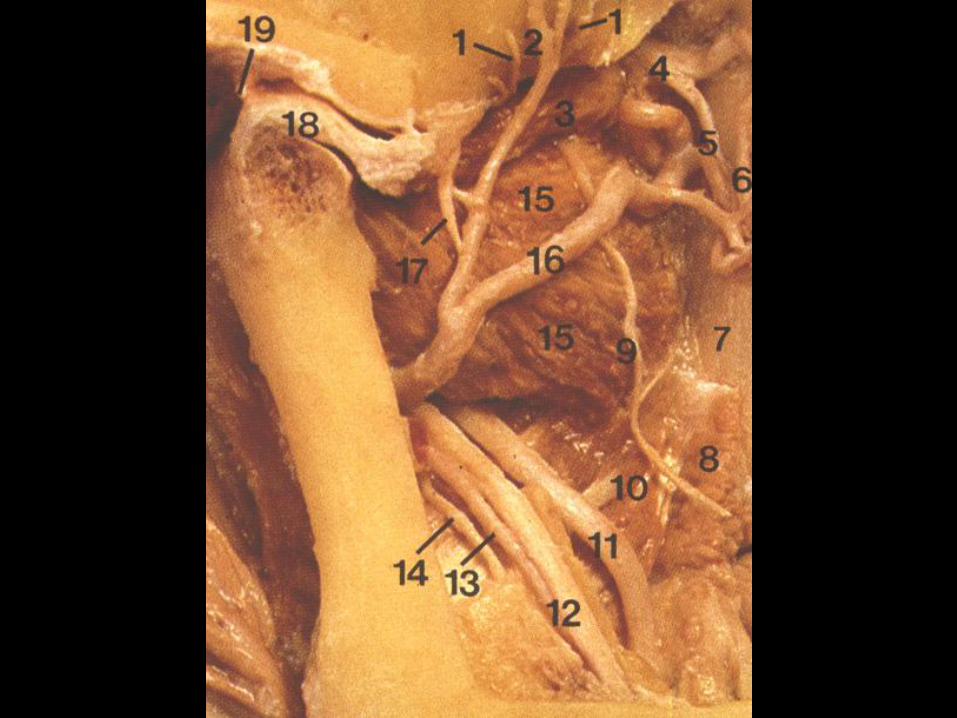
CAVERNOUS
SINUS
THROMBOSIS


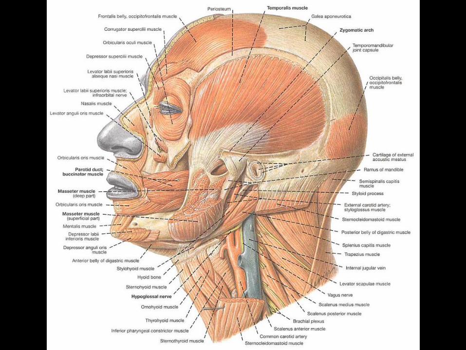
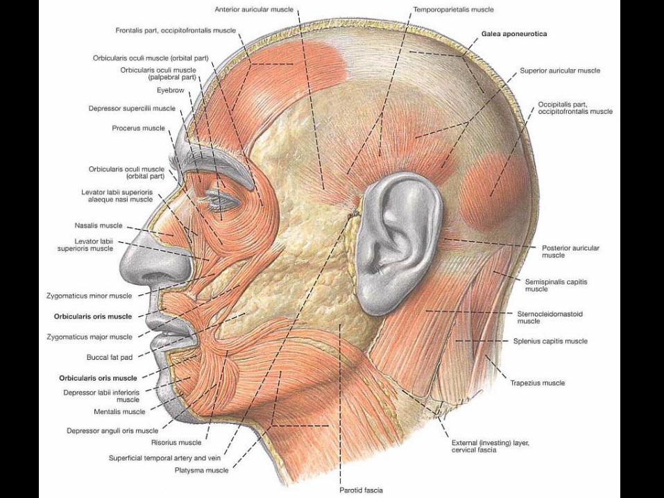
Facial Muscles
(muscles of facial expression) Not like most of the skeletal muscles , which usually
orginates from one bone and goes across the joint and
inserts into the bone distal to it.
e.g. Brachialis - a stong flexor of forearm distal 3/5 of the
anterior surface of the humerus
insertion: coronoid process of the ulna
Facial muscles originate from bony structure and insert
underneath the skin
Function: Facial expression
Since all facial muscles are derived from 2nd (hyoid) arch,
all facial muscles are innervated by facial N. (VII)

Temporoparietalis runs between frontal belley of epicranius and
ant. and sup auricular muscle of ear
innervated by temporal br. of facial N.
it can raise the ear
epicranius (both belleies) and temporoparietalis act together furrow the forehead
raise the eyebrows and widen the eyes
facial muscles-important character, act as group

Muscles of the mouth 1. Orbicularis oris-sphincter of oral commissure
2. Risorius-smileing muscle
originate from masseteric fascia insert into the skin at corner of mouth
3. Depressors of the lip
a. Depressor labii inferioris Quadrangular in shape
b. Depressor anguli oris- expressing
sadness Triangular in shape

continued
4. Elevators of the lip
a. levator labii superioris alaeque nasi
b. levator labii superioris
c. levator anguli oris lies deep to b assists in the formation of nasolabial furrow assists in expressing happiness
d. zygomaticus minor orgin: maxillary process of zygomatic bone
insertion: lateral to b’s insertion
e. zygomaticus major


Muscle of the cheek Buccinator-acessory muscle of mastication
quadangular-shaped muscle
occupying the space between
maxilla and mandible
Deeper than muscles of facial expression
It helps to blow air out of oral vestibule, as
blowing dust.


continued
Orgin: Man. baccal surface of alveolar
Max. processes
pterygomandibular raphe
Insertion: At the corner of mouth, upper
and lower fascicles decussate to
insert into lower and upper lip,
respectively.

Modiolus (hub of wheel)
1 cm lateral and slightly above the corner of
the mouth where 5 muscles interlace forming a
muscular node opposite to mand. 1st bicuspids
1. orbicularis oris
2. bucinnator
3. zygomaticus major
4. levator anguli oris
5. depressor anguli oris


Muscles surounding
the orbit 1. Orbicularis oculi-sphincter of eyelid
Orbital portion- forceful closure
Palpebral portion- light closure e.g. blinking
2. Corrugator- deep to the superomedial
aspect of orbicularis oculi,
at the medial aspect of the
eyebrow
3. Levator palpebrae superioris

Muscles of the ear and nose (fairly inconsequential)
Ear: auricularis anterior
auricularis superior
auricularis posterior
Nose: procerus
nasalis
depressor septi

Motor nerve of the face
FACIAL
NERVE


Exit from the skull
(Stylomastoid foramen)
1. Temporal branch
muscles above the zygomatic arch
2. Zygomatic branch- travel transversly across
face, zygomatic, orbital, infraorbital areas
3. Buccal branch - cheek
4. Mandibular branch
5. Cervical branch- platysma and posterior
belley of digastric
Damage to facial nerve
1. trauma
2. virus infection
3. unknown factors

Result:
-facial paralysis
-Bell’s palsy


Symptoms
1. eyelids cannot be properly closed
==> no lubrication of eye
2. drool of saliva from mouth
3. food cannot be properly held
masticated on affected side
4. facial distortion
due to contraction of unopposed
contralateral facial muscle

Mandibular block
– parotid gland region
– paralysis of the facial nerve
Facial N.
– leaves the cranial cavity via internal
acoustic meatus
– travels in temporal bone
– emerges through the stylomastoid foramen

Arterial supply of face
–mainly external caotid A
–some from internal carotid A
Facial blood supply
–anastomotic network

Facial artery
–ext. carotid ==> facial artery
inferior labial
superior labial
lateral nasal
angular A.
terminal br. of facial A
Superficial temporal A
–transverse facial

Branches of the
maxillary artery
infraorbital A-from the 3rd (or
pterygopalatine portion of maxillary
A)
buccal A- from the 2nd (or
pterygoid portion of maxillary A)
mental A- inferior alveolar


Branches of the
ophthalmic A
supraorbital A
supratrochlear A
zygomaticofacial A
dorsal nasal A

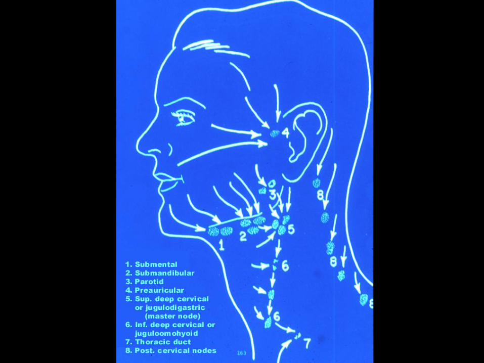
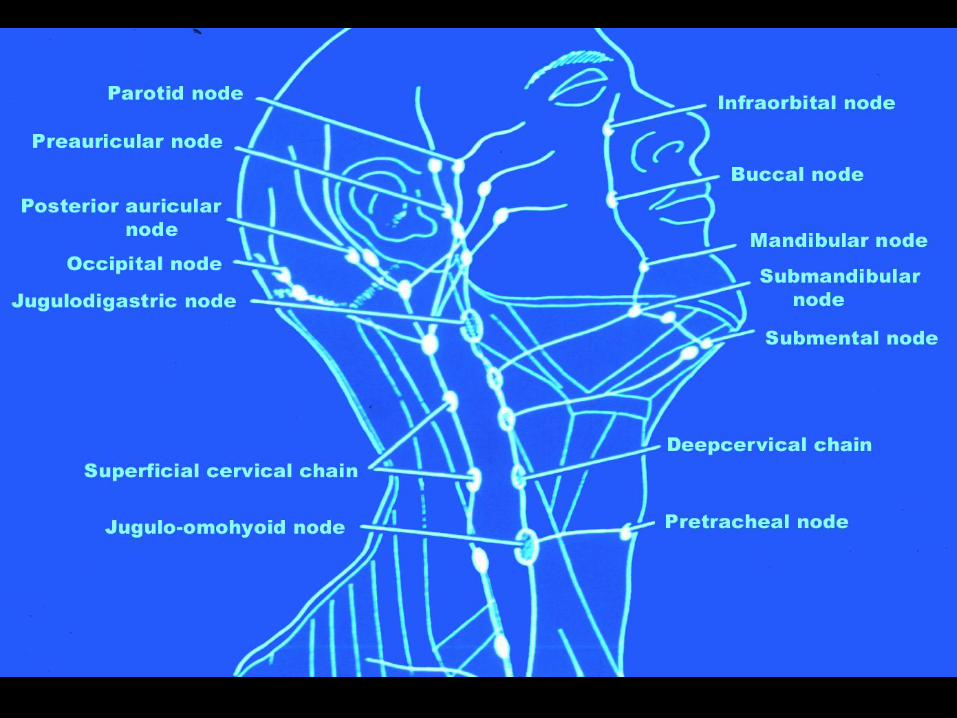
Venous Drainage (Veins in the cranial facial region)
supraorbital
posterior auricular
buccal
infraorbital
submental
superior and inferior lavial vein

Main Discharge 1. Facial V.
2. Superficial temporal V.
3. Maxillary V.
Both facial and retromandibular veins
drain into common facial vein
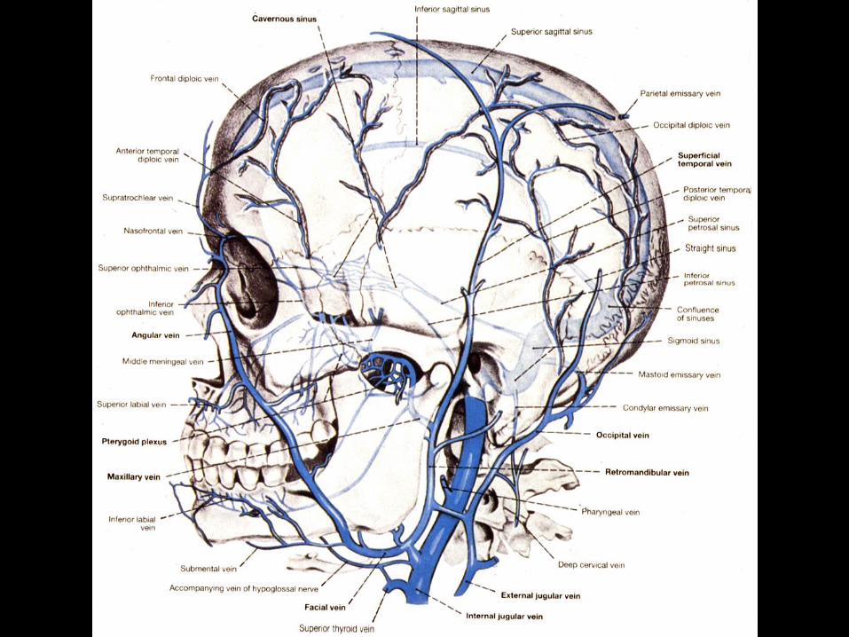

Veins of the face
General rule
–vein usually accompanies
the artery
–blood flow opposite
direction

Facial vein (it’s communicator)
upper 1/3 face-superior ophthalmic-
cavarnous sinous
middle 1/3 face-->upper lip-->infraorbital
vein--> pterygoid plexus
lower 1/3 and middle 1/3 --> facial
retromandibular --> common facial -->
internal jugular


Carotid sinus-BP- CN IX
Carotid body-Chem-CN IX
and X

Muscles of Mastication
Masseter
Temporalis
Medial pterygoid
Lateral pterygoid