Embed Size (px)
Citation preview

Eur J Clin Chem Clin Biochem 1996; 34:133-137 © 1996 by Walter de Gruyter · Berlin · New York
Nitro-X-Arginine Interferes with the Cadmium Reduction ofNitrate/Griess Reaction Method of Measuring Nitric Oxide Production1)
Kasem Nithipatikom, Phillip F. Pratt and William B. Campbell
Department of Pharmacology and Toxicology, Medical College of Wisconsin, Milwaukee, WI, USA
Summary: Nitro-£-arginine is used as an inhibitor of nitric oxide synthase in many biological systems. Nitric oxide(NO) is unstable and degrades to nitrite (NOJ") and nitrate (NOJ"). The colorimetric reaction of NO^~ with Griessreagent is commonly used to measure NOf. NOf may be measured as NO£" following reduction by cadmium orcadmium/copper. We found that bradykinin increased the formation of NO£~ by bovine coronary endothelial cells.Nitro-L-arginine further increased the formation of NOi". This increase is due to the interference of nitro-L-argininein determination of NOf by the cadmium reduction to NO^~ and Griess reagent reaction. Incubation of nitro-L-arginine with cadmium or cadmium/copper produced a product that reacts with Griess reagent to form a compoundthat has an absorption spectrum identical to the product formed by NO^" and Griess reagent. Caution must beexercised when using the NOf/NOf measurement by the Griess reaction to assess inhibition of nitric oxide synthaseby nitro-Z,-arginine.
Introduction
Nitric oxide (NO) is now recognized as an endothelium-derived vasodilator and a possible neurotransmitter (1).In biological systems, it is produced in very low concen-trations, and it has a very short half-life (< 6 s) (2). Un-der physiological conditions, NO may degrade to nitrite(NOi~) and also nitrate (NOJ"). This, however, representsonly one of its inactivation pathways. Detection ofNO^~ and NOJ" is commonly used as an index of nitricoxide synthase2) activity. A number of methods for NO,NO£~ and NOJ" detection have been reported. Measure-ments of reactive NO are made by chemiluminescenceusing the reaction of NO with ozone (3, 4) or luminol-hydrogen peroxide (5, 6) and by spectrophotometric as-says based on the reaction with oxyhaemoglobin (7) orFe-S proteins (8). For measuring the more stable NOfand NOJ", a Chromatographie method (7), a reaction ofNOf with Griess reagent followed by a spectrophoto-metric detection (9, 10), an automated system with aNQf-GWess reagent reaction (11, 12), and fluorometricmethod (13) have been reported. With these spectropho-tometric and fluorometric methods, NOJ~ has to be re-duced to NO^T by nitrate reductase or more commonlyby % metal such as cadmium prior to the reaction withthe reagents.During the study of the effects of nitro-L-arginine onNO release from cultured endothelial cells, the NO^YNOJ" concentration was determined by cadmium (Cd)
!) Funding Organization: The National Heart, Lung and Blood In-stitute, 9000 Rockville Pike, Bethesda, MD 20892, U.S.A.2) Enzyme: Nitric oxide synthase
reduction and Griess reagent reaction. We found that thesamples always produced more colour or apparentNOJ in the presence than in the absence of nitro-Z,-argi-nine. Preliminary data showed that the bradykinin stimu-lation of bovine coronary artery endothelial cellsincreased the production of NO^~. In the presence of ni-tro-L-arginine, the production of NOJ increased furtherrather than decreasing. This prompted us to investigatethe effects of nitro-Z,-arginine on this assay method andthe production of the interfering species in the assay.
Materials and MethodsMaterials
Sulphanilamide, N-(l-naphthyl)ethylenediamine dihydrochloride,and Cd powder (—100 mesh) were purchased from Aldrich Chemi-cal Co. (Milwaukee. WI); NaNO2, NaNO3, D-arginine and L-argi-nine from Sigma Chemical Co. (St. Louis, MO); nitro-L-argininefrom Sigma Chemical Co., Serva Feinbiochemica (Heidelberg,Germany), and Schweizerhalle (South Plainfield, NJ); bradykininfrom BACHEM Bioscience Inc. (Philadelphia, PA); [3H]£-argininefrom Amersham (Arlington Heights, IL); all other chemicals andreagents from Sigma Chemical Co. Amino-L-arginine was a giftfrom Dr. Owen Griffith (Department of Biochemistry, Medical Col-lege of Wisconsin, Milwaukee, WI). Distilled-deionized water wasused in this study. Precleaned plasticware with methanol and deion-ized water was used for all solutions.
Determinat ion of ni t r i te in bovine coronary arteryendothel ial cells
Bovine coronary artery endothelial cells were cultured onto 6-wellplates as previously described (14). The cells were washed withHepes buffer and then incubated in 3 ml of buffer containing nitro-L-arginine (30 μτηοΐ/l) or vehicle for 10 min at 37 °C. Then, brady-kinin (1 μιηοΐ/l) was added, and the incubation continued for 2 h.Following the incubation, the supernatant was removed for NOf/NOJ" measurement and the cells were frozen for protein determine-

134 Nithipatikom et al.: Nitro-L-arginine interferes nitrate measurement
tion (15). Total nitrite (NOf and NOf) was measured by the Griessreaction (see below). The results were expressed as total nitrite(pmol) and were normalized to the total amounts of protein. Cell-free samples did not have protein content and they were comparedat the same volumes and concentrations of chemicals.
Ni t r i c oxide synthase assay
Bovine coronary artery endothelial cells were grown to confluenceon 225 cm2 flasks and lysed with a pH 7.4 buffer containing: Hepes(11 mmol/1), sucrose (0.35 mol/1), EDTA (0.1 mmol/1), dithiothrei-tol (1 mmol/1), leupeptin (10 mg/1), aprotinin (2mg/l), soybeantrypsin inhibitor (10 mg/1), phenylmethylsulphonyl fluoride (10mg/1), NP-40 (10 g/1), and glycerol (100 g/1). The conversion of[3H]arginine to [3H]citrulline was used as an index of nitric oxidesynthase activity. The reaction volume was 100 μΐ in pH 7.5 buffercontaining: Tris-HCl (50 mmol/1), EDTA (0.1 mmol/1), EGTA (0.1mmol/1), mercaptoethanol (lg/1), leupeptin (100 μπιοΐ/ΐ), phenyl-methylsulphonyl fluoride (1 mmol/1), soybean trypsin inhibitor (10mg/1), aprotinin (2 mg/1), calmodulin (10 nmol/1), NADPH(1 mmol/1), tetraliydrobiopterin (3 μπιοΐ/ΐ), Ζ,-arginine (10 μπιοΐ/ΐ),[3H]L-arginine (7.4 kBq, 2.52 TBq/mmol), calcium chloride (2.5mmol/1), and 100 μg protein. Nitro-L-arginine was added to sometubes at a final concentration of 30 μπιοΐ/ΐ. All samples were incu-bated for 15 min at 37 °C in a shaking water bath and the reactionwas stopped by the addition of 1 ml of ice-cold pH 5.5 STOPbuffer containing: Hepes (20 mmol/1), EDTA (2 mmol/1), andEGTA (2 mmol/1). The [3H]citrulline was separated from the[3H]arginine by passing the sample over an equilibrated Dowexcation exchange column (Na+ form). Nitric oxide synthase activityis expressed as Bq of citrulline.
Ni t ra te /ni t r i te determination
All NOi", NOj and NOf/NOf standards and samples were pre-pared in pH 7.4 Hepes buffer consisting of Hepes (10 mmol/l),NaCl (150 mmol/1), KC1 (5 mmol/1), CaCl2 (2 mmol/1), MgCl2(1 mmol/1) and glucose (5 mmol/1). Nitrate was reduced to NOi"by incubating 1 ml of the solution with approximately 0.05 g ofCd powder for different periods of time, generally 15 min. To thisincubation, 5 μΐ of 10% HC1 were added, and the solution wascentrifuged for a few minutes to separate the Cd. The supernatantof 800 μΐ was transferred to a small tube. A 25 μΐ of 5 g/1 sulphanil-amide in 2 mol/1 HC1 was added and mixed. Then, 25 μΐ of 3 g/1naphthylethylenediamine dihydrochloride in 0.1 mol/1 HC1 wereadded, mixed and allowed to react for 15 min. Two aliquots of 350
μΐ were transferred to 96-well microplate and the absorbance wasmeasured at 540 nm on Microplate Autoreader (Bio-Tek Instru-ment, Winooski, VT). The absorbance was the average of two wellsfor each sample. Nitrite was treated the same way as NOf butwithout the Cd. Absorbance of Hepes buffer blanks treated withCd were subtracted from all measurements. Results were expressedas apparent NOJ (μπιοΐ/ΐ). . f
High performance liquid chromatography
Chromatography was performed on a 1090 Series II Hewlett Pack-ard Liquid Chromatograph (Hewlett Packard Co., Palo Alto, CA).Three types of columns and gradient programmes were used forseparation of nitro-L-arginine and its Cd catalyzed product(s);
(a) an ODS Ci8 column (5 μιη, 100 X 2.1 mm) (Hewlett PackardCo., Palo Alto, CA) with a 3 min 0 to 15% acetonitrile in waterelution gradient;
(b) a Nucleosil Cig column (5 μηι, 250 X 10 mm) (Phenornenex,Torrance, CA) with a 5 min 0 to 10% acetonitrile in water elutiongradient; and
(c) a Nucleosil 5 NH2 column (5 μπι, 250 X 4.6 mm) (Pheno-rnenex) with 60% acetonitrile/40% water with 0.1 mol/1 ammo-nium acetate elution solvent.
The absorbance was detected by a Hewlett Packard model 1040photodiode array detector, and the chromatograms were recordedat 275 mm with 10 nm bandwidth and reference wavelength of 450nm with 20 nm bandwidth. Full UV absorbance spectra (200^-400nm) were obtained during the HPLC analysis every 6 s and storedon a Hewlett Packard ChemStation for later analysis. The productpeak fraction was collected and reacted with Griess reagent orotherwise analyzed.
Results and Discussion
It has been shown that bradykinin stimulated the NOproduction of endothelial cells (16) and the nitro-L-argi-nine was a potent inhibitor of the nitric oxide synthase(17, 18). Preliminary data show that bradykinin stim-ulated total NOi (NOf + NOf) production by culturedendothelial cells (fig. la). However, preincubation of the
2000-1
Fig.J a) Effect of nitro-L-arginine and bradykinin on totalNOJ (NOJ + NOf) by bovine coronary endothelial cells. Thesedata are normalized to the total protein content. Also shown arecell-free incubations with bradykinin. The data for cell-free incub-ations are compared at the same volumes and concentrations ofchemicals. Each value represents the mean ± S. E. (n = 6).
b) Nitric oxide synthase activity (expressed as Bq per volume unit)of endothelial cells (control) and in the presence of nitro-L-argi-nine. (n = 3).
Nithipatikom et al.: Nitro-L-arginine interferes nitrate measurement 135
cells with the nitric oxide synthase inhibitor, nitro-L-arginine, increased instead of decreased NOJ pro-duction. In contrast, nitro-L-arginine markedly inhibitedthe nitric oxide synthase activity in the cells (fig. Ib).The total NOf (NOJ + NOf) in mfcdia (cell free) wasalso increased by nitro-L-arginine (fig. la). These dataindicate that nitro-L-arginine actually inhibits NO pro-duction by endothelial cells and suggests that nitro-L-arginine may interfere with the assay for NOf by cad-mium reduction and Griess reaction.
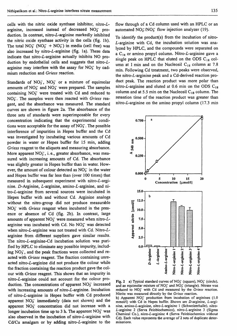
Standards of NOJ, NOf or a mixture of equimolaramounts of NOJ and NOf were prepared. The samplescontaining NOf were treated with Cd and reduced toNO£~. The samples were then reacted with Griess rea-gent, and the absorbance was measured. The standardcurves are shown in figure 2a. The absorbance of thethree sets of standards were superimposable for everyconcentration indicating that the experimental condi-tions were acceptable for the assay of NOS". The possibleinterference of impurities in Hepes buffer and the Cdwas investigated by incubating various amounts of Cdpowder in water or Hepes buffer for 15 min, addingGriess reagent to the aliquots and measuring absorbance.More apparent NOJ, i. e., greater absorbance, was mea-sured with increasing amounts of Cd. The absorbancewas slightly greater in Hepes buffer than in water. How-ever, the amount of colour detected as NOJ in the waterand Hepes buffer was far less than (over 100 times) thatmeasured in subsequent experiment with nitro-L-argi-nine. D-Arginine, L-arginine, amino-L-arginine, and ni-tro-L-arginine from several sources were incubated inHepes buffer with and without Cd. Arginine analogswithout the nitro-group did not produce measurableNO£~ with Griess reagent when incubated in the pres-ence or absence of Cd (fig. 2b). In contrast, largeamounts of apparent NO^~ were measured when nitro-L-arginine was incubated with Cd. No NO^" was detectedwhen nitro-L-arginine was not treated with Cd. Nitro-L-arginine from different suppliers gave similar results.The nitro-L-arginine-Cd incubation solution was puri-fied by HPLC to eliminate any possible impurity, includ-ing NOJ", and the peak fractions were collected and re-acted with Griess reagent. The fraction containing unre-acted nitro-L-arginine did not produce the colour whilethe fraction containing the reaction product gave the col-our with Griess reagent. This shows that an impurity innitro-L-arginine could not account for the colour pro-duction. The concentrations of apparent NOJ increasedwith increasing amounts of nitro-L-arginine. Incubationof nitro-L-arginine in Hepes buffer with Cd producedapparent NOJ immediately (data not shown) and theapparent NOf concentration did not increase with alonger incubation time up to 3 h, The apparent NOJ wasalso observed in the incubation of nitro-L-arginine withCd/Cu amalgam or by adding nitro-L-arginine to the
flow through of a Cd column used with an HPLC or anautomated NOj/NOf flow injection analyzer (19).
To identify the product(s) from the incubation of nitro-L-arginine with Cd, the incubation solution was ana-lyzed by HPLC, and the compounds were separated ona Cig or amino propyl column. Nitro-L-arginine gave asingle peak on HPLC that eluted on the ODS C18 col-umn at l min and on the Nucleosil Qg column at 7.8min. Following Cd treatment, two peaks were observed,the nitro-L-arginine peak and a Cd-derived reaction pro-duct peak. The reaction product was more polar thannitro-L-arginine and eluted at 0.6 min on the ODS Cigcolumn and at 5.5 min on the Nucleosil C18 column. Theretention time of the reaction product was greater thannitro-L-arginine on the amino propyl column (17.3 min
0.750-
0.500-
0.250-
0.0005 10 15
Concentration [μιηοΐ/ΐ]20
ι 12·5·J; 10.0-
**1 7.5-2·**S 5.0-
1 2.5-
0.0-
b
P.V.V! |i ι ι ι «
τ1·1·1·1·
Sj:;j
££:
||·χ·χ•X'X
:·:·£:·
Τ
ΧχΧ
•IvX°
χΧνx:x::x'i'x
1 1 1 1 1 1 1
B e B•a « -g»2 5 l
ls?iz
•g.*Jέ
Fig. 2 a) Typical standard curves of NO2 (square), N 3 (circle),and an equimolar mixture of NOf and NOf (triangle). Nitrate wasreduced to NOJ with Cd and measured by the Griess reaction.Nitrite was measured directly by the Griess reaction,b) Apparent NOf production from incubation of argintnes (1.0mmol/1) with Cd in Hepes buffer. Shown are D-arginine, L-argi-nine, amino-L-arginine, nitro-L-argininc l (Schweizerhalle), nitro-L-arginine 2 (Serva Feinbiochemica), nitro-L-arginine 3 (SigmaChemical Co.), nitro-L-arginine 4 (Serva Feinbiochcmica withoutCd). Each value represents the average of 2 sets of duplicate deter-minations.

136 Nithipatikom et al.: Nitro-L-arginine interferes nitrate measurement
8
10 15 20
t [min]
Fig. 3 a) Chromatogram of the incubation of nitro-I-argininewith Cd separated on the Nucleosil 5 NH2 column (see text). Thefirst peak (I) at 14.2 min corresponds to nitro-i.-arginine retentiontime, the second peak (II) at 17.3 min is the reaction product.
250 300 350 400
Wavelength [nm]
b) UV absorption spectra taken from the peaks of chromatogramsduring the analysis. The solid line (I) is the spectrum of nitro-L-arginine (the first peak) and the dashed line (II) is the spectrum ofthe reaction product (the second peak). The spectra are normalizedat the absorption maxima.
versus 14.2 min for nitro-L-arginine) (fig. 3a). The ab-sorption maximum of the reaction product was shiftedfrom 270 nm for nitro-L-arginine to 255 nm with a weakabsorption peak at 350 nm (fig. 3b). This product alsohad a light yellow colour. The retention time and UVabsorbance of this product did not match i-arginine orany of the arginine analogs used in this study. The reac-tion product of nitro-L-arginine with Cd was isolated byHPLC and added to the Griess reagent. It formed a col-oured adduct that had the same visible spectrum as theone formed in the reaction of NOi" with the Griess rea-gent (absorbance maxima of 543 nm). We therefore callthe colour or absorbance produced by the Cd-derivedreaction product "apparent NO^"". The identity of thisreaction product is not known.
In conclusion, the incubation of nitro-L-arginine withCd generates a product, possibly products, that appearsresponsible for the apparent NO^~ that was determined
by the reaction with Griess reagent. These data indicatethat nitro-L-arginine will interfere with the measurementof NOj". Detection of NO^~ and NO J" is commonly usedas an index of nitric oxide syrithase activity even thoughthese ions are not the only products of NO degradation(12, 20). Interference Of nitro-L-arginine with the detec-tion of NOf will further complicate this relationship.For this reason, caution should be used when using theNO^~/NOf measurement by the Griess reaction to assessinhibition of nitric oxide synthase by nitro-L-arginine.
AcknowledgementsThe authors thank Dr. Owen Griffith for his helpful suggestions,arginine analogs and numerous discussions and Ms. Gretchen Bargfor her secretarial assistance. Suppport was provided by grantsfrom the National Heart, Lung and Blood Institute (HL-37981 andHL-51055). The authors also acknowledge the generosity of LachatInstruments, Inc. for providing the QuikChem AE Ion Analyzerfor NOf and NOj measurements.
References1. Moncada S, Palmer RMJ, Higgs EA. Nitric oxide: physiology,
pathophysiology, and pharmacology. Pharmacol Rev 1991·43:109-42.
2. Keim M, Feelisch M, Spahr R, Piper HM, Noack E, SchraderJ. Quantitative and kinetic characterization of nitric oxide andEDRF released from cultured endothelial cells. Biochem Bio-phys Res Comm 1988; 154:236-41.
3. Palmer RM, Ferrige AG, Moncada S. Nitric oxide release ac-counts for the biological activity of endothelium-derived relax-ing factor. Nature (London) 1987; 327:524-6.
4. Hybertson BM, Dunham AJ, Thompson DC, Terada LS, Re-pine JE. Flow injection analysis of nitrite generated by neutro-phils and endothelial cells. Anal Lett 1994; 27:3081-93.
5. Kikuchi K, Nagono T, Hayakawa H, Hirata Y, Hirobe M. De-tection of nitric oxide production from a perfused organ byluminol-H202 system. Anal Chem 1993; 65:1794-9.
6. Kikuchi K, Nagono T, Hayakawa H, Hirata Y, Hirobe M. Realtime measurement of nitric oxide produced ex-vivo by lumi-nol-H202 chemiluminescence method. J Biol Chem 1993·268:23106-10.
7. Feelisch M, Noack EA. Correlation between nitric oxide for-mation during degradating of organic nitrates and activation ofguanylate cyclase. Eur J Pharmacol 1987; 139:19-30.
8. Stuehr DJ, Gross SS, Sakuma I, Levi R, Nathan CF. Activatedmurine macrophages secrete a metabolite of arginine with thebioactivity of endothelium-derived relaxing factor and thechemical reactivity of nitric oxide. J Exp Med 1989;169:1011-20.
9. Gutman SI, Hollywood CA. Simple, rapid method for deter-mining nitrates and nitrites in biological fluids [letter]. ClinChem 1992; 38:2152.
10. Cortas NK, Wakid NW. Determination of inorganic nitrate inhuman and urine by a kinetic cadmium-reduction method. ClinChem 1990; 36:1440-3.Green LC, Wagner DA, Glogowski J, Skipper PL, Wishnok JS,Tannenbaum SR. Analysis of nitrate, nitrite, and [l5N]nitrate inbiological fluids. Anal Biochem 1982; 126:131-8.
12. Tracey WR, Linden J, Peach MJ, Johns RA. Comparison ofspectrophotometric and biological assays for nitric oxide (NO)and endothelium-derived relaxing factor (EDRF): Nonspecif-icity of the diazotization reaction for NO and failure to detect
"EDRF. J Pharmacol Exp Ther 1990; 252:922-8.Misko TP, Schilling RJ, Salvemini D, Moore WM, Currie MG.A fluorometric assay for the measurement of nitrite in biologi-cal samples. Anal Biochem 1993; 214:11-6.
11
13

Nithipatikom et al.: Nitro-L-arginine interferes nitrate measurement 137
14. Revtyak GE, Johnson AR, Campbell WB. Cultured bovinecoronary arterial endothelial cells synthesize HETEs andprostacyclins. Am J Physiol 1988; 254:C8-C19.
15. Lowry OH, Rosebrough NJ, Fair AL, Randali RJ. Protein mea-surement with Folin phenol reagent. J Biol Chem 1951;193:265-75.
16. Schmidt HHHW, Zernikow B, Baeblich S, Böhme E. Basaland stimulated formation and release of L-arginine-derived ni-trogen oxides from cultured endothelial cells. J Pharmacol ExpTher 1990; 254:591-7.
17. Ishii K, Chang B, Kerwin JF, Huang Z, Murad F. N(0-Nitro-L-arginine: a potent inhibitor of endothelium-derived relaxingfactor formation. Eur J Pharmacol 1990; 176:219-23.
18. Mulsch A, Busse R. NG-Nitro-L-arginine (N5-[imino(nitroami-no)methyl]-L-ornithine) impairs endothelium-dependent dil-ations by inhibiting cytosolic nitric oxide synthesis from L-
arginine. Naunyn-Schmiedeberg's Arch Pharmacol 1990;341:143-7.
19. Pratt PF, Nithipatikom K, Campbell WB. Simultaneous deter-mination of nitrate and nitrite in biological samples by multi-channel flow injection analysis. Anal Biochem. 1995;231:383-6.
20. Mirza U, Chait BT, Landers HM. Monitoring reactions of nitricoxide with peptides and proteins by electrospray ionization-mass spectrometry. J Biol Chem 1995; 270:17185-8.
Received August 2 I/November 7, 1995Corresponding author: Kasem Nithipatikom, Ph. D., Departmentof Pharmacology and Toxicology, Medical College of Wisconsin,8701 Watertown Plank Road, Milwaukee, WI 53226, USA