Embed Size (px)
Citation preview
105Copyright © 2014 The Korean Society of Cardiology
Korean Circulation Journal
Introduction
Flow-induced shear stress plays a central role on endothelial re-sponse and mechano-signal transduction in the cardiovascular sys-tem. The mechanical shear force stimulates endothelial protein ac-
Original Article
http://dx.doi.org/10.4070/kcj.2014.44.2.105Print ISSN 1738-5520 • On-line ISSN 1738-5555
Nitric Oxide Synthase Inhibition Attenuates Cardiac Response to Hemodilution with Viscogenic Plasma ExpanderSurapong Chatpun, PhD1 and Pedro Cabrales, PhD2
1Institute of Biomedical Engineering, Faculty of Medicine, Prince of Songkla University, Songkhla, Thailand 2Department of Bioengineering, University of California, San Diego, CA, USA
Background and Objectives: Increased vascular wall shear stress by elevated plasma viscosity significantly enhances the endothelial ni-tric oxide synthase (eNOS) activity during an acute isovolemic hemodilution. Also the modulation of plasma viscosity has effects on the car-diac function that were revealed if a left ventricular (LV) pressure-volume (PV) measurement was used. The aim of this study was to assess cardiac function responses to nitric oxide synthase (NOS) inhibitors with the presence of an elevated plasma viscosity but a low hematocrit level. Furthermore, systemic parameters were monitored in a murine model.Materials and Methods: As test group five anesthetized hamsters were administered with N(G)-nitro-L-arginine methyl ester (L-NAME), NOS inhibitor, whereas five other hamsters were used as control group without L-NAME infusion. The dosage of L-NAME was 10 mg/kg. An isovolemic hemodilution was performed by 40% of estimated blood volume with 6% w/v dextran 2000 kDa, high viscosity plasma expanders (PEs) with viscosity 6.34 cP. LV function was measured and assessed using a 1.4 Fr PV conductance catheter.Results: The study results demonstrated that NOS inhibition prevented the normal cardiac adaptive response after hemodilution. The end-systolic pressure increased 14% after L-NAME infusion and maintained higher than at the baseline after hemodilution, whereas it gradually decreased in the animals without L-NAME infusion. The admission of L-NAME significantly decreased the maximum rate of ventricular pressure rise (+dP/dtmax), stroke volume and cardiac output after hemodilution if compared to the control group (p<0.05). Conclusion: This finding supports the presumption that nitric oxide induced by an increased plasma viscosity with the use of a high viscosity PE plays a major role in the cardiac function during an acute isovolemic hemodilution. (Korean Circ J 2014;44(2):105-112)
KEY WORDS: Nitric oxide; Endothelial nitric oxide synthase; Blood viscosity; Hemodilution; Ventricular function.
Received: October 22, 2013Revision Received: January 2, 2014Accepted: January 13, 2014Correspondence: Surapong Chatpun, PhD, Institute of Biomedical Engi-neering, Faculty of Medicine, Prince of Songkla University, Songkhla 90110, ThailandTel: 66-7445-1743, Fax: 66-7445-1744E-mail: [email protected]
• The authors have no financial conflicts of interest.
This is an Open Access article distributed under the terms of the Creative Commons Attribution Non-Commercial License (http://creativecommons.org/licenses/by-nc/3.0) which permits unrestricted non-commercial use, distribution, and reproduction in any medium, provided the original work is properly cited.
tivities including nitric oxide synthesis (NOS) by endothelial nitric oxide synthase (eNOS).1)2) Nitric oxide (NO) is an important mediator that has various physiological functions such as vascular tone reg-ulation, platelet activation prevention, vascular smooth muscle pro-liferation inhibition and myocardial contractility regulation.3-5) The inhibition of eNOS activity and endothelial dysfunction decreases the inotropic effect of ventricular myocytes and constricts blood vessels in a normal physiological range of hematocrit (Hct).6) Fur-thermore, the injection of N(G)-nitro-L-arginine methyl ester (L-NAME) or other NOS inhibitors induces high blood pressure and increases microvascular resistance to blood flow.7-9)
Vascular wall shear stress (WSS) is modulated by blood flow and blood viscosity. A previous study in the cremaster muscles of ham-sters using dextran with different molecular weights as an exchange solution showed that elevated plasma viscosity increased WSS and produced vasodilation.10) Similarly, Tsai et al.11) demonstrated that elevated plasma viscosity increased perivascular NO production in
106 NOS Inhibition on Cardiac Function during Acute Anemia
http://dx.doi.org/10.4070/kcj.2014.44.2.105 www.e-kcj.org
concert with the increased aortic eNOS protein expression during extreme hemodilution. Studies in awake animals treated with high viscosity plasma expanders (HVPEs) showed advantageous effects in the microvascular function in both hemorrhagic shock-resuscita-tion and acute extreme hemodilution models.12-14) These studies re-vealed that an elevated plasma viscosity positively correlates with increased WSS and NOS. The vascular network connected with the heart is a complex biological coupling. Therefore, the effects of in-creased plasma viscosity by plasma expanders (PEs) on the circula-tory system are being continually investigated in order to gain a better understanding related to novel PE development. Recently, us-ing a miniature pressure-volume (PV) conductance catheter in an acute hemodilution, it was found that HVPE provides better effects on cardiac function compared with low viscosity plasma expanders (LVPE).15) Furthermore, a recent study has demonstrated that animals hemodiluted with LVPE combined with a proper amount of NO do-nor had positive effects on cardiac function, which could not be observed in animals hemodiluted with LVPE only.16) The aim of this study was to assess the response of cardiac function to NOS inhibi-tors during an elevated plasma viscosity but with low Hct level.
Materials and Methods
Animal preparation The experiments were carried out in anesthetized male Golden
Syrian hamsters with a weight between 60 and 70 g (Charles River Laboratories; Boston, MA, USA). The animal handling and care fol-lowed the NIH Guideline for Care and Use of Laboratory Animals. The experimental study was approved by the animal care commit-tee of the University of California, San Diego. Animal surgery was performed following an intraperitoneal (i.p.) administration of so-dium pentobarbital (50 mg/kg). The left jugular vein was cannulat-ed to allow infusion fluid and the left femoral artery was catheter-ized for systemic blood pressure monitoring and blood withdrawal and sampling. In addition, a tracheotomy was conducted and the animal was cannulated with a polyethylene-90 tube to facilitate spontaneous breathing. The body core temperature was maintained at around 37°C by being placed in the supine position on a heating pad. During the experiment, if the animals responded to a toe pinching, a small bolus of sodium pentobarbital (10–15 mg/kg, i.p.) was administered for maintaining an anesthesia condition.
Inclusion criteriaAnimals under anesthesia were included in the experiments if
they had no signs of bleeding and their systemic parameters were within the normal range: 1) mean arterial pressure (MAP) above 80 mm Hg, 2) heart rate (HR) above 320 beats/minute, and 3) systemic
Hct level above 45%.
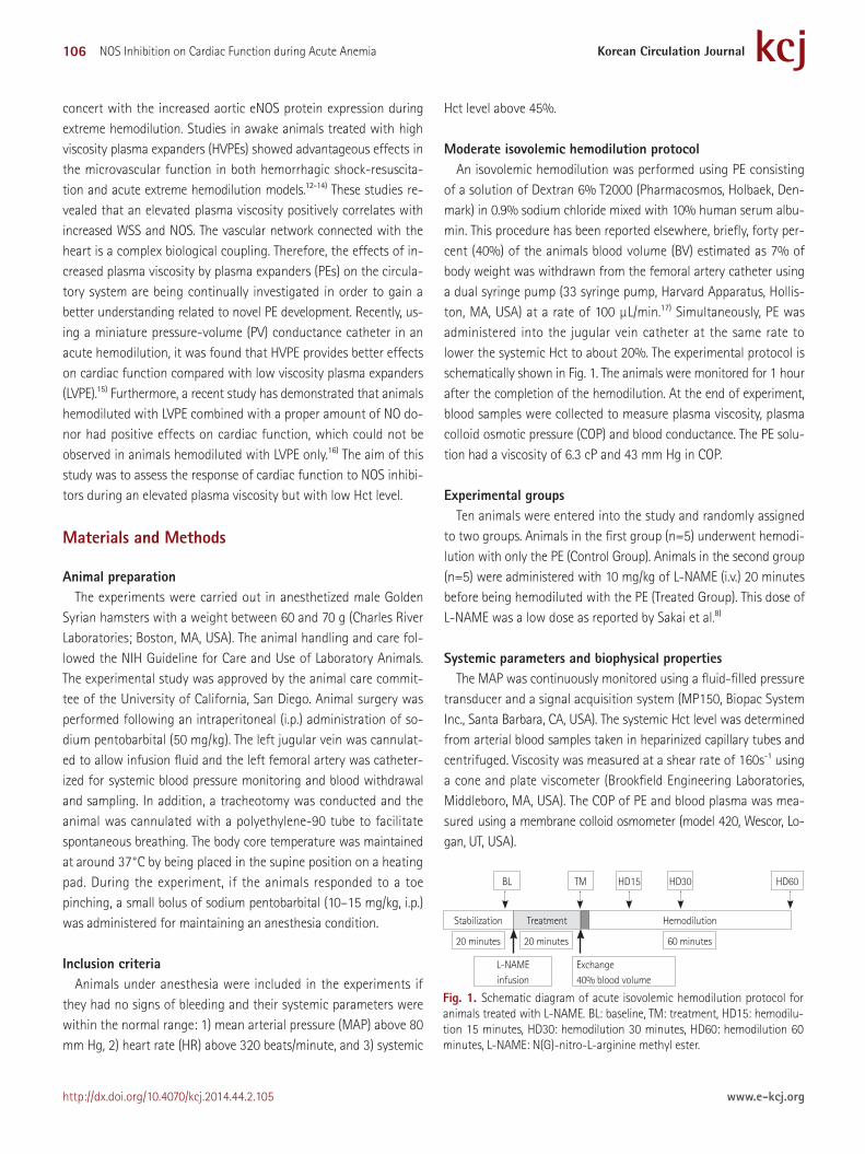
Moderate isovolemic hemodilution protocolAn isovolemic hemodilution was performed using PE consisting
of a solution of Dextran 6% T2000 (Pharmacosmos, Holbaek, Den-mark) in 0.9% sodium chloride mixed with 10% human serum albu-min. This procedure has been reported elsewhere, briefly, forty per-cent (40%) of the animals blood volume (BV) estimated as 7% of body weight was withdrawn from the femoral artery catheter using a dual syringe pump (33 syringe pump, Harvard Apparatus, Hollis-ton, MA, USA) at a rate of 100 µL/min.17) Simultaneously, PE was administered into the jugular vein catheter at the same rate to lower the systemic Hct to about 20%. The experimental protocol is schematically shown in Fig. 1. The animals were monitored for 1 hour after the completion of the hemodilution. At the end of experiment, blood samples were collected to measure plasma viscosity, plasma colloid osmotic pressure (COP) and blood conductance. The PE solu-tion had a viscosity of 6.3 cP and 43 mm Hg in COP.
Experimental groupsTen animals were entered into the study and randomly assigned
to two groups. Animals in the first group (n=5) underwent hemodi-lution with only the PE (Control Group). Animals in the second group (n=5) were administered with 10 mg/kg of L-NAME (i.v.) 20 minutes before being hemodiluted with the PE (Treated Group). This dose of L-NAME was a low dose as reported by Sakai et al.8)
Systemic parameters and biophysical propertiesThe MAP was continuously monitored using a fluid-filled pressure
transducer and a signal acquisition system (MP150, Biopac System Inc., Santa Barbara, CA, USA). The systemic Hct level was determined from arterial blood samples taken in heparinized capillary tubes and centrifuged. Viscosity was measured at a shear rate of 160s-1 using a cone and plate viscometer (Brookfield Engineering Laboratories, Middleboro, MA, USA). The COP of PE and blood plasma was mea-sured using a membrane colloid osmometer (model 420, Wescor, Lo-gan, UT, USA).
Fig. 1. Schematic diagram of acute isovolemic hemodilution protocol for animals treated with L-NAME. BL: baseline, TM: treatment, HD15: hemodilu-tion 15 minutes, HD30: hemodilution 30 minutes, HD60: hemodilution 60 minutes, L-NAME: N(G)-nitro-L-arginine methyl ester.
Stabilization Treatment Hemodilution
HD60HD30HD15TMBL
20 minutes 20 minutes
L-NAMEinfusion
Exchange40% blood volume
60 minutes
107Surapong Chatpun, et al.
http://dx.doi.org/10.4070/kcj.2014.44.2.105www.e-kcj.org
Cardiac functionThe closed-chest method was used to assess cardiac function.18)
Briefly, the right common carotid artery was exposed allowing to insert a 1.4 Fr PV conductance catheter (PV catheter; SPR-839, Mil-lar Instruments, Houston, TX, USA). The PV catheter was advanced passing through the aortic valve into the left ventricle. At baseline conditions and the end of the experiment, a small bolus (10 µL) of 15% hypertonic saline was administered intravenously to deter-mine the parallel volume. The left ventricular (LV) pressure and volume measurements were digitized and acquired at 1 kHz sam-pling rate (MPVS300, Millar Instruments, Houston, TX, USA and PowerLab 8/30, ADInstruments, Colorado Springs, CO, USA). Then the cardiac function data was analyzed with PVAN software (ver-sion 3.6, Millar Instruments, Houston, TX, USA). The selected 8-12 cardiac cycles were analyzed for cardiac function indices at each
time point.
Estimation of left ventricular blood volume The LV BV was measured in conductance units (relative volume
unit) and converted to actual BV (μL) using a known-volume cu-vette at the end of the experiment. The blood conductance at base-line was estimated from the baseline Hct level, using the pooling data of blood conductance and Hct relationship.17)
Statistical analysisThe results are depicted as mean±standard deviation. The data
pertaining to the same group were evaluated using the analysis of variance and Dunnett’s multiple comparison test for post hoc analy-ses. An unpaired, two-tailed t-test was performed to compare be-tween groups at each time point.
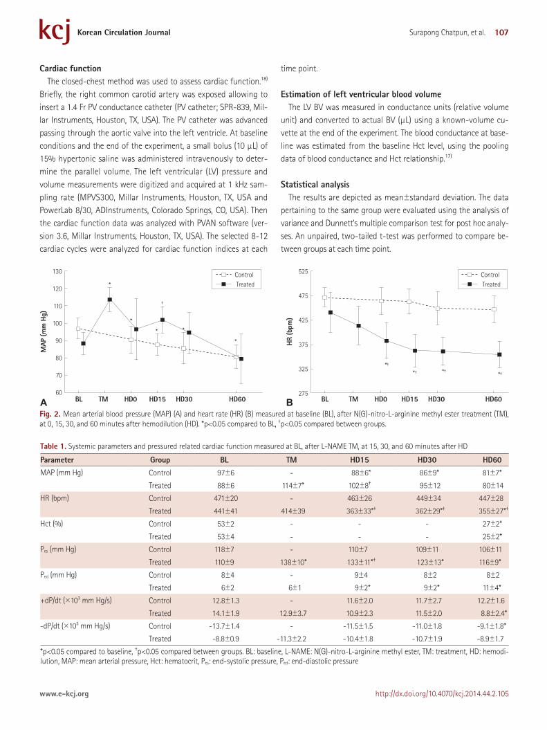
Fig. 2. Mean arterial blood pressure (MAP) (A) and heart rate (HR) (B) measured at baseline (BL), after N(G)-nitro-L-arginine methyl ester treatment (TM), at 0, 15, 30, and 60 minutes after hemodilution (HD). *p<0.05 compared to BL, †p<0.05 compared between groups.
130
120
110
100
90
80
70
60
*
†
*
* *
*
*†
*† *†*†
525
475
425
375
325
275
MAP
(mm
Hg)
HR (b
pm)
BL TM HD0 HD15 HD30 HD60 BL TM HD0 HD15 HD30 HD60
Control Treated
Control Treated
A B
Table 1. Systemic parameters and pressured related cardiac function measured at BL, after L-NAME TM, at 15, 30, and 60 minutes after HD
Parameter Group BL TM HD15 HD30 HD60MAP (mm Hg) Control 97±6 - 88±6* 86±9* 81±7*
Treated 88±6 114±7* 102±8† 95±12 80±14
HR (bpm) Control 471±20 - 463±26 449±34 447±28
Treated 441±41 414±39 363±33*† 362±29*† 355±27*†
Hct (%) Control 53±2 - - - 27±2*
Treated 53±4 - - - 25±2*
Pes (mm Hg) Control 118±7 - 110±7 109±11 106±11
Treated 110±9 138±10* 133±11*† 123±13* 116±9*
Ped (mm Hg) Control 8±4 - 9±4 8±2 8±2
Treated 6±2 6±1 9±2* 9±2* 11±4*
+dP/dt (×103 mm Hg/s) Control 12.8±1.3 - 11.6±2.0 11.7±2.7 12.2±1.6
Treated 14.1±1.9 12.9±3.7 10.9±2.3 11.5±2.0 8.8±2.4*
-dP/dt (×103 mm Hg/s) Control -13.7±1.4 - -11.5±1.5 -11.0±1.8 -9.1±1.8*
Treated -8.8±0.9 -11.3±2.2 -10.4±1.8 -10.7±1.9 -8.9±1.7
*p<0.05 compared to baseline, †p<0.05 compared between groups. BL: baseline, L-NAME: N(G)-nitro-L-arginine methyl ester, TM: treatment, HD: hemodi-lution, MAP: mean arterial pressure, Hct: hematocrit, Pes: end-systolic pressure, Ped: end-diastolic pressure
108 NOS Inhibition on Cardiac Function during Acute Anemia
http://dx.doi.org/10.4070/kcj.2014.44.2.105 www.e-kcj.org
All of the statistics were performed using GraphPad Prism 5.04 (GraphPad Software, San Diego, CA, USA). Statistical significance was determined at a p<0.05.
Results
Systemic parametersThe MAP (Fig. 2A) increased 30% from baseline to 20 minutes
after L-NAME administration. The effect was long lasting, for at least 30 minutes after hemodilution, and returned closer to baseline levels at 60 minutes after hemodilution. In contrast, the MAP in the con-trol group was at all points significantly lower after hemodilution than at baseline and was significantly lower than in the treated group where it was only at time point 15 minutes after hemodilu-tion. The HRs (Fig. 2B) in the treated group decreased by 15% from the baseline, mainly after hemodilution. In the control group, the HR remained near baseline values after hemodilution. The systemic Hct level decreased from 53±2% to 27±2% and 53±4% to 25±2% in the control group and the L-NAME-treated group, respectively.
Table 1 summarizes the systematic parameters in each group at the different time points. Blood viscosity in the control group after he-modilution was 3.61±0.24 cP whereas plasma viscosity was 2.12± 0.10 cP. These viscosities were not significantly different compared to that of the treated group.
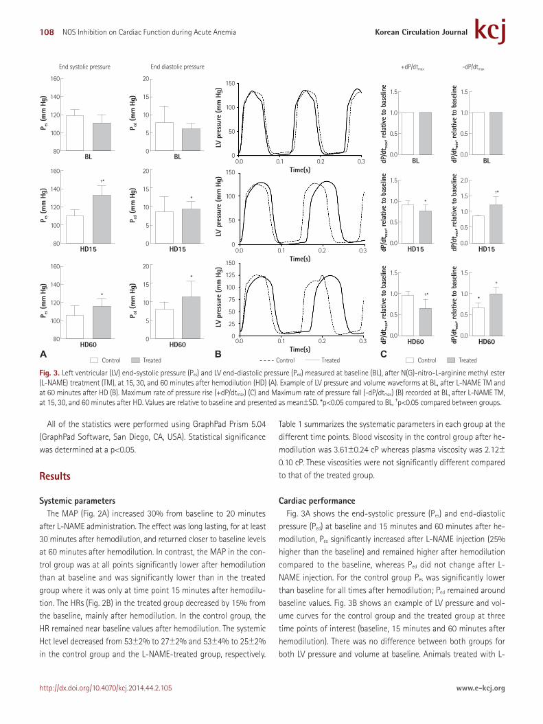
Cardiac performanceFig. 3A shows the end-systolic pressure (Pes) and end-diastolic
pressure (Ped) at baseline and 15 minutes and 60 minutes after he-modilution, Pes significantly increased after L-NAME injection (25% higher than the baseline) and remained higher after hemodilution compared to the baseline, whereas Ped did not change after L-NAME injection. For the control group Pes was significantly lower than baseline for all times after hemodilution; Ped remained around baseline values. Fig. 3B shows an example of LV pressure and vol-ume curves for the control group and the treated group at three time points of interest (baseline, 15 minutes and 60 minutes after hemodilution). There was no difference between both groups for both LV pressure and volume at baseline. Animals treated with L-
Fig. 3. Left ventricular (LV) end-systolic pressure (Pes) and LV end-diastolic pressure (Ped) measured at baseline (BL), after N(G)-nitro-L-arginine methyl ester (L-NAME) treatment (TM), at 15, 30, and 60 minutes after hemodilution (HD) (A). Example of LV pressure and volume waveforms at BL, after L-NAME TM and at 60 minutes after HD (B). Maximum rate of pressure rise (+dP/dtmax) (C) and Maximum rate of pressure fall (-dP/dtmax) (B) recorded at BL, after L-NAME TM, at 15, 30, and 60 minutes after HD. Values are relative to baseline and presented as mean±SD. *p<0.05 compared to BL, †p<0.05 compared between groups.
†*†*
†*
†
*
*
**
*
A
160
140
120
100
80
1.5
1.0
0.5
0.0
1.5
1.0
0.5
0.0
1.5
1.0
0.5
0.0
1.5
1.0
0.5
0.0
1.5
1.0
0.5
0.0
2.0
1.5
1.0
0.5
0.0
150
100
50
0
150
100
50
0
150
125
100
75
50
25
0
160
140
120
100
80
160
140
120
100
80
20
15
10
5
0
20
15
10
5
0
20
15
10
5
0
BL BL
HD60
HD15
BL
HD60
HD15HD15
HD60
BL
Time(s)
Time(s)
Time(s)
0.0 0.1 0.2 0.3
0.0 0.1 0.2 0.3
End diastolic pressure +dP/dtmax -dP/dtmaxEnd systolic pressure
0.0 0.1 0.2 0.3
HD15
HD60
P es (
mm
Hg)
dP/d
t max, r
elat
ive
to b
asel
ine
dP/d
t max, r
elat
ive
to b
asel
ine
dP/d
t max, r
elat
ive
to b
asel
ine
dP/d
t min, r
elat
ive
to b
asel
ine
dP/d
t min, r
elat
ive
to b
asel
ine
dP/d
t min, r
elat
ive
to b
asel
ine
LV p
ress
ure
(mm
Hg)
LV p
ress
ure
(mm
Hg)
LV p
ress
ure
(mm
Hg)
P es (
mm
Hg)
P es (
mm
Hg)
P ed (
mm
Hg)
P ed (
mm
Hg)
P ed (
mm
Hg)
Control Treated Control Treated Control TreatedB C
109Surapong Chatpun, et al.
http://dx.doi.org/10.4070/kcj.2014.44.2.105www.e-kcj.org
NAME had a higher LV pressure than the animals hemodiluted with HVPE only, especially at 15 minutes after hemodilution. Fig. 3C shows the maximum and minimum rates of pressure change (dP/dt) for the same points of interests. The maximum rate of pressure rise (+dP/dtmax) decreased after hemodilution in both groups when com-pared to the baseline. In the L-NAME treated group, +dP/dtmax was significantly lower than in the control group where it was only 60 minutes after hemodilution. As shown in Fig. 3C, the maximum rate of pressure fall (-dP/dtmax) in the L-NAME group was significantly higher than in the control group (p<0.05) for every time point after hemodilution. These parameters were also summarized in Table 1 at the time points of interest.
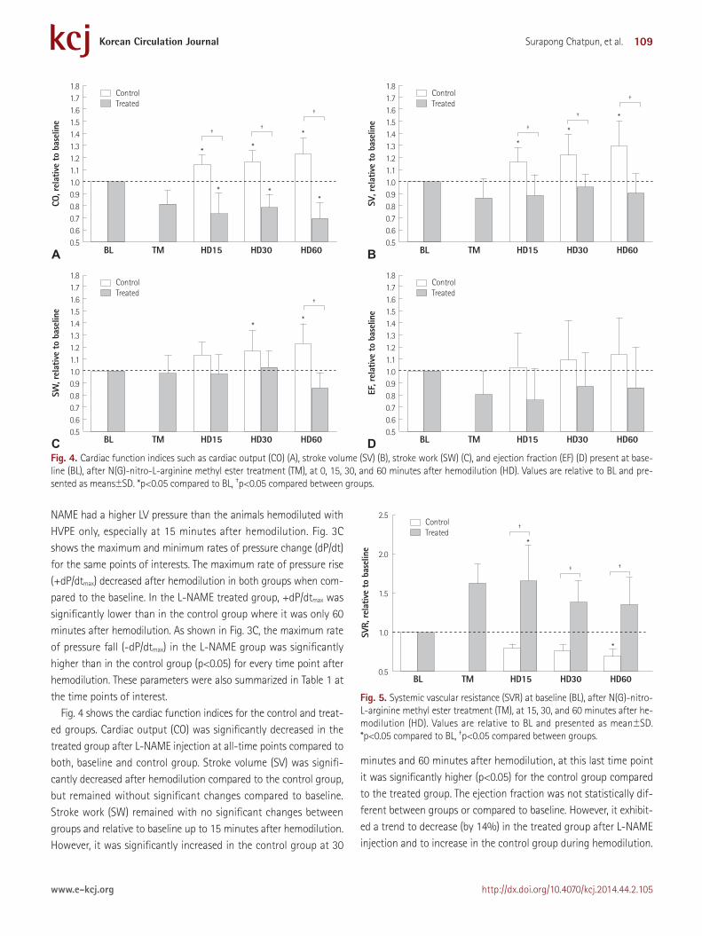
Fig. 4 shows the cardiac function indices for the control and treat-ed groups. Cardiac output (CO) was significantly decreased in the treated group after L-NAME injection at all-time points compared to both, baseline and control group. Stroke volume (SV) was signifi-cantly decreased after hemodilution compared to the control group, but remained without significant changes compared to baseline. Stroke work (SW) remained with no significant changes between groups and relative to baseline up to 15 minutes after hemodilution. However, it was significantly increased in the control group at 30
minutes and 60 minutes after hemodilution, at this last time point it was significantly higher (p<0.05) for the control group compared to the treated group. The ejection fraction was not statistically dif-ferent between groups or compared to baseline. However, it exhibit-ed a trend to decrease (by 14%) in the treated group after L-NAME injection and to increase in the control group during hemodilution.
Fig. 5. Systemic vascular resistance (SVR) at baseline (BL), after N(G)-nitro-L-arginine methyl ester treatment (TM), at 15, 30, and 60 minutes after he-modilution (HD). Values are relative to BL and presented as mean±SD. *p<0.05 compared to BL, †p<0.05 compared between groups.
2.5
2.0
1.5
1.0
0.5
SVR,
rela
tive
to b
asel
ine
BL TM HD15 HD30 HD60
Control Treated
†
† †
*
*
Fig. 4. Cardiac function indices such as cardiac output (CO) (A), stroke volume (SV) (B), stroke work (SW) (C), and ejection fraction (EF) (D) present at base-line (BL), after N(G)-nitro-L-arginine methyl ester treatment (TM), at 0, 15, 30, and 60 minutes after hemodilution (HD). Values are relative to BL and pre-sented as means±SD. *p<0.05 compared to BL, †p<0.05 compared between groups.
1.81.71.61.51.41.31.21.11.00.90.80.70.60.5
1.81.71.61.51.41.31.21.11.00.90.80.70.60.5
1.81.71.61.51.41.31.21.11.00.90.80.70.60.5
1.81.71.61.51.41.31.21.11.00.90.80.70.60.5
CO, r
elat
ive
to b
asel
ine
SW, r
elat
ive
to b
asel
ine
SV, r
elat
ive
to b
asel
ine
EF, r
elat
ive
to b
asel
ine
BL TM HD15 HD30 HD60
BL TM HD15 HD30 HD60
BL TM HD15 HD30 HD60
BL TM HD15 HD30 HD60
A
C
B
D
Control Treated
Control Treated
Control Treated
Control Treated
†††
††
†
†
**
* **
*
*
*
*
*
*
110 NOS Inhibition on Cardiac Function during Acute Anemia
http://dx.doi.org/10.4070/kcj.2014.44.2.105 www.e-kcj.org
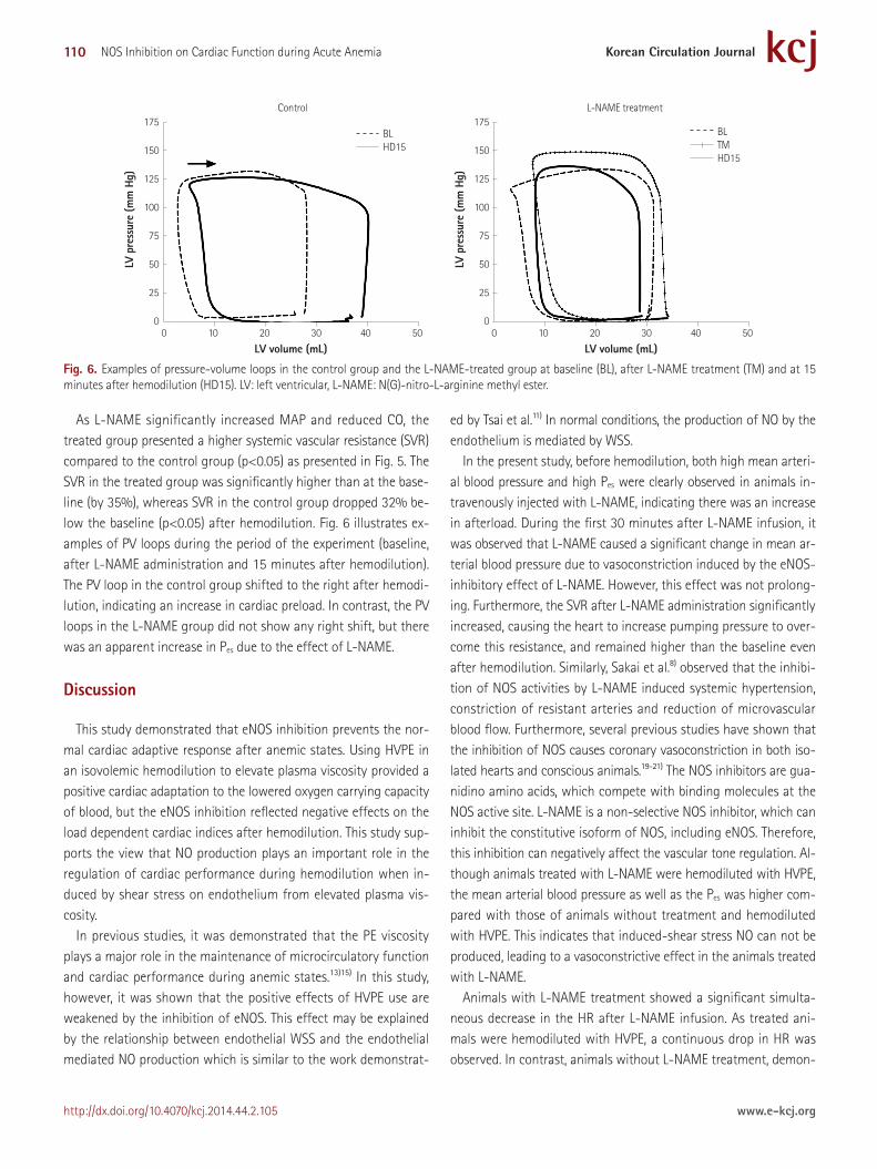
As L-NAME significantly increased MAP and reduced CO, the treated group presented a higher systemic vascular resistance (SVR) compared to the control group (p<0.05) as presented in Fig. 5. The SVR in the treated group was significantly higher than at the base-line (by 35%), whereas SVR in the control group dropped 32% be-low the baseline (p<0.05) after hemodilution. Fig. 6 illustrates ex-amples of PV loops during the period of the experiment (baseline, after L-NAME administration and 15 minutes after hemodilution). The PV loop in the control group shifted to the right after hemodi-lution, indicating an increase in cardiac preload. In contrast, the PV loops in the L-NAME group did not show any right shift, but there was an apparent increase in Pes due to the effect of L-NAME.
Discussion
This study demonstrated that eNOS inhibition prevents the nor-mal cardiac adaptive response after anemic states. Using HVPE in an isovolemic hemodilution to elevate plasma viscosity provided a positive cardiac adaptation to the lowered oxygen carrying capacity of blood, but the eNOS inhibition reflected negative effects on the load dependent cardiac indices after hemodilution. This study sup-ports the view that NO production plays an important role in the regulation of cardiac performance during hemodilution when in-duced by shear stress on endothelium from elevated plasma vis-cosity.
In previous studies, it was demonstrated that the PE viscosity plays a major role in the maintenance of microcirculatory function and cardiac performance during anemic states.13)15) In this study, however, it was shown that the positive effects of HVPE use are weakened by the inhibition of eNOS. This effect may be explained by the relationship between endothelial WSS and the endothelial mediated NO production which is similar to the work demonstrat-
ed by Tsai et al.11) In normal conditions, the production of NO by the endothelium is mediated by WSS.
In the present study, before hemodilution, both high mean arteri-al blood pressure and high Pes were clearly observed in animals in-travenously injected with L-NAME, indicating there was an increase in afterload. During the first 30 minutes after L-NAME infusion, it was observed that L-NAME caused a significant change in mean ar-terial blood pressure due to vasoconstriction induced by the eNOS-inhibitory effect of L-NAME. However, this effect was not prolong-ing. Furthermore, the SVR after L-NAME administration significantly increased, causing the heart to increase pumping pressure to over-come this resistance, and remained higher than the baseline even after hemodilution. Similarly, Sakai et al.8) observed that the inhibi-tion of NOS activities by L-NAME induced systemic hypertension, constriction of resistant arteries and reduction of microvascular blood flow. Furthermore, several previous studies have shown that the inhibition of NOS causes coronary vasoconstriction in both iso-lated hearts and conscious animals.19-21) The NOS inhibitors are gua-nidino amino acids, which compete with binding molecules at the NOS active site. L-NAME is a non-selective NOS inhibitor, which can inhibit the constitutive isoform of NOS, including eNOS. Therefore, this inhibition can negatively affect the vascular tone regulation. Al-though animals treated with L-NAME were hemodiluted with HVPE, the mean arterial blood pressure as well as the Pes was higher com-pared with those of animals without treatment and hemodiluted with HVPE. This indicates that induced-shear stress NO can not be produced, leading to a vasoconstrictive effect in the animals treated with L-NAME.
Animals with L-NAME treatment showed a significant simulta-neous decrease in the HR after L-NAME infusion. As treated ani-mals were hemodiluted with HVPE, a continuous drop in HR was observed. In contrast, animals without L-NAME treatment, demon-
Fig. 6. Examples of pressure-volume loops in the control group and the L-NAME-treated group at baseline (BL), after L-NAME treatment (TM) and at 15 minutes after hemodilution (HD15). LV: left ventricular, L-NAME: N(G)-nitro-L-arginine methyl ester.
175
150
125
100
75
50
25
0
175
150
125
100
75
50
25
0
LV p
ress
ure
(mm
Hg)
LV p
ress
ure
(mm
Hg)
0 10 20 30 40 50
Control L-NAME treatment
0 10 20 30 40 50
LV volume (mL) LV volume (mL)
BL HD15
BL TM HD15
111Surapong Chatpun, et al.
http://dx.doi.org/10.4070/kcj.2014.44.2.105www.e-kcj.org
strated a slight decrease in HR after hemodilution. With the study by Pabla and Curtis20) on the effects of NO modulation on cardiac arrhythmias in isolated rat hearts it was reported that L-NAME caused sinus bradycardia–similar to the results of other in vivo studies re-porting on the bradycardia effects of NOS.22)23) Recently, NO donor administration in animals hemodiluted with Dextran 70 kDa demon-strated the chronological effect of a 4% increase in the HR from the baseline, which was significantly increased when compared to ani-mals without NO donor administration.16) In addition, Bryan et al.24) have reported the effects on hemodynamics in a normal Hct level, that the pre-administration of S-nitro-N-acetylpenicillamine fol-lowed by L-NAME administration and vice versa can decrease sys-tolic blood pressure and mean arterial blood pressure, while the HR is increased.25) These previous studies emphasize the role of NO on HR. Therefore, it is important to note that the inhibition of NOS or the decrease in NO production coupled with hemodilution with HVPE can cause a negative effect on HR. However, controversial and unclear issues related to the chronological effects of NO still remain, especially in in vivo studies.26)
In the present study, the CO markedly decreased after L-NAME infusion followed by hemodilution with HVPE, while CO in the group without L-NAME treatment significantly increased from the base-line. This finding is in agreement with those of several studies, which have indicated that L-NAME decreases the CO in a normal level of Hct either in the short-term or long-term treatments with L-NAME.25)27)28) Furthermore, in this study, the L-NAME administration led to a re-duction in SV even after hemodilution. Especially at 15 minutes after hemodilution as shown by PV loops, it could be noticed that the end-diastolic volume decreased after hemodilution in animals with L-NAME treatment which led to lower SV. Generally, there is a com-pensatory mechanism for a reduction in oxygen transport capabil-ity–i.e., hemodilution by an increase in CO via either an increase in HR or an enhancement of SV. This compensation was still observed after hemodilution in animals without L-NAME treatment but not in animals treated with L-NAME. By administration through a cor-onary catheter, using non-selective NOS inhibitors such as N-nitro-L-arginine (L-NNA) in normal dogs an increment in the coronary blood flow was shown.29) In contrast, Biwer et al.28) demonstrated that long-term L-NAME-treated rats had not only a decrease in CO and SW but also a decrease in coronary blood flow, which potential-ly caused cardiac dysfunction. Therefore, it is a point of concern that using PEs in hemodilution with a stage of NOS impairment can lead to an attenuation of the cardiac compensatory mechanism.
The use of a non-selective NOS inhibitor, L-NNA, resulted in an in-crease in myocardium oxygen consumption in normal un-anesthe-tized dogs, whereas using L-NAME in anesthetized open-chest dogs did not present this.29)30) In the present study, after hemodilution
with HVPE, animals treated with L-NAME presented a lower SW and +dP/dtmax than animals without L-NAME treatment. This indicates an attenuation of energy to pump out blood from the chamber. How-ever, this attenuation of energy related to a decrease in SV and a smaller PV loop area. Therefore, it can be pointed out that an impair-ment of NOS significantly affects cardiac load-dependent indices after hemodilution. Also the heart seems to be less able to adjust to the change of reduced Hct content in animals treated with L-NAME.
The analogs of L-arginine were used to treat some patient’s con-ditions such as hypotension during septic shock. As a result of the presented study, it was demonstrated that the cardiac compensa-tory mechanism attenuated by L-NAME responded to hemodilu-tion. Therefore, it might not be beneficial to administer L-NAME in patients with low Hct and septic shock or with a required hemodi-lution regarding to the modalities of sepsis treatment. On the oth-er hand, for septic patients with L-NAME administration a high vis-cosity PE might not be suitable for volume replacement. However, in the present study L-NAME administration was performed as a short-term treatment and therefore it is necessary to look at long-term treatment effects on a hemodiluted condition. Furthermore, the study had several limiting issues such as a small number of ani-mals in each group, only two dosages of L-NAME, no oxygen de-livery assessment and the effects of anesthetics (sodium pentobar-bital) as a cardiac depressor.
In conclusion, it was shown that eNOS inhibition prevents the normal cardiac adaptive response after anemic states. Using high viscogenic PE in isovolemic hemodilution to elevate plasma viscos-ity provided a positive cardiac adaptation to the lowered oxygen carrying capacity of blood, but eNOS inhibition reflected negative ef-fects on load dependent cardiac indices.
AcknowledgmentsThe authors greatly thank Cynthia Walser for the surgical prepa-
ration of the animals. This work was supported by the Bioengineer-ing Research Partnership grant R24-HL64395, program project P01-HL071064, grant R01-HL62354 and R01-HL62318 and by the Faculty of Medicine, Prince of Songkla University grant 54-221-25-2-3.
References1. Boo YC. Shear stress stimulates phosphorylation of protein kinase A
substrate proteins including endothelial nitric oxide synthase in endo-thelial cells. Exp Mol Med 2006;38:453.
2. Corson MA, James NL, Latta SE, Nerem RM, Berk BC, Harrison DG. Phosphorylation of endothelial nitric oxide synthase in response to fluid shear stress. Circ Res 1996;79:984-91.
3. Shesely EG, Maeda N, Kim HS, et al. Elevated blood pressures in mice lacking endothelial nitric oxide synthase. Proc Natl Acad Sci U S A 1996;
112 NOS Inhibition on Cardiac Function during Acute Anemia
http://dx.doi.org/10.4070/kcj.2014.44.2.105 www.e-kcj.org
93:13176-81.4. Rastaldo R, Pagliaro P, Cappello S, et al. Nitric oxide and cardiac func-
tion. Life Sci 2007;81:779-93.5. Palmer RM, Rees DD, Ashton DS, Moncada S. L-arginine is the physio-
logical precursor for the formation of nitric oxide in endothelium-de-pendent relaxation. Biochem Biophys Res Commun 1988;153:1251-6.
6. Hsieh NK, Chang HR, Hu CT, Chen HI. Effects of nitric oxide donor and nitric oxide synthase inhibitor on the resistance, exchange and capaci-tance functions of the canine intestinal vasculature. Vascul Pharmacol 2008;48:122-8.
7. Fitch RM, Vergona R, Sullivan ME, Wang YX. Nitric oxide synthase inhi-bition increases aortic stiffness measured by pulse wave velocity in rats. Cardiovasc Res 2001;51:351-8.
8. Sakai H, Hara H, Tsai AG, Tsuchida E, Intaglietta M. Constriction of re-sistance arteries determines l-NAME-induced hypertension in a con-scious hamster model. Microvasc Res 2000;60:21-7.
9. Fozard JR, Part ML. Haemodynamic responses to NG-monomethyl-L-arginine in spontaneously hypertensive and normotensive Wistar-Kyo-to rats. Br J Pharmacol 1991;102:823-6.
10. de Wit C, Schäfer C, von Bismarck P, Bolz SS, Pohl U. Elevation of plas-ma viscosity induces sustained NO-mediated dilation in the hamster cremaster microcirculation in vivo. Pflugers Arch 1997;434:354-61.
11. Tsai AG, Acero C, Nance PR, et al. Elevated plasma viscosity in extreme hemodilution increases perivascular nitric oxide concentration and mi-crovascular perfusion. Am J Physiol Heart Circ Physiol 2005;288: H1730-9.
12. Cabrales P, Intaglietta M, Tsai AG. Increase plasma viscosity sustains microcirculation after resuscitation from hemorrhagic shock and con-tinuous bleeding. Shock 2005;23:549-55.
13. Cabrales P, Tsai AG. Plasma viscosity regulates systemic and microvas-cular perfusion during acute extreme anemic conditions. Am J Physiol Heart Circ Physiol 2006;291:H2445-52.
14. Cabrales P, Tsai AG, Intaglietta M. Increased plasma viscosity prolongs microhemodynamic conditions during small volume resuscitation from hemorrhagic shock. Resuscitation 2008;77:379-86.
15. Chatpun S, Cabrales P. Cardiac mechanoenergetic cost of elevated plasma viscosity after moderate hemodilution. Biorheology 2010;47: 225-37.
16. Chatpun S, Cabrales P. Exogenous intravascular nitric oxide enhances ventricular function after hemodilution with plasma expander. Life Sci 2012;90:39-46.
17. Chatpun S, Cabrales P. Effects of plasma viscosity modulation on car-
diac function during moderate hemodilution. Asian J Transfus Sci 2010; 4:102-8.
18. Pacher P, Nagayama T, Mukhopadhyay P, Bátkai S, Kass DA. Measure-ment of cardiac function using pressure-volume conductance cathe-ter technique in mice and rats. Nat Protoc 2008;3:1422-34.
19. Amezcua JL, Palmer RM, de Souza BM, Moncada S. Nitric oxide synthe-sized from L-arginine regulates vascular tone in the coronary circula-tion of the rabbit. Br J Pharmacol 1989;97:1119-24.
20. Pabla R, Curtis MJ. Effects of NO modulation on cardiac arrhythmias in the rat isolated heart. Circ Res 1995;77:984-92.
21. Rees DD, Palmer RM, Schulz R, Hodson HF, Moncada S. Characteriza-tion of three inhibitors of endothelial nitric oxide synthase in vitro and in vivo. Br J Pharmacol 1990;101:746-52.
22. Pfeiffer S, Leopold E, Schmidt K, Brunner F, Mayer B. Inhibition of nitric oxide synthesis by NG-nitro-L-arginine methyl ester (L-NAME): re-quirement for bioactivation to the free acid, NG-nitro-L-arginine. Br J Pharmacol 1996;118:1433-40.
23. Ward JE, Angus JA. Acute and chronic inhibition of nitric oxide syn-thase in conscious rabbits: role of nitric oxide in the control of vascular tone. J Cardiovasc Pharmacol 1993;21:804-14.
24. Bryan S, Alexander-Lindo R, McGrowder D. The effect of nitric oxide inhibitors and s-nitrosothiols on hemodynamic parameters in an ani-mal model. Open Access Animal Physiology 2011;3:1-8.
25. Crystal GJ, Zhou X, Halim AA, Alam S, El-Orbany M, Salem MR. Nitric oxide does not modulate whole body oxygen consumption in anesthe-tized dogs. J Appl Physiol (1985) 1999;86:1944-9.
26. Herring N, Paterson DJ. Endothelial nitric oxide synthase and heart rate. Circulation 2002;106:e5; author reply e5.
27. Widdop RE, Gardiner SM, Kemp PA, Bennett T. The influence of atro-pine and atenolol on the cardiac haemodynamic effects of NG-nitro-L-arginine methyl ester in conscious, Long Evans rats. Br J Pharmacol 1992;105:653-6.
28. Biwer LA, Broderick TL, Xu H, Carroll C, Hale TM. Protection against L-NAME-induced reduction in cardiac output persists even after cessa-tion of angiotensin-converting enzyme inhibitor treatment. Acta Physi-ol (Oxf) 2013;207:156-65.
29. Chen Y, Traverse JH, Du R, Hou M, Bache RJ. Nitric oxide modulates myocardial oxygen consumption in the failing heart. Circulation 2002; 106:273-9.
30. Crystal GJ, Zhou X. Nitric oxide does not modulate the increases in blood flow, O2 consumption, or contractility during CaCl2 administra-tion in canine hearts. Cardiovasc Res 1999;42:232-9.