Embed Size (px)
Citation preview

European Polymer Journal 55 (2014) 108–113
Contents lists available at ScienceDirect
European Polymer Journal
journal homepage: www.elsevier .com/locate /europol j
Nitric oxide sensitive fluorescent polymeric hydrogels showingnegligible interference by dehydroascorbic acid
http://dx.doi.org/10.1016/j.eurpolymj.2014.03.0270014-3057/� 2014 Elsevier Ltd. All rights reserved.
⇑ Corresponding authors. Tel.: +34 964728236; fax: +34 964728214.E-mail addresses: [email protected] (F. Galindo), [email protected]
(S.V. Luis).
Víctor Fabregat, M. Ángeles Izquierdo, M. Isabel Burguete, Francisco Galindo ⇑,Santiago V. Luis ⇑Universitat Jaume I, Departamento de Química Inorgánica y Orgánica, Av. Sos Baynat, s/n, E-12071 Castellón, Spain
a r t i c l e i n f o a b s t r a c t
Article history:Received 5 August 2013Received in revised form 3 February 2014Accepted 22 March 2014Available online 1 April 2014
Keywords:Nitric oxideFluorescence sensingProbesDehydroascorbic acidPolymeric hydrogels
A series of nitric oxide (NO) sensitive polymeric hydrogels have been synthesized and theirproperties described. The new materials are based on a poly(2-hydroxyethyl methacrylate)matrix entrapping the DAF-FM (4-amino-5-methylamino-20,70-difluorofluorescein)fluorescent probe. Some of the developed materials show sensitivities in the range ca.50–75 nM towards NO dissolved in aerated phosphate buffer (PBS), being nitrousanhydride the reactive species under such conditions. Remarkably, the sensitivity of thefluorescent polymers is not influenced by dehydroascorbic acid, a molecule of biologicalimportance, which has been reported to be a common interfering species capable to reactwith well known probes for NO.
� 2014 Elsevier Ltd. All rights reserved.
1. Introduction
Nitric oxide (NO) is a small diatomic molecule playingnumerous biological [1] and environmental roles [2].Regarding the importance of NO for Biological Chemistryit can be mentioned that it is a neurotransmitter [3], arelevant species for the Immune System [4] and also asignaling molecule triggering the muscular relaxationresponse [5]. In the context of Environmental Chemistry,NO is a pollutant originated from the combustion enginesof vehicles and hence a major concern for public authori-ties [6]. Hence, a great number of reports dealing withthe measurement of NO in both contexts have beenpublished during the last years. Electrochemistry [7] andfluorescence [8] based methodologies are the most popularapproaches to the analytical determination of NO concen-trations. Regarding the fluorescence based technologies, abig pool of molecular fluorescent probes with excellent
sensitivity towards NO have been reported [9–13]. Themajority of molecular probes are typically used in solutionbased applications; however some situations, e.g. environ-mental monitoring, require NO-sensitive probes to beincorporated into a solid material, in order to integratethem into a measuring device [14]. In comparison to thelarge number of molecular probes for NO, a limitednumber of fluorogenic and chromogenic solid materialssensitive to this species have been reported [15]. In thisregard, we have developed recently new NO-sensitivecomposite materials based on organogels [16] and poly-meric films [17–19].
One of the most popular fluorescent NO probes is thediamino fluorescein derivative DAF-FM (4-amino-5-methylamino-20,70-difluorofluorescein, Chart 1) [20], whichhas excellent sensitivity towards NO in oxygenatedaqueous medium [21]. The selectivity of some diaminoflu-orescein and diaminorhodamine based probes were inves-tigated by Sweedler and co-workers [22] who observedthat biologically relevant compounds, in particular dehy-droascorbic acid (DHAA), reacted to yield a fluorescentproduct that could be falsely identified as the NO adduct.

OO
F F
O
COO
NH2NHCH3
DAF-FM
_
_
Chart 1. Chemical structure of DAF-FM.
V. Fabregat et al. / European Polymer Journal 55 (2014) 108–113 109
Therefore, the development of fluorescent NO sensorsexhibiting low interference by DHAA, is highly desirable.
In this paper, we describe the encapsulation of DAF-FMinto a series of polymeric hydrogels based on 2-hydroxy-ethylmethacrylate (HEMA) and the study of the spectro-scopic and the analytical properties of the obtainedmaterials. The new polymers are reactive to NO in oxygen-ated media (nitrosating nitrous anhydride formed in suchconditions [1a]), displaying a fluorescent sensitivity analo-gous to the free probe in solution. Importantly, the newmaterials are insensitive to the potential interference ofDHAA (up to 1 mM). Hence we have developed a noveland very effective strategy to avoid the interference byDHAA, which could be of potential interest for the develop-ment of more precise NO-sensitive devices.
2. Experimental section
2.1. Materials and methods
2-Hydroxyethylmethacrylate (HEMA), 3-(acrylamide-propyl)trimethylamonium chloride (AAPTEA+), sodium2-acrylamido-2methyl-1-propensulfonate (AAMPS�), N-hydroxyethylacrylamide (HEAA), ethylenglycoldimethacry-late (EGDMA), polyethylenglycoldimethacrylate (PEGDMA),dehydroascorbic acid and sodium NONOate (sodium dieth-ylammonium (Z)-1-(N,N-diethylamino)diazen-1-ium-1,2-diolate) used in this study were obtained from Aldrich.Azobisisoburyronitrile (AIBN) was obtained from Flukaand DAF-FM from Invitrogen. Water used throughout thestudies was Millipore Q quality.
UV–Vis absorption spectra were recorded with a Hew-lett–Packard 8453 spectrophotometer. Fourier TransformInfrared (FT-IR) spectra were acquired using a FT/IR-6200type A JASCO spectrometer, with 4 cm�1 resolution and50 scan accumulation. Fourier Transform Raman (FT-Raman) spectra were recorded using a JASCO NRS-3100dispersive laser Raman spectrometer, with 4 cm�1 resolu-tion and 100 scans accumulation (kex = 632 nm). Thermo-gravimetric Analysis (TGA) were carried out using aMettler Toledo TG-STDA instrument (30–400 �C at a heat-ing rate of 5 �C/min). Steady-state fluorescence spectrawere recorded with a Spex Fluorog 3–11 equipped with a450 W xenon lamp. Fluorescence spectra were recordedin the front face mode. Time-resolved fluorescencemeasurements were done with the technique of time
correlated single photon counting (TCSPC) using an IBH-5000U apparatus. Samples were excited at 464 nm usinga nanoLED with a FWHM of 1.4 ns and a repetition rateof 100 kHz. Data were fitted to a single exponential modelafter deconvolution of the instrument response function byiterative deconvolution using the IBH DAS6 fluorescencedecay analysis fluorescence decay analysis software(monoexponential decays), where reduced v2 andweighted residuals serve as parameters for goodness ofthe fit. All the samples were measured under aeratedconditions.
2.2. Preparation and characterization of polymers
The preparation of the dye-loaded sensitive films wascarried out following the methodology previouslydescribed to prepare analogous materials [17,19,23]. Theappropriate weight composition of monomers and cross-linking agents was mixed in a vial with 1% of AIBN (totalweight). Then, DAF-FM (100 lg/mL) was added and thesample was stirred vigorously at room temperature untilcomplete dissolution of the reagents. Subsequently, thepolymerization mixture was placed in a mold, which con-sisted in two microscope glass slides separated by two thinlamellas (�120 lm thickness).
The polymerization reaction was conducted in an ovenat 85 �C for 15 min. The films were then removed mechan-ically from the molds and washed with a solution of PBSbuffer 0.1 M. All of the films were red-colored and highlytransparent. The polymers were characterized by FT-IR,FT-Raman, UV–Vis absorption and fluorescence spectros-copies (see Electronic Supporting Information, ESI). FilmI: FT-IR (cm�1): 3378, 2949, 1713, 1453, 1244, 1152,1072. FT-Raman (cm�1): 2938, 1722, 1634, 1446, 1296,972. UV–Vis (kmax, nm): 501. Fluorescence (kmax, nm):522 (kex = 500 nm). TGA (Tg, �C): 260 – 270. Film II: FT-IR(cm�1): 3429, 2944, 1717, 1442, 1247, 1153, 1072.FT-Raman (cm�1): 2948, 1718, 1639, 1455, 1283, 972.UV–Vis (kmax, nm): 500. Fluorescence (kmax, nm): 519(kex = 500 nm). TGA (Tg, �C): 250–255. Film III: FT-IR(cm�1): 3386, 2945, 1711, 1637, 1449, 1256, 1153, 1075.FT-Raman (cm�1): 2944, 1716, 1636, 1421, 1314, 1234,1100. UV–Vis (kmax, nm): 501. Fluorescence (kmax, nm):522 (kex = 500 nm). TGA (Tg, �C): 330–340. Film IV: FT-IR(cm�1): 3376, 2952, 1707, 1644, 1453, 1257, 1160, 1057.FT-Raman (cm�1): 2936, 1723, 1642, 1419, 1317, 1241,1005. UV–Vis (kmax, nm): 498. Fluorescence (kmax, nm):520 (kex = 500 nm). TGA (Tg, �C): 330 – 340. Film V: FT-IR(cm�1): 3407, 2931, 1716, 1647, 1454, 1244, 1153, 1075.FT-Raman (cm�1): 2942, 1718, 1642, 1419, 1311, 1241,1106. UV–Vis (kmax, nm): 496. Fluorescence (kmax, nm):516 (kex = 500 nm). TGA (Tg, �C): 320 – 330. Film VI:FT-IR (cm�1): 3379, 2951, 1705, 1647, 1450, 1159, 1081,983, 844. FT-Raman (cm�1): 2935, 1721, 1641, 1417,1310, 1236, 1104. UV–Vis (kmax, nm): 497. Fluorescence(kmax, nm): 518 (kex = 500 nm). TGA (Tg, �C): 320–330. FilmVII: FT-IR (cm�1): 3375, 2943, 1712, 1643, 1454, 1251,1153, 1069. FT-Raman (cm�1): 2942, 1717, 1636, 1414,1317, 1241, 1106, 1046. UV–Vis (kmax, nm): 498.Fluorescence (kmax, nm): 520 (kex = 500 nm). TGA (Tg, �C):330 – 340. Film VIII: FT-IR (cm�1): 3363, 2951, 1709,
110 V. Fabregat et al. / European Polymer Journal 55 (2014) 108–113
1639, 1450, 1244, 1153, 1075, 996, 640. FT-Raman (cm�1):2943, 1720, 1641, 1425, 1317, 1239, 1102. UV–Vis (kmax,nm): 500. Fluorescence (kmax, nm): 521 (kex = 500 nm).TGA (Tg, �C): 380–390.
2.3. Analytical determinations
Nitric oxide solutions were prepared from sodiumNONOate (diethylamine NONOate sodium salt hydrate) inphosphate buffer (PBS, 0.01 M, pH 7.36), as described pre-viously.[24] Polymer fragments (15 ± 1 mg) were intro-duced in the corresponding NO solutions and allowed toequilibrate for 1 h. The fluorescence spectra of the poly-mers were recorded before and after the reaction withNO (kex = 500 nm) and the limits of detection (LOD) werecalculated according to the method reported in the litera-ture (blank + 3r) [25]. Every emission measurement wasdone by triplicate and the average values were calculated.
2.4. Experiments with dehydroascorbic acid (DHAA)
The fluorescence emission spectra of a DAF-FM solution(10 lM) in PBS (0.01 M, pH 7.36) was recorded (kex = 500 -nm). Subsequently, DHAA was added to a final concentra-tion of 1 mM, the solution was allowed to equilibrate for1 h and the fluorescence emission of the solution wasrecorded. Next, sodium NONOate (1.9 mM) was added toobtain a saturated solution of nitric oxide [8a]. The sampleswere allowed to equilibrate for 1 h and the fluorescenceemission was recorded. Simultaneously, the same proce-dure was applied to 10 mg of film IV in 3 mL of PBS.
3. Results and discussion
3.1. Synthesis and characterization of dye-loaded films
The molecular probe DAF-FM was encapsulated in aseries of transparent polymeric films following previouslydescribed procedures [18,19,23]. Polymerization of thehighly hydrophilic monomer 2-hydroxyethyl methacrylate(HEMA) led to the polyHEMA hydrogel films used in thisstudy. In previous reports, different combinations of acrylicmonomers and cross-linkers have been used [18,19,23]. Inthe present study, a similar approach was employed for theimmobilization of DAF-FM, using HEMA as the main
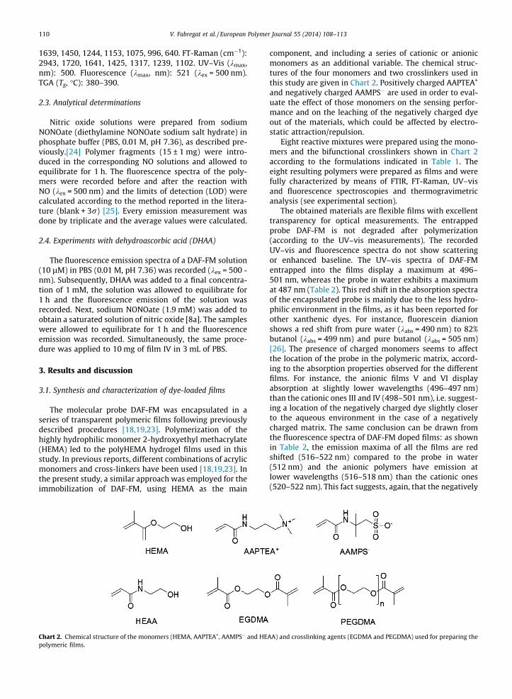
Chart 2. Chemical structure of the monomers (HEMA, AAPTEA+, AAMPS� and HEpolymeric films.
component, and including a series of cationic or anionicmonomers as an additional variable. The chemical struc-tures of the four monomers and two crosslinkers used inthis study are given in Chart 2. Positively charged AAPTEA+
and negatively charged AAMPS� are used in order to eval-uate the effect of those monomers on the sensing perfor-mance and on the leaching of the negatively charged dyeout of the materials, which could be affected by electro-static attraction/repulsion.
Eight reactive mixtures were prepared using the mono-mers and the bifunctional crosslinkers shown in Chart 2according to the formulations indicated in Table 1. Theeight resulting polymers were prepared as films and werefully characterized by means of FTIR, FT-Raman, UV–visand fluorescence spectroscopies and thermogravimetricanalysis (see experimental section).
The obtained materials are flexible films with excellenttransparency for optical measurements. The entrappedprobe DAF-FM is not degraded after polymerization(according to the UV–vis measurements). The recordedUV–vis and fluorescence spectra do not show scatteringor enhanced baseline. The UV–vis spectra of DAF-FMentrapped into the films display a maximum at 496–501 nm, whereas the probe in water exhibits a maximumat 487 nm (Table 2). This red shift in the absorption spectraof the encapsulated probe is mainly due to the less hydro-philic environment in the films, as it has been reported forother xanthenic dyes. For instance, fluorescein dianionshows a red shift from pure water (kabs = 490 nm) to 82%butanol (kabs = 499 nm) and pure butanol (kabs = 505 nm)[26]. The presence of charged monomers seems to affectthe location of the probe in the polymeric matrix, accord-ing to the absorption properties observed for the differentfilms. For instance, the anionic films V and VI displayabsorption at slightly lower wavelengths (496–497 nm)than the cationic ones III and IV (498–501 nm), i.e. suggest-ing a location of the negatively charged dye slightly closerto the aqueous environment in the case of a negativelycharged matrix. The same conclusion can be drawn fromthe fluorescence spectra of DAF-FM doped films: as shownin Table 2, the emission maxima of all the films are redshifted (516–522 nm) compared to the probe in water(512 nm) and the anionic polymers have emission atlower wavelengths (516–518 nm) than the cationic ones(520–522 nm). This fact suggests, again, that the negatively
AA) and crosslinking agents (EGDMA and PEGDMA) used for preparing the
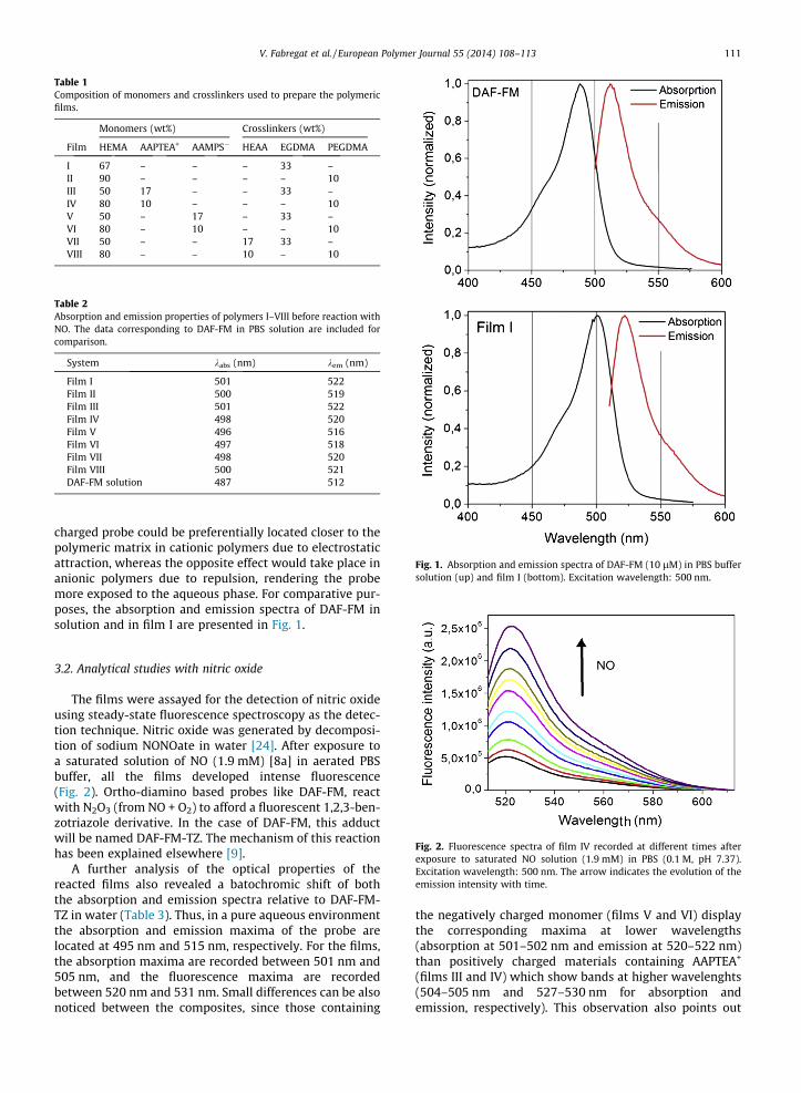
Table 1Composition of monomers and crosslinkers used to prepare the polymericfilms.
Monomers (wt%) Crosslinkers (wt%)
Film HEMA AAPTEA+ AAMPS� HEAA EGDMA PEGDMA
I 67 – – – 33 –II 90 – – – – 10III 50 17 – – 33 –IV 80 10 – – – 10V 50 – 17 – 33 –VI 80 – 10 – – 10VII 50 – – 17 33 –VIII 80 – – 10 – 10
Table 2Absorption and emission properties of polymers I–VIII before reaction withNO. The data corresponding to DAF-FM in PBS solution are included forcomparison.
System kabs (nm) kem (nm)
Film I 501 522Film II 500 519Film III 501 522Film IV 498 520Film V 496 516Film VI 497 518Film VII 498 520Film VIII 500 521DAF-FM solution 487 512
Fig. 1. Absorption and emission spectra of DAF-FM (10 lM) in PBS buffersolution (up) and film I (bottom). Excitation wavelength: 500 nm.
V. Fabregat et al. / European Polymer Journal 55 (2014) 108–113 111
charged probe could be preferentially located closer to thepolymeric matrix in cationic polymers due to electrostaticattraction, whereas the opposite effect would take place inanionic polymers due to repulsion, rendering the probemore exposed to the aqueous phase. For comparative pur-poses, the absorption and emission spectra of DAF-FM insolution and in film I are presented in Fig. 1.
Fig. 2. Fluorescence spectra of film IV recorded at different times afterexposure to saturated NO solution (1.9 mM) in PBS (0.1 M, pH 7.37).Excitation wavelength: 500 nm. The arrow indicates the evolution of theemission intensity with time.
3.2. Analytical studies with nitric oxide
The films were assayed for the detection of nitric oxideusing steady-state fluorescence spectroscopy as the detec-tion technique. Nitric oxide was generated by decomposi-tion of sodium NONOate in water [24]. After exposure toa saturated solution of NO (1.9 mM) [8a] in aerated PBSbuffer, all the films developed intense fluorescence(Fig. 2). Ortho-diamino based probes like DAF-FM, reactwith N2O3 (from NO + O2) to afford a fluorescent 1,2,3-ben-zotriazole derivative. In the case of DAF-FM, this adductwill be named DAF-FM-TZ. The mechanism of this reactionhas been explained elsewhere [9].
A further analysis of the optical properties of thereacted films also revealed a batochromic shift of boththe absorption and emission spectra relative to DAF-FM-TZ in water (Table 3). Thus, in a pure aqueous environmentthe absorption and emission maxima of the probe arelocated at 495 nm and 515 nm, respectively. For the films,the absorption maxima are recorded between 501 nm and505 nm, and the fluorescence maxima are recordedbetween 520 nm and 531 nm. Small differences can be alsonoticed between the composites, since those containing
the negatively charged monomer (films V and VI) displaythe corresponding maxima at lower wavelengths(absorption at 501–502 nm and emission at 520–522 nm)than positively charged materials containing AAPTEA+
(films III and IV) which show bands at higher wavelenghts(504–505 nm and 527–530 nm for absorption andemission, respectively). This observation also points out
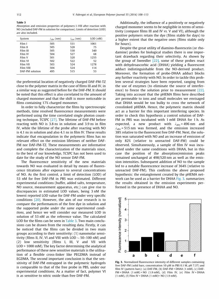
Table 3Absorption and emission properties of polymers I–VIII after reaction withNO (included DAF-FM in solution for comparison). Limits of detection (LOD)are also included.
System kabs (nm) kem (nm) LOD (nM)
Film I 505 531 1061Film II 505 528 75Film III 505 530 340Film IV 504 527 68Film V 501 520 1255Film VI 502 522 52Film VII 503 524 1278Film VIII 504 524 114DAF-FM solution 495 515 53
Fig. 3. Normalized fluorescence intensity of different samples containingfree DAF-FM (solid bars, concentration 10 lM in PBS 0.1 M, pH 7.37) andfilm IV (pattern bars): (a) DAF-FM, (b) DAF-FM + DHAA (1 mM), (c) DAF-FM + DHAA (1 mM) + NO (1.9 mM), (d) Film IV, (e) Film IV + DHAA(1 mM), (f) Film IV + DHAA (1 mM)+ NO (1.9 mM).
112 V. Fabregat et al. / European Polymer Journal 55 (2014) 108–113
the preferential location of negatively charged DAF-FM-TZclose to the polymer matrix in the case of films III and IV, ina similar way as suggested before for the DAF-FM. It shouldbe noted that this effect is also correlated to the amount ofcharged monomer and that the shift is more noticeable infilms containing 17% charged monomer.
In order to fully characterize the films by spectroscopicmethods, time resolved fluorescence measurements wereperformed using the time correlated single photon count-ing technique, TCSPC [27]. The lifetime of DAF-FM beforereacting with NO is 3.8 ns in solution and 3.9 ns in filmIV, while the lifetime of the probe after reacting with NOis 4.1 ns in solution and also 4.1 ns in film IV. These resultsindicate that encapsulation in the polymeric films has norelevant effect on the emission lifetimes of neither DAF-FM nor DAF-FM-TZ. These measurements are informativeand complete the characterization of the materials since,to the best of our knowledge, TCSPC has not been used todate for the study of the NO sensor DAF-FM.
The fluorescence sensitivity of the new materialstowards NO was evaluated in detail by means of fluores-cence titrations after exposure to several concentrationsof NO. As the first control, a limit of detection (LOD) of53 nM for free DAF-FM in PBS was estimated. Differentexperimental conditions (buffer, added salts, temperature,NO source, measurement apparatus, etc.) can give rise todiscrepancies in estimated LOD values, being 3 nM thelowest reported LOD value for DAF-FM under very specificconditions [20]. However, the aim of our research is tocompare the performances of the free dye in solution andthe supported probe under the same experimental condi-tions, and hence we will consider our measured LOD insolution of 53 nM as the reference value. The calculatedLOD for the films can be seen in Table 3. Two main conclu-sions can be drawn from the resulting data. Firstly, it canbe noticed that the films can be divided in two maingroups according to their sensitivity: (1) nanomolar sensi-tivity (films II, IV, VI and VIII with LOD � 50–100 nM) and(2) low sensitivity (films I, III, V and VII withLOD > 1000 mM). The key factor determining the analyticalperformance of these new sensitive materials is the utilisa-tion of a flexible cross-linker like PEGDMA instead ofEGDMA. The second important conclusion is that the sen-sitivity of DAF-FM entrapped in the polymeric hydrogelsis comparable to that of the free probe in PBS, under ourexperimental conditions. As a matter of fact, polymer VIis as sensitive to nitric oxide than free DAF-FM.
Additionally, the influence of a positively or negativelycharged monomer seems to be negligible in terms of sensi-tivity (compare films III and IV vs. V and VI), although thepositive polymers retain the dye (films stable for days) toa higher extent that the negative ones (films stable onlyfor hours).
Despite the great utility of diamino-fluorescein (or rho-damine) probes for biological studies there is one impor-tant drawback regarding their selectivity. As shown bythe group of Sweedler [22], some of these probes reactwith dehydroascorbic acid (DHAA) yielding a fluorescentadduct indistinguishable from the NO-derived product.Moreover, the formation of probe-DHAA adduct blocksany further reactivity with NO. In order to tackle this prob-lem several strategies have been reported, ranging fromthe use of enzymes (to eliminate the source of interfer-ence) to freeze the solution prior to measurement [22].Taking into account that the pHEMA films here describedare permeable to ions and small molecules we envisagedthat DHAA would be too bulky to cross the network ofcrosslinked pHEMA. Hence, the polymeric matrix shouldact as a barrier for this important interfering species. Inorder to check this hypothesis a control solution of DAF-FM in PBS was incubated with 1 mM DHAA for 1 h. Asexpected, a new product with kabs = 496 nm andkabs = 515 nm was formed, and the emission increased38% relative to the fluorescent free DAF-FM. Next, the solu-tion was saturated with NO and an increase of emission ofonly 82% (relative to unreacted DAF-FM) could beobserved. Simultaneously, a sample of film IV was incu-bated under the same conditions with DHAA, but in thiscase the position of the absorption/emission peaksremained unchanged at 498/520 nm as well as the emis-sion intensities. Subsequent addition of NO to the sampleled to a notable fluorescence increase of 364% (relative tounreacted DAF-FM). This confirms the above proposedhypothesis: the entanglement created by the pHEMA net-work can be used as a barrier for DHAA Fig. 3. summarizesthe results obtained in the emission experiments per-formed in the presence of DHAA and NO.

V. Fabregat et al. / European Polymer Journal 55 (2014) 108–113 113
4. Conclusions
In summary, a series of films have been synthesized byentrapping the fluorescent probe DAF-FM into poly(2-hydroxyethyl methacrylate) hydrogels. These materialsshow diverse sensitivities towards nitric oxide in aqueousaerated solution (PBS). The best results are achieved usinga low percentage of polyethyleneglycoldimethacrylate ascrosslinking agent, yielding polymers with sensitivities inthe range ca. 50–75 nM and comparable to the valuesobtained by the free probe in solution, under the sameexperimental conditions. Besides, the films are insensitiveto the presence of dehydroascorbic acid, a common inter-fering species with some diamino-probes used in Cell Biol-ogy for the analysis of NO.
Acknowledgements
Financial support from the Spanish MINECO (CTQ2009-09953 and CTQ2012-38543-C03-01) Generalitat Valenciana(PROMETEO/2012/020) and UJI (project P1�1B2012-41) areacknowledged. V.F. thanks the financial support from UJI(predoctoral fellowship). We thank the SCIC of the UJI fortechnical assistance with instrumental analyses.
Appendix A. Supplementary material
Supplementary data associated with this article can befound, in the online version, at http://dx.doi.org/10.1016/j.eurpolymj.2014.03.027.
References
[1] (a) Hall CN, Garthwaite J. Nitric Oxide 2009;21:92;(b) Lundberg JO, Weitzberg E, Gladwin MT. Nature Rev Drug Disc2008;7:156;(c) Pacher P, Beckam JS, Liaudet L. Physiol Rev 2007;87:315;(d) Murad F. Angew Chem Int Ed 1999;38:185;(e) Furchgott RF. Angew. Chem Int Ed 1999;38:1870;(f) Ignarro LJ. Angew Chem Int Ed 1999;38:1882;(h) Palmer RMJ, Ferrige AG, Moncada S. Nature 1987;327:524.
[2] Skalska K, Miller JS, Ledakowicz S. Sci Total Environ 2010;408:3976.[3] Garthwaite J. J Eur Neurosci 2008;27:2783.[4] Bogdan C. Nat Immunol 2001;2:907.[5] Ge ZD, Zhang HX, Fung PC, He GW. Cardiovasc Res 2000;46:547.[6] Gómez-García MA, Pitchon V, Kiennemann A. Environ Int
2005;31:445.[7] (a) Hetrick EM, Schoenfisch MH. Annu Rev Anal Chem 2009;2:2620;
(b) Privet BJ, Shin JH, Schoenfisch MH. Anal Chem 2010;82:4723;(c) Coneski PN, Schoenfisch MM. Chem Soc Rev 2012;41:3753;(d) Hunter RA, Storm WL, Coneski PN, Schoenfisch MM. Anal Chem2013;85:1957;(e) Borgmann S. Anal Bioanal Chem 2009;394:95.
[8] (a) Nagano T, Yoshimura T. Chem Rev 2002;102:1235;(b) McQuade LE, Lippard SL. Curr Opin Chem Biol 2010;13:43.;(c) Tonezetich ZJ, McQuade LE, Lippard SL. Inorg Chem2010;49:6338;(d) Lim MH, Lippard SJ. Acc Chem Res 2007;40:41;(e) Gomes A, Fernandes E, Lima E. J Fluores 2006;16:119;(f) Chen X, Tian X, Shin I, Yoon Y. Chem Soc Rev 2011;40:4783.
[9] (a) Kojima H, Nakatsubo N, Kikuchi K, Kawahara S, Kirino Y, NagoshiH, et al. Anal Chem 1998;70:2446;(b) Kojima H, Hirotani M, Nakatsubo N, Kikuchi K, Urano Y, HiguchiT, et al. Anal Chem 2001;73:1967;(c) Gabe Y, Ueno T, Urano Y, Kojima H, Nagano T. Anal Bioanal Chem2006;386:621;(d) Gabe Y, Urano Y, Kikuchi K, Kojima H, Nagano T. J Am Chem Soc2004;126:3357;(e) Sasaki E, Kojima H, Nishimatsu H, Urano Y, Kikuchi K, Hirata Y,et al. J Am Chem Soc 2005;127:3684;
(f) Izumi S, Urano Y, Hanaoka K, Terai T, Nagano T. J Am Chem Soc2009;131:10189;(g) Terai T, Urano Y, Izumi S, Kojima H, Nagano T. Chem Commun2012;48:2840.
[10] (a) Pluth MD, McQuade LE, Lippard SJ. Org Lett 2010;12:2318;(b) Lim MH, Xu D, Lippard SJ. Nat Chem Biol 2006;2:375;(c) Lim MH, Wong BA, Pitcock WH, Mokshagundam D, Baik MH,Lippard SJ. J Am Chem Soc 2006;128:14364;(d) Pluth MD, Chan MR, McQuade LE, Lippard SJ. Inorg Chem2011;50:9385. Mc.
[11] (a) Yang Y, Seideits SK, Adams MM, Lynch VM, Schmidt CE, AnslynEV, et al. J Am Chem Soc 2010;132:13114;(b) Zhang J, Boghossian AA, Barone PW, Rwei A, Kim JH, Lin D, et al. JAm Chem Soc 2011;133:567;(c) Kim JH, Heller DA, Jin H, Barone PW, Song C, Zhang J, et al. NatureChem 2009;1:473.
[12] (a) Zhang R, Ye Z, Wang G, Zhang W, Yuan J. Chem Eur J2010;16:6884;(b) Chen Y, Guo W, Ye Z, Wang G, Yuan J. Chem Commun2011;47:6266;(c) Lin LY, Lin XY, Lin F, Wong KT. Org Lett 2011;13:2216;(d) Yuan L, Lin W, Xie Y, Chen B, Zhu S. J Am Chem Soc2012;134:1305;(e) Seo EW, Han JH, Heo CH, Shin JH, Kim HM, Cho BR. Chem Eur J2012;18:12388;(f) Yu H, Xiao Y, Jin L. J Am Chem Soc 2012;134:17486;(g) Yu H, Jin L, Dai Y, Li H, Xiao Y. New J Chem 2013;37:1688.
[13] (a) Galindo F, Kabir N, Gavrilovic J, Russell DA. Photochem PhotobiolSci 2008;7:126;(b) Marín MJ, Thomas P, Fabregat V, Luis SV, Russell DA, Galindo F.ChemBioChem 2011;12:2471.
[14] Optical Sensors: industrial, environmental and diagnosticapplications. In: Narayanaswamy R, Wolfbeis OS, editors. Springer,Berlin; 2004.
[15] (a) Aylott JW, Richardson DJ, Russell DA. Chem Mater 1997;9:2261;(b) Hedges DHP, Richardson DJ, Russell DA. Langmuir 2004;20:1901;(c) Dacres H, Narayanaswamy R. Sens Act B 2005;107(14):222;(d) Zguris J, Pishko MV. Sens Act B 2006;115:503;(e) Do L, Smith RC, Tennyson AG, Lippard SJ. Inorg Chem 2006;45:8998;(f) Smith RC, Tennyson AG, Lippard SJ. Inorg Chem 2006;45:6222;(g) Smith RC, Tennyson AG, Won AC, Lippard SJ. Inorg Chem2006;45:9367;(h) Barker SL, Clark HA, Swallen SF, Kopelman R. Anal Chem1999;71:1767;(i) Amemori S, Matsusaki M, Akashi M. Chem Lett 2010;39:42;(j) Hammond VJ, Aylott JW, Greenway GM, Watts P, Webster A, Wiles C.Analyst 2008;133:71;(k) Henderson JR, Fulton DA, McNeil CJ, Manning P. BiosensBioelectronics 2009;24:3608.
[16] Wadhavane PD, Izquierdo MA, Burguete MI, Galindo F, Luis SV. SoftMatter 2012;8:4373.
[17] Fabregat V, Izquierdo MA, Burguete MI, Galindo F, Luis SV. InorgChim Acta 2012;381:212.
[18] Burguete MI, Fabregat V, Galindo F, Izquierdo MA, Luis SV. Eur PolymJ 2009;45:1516.
[19] Bru M, Burguete MI, Galindo F, Luis SV, Marín MJ, Vigara L.Tetrahedron Lett 2006;47:1787.
[20] Kojima H, Urano Y, Kikuchi K, Higuci T, Hirata Y, Nagano T. AngewChem Int Ed 1999;38:3209.
[21] Under oxygenated conditions. NO is converted to nitrous anhydride(N2O3), which is the nitrosating species.
[22] (a) Zhang X, Kim WS, Hatcher N, Potgieter K, Moroz LL, Gillette R,et al. J Biol Chem 2002;277:48472;(b) Ye X, Kim WS, Rubakhin SS, Sweedler JV. Analyst 2004;129:1200;(c) Kim WS, Ye X, Rubakhin SS, Sweedler JV. Anal Chem2006;78:1859;(d) Ye X, Rubakhin SS, Sweedler JV. Analyst 2008;133:423.
[23] (a) Galindo F, Lima JC, Luis SV, Parola AJ, Pina F. Adv Funct Mater2005;15:54;(b) Galindo F, Lima JC, Luis SV, Melo MJ, Parola AJ, Pina F. J MaterChem 2005;15:2840.
[24] Griveau S, Dumezy C, Seguin J, Chabot GG, Scherman D, Bedioni F.Anal Chem 2007;79:1030.
[25] Valcárcel M. Principles of Analytical Chemistry: ATextbook. Berlin: Springer-Verlag; 2000.
[26] Mchedlov-Petrossyan NO, Tychina ON, Berezhnaya TA, Alekseeva VI,Savvina LP. Dyes and Pigments 1999;43:33.
[27] Lakowicz JR. Principles of Fluorescence Spectroscopy. 3rd ed. NewYork: Springer Science + Business Media; 2006.