Embed Size (px)
Citation preview

University of Veterinary Medicine Hannover
Institute for Animal Ecology and Cell Biology
Division of Cell Biology
Nitric oxide (NO)- and carbon monoxide (CO)-
mediated signal transduction in a co-culture system
of microglia and human model neurons
THESIS
submitted in partial fulfillment of the requirements for the degree
- DOCTOR RERUM NATURALIUM -
(DR. RER. NAT.)
by
Hannah Christina Scheiblich, M.Sc.
Cologne, Germany
Hannover 2015


Supervisor: Prof. Dr. Gerd Bicker
Division of Cell Biology, Institute for Animal Ecology and Cell Biology, University of Veterinary Medicine Hannover, Germany
Supervision group: Prof. Dr. Gerd Bicker
PD Dr. Michael Stern Division of Cell Biology, Institute for Animal Ecology and Cell Biology, University of Veterinary Medicine Hannover, Germany
1st Evaluation: Prof. Dr. Gerd Bicker Division of Cell Biology, Institute for Animal Ecology and Cell Biology, University of Veterinary Medicine Hannover, Germany
2nd Evaluation: Apl. Prof. Dr. Manuela Gernert Department of Pharmacology, Toxicology and Pharmacy, University of Veterinary Medicine Hannover, Germany
Date of final exam: 09.11.2015
The present work was supported by a grant from the German Research Foundation
(DFG) to Gerd Bicker (FOR 1103, BI 262/16-2).


Dedicated to
my family

Table of Contents I
Table of Contents
List of publications .............................................................................. III
Abbreviations ....................................................................................... V
Abstract ...............................................................................................VII
Zusammenfassung ..............................................................................IX
Introduction ........................................................................................... 1
Gatekeepers of the CNS: Microglia in health and disease ...................................... 2
Cell-to-cell communication signaling ....................................................................... 5
Nitric oxide: the gas with a dual character ...................................................................... 6
Carbon monoxide: a novel agent to counteract inflammation? ....................................... 8
Feedback between nitric oxide and carbon monoxide generation ................................... 9
Aims of the dissertation ..................................................................... 12
Publications ......................................................................................... 13
Authors’ contributions ............................................................................................ 13
Nitric oxide / cyclic GMP regulates motility of a microglial cell line and primary
microglia ................................................................................................................ 15
Nitric oxide regulates antagonistically phagocytic and neurite outgrowth inhibiting
capacities of microglia ........................................................................................... 16
Regulation of microglial migration, phagocytosis and neurite outgrowth by HO-
1/CO signaling ....................................................................................................... 17
Enhanced neurite outgrowth of human model (NT2) neurons by small-molecule
inhibitors of Rho/ROCK signaling .......................................................................... 18
Discussion ........................................................................................... 19
BV-2 cells as model system for primary microglia ................................................. 21
Microglial activation: why could carbon monoxide be beneficial for the inflamed
brain? .................................................................................................................... 22
Reciprocity of nitric oxide and carbon monoxide in regulating microglial cell
migration ............................................................................................................... 24
Antagonistic regulation of microglial phagocytosis by nitric oxide and carbon
monoxide ............................................................................................................... 26
Neurite outgrowth of human neurons is regulated by microglia ............................. 28
II Table of Contents
Conclusion .......................................................................................... 31
References ........................................................................................... 32
Acknowledgements ............................................................................ 41
Eidesstattliche Erklärung ................................................................... 43
List of publications III
List of publications
Parts of this thesis were already published
SCHEIBLICH H, BICKER G (2015) Nitric oxide regulates antagonistically phagocytic and
neurite outgrowth inhibiting capacities of microglia. Developmental Neurobiology, accepted
for publication. DOI: 10.1002/dneu.22333
ROLOFF F, SCHEIBLICH H, DEWITZ C, DEMPEWOLF S, STERN M, BICKER G (2015) Enhanced
neurite outgrowth of human model (NT2) neurons by small-molecule inhibitors of Rho/ROCK
signaling. PLoS ONE 10(2): e0118536. DOI: 10.1371/journal.pone.0118536
SCHEIBLICH H, BICKER G (2015) Regulation of microglial migration, phagocytosis, and
neurite outgrowth by HO-1/CO signaling. Developmental Neurobiology 75(8): 854-876. DOI:
10.1002/dneu.22253
SCHEIBLICH H, ROLOFF F, SINGH V, STANGEL M, STERN M, BICKER G (2014) Nitric
oxide / cyclic GMP regulates motility of a microglial line and primary microglia. Brain
Research 1564: 9-21. DOI: 10.1016/j.brainres.2014.03.048
Other publications (not relevant for thesis)
SIJU KP, REIFENRATH A, SCHEIBLICH H, NEUPERT S, PREDEL R, HANSSON B, SCHACHTNER
J, IGNELL R (2013) Neuropeptides in the antennal lobes of the yellow fever mosquito, Aedes
aegypti. The Journal of Comparative Neurology, 522(3), 592-608. DOI: 10.1002/cne.23434
EICKHOFF R, LORBEER RA, SCHEIBLICH H, HEISTERKAMP A, MEYER H, STERN M, BICKER G
(2012) Scanning Laser Optical Tomography resolves structural plasticity during regeneration
in an insect brain. PLOS ONE, 7, e41236. DOI: 10.1371/journal.pone.0041236
STERN M, SCHEIBLICH H, EICKHOFF R, DIDWISCHUS N, BICKER G (2012) Regeneration of
olfactory afferent axons in the locust brain. The Journal of Comparative Neurology, 520(4),
679-693. DOI: 10.1002/cne.22770

IV List of publications
Conference contributions regarding this thesis
SCHEIBLICH H, BICKER G (2015) Nitric oxide-mediated microglial phagocytosis: why could
carbon monoxide be good for the inflamed brain?, XII European Meeting on Glial Cells in
Health and Disease, T12-62A, Bilbao, Spain.
SCHEIBLICH H, BICKER G (2015) Nitric oxide-mediated microglial phagocytosis and why
carbon monoxide could be good for the inflamed brain, Symposium „Advances in Research
on Neurodegenerative Disease with a Focus on Dementias“, Halle (Saale), Germany.
SCHEIBLICH H, BICKER G (2015) Anti-inflammatory role of heme oxygenase-1 / carbon
monoxide in functional assays using co-cultures of microglia and human model neurons, 11th
Göttingen Meeting of the German Neuroscience Society, T12-3A, Göttingen, Germany.
SCHEIBLICH H, ROLOFF F, SINGH V, STANGEL M, STERN M, BICKER G (2014) Nitric oxide /
cyclicGMP regulates motility of a microglial line and primary microglia, 9th FENS Forum of
Neuroscience, B021, Milan, Italy.
SCHEIBLICH H, ROLOFF F, SINGH V, STANGEL M, STERN M, BICKER G (2014) Nitric oxide /
cyclicGMP regulates motility of microglia in vitro, 2nd International Workshop of Veterinary
Neuroscience, Hannover, Germany.
SCHEIBLICH H, ROLOFF F, SINGH V, STANGEL M, STERN M, BICKER G (2013) Nitric oxide /
cyclicGMP regulates motility of a microglial cell line, 10th Göttingen Meeting of the German
Neuroscience Society, T9‐5B, Göttingen, Germany.

Abbreviations V
Abbreviations
Ca2+ calcium
cGMP cyclic guanosine monophosphate / zyklisches
Guanosinmonophosphat
CNGC cyclic nucleotide gated ion channels
CNS central nervous system
CO carbon monoxide / Kohlenmonoxid
CORM CO-releasing molecule / CO-freisetzendes Molekül
CORM-II CO-releasing molecule: Tricarbonyldichlororuthenium(II) dimer
Fe2+ ferrous iron
GMP guanosine monophosphate
GTP guanosine triphosphate
HO heme oxygenase
HO-1 heme oxygenase-1 / Hämoxigenase-1
iNOS inducible nitric oxide synthase / induzierbare NO-Synthase
LPS lipopolysaccharide
NF-ƙB nuclear factor kappa-light-chain enhancer of activated B cells
NO nitric oxide / Stickstoffmonoxid
NOS nitric oxide synthase
Nrf2 nuclear factor-erythroid 2-related factor 2
NT2 human Ntera2/D1 precursor cells
O2- superoxide
ONOO- peroxynitrite
PDE cyclic nucleotide phosphodiesterase
PHOX phagocytic oxidase
PKG protein kinase G
PRRs pattern-recognition receptors
PS phosphatidylserine
RhoA Ras homolog gene family, member A GTPase

VI Abbreviations
ROCK Rho-associated coiled coil forming protein serine/threonine
kinase
sGC soluble guanylyl cyclase / lösliche Guanylatzyklase
TLR Toll-like receptor
Xkr8 Xk-related protein 8
ZNS Zentralnervensystem

Abstract VII
Abstract
Hannah Scheiblich
Nitric oxide (NO)- and carbon monoxide (CO)-mediated signal transduction in a
co-culture system of microglia and human model neurons
Inflammation within the brain is usually accompanied by the activation of microglia
and is commonly associated with various neurodegenerative diseases. Microglia are
the resident immune effector cells of the central nervous system (CNS), initiating a
range of cellular responses for host defense upon recognizing damage or danger
signals. However, it is widely accepted that microglia play a dual role in mediating the
immune response since it has been shown that failed regulation of microglial
activation can exacerbate the progression of neurodegeneration. The aim of this
thesis was to investigate the regulation of different cellular characteristics of activated
microglia in response to the two gaseous messenger molecules nitric oxide (NO) and
carbon monoxide (CO).
In the first part of this thesis, mechanistic links between lipopolysaccharide (LPS)-
induced inflammation, NO signaling, microglial activation, and cell migration were
explored in an in vitro approach of cells from the microglial cell line BV-2 and rodent
primary microglia. The migration pattern of BV-2 cells versus primary microglia was
investigated by employing small bioactive enzyme activators and inhibitors of the
NO/cGMP signaling cascade. Despite some differences in the threshold towards
stimulation with the chemical agents, it could be demonstrated that NO positively
regulates the cell migration of microglia via cyclic guanosine monophosphate
(cGMP), thereby leading to cytoskeletal rearrangement. Moreover, the microglial cell
line BV-2 was established as an adequate model system for primary microglia.
The second part of this thesis evaluated the regulatory effects and cross-talk
between NO and CO on certain aspects of microglial biology, including activation,
migration, phagocytosis and neuron-interaction. Data from my cell culture model
show that LPS-stimulated microglia increase their NO production via activation of the
inducible NO synthase (iNOS), resulting in an increase in their phagocytotic activity

VIII Abstract
through self-stimulation by a mechanism independent of the sGC/cGMP pathway.
Stimulation of the CO-generating enzyme heme oxygenase-1 (HO-1) and application
of a CO-donor prevented the production of NO during LPS stimulation, and
attenuated microglial migration, and phagocytosis in a model of acute inflammation.
LPS activation of microglia inhibited the neurite outgrowth of adjacent human
neurons, but the neurite outgrowth reduction could be antagonized by addition of a
CO-donor or induction of HO-1. Since LPS stimulation of microglia affected the
neurite length of adjacent neurons without required cell-cell contact, the effects were
mediated by diffusible factors. One likely candidate is NO which is released in
excessive amounts upon LPS-stimulation of microglia and slowed the neurite
outgrowth of co-cultured neurons.
Finally, in the third part of this thesis evidence has been found that the neurite
outgrowth retarding effect seems to be regulated downstream of NO and CO via the
RhoA (Ras homolog gene family, member A GTPase) / ROCK (Rho-associated
coiled coil forming protein serine/threonine kinase) signaling cascade. Treatment with
RhoA/ROCK inhibiting agents, like the common pain reliever Ibuprofen, greatly
decreased RhoA activation and promoted neurite outgrowth of human neurons.
Conversely, activation of RhoA/ROCK resulted in growth cone collapse.
Taken together, the present thesis provides first insights how cellular responses of
microglia are modulated by the two gases NO and CO and the underlying molecular
signal transduction mechanisms.

Zusammenfassung IX
Zusammenfassung
Hannah Scheiblich
Stickstoffmonoxid (NO)- und Kohlenmonoxid (CO)-vermittelte
Signaltransduktion in einem Ko-Kulturmodell aus Mikroglia und humanen
Modellneuronen
Entzündungsprozesse im Gehirn, die im Wesentlichen mit der Aktivierung spezieller
Immunzellen, den sogenannten Mikroglia einhergehen, spielen bei einer Vielzahl
neurodegenerativer Erkrankungen eine wesentliche Rolle. Mikroglia sind die
residenten Immuneffektorzellen des Zentralnervensystems (ZNS), deren Funktion in
der immunologischen Überwachung des Nervengewebes liegt. Infolge
pathologischer Veränderungen initiieren Mikroglia eine Reihe an zellulären
Immunantworten, die als Verteidigungsstrategie und somit dem Schutz des ZNS
dienen. Dennoch ist die Aktivierung der Mikroglia während inflammatorischer
Prozesse als kritisch zu beurteilen, da eine fehlerhafte Regulierung der mikroglialen
Immunantwort die Pathogenese neurodegenerativer Prozesse verschlimmern kann.
Ziel dieser Arbeit war es, die Relevanz von Stickstoffmonoxid (NO) und
Kohlenmonoxid (CO) sowie ihrer vermittelnden Signaltransduktionswege auf die
Regulierung zellulärer Reaktionen der Mikroglia zu untersuchen.
Um das Migrationsverhalten der Mikroglia sowie die regulatorischen Effekte von NO
und CO auf die Zellmigration zu erforschen, habe ich einen in vitro Versuchsansatz
verwendet, bei dem das Einwandern der Zellen in eine Kratzwunde quantifiziert
wurde. Dabei wurden Zellen der murinen mikroglialen Zelllinie BV-2 sowie primäre
Mikroglia aus der Ratte untersucht und miteinander verglichen. Die Charakterisierung
des Migrationsverhaltens erfolgte durch den Einsatz einer Reihe von
pharmakologischen Wirkstoffen, die den NO/cGMP- sowie den HO-1/CO-Signalweg
auf verschiedenen Ebenen der Signaltransduktion manipulieren. Meine Ergebnisse
weisen deutliche Evidenzen auf, dass NO durch die Aktivierung der löslichen
Guanylatzyklase (sGC) die Bildung von zyklischem Guanosinmonophosphat (cGMP)
stimuliert und so die mikrogliale Zellmigration fördert. Im Gegensatz dazu habe ich

X Zusammenfassung
durch die Aktivierung der Hämoxygenase-1 (HO-1) sowie die Applikation eines CO-
freisetzenden Moleküls (CORM) die HO-1/CO-Kaskade als einen antagonistischen
Modulator der NO/cGMP-Signaltransduktion bei der Regulierung der mikroglialen
Zellmigration identifiziert. Trotz einiger Unterschiede bezüglich der
Konzentrationsbereiche, in denen die pharmakologischen Wirkstoffe eingesetzt
werden mussten, konnte die mikrogliale Zelllinie BV-2 als geeignetes Modellsystem
etabliert werden, um den Einsatz primärer Mikroglia in unseren Versuchsansätzen zu
reduzieren.
In einem weiteren Teil dieser Arbeit habe ich den Fokus auf die regulatorischen
Effekte und die Wechselbeziehung zwischen NO und CO während der Regulierung
der Aktivierung und Phagozytose von Mikroglia gelegt. Meine Ergebnisse zeigen,
dass Stimulation der Mikroglia mittels Lipopolysacchariden (LPS) zu einer deutlich
gesteigerten NO-Freisetzung durch Aktivierung der induzierbaren NO-Synthase
(iNOS) führt. Durch Aktivierung des CO-generierenden Enzyms HO-1 sowie den
Einsatz von CORM konnte die LPS-induzierte iNOS Expression und NO-Produktion
vollständig geblockt werden. Um die Regulierung der Phagozytose der Mikroglia zu
charakterisieren habe ich einen Phagozytose-Assay entwickelt, bei dem Mikroglia
und humane Modellneurone ko-kultiviert wurden. Ich konnte zeigen, dass exogenes
NO nicht nur die iNOS zur endogenen NO-Produktion stimuliert, sondern auch die
Phagozytose-Aktivität der Mikroglia durch Selbststimulation erhöht. Durch Induktion
der HO-1 sowie eine Behandlung mit CORM konnte ich erstmals zeigen, dass der
HO-1/CO-Signalweg die Phagozytose-Aktivität der Mikroglia unter akuten
Entzündungsbedingungen deutlich herunter reguliert.
In einem Versuchsansatz zur Bestimmung des Neuritenwachstums, führte die
Aktivierung der Mikroglia mittels LPS zu einer Inhibition der auswachsenden Neuriten
benachbarter humaner Modellneurone. Diesem inhibitorischen Effekt konnte durch
die Induktion der HO-1 sowie der Zugabe von CORM vollständig entgegengewirkt
werden. Da kein direkter Zell-Zell-Kontakt zwischen Mikroglia und Neuronen für die
Inhibition des Neuritenwachstums erforderlich war, stellten wir die Hypothese auf,
dass diffusionsfähige Faktoren zu diesem negativen Effekt führen müssen. In
weiteren Analysen konnte ich zeigen, dass NO, welches von Mikroglia freigesetzt
Zusammenfassung XI
wird, für den hemmenden Effekt auf das Neuritenwachstum ko-kultivierter Neurone
verantwortlich war.
Im abschließenden Teil dieser These wiesen unsere Ergebnisse deutliche Evidenzen
auf, dass der hemmende Effekt auf das Neuritenwachstum auf einer
nachgeschalteten Ebene der NO- und CO-Signaltransduktion reguliert werden
könnte, nämlich über die RhoA (Ras homolog gene family, member A GTPase) /
ROCK (Rho-associated coiled coil forming protein serine/threonine kinase) -
Signalkaskade. Behandlungen mit Inhibitoren des RhoA/ROCK-Signalwegs, wie
beispielsweise dem gängigen Schmerzmittel Ibuprofen, führten zu einer
verminderten RhoA-Aktivität und förderten so das Neuritenwachstum der humanen
Neurone. Umgekehrt führte die Aktivierung des RhoA/ROCK-Signalwegs zu einem
Zusammenfall des Wachstumskegels.
Die vorliegende Arbeit bietet fundamentale Einblicke in die
Signaltransduktionsmechanismen, die der Regulierung verschiedener zellulärer
Immunantworten von aktivierten Mikroglia via NO und CO zu Grunde liegen. Mit Hilfe
dieses Wissens könnte es möglich werden, die Mikroglia als Schlüssel für die
Entwicklung effizienter Therapiestrategien bei der Behandlung neurodegenerativer
und neuroinflammatorischer Erkrankungen des ZNS zu bestimmen.


Introduction 1
Introduction
Inflammation within the central nervous system (CNS) has been implicated as a
common denominator driving the progression of multiple neurodegenerative
diseases, including Alzheimer’s disease, Parkinson’s disease, multiple sclerosis,
amyotrophic lateral sclerosis, HIV-associated dementia, frontotemporal dementia,
and stroke (for review see: Block and Hong, 2005). There are several cell types that
have been linked to the modulation of inflammation-mediated neurodegeneration, but
to date it is widely accepted that the unregulated response of microglia is associated
with the inflammation-mediated neurodegeneration. As resident immune effector
cells of the CNS, microglia are responsible for brain homeostasis and surveillance
under physiological conditions. However, microglia become rapidly activated in
response to pathological threats (Kreutzberg, 1996).
Under inflammatory conditions, microglia initiate a range of host defense
mechanisms, including the up-regulation of the inducible isoform of the nitric oxide
synthase (iNOS), leading to the generation of high levels of nitric oxide (NO)
(Minghetti and Levi, 1998; Nathan, 1992; Vicente et al., 2001; Vincent, 1994). Even
though microglia-derived NO serves as a cellular defense mechanism at the onset of
inflammation, detrimental side effects may occur as a consequence to the excessive
overproduction of NO by uncontrolled microglial activation. In contrast to NO, the gas
carbon monoxide (CO) has emerged to exert cytoprotective properties (Baranano
and Snyder, 2001; Motterlini and Otterbein, 2010; Soares and Bach, 2009). Although
CO is reported to counteract inflammation and the progression of neurodegeneration,
the knowledge about how CO can regulate cellular characteristics of activated
microglia during these processes is quite limited. Thus, it is of great importance to
understand the signaling pathways that are involved in inflammation and in the
regulation of cellular characteristics driving excessive neuronal loss. Insights into
microglial biology might contribute to pinpoint microglia as a key pharmacological
target for controlling acute inflammatory conditions and neurodegenerative processes
(Block and Hong, 2005; Block et al., 2007; Greter and Merad, 2013; Rock and
Peterson, 2006).
2 Introduction
This thesis examines the regulation of microglial activation including cellular
characteristics such as cell migration, phagocytosis of neurons, and their impact on
neurite outgrowth of adjacent neurons with respect to two major different signaling
cascades. The first section (publication 1, 2) addresses the NO/cGMP signaling
pathway. It is focused on the regulation of microglial cell migration, phagocytic
activity and the impact of microglia on adjacent neurons by using small bioactive
enzyme activators and inhibitors of the NO/cGMP signaling cascade. My doctoral
thesis showed that all of these cellular characteristics of reactive microglia were
regulated by NO. The second section (publication 3) focused on the interrelationship
between NO/cGMP and HO-1/CO signaling in the regulation of microglial features
under inflammatory conditions. Here, our research group showed for the first time
that chemical manipulation of HO-1/CO signaling down-regulates inflammation-
mediated microglial phagocytosis. Moreover, we outlined important initial steps for
designing novel CO-based strategies to medicate excessive inflammation and
microglia-mediated neurodegeneration in the nervous system. In support of the first
two sections, the third section of this thesis (publication 4) presents evidence that
neurite outgrowth of human model neurons is regulated downstream the NO pathway
via the RhoA/ROCK cascade. Here, we showed for the first time that treatment of
human neurons with the commercial pain reliever Ibuprofen enhanced the neurite
growth capacity indicating that inhibition of RhoA/ROCK can overcome the
regeneration inhibitory effects of the CNS environment.
Gatekeepers of the CNS: Microglia in health and disease
Within the CNS, microglia are generally considered as specialized immune
surveillance cells that play a crucial role in mediating brain homeostasis and the
innate immune response against a wide range of pathogenic factors or neuronal
trauma. Depending on their physiological activation level, microglia can be found in
three different characteristic states: (1) resting, ramified microglia found in the healthy
CNS, (2) activated non-phagocytic microglia found in areas of inflammation, and

Introduction 3
(3) reactive, phagocytic microglia found in areas of persisting infection and traumatic
injury (Fig. 1) (Streit et al., 1999).
Under normal physiological conditions microglia exist in a ‘resting' or ramified state
dispersed in all regions of the CNS with cell densities varying between 5% in the
cortex and corpus callosum and 12% in the substantia nigra (Lawson et al., 1990).
Resting microglia are morphologically characterized by a small cell soma with many
thin and highly branched cellular processes extending in all directions. While the
position of the cell soma of resting microglia remains to stay stationary, the
processes constantly move through their microenvironment to scan the brain for
damage or danger signals (Nimmerjahn et al., 2005; Ohsawa and Kohsaka, 2011).
Thereby, every microglia cell covers a separated surveillance territory of about 15-30
µm per cell with a motion speed of up to 1.5 µm/min (Kettenmann and Verkhratsky,
2011). Moreover, microglial processes send out small protrusions which elongate
and retract by 2-3 µm/min (Kettenmann and Verkhratsky, 2011). Thus, the processes
of resting microglia are the most rapidly moving structures within the healthy CNS
enabling microglia to completely scan the brain every several hours and to promptly
respond to environmental changes. To communicate with neighboring cells,
microglial processes directly contact neuronal structures, blood vessels, and
surrounding astrocytes, but avoid contacting adjacent microglia.
For communication and surveillance microglia are equipped with a wealth of cell
surface molecules and pattern-recognition receptors (PRRs) to be able to detect and
discriminate between a wide range of self and non-self stimuli (for review see:
Kierdorf and Prinz, 2013). This enables microglia to sense and respond to an array of
factors, ranging from structural components of exogenous pathogens (pathogen-
associated molecular pattern, PAMP) to abnormal concentrations of soluble and
insoluble endogenous molecules released by damaged cells (damage-associated
molecular pattern, DAMP) (Hanisch and Kettenmann, 2007). The most investigated
and well-studied PRRs are the Toll-like receptor (TLR) family which is known to
consist of 11 members in mammals (Akira and Takeda, 2004). TLRs recognize
structural conserved molecules broadly shared by pathogenic microorganisms like
the lipopolysaccharide (LPS), a component of the outer membrane of Gram-negative

4 Introduction
bacteria. Stimulation initiates a range of host defense mechanisms including dramatic
morphological and functional alterations, by which the cells change from resting
ramified to more reactive cells (Fig. 1) (Hanisch and Kettenmann, 2007; Kreutzberg,
1996; Streit et al., 1999).
Figure 1: Microglial activation states and cellular alterations throughout an activation process.
The “resting” microglia constantly scan their microenvironment for endogenous and exogenous signals
for homeostatic surveillance. Recognition of damage (neuronal) or danger (viral/bacterial) signals such
as nitric oxide (NO) initiates the activation of microglia including dramatic morphological and functional
alterations. Activated or “reactive” microglia become a more amoeboid phenotype to penetrate the
tissue. Depending on the challenging stimuli, reactive microglia are either able to release
neuroprotective factors including carbon monoxide (CO) aiding in brain repair or neurotoxic factors like

Introduction 5
◄ NO that enhance oxidative stress. Persistent activation of reactive microglia further shifts the cells
to “phagocytic microglia” with the ability to engulf damaged neuronal tissue or invading pathogens.
Reactive microglia retract their processes to become a motile amoeboid shape,
penetrate the tissue, gather around the region of the insult and change their reaction
pattern (Kreutzberg, 1996). Depending on their activation stimulus microglia secrete
a myriad of factors that may have either pro- or anti-inflammatory properties (Lee et
al., 2006; Liao, 2004; Liu and Hong, 2003; Morgan et al., 2004; Moss and Bates,
2001; Polazzi et al., 2001). Pro-inflammatory factors mainly exert neurotoxic features
that enhance oxidative stress and induce apoptotic cascades eventually leading to
the progressive loss of neuronal structure and functions. In contrast,
anti-inflammatory responses have rather neuroprotective/neurotrophic activities
aiding in brain repair and regeneration.
However, when a brain insult persists microglia undergo further transformation
changing from reactive to phagocytic microglia (Fig. 1) (Streit et al., 1999) with the
ability to clean up debris, phagocytize invading microorganisms or engulf damaged
neuronal material. In this context, the unregulated activation of microglia and
subsequently the self-sustained cycle of inflammatory responses including the
engulfment of otherwise healthy neuronal structures have been associated with the
pathogenesis of multiple neurodegenerative diseases (Block and Hong, 2005). It is
therefore necessary to understand the conditions and the molecular mechanisms
regulating the balance between the neuroprotective and neurotoxic actions of
microglia.
Cell-to-cell communication signaling
In the present thesis, I will restrict my focus on two gaseous signaling messengers,
nitric oxide and carbon monoxide, which may be crucial regulators in determining the
outcomes of microglial activation and to regulate their reaction pattern under
inflammatory conditions.
6 Introduction
Nitric oxide: the gas with a dual character
Initially, nitric oxide (NO) was discovered as an important gaseous signaling molecule
that plays a key role in neurotransmission, vasodilatation, and development;
however, in the past few years glia-derived NO has been shown to be involved in the
pathogenesis of inflammation followed by excessive tissue damage (Block and Hong,
2005; Block et al., 2007; Liu and Hong, 2003; Neher et al., 2012).
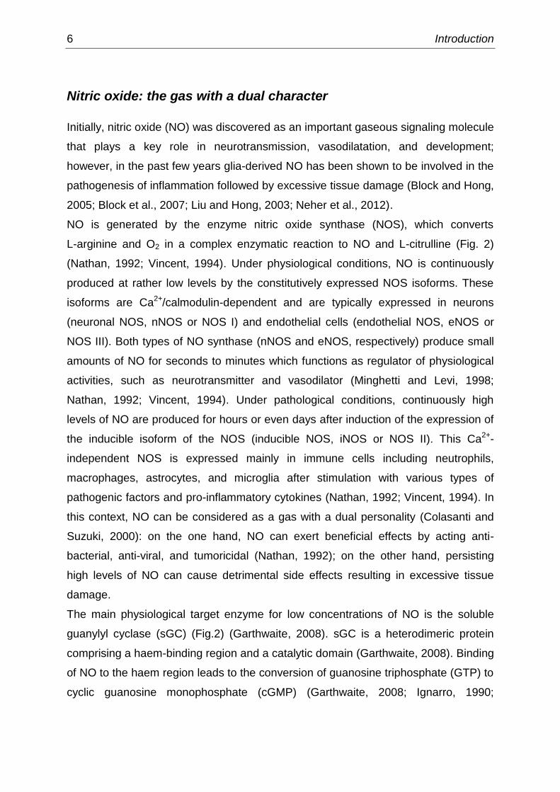
NO is generated by the enzyme nitric oxide synthase (NOS), which converts
L-arginine and O2 in a complex enzymatic reaction to NO and L-citrulline (Fig. 2)
(Nathan, 1992; Vincent, 1994). Under physiological conditions, NO is continuously
produced at rather low levels by the constitutively expressed NOS isoforms. These
isoforms are Ca2+/calmodulin-dependent and are typically expressed in neurons
(neuronal NOS, nNOS or NOS I) and endothelial cells (endothelial NOS, eNOS or
NOS III). Both types of NO synthase (nNOS and eNOS, respectively) produce small
amounts of NO for seconds to minutes which functions as regulator of physiological
activities, such as neurotransmitter and vasodilator (Minghetti and Levi, 1998;
Nathan, 1992; Vincent, 1994). Under pathological conditions, continuously high
levels of NO are produced for hours or even days after induction of the expression of
the inducible isoform of the NOS (inducible NOS, iNOS or NOS II). This Ca2+-
independent NOS is expressed mainly in immune cells including neutrophils,
macrophages, astrocytes, and microglia after stimulation with various types of
pathogenic factors and pro-inflammatory cytokines (Nathan, 1992; Vincent, 1994). In
this context, NO can be considered as a gas with a dual personality (Colasanti and
Suzuki, 2000): on the one hand, NO can exert beneficial effects by acting anti-
bacterial, anti-viral, and tumoricidal (Nathan, 1992); on the other hand, persisting
high levels of NO can cause detrimental side effects resulting in excessive tissue
damage.
The main physiological target enzyme for low concentrations of NO is the soluble
guanylyl cyclase (sGC) (Fig.2) (Garthwaite, 2008). sGC is a heterodimeric protein
comprising a haem-binding region and a catalytic domain (Garthwaite, 2008). Binding
of NO to the haem region leads to the conversion of guanosine triphosphate (GTP) to
cyclic guanosine monophosphate (cGMP) (Garthwaite, 2008; Ignarro, 1990;
Introduction 7
Moncada et al., 1989). cGMP itself activates downstream effectors such as protein
kinases G (PKG) (Hofmann et al., 2006), cyclic nucleotide gated ion channels
(CNGC) (Bender and Beavo, 2006), and cyclic nucleotide phosphodiesterases (PDE)
(Garthwaite, 2008), which in turn modulate the activity of an array of intracellular
signaling molecules, thereby regulating neurotransmission, proliferation, migration,
differentiation, axon outgrowth, and axon guidance.
Figure 2: The NO/cGMP signaling cascade. In immune cells such as microglia the inducible nitric
oxide synthase (iNOS) has been identified to synthesize high levels of NO in response to
immunological stimuli including lipopolysaccharide (LPS). Upon induction iNOS converts L-arginine
and O2 to L-citrulline and NO. NO binds to the soluble guanylyl cyclase (sGC) which leads to the
conversion of guanosine triphosphate (GTP) to cyclic guanosine monophosphate (cGMP). cGMP itself
has the potential to activate downstream effectors such as protein kinase G (PKG), cyclic nucleotide
gated ion channels (CNGC), and cyclic nucleotide phosphodiesterases (PDE), which converts cGMP
to guanosine monophosphate (GMP).
However, several studies focused on cGMP-independent mechanisms of NO’s
actions in the pathogenesis of a number of neurodegenerative diseases
(Madhusoodanan and Murad, 2007). These cGMP-independent actions include
oxidation of thiols, S-nitrosylation, and nitration of proteins which may play a role in
neuronal cell death. Moreover, NO can react with concomitantly produced superoxide
anions to form highly toxic compounds like peroxynitrite (ONOO-) (Brown and Neher,
2010; Neher et al., 2012). The presence of ONOO- then causes DNA damage,
apoptosis and consequently neuronal loss. In addition to this, neurons are
remarkably sensitive to sustained high levels of NO, causing inhibition of neuronal
respiration and subsequently depolarization and glutamate release followed by
excitotoxic death (Bal-Price and Brown, 2001; McNaught and Brown, 1998).

8 Introduction
A better understanding of the molecular mechanisms regulated by NO might provide
an important initial step for designing novel strategies to medicate detrimental side
effects such as neurodegeneration caused by the excessive release of NO.
Carbon monoxide: a novel agent to counteract inflammation?
In the last few years, carbon monoxide (CO) has been emerged as a gaseous
messenger molecule with anti-inflammatory, anti-apoptotic and cytoprotective
properties (Bach, 2005; Baranano and Snyder, 2001; Motterlini and Otterbein, 2010;
Soares and Bach, 2009). However, to date there is only quite limited knowledge
about how CO can affect cellular characteristics of reactive microglia and how CO
exert its anti-inflammatory effects on inflamed cells.
CO is generated by the rate-limiting enzyme heme oxygenase (HO) during the
physiological degradation of heme to biliverdin-IX, ferrous iron (Fe2+), and CO (Fig. 3)
(Choi and Alam, 1996; Maines, 1997; Ryter et al., 2002). Three isoforms of HO have
been characterized: an inducible isoform (HO-1) which is expressed in response to
all kinds of agents and stimuli that have the ability to cause oxidative stress, as well
as inflammogenes such as membrane components of germs (Maines, 1997;
Otterbein and Choi, 2000); a constitutively expressed isoform (HO-2) which is
responsive only to adrenal glucocorticoids (Maines, 1997) and Ca2+/calmodulin
(Boehning et al., 2004); and an elusive and poorly understood catalytically inactive
isoform (HO-3) which is thought to act in oxygen sensing (Hayashi et al., 2004).
However, there is extensive evidence suggesting HO-1 as the major key regulator of
the cytoprotective effects against oxidative stress (Keyse and Tyrrell, 1987, 1989;
Nath et al., 1992) as well as to counteract inflammation (Bach, 2005) and the
progression of neurodegeneration (Syapin, 2008). The mechanisms by which HO-1
exerts its cytoprotective and anti-inflammatory properties are not fully understood, but
implicate a functional role for each of the three catalytic by-products of heme
degradation: biliverdin-IX, Fe2+, and CO (Bach, 2005; Otterbein and Choi, 2000;
Otterbein et al., 2003). To date, there is no other enzyme identified yet that is
affected by such an impressive number of stimuli, as HO-1 does, and that mediates

Introduction 9
the therapeutic actions of other molecules, suggesting that HO-1 functioning as a
“therapeutic amplification funnel” (Fig. 3) (Bach, 2005).
Figure 3: Heme oxygenase-1 (HO-1) as a
therapeutic funnel. HO-1 can be induced
by an impressive number of stimuli amongst
others inflammatory signals, oxidative stress
agents, nitric oxide (NO), carbon monoxide
(CO), and heme. Activation of HO-1 results
in the degradation of heme to biliverdin
(which is consequently reduced to
bilirubin by the biliverdin reductase), CO,
and ferrous iron (Fe2+
). All three components
have the potential to act anti-inflammatory,
or cytoprotective. However, HO-1 has been
implicated to be the real mediator of the
cytoprotective properties. Because HO-1
mediates the actions of other molecules, the
cytoprotective enzyme has been suggested
to function as a therapeutic amplification
funnel.
In addition to its cytoprotective properties, there is increasing evidence for HO-1/CO
signaling as poor activator of the cGMP-synthesizing enzyme sGC. CO stimulates
sGC at ~100-fold lower efficiency than NO, suggesting that the presence of
exogenous CO can modulate the outcomes of NO signaling (Baranano and Snyder,
2001; Kharitonov et al., 1995; Knipp and Bicker, 2009).
A better understanding about how HO-1/CO signaling could develop its
cytoprotective properties on inflamed microglia might provide important insights in
regulating neurodegenerative processes in the nervous system.
Feedback between nitric oxide and carbon monoxide generation
It is becoming increasingly evident that iNOS/NO and HO-1/CO signaling are tightly
linked, mutually affect the induction of each other, and share many of their chemical

10 Introduction
properties (Maines, 1997). The fact, that NO causes HO-1 up-regulation while in
parallel HO-1 induction resulted in the down-regulation of iNOS, led various scientists
to investigate the interrelationship of NO and CO under inflammatory conditions
(Fig. 4). Inflammation is usually accompanied by the induction of iNOS via the
nuclear factor NF-ƙB (nuclear factor kappa-light-chain-enhancer of activated B cells)
and subsequently the generation of NO (Lee et al., 2004; Wang et al., 2002). NO
itself causes oxidative stress, which has been implicated in the activation of the
transcription factor Nrf2 (nuclear factor-erythroid 2-related factor 2) (Terazawa et al.,
2013). Nrf2 regulates the expression of various cytoprotective genes including HO-1,
leading to an enhanced HO-1 induction (Itoh et al., 1997). In addition to Nrf2, NF-ƙB
is an important component of HO-1 induction in response to a diversity of pro-
inflammatory stimuli (Lee and Suk, 2007; Lu et al., 2010).
Figure 4: Feedback between nitric oxide (NO) and carbon monoxide (CO) generation.
Lipopolysaccharide (LPS) induces inducible NO synthase (iNOS) expression and NO production via
NF-ƙB (nuclear factor kappa-light-chain-enhancer of activated B cells). NO itself causes the activation
of Nrf2 (nuclear factor-erythroid 2-related factor 2) which regulates the expression of the heme
oxygenase-1 (HO-1). In parallel, LPS activates HO-1 via NF-ƙB. Induction of HO-1 leads to the
degradation of heme to ferrous iron (Fe2+
), CO and Biliverdin (which is converted to Bilirubin) with all
three by-products having the capacity to block iNOS expression and subsequently to reduce NO
production.
Introduction 11
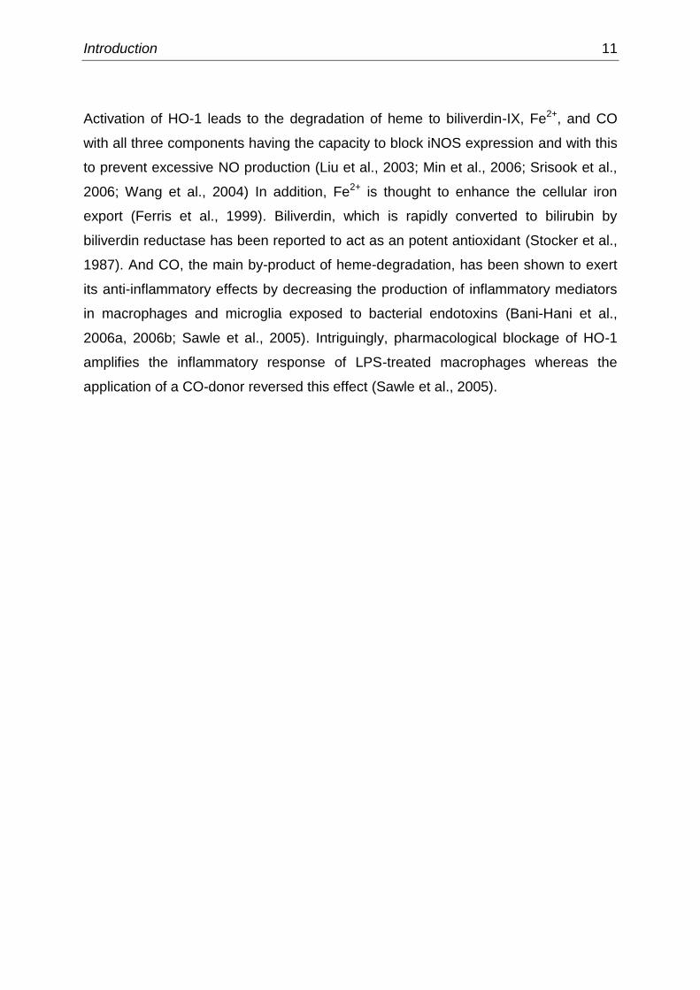
Activation of HO-1 leads to the degradation of heme to biliverdin-IX, Fe2+, and CO
with all three components having the capacity to block iNOS expression and with this
to prevent excessive NO production (Liu et al., 2003; Min et al., 2006; Srisook et al.,
2006; Wang et al., 2004) In addition, Fe2+ is thought to enhance the cellular iron
export (Ferris et al., 1999). Biliverdin, which is rapidly converted to bilirubin by
biliverdin reductase has been reported to act as an potent antioxidant (Stocker et al.,
1987). And CO, the main by-product of heme-degradation, has been shown to exert
its anti-inflammatory effects by decreasing the production of inflammatory mediators
in macrophages and microglia exposed to bacterial endotoxins (Bani-Hani et al.,
2006a, 2006b; Sawle et al., 2005). Intriguingly, pharmacological blockage of HO-1
amplifies the inflammatory response of LPS-treated macrophages whereas the
application of a CO-donor reversed this effect (Sawle et al., 2005).

12 Aims of the dissertation
Aims of the dissertation
Inflammation within the CNS is usually characterized by the activation of microglia
and has been closely associated with the pathogenesis of several neurological
disorders with a neurodegenerative component. Microglia play a dual role in
mediating the immune response after infections or injuries. Depending on their
activation state, they are either able to amplify inflammatory conditions by the release
of pro-inflammatory molecules, or to protect the neuronal tissue by the release of
anti-inflammatory cytokines and the engulfment of infected cells. Insights into
mechanisms of activation, migration, phagocytosis, and neuron-interaction may help
to pinpoint microglia as a key pharmacological target in the treatment of
neurodegeneration.
Specific aims of this thesis were:
(1) to introduce the murine microglial cell line BV-2 as a useful model system for
primary microglia
(2) to develop an in vitro cell migration assay for the investigation whether
NO/cGMP signaling could modulate microglial movement and to test for
potential interactions with the HO-1/CO signaling cascade
(3) to develop an in vitro phagocytosis assay with microglia and human model
neurons to quantify possible anti-inflammatory and cytoprotective properties of
the HO-1/CO cascade and to determine the effects of NO signaling
(4) to devise a suitable co-culture assay to investigate potential crosstalk between
microglial activation, pharmacological manipulation of activated microglia, and
its impact on neurite outgrowth of developing neurons
(5) and to elucidate the mechanisms contributing to these effects
Publications 13
Publications
This thesis was prepared as a cumulative dissertation comprising four original
publications. Three of these publications are first-authored. For a better
understanding, articles are not presented in their chronological order of publication
date.
Authors’ contributions
1) Scheiblich H, Roloff F, Singh V, Stangel M, Stern M, Bicker G (2014) Nitric
oxide / cyclic GMP regulates motility of a microglial cell line and primary
microglia, Brain Research 1564: 9-21. DOI: 10.1016/j.brainres.2014.03.048
designed experiments: HS, MS, GB; performed experiments: HS; isolated
primary microglia: VS; analyzed data: HS, GB; wrote the article: HS (with input
from GB); corrected and improved the manuscript: all authors
Some data contributing to this publication have already been collected during my
masters’ thesis (2012) entitled “Zellmigrations-Assay an primären Mikroglia und der
mikroglialen Zelllinie BV-2” at the University of Veterinary Medicine Hannover,
Germany.
2) Scheiblich H, Bicker G (2015) Nitric oxide regulates antagonistically
phagocytic and neurite outgrowth inhibiting capacities of microglia,
Developmental Neurobiology, accepted. DOI: 10.1002/dneu.22333
designed experiments: HS, GB; performed experiments: HS; analyzed data:
HS, GB; wrote the article: HS (with input from GB); corrected and improved
the manuscript: all authors

14 Publications
3) Scheiblich H, Bicker G (2015) Regulation of microglial migration,
phagocytosis, and neurite outgrowth by HO-1/CO signaling,
Developmental Neurobiology 75(8): 854-876. DOI: 10.1002/dneu.22253
designed experiments: HS, GB; performed experiments: HS; analyzed data:
HS, GB; wrote the article: HS (with input from GB); corrected and improved
the manuscript: all authors
4) Roloff F, Scheiblich H, Dewitz C, Dempewolf S, Stern M, Bicker G (2015)
Enhanced neurite outgrowth of human model (NT2) neurons by small-
molecule inhibitors of Rho/ROCK signaling, PLoS ONE 10(2): e0118536.
DOI: 10.1371/journal.pone.0118536
designed experiments: FR, GB; performed experiments: FR, HS (RhoA pull-
down activation assay and western blot analysis), CD, SD; analyzed data: FR,
HS (RhoA pull-down activation assay and western blot analysis), CD, SD, GB;
wrote the article: FR (with input from GB); corrected and improved the
manuscript: all authors
Publications 15
Publication 1
Nitric oxide / cyclic GMP regulates motility of a microglial cell line
and primary microglia
Hannah Scheiblich, Frank Roloff, Vikramjeet Singh, Martin Stangel, Michael Stern,
Gerd Bicker (2014) Nitric oxide / cyclic GMP regulates motility of a microglial
cell line and primary microglia. Brain Research 1564: 9-21.
http://www.sciencedirect.com/science/article/pii/S0006899314004521
DOI: 10.1016/j.brainres.2014.03.048
Abstract
Microglia are the resident immune cells of the brain, which become rapidly activated and
migrate to the site of insult in brain infection and disease. Activated microglia generate large
amounts of the highly reactive messenger molecule nitric oxide (NO). NO is able to raise
cyclic GMP levels via binding to soluble guanylyl cyclase. We investigated potential
mechanistic links between inflammation, NO signaling, and microglial migration. To monitor
cell migration, we used a scratch wound assay and compared results obtained in the BV-2
microglial line to primary microglia. Incubation with lipopolysaccharide (LPS) as stimulator of
acute inflammatory processes enhanced migration of both microglial cell types. LPS
activated NO production in BV-2 cells and application of an NO donor increased BV-2 cell
migration while an NO scavenger reduced motility. Pharmacological inhibition of soluble
guanylyl cyclase and the resulting decrease in motility can be rescued by a membrane
permeant analog of cGMP. Despite differences in the threshold towards stimulation with the
chemical agents, both BV-2 cells and primary microglia react in a similar way. The important
role of NO/cGMP as positive regulator of microglial migration, the downstream targets of the
signaling cascade, and resulting cytoskeletal changes can be conveniently investigated in a
microglial cell line.

16 Publications
Publication 2
Nitric oxide regulates antagonistically phagocytic and neurite
outgrowth inhibiting capacities of microglia
Hannah Scheiblich, Gerd Bicker (2015) Nitric oxide regulates antagonistically
phagocytic and neurite outgrowth inhibiting capacities of microglia.
Developmental Neurobiology, accepted for publication.
http://onlinelibrary.wiley.com/doi/10.1002/dneu.22333/abstract
DOI: 10.1002/dneu.22333
Abstract
Traumatic injury or the pathogenesis of some neurological disorders is accompanied by
inflammatory cellular mechanisms, mainly resulting from the activation of CNS resident
microglia. Under inflammatory conditions, microglia upregulate the inducible isoform of NOS
(iNOS), leading to the production of high concentrations of the radical molecule nitric oxide
(NO). At the onset of inflammation, high levels of microglial-derived NO may serve as a
cellular defense mechanism helping to clear the damaged tissue and combat infection of the
CNS by invading pathogens. However, the excessive overproduction of NO by activated
microglia has been suggested to govern the inflammation-mediated neuronal loss causing
eventually complete neurodegeneration.
Here, we investigated how NO influences phagocytosis of neuronal debris by BV-2 microglia,
and how neurite outgrowth of human NT2 model neurons is affected by microglial-derived
NO. The presence of NO greatly increased microglial phagocytic capacity in a model of acute
inflammation comprising lipopolysaccharide (LPS)-activated microglia and apoptotic neurons.
Chemical manipulations suggested that NO upregulates phagocytosis independently of the
sGC/cGMP pathway. Using a transwell system, we showed that reactive microglia inhibit
neurite outgrowth of human neurons via the generation of large amounts of NO over effective
distances in the millimeter range. Application of a NOS blocker prevented the LPS-induced
NO production, totally reversed the inhibitory effect of microglia on neurite outgrowth, but
reduced the engulfment of neuronal debris. Our results indicate that a rather simple notion of
treating excessive inflammation in the CNS by NO synthesis blocking agents has to consider
functionally antagonistic microglial cell responses during pharmaceutic therapy.

Publications 17
Publication 3
Regulation of microglial migration, phagocytosis and neurite
outgrowth by HO-1/CO signaling
Hannah Scheiblich, Gerd Bicker (2015) Regulation of microglial migration,
phagocytosis and neurite outgrowth by HO-1/CO signaling. Developmental
Neurobiology 75(8): 854-876.
http://onlinelibrary.wiley.com/doi/10.1002/dneu.22253/abstract
DOI: 10.1002/dneu.22253
Abstract
Clearance of infected and apoptotic neuronal corpses during inflammatory conditions is a
fundamental process to create a favorable environment for neuronal recovery. Microglia are
the resident immune cells and the predominant phagocytic cells of the CNS, showing a
multitude of cellular responses upon activation. Here, we investigated in functional assays
how the CO generating enzyme heme oxygenase 1 (HO-1) influences BV-2 microglial
migration, clearance of debris, and neurite outgrowth of human NT2 neurons. Stimulation of
HO-1 activity attenuated microglial migration in a scratch wound assay, and phagocytosis in
a cell culture model of acute inflammation comprising lipopolysaccharide (LPS)-activated
microglia and apoptosis-induced neurons. Application of a CO donor prevented the
production of NO during LPS stimulation, and reduced microglial migration and engulfment of
neuronal debris. LPS-activated microglia inhibited neurite elongation of human neurons
without requiring direct cell-cell surface contact. The inhibition of neurite outgrowth was
totally reversed by application of exogenous CO or increased internal CO production through
supply of the substrate hemin to HO. Our results point towards a vital cytoprotective role of
HO-1/CO signaling after microglial activation. In addition, they support a therapeutic potential
of CO releasing chemical agents in the treatment of excessive inflammatory conditions in the
CNS.

18 Publications
Publication 4
Enhanced neurite outgrowth of human model (NT2) neurons by
small-molecule inhibitors of Rho/ROCK signaling
Frank Roloff, Hannah Scheiblich, Carola Dewitz, Silke Dempewolf, Michael Stern,
Gerd Bicker (2015) Enhanced neurite outgrowth of human model (NT2) neurons
by small-molecule inhibitors of Rho/ROCK signaling. PLoS ONE 10(2):
e0118536.
http://journals.plos.org/plosone/article?id=10.1371/journal.pone.0118536
DOI: 10.1371/journal.pone.0118536
Abstract
Axonal injury in the adult human central nervous system often results in loss of sensation
and motor functions. Promoting regeneration of severed axons requires the inactivation of
growth inhibitory influences from the tissue environment and stimulation of the neuron
intrinsic growth potential. Especially glial cell derived factors, such as chondroitin sulfate
proteoglycans, Nogo-A, myelin-associated glycoprotein, and myelin in general, prevent axon
regeneration. Most of the glial growth inhibiting factors converge onto the Rho/ROCK
signaling pathway in neurons. Although conditions in the injured nervous system are clearly
different from those during neurite outgrowth in vitro, here we use a chemical approach to
manipulate Rho/ROCK signalling with small-molecule agents to encourage neurite outgrowth
in cell culture. The development of therapeutic treatments requires drug testing not only on
neurons of experimental animals, but also on human neurons. Using human NT2 model
neurons, we demonstrate that the pain reliever Ibuprofen decreases RhoA (Ras homolog
gene family, member A GTPase) activation and promotes neurite growth. Inhibition of the
downstream effector Rho kinase by the drug Y-27632 results in a strong increase in neurite
outgrowth. Conversely, activation of the Rho pathway by lysophosphatidic acid results in
growth cone collapse and eventually to neurite retraction. Finally, we show that blocking of
Rho kinase, but not RhoA results in an increase in neurons bearing neurites. Due to its anti-
inflammatory and neurite growth promoting action, the use of a pharmacological treatment of
damaged neural tissue with Ibuprofen should be explored.

Discussion 19
Discussion
This thesis provides a comprehensive overview how the two gaseous messengers,
NO and CO, influence certain aspects of microglia biology. For reasons of
importance to medical treatment of neurodegenerative processes, I took advantage
of performing experimental manipulations of the NO and CO signaling cascades in
cell culture assays. I focused on the possibility to control different characteristics of
microglia under basal conditions and after LPS-induced activation. Moreover,
molecular mechanisms contributing to the detrimental effects of activated microglia
could be elucidated.
Key findings of this work are:
the BV-2 microglial cell line is a useful model for primary microglia in assays
where robust microglial cell behavior is required
NO/cGMP signaling positively regulates migration of primary microglia and
BV-2 cells and causes cytoskeletal changes in both cell types
migratory response of microglia is dually regulated by antagonistic interacting
iNOS/NO/cGMP and HO-1/CO/cGMP signal transduction pathways
NO signaling enhances phagocytic activity of microglia in a co-culture system
with apoptosis-induced human NT2 (Ntera2/D1 precursor cells) model
neurons
stimulation of HO-1/CO signaling prevents the LPS-induced NO production
and may thus be a powerful modulator of inflammation-activated microglia
chemical manipulation of HO-1/CO signaling down-regulates inflammation-
mediated microglial phagocytosis and represent an important initial step for
designing novel CO-donor based strategies to medicate excessive
inflammation in the nervous system

20 Discussion
LPS-activated microglia inhibit neurite elongation of human neurons without
required cell-cell surface contact via the release of NO
stimulation of HO-1/CO signaling and inhibition of iNOS induction totally
reverse the detrimental effects of LPS-activated microglia on neurite outgrowth
of the model neurons and may thus be helpful to facilitate neurite outgrowth
for the repair of injured neural connections
in vitro treatment of developing model neurons with RhoA/ROCK inhibiting
agents greatly increase neurite outgrowth, indicating that the regulation is
caused downstream of the NO/cGMP pathway
The following discussion will focus on these outcomes and provide some
perspectives for future experiments.

Discussion 21
BV-2 cells as model system for primary microglia
The isolation of primary microglia from rat brains is a time consuming procedure
yielding rather low cell numbers with a limited proliferation capacity for in vitro
culturing. The use of a microglial cell line might thus serve as a potential method to
replace in vivo experiments and to reduce the high impact of animal consumption in
research (Henn et al., 2009). Most work on microglial activation, cell signaling, and
function has thus been performed using rapidly proliferating microglial cell lines such
as N9 (Corradin et al., 1993) and BV-2 (Blasi et al., 1990).
In this study, I used the in vitro scratch wound assay to introduce the immortalized
murine microglial cell line BV-2 (Blasi et al., 1990) as a valid substitute for primary
microglia (Scheiblich et al., 2014). Cell migration behavior of BV-2 cells versus
primary microglia was analyzed by chemical manipulation of the NO/cGMP signaling
cascade (Fig. 2). I found, that despite differences in the threshold towards stimulation
with the chemical agents, both BV-2 cells and primary microglia reacted in a similar
way upon manipulation (Scheiblich et al., 2014). Moreover, the reaction patterns of
our two microglial cell types were in line with several other cell culture and in vivo
studies investigating the effects of NO/cGMP on microglial cell motility upon chemical
manipulation (Chen et al., 2000; Dibaj et al., 2010; Duan et al., 2009; Haynes et al.,
2006; Ohsawa et al., 2007). In addition to their migration behavior, I found that BV-2
cells release NO time- and dose-dependently upon LPS challenge similar than
primary microglia (Blasi et al., 1990; Henn et al., 2009; Horvath et al., 2008; Stansley
et al., 2012). In another part of this approach, I showed that BV-2 cells have the
potential to engulf apoptosis-induced neurons and to influence neurite outgrowth of
human NT2 model neurons (Scheiblich and Bicker, 2015a, 2015b). Nevertheless, for
a straightforward comparison of both cell types, it should be kept in mind that primary
microglia are a heterogeneous population isolated from different brain regions with
diverse reaction patterns, whereas BV-2 cells perform more uniformly (see
discussion in Scheiblich et al., 2014).
However, the robust response in our in vitro assays, the cellular homogeneity, and
the unlimited proliferation potential make the BV-2 microglial cell line an ideal model
for the investigation of microglial functions during acute inflammatory conditions.
22 Discussion
Microglial activation: why could carbon monoxide be beneficial for
the inflamed brain?
One of the first responses after a pathological event is the activation of microglia
which are prepared to recognize and discriminate between a wide range of
pathogenic stimuli with a high level of fine-tuned responsiveness (Hanisch and
Kettenmann, 2007). In general, microglial activation is thought to be protective to the
brain parenchyma. However, many studies have concluded that detrimental side
effects may eventuate in collateral damage following excessive periods of microglial
activation (Bal-Price and Brown, 2001; Banati et al., 1993; Bolaños et al., 1997). The
mechanisms by which reactive microglia drive neurodegenerative processes are only
partly known, but implicate the release of microglia-derived NO (Bal-Price and
Brown, 2001; Bolaños et al., 1997; Chao et al., 1996; Loihl and Murphy, 1998).
In line with previous studies (Arimoto and Bing, 2003; Boje and Arora, 1992; Brown,
2007; Hunot et al., 1996), results of the present thesis support evidence that
microglial activation, the induction of iNOS and the sustained release of NO might
potentially play a role in regulating neurodegenerative conditions. I found that
treatment of cultured microglia with the inflammogen LPS not only increased the
protein expression of iNOS and subsequently the microglia-derived NO production
(Fig. 5), but also up-regulated cellular characteristics of microglia, such as cell
migration, phagocytic activity, and neurite growth retarding effects on adjacent
neurons (Scheiblich and Bicker, 2015a, 2015b; Scheiblich et al., 2014). By contrast,
inhibition of iNOS expression and NO production provided more neuroprotective
effects by down-regulating these cellular characteristics (Scheiblich and Bicker,
2015b).
During the last decades the CO generating enzyme HO-1 has been emerged as a
novel target to counteract inflammation and the progression of neurodegenerative
diseases (Baranano and Snyder, 2001; Motterlini and Otterbein, 2010; Soares and
Bach, 2009; Syapin, 2008). I found, that in LPS-activated microglia, the chemical
manipulation of the HO-1 signaling cascade inhibited the induction of the iNOS
protein expression and subsequently decreased the NO production (Fig. 5)
(Scheiblich and Bicker, 2015a). In addition, chemical manipulation of HO-1/CO

Discussion 23
signaling down-regulated cellular characteristics of reactive microglia including cell
migration, phagocytic activity, and the retarding effects on neurite outgrowth of co-
cultured neurons (Scheiblich and Bicker, 2015a). Intriguingly, treatment with LPS or
exogenous NO induced the expression of HO-1 (Scheiblich and Bicker, 2015a;
Terazawa et al., 2013), suggesting reciprocal interactions between iNOS and HO-1
mediated signal transduction (Fig. 5). In support of our observations, it has been
already demonstrated that all three catalytic by-product (biliverdin, Fe2+, and CO)
arisen from HO-1 degradation of heme have the potential to block iNOS expression
and subsequently NO production (Liu et al., 2003; Min et al., 2006; Srisook et al.,
2006; Wang et al., 2004). Nevertheless, CO seems to be the main inhibitor of iNOS
expression and NO generation since CO has the potential to bind to the heme group
of iNOS and with that to inhibit the electron transfer reaction which is required for the
production of NO (Turcanu et al., 1998a, 1998b).
Figure 5: Interaction between nitric oxide (NO) and carbon monoxide (CO) signaling in
microglia. Chemical manipulation of HO-1/CO signal transduction suggests reciprocal interactions
with iNOS/NO signaling. Upon detecting inflammogenes such as lipopolysaccharide (LPS) microglial
inducible NO synthase (iNOS) synthesize high levels of NO. Similar to iNOS, heme oxygenase-1
(HO-1) is induced by LPS to produce CO. Moreover, HO-1 can be activated by application of the HO-
substrate hemin. The presence of CO markedly decreases iNOS induction and subsequently NO
production. Representative western blots of microglia treated for 24 h with different chemical
compounds interfering with NO and CO signal transduction revealed our findings.
Findings of the present work support a therapeutic potential of NO synthesis blocking
agents and CO-releasing molecules (CORMs) to medicate excessive inflammation in

24 Discussion
the nervous system. However, for a straightforward statement one must ask whether
LPS-induced microglial activation in culture accurately reflect microglial functions in
the intact nervous system (Hanisch and Kettenmann, 2007). LPS stimulation has
long been considered as the gold standard for microglial activation, but stimulation
with the bacterial endotoxin results in a massive all-or-none defense reaction. In the
CNS the precise nature of microglial activation is usually context dependent and
implicates the adaptation to even tiny environmental changes via many sub-states on
the way from resting to phagocytic microglia. Thus, the development of suitable
therapies requires further investigation in relevant in vitro and in vivo models of
inflammation-mediated neurodegeneration.
Reciprocity of nitric oxide and carbon monoxide in regulating
microglial cell migration
Upon activation microglial migration from their resting location in the CNS to the site
of insult is an essential component of the cellular response under pathological
conditions (Chen et al., 2000; Dibaj et al., 2010; Duan et al., 2009; Kreutzberg,
1996). A series of studies recently addressed various molecules initiating microglial
movement in abnormal concentrations including adenosine triphosphate (ATP) (Duan
et al., 2009), cannabinoids (Walter et al., 2003), chemokines (Cross and Woodroofe,
1999), and lysophosphatidic acid (LPA) (Schilling et al., 2004).
We and others found that NO released by degenerating neurons and activated
microglia itself acts as a modulator and chemoattractant to microglial migration
resulting in the recruitment of the cells toward the insult (Chen et al., 2000; Dibaj et
al., 2010; Duan et al., 2009; Haynes et al., 2006; Ohsawa et al., 2007; Scheiblich et
al., 2014). Interestingly, low concentrations of NO enhanced the movement of
migrating microglia while high concentrations acted as a stop signal to may cause
accumulation at the lesion site. Application of small bioactive enzyme activators and
inhibitors of the NO/cGMP signaling cascade indicated that NO positively modulates
migration of microglia via cGMP (Fig. 6) (Scheiblich et al., 2014). However, the
outcome of cellular signal transduction is complex because multiple signaling
Discussion 25
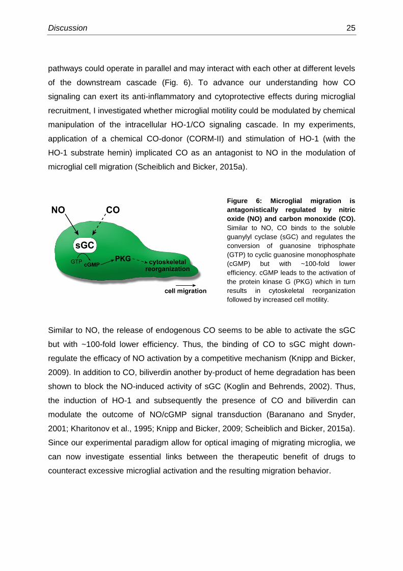
pathways could operate in parallel and may interact with each other at different levels
of the downstream cascade (Fig. 6). To advance our understanding how CO
signaling can exert its anti-inflammatory and cytoprotective effects during microglial
recruitment, I investigated whether microglial motility could be modulated by chemical
manipulation of the intracellular HO-1/CO signaling cascade. In my experiments,
application of a chemical CO-donor (CORM-II) and stimulation of HO-1 (with the
HO-1 substrate hemin) implicated CO as an antagonist to NO in the modulation of
microglial cell migration (Scheiblich and Bicker, 2015a).
Figure 6: Microglial migration is
antagonistically regulated by nitric
oxide (NO) and carbon monoxide (CO).
Similar to NO, CO binds to the soluble
guanylyl cyclase (sGC) and regulates the
conversion of guanosine triphosphate
(GTP) to cyclic guanosine monophosphate
(cGMP) but with ~100-fold lower
efficiency. cGMP leads to the activation of
the protein kinase G (PKG) which in turn
results in cytoskeletal reorganization
followed by increased cell motility.
Similar to NO, the release of endogenous CO seems to be able to activate the sGC
but with ~100-fold lower efficiency. Thus, the binding of CO to sGC might down-
regulate the efficacy of NO activation by a competitive mechanism (Knipp and Bicker,
2009). In addition to CO, biliverdin another by-product of heme degradation has been
shown to block the NO-induced activity of sGC (Koglin and Behrends, 2002). Thus,
the induction of HO-1 and subsequently the presence of CO and biliverdin can
modulate the outcome of NO/cGMP signal transduction (Baranano and Snyder,
2001; Kharitonov et al., 1995; Knipp and Bicker, 2009; Scheiblich and Bicker, 2015a).
Since our experimental paradigm allow for optical imaging of migrating microglia, we
can now investigate essential links between the therapeutic benefit of drugs to
counteract excessive microglial activation and the resulting migration behavior.

26 Discussion
Antagonistic regulation of microglial phagocytosis by nitric oxide
and carbon monoxide
A key function of microglia upon reaching the inflammatory epicenter, but also in
non-inflammatory environments is debris clearance (Neumann et al., 2009), including
phagocytosis of apoptotic neurons. However, the inflammation-related microglia-
mediated excessive uptake of healthy neurons is considered to be a characteristic
feature in the onset and progression of neurodegenerative processes (Glass et al.,
2010; Lucin and Wyss-Coray, 2009). In general, microglial phagocytosis has been
summarized in a three-step model: find-me, eat-me, and digest-me (for review see:
Savill et al., 2002; Sierra et al., 2013). As mentioned earlier in this thesis, the first
step (‘find-me‘) is triggered by signals released from damaged neurons including NO
to attract microglia (see discussion in Scheiblich et al., 2014). The second step (‘eat-
me’) includes the recognition and engulfment of the apoptotic cell (see discussion in
Scheiblich and Bicker, 2015a, 2015b), triggered by the interaction of microglial
receptors and their ligands on the membrane of the target cell, such as
phosphatidylserine (PS) (Neher et al., 2012). The third step (‘digest-me’) implicates
the full degradation of the apoptotic cell in the phagolysosome (Sierra et al., 2013).
The mechanisms regulating the engulfment of healthy neurons by inflamed microglia
are not completely understood but implicate a significant role for NO (for review see:
Block and Hong, 2005; Brown, 2007). In line with this, results from the present thesis
argue for a regulating impact of NO as enhancer of microglial phagocytosis (Fig. 7)
(Scheiblich and Bicker, 2015b). I found that beside LPS-stimulated microglia,
microglia challenged with exogenous NO increased their NO production via iNOS
induction. The presence of NO enhanced the phagocytic activity through
self-stimulation independent of the source of NO (Scheiblich and Bicker, 2015b).
Strong evidence has accumulated that the activation of microglia through Toll-like
receptor 4 results in the induction of iNOS and the assembly of the phagocytic
oxidase (PHOX), resulting in the production of NO and superoxide (O2-), respectively
(Brown and Neher, 2010; Neher et al., 2012). NO and O2- subsequently react to form
ONOO- which causes apoptosis and consequently a disrupted lipid asymmetry of the
plasma membrane followed by the externalization of PS to the outer membrane

Discussion 27
leaflet of neurons (Neher et al., 2012). The process which is known as scrambling
has been considered a characteristic feature of apoptosis and requires the activity of
a membrane protein called scramblase (for review see: Mariño and Kroemer, 2013).
Xk-related protein 8 (Xkr8) has recently been identified as the enzyme responsible
for the apoptosis-induced lipid scrambling (Suzuki et al., 2013). The mechanisms of
apoptosis-induced PS exposure is poorly understood, but implicated the presence of
caspase recognition sites of Xkr8 and caspase cleavage. Since ONOO--induced
apoptosis has been reported to activate several caspases (Zhuang and Simon,
2000), it is likely that similar mechanisms regulate microglia-mediated PS exposure
of adjacent and otherwise healthy neurons. The exposure of PS to the outer
membrane leaflet of neurons is then recognized by microglia as an eat-me signal,
thereby causing engulfment (Neher et al., 2012). Inhibition of iNOS and PHOX
prevented the formation of ONOO-, exposure of PS and phagocytosis (Kumar et al.,
2014; Mander and Brown, 2005; Scheiblich and Bicker, 2015b).
Figure 7: Nitric oxide (NO)-mediated microglial (green) phagocytosis of neuronal (magenta)
debris is down-regulated by the induction of heme oxygenase-1 (HO-1) / carbon monoxide (CO)
signaling. Detection of inflammogenes such as lipopolysaccharide (LPS) caused the increased
expression of the inducible NO synthase (iNOS) and activation of the phagocytic oxidase to produce
NO and superoxide (O2-), respectively. NO and O2
- subsequently react to form peroxynitrite (ONOO
-)
leading to the exposure of phosphatidylserine (PS) in neurons. Recognition of PS on the outer
membrane leaflet of neurons and binding to the microglial phosphatidylserine-receptor (PS-R) results
in the engulfment of the neuron. Induction of HO-1/CO signaling inhibits LPS-induced iNOS induction
and NO production resulting in a blocked phagocytic activity.

28 Discussion
Moreover, to the best of current knowledge, it was shown for the first time that the
phagocytosis enhancing effect of microglia activation could be overcome by the
induction of HO-1/CO signaling and treatment with a CO-releasing molecule (CORM)
(Scheiblich and Bicker, 2015a). Since HO-1/CO signaling effectively prevented LPS-
induced iNOS expression and subsequently NO production of microglia, my results
suggest a functional antagonism between NO and CO signaling in the regulation of
microglial phagocytosis (Fig. 7).
However, beside its detrimental side effects NO might aid in the removal of already
damaged cells and in initiating repair mechanisms under injured conditions. There is
therefore the need to carefully evaluate the exact time frame in which NO acts
beneficial by enhancing the clearance of wounded nervous tissue to promote
functional recovery, and in which NO exerts more detrimental side effects such as
excessive inflammation and loss of healthy neurons.
Neurite outgrowth of human neurons is regulated by microglia
Traumatic damage to the nervous system including spinal cord injury (SCI) causes
extensive inflammation and the invasion of microglia in and around the affected
regions. Previous work has shown, that activation and accumulation of microglia
around the epicenter of the lesion is thought to be involved in the quite limited axonal
recovery of the CNS (Kitayama et al., 2011; Popovich et al., 1999). The molecular
mechanisms blocking neurite regrowth and regeneration are not completely
understood but they implicate microglia-derived substances acting as axon growth
inhibitors.
Results of the present thesis support evidence that activation of microglia attenuate
neurite outgrowth of co-cultured neurons without direct cell-cell surface contact.
Thus, we hypothesized that microglia-derived secreted molecules must be
responsible for the poor axonal elongation (Fig. 8 A). By performing experimental
manipulations I found that the induction of microglial iNOS and the presence of NO
were responsible for blocking neurite growth following growth cone collapse
(Scheiblich and Bicker, 2015b). Intriguingly, inhibition of iNOS/NO production via

Discussion 29
induction of HO-1/CO signaling not only attenuated the inflammatory response of
reactive microglia (see discussion before) but also allowed for neurite outgrowth
(Scheiblich and Bicker, 2015a). In support of these observations we found, that the
regulation of neurite outgrowth is caused downstream the NO pathway (Fig. 8 B)
(Roloff et al., 2015).
Figure 8: Neurite outgrowth of human model neurons (magenta) is regulated by microglia
(green). (A) Microglial activation result in the induction of the inducible nitric oxide synthase (iNOS)
and subsequently in the release of nitric oxide (NO). The presence of NO blocks neurite outgrowth of
the neurons. However, induction of heme oxygenase-1 (HO-1) and the release of carbon monoxide
(CO) rescue the detrimental effects of microglia-derived NO on neurite outgrowth. (B) Sine both, NO
and CO, have the potential to bind to the soluble guanylyl cyclase (sGC) and thus to regulate the
conversion of guanosine triphosphate (GTP) to cyclic guanosine monophosphate (cGMP) by the
protein kinase G (PKG) it is likely that considerable parts of neurite growth are regulated downstream
the sGC/cGMP cascade. In support of this, treatment with the commercial pain reliever Ibuprofen
(RhoA (Ras homolog gene family, member A GTPase) and Rho kinase ROCK (Rho-associated coiled
coil forming protein serine/threonine kinase) inhibiting agent) greatly increased neurite length of the
human model neurons while activation of RhoA/ROCK result in growth cone collapse followed by the
inhibition of neurite growth.
We and others revealed a pivotal role for RhoA (Ras homolog gene family, member
A GTPase) and the Rho kinase ROCK (Rho-associated coiled coil forming protein
serine/threonine kinase) signaling on neurite outgrowth of neurons (Boomkamp et al.,
2012; DeGeer and Lamarche-Vane, 2013; Tönges et al., 2011; Wu et al., 2009).
Treatment with RhoA/ROCK inhibiting agents increased neurite length of the human
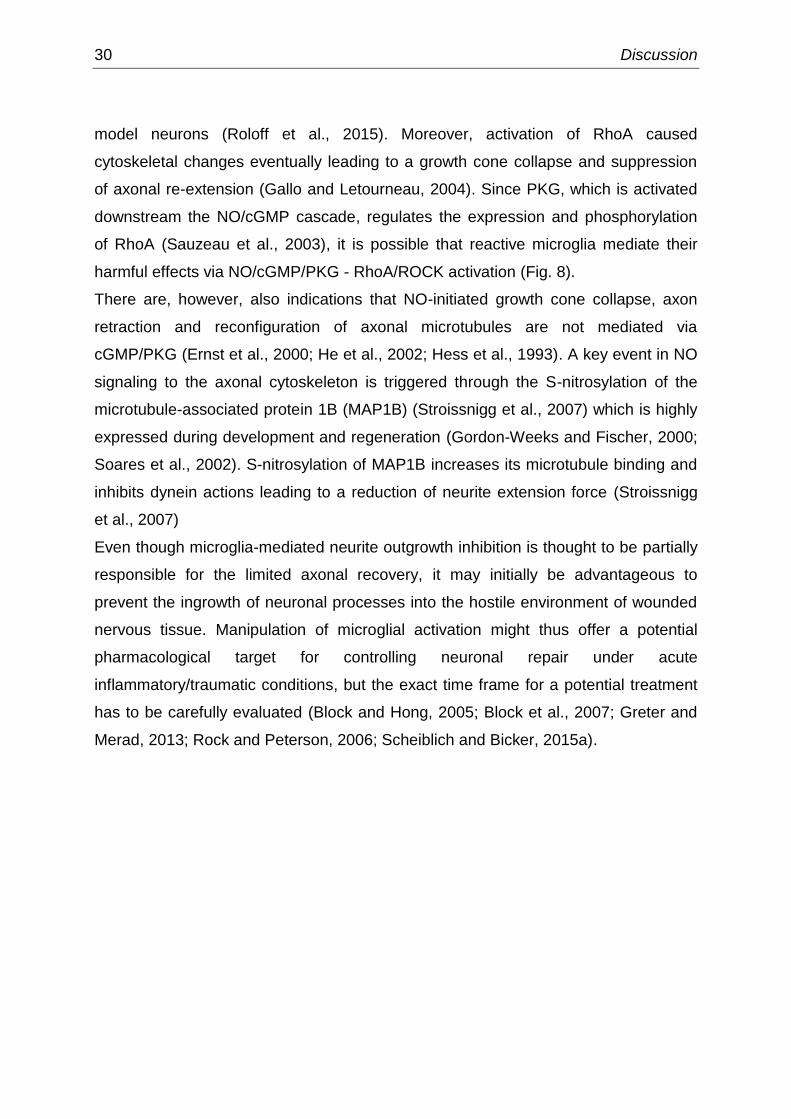
30 Discussion
model neurons (Roloff et al., 2015). Moreover, activation of RhoA caused
cytoskeletal changes eventually leading to a growth cone collapse and suppression
of axonal re-extension (Gallo and Letourneau, 2004). Since PKG, which is activated
downstream the NO/cGMP cascade, regulates the expression and phosphorylation
of RhoA (Sauzeau et al., 2003), it is possible that reactive microglia mediate their
harmful effects via NO/cGMP/PKG - RhoA/ROCK activation (Fig. 8).
There are, however, also indications that NO-initiated growth cone collapse, axon
retraction and reconfiguration of axonal microtubules are not mediated via
cGMP/PKG (Ernst et al., 2000; He et al., 2002; Hess et al., 1993). A key event in NO
signaling to the axonal cytoskeleton is triggered through the S-nitrosylation of the
microtubule-associated protein 1B (MAP1B) (Stroissnigg et al., 2007) which is highly
expressed during development and regeneration (Gordon-Weeks and Fischer, 2000;
Soares et al., 2002). S-nitrosylation of MAP1B increases its microtubule binding and
inhibits dynein actions leading to a reduction of neurite extension force (Stroissnigg
et al., 2007)
Even though microglia-mediated neurite outgrowth inhibition is thought to be partially
responsible for the limited axonal recovery, it may initially be advantageous to
prevent the ingrowth of neuronal processes into the hostile environment of wounded
nervous tissue. Manipulation of microglial activation might thus offer a potential
pharmacological target for controlling neuronal repair under acute
inflammatory/traumatic conditions, but the exact time frame for a potential treatment
has to be carefully evaluated (Block and Hong, 2005; Block et al., 2007; Greter and
Merad, 2013; Rock and Peterson, 2006; Scheiblich and Bicker, 2015a).

Conclusion 31
Conclusion
This thesis describes the interrelationship between NO and CO in regulating cellular
characteristics of microglia under basal conditions and after activation. For reasons
of reducing animal consumption in research the BV-2 cell line was introduced as
model system for primary microglia in functional assays. Our experiments on
microglial activation, cell migration, phagocytosis of neuronal fragment, and the
impact of microglia on neurite outgrowth of human model neurons contribute to better
understand the signaling pathways underlying microglial modulation in pathogenic
conditions (Fig. 9). Moreover, results of the present thesis support a therapeutic
potential of NO synthesis blocking agents and CO-releasing molecules (CORMs) to
medicate excessive inflammation within the CNS and to control the inflammatory
responsiveness of reactive microglia.
Figure 9: Summary of the key findings of the present thesis. Cellular characteristics of microglia
(green), including activation, migration, phagocytosis, and the impact on neurite outgrowth of human
model neurons (magenta), are dually regulated by antagonistic interacting nitric oxide (NO) and
carbon monoxide (CO) signal transduction pathways.

32 References
References
Akira, S., and Takeda, K. (2004). Toll-like receptor signalling. Nat. Rev. Immunol. 4, 499– 511.
Arimoto, T., and Bing, G. (2003). Up-regulation of inducible nitric oxide synthase in the substantia nigra by lipopolysaccharide causes microglial activation and neurodegeneration. Neurobiol. Dis. 12, 35–45.
Bach, F.H. (2005). Heme oxygenase-1: a therapeutic amplification funnel. FASEB J. 19, 1216–1219.
Bal-Price, A., and Brown, G.C. (2001). Inflammatory neurodegeneration mediated by nitric oxide from activated glia-inhibiting neuronal respiration, causing glutamate release and excitotoxicity. J. Neurosci. 21, 6480–6491.
Banati, R.B., Gehrmann, J., Schubert, P., and Kreutzberg, G.W. (1993). Cytotoxicity of microglia. Glia 7, 111–118.
Bani-Hani, M.G., Greenstein, D., Mann, B.E., Green, C.J., and Motterlini, R. (2006a). A carbon monoxide-releasing molecule (CORM-3) attenuates lipopolysaccharide-and interferon-gamma-induced inflammation in microglia. Pharmacol. Rep. 58, 132-144.
Bani-Hani, M.G., Greenstein, D., Mann, B.E., Green, C.J., and Motterlini, R. (2006b). Modulation of thrombin-induced neuroinflammation in BV-2 microglia by carbon monoxide-releasing molecule 3. J. Pharmacol. Exp. Ther. 318, 1315–1322.
Baranano, D.E., and Snyder, S.H. (2001). Neural roles for heme oxygenase: Contrasts to nitric oxide synthase. Proc. Natl. Acad. Sci. 98, 10996–11002.
Bender, A.T., and Beavo, J.A. (2006). Cyclic nucleotide phosphodiesterases: molecular regulation to clinical use. Pharmacol. Rev. 58, 488–520.
Blasi, E., Barluzzi, R., Bocchini, V., Mazzolla, R., and Bistoni, F. (1990). Immortalization of murine microglial cells by a v-raf / v-myc carrying retrovirus. J. Neuroimmunol. 27, 229–237.
Block, M.L., and Hong, J.-S. (2005). Microglia and inflammation-mediated neurodegeneration: Multiple triggers with a common mechanism. Prog. Neurobiol. 76, 77–98.
Block, M.L., Zecca, L., and Hong, J.-S. (2007). Microglia-mediated neurotoxicity: uncovering the molecular mechanisms. Nat Rev Neurosci 8, 57–69.
Boehning, D., Sedaghat, L., Sedlak, T.W., and Snyder, S.H. (2004). Heme oxygenase-2 is activated by calcium-calmodulin. J. Biol. Chem. 279, 30927–30930.
Boje, K.M., and Arora, P.K. (1992). Microglial-produced nitric oxide and reactive nitrogen oxides mediate neuronal cell death. Brain Res. 587, 250–256.
References 33
Bolaños, J.P., Almeida, A., Stewart, V., Peuchen, S., Land, J.M., Clark, J.B., and Heales,
S.J. (1997). Nitric oxide‐mediated mitochondrial damage in the brain: Mechanisms and implications for neurodegenerative diseases. J. Neurochem. 68, 2227–2240.
Boomkamp, S.D., Riehle, M.O., Wood, J., Olson, M.F., and Barnett, S.C. (2012). The development of a rat in vitro model of spinal cord injury demonstrating the additive effects of Rho and ROCK inhibitors on neurite outgrowth and myelination. Glia 60, 441–456.
Brown, G. (2007). Mechanisms of inflammatory neurodegeneration: iNOS and NADPH oxidase. Biochem. Soc. Trans. 35, 1119–1121.
Brown, G.C., and Neher, J.J. (2010). Inflammatory neurodegeneration and mechanisms of microglial killing of neurons. Mol. Neurobiol. 41, 242–247.
Chao, C.C., Hu, S., Sheng, W.S., Bu, D., Bukrinsky, M.I., and Peterson, P.K. (1996). Cytokine‐stimulated astrocytes damage human neurons via a nitric oxide mechanism. Glia 16, 276–284.
Chen, A., Kumar, S.M., Sahley, C.L., and Muller, K.J. (2000). Nitric oxide influences injury- induced microglial migration and accumulation in the leech CNS. J. Neurosci. 20, 1036–1043.
Choi, A., and Alam, J. (1996). Heme oxygenase-1: function, regulation, and implication of a novel stress-inducible protein in oxidant-induced lung injury. Am. J. Respir. Cell Mol. Biol. 15, 9–19.
Colasanti, M., and Suzuki, H. (2000). The dual personality of NO. Trends Pharmacol. Sci. 21, 249–252.
Corradin, S.B., Mauël, J., Donini, S.D., Quattrocchi, E., and Ricciardi‐Castagnoli, P. (1993). Inducible nitric oxide synthase activity of cloned murine microglial cells. Glia 7, 255– 262.
Cross, A.K., and Woodroofe, M.N. (1999). Chemokines induce migration and changes in actin polymerization in adult rat brain microglia and a human fetal microglial cell line in vitro. J. Neurosci. Res. 55, 17–23.
DeGeer, J., and Lamarche-Vane, N. (2013). Rho GTPases in neurodegeneration diseases. Exp. Cell Res. 319, 2384–2394.
Dibaj, P., Nadrigny, F., Steffens, H., Scheller, A., Hirrlinger, J., Schomburg, E.D., Neusch, C., and Kirchhoff, F. (2010). NO mediates microglial response to acute spinal cord injury under ATP control in vivo. Glia 58, 1133–1144.
Duan, Y., Sahley, C.L., and Muller, K.J. (2009). ATP and NO dually control migration of microglia to nerve lesions. Dev. Neurobiol. 69, 60–72.
Elliott, M.R., Chekeni, F.B., Trampont, P.C., Lazarowski, E.R., Kadl, A., Walk, S.F., Park, D., Woodson, R.I., Ostankovich, M., and Sharma, P. (2009). Nucleotides released by apoptotic cells act as a find-me signal to promote phagocytic clearance. Nature 461, 282–286.
34 References
Ernst, A.F., Gallo, G., Letourneau, P.C., and McLoon, S.C. (2000). Stabilization of growing retinal axons by the combined signaling of nitric oxide and brain-derived neurotrophic factor. J. Neurosci. 20, 1458–1469.
Ferris, C.D., Jaffrey, S.R., Sawa, A., Takahashi, M., Brady, S.D., Barrow, R.K., Tysoe, S.A., Wolosker, H., Barañano, D.E., and Doré, S. (1999). Haem oxygenase-1 prevents cell death by regulating cellular iron. Nat. Cell Biol. 1, 152–157.
Gallo, G., and Letourneau, P.C. (2004). Regulation of growth cone actin filaments by guidance cues. J. Neurobiol. 58, 92–102.
Garthwaite, J. (2008). Concepts of neural nitric oxide-mediated transmission. Eur. J. Neurosci. 27, 2783–2802.
Glass, C.K., Saijo, K., Winner, B., Marchetto, M.C., and Gage, F.H. (2010). Mechanisms underlying inflammation in neurodegeneration. Cell 140, 918–934.
Gordon-Weeks, P.R., and Fischer, I. (2000). MAP1B expression and microtubule stability in growing and regenerating axons. Microsc. Res. Tech. 48, 63–74.
Greter, M., and Merad, M. (2013). Regulation of microglia development and homeostasis. Glia 61, 121–127.
Hanisch, U.-K., and Kettenmann, H. (2007). Microglia: active sensor and versatile effector cells in the normal and pathologic brain. Nat. Neurosci. 10, 1387–1394.
Hayashi, S., Omata, Y., Sakamoto, H., Higashimoto, Y., Hara, T., Sagara, Y., and Noguchi, M. (2004). Characterization of rat heme oxygenase-3 gene. Implication of processed pseudogenes derived from heme oxygenase-2 gene. Gene 336, 241–250.
Haynes, S.E., Hollopeter, G., Yang, G., Kurpius, D., Dailey, M.E., Gan, W.-B., and Julius, D. (2006). The P2Y12 receptor regulates microglial activation by extracellular nucleotides. Nat. Neurosci. 9, 1512–1519.
He, Y., Yu, W., and Baas, P.W. (2002). Microtubule reconfiguration during axonal retraction induced by nitric oxide. J. Neurosci. 22, 5982–5991.
Henn, A., Lund, S., Hedtjärn, M., Schrattenholz, A., Pörzgen, P., and Leist, M. (2009). The suitability of BV2 cells as alternative model system for primary microglia cultures or for animal experiments examining brain inflammation. ALTEX 26, 83–94.
Hess, D.T., Patterson, S.I., Smith, D.S., and Skene, J.P. (1993). Neuronal growth cone collapse and inhibition of protein fatty acylation by nitric oxide. Nature 366, 562–565.
Hofmann, F., Feil, R., Kleppisch, T., and Schlossmann, J. (2006). Function of cGMP- dependent protein kinases as revealed by gene deletion. Physiol. Rev. 86, 1–23.
Horvath, R.J., Nutile-McMenemy, N., Alkaitis, M.S., and DeLeo, J.A. (2008). Differential migration, LPS-induced cytokine, chemokine, and NO expression in immortalized BV- 2 and HAPI cell lines and primary microglial cultures. J. Neurochem. 107, 557–569.

References 35
Hunot, S., Boissiere, F., Faucheux, B., Brugg, B., Mouatt-Prigent, A., Agid, Y., and Hirsch, E. (1996). Nitric oxide synthase and neuronal vulnerability in Parkinson’s disease. Neuroscience 72, 355–363.
Ignarro, L.J. (1990). Nitric oxide. A novel signal transduction mechanism for transcellular communication. Hypertension 16, 477–483.
Itoh, K., Chiba, T., Takahashi, S., Ishii, T., Igarashi, K., Katoh, Y., Oyake, T., Hayashi, N., Satoh, K., and Hatayama, I. (1997). An Nrf2/small Maf heterodimer mediates the induction of phase II detoxifying enzyme genes through antioxidant response elements. Biochem. Biophys. Res. Commun. 236, 313–322.
Kettenmann, H., and Verkhratsky, A. (2011). [Neuroglia--living nerve glue]. Fortschritte Neurol. Psychiatr. 588–597.
Keyse, S.M., and Tyrrell, R.M. (1987). Both near ultraviolet radiation and the oxidizing agent hydrogen peroxide induce a 32-kDa stress protein in normal human skin fibroblasts. J. Biol. Chem. 262, 14821–14825.
Keyse, S.M., and Tyrrell, R.M. (1989). Heme oxygenase is the major 32-kDa stress protein induced in human skin fibroblasts by UVA radiation, hydrogen peroxide, and sodium arsenite. Proc. Natl. Acad. Sci. 86, 99–103.
Kharitonov, V.G., Sharma, V.S., Pilz, R.B., Magde, D., and Koesling, D. (1995). Basis of guanylate cyclase activation by carbon monoxide. Proc. Natl. Acad. Sci. 92, 2568– 2571.
Kierdorf, K., and Prinz, M. (2013). Factors regulating microglia activation. Front. Cell. Neurosci. 7:44.
Kitayama, M., Ueno, M., Itakura, T., and Yamashita, T. (2011). Activated Microglia Inhibit Axonal Growth through RGMa. PLoS ONE 6, e25234.
Knipp, S., and Bicker, G. (2009). Regulation of enteric neuron migration by the gaseous messenger molecules CO and NO. Development 136, 85–93.
Koglin, M., and Behrends, S. (2002). Biliverdin IX is an endogenous inhibitor of soluble guanylyl cyclase. Biochem. Pharmacol. 64, 109–116.
Kreutzberg, G.W. (1996). Microglia: a sensor for pathological events in the CNS. Trends Neurosci. 19, 312–318.
Kumar, A., Chen, S.-H., Kadiiska, M.B., Hong, J.-S., Zielonka, J., Kalyanaraman, B., and Mason, R.P. (2014). Inducible nitric oxide synthase is key to peroxynitrite-mediated, LPS-induced protein radical formation in murine microglial BV2 cells. Free Radic. Biol. Med. 73, 51-59.
Lawson, L., Perry, V., Dri, P., and Gordon, S. (1990). Heterogeneity in the distribution and morphology of microglia in the normal adult mouse brain. Neuroscience 39, 151–170.
Lee, S., and Suk, K. (2007). Heme oxygenase-1 mediates cytoprotective effects of immunostimulation in microglia. Biochem. Pharmacol. 74, 723–729.

36 References
Lee, J.Y., Jhun, B.S., Oh, Y.T., Lee, J.H., Choe, W., Baik, H.H., Ha, J., Yoon, K.-S., Kim, S.S., and Kang, I. (2006). Activation of adenosine A3 receptor suppresses lipopolysaccharide-induced TNF-α production through inhibition of PI 3-kinase/Akt and NF-κB activation in murine BV2 microglial cells. Neurosci. Lett. 396, 1–6.
Lee, S.M., Yune, T.Y., Kim, S.J., Kim, Y.C., Oh, Y.J., Markelonis, G.J., and Oh, T.H. (2004). Minocycline inhibits apoptotic cell death via attenuation of TNF-α expression following iNOS/NO induction by lipopolysaccharide in neuron/glia co-cultures. J. Neurochem. 91, 568–578.
Liao, H. (2004). Tenascin-R plays a role in neuroprotection via its distinct domains that coordinate to modulate the microglia function. J. Biol. Chem. 280, 8316–8323.
Liu, B., and Hong, J.-S. (2003). Role of microglia in inflammation-mediated neurodegenerative diseases: mechanisms and strategies for therapeutic intervention. J. Pharmacol. Exp. Ther. 304, 1–7.
Liu, Y., Zhu, B., Wang, X., Luo, L., Li, P., Paty, D.W., and Cynader, M.S. (2003). Bilirubin as a potent antioxidant suppresses experimental autoimmune encephalomyelitis: implications for the role of oxidative stress in the development of multiple sclerosis. J. Neuroimmunol. 139, 27–35.
Loihl, A.K., and Murphy, S. (1998). Expression of nitric oxide synthase-2 in glia associated with CNS pathology. Prog. Brain Res. 118, 253–267.
Lu, D.-Y., Tsao, Y.-Y., Leung, Y.-M., and Su, K.-P. (2010). Docosahexaenoic acid suppresses neuroinflammatory responses and induces heme oxygenase-1 expression in BV-2 microglia: implications of antidepressant effects for Omega-3 fatty acids. Neuropsychopharmacology 35, 2238–2248.
Lucin, K.M., and Wyss-Coray, T. (2009). Immune activation in brain aging and neurodegeneration: too much or too little? Neuron 64, 110–122.
Madhusoodanan, K., and Murad, F. (2007). NO-cGMP signaling and regenerative medicine involving stem cells. Neurochem. Res. 32, 681–694.
Maines, M.D. (1997). The heme oxygenase system: a regulator of second messenger gases. Annu. Rev. Pharmacol. Toxicol. 37, 517–554.
Mander, P., and Brown, G.C. (2005). Activation of microglial NADPH oxidase is synergistic with glial iNOS expression in inducing neuronal death: a dual-key mechanism of inflammatory neurodegeneration. J. Neuroinflammation 2, 20.
Mariño, G., and Kroemer, G. (2013). Mechanisms of apoptotic phosphatidylserine exposure. Cell Res. 23, 1247-1248.
McNaught, K.S.P., and Brown, G.C. (1998). Nitric oxide causes glutamate release from brain synaptosomes. J. Neurochem. 70, 1541–1546.
Min, K.-J., Yang, M., Kim, S.-U., Jou, I., and Joe, E. (2006). Astrocytes induce hemeoxygenase-1 expression in microglia: a feasible mechanism for preventing excessive brain inflammation. J. Neurosci. 26, 1880–1887.
References 37
Minghetti, L., and Levi, G. (1998). Microglia as effector cells in brain damage and repair: focus on prostanoids and nitric oxide. Prog. Neurobiol. 54, 99–125.
Moncada, S., Palmer, R., and Higgs, E. (1989). The biological significance of nitric oxide formation from L-arginine. Biochem Soc Trans 17, 642–644.
Morgan, S.C., Taylor, D.L., and Pocock, J.M. (2004). Microglia release activators of neuronal proliferation mediated by activation of mitogen-activated protein kinase, phosphatidylinositol-3-kinase/Akt and delta-Notch signalling cascades: Microglia enhance neuronal survival and proliferation. J. Neurochem. 90, 89–101.
Moss, D.W., and Bates, T.E. (2001). Activation of murine microglial cell lines by lipopolysaccharide and interferon-gamma causes NO-mediated decreases in mitochondrial and cellular function. Eur. J. Neurosci. 13, 529–538.
Motterlini, R., and Otterbein, L.E. (2010). The therapeutic potential of carbon monoxide. Nat. Rev. Drug Discov. 9, 728–743.
Nath, K.A., Balla, G., Vercellotti, G.M., Balla, J., Jacob, H.S., Levitt, M., and Rosenberg, M.E. (1992). Induction of heme oxygenase is a rapid, protective response in rhabdomyolysis in the rat. J. Clin. Invest. 90, 267-270.
Nathan, C. (1992). Nitric oxide as a secretory product of mammalian cells. FASEB J. 6, 3051–3064.
Neher, J.J., Neniskyte, U., and Brown, G.C. (2012). Primary phagocytosis of neurons by inflamed microglia: potential roles in neurodegeneration. Front. Pharmacol. 3:27.
Neumann, H., Kotter, M.R., and Franklin, R.J.M. (2009). Debris clearance by microglia: an essential link between degeneration and regeneration. Brain 132, 288–295.
Nimmerjahn, A., Kirchhoff, F., and Helmchen, F. (2005). Resting microglial cells are highly dynamic surveillants of brain parenchyma in vivo. Science 308, 1314–1318.
Ohsawa, K., and Kohsaka, S. (2011). Dynamic motility of microglia: Purinergic modulation of microglial movement in the normal and pathological brain. Glia 59, 1793–1799.
Ohsawa, K., Irino, Y., Nakamura, Y., Akazawa, C., Inoue, K., and Kohsaka, S. (2007). Involvement of P2X4 and P2Y12 receptors in ATP-induced microglial chemotaxis. Glia 55, 604–616.
Otterbein, L.E., and Choi, A.M. (2000). Heme oxygenase: colors of defense against cellular stress. Am. J. Physiol.-Lung Cell. Mol. Physiol. 279, L1029–L1037.
Otterbein, L.E., Soares, M.P., Yamashita, K., and Bach, F.H. (2003). Heme oxygenase-1: unleashing the protective properties of heme. Trends Immunol. 24, 449–455.
Polazzi, E., Gianni, T., and Contestabile, A. (2001). Microglial cells protect cerebellar granule neurons from apoptosis: Evidence for reciprocal signaling. Glia 36, 271–280.
Popovich, P.G., Guan, Z., Wei, P., Huitinga, I., van Rooijen, N., and Stokes, B.T. (1999). Depletion of hematogenous macrophages promotes partial hindlimb recovery and

38 References
neuroanatomical repair after experimental spinal cord injury. Exp. Neurol. 158, 351– 365.
Rock, R.B., and Peterson, P.K. (2006). Microglia as a pharmacological target in infectious and inflammatory diseases of the brain. J. Neuroimmune Pharmacol. 1, 117–126.
Roloff, F., Scheiblich, H., Dewitz, C., Dempewolf, S., Stern, M., and Bicker, G. (2015). Enhanced neurite outgrowth of human model (NT2) neurons by small-molecule inhibitors of Rho/ROCK signaling. PLoS ONE 10, e0118536.
Ryter, S.W., Otterbein, L.E., Morse, D., and Choi, A.M. (2002). Heme oxygenase/carbon monoxide signaling path-ways: Regulation and functional significance. In Oxygen/Nitrogen Radicals: Cell Injury and Disease, (Springer), 249–263.
Sauzeau, V., Rolli-Derkinderen, M., Marionneau, C., Loirand, G., and Pacaud, P. (2003). RhoA expression is controlled by nitric oxide through cGMP-dependent protein kinase activation. J. Biol. Chem. 278, 9472–9480.
Savill, J., Dransfield, I., Gregory, C., and Haslett, C. (2002). A blast from the past: clearance of apoptotic cells regulates immune responses. Nat. Rev. Immunol. 2, 965–975.
Sawle, P., Foresti, R., Mann, B.E., Johnson, T.R., Green, C.J., and Motterlini, R. (2005). Carbon monoxide-releasing molecules (CO-RMs) attenuate the inflammatory response elicited by lipopolysaccharide in RAW264.7 murine macrophages. Br. J. Pharmacol. 145, 800–810.
Scheiblich, H., and Bicker, G. (2015a). Regulation of microglial migration, phagocytosis, and neurite outgrowth by HO-1/CO signaling. Dev. Neurobiol. 75, 854–876.
Scheiblich, H., and Bicker, G. (2015b). Nitric oxide regulates antagonistically phagocytic and neurite outgrowth inhibiting capacities of microglia. Dev. Neurobiol. in press.
Scheiblich, H., Roloff, F., Singh, V., Stangel, M., Stern, M., and Bicker, G. (2014). Nitric oxide/cyclic GMP signaling regulates motility of a microglial cell line and primary microglia in vitro. Brain Res. 1564, 9–21.
Schilling, T., Stock, C., Schwab, A., and Eder, C. (2004). Functional importance of Ca2+‐activated K+ channels for lysophosphatidic acid‐induced microglial migration. Eur. J. Neurosci. 19, 1469–1474.
Sierra, A., Abiega, O., Shahraz, A., and Neumann, H. (2013). Janus-faced microglia: beneficial and detrimental consequences of microglial phagocytosis. Front. Cell. Neurosci. 7:6.
Soares, M.P., and Bach, F.H. (2009). Heme oxygenase-1: from biology to therapeutic potential. Trends Mol. Med. 15, 50–58.
Soares, S., Boxberg, Y. Von, Lombard, M., Ravaille‐Veron, M., Fischer, I., Eyer, J., and Nothias, F. (2002). Phosphorylated MAP1B is induced in central sprouting of primary afferents in response to peripheral injury but not in response to rhizotomy. Eur. J. Neurosci. 16, 593–606.

References 39
Srisook, K., Han, S.-S., Choi, H.-S., Li, M.-H., Ueda, H., Kim, C., and Cha, Y.-N. (2006). CO from enhanced HO activity or from CORM-2 inhibits both O2− and NO production and downregulates HO-1 expression in LPS-stimulated macrophages. Biochem. Pharmacol. 71, 307–318.
Stansley, B., Post, J., and Hensley, K. (2012). A comparative review of cell culture systems for the study of microglial biology in Alzheimer’s disease. J. Neuroinflammation 9, 115.
Stocker, R., Yamamoto, Y., McDonagh, A.F., Glazer, A.N., and Ames, B.N. (1987). Bilirubin is an antioxidant of possible physiological importance. Science 235, 1043–1046.
Streit, W.J., Walter, S.A., and Pennell, N.A. (1999). Reactive microgliosis. Prog. Neurobiol. 57, 563–581.
Stroissnigg, H., Trančíková, A., Descovich, L., Fuhrmann, J., Kutschera, W., Kostan, J., Meixner, A., Nothias, F., and Propst, F. (2007). S-nitrosylation of microtubule- associated protein 1B mediates nitric-oxide-induced axon retraction. Nat. Cell Biol. 9, 1035–1045.
Suzuki, J., Denning, D.P., Imanishi, E., Horvitz, H.R., and Nagata, S. (2013). Xk-related protein 8 and CED-8 promote phosphatidylserine exposure in apoptotic cells. Science 341, 403–406.
Syapin, P. (2008). Regulation of haeme oxygenase‐1 for treatment of neuroinflammation and brain disorders. Br. J. Pharmacol. 155, 623–640.
Terazawa, R., Akimoto, N., Kato, T., Itoh, T., Fujita, Y., Hamada, N., Deguchi, T., Iinuma, M., Noda, M., Nozawa, Y., et al. (2013). A kavalactone derivative inhibits lipopolysaccharide-stimulated iNOS induction and NO production through activation of Nrf2 signaling in BV2 microglial cells. Pharmacol. Res. 71, 34–43.
Tönges, L., Koch, J.-C., Bähr, M., and Lingor, P. (2011). ROCKing regeneration: Rho kinase inhibition as molecular target for neurorestoration. Front. Mol. Neurosci. 4:39.
Turcanu, V., Dhouib, M., and Poindron, P. (1998a). Heme oxygenase inhibits nitric oxide synthase by degrading heme: a negative feedback regulation mechanism for nitric oxide production. Transplant. Proc. 30, 4184–4185.
Turcanu, V., Dhouib, M., and Poindron, P. (1998b). Nitric oxide synthase inhibition by haem oxygenase decreases macrophage nitric-oxide-dependent cytotoxicity: a negative feedback mechanism for the regulation of nitric oxide production. Res. Immunol. 149, 741–744.
Vicente, A.M., Guillén, M.I., and Alcaraz, M.J. (2001). Modulation of haem oxygenase-1 expression by nitric oxide and leukotrienes in zymosan-activated macrophages. Br. J. Pharmacol. 133, 920–926.
Vincent, S.R. (1994). Nitric oxide: a radical neurotransmitter in the central nervous system. Prog. Neurobiol. 42, 129–160.
40 References
Walter, L., Franklin, A., Witting, A., Wade, C., Xie, Y., Kunos, G., Mackie, K., and Stella, N. (2003). Nonpsychotropic cannabinoid receptors regulate microglial cell migration. J. Neurosci. 23, 1398–1405.
Wang, M.-J., Lin, W.-W., Chen, H.-L., Chang, Y.-H., Ou, H.-C., Kuo, J.-S., Hong, J.-S., and Jeng, K.-C.G. (2002). Silymarin protects dopaminergic neurons against lipopolysaccharide-induced neurotoxicity by inhibiting microglia activation. Eur. J. Neurosci. 16, 2103–2112.
Wang, W.W., Smith, D.L., and Zucker, S.D. (2004). Bilirubin inhibits iNOS expression and NO production in response to endotoxin in rats. Hepatology 40, 424–433.
Wu, D., Yang, P., Zhang, X., Luo, J., Haque, M.E., Yeh, J., Richardson, P.M., Zhang, Y., and Bo, X. (2009). Targeting a dominant negative rho kinase to neurons promotes axonal outgrowth and partial functional recovery after rat rubrospinal tract lesion. Mol. Ther. 17, 2020–2030.
Zhuang, S., and Simon, G. (2000). Peroxynitrite-induced apoptosis involves activation of multiple caspases in HL-60 cells. Am. J. Physiol.-Cell Physiol. 279, C341–C351.

Acknowledgements 41
Acknowledgements
There are numerous people who have supported me throughout my doctoral work,
from scientists and colleagues to family and friends.
First and foremost, I thank my supervisor Prof. Dr. Gerd Bicker who offered me the
opportunity to work on this fruitful project, for his unwavering support and guidance
throughout my education, and for his never-ending patience. My time in your
laboratory and the freedom to express my own ideas has transformed me from an
unknowing student to a true and enthusiastic scientist. I really appreciate that you
always believed in my research abilities and taught me to see the bigger picture in
my research. Thank you so much, Gerd!
Secondly, I thank my 2nd evaluator Apl. Prof. Dr. Manuela Gernert who gave me
incredible insights into epilepsy during my master’s course and now kindly agreed to
review my doctoral thesis.
Special thanks are addressed to my co-supervisor PD Dr. Michael Stern who always
had his door and ears open for me. Thank you for very constructive discussions and
helpful suggestions. I always felt welcome and supported by you as you ever had a
hand full of inventiveness! Thank you for teaching me to “think outside the box”!
I thank my co-authors Dr. Vikram Singh and Prof. Dr. Martin Stangel from the
Hannover Medical School who kindly provided me with primary microglia. Thanks to
my colleagues from the Forschergruppe FOR1103 for their contributions regarding
glial culture. Moreover, I really appreciate Prof. Dr. Marcel Leist for the gift of the
BV-2 cell line.
I also thank my former fellow students and the Bicker lab as a whole, but especially
appreciate the support of Dr. Frank Roloff and Saime Tan. Frank, thank you for
teaching me all the needful hinds regarding cell culture. Saime, thank you for always

42 Acknowledgements
having open ears, your technical support, and all the enjoyable coffee breaks during
the last years.
Thanks are also addressed to Prof. Dr. Gerhard Breves who offered me the
opportunity to have my own one-man office and for the shared barbecue activities
during the last years.
Finally, I thank my family and friends without which this work could never have been
performed. My Mom and Dad for always being there for me, and believing in me at
every stage of my life. Thank you for your love, confidence, encouragement, and
never-ending mental support! My sister for always having open ears, and for giving
advice in every condition of life. Thank you for having given birth to my beloved
nephew who really makes me proud! And last, but never ever least, a special big
thank to my friend Anne, who has been a constant strength and steady support not
only in stressful situations but also in our everyday actions. Thank you so much for
teaching me how to deal with pressure and tension, your help to stay focused, your
mental support, and your confidence and love!
THANK YOU!
Eidesstattliche Erklärung 43
Eidesstattliche Erklärung
Hiermit erkläre ich, dass ich die Dissertation „Nitric oxide (NO)- and carbon
monoxide (CO)-mediated signal transduction in a co-culture system of
microglia and human model neurons” selbstständig verfasst habe. Ich habe keine
entgeltliche Hilfe von Vermittlungs- bzw. Beratungsdiensten (Promotionsberater oder
anderer Personen) in Anspruch genommen. Niemand hat von mir unmittelbar oder
mittelbar entgeltliche Leistungen für Arbeiten erhalten, die im Zusammenhang mit
dem Inhalt der vorgelegten Dissertation stehen.
Ich habe die Dissertation an folgenden Institutionen angefertigt:
Stiftung Tierärztliche Hochschule Hannover
Institut für Tierökologie und Zellbiologie, AG Zellbiologie,
Bischofsholer Damm 15/102, 30173 Hannover
Die Dissertation wurde bisher nicht für eine Prüfung oder Promotion oder für einen
ähnlichen Zweck zur Beurteilung eingereicht. Ich versichere, dass ich die
vorstehenden Angaben nach bestem Wissen vollständig und der Wahrheit
entsprechend gemacht habe.
Hannover, den 11. September 2015 ____________________
(Hannah C. Scheiblich)