Embed Size (px)
Citation preview
Development of Toxoplasma gondii Calcium-Dependent ProteinKinase 1 (TgCDPK1) Inhibitors with Potent Anti-ToxoplasmaActivity
Steven M. Johnson1, Ryan C. Murphy1, Jennifer A. Geiger4, Amy E. DeRocher4,Zhongsheng Zhang3, Kayode K. Ojo2, Eric T. Larson3, B. Gayani K. Perera1, Edward J.Dale1, Panqing He2, Molly C. Reid2, Anna M.W. Fox2, Natascha R. Mueller2, Ethan A.Merritt3, Erkang Fan3, Marilyn Parsons4,5, Wesley C. Van Voorhis2,5,*, and Dustin J. Maly1,*
1Department of Chemistry, University of Washington, Seattle, Washington, USA2Division of Allergy and Infectious Diseases, Department of Medicine, University of Washington,Seattle, Washington, USA3Department of Biochemistry, University of Washington, Seattle, Washington, USA4Seattle Biomedical Research Institute, Seattle, Washington, USA5Department of Global Health, University of Washington, Seattle, Washington, USA
AbstractToxoplasmosis is a disease of prominent health concern that is caused by the protozoan parasite,Toxoplasma gondii. Proliferation of T. gondii is dependent on its ability to invade host cells,which is mediated, in part, by calcium-dependent protein kinase 1 (CDPK1). We have developedATP competitive inhibitors of TgCDPK1 that block invasion of parasites into host cells,preventing their proliferation. The presence of a unique glycine gatekeeper residue in TgCDPK1permits selective inhibition of the parasite enzyme over human kinases. These potent TgCDPK1inhibitors do not inhibit the growth of human cell lines and represent promising candidates astoxoplasmosis therapeutics.
IntroductionToxoplasma gondii is a food and waterborne pathogen that can infect humans and all warm-blooded animals.1, 2 It is acquired by consumption of undercooked meat bearing tissue cystsor by ingesting foods or water contaminated with oocysts shed by infected felines. Theoocysts are highly infectious and environmentally stable, making infection by this route aserious concern in areas where the water supply is not safe and secure. During the initialinfection, the parasites proliferate rapidly as tachyzoites until controlled by the immunesystem. At that point, the parasite transforms into the bradyzoite, a slow growing stage,establishing a reservoir of tissue cysts in the brain and other tissues. Periodically, the tissuecysts rupture, releasing tachyzoites that again replicate rapidly. If not brought under control
Correspondence should be addressed to: DJM (Tel: 206-543-1653. Fax 206-685 7002. [email protected]) or WCVV (Tel:206-543-2447. Fax: 206-616-4898. [email protected]).The authors are solely responsible for the content.Supporting Information:Tabulation of IC50-fold differences between human kinases and TgCDPK1; graphical comparison of SRC and TgCDPK1 enzymaticIC50 results; Tg cell proliferation EC50 shifts with Gly128Met TgCDPK1 mutant for compounds 15o and 16n; synthesis andcharacterization data for all compounds. This material is available free of charge via the Internet at http://pubs.acs.org.
NIH Public AccessAuthor ManuscriptJ Med Chem. Author manuscript; available in PMC 2013 March 8.
Published in final edited form as:J Med Chem. 2012 March 8; 55(5): 2416–2426. doi:10.1021/jm201713h.
NIH
-PA Author Manuscript
NIH
-PA Author Manuscript
NIH
-PA Author Manuscript

by the immune system, this can cause re-emergence of the disease. The result inimmunocompromised individuals is toxoplasmic encephalitis. In some regions of the world,T. gondii infections even appear to be problematic in immunocompetent individuals, such asfoci in Brazil where up to 17% of individuals suffer from ocular toxoplasmosis3 and inFrench Guiana where severely lifethreatening manifestations of infection have been seen inimmunocompetent patients.4 A recent study suggests that a large fraction of individuals withocular toxoplasmosis also have tachyzoites in the blood.5 When initial infection with T.gondii occurs during pregnancy, it can be vertically transmitted, often leading to birthdefects or miscarriage. A recent review of the literature illuminates the high prevalence of T.gondii infection in women of childbearing age.6 Approximately 11% of the U.S. populationis seropositive for T. gondii, with most studies of European populations reporting 20–35%seropositivity.1, 7 In less developed countries, these rates can reach 50–75%.6 Thus, manyindividuals who become immunocompromised are at risk for developing acutetoxoplasmosis.
In immunocompetent individuals, toxoplasmosis is usually asymptomatic, but can appear asmild flu like symptoms in some instances. For these individuals, recovery usually occurswithout anti-microbial treatment. However, the fact that toxoplasma infection isepidemiologically associated with schizophrenia suggests that some immunocompetentindividuals may also suffer from other adverse health effects.8 For immunocompromisedindividuals, intensive treatment is often required for the infection, with additionalsuppressive therapy necessary for the duration of the immunosuppression. This treatmentcan be for life in the case of patients with AIDS, though with immune reconstitution afterhighly active antiretroviral therapy, suppressive treatment can be stopped. Before the era ofhighly effective antiretrovirals, toxoplasmic encephalitis was the initial AIDS-definingillness in up to 33% of cases.9 First line therapy for toxoplasmosis typically involves acombination regimen of pyrimethamine with a sulfa (sulfonamide) drug, such assulfadiazine.2 For patients with sensitivity to sulfa drugs, clindamycin can be administeredin lieu of sulfadiazine. Leucovorin (folinic acid) is coadministered to mitigate the toxiceffects that pyrimethamine has on bone marrow. Additionally, pyrimethamine is teratogenicand is thus contraindicated for use in women during their first trimester of pregnancy. Whilenot as effective as pyrimethamine and sulfonamides, spiramycin is recommended in thesecircumstances and has proven moderately effective at reducing congenitaltransmission.2, 10, 11 Unfortunately, spiramycin has yet to gain FDA approval in the UnitedStates. While other T. gondii anti-parasitic drugs are available, these agents also havesignificant drawbacks. Because of the toxicity associated with current toxoplasmosistherapeutics, complicated dosing regimens, and decreased effectiveness of second-linetreatments when pyrimethamine and sulfonamides are contraindicated, there is the need todevelop new T. gondii anti-parasitic drugs that are non-toxic to humans and possess simplerdosing profiles.
In developing new toxoplasmosis therapeutics, we are exploring enzyme targets that areinvolved in calcium-regulated biological processes, such as host cell invasion, glidingmotility, and exocytosis.12, 13 A key component of the signaling pathways that regulatethese events is the calcium-dependent protein kinase, CDPK1. As calcium levels increase,CDPK1 is activated, leading to increased gliding and motility, which is important for bothparasite invasion and egress.14 Because T. gondii is an obligate intracellular parasite thatrequires invasion of mammalian host cells to proliferate, TgCDPK1 represents a promisingdrug target for the development of anti-parasitic agents. We previously developed severalATP-competitive inhibitors of TgCDPK1 enzymatic activity and confirmed that TgCDPK1inhibition prevents invasion of T. gondii into host cells, blocking parasite proliferation.15, 16
A critical consideration of this anti-parasitic strategy is to minimize perturbation of off-target mammalian signaling pathways by selectively targeting TgCDPK1 over the 518
Johnson et al. Page 2
J Med Chem. Author manuscript; available in PMC 2013 March 8.
NIH
-PA Author Manuscript
NIH
-PA Author Manuscript
NIH
-PA Author Manuscript

kinases present in humans. We were able to accomplish this goal by exploiting a uniquesequence and structural variation in the ATPbinding cleft of TgCDPK1, where the presenceof a small glycine gatekeeper residue permits large hydrophobic substituents displayed fromthe C-3 position of the pyrazolopyrimidine scaffold to occupy an adjacent hydrophobicpocket (Figures 1 and 2). Human kinases contain gatekeeper residues with larger side chainsthat sterically occlude access to this pocket. Based on structure-activity relationships fromour previous studies,15, 16 we have developed an optimized panel of TgCDPK1 inhibitors.Numerous compounds from this panel are extremely potent inhibitors of TgCDPK1 activityin vitro and block T. gondii host cell invasion and proliferation. Several lead candidateswere further shown to be highly selective for TgCDPK1 over a panel of human kinases andadditionally do not inhibit the growth of human cell lines, suggesting this anti-parasiticstrategy could prove non-toxic to mammalian systems.
Results and DiscussionMolecular Design and Synthesis
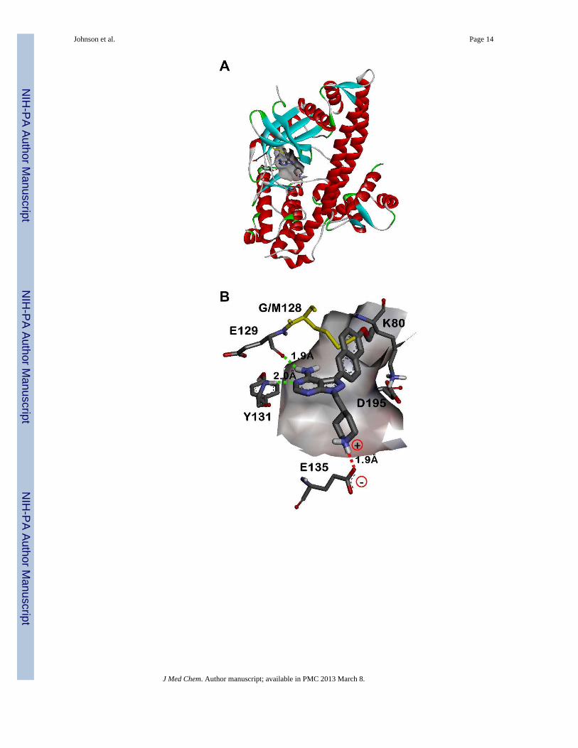
We have previously shown that pyrazolopyrimidine-based molecules, variably substituted atthe R1 and R2 positions of the core scaffold (Figure 1), are potent inhibitors of TgCDPK1enzymatic activity.16 In that study, two distinct molecular series were developed to optimizecompounds for inhibition of TgCDPK1 enzymatic activity. The first series exploredvariation of the R2 substructure in the context of a naphthylmethylene R1-bearingpyrazolopyrimidine core scaffold (substituent 10 in Figure 1). From that series, severalpiperidine-containing R2 substructures were found that confer potent inhibition ofTgCDPK1 enzymatic activity; the best being the 4-piperidinemethyl R2 substructure ofanalogue 10n (TgCDPK1 IC50=15 nM). X-ray crystallographic analysis showed that the 4-piperidinemethyl group orients towards the αD-helix and makes a solvent exposed saltbridge with the Glu135 side chain carboxylate (as exemplified in the co-crystal structure ofcompound 15n bound to TgCDPK1, presented in Figure 2B). The second series of inhibitorsevaluated variation at the R1 position and identified several groups that were superior to thenaphthylmethylene substructure for conferring potent inhibition of TgCDPK1 activity.These contain R1 aryl groups directly linked to the pyrazolopyrimidine core (i.e., through aCaryl-Caryl linkage), which orients the R1 substructures directly towards the Gly128gatekeeper residue and into an adjacent hydrophobic pocket (Figure 2). This increasesselectivity for TgCDPK1 over potential off-target human kinases, which primarily containthreonine and larger gatekeeper residues that hinder access to this pocket.
Based on the structure-activity relationships generated in our previous study, we havedesigned, synthesized, and evaluated a subsequent panel of lead TgCDPK1 enzymeinhibitors for their ability to prevent the invasion of T. gondii parasites into host cells. In thefirst part of this study, we have investigated a panel of R1 groups in the context of iPr-, tBu-,and 4- piperidinemethyl-substituents at the R2 position (series a, b, and n in Figure 1 andTable 1). To impart selective inhibition for TgCDPK1 over human kinases, our effortsfocused on R1 substructures that occupy the enlarged hydrophobic pocket next to the Gly128gatekeeper residue, such as substituted phenyls, indoles, indazoles, naphthyls, andquinolines (Figure 1). From previous structural studies,15 it appeared that increased bindingaffinity could be gained by extending the R1 substructure more deeply into the adjacenthydrophobic pocket (Figure 2C). To explore this possibility, analogues with extended R1groups (series 14–25) were synthesized. Analysis of various R2 substructures in the contextof multiple R1 groups allows the interdependence of these two positions to be explored.
Syntheses of pyrazolopyrimidine compounds with iPr- or tBu-groups at the R2 position(series a and b) are outlined in Scheme 1. Detailed procedures and characterization data forall compounds are presented in the Supporting Information. Microwave-assisted Suzuki-
Johnson et al. Page 3
J Med Chem. Author manuscript; available in PMC 2013 March 8.
NIH
-PA Author Manuscript
NIH
-PA Author Manuscript
NIH
-PA Author Manuscript

Miyaura reactions were employed for the coupling of R1 boronic acids or boronate pinacolesters to the respective pyrazolopyrimidine halide intermediates 26 and 27.16, 17 Forcompounds 17a–25a, containing extended R1 substructures, Suzuki-Miyaura coupling of theTBDMS-protected boronic acid conveniently afforded the deprotected naphthol intermediatedirectly, which was subsequently alkylated with the respective R′-halide. Synthesis of the 4-piperidinemethyl-pyrazolopyrimidine compounds (series n) is outlined in Scheme 2. Thepyrazolopyrimidine core scaffold intermediate 32 was synthesized according to previouslyreported procedures.16, 17 Pyrazolopyrimidine 32 was then alkylated with mesylate 30 toafford the key intermediate 34. Suzuki-Miyaura coupling of the respective R1 boronic acidsor boronate pinacol esters, and subsequent naphthol alkylations, were performed asdescribed above for the series a and b compounds. Boc-containing compounds weredeprotected using 50% TFA in CH2Cl2 and converted to their HCl salts for enzymatic andcell-based evaluation (results are discussed below and presented in Tables 1 and 2).
From the compounds described in Table 1, the 6-ethoxynaphthyl R1 group (15) wasidentified as the best substructure for conferring potent inhibition of TgCDPK1 enzymaticactivity and T. gondii cell proliferation (vide infra). Therefore, a second series of compoundscontaining a 6-ethoxynaphthyl R1 and variable R2 substructures was generated (Figure 1 –substructures a-w). Syntheses of these compounds are presented in Scheme 3. For thepiperidinyl-amide and sulfonamide compounds (15l, 15m, 15q, and 15r), thepyrazolopyrimidine intermediate core 32 was alkylated with mesylates 36, 37, 40 and 41,respectively, followed by microwave-assisted Suzuki-Miyaura coupling with 6-ethoxynaphthyl-2-boronic acid as described above (Scheme 3A). Compounds 15o and 15pwere synthesized by reductive alkylation of 15n with formaldehyde and acetaldehyde,respectively, using sodium cyanoborohydride (Scheme 3B). The remaining series 15compounds were synthesized by first appending the 6- ethoxynaphthyl R1 substructure tothe pyrazolopyrimidine core scaffold 32 through microwaveassisted Suzuki-Miyauracouplings, affording intermediate 44. Mitsunobu alkylation of the remaining R2 groupsusing resin-bound PPh3, followed by Boc-deprotection with TFA/CH2Cl2, affordedcompounds 15c, 15e, 15g, and 15s. Reductive alkylation of the deprotected intermediates,with respective R2-aldehydes, provided final compounds 15d, 15f, 15h-k, and 15tw(compounds 15k and 15w required additional Boc-deprotection). All compounds werepurified by preparatory RP-HPLC and amine-containing compounds were converted to theirHCl salts for enzymatic and cell-based evaluation (results are discussed below).
Pyrazolopyrimidines are potent inhibitors of TgCDPK1 enzymatic activityCompounds were first evaluated for their ability to inhibit the in vitro enzymatic activity ofwild type T. gondii CDPK1. Inhibition was determined using a previously reportedluminescence-based kinase assay.16 Although a large percentage of the compounds testeddisplayed very potent inhibition of TgCDPK1, several notable trends were observed. Whilethe absence of a hydrophobic substituent at the R1 position renders these pyrazolopyrimidinecompounds inactive against TgCDPK1 (the IC50 values for 1b and 1n are >5 μM, Table 1),incorporation of R1 substructures as small as a phenyl group confers potent enzymaticinhibition. Overall, 86% of the compounds tested have IC50 values <25 nM againstTgCDPK1 (Table 1). When compared with results obtained in our previous study,16 it isevident that coupling of an R1 substructure to the pyrazolopyrimidine core through a directCaryl-Caryl linkage provides analogues with superior potency relative to those that contain amethylene spacer (for example, series 10). As observed from crystallographic studies, thisdirect linkage orients the R1 substructures towards the adjacent pocket next to the Gly128gatekeeper residue, allowing large hydrophobic groups to make extensive contacts (Figure2).18 In the context of the 6- ethoxynaphthyl R1 substructure (series 15), it appears that awide variety of substitutions at the R2 position are well tolerated for maintaining potent
Johnson et al. Page 4
J Med Chem. Author manuscript; available in PMC 2013 March 8.
NIH
-PA Author Manuscript
NIH
-PA Author Manuscript
NIH
-PA Author Manuscript

TgCDPK1 enzymatic inhibition (Table 3), which is in contrast to analogues that contain anapthylmethylene group at this position. For example, series 15 compounds (6-ethoxynaphthyl R1) that are alkylated, acetylated, or sulfonylated on the piperidine rings oftheir R2 substructures maintain low nanomolar inhibition of TgCDPK1 enzymatic activity,whereas series 10 analogues (naphthylmethylene R1) containing similar modificationsdemonstrate IC50 values in the high nanomolar range. Consistently potent inhibition across awide range of R1 and R2 substructures suggests there is ample chemical space at eitherposition to exploit for optimizing the pharmacological properties of these inhibitors.
To determine how a larger gatekeeper residue affects compound binding, inhibitors weretested against a TgCDPK1 mutant enzyme that contains methionine at this position(Gly128Met). Molecular modeling predicts that the increased steric bulk of this residueshould clash with large R1 substructures (Figure 2B). In addition, the Gly128Met mutantwas selected as a drug-resistant mutant because it maintains comparable enzymatic activityto wild type TgCDPK1 and is able to complement for loss of endogenous enzyme activity inT. gondii parasites. In nearly all cases, the presence of the larger methionine side chainabolishes the inhibitory activity of these molecules (IC50 values are generally >3 μM). Evenfor compounds 22n, 24n, 25n, 15h, 15k, and 15s-w, which show some activity againstGly128Met TgCDPK1, the differences in IC50 values between the wild type and mutantenzymes are >250-fold (with the exception of 15w, which displays a 68-fold difference).Thus, the presence of a small gatekeeper residue provides a distinct preference for binding tothe wild type enzyme. These results are promising for the development ofpyrazolopyrimidine inhibitors as potential anti-parasitic drugs because it suggests we shouldbe able to obtain selectivity for TgCDPK1 over human kinases, which do not containgatekeeper residues as small as Gly or Ala.
Lead compounds selectively inhibit TgCDPK1 over human kinases and do not inhibitgrowth of human cell lines
While compound evaluation in the Gly128Met TgCDPK1 enzymatic assay was an importantsurrogate for gauging potential inhibition of off-target kinases that contain larger gatekeeperresidues in an otherwise identical binding site, further evaluation was performed against apanel of human kinases. Compounds were first tested against SRC kinase, as priorselectivity studies have demonstrated that similar pyrazolopyrimidine-based inhibitorspreferentially target this enzyme.19–21 Since SRC contains a threonine gatekeeper residue,which is one of the smallest amino acid side chains present at this position in human kinases,we selected this enzyme as a surrogate for probing potential off-target kinase activity.Inhibition of this enzyme serves as a first-pass filter to prioritize lead candidates forsubsequent cell-based evaluation. Inhibition values were determined using a previouslyreported radioactive kinase assay.16
The activities of the first series of compounds against SRC are shown in Table 1. While wehad initially envisioned the R1 substructure providing the primary determinant for obtainingselective inhibition of TgCDPK1 over human kinases (i.e., a steric clash of the R1substructures with the larger gatekeeper residue side chains obstructs ligand binding), thisappears to be only partially true. For 93% of the compounds tested, IC50 values are at least25-fold higher for SRC than for TgCDPK1 (Table 1). Thus, substitution at the R1substructure is a significant determinant of selective inhibition of TgCDPK1. However,further examination of the results presented in Table 1, where we have evaluated R1substructures in the context of iPr (a), tBu (b), and 4-piperidinemethyl (n) R2 substituents,demonstrates a striking trend where the iPr- and tBucontaining compounds are consistentlyless selective for TgCDPK1 over SRC than the 4- piperidinemethyl analogues (differencesare readily observed in the inhibition heat maps in Tables 2A and B, and Figure S1 in theSupporting Information). The second compound series, where we have evaluated R2
Johnson et al. Page 5
J Med Chem. Author manuscript; available in PMC 2013 March 8.
NIH
-PA Author Manuscript
NIH
-PA Author Manuscript
NIH
-PA Author Manuscript

substructures in the context of a 6-ethoxynapthyl (15) group at the R1 position, was testedagainst SRC to further probe this interesting phenomenon (Table 3). In this series, it appearsthat compounds which contain a methylene spacer adjacent to the pyrazolopyrimidine coredisplay greater selectivity for TgCDPK1 inhibition over SRC. 4- piperidinemethyl-containing compounds (n-r) are particularly selective and display no inhibition of SRCactivity at compound concentrations upwards of 10 μM. Thus, it is evident that the degree ofselective inhibition of TgCDPK1 over SRC results from a synergy between the R1 and R2substructures. A more detailed discussion of the structural underpinnings for this synergywill be presented in a forthcoming X-ray crystallography manuscript.18
Several lead compounds (14a, 14n, 15a, 15h, 15n, 15o, and 16n) were further evaluated inan expanded panel of human kinases that all contain threonine gatekeeper residues (ABL,LCK, p38α, EPHA3, CSK, and EGFR). In general, similar inhibition trends were observedfor ABL, p38α, EPHA3, CSK, and EGFR as described above (Table 4). In comparison toSRC, compounds generally display increased inhibition of LCK, equipotency against ABLand EGFR, and decreased inhibition of p38α, EPHA3, and CSK, which is similar to trendsthat have been previously reported.19 Importantly, we generally observe >1000-folddifferences between IC50 values for TgCDPK1 over human kinases for our lead inhibitors(14n, 15h, 15n, 15o, and 16n; refer to Table S1 in the Supporting Information for specificselectivity ratios between the human kinase IC50 values compared to TgCDPK1). Whilethese compounds have only been tested against a small panel of enzymes that wereenvisioned to be of primary concern for inhibition, these results suggest our lead candidateT. gondii therapeutics should interact minimally with potential off-target human kinases.
As an indicator for potential host cell toxicity during toxoplasmosis therapy, we furtherevaluated our panel of TgCDPK1 inhibitors (14a, 14n, 15a, 15h, 15n. 15o, and 16n) fortheir ability to inhibit the growth of human neutrophil (HL-60) and lymphocyte (CRL-8155)cell lines. Assays were performed with a similar procedure as previously reported.16 Thesecompounds exhibited no inhibition of cell growth at concentrations up to 10 μM (Table 4).To better define potential therapeutic windows for our toxoplasmosis drug candidates, weare performing growth inhibition studies using higher compound concentrations andidentifying drug levels required to clear parasitic infection in mammalian challenge models.Results from these studies will be presented elsewhere.
Potent TgCDPK1 enzymatic inhibitors block the proliferation of T. gondii parasitesHaving developed compounds that selectively inhibit TgCDPK1 over a panel of humankinases and do not inhibit the growth of human cell lines, we further investigated the mostpotent TgCDPK1 enzymatic inhibitors (IC50 < 25 nM) for their efficacy in blocking theinvasion of T. gondii parasites into human foreskin fibroblast cells. Since T. gondii is anobligate intracellular parasite, inhibition of host cell invasion blocks parasite replication,which was measured as a surrogate according to a slightly modified version of a previouslyreported procedure.15 In these cellular assays, several prominent trends were observed.Notably, compounds 1b and 1n, which do not contain an R1 substituent and are inactiveagainst TgCDPK1 enzymatic activity, do not block T. gondii cell invasion/proliferation. Ofthe compounds that do potently inhibit TgCDPK1 enzymatic activity (IC50 < 25 nM), animpressive 84% also effectively block T. gondii cell invasion/proliferation (EC50 < 1 μM).Importantly, no inhibitor toxicity was observed against the human foreskin fibroblasts usedin this assay. Thus, it seems unlikely that the decreased parasite growth is an artifact of hostcell inhibition. Many of the 4-piperidinemethyl compounds are potent inhibitors of T. gondiiproliferation. In particular, compounds bearing the 6- ethoxynaphthyl R1 substructure (series15) are potent enzymatic and cell proliferation inhibitors across nearly the entire R2substructure panel. However, compounds containing an iPr or tBu group at the R2 position(series a and b, respectively) are generally more potent inhibitors in the cell proliferation
Johnson et al. Page 6
J Med Chem. Author manuscript; available in PMC 2013 March 8.
NIH
-PA Author Manuscript
NIH
-PA Author Manuscript
NIH
-PA Author Manuscript

assay than their 4-piperidinemethyl analogues (series n). This is readily observed in Figure3A and in the inhibition heat map presented in Table 2C. The a and b series of inhibitors aresignificantly more hydrophobic than the 4-piperidinemethyl-containing compounds (thepiperidine amine would be protonated under physiological conditions), which may increasetheir membrane permeability. In addition, the greater potential of pyrazolopyrimidineinhibitors with iPr and tBu substituents at the R2 position to inhibit off-target mammalianand parasitic kinases (as suggested by their more potent inhibition of SRC and other humankinases), may lead to enhanced cellular activity. While it is interesting to speculate on thebiophysical underpinnings of these differences, SAR rationalizations in these cellular assaysare likely to be complicated and beyond the scope of this manuscript.
Three compounds (15n, 15o, and 16n) were evaluated in cell-based experiments using T.gondii parasites expressing the Gly128Met TgCDPK1 gatekeeper mutant, to further validatethat TgCDPK1 is the primary target for the observed anti-parasitic activity. The Gly128MetTgCDPK1 variant is functionally active and complements the loss of endogenous wild typeenzyme activity, permitting parasite invasion and proliferation in host cells. For all threecompounds, a significant increase in T. gondii proliferation EC50 (8- to 26-fold) wasobserved for parasites expressing the Gly128Met TgCDPK1 mutant over those expressingwild type TgCDPK1 at similar levels or non-transfected parasites (Figure 3B and FiguresS3A and S3B in the Supporting Information). The observed inhibitory effects at highercompound concentrations (EC50 ~1 to 3 μM for the Gly128Met TgCDPK1 expressingparasites) could indicate off-target inhibition of another parasite kinase. However, thedramatic EC50 shifts seen for all three inhibitors demonstrate that TgCDPK1 is the primarytarget for blocking parasite invasion and proliferation.
ConclusionsIn the present study, we have evaluated over 70 ATP-competitive inhibitors of TgCDPK1for their utility as potential toxoplasmosis therapeutics. We found that inhibition ofTgCDPK1 enzymatic activity correlates strongly with inhibition of T. gondii proliferation.Of the 64 compounds that are potent inhibitors of TgCDPK1 enzymatic activity in vitro(IC50 < 25 nM), 83% exhibit EC50 values of <1 μM in a parasite invasion/proliferationassay. Furthermore, 38% exhibit EC50 values <100 nM. In a rescue experiment, expressionof the drug-resistant Gly128Met TgCDPK1 enzyme in parasites verified that inhibition ofendogenous wild type CDPK1 is the primary mechanism of action for these compounds. Byvirtue of the small glycine gatekeeper residue in wild type TgCDPK1, these “bumped”kinase inhibitors are able to selectively inhibit the parasite enzyme over human kinases,which contain gatekeeper residues larger than glycine that sterically hinder inhibitorbinding. This selectivity is mimicked in cellular assays where compounds potently inhibit T.gondii proliferation, but do not affect the growth of human cell lines. Compounds exhibitinglarge therapeutic windows between inhibition of T. gondii proliferation and human cellgrowth (e.g. >100–1000x) are undergoing additional pre-clinical drug development testingto evaluate pharmacological properties such as solubility, pharmacokinetics,pharmacodynamics, and metabolism. The results obtained here poise us for future studies toevaluate lead candidates possessing favorable properties in parasitic challenge models inmice as a therapeutic proof of principle.
Experimental ProceduresGeneral synthetic methods
Unless otherwise stated, all chemicals were purchased from commercial suppliers and usedwithout further purification. Reaction progress was monitored by thin-layer chromatographyon silica gel 60 F254 coated glass plates (EM Sciences). Chromatography was performed
Johnson et al. Page 7
J Med Chem. Author manuscript; available in PMC 2013 March 8.
NIH
-PA Author Manuscript
NIH
-PA Author Manuscript
NIH
-PA Author Manuscript

using an IntelliFlash 280 automated flash chromatography system, eluting on prepackedVarian SuperFlash silica gel columns with hexanes/EtOAc or CH2Cl2/MeOH gradientsolvent systems. For preparatory HPLC purification, samples were chromatographicallyseparated using a Varian Dynamax Microsorb 100-5 C18 column (250 mm × 21.4 mm),eluting with H2O/CH3CN or H2O/MeOH gradient solvent systems (+0.05% TFA). Thepurity of all final compounds was determined by two analytical RP-HPLC methods, usingan Agilent ZORBAX SB-C18 (2.1 mm × 150 mm) or Varian Microsorb-MV 100-5 C18column (4.6 mm × 150 mm), and eluting with either H2O/CH3CN or H2O/MeOH gradientsolvent systems (+0.05% TFA) run over 30 min. Products were detected by UV at λ=254nm, with all final compounds displaying >95% purity. NMR spectra were recorded onBruker 300 or 500 MHz spectrometers at ambient temperature. Chemical shifts are reportedin parts per million (δ) and coupling constants in Hz. 1H-NMR spectra were referenced tothe residual solvent peaks as internal standards (7.26 ppm for CDCl3, 2.50 ppm for d6-DMSO, and 3.34 ppm for CD3OD). Mass spectra were recorded with a Bruker EsquireLiquid Chromatograph - Ion Trap Mass Spectrometer. Inhibitors were synthesized throughseveral different routes, as represented in Schemes 1–3. Syntheses of compounds 1b, 3a, 4a,5a, 10a, 10b, 10n, 11a, 11b, 12a, 14a, 15a, 26, 27, 31, and 32 have been previouslyreported.16, 17 All other synthesis and compound characterization data is presented in theSupporting Information.
T. gondii CDPK1 enzymatic assaysExpression, purification, and enzymatic evaluation of wild type and Gly128Met gatekeepermutant TgCDPK1 was performed as described previously.15, 16 Briefly, enzymatic reactionswere performed with 4 nM of either wild type or Gly128Met TgCDPK1 in assay buffercontaining 20 mM HEPES (pH=7.5), 0.1% BSA, 10 mM MgCl2, 1 mM EGTA, 2 mMCaCl2, 10 μM ATP, and 40 μM Syntide-2 peptide substrate (peptide sequence:PLARTLSVAGLPGKKOH). After incubating for 90 min at 30 °C, the enzymatic reactionswere terminated by adding EGTA to a final concentration of 5 mM. The amount of ATPremaining in solution was evaluated using the Kinase Glo luciferase assay from Promega,with sample luminescence read using a Microbeta 2 plate reader (Perkin Elmer, Waltham,MA). Results were converted to percent inhibition and IC50 values were calculated usingnon-linear regression analysis in GraphPad Prism. Compounds were evaluated in triplicatein 8 point dilutions (3-fold dilution series) during the enzymatic reactions.
Human kinase enzymatic assaysAll compounds were evaluated in a primary counter-screen against SRC kinase using eitherthe truncated catalytic kinase domain (SRCKD) or the full length three domain enzyme(SRC3D). Results from compounds tested with both KD and 3D enzymes demonstrated nosignificant difference in IC50 values, and are thus reported together simply as inhibition of“SRC” kinase. Lead compounds were further evaluated against a small panel of additionalhuman kinases – ABL, LCK, p38α, EPHA3, CSK, and EGFR. Compounds were evaluatedin 10 point, 3-fold dilution series ranging from 10 μM to 0.5 nM during the enzymaticreactions, as per previously reported procedures.16 Results were converted to percentinhibition and IC50 values were calculated using non-linear regression analysis in GraphPadPrism. Experiments were performed in triplicate or quadruplicate. Assay buffers, enzymeconcentrations, substrate peptide sequences and concentrations, and enzymatic reactiontimes are listed in the assay-specific details presented below. All assays were performedusing <5 μM ATP ([ATP] ≪ Km).
1. SRC – Kinase concentration during the enzymatic reaction: 1 nM for SRCKD or 2nM for SRC3D. Assay buffer: 33.5 mM HEPES, pH=7.5, 6.7 mM MgCl2, 1.7 mMEGTA, 67 mM NaCl, 2 mM Na3VO4, 0.08 mg/ml BSA, γ32P ATP (0.2 μCi/well).
Johnson et al. Page 8
J Med Chem. Author manuscript; available in PMC 2013 March 8.
NIH
-PA Author Manuscript
NIH
-PA Author Manuscript
NIH
-PA Author Manuscript

Enzymatic reaction time: 30 min for SRCKD or 60 min for SRC3D. Substratepeptide sequence and concentration: Ac- EIYGEFKKK-OH (100 μM).
2. ABL – Kinase concentration during the enzymatic reaction: 1 nM for ABLKD or 2nM for ABL3D. Assay buffer: 33.5 mM HEPES, pH=7.5, 6.7 mM MgCl2, 1.7 mMEGTA, 67 mM NaCl, 2 mM Na3VO4, 0.08 mg/ml BSA, γ32P ATP (0.2 μCi/well).Enzymatic reaction time: 30 min for ABLKD and 60 min for ABL3D. Substratepeptide sequence and concentration: Ac-EAIYAAPFAKKK-OH (100 μM).
3. LCK – Kinase concentration during the enzymatic reaction: 10 nM. Assay buffer:75 mM HEPES, pH=7.5, 15 mM MgCl2, 3.75 mM EGTA, 150 mM NaCl, 2 mMNa3VO4, 0.08 mg/mL BSA, γ32P ATP (0.2 μCi/well). Enzymatic reaction time: 60min. Substrate peptide sequence and concentration: Ac-EIYGEFKKK-OH (100μM).
4. p38α – Kinase concentration during the enzymatic reaction: 2 nM. Assay buffer: 75mM HEPES, pH=7.5, 15 mM MgCl2, 3.75 mM EGTA, 150 mM NaCl, 2 mMNa3VO4, 0.08 mg/mL BSA, 1.9 mM BME, γ32P ATP (0.2 μCi/well). Enzymaticreaction time: 180 min. Substrate peptide and concentration: Myelin Basic Protein(0.2 mg/mL).
5. EPHA3 – Kinase concentration during the enzymatic reaction: 10 nM. Assaybuffer: 30 mM HEPES, pH=7.5, 38 mM MgCl2, 630 μM EGTA, 2 mM Na3VO4,40 μg/ml BSA, γ32P ATP (0.2 μCi/well). Enzymatic reaction time: 120 min.Substrate peptide and concentration: Myelin Basic Protein (0.2 mg/mL).
6. CSK – Kinase concentration during the enzymatic reaction: 5 nM. Assay buffer: 75mM HEPES, pH=7.5, 15 mM MgCl2, 3.75 mM EGTA, 150 mM NaCl, 2 mMNa3VO4, 0.2 mg/mL BSA, γ32P ATP (0.2 μCi/well). Enzymatic reaction time: 180min. Substrate peptide sequence and concentration: Ac-KKKKEEIYFFF-OH (130μM).
7. EGFR – Kinase concentration during the enzymatic reaction: 1 nM. Assay buffer:37.5 mM Tris, pH=7.5, 15 mM MgCl2, 0.75 mM EGTA, 0.75 mM Na3VO4,0.015% Triton X-100, 3.75 mM DTT, 0.08 mg/mL BSA, 2 mM ATP, γ32P ATP(2.0 μCi/well). Enzymatic reaction time: 30 min. Substrate peptide andconcentration: Poly Glu-Tyr substrate (0.2 mg/mL).
Human cell growth inhibition assaysLead compounds were evaluated for potential toxicity against two human cell lines: HL- 60(neutrophil) and CRL-8155 (lymphocytic) cells. Cells were grown in either IMDM (HL-60)or RPMI-1640 (CRL-8155) growth media supplemented with 10% heat inactivated fetal calfserum and 2 mM L-glutamine. HL-60 growth medium additionally contained 25 mMHEPES and 1% penicillin/streptomycin. CRL-8155 growth medium additionally contained10 mM HEPES, 1 mM sodium pyruvate, 4.5 g/L glucose and 1.5 g/L sodium bicarbonate.Cells were grown in the presence of 10 μM test compound for 48 or 72 hours at 37°C and5% CO2 in 96- well flat-bottom plates (Corning). Growth was quantified using Alamar Blueas a developing reagent and detecting sample absorbance at λ = 570 nm (600 nm referencewavelength). Percent growth inhibition by test compounds were calculated based on culturesincubated with DMSO negative and tipifarnib (R115777) positive controls (0% and 100%growth inhibition, respectively). All assays were performed in triplicate.
T. gondii cell proliferation assaysThe invasion assay was performed as previously described,15 with slight modifications toimprove assay sensitivity and reliability. Compounds were diluted in DMEM maintaining
Johnson et al. Page 9
J Med Chem. Author manuscript; available in PMC 2013 March 8.
NIH
-PA Author Manuscript
NIH
-PA Author Manuscript
NIH
-PA Author Manuscript

0.5% DMSO. T. gondii clonal parasites (103) expressing β-galactosidase as a reporter(genotype RHΔhxgprt, β-galactosidase, GFP15 for the standard protocol) were mixed withthe medium containing the compounds (200 μl) and incubated at 37°C, 5% CO2, for ~5min.The parasite/compound mixture was added to 96-well plates containing confluent humanfibroblast cell layers (from which growth media was aspirated) and incubated for 44 hours at37°C and 5% CO2. As a control, a dilution series of T. gondii (103-0) parasites was grown inthe same conditions described above, but without compound. Plates were visually inspectedfor evidence of cytotoxic effects on fibroblasts. β-galactosidase was then assayed usingchlorophenol red β-galactopyranose (Sigma) as a substrate.22 Plates were developed for ~1.5hours at 37°C. Absorbance was measured at 595 nm on a SpectraMax M2 (MolecularDevices) microplate reader. Each experiment was performed in triplicate and experimentsyielding EC50 values < 0.5 μM were repeated at least once. For assays to test the role of thegatekeeper residue, the above procedure was followed except that three T. gondii cell linesexpressing β-galactosidase in either a “wild type” background (as above in the standardassay) or also expressing HA-TgCDPK1 or HA-Gly128Met TgCDPK1 or were assayed inparallel.
Supplementary MaterialRefer to Web version on PubMed Central for supplementary material.
AcknowledgmentsWe are grateful for the technical assistance of Suzanne Scheele from the Parsons lab. This work was funded by theNational Institute of General Medical Sciences Grant R01GM086858 (D.J.M.) and the National Institute of Allergyand Infectious Diseases Grants R01AI080625 (W.C.V.V.) and R01AI067921 (E.A.M. and W.C.V.V.). J.A.G. wassupported by a training grant from the National Institute of Allergy and Infectious Diseases T32AI007509.
Abbreviations List
ATP adenosine triphosphate
Boc tert-butyloxycarbonyl
CDPK1 calciumdependent protein kinase 1
CSK C-terminal SRC kinase
DIAD diisopropyl azodicarboxylate
DME dimethoxyethane
EC50 half maximal effective concentration
EGFR epidermal growth factor receptor kinase
EPHA3 EPH receptor A3 kinase
IC50 half maximal inhibitory concentration
LCK lymphocyte-specific protein tyrosine kinase
TBDMS t-butyldimethylsilyl
References1. Tenter AM, Heckeroth AR, Weiss LM. Toxoplasma gondii: from animals to humans. Int J Parasitol.
2000; 30:1217–1258. [PubMed: 11113252]2. Schwartzman, JD.; Maguire, JH. Tropical Infectious Diseases: Principles, Pathogens and Practice. 3.
W.B. Saunders; Edinburgh: 2011. CHAPTER 103 - Toxoplasmosis; p. 722-728.
Johnson et al. Page 10
J Med Chem. Author manuscript; available in PMC 2013 March 8.
NIH
-PA Author Manuscript
NIH
-PA Author Manuscript
NIH
-PA Author Manuscript
3. Jones JL, Muccioli C, Belfort R Jr, Holland GN, Roberts JM, Silveira C. Recently acquiredToxoplasma gondii infection Brazil. Emerg Infect Dis. 2006; 12:582–587. [PubMed: 16704805]
4. Demar M, Hommel D, Djossou F, Peneau C, Boukhari R, Louvel D, Bourbigot AM, Nasser V,Ajzenberg D, Darde ML, Carme B. Acute toxoplasmoses in immunocompetent patients hospitalizedin an intensive care unit in French Guiana. Clin Microbiol Infect. [Online early access]. PublishedOnline: September 29, 2011. 10.1111/j.1469–0691.2011.03648.x
5. Silveira C, Vallochi AL, Rodrigues da Silva U, Muccioli C, Holland GN, Nussenblatt RB, BelfortR, Rizzo LV. Toxoplasma gondii in the peripheral blood of patients with acute and chronictoxoplasmosis. Br J Ophthalmol. 2011; 95:396–400. [PubMed: 20601663]
6. Pappas G, Roussos N, Falagas ME. Toxoplasmosis snapshots: global status of Toxoplasma gondiiseroprevalence and implications for pregnancy and congenital toxoplasmosis. Int J Parasitol. 2009;39:1385–1394. [PubMed: 19433092]
7. Jones JL, Kruszon-Moran D, Sanders-Lewis K, Wilson M. Toxoplasma gondii infection in theUnited States, 1999–2004, decline from the prior decade. Am J Trop Med Hyg. 2007; 77:405–410.[PubMed: 17827351]
8. Yolken RH, Torrey EF. Are some cases of psychosis caused by microbial agents? A review of theevidence. Mol Psychiatry. 2008; 13:470–479. [PubMed: 18268502]
9. Dubey JP, Jones JL. Toxoplasma gondii infection in humans and animals in the United States. Int JParasitol. 2008; 38:1257–1278. [PubMed: 18508057]
10. Montoya JG, Liesenfeld O. Toxoplasmosis. Lancet. 2004; 363:1965–76. [PubMed: 15194258]11. Montoya JG, Remington JS. Management of Toxoplasma gondii infection during pregnancy. Clin
Infect Dis. 2008; 47:554–566. [PubMed: 18624630]12. Nagamune K, Sibley LD. Comparative genomic and phylogenetic analyses of calcium ATPases
and calcium-regulated proteins in the apicomplexa. Mol Biol Evol. 2006; 23:1613– 1627.[PubMed: 16751258]
13. Lourido S, Shuman J, Zhang C, Shokat KM, Hui R, Sibley LD. Calciumdependent protein kinase 1is an essential regulator of exocytosis in Toxoplasma. Nature. 2010; 465:359–362. [PubMed:20485436]
14. Kieschnick H, Wakefield T, Narducci CA, Beckers C. Toxoplasma gondii attachment to host cellsis regulated by a calmodulin-like domain protein kinase. J Biol Chem. 2001; 276:12369–12377.[PubMed: 11154702]
15. Ojo KK, Larson ET, Keyloun KR, Castaneda LJ, Derocher AE, Inampudi KK, Kim JE, ArakakiTL, Murphy RC, Zhang L, Napuli AJ, Maly DJ, Verlinde CL, Buckner FS, Parsons M, Hol WG,Merritt EA, Van Voorhis WC. Toxoplasma gondii calcium-dependent protein kinase 1 is a targetfor selective kinase inhibitors. Nat Struct Mol Biol. 2010; 17:602–607. [PubMed: 20436472]
16. Murphy RC, Ojo KK, Larson ET, Castellanos-Gonzalez A, Perera BG, Keyloun KR, Kim JE,Bhandari JG, Muller NR, Verlinde CL, White AC Jr, Merritt EA, Van Voorhis WC, Maly DJ.Discovery of Potent and Selective Inhibitors of Calcium-Dependent Protein Kinase 1 (CDPK1)from C. parvum and T. gondii. ACS Med Chem Lett. 2010; 1:331–335. [PubMed: 21116453]
17. Bulawa, CE.; Devit, M.; Elbaum, D. Preparation of pyrazolopyrimidinamines as modulators ofprotein trafficking. WO. 2009062118A2. 2009.
18. Larson ET, Ojo KK, Murphy RC, Johnson SM, Zhang Z, Kim JE, Leibly DJ, Fox AMW, ReidMC, Dale EJ, Perera BGK, Kim J, Hewitt SN, Hol WGJ, Verlinde CLMJ, Fan E, Van VoorhisWC, Maly DJ, Merritt EA. Unpublished results.
19. Bain J, Plater L, Elliott M, Shpiro N, Hastie CJ, McLauchlan H, Klevernic I, Arthur JS, Alessi DR,Cohen P. The selectivity of protein kinase inhibitors: a further update. Biochem J. 2007; 408:297–315. [PubMed: 17850214]
20. Hanke JH, Gardner JP, Dow RL, Changelian PS, Brissette WH, Weringer EJ, Pollok BA, ConnellyPA. Discovery of a novel, potent, and Src family-selective tyrosine kinase inhibitor. Study of Lck-and FynT-dependent T cell activation. J Biol Chem. 1996; 271:695–701. [PubMed: 8557675]
21. Liu Y, Bishop A, Witucki L, Kraybill B, Shimizu E, Tsien J, Ubersax J, Blethrow J, Morgan DO,Shokat KM. Structural basis for selective inhibition of Src family kinases by PP1. Chem Biol.1999; 6:671–678. [PubMed: 10467133]
Johnson et al. Page 11
J Med Chem. Author manuscript; available in PMC 2013 March 8.
NIH
-PA Author Manuscript
NIH
-PA Author Manuscript
NIH
-PA Author Manuscript

22. Seeber F, Boothroyd JC. Escherichia coli beta-galactosidase as an in vitro and in vivo reporterenzyme and stable transfection marker in the intracellular protozoan parasite Toxoplasma gondii.Gene. 1996; 169:39–45. [PubMed: 8635747]
Johnson et al. Page 12
J Med Chem. Author manuscript; available in PMC 2013 March 8.
NIH
-PA Author Manuscript
NIH
-PA Author Manuscript
NIH
-PA Author Manuscript

Figure 1.Legend of the R1 and R2 substructures of compounds evaluated in this study.
Johnson et al. Page 13
J Med Chem. Author manuscript; available in PMC 2013 March 8.
NIH
-PA Author Manuscript
NIH
-PA Author Manuscript
NIH
-PA Author Manuscript
Johnson et al. Page 14
J Med Chem. Author manuscript; available in PMC 2013 March 8.
NIH
-PA Author Manuscript
NIH
-PA Author Manuscript
NIH
-PA Author Manuscript

Figure 2.X-ray crystallographic structure of compound 15n bound to wild type T. gondii CDPK1(PDB accession code: 3SX9).18 A) Complete view of the 15n•TgCDPK1 co-crystalstructure. B) Expanded view of the ATP binding site from panel A (protein backboneribbons have been removed for clarity). The backbone carbonyl of Glu129 and NH ofTyr131 hydrogen bond with the exocyclic amine and adjacent endocyclic nitrogen of thepyrazolopyrimidine core, respectively (green dashed lines). The Glu135 side chaincarboxylate makes a solvent exposed salt bridge with the piperidine R2 ring, which isprotonated under physiological conditions (red dashed line). The R1 aryl substructure orientstowards the Gly128 gatekeeper residue and into the adjacent hydrophobic pocket. Themutant Met128 residue (yellow) has been overlayed to show the steric clash that wouldoccur between a larger gatekeeper side chain with the inhibitor R1 substructures. Thecatalytic Lys80 and Asp195 side chains are also shown for context. C) Rotated, side/bottomview of the ATP binding site from panel B.
Johnson et al. Page 15
J Med Chem. Author manuscript; available in PMC 2013 March 8.
NIH
-PA Author Manuscript
NIH
-PA Author Manuscript
NIH
-PA Author Manuscript

Figure 3.Enzymatic and cellular inhibition of TgCDPK1. A) Correlation between T. gondii cellproliferation EC50 and TgCDPK1 enzymatic IC50 results. The solid lines at EC50 >6.25 μMand IC50 >5 μM represent the upper detection limits for compounds tested in the respectiveassays. Of the 62 compounds displaying a TgCDPK1 enzymatic IC50 <0.025 μM, 86% arealso potent inhibitors of T. gondii invasion/proliferation (EC50 <1 μM, points in bottom leftquadrant). B) Comparison of EC50 curves between wild type T. gondii (open squares) andparasites expressing either wild type TgCDPK1 (open circles) or the drug resistantGly128Met TgCDPK1 mutant (closed circles). Expression of the drug resistant Gly128MetTgCDPK1 mutant rescues cells from the potent anti-proliferative effects of inhibitor 15n.
Johnson et al. Page 16
J Med Chem. Author manuscript; available in PMC 2013 March 8.
NIH
-PA Author Manuscript
NIH
-PA Author Manuscript
NIH
-PA Author Manuscript

Scheme 1.General synthetic procedures for compound series a and b. i) Na2CO3, Pd(PPh3)4, boronicacid or boronate pinacol ester, H2O/DME, 80°C; ii) TFA/CH2Cl2 (for Boc-protected R1substructures 6 and 7); iii) R′-halide, K2CO3, DMF, room temperature or 80 °C.
Johnson et al. Page 17
J Med Chem. Author manuscript; available in PMC 2013 March 8.
NIH
-PA Author Manuscript
NIH
-PA Author Manuscript
NIH
-PA Author Manuscript
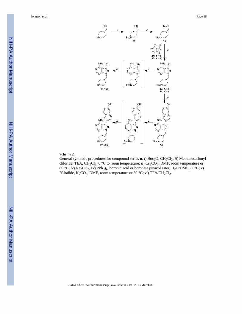
Scheme 2.General synthetic procedures for compound series n. i) Boc2O, CH2Cl2; ii) Methanesulfonylchloride, TEA, CH2Cl2, 0 °C to room temperature; ii) Cs2CO3, DMF, room temperature or80 °C; iv) Na2CO3, Pd(PPh3)4, boronic acid or boronate pinacol ester, H2O/DME, 80°C; v)R′-halide, K2CO3, DMF, room temperature or 80 °C; vi) TFA/CH2Cl2.
Johnson et al. Page 18
J Med Chem. Author manuscript; available in PMC 2013 March 8.
NIH
-PA Author Manuscript
NIH
-PA Author Manuscript
NIH
-PA Author Manuscript

Scheme 3.General synthetic procedures for compound series 15. A) i) Ac2O, TEA, CH2Cl2, thenmethanesulfonyl chloride; ii) Methanesulfonyl chloride, TEA, CH2Cl2; iii) Cs2CO3, DMF,80 °C; iv) Na2CO3, Pd(PPh3)4, boronic acid or boronate pinacol ester, H2O/DME, 80 °C inmicrowave. B) i) Na2CO3, Pd(PPh3)4, 6-ethoxynaphthalene-2-boronic acid, H2O/DME, 110°C; ii) Boc-R2- OH, PS-PPh3, DIAD, THF, room temperature to 70 °C; iii) TFA/CH2Cl2; iv)sodium methoxide in MeOH, then 2% AcOH, R″-aldehyde, NaBH3CN.
Johnson et al. Page 19
J Med Chem. Author manuscript; available in PMC 2013 March 8.
NIH
-PA Author Manuscript
NIH
-PA Author Manuscript
NIH
-PA Author Manuscript

NIH
-PA Author Manuscript
NIH
-PA Author Manuscript
NIH
-PA Author Manuscript
Johnson et al. Page 20
Tabl
e 1
Enzy
mat
ic a
ssay
(IC
50) a
nd T
. gon
dii p
rolif
erat
ion
(EC
50) r
esul
ts fo
r com
poun
ds w
ith v
aria
ble
R1 s
ubst
ruct
ures
(1-2
5) a
cros
s the
R2 s
erie
s a, b
, and
n.
All
resu
lts a
re th
e av
erag
es o
f at l
east
thre
e as
says
. Hea
t map
repr
esen
tatio
n of
IC50
and
EC
50 re
sults
are
pre
sent
ed in
Tab
les 2
AC
.
Com
poun
d
Kin
ase
Enz
ymat
ic IC
50 (μ
M)
T. G
ondi
i Pro
lifer
atio
n E
C50
(μM
)Tg
CD
PK1
TgC
DPK
1 (G
128M
)SR
C
1b
>5>3
>10
>6.2
5
n>5
>3>1
0>6
.25
2b
0.00
45>3
0.13
0.23
n0.
058
>3>1
0
3a
0.01
9>3
1.3
0.51
b0.
0073
>30.
580.
041
4a
0.04
0>3
5.1
5
a0.
0050
>30.
650.
080
b0.
0041
>30.
180.
0030
n0.
0022
>3>1
01.
7
6a
0.01
2>3
1.5
0.15
n0.
081
>3>1
0
7a
0.00
91>3
1.2
0.27
n0.
018
>3>1
0>6
.25
8a
0.00
36>3
0.29
0.12
b0.
013
>30.
520.
044
9b
0.00
25>3
2.1
0.12
n0.
0037
>3>1
0>6
.25
10
a0.
13>3
8.8
b0.
031
>32.
2
n0.
015
>3>1
02.
2
J Med Chem. Author manuscript; available in PMC 2013 March 8.

NIH
-PA Author Manuscript
NIH
-PA Author Manuscript
NIH
-PA Author Manuscript
Johnson et al. Page 21
Com
poun
d
Kin
ase
Enz
ymat
ic IC
50 (μ
M)
T. G
ondi
i Pro
lifer
atio
n E
C50
(μM
)Tg
CD
PK1
TgC
DPK
1 (G
128M
)SR
C
11
a0.
0050
>30.
065
0.07
2
b0.
0079
>30.
120.
35
n0.
0025
>3>1
00.
63
12a
0.02
4>3
0.20
0.52
n0.
018
>3>1
0>6
.25
13b
0.00
31>3
4.1
0.11
n0.
0054
>3>1
0>6
.25
14
a0.
0060
>30.
670.
021
b0.
020
>32.
00.
010
n0.
0049
>3>1
00.
41
15
a0.
0050
>30.
380.
018
b0.
033
>32.
2
n0.
0025
>3>1
00.
052
16n
0.00
34>3
>10
0.05
8
17a
0.01
0>3
0.55
0.33
n0.
0038
>3>1
00.
50
18a
0.00
06>3
0.20
0.01
0
n0.
0030
>3>1
00.
12
19a
0.00
32>3
0.30
>6.2
5
n0.
0026
>3>1
00.
16
20
a0.
0054
>30.
260.
31
b0.
20>3
1.1
n0.
0037
>3>1
00.
21
21a
0.00
09>3
0.78
0.09
0
n0.
0037
>3>1
00.
060
J Med Chem. Author manuscript; available in PMC 2013 March 8.

NIH
-PA Author Manuscript
NIH
-PA Author Manuscript
NIH
-PA Author Manuscript
Johnson et al. Page 22
Com
poun
d
Kin
ase
Enz
ymat
ic IC
50 (μ
M)
T. G
ondi
i Pro
lifer
atio
n E
C50
(μM
)Tg
CD
PK1
TgC
DPK
1 (G
128M
)SR
C
22a
0.00
08>3
0.04
10.
0033
n0.
0023
0.94
3.1
0.13
23a
0.01
1>3
0.28
0.14
n0.
0013
>31.
81.
1
24a
0.00
55>3
0.28
0.01
2
n0.
0007
1.3
0.38
0.10
25a
0.00
40>3
0.27
0.01
3
J Med Chem. Author manuscript; available in PMC 2013 March 8.

NIH
-PA Author Manuscript
NIH
-PA Author Manuscript
NIH
-PA Author Manuscript
Johnson et al. Page 23
Table 2
A) Heat map representation of wild type TgCDPK1 enzymatic IC50 values from Table 1. B) Heat maprepresentation of SRC enzymatic IC50 values from Table 1. C) Heat map representation of wild type T. gondiiproliferation EC50 values from Table 1. Blue represents compounds with desirable IC50 or EC50 values,transitioning to red for compounds with undesirable activities.
J Med Chem. Author manuscript; available in PMC 2013 March 8.

NIH
-PA Author Manuscript
NIH
-PA Author Manuscript
NIH
-PA Author Manuscript
Johnson et al. Page 24
Table 3
Enzymatic assay (IC50) and T. gondii proliferation (EC50) results for compounds with variable R2 (a-w)substructures and a 6-ethoxynaphthyl group (series 15) at the R1 position. All results are the averages of atleast three assays.
Compound 15-
Kinase Enzymatic IC50 (μM)
T. Gondii Proliferation EC50 (μM)TgCDPK1 TgCDPK1 (G128M) SRC
a 0.0050 >3 0.37 0.018
b 0.033 >3 2.2
c 0.013 >3 >10 1.0
d 0.012 >3 >10 0.96
e 0.0091 >3 >10 0.97
f 0.0081 >3 >10 0.86
g 0.0026 >3 5.0 0.045
h 0.0024 2.6 5.6 0.050
i 0.0034 >3 8.7 0.090
j 0.0043 >3 >10 0.16
k 0.0043 0.98 0.73 0.34
l 0.0061 >3 1.6 0.029
m 0.0028 >3 0.81 0.035
n 0.0025 >3 >10 0.052
o 0.0029 >3 >10 0.14
p 0.0050 >3 >10 0.33
q 0.011 >3 >10 0.20
r 0.032 >3 >10
s 0.0019 1.5 3.3 0.14
t 0.0022 1.4 5.2 0.049
u 0.0027 2.0 6.3 0.10
v 0.0030 2.9 6.7 0.18
w 0.0043 0.27 8.2 1.2
J Med Chem. Author manuscript; available in PMC 2013 March 8.

NIH
-PA Author Manuscript
NIH
-PA Author Manuscript
NIH
-PA Author Manuscript
Johnson et al. Page 25
Tabl
e 4
Enzy
mat
ic a
ssay
resu
lts (I
C50
) for
an
expa
nded
pan
el o
f hum
an k
inas
es a
nd g
row
th in
hibi
tion
(GI 5
0) o
f hum
an c
ell l
ines
. All
resu
lts a
re th
e av
erag
es o
f at
leas
t thr
ee a
ssay
s.
Com
poun
d
Kin
ase
Enz
ymat
ic IC
50 (μ
M)
Hum
an C
ells
GI 5
0 (μM
)
TgC
DPK
1SR
CA
BL
LC
Kp3
8αE
PHA
3C
SKE
GFR
HL
-60
CR
L-8
155
14a
0.00
600.
670.
820.
079
>10
1.6
2.6
0.51
>10
>10
14n
0.00
49>1
0>1
00.
62>1
0>1
0>1
0>1
0>1
0>1
0
15a
0.00
500.
381.
70.
052
>10
3.7
3.1
0.70
>10
>10
15h
0.00
245.
6>1
00.
12>1
0>1
010
>10
>10
>10
15n
0.00
25>1
0>1
00.
96>1
0>1
010
>10
>10
>10
15o
0.00
29>1
04.
63.
3>1
0>1
0>1
0>1
0>1
0>1
0
16n
0.00
34>1
0>1
0>1
0>1
0>1
0>1
0>1
0>1
0>1
0
J Med Chem. Author manuscript; available in PMC 2013 March 8.