Embed Size (px)
Citation preview

Nf2/Merlin controls progenitor homeostasisand tumorigenesis in the liver
Samira Benhamouche,1,3 Marcello Curto,1,3 Ichiko Saotome,1 Andrew B. Gladden,1 Ching-Hui Liu,1
Marco Giovannini,2 and Andrea I. McClatchey1,4
1Department of Pathology, Massachusetts General Hospital Center for Cancer Research, Harvard Medical School, Charlestown,Massachusetts 02129, USA; 2Center for Neural Tumor Research, House Ear Institute, Los Angeles California 90057, USA
The molecular signals that control the maintenance and activation of liver stem/progenitor cells are poorlyunderstood, and the role of liver progenitor cells in hepatic tumorigenesis is unclear. We report here that liver-specific deletion of the neurofibromatosis type 2 (Nf2) tumor suppressor gene in the developing or adult mousespecifically yields a dramatic, progressive expansion of progenitor cells throughout the liver without affectingdifferentiated hepatocytes. All surviving mice eventually developed both cholangiocellular and hepatocellularcarcinoma, suggesting that Nf2�/� progenitors can be a cell of origin for these tumors. Despite the suggested linkbetween Nf2 and the Hpo/Wts/Yki signaling pathway in Drosophila, and recent studies linking the correspondingMst/Lats/Yap pathway to mammalian liver tumorigenesis, our molecular studies suggest that Merlin is not a majorregulator of YAP in liver progenitors, and that the overproliferation of Nf2�/� liver progenitors is instead driven byaberrant epidermal growth factor receptor (EGFR) activity. Indeed, pharmacologic inhibition of EGFR blocks theproliferation of Nf2�/� liver progenitors in vitro and in vivo, consistent with recent studies indicating that theNf2-encoded protein Merlin can control the abundance and signaling of membrane receptors such as EGFR.Together, our findings uncover a critical role for Nf2/Merlin in controlling homeostasis of the liver stem cell niche.
[Keywords: EGFR; Merlin; NF2; cholangiocellular carcinoma; hepatocellular carcinoma; liver progenitor]
Supplemental material is available at http://www.genesdev.org.
Received April 17, 2010; revised version accepted June 18, 2010.
The remarkable regenerative capacity of the mammalianliver is well known (Fausto et al. 2006; Michalopoulos2007). In response to liver cell loss, existing hepatocytesand cholangiocytes (bile duct cells) re-enter the cell cycleto maintain or restore the original liver volume and bil-iary tree. When pathological or experimental conditionsthat block hepatocyte proliferation also exist, facultativeliver progenitor cells, known in rodents as ‘‘oval cells’’(OCs) for their morphological appearance (Farber 1956),emerge and expand from the most terminal branches ofthe biliary tree (Fausto and Campbell 2003; Fausto 2004;Roskams et al. 2004; Alison 2005; Theise 2006). Likeembryonic hepatoblasts (HBs), OCs are considered to bebipotential, and can give rise to both hepatocytes andcholangiocytes (Evarts et al. 1987; Sell 2001). Cells thatare thought to be equivalent to OCs have been identifiedin humans, and are presumed to also be liver progenitors(Roskams et al. 2004). However, it has not been possibleto define the origin, potential, or molecular features of hu-man liver progenitor cells.
Chemically induced liver tumors in mice often featurean initial expansion of OCs, suggesting that they can bethe cell of origin of at least some liver tumors (Sell 2001;Roskams 2006). However, genetically defined animalmodels that feature primary OC expansion are rare(Jakubowski et al. 2005). Instead, OCs appear in somegenetically engineered models of liver tumorigenesis, butonly secondary to hepatocellular dysplasia/neoplasia andinflammation (Sandgren et al. 1989; Santoni-Rugiu et al.1996; Lu et al. 2010; Song et al. 2010), precluding aninvestigation of the molecular signals that govern OCproliferation, identification of the OC niche, and delin-eation of the link between OCs and liver tumorigenesis.
The paucity of genetically defined mouse models hasalso impeded progress in defining the cellular and molec-ular bases of liver cancer, the third leading cause of cancerdeath in humans (Llovet et al. 2003). The two mostcommon types of liver cancer are hepatocellular carci-noma (HCC) and cholangiocarcinoma (CC; bile ductcancer); mixed tumors with features of both HCC andCC also occur (Llovet et al. 2003; Blechacz and Gores2008). Rodent models of liver tumorigenesis have tradi-tionally relied on chemical induction, which yields HCCalmost exclusively and CC only rarely (Lee et al. 1997;Sell 2001). Some transgenic models of HCC have been
3These authors contributed equally to this work.4Corresponding author.E-MAIL [email protected]; FAX (617) 726-7808.Article published online ahead of print. Article and publication date areonline at http://www.genesdev.org/cgi/doi/10.1101/gad.1938710.
1718 GENES & DEVELOPMENT 24:1718–1730 � 2010 by Cold Spring Harbor Laboratory Press ISSN 0890-9369/10; www.genesdev.org
Cold Spring Harbor Laboratory Press on October 14, 2018 - Published by genesdev.cshlp.orgDownloaded from

developed, but these involve overexpression of specificoncogenes throughout the liver (Sandgren et al. 1989;Murakami et al. 1993; Dong et al. 2007); geneticallyengineered mouse models of CC are rare (Kiguchi et al.2001; Xu et al. 2006). Few examples of liver tumorigenesisin tumor suppressor mutant strains of mice have beenreported (Colnot et al. 2004; Xu et al. 2006; Zhou et al.2009; Lu et al. 2010; Song et al. 2010). However, micecarrying a heterozygous mutation in the neurofibromatosistype 2 (NF2) tumor suppressor gene spontaneously developboth HCC and CC (McClatchey et al. 1998; Kalamarideset al. 2002). Liver tumor initiation and progression was notstudied in these animals because of the predominant de-velopment of osteosarcomas and other tumors.
The NF2-encoded protein Merlin is closely relatedto the membrane:cytoskeleton-linking proteins Ezrin,Radixin, and Moesin (the ERM proteins), and can associatewith a number of membrane-associated proteins and recep-tors (McClatchey and Fehon 2009). Recent studies revealthat Merlin can control the abundance of certain mem-brane receptors in mammalian cells and in flies, includingthe epidermal growth factor receptor (EGFR) (Maitra et al.2006; Curto et al. 2007; Cole et al. 2008; Lallemand et al.2009). In mammalian cells, Merlin can block EGFR inter-nalization and signaling specifically in response to cell:cellcontact, providing one explanation for the loss of contact-dependent inhibition of proliferation exhibited by Nf2-deficient cells (Morrison et al. 2001; Lallemand et al.2003; McClatchey and Giovannini 2005; Okada et al.2005; Curto et al. 2007; Morris and McClatchey 2009).Indeed, pharmacologic inhibitors of EGFR block the over-proliferation of Nf2�/� cells in culture and within Nf2�/�
renal tumors in vivo (Curto et al. 2007). Increasing evi-dence indicates that physical communication betweenstem cells and the surrounding niche regulates their pro-liferation and ability to divide asymmetrically, whichcould render them particularly sensitive to Nf2 loss (Fuchset al. 2004; Yamashita et al. 2005; Scadden 2006).
We wanted to study the role of Nf2/Merlin in control-ling proliferation in the developing and adult mouse liverin order to better understand the predisposition of Nf2heterozygous mutant mice to liver tumorigenesis and thepotential role of Nf2-regulated pathways in human livermalignancies. We found that deletion of Nf2 in the de-veloping liver leads to a rapid and dramatic expansion ofliver progenitor cells throughout the liver, and subse-quently to the development of CC and metastatic HCC.Loss of Nf2 in the adult liver yields equivalent histopath-ological lesions, suggesting that Nf2 is required to bothestablish and maintain the hepatic progenitor cell niche;all of these mice also go on to develop both CC and HCC.Notably, Nf2�/� hepatocytes in these models remainedquiescent, and were even able to participate in normalliver regeneration, indicating that progenitor cells areuniquely sensitive to Nf2 loss. Despite recently estab-lished links between the Drosophila Nf2 ortholog and theHpo/Wts/Yki proliferation control pathway in flies(Hamaratoglu et al. 2006; Hamaratoglu et al. 2009;Baumgartner et al. 2010; Genevet et al. 2010; Yu et al.2010), and between the corresponding mammalian Mst/
Lats/Yap pathway in hepatocyte quiescence and tumori-genesis (Zender et al. 2006; Dong et al. 2007; Zhou et al.2009; Lu et al. 2010; Song et al. 2010), our studies suggestthat Merlin is not a major regulator of Mst/Lats/Yap inmouse liver progenitors. Instead, our studies suggest that,as in other cell types, a key function of Merlin in thesecells is to negatively regulate the activity of EGFR. Thus,pharmacologic inhibition of EGFR blocks the overprolif-eration of liver progenitors in this model, consistent withstudies that conclude that EGFR signaling promotes OCproliferation (Evarts et al. 1993; Nagy et al. 1996) and livertumorigenesis in humans and rodents (Jhappan et al.1990; Breuhahn et al. 2006; Tanabe et al. 2008). Thesestudies yield novel insights into the cellular and molec-ular basis of liver progenitor homeostasis, the biologicalfunction of the Nf2 tumor suppressor, and the relation-ship between liver progenitors and tumorigenesis.
Results
Nf2 deletion causes liver progenitor expansionand hepatomegaly
To isolate the consequences of Nf2 deficiency to the liver,we crossed conditional Nf2 mutant (Nf2lox/lox) mice totransgenic Alb-Cre mice in which the albumin promoterdrives expression of the Cre recombinase in the developingand adult liver (Postic and Magnuson 2000). Liver-specificdeletion of Nf2 led to marked abdominal enlargementbeginning at 6–8 wk of age due to massive hepatomegaly,with livers representing up to one-third of the total bodyweight (Fig. 1A,B; Supplemental Fig. 1A,C). Surprisingly,histological examination revealed that this was not dueto an increase in hepatocyte number or size, but insteadto extensive hyperplasia of undifferentiated cells that fitthe morphological description of facultative mouse liverprogenitor cells known as OCs (Fig. 1C,D). These lesionsoriginated from the portal tracts and infiltrated thesurrounding parenchyma, leaving the centrilobular veinslargely intact (Fig. 1D). Indeed, while cells within theselesions were highly proliferative, islands of neighboringNf2�/� hepatocytes remained quiescent and exhibited noapparent defects (Fig. 1D,F). Apoptotic cells were notdetected in the lesions or in neighboring hepatocytes(data not shown).
The albumin promoter becomes active in HBs atembryonic day 9.5 (E9.5) in the developing mouse liver,and its activity is maintained postnatally in maturehepatocytes but not in mature cholangiocytes (Shiojiri1981). Bona fide OCs have not been described in theembryo, but their correspondence with HBs within theprimitive biliary tree has been suggested (Shiojiri et al.1991). In fact, periductular hyperplasia is already detect-able in newborn Alb-Cre;Nf2lox/lox livers, which are other-wise normal (Fig. 2B). Thus, the earliest histopathologicalconsequences of Nf2 deficiency are restricted to the pro-spective locations of the adult liver stem cell niches fromwhich OCs arise (Theise et al. 1999; Kuwahara et al. 2008;Sell and Leffert 2008), suggesting that Merlin plays animportant role in the initial establishment of the liverprogenitor niche.
Nf2 deficiency in the mouse liver
GENES & DEVELOPMENT 1719
Cold Spring Harbor Laboratory Press on October 14, 2018 - Published by genesdev.cshlp.orgDownloaded from

The expansion of OC-containing lesions in postnatalAlb-Cre;Nf2lox/lox livers is progressive, yielding cords thatradiate from and bridge the portal tracts to strikinglydelineate the polygonal architecture of hepatic lobuli (Fig.2C). As proliferation of OCs and associated cholangio-cyte-like cells proceeds, immature neoductuli are formed(Fig. 2B). Despite some regional variation, the phenotypicpenetrance is complete, ultimately leading to a diffuseand massive OC expansion that surrounds and progres-sively compromises islands of normal-appearing hepato-cytes (Fig. 2C,D). Thus, Nf2 loss is sufficient for thesustained overproliferation of liver progenitors in vivo.
Consistent with classic models of OC hyperplasia inthe mouse, cells throughout the Alb-Cre;Nf2lox/lox liverlesions were marked by anti-pan-cytokeratin (panCK),A6, and anti-CD34 antibodies (Fig. 3B,C,E,F,H,I,K,L,N;Omori et al. 1997; Crosby et al. 2001; Jakubowski et al.2005; Kofman et al. 2005; Lee et al. 2006). In contrast, inthe wild-type liver, expression of the panCK and A6
antigens are restricted to bile duct epithelial cells (BECs)(Fig. 3D,J,M); likewise, CD34 is normally expressed onlyby BECs and endothelial cells (Fig. 3G,J). Cells within thelesions did not express a-smooth muscle actin, as wouldbe seen upon activation of hepatic stellate cells duringfibrosis, and we did not detect infiltrating lymphocytesor other signs of inflammation (data not shown). Takentogether, these observations suggest that deletion ofNf2 yields an early, primary, and specific expansion ofliver progenitor cells without affecting differentiatedhepatocytes.
Deregulated EGFR signaling drives the overproliferationof Nf2�/� liver progenitors in vitro and in vivo
Recent studies suggest that Merlin and the related tumorsuppressor Expanded can function together to regulatethe Hpo/Wts/Yki tumor suppressor pathway in Drosoph-ila, but it is not clear whether they are key regulators ofthe analogous mammalian Mst/Lats/Yap pathway (Edgar2006; Hamaratoglu et al. 2006, 2009; Baumgartner et al.2010; Genevet et al. 2010; Yu et al. 2010). If Merlinregulates Mst/Lats/Yap in mammalian liver cells, thepresence or absence of Nf2 should confer changes in thephosphorylation and/or localization of the transcriptionalactivator Yap1 (Zhao et al. 2008). However, the localiza-tion and phosphorylation of Yap1 is not altered by en-dogenous or exogenous Merlin and/or Expanded in cul-tured OCs derived from early Alb-Cre;Nf2lox/lox lesions(for their derivation and characterization, see the Mate-rials and Methods; Supplemental Fig. 2) or HBs, even atvery high cell density (Supplemental Figs. 3A–E, 4B,C; JGervais and AI McClatchey, unpubl.). In contrast, exog-enous Lats did yield a clear increase in cytoplasmic Yap1in both HBs and OCs (Supplemental Fig. 3E,F; data not
Figure 1. Targeted deletion of Nf2 in the mouse liver results inmassive hepatic enlargement due to OC hyperproliferation. (A)Hepatomegaly exhibited by the liver of a 20-wk-old Alb-Cre;Nf2lox/lox mouse (right) relative to the liver from a control Nf2lox/lox
littermate (left). (B) Distribution of liver weight:body weightratios in Alb-Cre;Nf2lox/lox (red) versus control (black) micebetween 1 and 4 mo of age. Livers of Alb-Cre;Nf2lox/lox micecomprise up to one-third of the body mass. (C,D) Hematoxylin-and eosin-stained paraffin sections of 9-wk-old littermates re-veals marked periportal expansion of OCs throughout Alb-
Cre;Nf2lox/lox (D) but not control (C) livers. Portal veins (arrow-heads) and centrilobular veins (asterisks) are denoted (2003).(E,F) As revealed by BrdU incorporation, proliferation in Alb-Cre;Nf2lox/lox livers is limited to the OCs, and is not seen insurrounding hepatocytes (shown in F). (E) BrdU-incorporatingcells are essentially undetectable in the control liver. A singleBrdU pulse was given 2 h before sacrifice (4003).
Figure 2. Evolution of OC hyperplasia in Alb-Cre;Nf2lox/lox
mice. Hematoxylin- and eosin-stained sections reveal that thick-ening of portal spaces (arrowheads) is already detectable inneonatal (postnatal day 3 [P3]) Alb-Cre;Nf2lox/lox livers (B) com-pared with Nf2lox/lox control littermate livers (A) (6003). (C,D)Massive and progressive expansion of OCs (asterisks) in the liverof 3-wk-old (C) and 15-wk-old (D) Alb-Cre;Nf2lox/lox mice entrapsresidual foci of perivenous hepatocytes (arrows) (2003).
Benhamouche et al.
1720 GENES & DEVELOPMENT
Cold Spring Harbor Laboratory Press on October 14, 2018 - Published by genesdev.cshlp.orgDownloaded from

shown). Similarly, the expression of Yap1 target geneswas not altered by the presence or absence of Merlin inconfluent OCs or HBs (Supplemental Fig. 4A,B). Mostimportantly, shRNA-mediated knockdown of Yap1 didnot rescue the loss of contact-dependent inhibition ofproliferation exhibited by cultured Nf2�/� OCs (Supple-
mental Fig. 4C,D). Thus, Merlin does not appear to be amajor regulator of Yap1 in liver progenitors, and Yap1does not mediate the overproliferation of Nf2�/� liverprogenitors.
We found previously that Merlin can associate with andnegatively regulate EGFR internalization and signaling
Figure 3. Expression of classic OC markers in Nf2�/� liver lesions. (A–C) Immunohistochemical analysis using an anti-panCKantibody reveals prominent and progressive staining of the expanding OC population in the liver of 3-wk-old (B) (6003) and 9-wk-old (C)Alb-Cre;Nf2lox/lox mice (2003). (A) In contrast, in the control Nf2lox/lox liver, only the portal bile duct cells express panCK antigens(6003). (D–L) Immunofluorescent detection of panCK and CD34 reveals prominent expression throughout the expanding lesions in3-wk-old (E,H,K) and 9-wk-old (F,I,L) Alb-Cre;Nf2lox/lox livers, compared with the control Nf2lox/lox liver (D,G,J ). (G,J ) CD34 staining ofportal vein endothelium is also apparent in the control. Costaining of the lesions with anti-CD34 and anti-panCK antibodies is shownin K and L. (M,N ) The well-known A6 antibody, which shares epitopes with the panCK antibody (Kofman et al. 2005; Jelnes et al. 2007),similarly exhibits prominent staining throughout the lesions (N ), but detects only BECs in the normal liver (M).
Nf2 deficiency in the mouse liver
GENES & DEVELOPMENT 1721
Cold Spring Harbor Laboratory Press on October 14, 2018 - Published by genesdev.cshlp.orgDownloaded from
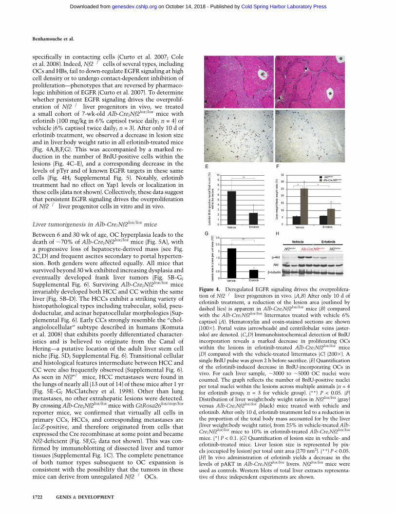
specifically in contacting cells (Curto et al. 2007; Coleet al. 2008). Indeed, Nf2�/� cells of several types, includingOCs and HBs, fail to down-regulate EGFR signaling at highcell density or to undergo contact-dependent inhibition ofproliferation—phenotypes that are reversed by pharmaco-logic inhibition of EGFR (Curto et al. 2007). To determinewhether persistent EGFR signaling drives the overprolif-eration of Nf2�/� liver progenitors in vivo, we treateda small cohort of 7-wk-old Alb-Cre;Nf2lox/lox mice witherlotinib (100 mg/kg in 6% captisol twice daily; n = 4) orvehicle (6% captisol twice daily; n = 3). After only 10 d oferlotinib treatment, we observed a decrease in lesion sizeand in liver:body weight ratio in all erlotinib-treated mice(Fig. 4A,B,F,G). This was accompanied by a marked re-duction in the number of BrdU-positive cells within thelesions (Fig. 4C–E), and a corresponding decrease in thelevels of pTyr and of known EGFR targets in these samecells (Fig. 4H; Supplemental Fig. 5). Notably, erlotinibtreatment had no effect on Yap1 levels or localization inthese cells (data not shown). Collectively, these data suggestthat persistent EGFR signaling drives the overproliferationof Nf2�/� liver progenitor cells in vitro and in vivo.
Liver tumorigenesis in Alb-Cre;Nf2lox/lox mice
Between 6 and 30 wk of age, OC hyperplasia leads to thedeath of ;70% of Alb-Cre;Nf2lox/lox mice (Fig. 5A), witha progressive loss of hepatocyte-derived mass (see Fig.2C,D) and frequent ascites secondary to portal hyperten-sion. Both genders were affected equally. All mice thatsurvived beyond 30 wk exhibited increasing dysplasia andeventually developed frank liver tumors (Fig. 5B–G;Supplemental Fig. 6). Surviving Alb-Cre;Nf2lox/lox miceinvariably developed both HCC and CC within the sameliver (Fig. 5B–D). The HCCs exhibit a striking variety ofhistopathological types including trabecular, solid, pseu-doductular, and acinar hepatocellular morphologies (Sup-plemental Fig. 6). Early CCs strongly resemble the ‘‘chol-angiolocellular’’ subtype described in humans (Komutaet al. 2008) that exhibits poorly differentiated character-istics and is believed to originate from the Canal ofHering—a putative location of the adult liver stem cellniche (Fig. 5D; Supplemental Fig. 6). Transitional cellularand histological features intermediate between HCC andCC were also frequently observed (Supplemental Fig. 6).As seen in Nf2+/� mice, HCC metastases were found inthe lungs of nearly all (13 out of 14) of these mice after 1 yr(Fig. 5E–G; McClatchey et al. 1998). Other than lungmetastases, no other extrahepatic lesions were detected.By crossing Alb-Cre;Nf2lox/lox mice with GtRosa26lox/stop/lox
reporter mice, we confirmed that virtually all cells inprimary CCs, HCCs, and corresponding metastases arelacZ-positive, and therefore originated from cells thatexpressed the Cre recombinase at some point and becameNf2-deficient (Fig. 5F,G; data not shown). This was con-firmed by immunoblotting of dissected liver and tumortissues (Supplemental Fig. 1C). The complete penetranceof both tumor types subsequent to OC expansion isconsistent with the possibility that the tumors in thesemice can derive from unregulated Nf2�/� OCs.
Figure 4. Deregulated EGFR signaling drives the overprolifera-tion of Nf2�/� liver progenitors in vivo. (A,B) After only 10 d oferlotinib treatment, a reduction of the lesion area (outlined bydashed lies) is apparent in Alb-Cre;Nf2lox/lox mice (B) comparedwith the Alb-Cre;Nf2lox/lox littermates treated with vehicle 6%captisol (A). Hematoxylin and eosin-stained sections are shown(1003). Portal veins (arrowheads) and centrilobular veins (aster-isks) are denoted. (C,D) Immunohistochemical detection of BrdUincorporation reveals a marked decrease in proliferating OCswithin the lesions in erlotinib-treated Alb-Cre;Nf2lox/lox mice(D) compared with the vehicle-treated littermates (C) (2003). Asingle BrdU pulse was given 2 h before sacrifice. (E) Quantificationof the erlotinib-induced decrease in BrdU-incorporating OCs invivo. For each liver sample, ;3000 to ;5000 OC nuclei werecounted. The graph reflects the number of BrdU-positive nucleiper total nuclei within the lesions across multiple animals (n = 4for erlotinib group; n = 3 for vehicle group). (**) P < 0.05. (F)Distribution of liver weight:body weight ratios in Nf2lox/lox (gray)versus Alb-Cre;Nf2lox/lox (black) mice treated with vehicle anderlotinib. After only 10 d, erlotinib treatment led to a reduction inthe proportion of the total body mass accounted for by the liver(liver weight:body weight ratio), from 25% in vehicle-treated Alb-
Cre;Nf2lox/lox mice to 10% in erlotinib-treated Alb-Cre;Nf2lox/lox
mice. (*) P < 0.1. (G) Quantification of lesion size in vehicle- anderlotinib-treated mice. Liver lesion size is represented by pix-els (occupied by lesion) per total unit area (270 nm2). (**) P < 0.05.(H) In vivo administration of erlotinib yields a decrease in thelevels of pAKT in Alb-Cre;Nf2lox/lox livers. Nf2lox/lox mice wereused as controls. Western blots of total liver extracts representa-tive of three independent experiments are shown.
Benhamouche et al.
1722 GENES & DEVELOPMENT
Cold Spring Harbor Laboratory Press on October 14, 2018 - Published by genesdev.cshlp.orgDownloaded from

OC proliferation and tumorigenesis upon Nf2 deletionin adult mice: role of proliferative stimuli
To determine whether loss of Nf2 must occur in thecontext of the developing liver in order for OC expansionand tumorigenesis to ensue, we deleted Nf2 in the adultliver by injecting a Cre-expressing adenovirus (Ad-Cre)into 8-wk-old Nf2lox/lox mice (Fig. 6A). After 8–12 mo,only mild periportal hyperplasia was detected in thelivers of these mice (Fig. 6B,D); hepatocyte alterationswere not detectable, and neither CC nor HCC developed.We reasoned that the highly proliferative context of thedeveloping liver in Alb-Cre;Nf2lox/lox embryos might playa critical role in the response to Nf2 loss, as the normaladult liver is mitotically quiescent. Therefore, we in-duced proliferation in the liver of Ad-Cre-injected micevia partial hepatectomy (PHx) (Higgins and Anderson1931). As seen in Alb-Cre;Nf2lox/lox mice, all of thesemice developed marked primary OC hyperplasia and sub-sequent HCC and CC (Fig. 6C,E; data not shown). OChyperplasia was never seen upon PHx of control mice. Asan alternative strategy, we generated Mx1-Cre;Nf2lox/lox
mice in which Cre expression can be induced throughoutthe liver upon stimulation of the interferon pathway viaintraperitoneal injection of polyIC (polyinosinic–polycy-tidylic acid) (Fig. 6F; Kuhn et al. 1995). Again, Cre-mediated inactivation of Nf2 in adult Mx1-Cre;Nf2lox/lox
mice resulted in only a modest activation of OCs after 5–8mo, despite clear evidence of Nf2 recombination/lossthroughout the liver (Fig. 6G; Supplemental Fig. 1B).However, performing a PHx after polyIC induction led
Figure 5. Metastatic liver tumor development in Alb-Cre;
Nf2lox/lox mice. (A) Survival of Alb-Cre;Nf2lox/lox mice. Whilemany animals died within a few months of age, likely dueto compromised liver function, those that survived beyond;30 wk all developed frank HCC and CC. (B) Multiple tumorsare macroscopically evident in a 64-wk-old Alb-Cre;Nf2lox/lox
liver. (C,D) Hematoyxlin and eosin staining reveals both CC andHCC neoplasias in the liver of an 83-wk-old Alb-Cre;Nf2lox/lox;
GtRosa26lox/stop/lox mouse. (C) 1003. (D) 4003. (E–G) Lungmetastases from the liver shown in C. Hematoxylin and eosinstaining (403). HCC metastases in the lung exhibit LacZ
expression, as revealed by whole-mount (F) and paraffin sec-tion (G).
Figure 6. Deletion of Nf2 in the adult liver yields OC hyper-proliferation and tumor development that are promoted by PHx.(A) Two-month-old Nf2lox/lox mice were injected with Ad-Cre,followed 2 wk later by two-third PHx or sham operation, and weresacrificed 8 mo to 1 yr post-PHx. The liver from nonhepatecto-mized mice (B) reveals only mild periportal OC hyperplasia(arrowhead) (D). In contrast, Ad-Cre-injected mice develop multi-ple liver tumors (C) of both CC and HCC types (E) when subjectedto proliferative stimuli induced by PHx. (D,E) Hematoxylin andeosin-stained sections (2003). (F) Two-month-old Mx1-Cre;Nf2lox/lox mice were intraperitoneally injected with polyIC, sub-jected to PHx or sham operation after 2 wk, and sacrificed at 7–8mo. (G,H) Hematoxylin and eosin-stained sections reveal mildperiportal OC hyperplasia (arrowhead) in control, nonhepatec-tomized mice (G), and both CC and HCC in mice subjected toPHx (H) (4003). (I) As in Alb-Cre;Nf2lox/lox livers, lesions inhepatectomized, polyIC-induced Mx1-Cre;Nf2lox/lox livers expressthe OC markers panCK (red) and CD34 (green) (4003).
Nf2 deficiency in the mouse liver
GENES & DEVELOPMENT 1723
Cold Spring Harbor Laboratory Press on October 14, 2018 - Published by genesdev.cshlp.orgDownloaded from

to a marked increase in OC hyperplasia, as well as HCCand CC development with complete penetrance (Fig.6H,I). Notably, after PHx, liver regeneration itself occursnormally in the absence of Merlin, and Nf2�/� hepatocytesre-enter a quiescent state after regeneration is complete;this is followed by the appearance and expansion of OCs.Thus, the proliferative stimulus provided by PHx appearsto specifically unleash the overproliferation of otherwisequiescent Nf2�/� liver progenitors, and is not requiredfor their continued proliferation. This is consistent witha role for Merlin in down-regulating growth factor receptoroutput in liver cells. In fact, intraperitoneal injection ofEGF into Mx1-Cre;Nf2lox/lox mice yielded a ‘‘superinduc-tion’’ of phosphorylated EGFR and the known EGFRsignaling targets pSTAT3 and pAKT in the Nf2�/�
(polyIC-induced) relative to the control liver (no polyIC-mediated induction of Cre expression) (Supplemental Fig.7). Taken together, these findings suggest that Merlinfunction is required to establish and maintain quiescenceof liver progenitor cells.
Stem/progenitor cell-derived tumorigenesis
The overproliferation of OCs preceding the developmentof both HCC and CC in all three models suggests that thetumors originate from these liver progenitor cells. Todetermine whether undifferentiated Nf2�/� liver progen-itors can become neoplastic, we generated clonal lines ofNf2-expressing (wild-type) or Nf2-deficient HBs that alsoexpressed a lacZ reporter from E14.5 embryonic mouselivers, and transplanted them into the livers of adult im-munocompromised nu/nu mice (Supplemental Fig. 1D).Five weeks post-transplantation, lacZ+/Nf2-expressingHBs were scattered uniformly throughout the liver (Fig.7A,B). In contrast, livers from mice transplanted withNf2-deficient HBs revealed multiple foci of proliferatingundifferentiated cells that progressed within only 3–4 wkto form larger neoplasias that exhibited both cholangio-cytic and hepatocytic features (Fig. 7C–F). These datasupport a cell-autonomous requirement for Merlin incontrolling liver progenitor proliferation and neoplastictransformation; they also indicate that liver neoplasia canarise from clonal Nf2�/� liver progenitors.
Discussion
The results presented here provide the first example ofa single gene (Nf2) whose disruption is sufficient to effecta primary expansion of both embryonic and adult liverprogenitor cells in vivo, and to reproducibly yield bothmajor forms of liver cancer. The lesions that arise upondeletion of Nf2 in embryonic or adult mouse liverstrongly resemble classic models of chemically inducedOC expansion both histologically and in terms of markerexpression (Omori et al. 1997; Jakubowski et al. 2005;Kofman et al. 2005; Lee et al. 2006). Our phenotype alsoclosely resembles the OC expansion seen upon trans-genic overexpression of the TNF-like cytokine TWEAK(Jakubowski et al. 2005). However, in contrast to theNf2�/� liver, lesions in TWEAK-overexpressing livers
exhibit increased apoptosis and are not progressive. In-deed, tumors do not develop in these mice. The preciseorigin of OCs has been long debated; most studiesconclude that they arise from unidentified intrahepaticstem cells (Fausto and Campbell 2003; Fausto 2004;Theise 2006), although a hematopoietic origin has beensuggested (Petersen et al. 1999; Thorgeirsson and Grisham2006). Recently, label retention studies have partially sub-stantiated the long-held notion that OCs arise from intra-ductal and periductal locations within the most proximalbranches of the biliary tree (Kuwahara et al. 2008); this isprecisely where OC expansion is first seen in Nf2-deficientlivers. In fact, the initial appearance of OCs in newbornanimals, despite more pervasive expression of Alb-Cre, sug-gests that Nf2 is specifically required for OC quiescenceduring the establishment of the OC niche. Together withthe development of additional sophisticated genetic tools,this model could be used to define the cellular origin of theOC and the physical location of its niche.
Our data suggest that Nf2 mutant liver tumors arederived from Nf2�/� progenitors for the following reasons:First, OC expansion always precedes the development ofboth HCC and CC, regardless of whether Nf2 loss occursin the embryo or adult. Second, lesions in all three models(Alb-Cre;Nf2lox/lox, Ad-Cre-injected Nf2lox/lox, and polyIC-induced Mx1-Cre;Nf2lox/lox) always contain transitionalcellular morphologies with both hepatocytic and biliaryfeatures. Third, transplantation of clonal Nf2�/� HBsyielded the expansion of undifferentiated cells followedby the development of neoplasias bearing cholangiocyticand hepatocytic features, indicating that Nf2�/� liverprogenitors can initiate liver tumors. We cannot excludethe possibility that HCCs can develop from differentiatedhepatocytes in the Nf2-deficient liver, given that hepato-cytes continue to express Alb-Cre. However, Nf2 de-ficiency is clearly not sufficient for the overproliferationor transformation of differentiated hepatocytes, as Alb-Cre;Nf2lox/lox, Ad-Cre-injected Nf2lox/lox, and inducedMx1-Cre;Nf2lox/lox mice harbor many Nf2-deficient hepa-tocytes that do not exhibit gross morphological alterationsor aberrant BrdU incorporation in vivo; in fact, liverregeneration occurs normally in these mice, indicatingthat, even after receiving a proliferative stimulus andundergoing cell division, Nf2�/� hepatocytes can appropri-ately re-enter a quiescent state. This suggests that eitheradditional mutations cooperate with Nf2 loss to drive thedevelopment of HCC in this model, or HCC develops fromNf2�/� progenitors that adopt an improperly differentiatedstate. Indeed, recent studies document the development ofHCC from OCs in carcinogen-treated rats, and suggestthat a subset of human HCCs exhibit a gene expressionsignature that is similar to that of both rodent OCs andHBs (Lee et al. 2006; Andersen et al. 2010).
These models of liver-specific Nf2 deficiency providea unique opportunity to identify the molecular signalsthat regulate homeostasis of both embryonic and adultliver progenitors. We found that deletion of Nf2 in theliver, as in other cell types, yields deregulated EGFRsignaling, and that pharmacologic EGFR inhibitors re-verse the overproliferation of Nf2�/� progenitors in vitro
Benhamouche et al.
1724 GENES & DEVELOPMENT
Cold Spring Harbor Laboratory Press on October 14, 2018 - Published by genesdev.cshlp.orgDownloaded from

and in vivo (Fig. 4; Curto et al. 2007). This, together withthe superinduction of EGFR activity in the induced Mx-Cre;Nf2lox/lox liver, strongly supports a primary role foraberrant EGFR activity in OC expansion and tumorigen-esis in the absence of Nf2, although other receptors mayalso be important (McClatchey and Giovannini 2005;Maitra et al. 2006). This is consistent with many studiesthat have indicated a causal role for EGFR signaling in OCproliferation and liver tumorigenesis in humans and mice(Jhappan et al. 1990; Evarts et al. 1993; Nagy et al. 1996;Yoon et al. 2004; Breuhahn et al. 2006; Blechacz andGores 2008). Although NF2 mutations have not yet beenreported in human liver cancers, our data argue fora systematic mutational analysis across a large, diversepanel of liver tumors; indeed, NF2 mutations could beunique to a subset of tumors that are of progenitor cellorigin (Lee et al. 2006; Roskams 2006; Sell and Leffert2008; Andersen et al. 2010). In fact, the broad conse-quences of Nf2 loss in development and tumorigenesis inthe mouse predict an underappreciated role for NF2mutations in human cancer. As such, our discovery thatkidney-specific Nf2 deletion yields renal carcinomas inthe mouse has been complemented recently by thediscovery of NF2 mutations in a subset of sporadic humanrenal tumors (Forbes et al. 2008; Morris and McClatchey2009; Dalgliesh et al. 2010).
Despite the recent assertion that a critical function ofMerlin is to negatively regulate the Hpo/Wts/Yki tumorsuppressor pathway in Drosophila (Hamaratoglu et al.2006, 2009; Baumgartner et al. 2010; Genevet et al. 2010;Yu et al. 2010), together with recent studies linking thecorresponding mammalian Mst/Lats/Yap pathway toliver tumorigenesis in mice and humans (Zender et al.2006; Dong et al. 2007; Zhou et al. 2009; Lu et al. 2010;Song et al. 2010), our studies suggest an importantfunction for Merlin in the liver that is independent ofthis pathway. Indeed, the reported consequences of inac-tivating the Mst/Lats/Yap pathway in the liver, either byeliminating Mst1/2 or overexpressing YAP, are quitedistinct from that of deleting Nf2 (Camargo et al. 2007;Dong et al. 2007; Zhou et al. 2009; Lu et al. 2010; Songet al. 2010). In fact, overexpression of YAP or deletion of itsnegative regulators, Mst1 and Mst2, in the liver primarilyaffects hepatocytes, yielding aberrant hepatocyte morphol-ogy, proliferation, and, eventually, HCC that is not pre-
ceded by OC expansion (Camargo et al. 2007; Zhou et al.2009; Lu et al. 2010; Song et al. 2010). In contrast, deletionof Nf2 in the liver specifically yields an early and dramaticexpansion of progenitor cells without detectable alter-ations in neighboring hepatocytes, including any increasein nuclear Yap1 (data not shown). Instead, high levels ofcytoplasmic and nuclear Yap1 were apparent in some HCCfoci and metastases and not in others (Supplemental Fig.8), suggesting that Nf2 deficiency is not sufficient to yieldYap1 activation in hepatocytes. Instead, it is possible thatother mutations cooperate with Nf2 loss to achieve fullYAP activation. Similarly, we could not detect a major rolefor Merlin in regulating Yap1 in OCs, and we found thatYap1 was not required for the overproliferation of culturedOCs. Instead, we provide evidence that the overprolifera-tion of Nf2�/� liver progenitors in vitro and in vivo isEGFR-dependent.
Our previous molecular studies indicate that Merlincan mediate contact-dependent inhibition of proliferationby coordinating the establishment of cell:cell contactwith down-regulation of EGFR signaling (Curto et al.2007; Cole et al. 2008). In vivo, all cells are in contact, andcontact-dependent inhibition of proliferation must beoverridden and re-established; for example, during thedevelopmental expansion of tissues. Stem/progenitor cellsmust be constantly poised to override contact-dependentinhibition of proliferation, and may therefore be particu-larly sensitive to loss of Merlin function. Indeed, increas-ing evidence supports a critical role for physical commu-nication between progenitor cells and their niche ingoverning stem cell division (Fuchs et al. 2004; Scadden2006). Thus, studies of the relationship between germlinestem cells and their niche in Drosophila underscore theimportance of reciprocal coordination between intercel-lular contacts and receptor signaling in controlling nichehomeostasis (Yamashita et al. 2005). Similarly, it has beensuggested that cadherin-mediated communication be-tween hematopoietic stem cells and the bone marrowniche in mammals governs their proliferation (Adamset al. 2006). Notably, a recent study revealed that Merlinis necessary for the function and structure of the hema-topoietic stem cell niche, and, consequently, for limitinghematopoietic stem cell numbers (Larsson et al. 2008).Most studies conclude that the adult liver progenitor cellresides near or at the Canal of Hering, where cholangiocytes
Figure 7. Transplantation of clonal Nf2�/� HBsinto the liver of immunocompromised mice yieldsOC hyperplasia and cholangiocellular tumors. (A,B)Five weeks after transplantation into the liver ofnude mice, control HBs are distributed uniformlythroughout the liver parenchyma, as indicated byLacZ expression detected by whole-mount (a sectionof the tissue shown in A is presented in B). (C–F) Fiveweeks after transplantation, Ad-Cre-infected, Nf2-deficient HBs form multiple foci of undifferentiatedLacZ-positive cells (C) that progress to neoplasias (E)exhibiting both hepatocytic and cholangiocytic fea-tures (D,F). Hematoxylin and eosin-stained sectionsare shown in D and F. (B,D) 2003. (F) 4003.
Nf2 deficiency in the mouse liver
GENES & DEVELOPMENT 1725
Cold Spring Harbor Laboratory Press on October 14, 2018 - Published by genesdev.cshlp.orgDownloaded from

and hepatocytes come into physical contact with oneanother (Theise et al. 1999; Paku et al. 2001; Kuwaharaet al. 2008). Our studies reveal a key role for Merlin inestablishing and maintaining the liver progenitor cellniche. Although further studies will be required to moreprecisely define the role of Merlin in regulating liverprogenitor proliferation in vivo, we speculate that it mayfunction to coordinate physical contacts and growthfactor signaling among different cell types in this uniquesetting of intercellular communication.
Materials and methods
Animals
Homozygous Nf2lox/lox mice (FVB/N) were crossed with trans-genic mice expressing the Cre recombinase under a liver-specificalbumin promoter (Alb-Cre) (B6.Cg-Tg[Alb-cre]21Mgn/J; JacksonLaboratories) (Giovannini et al. 2000; Postic and Magnuson 2000)or the Interferon-a-inducible Mx1 promoter (Mx1-Cre) (Tg[Mx1-cre]1Cgn/J; Jackson Laboratories) (Kuhn et al. 1995). To monitorCre-mediated excision, mice were crossed to the reporter strainGtRosa26lox/stop/lox (B6;129-Gt[ROSA]26Sortm1Sho/J; Jackson Lab-oratories) in which removal of a floxed stop cassette allowsexpression of the b-galactosidase/LacZ reporter upon Cre-medi-ated recombination (Mao et al. 1999). Mixed-gender animals wereused for all experiments. Immunocompromised 5-wk-old femalenu/nu mice used for transplantation experiments were obtainedfrom the Massachusetts General Hospital (MGH) Cox7 facility.
Animal procedures
All animal procedures were performed according to federal andinstitutional guidelines, and were approved by the MGH Sub-committee on Research Animal Care. BrdU (Sigma) was injectedintraperitoneally at 100 mg/kg body weight, and mice weresacrificed after 2 h. Activation of the Mx1 promoter was inducedvia two intraperitoneal injections of 250 mg of polyIC (Sigma) at48-h intervals. EGF (Peprotech) was injected intraperitoneally at10 mg/g body weight, and mice were sacrificed after 30 min.Deletion of Nf2 in adult Nf2lox/lox mouse livers was induced viatail vein injection of 150 3 106 plaque-forming units (pfu) of aCre-expressing adenovirus (Ad5CMV-Cre; University of IowaGene Transfer Vector Core). Compensatory liver cell prolifera-tion was induced by two-third PHx performed according to thatdescribed previously in the rat (Higgins and Anderson 1931).
Erlotinib (ChemieTek) was solubilized at 10 mg/mL in a 6%w/v aqueous solution of Captisol (CyDex Pharmaceuticals). FourAlb-Cre;Nf2lox/lox and three Nf2lox/lox control mice were treatedby intraperitoneal injection of erlotinib at 100 mg/kg bodyweight once every 12 h for 10 d. Three Alb-Cre;Nf2lox/lox andthree Nf2lox/lox control mice were treated on the same schedulewith vehicle alone, with no detectable effect.
Transplantation of 3 3 106 HBs into the livers of 8- to 9-wk-oldfemale nude mice was performed via intrasplenic injection(Ponder et al. 1991). To increase engraftment of transplantedHBs into the liver (Laconi et al. 1998), a proliferative stimuluswas provided by a two-third PHx performed at the time of thesplenic injection.
Cell culture
Clonal HB lines were established according to the methoddescribed by Strick-Marchand and Weiss (2002) with some
modifications. The liver of E14.5 embryos were excised, minced,and dissociated in Liver Dissociation Medium (Gibco-Invitrogen)(30 + 30 min rocking at 37°C), and the cell suspension was platedon two collagen I-coated, 100-mm dishes in F12-DMEM medium(1:1) containing 10% FBS, 100 ng/mL EGF, 60 ng/mL IGF-II, and10 mg/mL insulin. Cultures were fed with fresh medium twicea week until epithelial clones appeared. Several clones wereisolated using cloning cylinders and were further expanded. Nf2deletion in cultured HBs was achieved via adenoviral expressionof the Cre-recombinase as described for mouse embryonicfibroblasts (Lallemand et al. 2003). Nf2�/� OC lines (OCs, alsodubbed ‘‘liver-derived cells’’ or LDCs) (Curto et al. 2007) wereobtained from the liver of a 12-wk-old AlbCre;Nf2lox/lox mousewith marked oval/biliary cell hyperplasia without detectablehepatocellular dysplasia or neoplasia. The liver was excised,minced, and dissociated as described above, and cells werecultured in 10% FBS-DMEM. Clonal cell lines with uniformepithelial morphologies were established by limiting dilution.The generation and use of adenoviral vectors expressing Nf2wt
have been described (Lallemand et al. 2003). Infections withretroviral vectors encoding Lats2 (Zhang et al. 2008)—shRNAstargeting Yap (sh1877; sh1883; a kind gift of Dr. Lars Zender,Helmholtz Center for Infection Research, Braunschweig, Germany)and Nf2—were performed as described (Cole et al. 2008). Growthcurves were performed as described (Curto et al. 2007).
Histology and immunohistochemistry
Freshly dissected tissues were fixed in 3.7% formaldehyde–PBS(phosphate-buffered saline), processed, and paraffin-embedded.Five-micron sections were stained with hematoxylin and eosinor processed further for immunohistochemistry. Antigen retrievalwas done by boiling in 10 mM citrate buffer (20 min) for Yap1detection, and 0.1% trypsin (20 min at 37°C) for panCK detection.Sections were incubated for 10 min in 0.3% H2O2-methanol;blocked 1 h in PBS, 1% BSA, and 10% normal goat serum; andincubated overnight at 4°C with the following primary antibodies:anti-Yap1 (1:40; Cell Signaling #4912), anti-panCK (1:1000; Dako#Z0622). BrdU-labeled cells were detected using a kit (Zymed)following the manufacturer’s instructions. The Vectastain Elitekit (Vector Laboratories) was used for HRP-based chromogenicdetection of secondary antibodies. Whole-mount LacZ staining of2- to 3-mm-thick sections was used to detect b-galactosidaseactivity in liver and lung tissues. Freshly dissected tissues werefixed (0.2% glutaraldehyde, 5 mM EGTA, 2 mM MgCl2, 0.1 MNaPO4 buffer at pH 7.3), washed three times in wash buffer (2 mMMgCl2, 0.02% NP-40, 0.01% deoxycholate, 0.1 M NaPO4 buffer atpH 7.3), stained overnight in wash buffer (1 mg/mL X-gal, 5 mMK-ferricyanide, 5 mM K-ferrocyanide) at 37°C with gentle agita-tion, and post-fixed in 3.7% formaldehyde–PBS. Some specimenswere processed further and paraffin-embedded, and 10-mm sec-tions were counterstained with eosin or nuclear fast red forhistological examination.
Immunofluorescent detection of CD34, panCK, and A6 werecarried out on cryosections of frozen, OCT-embedded tissue.Sections were fixed in methanol/acetone (1:1, v/v), blockedin 10% goat serum in PBS for 1 h, and incubated overnight at4°C with primary rat anti-mouse CD34 (1:100; BD-Pharmingen550537), rabbit anti-cytokeratin (wide spectrum screening; anti-panCK) (1:200; Dako Z0622), and rat anti-A6 (1:40; kindly pro-vided by Valentina Factor) in 1% BSA-PBS, followed by secondaryanti-rat Alexa488 (1:200; Molecular Probes) and secondary Cy3-conjugated anti-rabbit (1:200; Jackson Immunoresearch Laborato-ries), respectively, and mounted (Vectashield, Vector Laboratories).Nuclei were stained with DAPI. Two-dimensional epifluorescenceimages were acquired on a Nikon Eclypse 90i microscope with
Benhamouche et al.
1726 GENES & DEVELOPMENT
Cold Spring Harbor Laboratory Press on October 14, 2018 - Published by genesdev.cshlp.orgDownloaded from

a Q-Imaging 2000R camera and NIS AR2.3 software. Final imageswere prepared using Adobe Photoshop CS3.
Immunofluorescence
OCs or HBs were plated on glass coverslips and infected with Ad-Nf2wt (5 multiplicity of infection [m.o.i.]) or Ad-Cre (20 m.o.i.),respectively, when subconfluent. Four days to 5 d after reachingconfluence, cells were fixed for 15 min at room temperature in4% PFA-cytoskeletal buffer (10 mM 2-[N-Morpholino]ethane-sulfonic acid sodium salt [MES] at pH 6.3, 138 mM KCl, 3 mMMgCl2, 2 mM EGTA), and were subsequently permeabilized for15 min in 0.2% Triton X-100 in PBS. Coverslips were blocked in10% goat serum in PBS for 1 h and were incubated overnight at4°C with anti-Yap1 (100; Cell Signaling #49121) in 1% BSA-PBS.Costaining for Merlin was performed by conjugation of the anti-NF2 antibody (1:400; Santa Cruz Biotechnology #sc-331) withthe Zenon Rabbit IgG labeling kit (Molecular Probes) followingthe manufacturer’s instructions. Coverslips were rinsed in PBS,incubated for 1 h at room temperature in FITC- or Cy3-conjugatedsecondary antibodies (Jackson Immunoresearch Laboratories), andmounted (Vectashield, Vector Laboratories). Nuclei were stainedwith DAPI. Two-dimensional epifluorescence images were ac-quired on a Nikon Eclypse 90i microscope with a Q-Imaging2000R camera and NIS AR2.3 software. Final images were pre-pared using Adobe Photoshop CS3.
Immunoblotting
Liver tissue was homogenized at 4°C in 10 vol (w/v) of RIPAbuffer (1% Triton X-100, 0.5% Na-deoxycholate, 0.1% SDS, 50mM Tris-HCl at pH 7.4, 140 mM NaCl, 1 mM EDTA, 1 mMEGTA, 1 mM PMSF, 1 mM Na3VO4, 10 mg/mL each aprotinin,pepstatin, leupeptin) using a Teflon Potter-Elvehjem tissue grinder.Protein extracts from liver (120 mg per lane) or cultured cells (30 mgper lane) were analyzed by Western blotting using the followingprimary antibodies: EGFR-pY992 (#2235), EGFR-pY1068 (#2234),STAT3-pY705 (#9145), AKT-pS473 (#4060), YAP-pS127 (#4911),and YAP (#4912), all at 1:1000 (Cell Signaling); NF2 (#sc-331) at1:5,000 and EGFR (#sc-03) at 1:1000 (Santa Cruz Biotechnology);Lats2 (#A300-479A) at 1:1000 (Bethyl); Orc2 (#920-4-41) at 1:1000(BD-Pharmingen). HRP-conjugated secondary anti-mouse andanti-rabbit antibodies were from Amersham. For nuclear/cytoplas-mic fractionation, we used the NE-PER kit from Pierce followingthe manufacturer’s instructions.
PCR
Genotyping PCR from tissues and cells was performed as de-scribed for Nf2 (Giovannini et al. 2000), Cre recombinase, andGtRosa26 (Jackson Laboratory; http://jaxmice.jax.org/pub-cgi/protocols/protocols.sh?objtype=prot_list). For semiquantitativeand real-time quantitative RT–PCR, total cellular RNA wasextracted with TRIzol (Invitrogen), and reverse-transcribed withMMLV-RT (Promega) using oligo-dT primers. SemiquantitativeRT–PCR was carried out as described in Tanimizu et al. (2003)using primers described therein. Fast Start Universal SYBR Greenmix (Roche Applied Science) was used to amplify 0.5 mL of the RTreaction in a final 25-mL volume. Triplicate samples were run ona LightCycler 480 System (Roche Applied Science). Cycling condi-tions were denaturation for 15 sec at 95°C, annealing for 1 min at60°C, and extension for 1 min at 60°C, 40 cycles. Expression of theb-actin gene was used as an internal control. Primer sequences in59 to 39 orientation were as follows: AFP, f-TTCGTATTCCAACAGGAGGC and r-CAGACTTCCTGGTCCTGGGC; bActin, f-CTAAGGCCAACCGTGAAAAG and r-ACCAGAGGCATACAGGG
ACA; Birc2, f-GAAGAAAATGCTGACCCTACAGA and r-GCTCATCATGACGACATCTTTC; Birc3, f-AGAGAGGAGCAGATGGAGCA and r-TTTGTTCTTCCGGATTAGTGC; Sox4, f-CCTCGCTCTCCTCGTCCT and r-TCGTCTTCGAACTCGTCGT; Myc,f-CCTAGTGCTGCATGAGGAGA and r-TCTTCCTCATCTTCTTGCTCTTC.
Acknowledgments
We gratefully acknowledge Othon Iliopoulos and all members ofthe McClatchey laboratory for helpful discussions and com-ments on the manuscript, Valentina Factor (NIH, Bethesda) forthe A6 antibody, Lars Zender for the Yap shRNA expressionvectors, Daniel Haber and Jianmin Zhang for the Lats expressionvectors, and Anna Levitz and the Pathology Core Facility forhistological support. This work was supported by The Interna-tional Human Frontier Science Program Organization to S.B.,and by grants from the Tucker-Gosnell Foundation, Departmentof Defense (DOD), National Institutes of Health (NIH), and theChildren’s Tumor Foundation (CTF) to A.I.M.
References
Adams GB, Chabner KT, Alley IR, Olson DP, SzczepiorkowskiZM, Poznansky MC, Kos CH, Pollak MR, Brown EM,Scadden DT. 2006. Stem cell engraftment at the endostealniche is specified by the calcium-sensing receptor. Nature
439: 599–603.Alison MR. 2005. Liver stem cells: Implications for hepatocar-
cinogenesis. Stem Cell Rev 1: 253–260.Andersen JB, Loi R, Perra A, Factor VM, Ledda-Columbano GM,
Columbano A, Thorgeirsson SS. 2010. Progenitor-derivedhepatocellular carcinoma model in the rat. Hepatology 51:1401–1409.
Baumgartner R, Poernbacher I, Buser N, Hafen E, Stocker H.2010. The WW domain protein Kibra acts upstream of Hippoin Drosophila. Dev Cell 18: 309–316.
Blechacz B, Gores GJ. 2008. Cholangiocarcinoma: Advances inpathogenesis, diagnosis, and treatment. Hepatology 48: 308–321.
Breuhahn K, Longerich T, Schirmacher P. 2006. Dysregulation ofgrowth factor signaling in human hepatocellular carcinoma.Oncogene 25: 3787–3800.
Camargo FD, Gokhale S, Johnnidis JB, Fu D, Bell GW, JaenischR, Brummelkamp TR. 2007. YAP1 increases organ size andexpands undifferentiated progenitor cells. Curr Biol 17:2054–2060.
Cole BK, Curto M, Chan AW, McClatchey AI. 2008. Localiza-tion to the cortical cytoskeleton is necessary for Nf2/merlin-dependent epidermal growth factor receptor silencing. Mol
Cell Biol 28: 1274–1284.Colnot S, Decaens T, Niwa-Kawakita M, Godard C, Hamard G,
Kahn A, Giovannini M, Perret C. 2004. Liver-targeteddisruption of Apc in mice activates b-catenin signaling andleads to hepatocellular carcinomas. Proc Natl Acad Sci 101:17216–17221.
Crosby HA, Kelly DA, Strain AJ. 2001. Human hepatic stem-like cells isolated using c-kit or CD34 can differentiate intobiliary epithelium. Gastroenterology 120: 534–544.
Curto M, Cole BK, Lallemand D, Liu CH, McClatchey AI. 2007.Contact-dependent inhibition of EGFR signaling by Nf2/Merlin. J Cell Biol 177: 893–903.
Dalgliesh GL, Furge K, Greenman C, Chen L, Bignell G, ButlerA, Davies H, Edkins S, Hardy C, Latimer C, et al. 2010.Systematic sequencing of renal carcinoma reveals inactiva-tion of histone modifying genes. Nature 463: 360–363.
Nf2 deficiency in the mouse liver
GENES & DEVELOPMENT 1727
Cold Spring Harbor Laboratory Press on October 14, 2018 - Published by genesdev.cshlp.orgDownloaded from

Dong J, Feldmann G, Huang J, Wu S, Zhang N, Comerford SA,Gayyed MF, Anders RA, Maitra A, Pan D. 2007. Elucidationof a universal size-control mechanism in Drosophila andmammals. Cell 130: 1120–1133.
Edgar BA. 2006. From cell structure to transcription: Hippoforges a new path. Cell 124: 267–273.
Evarts RP, Nagy P, Marsden E, Thorgeirsson SS. 1987. A pre-cursor-product relationship exists between oval cells andhepatocytes in rat liver. Carcinogenesis 8: 1737–1740.
Evarts RP, Hu Z, Fujio K, Marsden ER, Thorgeirsson SS. 1993.Activation of hepatic stem cell compartment in the rat: Roleof transforming growth factor a, hepatocyte growth factor,and acidic fibroblast growth factor in early proliferation. CellGrowth Differ 4: 555–561.
Farber E. 1956. Similarities in the sequence of early histologicalchanges induced in the liver of the rat by ethionine,2-acetylamino-fluorene, and 39-methyl-4-dimethylaminoazo-benzene. Cancer Res 16: 142–148.
Fausto N. 2004. Liver regeneration and repair: Hepatocytes,progenitor cells, and stem cells. Hepatology 39: 1477–1487.
Fausto N, Campbell JS. 2003. The role of hepatocytes and ovalcells in liver regeneration and repopulation. Mech Dev 120:117–130.
Fausto N, Campbell JS, Riehle KJ. 2006. Liver regeneration.Hepatology 43: S45–S53. doi: 10.1002/hep.20969.
Forbes SA, Bhamra G, Bamford S, Dawson E, Kok C, Clements J,Menzies A, Teague JW, Futreal PA, Stratton MR. 2008. Thecatalogue of somatic mutations in cancer (COSMIC). CurrProtoc Hum Genet 57: 10.11.1–10.11.26. doi: 10.1002/0471142905.hg1011s57.
Fuchs E, Tumbar T, Guasch G. 2004. Socializing with theneighbors: Stem cells and their niche. Cell 116: 769–778.
Genevet A, Wehr MC, Brain R, Thompson BJ, Tapon N. 2010.Kibra is a regulator of the Salvador/Warts/Hippo signalingnetwork. Dev Cell 18: 300–308.
Giovannini M, Robanus-Maandag E, van der Valk M, Niwa-Kawakita M, Abramowski V, Goutebroze L, Woodruff JM,Berns A, Thomas G. 2000. Conditional biallelic Nf2 muta-tion in the mouse promotes manifestations of human neu-rofibromatosis type 2. Genes Dev 14: 1617–1630.
Hamaratoglu F, Willecke M, Kango-Singh M, Nolo R, Hyun E,Tao C, Jafar-Nejad H, Halder G. 2006. The tumour-suppressorgenes NF2/Merlin and Expanded act through Hippo signallingto regulate cell proliferation and apoptosis. Nat Cell Biol 8:27–36.
Hamaratoglu F, Gajewski K, Sansores-Garcia L, Morrison C, TaoC, Halder G. 2009. The Hippo tumor-suppressor pathwayregulates apical-domain size in parallel to tissue growth.J Cell Sci 122: 2351–2359.
Higgins GM, Anderson RM. 1931. Experimental pathology ofthe liver, I: Restoration of the liver of the white rat fol-lowing partial surgical removal. Arch Pathol (Chic) 12: 186–202.
Jakubowski A, Ambrose C, Parr M, Lincecum JM, Wang MZ,Zheng TS, Browning B, Michaelson JS, Baetscher M, Wang B,et al. 2005. TWEAK induces liver progenitor cell prolifera-tion. J Clin Invest 115: 2330–2340.
Jelnes P, Santoni-Rugiu E, Rasmussen M, Friis SL, Nielsen JH,Tygstrup N, Bisgaard HC. 2007. Remarkable heterogeneitydisplayed by oval cells in rat and mouse models of stemcell-mediated liver regeneration. Hepatology 45: 1462–1470.
Jhappan C, Stahle C, Harkins RN, Fausto N, Smith GH, MerlinoGT. 1990. TGFa overexpression in transgenic mice inducesliver neoplasia and abnormal development of the mammarygland and pancreas. Cell 61: 1137–1146.
Kalamarides M, Niwa-Kawakita M, Leblois H, Abramowski V,Perricaudet M, Janin A, Thomas G, Gutmann DH, GiovanniniM. 2002. Nf2 gene inactivation in arachnoidal cells is rate-limiting for meningioma development in the mouse. Genes
Dev 16: 1060–1065.Kiguchi K, Carbajal S, Chan K, Beltran L, Ruffino L, Shen J,
Matsumoto T, Yoshimi N, DiGiovanni J. 2001. Constitutiveexpression of ErbB-2 in gallbladder epithelium results indevelopment of adenocarcinoma. Cancer Res 61: 6971–6976.
Kofman AV, Morgan G, Kirschenbaum A, Osbeck J, Hussain M,Swenson S, Theise ND. 2005. Dose- and time-dependent ovalcell reaction in acetaminophen-induced murine liver injury.Hepatology 41: 1252–1261.
Komuta M, Spee B, Vander Borght S, De Vos R, Verslype C, AertsR, Yano H, Suzuki T, Matsuda M, Fujii H, et al. 2008.Clinicopathological study on cholangiolocellular carcinomasuggesting hepatic progenitor cell origin. Hepatology 47:1544–1556.
Kuhn R, Schwenk F, Aguet M, Rajewsky K. 1995. Inducible genetargeting in mice. Science 269: 1427–1429.
Kuwahara R, Kofman AV, Landis CS, Swenson ES, BarendswaardE, Theise ND. 2008. The hepatic stem cell niche: Identifi-cation by label-retaining cell assay. Hepatology 47: 1994–2002.
Laconi E, Oren R, Mukhopadhyay DK, Hurston E, Laconi S, PaniP, Dabeva MD, Shafritz DA. 1998. Long-term, near-total liverreplacement by transplantation of isolated hepatocytes inrats treated with retrorsine. Am J Pathol 153: 319–329.
Lallemand D, Curto M, Saotome I, Giovannini M, McClatcheyAI. 2003. NF2 deficiency promotes tumorigenesis andmetastasis by destabilizing adherens junctions. Genes
Dev 17: 1090–1100.Lallemand D, Manent J, Couvelard A, Watilliaux A, Siena M,
Chareyre F, Lampin A, Niwa-Kawakita M, Kalamarides M,Giovannini M. 2009. Merlin regulates transmembrane re-ceptor accumulation and signaling at the plasma membranein primary mouse Schwann cells and in human schwanno-mas. Oncogene 28: 854–865.
Larsson J, Ohishi M, Garrison B, Aspling M, Janzen V, AdamsGB, Curto M, McClatchey AI, Schipani E, Scadden DT. 2008.Nf2/merlin regulates hematopoietic stem cell behavior byaltering microenvironmental architecture. Cell Stem Cell 3:221–227.
Lee JH, Rim HJ, Sell S. 1997. Heterogeneity of the ‘oval-cell’response in the hamster liver during cholangiocarcinogenesisfollowing Clonorchis sinensis infection and dimethylnitro-samine treatment. J Hepatol 26: 1313–1323.
Lee JS, Heo J, Libbrecht L, Chu IS, Kaposi-Novak P, Calvisi DF,Mikaelyan A, Roberts LR, Demetris AJ, Sun Z, et al. 2006. Anovel prognostic subtype of human hepatocellular carci-noma derived from hepatic progenitor cells. Nat Med 12:410–416.
Llovet JM, Burroughs A, Bruix J. 2003. Hepatocellular carci-noma. Lancet 362: 1907–1917.
Lu L, Li Y, Kim SM, Bossuyt W, Liu P, Qiu Q, Wang Y, Halder G,Finegold MJ, Lee JS, et al. 2010. Hippo signaling is a potent invivo growth and tumor suppressor pathway in the mamma-lian liver. Proc Natl Acad Sci 107: 1437–1442.
Maitra S, Kulikauskas RM, Gavilan H, Fehon RG. 2006. Thetumor suppressors Merlin and Expanded function coopera-tively to modulate receptor endocytosis and signaling. Curr
Biol 16: 702–709.Mao X, Fujiwara Y, Orkin SH. 1999. Improved reporter strain for
monitoring Cre recombinase-mediated DNA excisions inmice. Proc Natl Acad Sci 96: 5037–5042.
Benhamouche et al.
1728 GENES & DEVELOPMENT
Cold Spring Harbor Laboratory Press on October 14, 2018 - Published by genesdev.cshlp.orgDownloaded from

McClatchey AI, Fehon RG. 2009. Merlin and the ERM proteins–regulators of receptor distribution and signaling at the cellcortex. Trends Cell Biol 19: 198–206.
McClatchey AI, Giovannini M. 2005. Membrane organizationand tumorigenesis—the NF2 tumor suppressor, Merlin.Genes Dev 19: 2265–2277.
McClatchey AI, Saotome I, Mercer K, Crowley D, Gusella JF,Bronson RT, Jacks T. 1998. Mice heterozygous for a mutationat the Nf2 tumor suppressor locus develop a range of highlymetastatic tumors. Genes Dev 12: 1121–1133.
Michalopoulos GK. 2007. Liver regeneration. J Cell Physiol 213:286–300.
Morris ZS, McClatchey AI. 2009. Aberrant epithelial morphol-ogy and persistent epidermal growth factor receptor signal-ing in a mouse model of renal carcinoma. Proc Natl Acad Sci
106: 9767–9772.Morrison H, Sherman LS, Legg J, Banine F, Isacke C, Haipek CA,
Gutmann DH, Ponta H, Herrlich P. 2001. The NF2 tumorsuppressor gene product, merlin, mediates contact inhibitionof growth through interactions with CD44. Genes Dev 15:968–980.
Murakami H, Sanderson ND, Nagy P, Marino PA, Merlino G,Thorgeirsson SS. 1993. Transgenic mouse model for syner-gistic effects of nuclear oncogenes and growth factors intumorigenesis: Interaction of c-myc and transforming growthfactor a in hepatic oncogenesis. Cancer Res 53: 1719–1723.
Nagy P, Bisgaard HC, Santoni-Rugiu E, Thorgeirsson SS. 1996. Invivo infusion of growth factors enhances the mitogenicresponse of rat hepatic ductal (oval) cells after administrationof 2-acetylaminofluorene. Hepatology 23: 71–79.
Okada T, Lopez-Lago M, Giancotti FG. 2005. Merlin/NF-2mediates contact inhibition of growth by suppressing re-cruitment of Rac to the plasma membrane. J Cell Biol 171:361–371.
Omori N, Omori M, Evarts RP, Teramoto T, Miller MJ, HoangTN, Thorgeirsson SS. 1997. Partial cloning of rat CD34cDNA and expression during stem cell-dependent liver re-generation in the adult rat. Hepatology 26: 720–727.
Paku S, Schnur J, Nagy P, Thorgeirsson SS. 2001. Origin andstructural evolution of the early proliferating oval cells in ratliver. Am J Pathol 158: 1313–1323.
Petersen BE, Bowen WC, Patrene KD, Mars WM, Sullivan AK,Murase N, Boggs SS, Greenberger JS, Goff JP. 1999. Bonemarrow as a potential source of hepatic oval cells. Science
284: 1168–1170.Ponder KP, Gupta S, Leland F, Darlington G, Finegold M,
DeMayo J, Ledley FD, Chowdhury JR, Woo SL. 1991. Mousehepatocytes migrate to liver parenchyma and function in-definitely after intrasplenic transplantation. Proc Natl Acad
Sci 88: 1217–1221.Postic C, Magnuson MA. 2000. DNA excision in liver by an
albumin-Cre transgene occurs progressively with age. Gen-
esis 26: 149–150.Roskams T. 2006. Liver stem cells and their implication in
hepatocellular and cholangiocarcinoma. Oncogene 25: 3818–3822.
Roskams TA, Theise ND, Balabaud C, Bhagat G, Bhathal PS,Bioulac-Sage P, Brunt EM, Crawford JM, Crosby HA, DesmetV, et al. 2004. Nomenclature of the finer branches of thebiliary tree: Canals, ductules, and ductular reactions inhuman livers. Hepatology 39: 1739–1745.
Sandgren EP, Quaife CJ, Pinkert CA, Palmiter RD, Brinster RL.1989. Oncogene-induced liver neoplasia in transgenic mice.Oncogene 4: 715–724.
Santoni-Rugiu E, Nagy P, Jensen MR, Factor VM, ThorgeirssonSS. 1996. Evolution of neoplastic development in the liver of
transgenic mice co-expressing c-myc and transforminggrowth factor-a. Am J Pathol 149: 407–428.
Scadden DT. 2006. The stem-cell niche as an entity of action.Nature 441: 1075–1079.
Sell S. 2001. Heterogeneity and plasticity of hepatocyte lineagecells. Hepatology 33: 738–750.
Sell S, Leffert HL. 2008. Liver cancer stem cells. J Clin Oncol 26:2800–2805.
Shiojiri N. 1981. Enzymo- and immunocytochemical analysesof the differentiation of liver cells in the prenatal mouse.J Embryol Exp Morphol 62: 139–152.
Shiojiri N, Lemire JM, Fausto N. 1991. Cell lineages and ovalcell progenitors in rat liver development. Cancer Res 51:2611–2620.
Song H, Mak KK, Topol L, Yun K, Hu J, Garrett L, Chen Y,Park O, Chang J, Simpson RM, et al. 2010. MammalianMst1 and Mst2 kinases play essential roles in organ sizecontrol and tumor suppression. Proc Natl Acad Sci 107:1431–1436.
Strick-Marchand H, Weiss MC. 2002. Inducible differentiationand morphogenesis of bipotential liver cell lines from wild-type mouse embryos. Hepatology 36: 794–804.
Tanabe KK, Lemoine A, Finkelstein DM, Kawasaki H, Fujii T,Chung RT, Lauwers GY, Kulu Y, Muzikansky A, Kuruppu D,et al. 2008. Epidermal growth factor gene functional poly-morphism and the risk of hepatocellular carcinoma inpatients with cirrhosis. JAMA 299: 53–60.
Tanimizu N, Nishikawa M, Saito H, Tsujimura T, Miyajima A.2003. Isolation of hepatoblasts based on the expression ofDlk/Pref-1. J Cell Sci 116: 1775–1786.
Theise ND. 2006. Gastrointestinal stem cells. III. Emergent themesof liver stem cell biology: Niche, quiescence, self-renewal, andplasticity. Am J Physiol Gastrointest Liver Physiol 290: G189–G193. doi: 10.1152/ajpgi.00041.2005.
Theise ND, Saxena R, Portmann BC, Thung SN, Yee H,Chiriboga L, Kumar A, Crawford JM. 1999. The canals ofHering and hepatic stem cells in humans. Hepatology 30:1425–1433.
Thorgeirsson SS, Grisham JW. 2006. Hematopoietic cells ashepatocyte stem cells: A critical review of the evidence.Hepatology 43: 2–8.
Xu X, Kobayashi S, Qiao W, Li C, Xiao C, Radaeva S, Stiles B,Wang RH, Ohara N, Yoshino T, et al. 2006. Induction ofintrahepatic cholangiocellular carcinoma by liver-specificdisruption of Smad4 and Pten in mice. J Clin Invest 116:1843–1852.
Yamashita YM, Fuller MT, Jones DL. 2005. Signaling in stemcell niches: Lessons from the Drosophila germline. J Cell Sci
118: 665–672.Yoon JH, Gwak GY, Lee HS, Bronk SF, Werneburg NW, Gores GJ.
2004. Enhanced epidermal growth factor receptor activationin human cholangiocarcinoma cells. J Hepatol 41: 808–814.
Yu J, Zheng Y, Dong J, Klusza S, Deng WM, Pan D. 2010. Kibrafunctions as a tumor suppressor protein that regulates Hipposignaling in conjunction with Merlin and Expanded. Dev
Cell 18: 288–299.Zender L, Spector MS, Xue W, Flemming P, Cordon-Cardo C,
Silke J, Fan ST, Luk JM, Wigler M, Hannon GJ, et al. 2006.Identification and validation of oncogenes in liver cancerusing an integrative oncogenomic approach. Cell 125: 1253–1267.
Zhang J, Smolen GA, Haber DA. 2008. Negative regulationof YAP by LATS1 underscores evolutionary conservationof the Drosophila Hippo pathway. Cancer Res 68: 2789–2794.
Nf2 deficiency in the mouse liver
GENES & DEVELOPMENT 1729
Cold Spring Harbor Laboratory Press on October 14, 2018 - Published by genesdev.cshlp.orgDownloaded from

Zhao B, Lei QY, Guan KL. 2008. The Hippo–YAP pathway: Newconnections between regulation of organ size and cancer.Curr Opin Cell Biol 20: 638–646.
Zhou D, Conrad C, Xia F, Park JS, Payer B, Yin Y, Lauwers GY,Thasler W, Lee JT, Avruch J, et al. 2009. Mst1 and Mst2maintain hepatocyte quiescence and suppress hepatocellularcarcinoma development through inactivation of the Yap1oncogene. Cancer Cell 16: 425–438.
Benhamouche et al.
1730 GENES & DEVELOPMENT
Cold Spring Harbor Laboratory Press on October 14, 2018 - Published by genesdev.cshlp.orgDownloaded from

10.1101/gad.1938710Access the most recent version at doi: originally published online July 30, 201024:2010, Genes Dev.
Samira Benhamouche, Marcello Curto, Ichiko Saotome, et al. liver
/Merlin controls progenitor homeostasis and tumorigenesis in theNf2
Material
Supplemental
http://genesdev.cshlp.org/content/suppl/2010/07/21/gad.1938710.DC1
Related Content
Genes Dev. August , 2010 24: 1673-1679
Chunling Yi and Joseph L. KissilMerlin in organ size control and tumorigenesis: Hippo versus EGFR?
References
http://genesdev.cshlp.org/content/24/16/1718.full.html#related-urls
Articles cited in:
http://genesdev.cshlp.org/content/24/16/1718.full.html#ref-list-1This article cites 84 articles, 27 of which can be accessed free at:
License
ServiceEmail Alerting
click here.right corner of the article or
Receive free email alerts when new articles cite this article - sign up in the box at the top
Copyright © 2010 by Cold Spring Harbor Laboratory Press
Cold Spring Harbor Laboratory Press on October 14, 2018 - Published by genesdev.cshlp.orgDownloaded from