Embed Size (px)
Citation preview

Inside This IssueFrom the Chair ........................................... 1Stem Cells .................................................... 1Dr. Lefkowitch ............................................ 3Anatomic Pathology .................................. 3Gundersen Lab ........................................... 6New Faculty ................................................ 8Charlie Noback In Memorium ................ 9Our Predecessors ....................................... 9Microscopy ................................................ 11
From the ChairmanThe Spring of the year is always busy as we prepare our budget for the coming year. This year was exceptionally so with the added activity resulting from the stimulus package. I would like to thank the entire administrative staff of the department for enabling us to get out the budget and get out the grants.
The past quarter has seen a substantial change in the academic administration of the department. Alain Borczuk, Professor of Clinical Pathology and Cell Biology has been named Vice-Chairman for Anatomic Pathology. He will be assisted by Edward Kritchevski, MBA, Administrative Direc-tor of Anatomic Pathology. Chuck Mar-boe will continue as Vice-Chairman for Education and will add responsibilities in career development of clinical faculty (see article below). Steve Spitalnik remains Vice-Chairman and Director of Labora-tories and Carlos Cordon-Cardo as Vice-Chairman for Translational Research.
The past week has seen the departure of Samih Nasr, MD from Renal Pathology for a position at Mayo and the appointment of Leal Herlitz, MD as Assistant Professor in that division. Two specialists in GI patholo-gy, Roger Moreira, MD and Fei Bao, MD will be joining us as Assistant Professors in July. In Laboratory Medicine, Harold Kaplan, MD has retired. Dr. Kaplan was a leader in transfusion medicine for many years and a pioneer in the study of errors in blood bank-ing and their elimination. Two new faculty members, Alexander Rai, PhD and Jennifer Daniel-Johnson, MD will join us in clinical chemistry and transfusion medicine respec-tively. A nationwide search for additional faculty members in Cell Biology has led to the hiring of Ulrich Hengst, PhD and Julie Canman, PhD. Dr. Hengst will start in Oc-tober and Dr. Canman in February. I would like to thank Richard Vallee, Gregg Gunder-sen, Ron Liem, Liza Pon, Gil Di Paolo, Ying-hui Mao and Fred Chang (Microbiology) for their work in selecting Ulrich and Julie from a pool of almost 300 applicants.
We are pleased to present this issue of the Columbia Pathology and Cell Biology Report. De-spite the difficult times, our Department continues to thrive. This issue describes some of the growing work our members are doing in stem cell research. We also welcome new faculty and administrators. Our normal format of a column on each of our three divisions – Anatomic Pathology, Clinical Pathology and Cell and Molecular Biology has been put aside for this is-sue. The pressure of writing for the Stimulus grants was too great to ask our members to write more. We will return to that format in the next issue. In this issue, our Chair tells us how we are doing financially and we recognize the efforts of our staff. We are pleased to congratulate Jay Lefkowich for the forthcoming publication of not one, but two books.
Continued on page 6
Stem Cells Proliferate in Pathology and Cell BiologyBy James Goldman Interim Director of the Stem Cell Initiative
Stem cells are featured prominently in cur-rent news reports because they hold promise for ameliorating diabetes, neurodegenera-tive disorders, vascular diseases that dam-age the heart, brain, and other organs and for generating new livers, bones, cartilage, bone marrow, skin, teeth, and nerve cells. And did I mention new strategies to treat cancer? Despite the potential of stem cells, there remains a huge amount of work to do before we can translate our basic knowledge of these cells into therapeutic applications.
Columbia University Medical Center has recently announced a new Stem Cell Initia-tive focusing on the identification and gen-eration of stem cells in many organs and the therapeutic uses of stem cells for treating human diseases. This Initiative was pro-posed for several reasons. First, the science of stem cell biology is progressing rapidly. We now understand something about the basic molecular biological pathways and controls underlying stem cell function and in some organs, how stem cells develop into mature cells. We also are starting to be able to generate stem-like cells from adult tissues and then to differentiate these immature cells into differentiated cells. Second, New York State has allocated funds for stem cell research, some of which have already been disbursed to CUMC and other NY institu-
The Art of Cell Biology
This splendid image comes from new faculty member Julie Canman who will join us in Febru-ary. It is an early C. elegans embryo undergoing the first mitotic division fixed and stained for microtubules (green), DNA (blue), and a micro-tubule motor essential for cell division (red). Julie is profiled in this issue.
Continued on page 2
Volume 2, Issue 1 Fall 2009

tions. Third, a large number of laboratories (over 70) across the CUMC campus work on stem cells or stem-like cells in a variety of or-gan systems, including the nervous system, skin, pancreas, gastrointestinal tract, eye, bone marrow, cardiovascular tissues, bone, cartilage, and teeth. Other laboratories work on cancer stem cells. Many of these laboratories share common goals, ideas, and techniques. Thus, one purpose of the Initia-tive is to promote communication and shar-ing of resources among laboratories.
Members of the Department of Pathol-ogy and Cell Biology have been taking major roles in developing new approaches to stem cell biology. Asa Abeliovich has developed methods for generating dopa-minergic neurons from embryonic stem cells, using combinations of transcription factors and miRNAs. A major goal is to understand and treat Parkinson disease. Peter Canoll is interested in the genesis of gliomas, and has developed experimental models of brain tumors from glial precur-sor cells in the adult central nervous sys-tem. Fiona Doetsch studies basic proper-ties of neural stem cells to understand the mechanisms by which they develop into neurons in the adult central nervous sys-tem. She has found that stem cells in the adult brain have properties of astrocytes and exist in specialized niches, and she is working to understand what regulates the transformation of stem cells into neurons. Michael Gershon investigates the develop-ment of the enteric nervous system from pluripotent cells of the neural crest. James Goldman, the interim head of the Stem Cell Initiative, studies glial precursors in the central nervous system, to understand how they can be used to repair tissues in demyelinating diseases like multiple scle-rosis. Christopher Henderson and Hynek Wichterle have recently turned skin fibro-blasts from a patient with amyotrophic lateral sclerosis back into stem-like cells and from there into motor neurons. Trans-forming a patient’s own skin fibroblasts, or other cells, into a cell type that is affected by that individual’s disease is attractive for several reasons. Scientists could study the evolution and cellular pathology of the disease in cells of the individual’s own genetic background, and theoretically us-ing the patient’s own cells would solve im-munological problems of transplantation.
Tae-Wan Kim is using embryonic stem cell technology to differentiate ES cells into neurons of the cerebral cortex for his studies of Alzheimer disease. Edward Laufer is investigating the nature of stem cells in the adrenal cortex, an organ that has not been well studied from a stem cell point of view. David Owens has identi-fied populations of multipotent epidermal stem cells in the hair follicles of adult skin. He is interested in the lineages of cells to which these populations give rise and also how these stem cells might participate in the evolution of skin cancers. Dominique Toran-Allerand studies estrogen receptors in the central nervous system. She thinks that a receptor for 17-alpha-estradiol may be important in regulating the genesis of new neurons in the adult brain. Richard Vallee studies basic mechanisms by which neuronal precursors of the embryonic ven-tricular zone migrate into the cortex. He focuses on molecular motors, since muta-tions in motor genes can cause abnormal cortical development.
New knowledge about stem cells will un-doubtedly contribute to a better under-standing of cancer biology and treatment. A number of Pathology researchers and clinicians who work on cancer are incor-porating findings in stem cell biology into their own studies or are discovering genes and signaling pathways important for the normal regulation of stem cell development that are dysregulated in carcinogenesis.
Anniversaries – 25 YearsYes, there are those who have served a long time. When these people started the neigh-borhood was suffering, crack and AIDS were the great subjects and Don Tapley was the Dean. Thanks for hanging in there and serving so well.
Neelwatie Khan Bookkeeper
Charles M. Marboe, MD
Vivette D. D’Agati, MD
Kathleen M. O’Toole, MD
Our Antique Microscope
The Department’s Antique Microscope stored in dusty splendor in Mike Shelanski’s office is a Powell and Lealand binocular made about 1870. It was highly regarded in its time but was soon outstripped by Zeiss. The company made microscopes in London from the 1840s to 1924. We do not yet know to whom it belonged. Attempts to see anything through it failed. Others are welcome to try.
Stem Cells Proliferate continued from page 1
A Powell and Lealand microscope appears in this drawing of Louis Pasteur visiting the Whitbread brewery laboratories in 1871. Their beer had gone sour and Pasteur showed them that bacteria rather than yeast were growing in their fermentation vats. An intense nationalist, he hated the fact that German beer was superior and set out to improve French and English beer.
�

Dr. Jay Lefkowitch has two new books com-ing out next year and so the Newsletter de-cided to note the occasion and to interview him. Dr. Lefkowitch (aka Jay) has been a member of the Department for years and has had a legendary career (far from over) in pathology, teaching, and writing – all ac-complished with sheer good humor.
Cell Biology and Pathology Reports (CPR): Two books?
JHL: It just worked out that way. They are new editions, but still a lot of work. The first is Scheuer’s Liver Biopsy Interpreta-tion. We did the last issue with Dr. Scheuer when he was still alive but since he passed away in �006, I have taken it on. I should say that this is a 40 year tradition, since Peter Scheuer started it in 1968. Longevity and continuity are important in medicine. Since he was such an important figure the next edition of the book and its future edi-tions will be named after him.
CPR: Can you give us some idea of how widely used this book has been?
JHL: It is the most widely used bench text of liver pathology in the world, and, I am happy to say it won an award from the British So-ciety of Authors as the best medical book of �006. The new edition will be even better.
CPR: And the second book?
JHL: The second book is a joint effort with other members of the department and is an Anatomic Pathology Board Review. The first issue came out in �006 and was
Dr. Jay Lefkowitch – the man, the author, the teacher and THAT OFFICE!
hugely successful–among budding pathol-ogists anyway. It didn’t make the New York Times, but if the Times had a Best Sellers List in Pathology, it would have. The new book will have a new focus in each area, new questions and will also, I hope, sell widely. It is a total departmental effort, with which I am very pleased.
CPR: Passing to a less substantive issue, many of us have noted that your office is, shall we say, a little messy, with books, papers and slides everywhere. How do you function?
JHL: Just fine thank you. I know where ev-erything is. I have a very spacial memory and I can pick out anything I need. This of-fice is a work of art and it is still recovering from the time the departmental adminis-tration renovated it a few years ago.
CPR: You mean a sort of sculpture of pa-per? Well, let’s just check out that spacial memory. (At this point CPR’s crack report-
er yanked two papers out of separate piles). Let me read you an author and see if you can remember the paper. How about Anna-Marie Roque-Afonso?
JHL: Ah, yes, that would be the �008 paper on chicken-pox associated fulminant hepa-titis from those nice folks in France. From Liver Transplantation, I believe. Good pa-per, that. The patient lived.
PCR: Lucky guess. How about this paper by the Banff Working Group?
JHL: Child’s play! That would be the �006 paper on Late Liver Allograft Dysfunction…
CPR: You are a scary person. You do know that, right?
JHL: Hey, Tiger Woods knows the 7th hole at Doral; I know liver.
CPR: Yeah, but Tiger probably has a nice neat office. Full of trophies and checks waiting to be cashed.
JHL: Yeah, but our book won a British prize and Tiger didn’t make the cut at the British Open.
CPR: You are not alone in your accumulation of paper artifacts. Dr. Lloyd Greene is giving you a run for best paper laden office…
JHL: Lloyd is a fine scientist, but he doesn’t have my panache with paper. I mean, in his office, there is actually a chair that you can sit in. The man’s an also ran, no competi-tion! You hear that Greene!
CPR: Easy big guy, easy!
3

Dr. Chuck Marboe is stepping down from his position of Vice-Chair of Anatomic Pathology to develop another aspect of the Department’s programs. We, of course, thank Chuck for his many years of ser-vice, but his new focus is also interesting. His new title (something of a mouthful) is Vice Chairman and Director of Education and Professional Development; Director, Pathology Residency Training Program. In addition to participating in Residency training, Chuck will take on the Depart-ment’ s new initiatives in Africa, particu-larly in Ethiopia. Our efforts have been focused in Ethiopia (see the last issue of the Newsletter). We have already collabo-rated with Dr. Teklu at Gondar and with the Department of Pathology at Addis Ababa University and Black Lion Hos-pital (with the country’s only pathology residency training program). One focus will be computer-based learning and case studies. Chuck will expand his efforts to Tanzania and Kenya where in addition to teaching, we hope, eventually to develop scientific collaborations. This opportu-nity comes at an opportune moment be-cause Chuck’s wife, Barbara Hewson, has recently become Director of Urban Fi-nance for UN Habitat, which specializes in grants for slum renewal. Ms. Hewson has a banking background and will be based in Nairobi. Congratulations to her. Anyone interested in becoming involved in our budding African efforts can con-tact Chuck or Mike Shelanski.
But do not worry that Anatomic Pathology will be left untended. The new Vice-Chair is Dr. Alain Borczuk. Assisting him, as Administrative Director of the Anatomic Pathology laboratories will be Edward Kritchevski. Alain got his MD at Cornell and did his residency at Albert Einstein. He has been in the Department of Pathology and Cell Biology since 1999. Alain’s special-ties are surgical pathology and lung pathol-ogy with special interest in lung carcinoma, interstitial lung disease and mesothelioma. His research was featured in an earlier Newsletter. (These can all be found online at http://pathology.columbia.edu/). As part of his new administrative responsibilities Dr. Borczuk is planning to centralize Anatomic Pathology’s resources to improve the work-flow of the Department’s clinical anatomic pathology services. An essential part of this effort will be the introduction of bar coding, to facilitate efficient analysis and, above all, to maximize patient safety.
Please welcome Edward Kritchevski to the Department of Pathology and Cell Biol-ogy as our new Administrative Director of Anatomic Laboratories. In this position, Edward will lead a management team de-voted to maintaining and improving Ana-tomic Pathology services.
Edward comes to us from a busy laboratory in North Carolina where his efforts trans-formed that laboratory, introducing modern laboratory and workflow technology. This
Big Changes in Anatomic Pathology
experience and his prior work at Horizon Molecular Medicine Clinical Laboratories and Northside Hospital in Atlanta, Georgia gives him a unique perspective on the op-eration and performance improvement of diverse anatomic pathology services.
We all look forward to working with Ed-ward and support his efforts towards a smooth transition and successful tenure here at Columbia. Edward, a bibliophile, has already seen Columbia’s outstanding rare books collection.
Dr. Chuck Marboe Dr. Alain Borczuk and Edward Kritchevski
Edward Laufer Differentiation of ES Cells into Adrenocortical Lineages
Fiona DoetschMolecular Profiling and Differentiation Potential of Purified Adult NSC
Asa AbeliovichIntracellular Signaling Cascades in iPS Reprogramming
Asa AbeliovichHuman iPS Cell-Based Models for Neurodegeneration
Hynek WichterleHuman iPS Cells as a Model to Study ALS Pathogenesis
NYSTEM Awards– New York State Stem Cell Initiative
4

Two new clinical pathologists have joined the department. Although they have been here for months, The Newsletter thought a broader introduction would be useful.
Dr. Rosanna G Abellar comes to us from a perinatal and pediatric fellowship at Brown University Medical School. Prior to that she did a bone and soft tissue fel-lowship at the University of Washington Medical School. She was Chief Resident at Saint Barnabas Medical Center in Liv-ingston, New Jersey and did her medical degree at the University of the City of Ma-nilla. Dr. Abellar is particularly interested in placental biology.
Dr. John Crapanzano has assumed duties in surgical pathology and cytology. He is also heavily involved in Resident teaching. A na-tive of Louisiana, John received his medical degree from the Louisiana State University Medical Center and did his internship and residency in Anatomic and Clinical Pathol-ogy at Charity Hospital in New Orleans. Since then John has completed a surgical pathology fellowship at Vanderbilt and on-cological and cytopathology fellowships at Memorial Sloan-Kettering.
Asa Abeliovich Generation and Analysis of AD Patient-Derived iPS Cells: Novel Therapeutic Approaches and Disease Models Fidelity Foundation
Asa AbeliovichAutophagy and Protein Degradation in Parkinson’s Disease Models National Institute of Neurological Diseases and Stroke (NINDS) RO1
Yinghui Mao Microtubule Regulation in Mitosis American Cancer Society
Alan Tenney and Christopher HendersonTranscriptional Control of Target Muscle Innervation by Facial Motor Neurons NINDS-Post-doctoral Fellowship
New Clinical Faculty
New GrantsGilbert Di PaoloAssessing the Effects of Synj1 Haploinsufficiency in Alzheimer’s Disease Models National Institute of Aging (RO3)
Edward LauferMolecular Regulation of Adrenal Cortex Homeostasis and Remodeling NIH (RO1)
Michael GershonNeural Control of Gastrointestinal Activity NINDS (RO1)
Tae-Wan Kim Screening of Small Molecule Agonists of Phosphatidylinositol 4-Kinase Institute for the Study of Aging
Carol A. Mason Growth and Guidance of Retinal Axons National Eye Institute (RO1)
A Contribution in Honor of Dr. Dan Fink
Notable Contributions
The Department is pleased to acknowledge that Dr. Robert DeCresce, Chair of the De-partment of Pathology at Rush University Medical Center and a graduate of The Col-lege of Physicians and Surgeons has made a generous contribution to sustain a P&S Commencement Prize honoring Dr. Dan Fink. The prize is called the Daniel J. Fink Commencement Prize and is awarded to the student who best exemplifies Dr. Fink’s enthusiasm for the study and practice of medicine. Dr. DeCresce writes: Dan and I met when we started medical school at P&S in 1971 and remained close friends over the years. Knowing his loyalty and devotion to the Pathology Department at Columbia, I thought the best way I could honor him was through a graduation prize in his name. This year’s recipient was Dr. Mark D. Ewalt, who is happily now a Resi-dent in our program.
We are grateful to Dr. DeCresce and, with him, we still mourn the loss of Dan Fink.
�
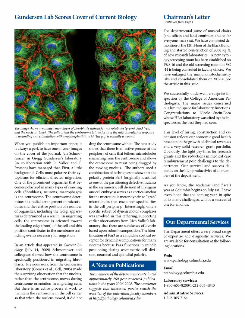
When you publish an important paper, it is always a perk to have one of your images on the cover of the journal. Jan Schmo-ranzer in Gregg Gundersen’s laboratory (in collaboration with R. Vallee and T. Pawson) have managed that. First, a little background: Cells must polarize their cy-toplasms for efficient directed migration. One of the prominent organelles that be-comes polarized in many types of crawling cells (fibroblasts, neurons, macrophages) is the centrosome. The centrosome deter-mines the radial arrangement of microtu-bules and the relative position of a number of organelles, including the Golgi appara-tus is determined as a result. In migrating cells, the centrosome is oriented toward the leading edge (front) of the cell and this position contributes to the membrane traf-ficking events necessary for migration.
In an article that appeared in Current Bi-ology (July 14, �009) Schmoranzer and colleagues showed how the centrosome is specifically positioned in migrating fibro-blasts. Previous work from the Gundersen laboratory (Gomes et al., Cell, �00�) made the surprising observation that the nucleus, rather than the centrosome, moves during centrosome orientation in migrating cells. But there is an active process at work to maintain the centrosome in the cell center so that when the nucleus moved, it did not
drag the centrosome with it. The new study shows that there is an active process at the periphery of cells that tethers microtubules emanating from the centrosome and allows the centrosome to resist being dragged by the moving nucleus. The authors used a combination of techniques to show that the polarity protein Par3 (originally identified as one of the partitioning defective mutants in the asymmetric cell division of C. elegans one cell embryos) serves as a cortical anchor for the microtubule motor dynein to “grab” microtubules that encounter specific sites in the cell periphery. Interestingly, only a specific subset of dynein motor complexes was involved in this tethering, supporting earlier observations from Rich Vallee’s lab-oratory that there are subclasses of dynein based upon subunit composition. The iden-tification of Par3 as a candidate cortical re-ceptor for dynein has implications for many systems because Par3 functions in spindle positioning during asymmetric cell divi-sion, neuronal and epithelial polarity.
Gundersen Lab Scores Cover of Current Biology
The departmental game of musical chairs (and offices and labs) continues and so far everyone has a seat. We have completed de-molition of the 1�th Floor of the Black Build-ing and started construction of 8000 sq. ft. of new research laboratories. A new cytol-ogy screening room has been established on P&S 16 and the old screening room on VC -14 is being converted to faculty offices. We have enlarged the immunohistochemistry labs and consolidated them on VC-14. See the article in this issue.
We successfully underwent a surprise in-spection by the College of American Pa-thologists. The major issues concerned our limited space for laboratory functions. Congratulations to Nicole Suciu-Foca whose HLA laboratory was cited by the in-spectors as the best they had seen.
This level of hiring, construction and ex-pansion reflects our economic good health based upon the growth of clinical revenues and a very solid research grant portfolio. Obviously, the tight pay-lines for research grants and the reductions in medical care reimbursement pose challenges to the de-partment. Our survival and success de-pends on the high productivity of all mem-bers of the department.
As you know, the academic (and fiscal) year at Columbia begins on July 1st. I have every hope that the coming year, in spite of its many challenges, will be a successful one for all of us.
Chairman’s Letter Continued from page 1
The image shows a wounded monolayer of fibroblasts stained for microtubules (green), Par3 (red) and the nucleus (blue). The cells orient the centrosome (at the focus of the microtubules) in response to wounding and stimulation with lysophosphatidic acid. The gap is actually a wound.
The Department offers a very broad range of expertise and diagnostic services. We are available for consultation at the follow-ing locations.
Web: www.pathology.columbia.edu
Email: [email protected]
Laboratory services: 1-800-6�3-8�00/1-�1�-30�-4840
Administrative Services: 1-�1�-30�-7164
Our Departmental Services
The members of the department contributed approximately 260 peer reviewed publica-tions in the years 2006-2008. The newsletter suggests that interested parties search the websites of the individual faculty members at http://pathology.columbia.edu/
A Note on Publications
6

The Department is pleased to reward the great efforts of the following faculty members. In these difficult times, the dedication of the faculty and the staff sustain us all.
The following faculty have been promoted:
Promotions
Corrine Abate-Shen, PhD has been pro-moted to Professor of Urologic Sciences and Pathology & Cell Biology (in Urology & the Herbert Irving Cancer Center), with tenure.
Rando Allikments, MD has been promot-ed to Associate Professor of Ophthamalogy and Pathology & Cell Biology with tenure.
Ottavio Arancio, MD, PhD has been pro-moted to Associate Professor of Pathology & Cell Biology (in the Taub Institute).
Alain Borczuk, MD has been promoted to Professor of Clinical Pathology & Cell Bi-ology. Alain is also the new Vice Chair of Anatomic Pathology-see separate article.
Thomas G. Diacovo, MD has been pro-moted to Associate Professor of Pediatrics and Pathology & Cell Biology.
Phyllis Faust, MD, PhD is newly pro-moted to Associate Professor of Clinical Pathology & Cell Biology.
Honors and Awards
Department members have received a number of awards in the last months. The Newsletter suspects that there are more but the usual shyness of our members prevents them from sending us the information! Nevertheless, we are pleased to announce the following:
Deborah W. Sevilla, MD, has been named Associate Program Director for the Fellow-ship in Hematology (Hematopathology).
Patrice Spitalnik MD has been elected to The Glenda Garvey Teaching Academy. This organization, named after legendary teacher Glenda Garvey, promotes research in new teaching methods and instructional material. The Newsletter congratulates Pa-trice for all of her many teaching activities and election to the Teaching Academy.
Ramon Parsons, MD, PhD was elected to the Association of American Physicians in April. The Association of American Physi-cians is a nonprofit, professional organiza-tion founded in 188� by seven physicians, including Dr. William Osler, for “the ad-vancement of scientific and practical med-icine.” Now the Association is composed of over 1000 active members and approxi-mately 600 emeritus and honorary mem-bers from the United States, Canada, and other countries.
Carlos Cordon-Cardo has won the Cris-tóbal Gabarrón Prize for Science and Re-search �009. Dr. Cordon-Cardo was awarded this prize, one of Spain’s most important, for his “pioneering contributions to the field of molecular pathology of cancer, an emerging biomedical discipline that promises to revo-lutionize the diagnosis of tumors and boost personalized cancer therapies.” Dr. Cordon-Cardo was featured in an earlier Newsletter.
This has been a very busy time for our excellent Grant Coordinators. In addition to the nor-mal schedule of grant and budget submissions our heroes (heroines?) have coped with all of the Stimulus submissions. We should all keep in mind that each of the �7 NIH Institutes has variations on the � or 6 main funding mechanisms, making the task even more complicated. The Newsletter was able to get before and after pictures, the first during the height of the submission panic and the second after things had calmed to merely frenetic. From left to right (right panel), Josie Salcedo, Steve Russo, Irene D’Silva (who smiles no matter what) and Frances Antonetty. The Department thanks you for hard work in difficult times.
Grants Administration- Heroic Actions
Continued on page 11
Tae-Wan Kim, PhD, has been promoted to Associate Professor of Pathology & Cell Biology with tenure.
Alexander Kratz, MD has been promoted to Associate Professor of Clinical Pathol-ogy & Cell Biology.
Brynn Levy, PhD has been promoted to Associate Professor of Clinical Pathology & Cell Biology.
Thomas Ludwig, PhD has been promoted to Associate Professor of Clinical Pathol-ogy & Cell Biology (in the Institute for Can-cer Genetics).
Laura Pasqualucci, PhD is now Associate Professor of Clinical Pathology & Cell Bi-ology (in the Institute for Cancer Genetics).
Anjali Saqi, MD, has been promoted to Associate Professor of Clinical Pathology & Cell Biology.
Darrell Yamashiro, MD, PhD has been promoted to Associate. Professor of Pe-diatrics and Pathology & Cell Biology, (in Surgery) at NYPH/CUMC.
7

Traditionally, protein synthesis is consid-ered to occur perinuclearly in the cell body, but in highly polarized cells such as neurons some mRNAs are selectively transported to the periphery. Local protein synthe-sis within axons and dendrites is a tightly controlled process in response to extracel-lular stimuli. Over the last couple of years, several axonal mRNAs as well as stimuli that trigger their translation have been de-scribed: the guidance cue Semphorin3A causes growth cone collapse in sensory neurons by triggering the local synthesis of RhoA; the mRNA for the transcription fac-tor CREB is locally translated in response to NGF, and CREB then translocates to the nucleus and supports neuronal survival. Most recently, we also found that netrin-1 and NGF trigger axonal elongation via the local translation of Par3, a key component of the PAR polarity complex.
Despite this recent progress, critical ques-tions remain, such as the physiological rel-evance of local translation for the develop-ment and function of the nervous system and associated pathologies, as well as the rationale for why only some mRNAs are locally translated while the vast majority of axonal proteins are transported from the cell body.
I am new to the Department and am pleased to be given this opportunity to in-troduce myself. I am a cell biologist and geneticist interested in understanding cell division, specifically how the contents of one cell are divided accurately into two daughter cells.
I was instantly fascinated with cell divi-sion as an undergraduate at the University of Wisconsin at Madison upon learning about Ray Rappaport’s experiments from the early 1960’s regarding the positioning of the cell division plane. I became hooked on cell division and have continued to work on this particular phenomenon ever since.
After receiving my bachelor’s degree, I got my first job as a research technician in Bill Bement’s laboratory at UW. We worked with Xenopus embryos as a model system to study the contractile molecules required to drive cell division. There I focused on understanding the interaction between two core cytoskeletal networks that regu-
New Basic Science Faculty
Julie Canman and Ulrich Hengst – two new basic scientists to join the Department
Despite the recession, our department is not retracting and the best evidence of this is our commitment to new faculty, both in basic science and in clinical areas (see the other profiles). Here, Julie and Ulrich describe their backgrounds and research excitement. That is Julie’s photo in The Art of Cell Biology – a regular feature of the Newsletter. First, Julie:
late cell division, the actin and microtu-bule cytoskeletons, which remain a subject of my research today.
My investigations in Bill’s lab increased my desire to study cell division, so Bill encour-aged me to apply to graduate school at the University of North Carolina at Chapel Hill (Go Heels!) to work with Dr. Ted (Ed-ward) Salmon. In Ted’s lab I learned to use high resolution light microscopy and live cell imaging to examine the microtubule cytoskeleton during cell division in mam-malian cultured cells. I was fascinated by how a cell knows where to divide and I dis-covered that despite the presence of a bipo-lar mitotic spindle in most dividing cells, monopolar mitotic spindles retain the competency to position the division plane.
As a post-doc, I wanted to build on my skills as a microscopist and learn condi-tional genetics, with the goal of combin-ing the two to understand the molecular mechanisms of cell division. I therefore moved to the University of Oregon at Eu-gene to work with Bruce Bowerman and learned to love the amazing soil-dwelling worm C. elegans. This organism is opti-cally clear and thus perfect for combining molecular analysis with live cell imaging assays- critical for studying the transient process of cell division. I screened through a collection of mutants for those with de-fects specifically in the first embryonic cell divisions. I then moved to the University of California at San Diego to work with Dr. Karen Oegema to characterize my mutants and understand the nature of the cell di-vision defects at a molecular level. I was able to identify a negative regulatory cas-cade that is essential for cell division using combinatorial forward and reverse genet-ics approach.
At Columbia, my lab will use a multidis-ciplinary approach of genetics and in vivo microscopy-based assays to understand the molecular mechanisms by which cells control the temporal and spatial regulation of cell division. I am excited about joining the Department in February and look for-ward to many collaborative projects.
Ulrich’s interests are in localized translation of mRNA, but we will let him explain:
Continued on page 11
8
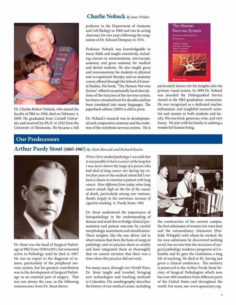
Dr. Stout was the head of Surgical Pathol-ogy at P&S from 19�8 to19�1, but remained active in Pathology until he died in 1967. He was an expert in the diagnosis of tu-mors, particularly of the peripheral ner-vous system, but his greatest contribution was in the development of Surgical Pathol-ogy as an essential part of surgery. That was not always the case, as the following reminiscence from Dr. Stout shows:
Our PredecessorsArthur Purdy Stout (1885-1967) By Alain Borczuk and Richard Kessin
When I first studied pathology I was told that it was possible to have a cancer of the lung but I was never shown the lungs of a person who had died of lung cancer nor during my en-tire four years in the medical school did I ever have a chance to examine a patient with lung cancer. How different from today when lung cancer stands high on the list of the causes of death, particularly among war veterans, thanks largely to the enormous increase of cigarette smoking. A. Purdy Stout, 1961
Dr. Stout understood the importance of histopathology in the understanding of disease and used this to bridge clinical pre-sentation and patient outcome by careful morphologic assessment and classification. These insights, like the one above, led to observations that form the basis of surgical pathology and we practice them so readily and have integrated them so thoroughly that we cannot envision that there was a time when this practice did not exist.
For many years, through two World Wars, Dr. Stout taught and traveled, bringing new technologies and teaching methods to Columbia. His autobiography describes the history of our medical center, including
the construction of the current campus, the first admission of women (we were late) and the extraordinary characters (Pen-field, Whipple) with whom he worked. By his own admission he discovered nothing novel, but we owe him the structure of sur-gical pathology residency programs at Co-lumbia and he gave the institution a long life of teaching. He died at 8�, having just given a clinical conference. His memory is preserved in the Arthur Purdy Stout So-ciety of Surgical Pathologists which now has over 400 members from different parts of the United States and throughout the world. For more, see: www.apssociety.org.
Dr. Charles Robert Noback, who joined the faculty of P&S in 1949, died on February 4, �009. He graduated from Cornell Univer-sity and received his Ph.D. in 194� from the University of Minnesota. He became a full
professor in the Department of Anatomy and Cell Biology in 1968 and was its acting chairman for two years following the resig-nation of Dr. Edward Dempsey in 1974.
Professor Noback was knowledgeable in many fields and taught extensively, includ-ing courses in neuroanatomy, microscopic anatomy, and gross anatomy for medical and dental students. He also taught gross and neuroanatomy for students in physical and occupational therapy, and an anatomy course offered through the School of Gener-al Studies. His book, “The Human Nervous System” offered exceptionally lucid descrip-tions of the function of the nervous system, has been a standard text for decades and has been translated into many languages. The paperback edition (�00�) is still in print.
Dr. Noback’s research was in developmen-tal and comparative anatomy and the evolu-tion of the vertebrate nervous system. He is
Charlie Noback By Joan Witkin
particularly known for his insights into the primate visual system. In 1999 Dr. Noback was awarded the Distinguished Service Award at the P&S graduation ceremonies. He was recognized as a dedicated teacher, enthusiastic and insightful research scien-tist and mentor to both students and fac-ulty. His was kind, generous, wise, and very funny. We join with his family in saluting a wonderful human being.
9

The immunohistochemistry labs have moved to an improved location on VC14 and recently celebrated with a lunch for the Department. More important than the move, perhaps, is the new equipment that has been installed. There are now four (count ‘em) Ultra-Ventana automated ma-chines capable of multiple histochemical analyses or in situ hybridization on DNA or RNA samples from paraffin sections. These machines are the most advanced available and give the staff unparalleled analytic capacity. Fabrizio Remotti, MD,
Immunohistochemistry and in situ Hybridization laboratory services moves to new and better location.
Director of the Facility says that if these machines were cars they would be Ferraris. Well, of course they would.
In addition to Fabrizio, the center is co-Di-rected by Mahesh Mansukhani, MD and supervised by Suzy Alexander, MS (ASCP). Suzy, who came to us three years ago from Cornell, reminds us that the service can also be used by researchers. The staff is rounded out by Ritchie Alsberry, Yuis J. Jimenez-Cortez, and Leonore C. Peruyero, all of whom appear in the photo.
Left to right: Jibin Chacko, Fabrizio Remotti, Suzy Alexander, Ritchie Alsberry, Dr. Shelanski,Leonore C. Peruyero and Yuis Cortez. A new Ultra-Ventana automated machine is shown at left.
New Recruitment Brochure for PhD Students
We are welcoming new students into our Pathobiology and Molecular Medicine Ph.D program. To make sure that there are more applicants in the coming year, we have created a four page brochure that con-tains a great deal of information and links to our Institutes and Centers. The contents will be posted to our website, which is un-dergoing development. We distribute these brochures at recruitment fairs, where, even in a digital age, people like something in hand. Our faculty should also distribute these brochures when they travel. For cop-ies, see Rich Kessin or Ron Liem. We thank Fiona Doetsch for the beautiful image on the cover.
Honors and Awards Continued from page 7
The Departmental Website (http://pathol-ogy.columbia.edu/) is going to be managed more actively. To that end, the management of the site is now in the hands of Columbia’s own Web Design Studio, who are easy to access on the �0th floor of PH. We have adapted the architecture of the Website to make it a simple matter for individual users to update their sections of the site. We have also added ways to count numbers of hits.
You should already have received requests to update research or clinical profiles. Mr. Ping Feng is managing this effort and he will contact you at least yearly. Ping
(pf�013) updates your references for you, so this is not onerous.
Our site will adhere to certain aesthetic de-sign principles, which will be determined by Richard Miller, who also designs the News-letter. The idea is that programmers should program and designers should design.
The Newsletter would like to thank Joann Li for overseeing the basic architecture of the site. She will now have a little more time for the important duty of keeping us solvent. Questions about the site and how you can manage your part should be ad-dressed to Rich Kessin.
Website Revisions
On the publication front, Dr. Shi Du Yan and colleagues at Columbia and elsewhere have published an extraordinary story in Nature Medicine (Oct. 10, �008) which received an interesting News and Views commentary entitled Portal to Alzheim-er’s Disease. The paper found that that the cognition of mice fated to develop an Al-zheimer’s like syndrome was much delayed if the normal self-destruction capability of mitochondria was blocked.
10

Virtually all data supporting a role of local protein translation in axon development have been obtained from primary culture experiments, and no direct in vivo evidence exists to support the notion that axonal translation is required for the formation of the nervous system. To understand the in vivo relevance of local mRNA transla-tion, we will create a mouse model in which the localization of Par3 mRNA can be re-stricted to the cell body. We chose Par3, (see Gundersen lab work on page 6), because we have previously shown that axonal transla-tion is required for attractive axon growth of commissural interneurons and sensory neurons in vitro. In the initial analysis we will concentrate on the role of axonal Par3 mRNA in commissural axon growth and guidance. However, this mouse model will also allow us to study the role of local trans-lation in the mature or aging nervous sys-tem as well as in non-neuronal cells. Local translation may not be exclusive to neurons but might be a general mechanism for the spatial regulation of protein activity. For example, polarization is crucial for normal cell behavior and is disrupted in cancer cells, which leads us to ask whether local Par3 synthesis plays a role in tissue homeo-stasis and tumorigenesis.
It is not clear why some proteins are syn-thesized in axons while the majority are transported from the cell body. Conceiv-ably, locally synthesized proteins might be post-translationally processed in a unique way, and this differential modification might confer distinct properties to the proteins. We will address this question by comparing the glycosylation pattern of cell body-derived and locally synthesized pro-teins. Additionally, we will characterize the glycosylation machinery in axons and growth cones. Curiously, no Golgi appa-ratus in axons and growth cones has been identified, leading to the question whether and how locally translated membrane pro-teins are glycosylated. Interestingly, some Golgi markers label vesicular structures in growth cones and loss of function of one of these proteins causes retrograde axonopa-thy leading to hereditary spastic paraple-gia. Thus, intra-axonal glycosylation ap-pears to be required for axon development or maintenance, and its study will have implications for our understanding of an important neurological disorder.
Hengst continued from page 8
When several faculty members in the De-partment of Anatomy and Cell Biology won a shared instrumentation grant for a new confocal microscope in 1996, they decided to share the wealth and make the instrument available to all Columbia researchers. Work-ing with the Herbert Irving Comprehensive Cancer Center (HICCC), they also obtained funding for a technician to train users. A core facility began with those decisions.
Today, the Confocal and Specialized Mi-croscopy Facility offers five advanced mi-croscope systems for imaging and manip-ulating cells and tissues. The emphasis is on high-resolution 3D imaging of fluores-cence: researchers can use wide-field mi-croscopy, deconvolution, and three types of confocal imaging: spinning-disk, laser scanning, and multiphoton excitation. Last year, we added a laser capture microdissec-tion system, which also allows cellular and subcellular ablation experiments. We also do color imaging of histological staining.
The facility has been directed by Liza Pon since 1996, and I have managed it since 1997, except for a hiatus during which I completed my Ph.D. in our department. Our technician, Laurel Coffey, brings a deep understanding of physics as well as a talent for training. Unfortunately for us, she will be pursuing graduate studies in physics at Brandeis this fall.
The facility has had a long and productive relationship with the Department of Pa-thology and Cell Biology. The Department provided space to the facility for many years before our move to the ICRC in �007. Basic and clinical researchers from the Depart-ment use the facility. Among the more than �40 publications and 8 journal covers us-ing data acquired in the facility, one of our
proudest productions was an image gener-ated in a collaboration between the Liem and Gundersen labs (Ho et al., 1998, “Novel features of intermediate filament dynamics revealed by green fluorescent protein chi-meras.” J. Cell Sci. 111:1767). This image of intermediate filaments in a mitotic cell dec-orated the official T-shirt of the American Society for Cell Biology in 1999.
Originally housed in an 8-by-10-foot dark-room, we now have a generous space in the Irving Cancer Research Center. Just as important as space for housing these instruments, however, is the ability to help scientists use them to the best advantage. To that end, we have maintained a strong orientation towards service. Every user can get help with the details of capturing a good image, as well as with questions about experimental design, sample prepa-ration, and data analysis.
As electron microscopists have long known, imaging is not only for generating “pretty pictures;” it can produce quantitative data for testing hypotheses. Like a genome se-quence or a microarray, digital images are rich in data, but deriving meaningful in-formation from them can be challenging. To facilitate this anaylsis, we help research-ers use software such as ImageJ to collect and examine data from their images.
To find out more, see our page under Shared Resources at http://hiccc.columbia.edu/. To contact us, e-mail [email protected], or call 8�1-4613.
The Confocal and Specialized Microscopy CoreBy Theresa Swayne, Ph.D
Multiphoton excitation confocal microscope. This is a Zeiss LSM510 NLO.
Laurel Coffey (left) and Theresa Swayne
11

Incoming Residents
Ashleigh Allen, MD (College of Physicians & Surgeons). Ashleigh receivde her BA from Rutgers with majors in chemistry and mathematics. She was Phi Beta Kappa and won the Department of Chemistry’s Har-old A. Fales Memorial Award, the Charles Pine Award for Physical Science, and the Pfizer Award for Outstanding Research in Chemistry. AP/CP.
Mark Ewalt, MD (College of Physicians & Sur-geons). Mark received his BS from Brown Uni-versity in Biology. He was a Howard Hughes Research Fellow in the year �000followed by two years experience as an analyst with Ac-centure LLP, Wellesley, MA. He was an NIH Fellow at Columbia �006 with interest in he-matopathology. Dr. Ewalt was also awarded the first Dan Fink Award for excellence in Pa-thology while in medical school. AP/CP
Kamraan Gill, MD (Duke). Kamraan re-ceived his BA from Rutgers with majors in Biology and Music. He won the Distin-guisehed Scholars Award and was presi-dent of the Rutgers Biochemistry Society. Dr. Gill did a one year research project at Duke in the Center for Cognitive Neuro-science on the biological basis for tonal fea-tures of music. AP/CP.
Elijah Luyten, MD (U. Mass-AOA). Elijah received his BA from U.C. Santa Cruz where his major was Chinese. Dr. Luyten did ad-ditional undergraduate studies at Qinghua University, Beijing, and Peking Univer-sity, Beijing, and post-baccalaureate study at Harvard University. He did his General Surgery Residency at NYPH-Columbia University with research on a mouse model of antibody-mediated rejection. CP.
Huy Phu Pham, MD (Rosalind Franklin/Chicago Medical School). Huy Phu received his BS from the University of California Berkeley where he received high honors for his bioengineering major. AP/CP.
Jean Hou, MD (Georgetown). Jean received her BS from Johns Hopkins in Biology. She has twelve years experience at the Labora-tory of Molecular Medicine and Neurosci-ences at NIH and became Head of the Vi-ral Diagnostic Unit. She received the NIH Directors Award and other awards from NIH. Dr. Hou has multiple peer-reviewed publications and book chapters. AP/CP.
Volume 2, Issue 1 Fall 2009
ChairmanDr. Michael L. ShelanskiDelafield Professor of PathologyPathology and Cell Biology
EditorDr. Richard H. KessinProfessorPathology and Cell Biology
Design and productionRichard V. MillerCUMC IT
The Columbia Pathology and Cell Biology Report is a publication of the Department of Pathology and Cell Biology at the Columbia University Medical Center. If you have comments or questions, contact: Dr. Richard H. Kessinat �1�.30�.�6�3Email: rhk�@columbia.edu
http://pathology.columbia.edu/