Embed Size (px)
Citation preview

REVIEWpublished: 28 January 2021
doi: 10.3389/fnagi.2021.623751
Edited by:
Eszter Farkas,University of Szeged, Hungary
Reviewed by:Arthur Liesz,
Ludwig Maximilian University ofMunich, Germany
Anusha Mishra,Oregon Health and Science
University, United States
*Correspondence:Johannes Boltze
Received: 30 October 2020Accepted: 04 January 2021Published: 28 January 2021
Citation:Boltze J, Aronowski JA, Badaut J,
Buckwalter MS, Caleo M, Chopp M,Dave KR, Didwischus N,
Dijkhuizen RM, Doeppner TR,Dreier JP, Fouad K, Gelderblom M,Gertz K, Golubczyk D, Gregson BA,
Hamel E, Hanley DF, Härtig W,Hummel FC, Ikhsan M, Janowski M,
Jolkkonen J, Karuppagounder SS,Keep RF, Koerte IK, Kokaia Z, Li P,
Liu F, Lizasoain I, Ludewig P,Metz GAS, Montagne A, Obenaus A,
Palumbo A, Pearl M, Perez-Pinzon M,Planas AM, Plesnila N, Raval AP,
Rueger MA, Sansing LH, Sohrabji F,Stagg CJ, Stetler RA, Stowe AM,
Sun D, Taguchi A, Tanter M, Vay SU,Vemuganti R, Vivien D, Walczak P,
Wang J, Xiong Y and Zille M(2021) New Mechanistic Insights,Novel Treatment Paradigms, and
Clinical Progress inCerebrovascular Diseases.
Front. Aging Neurosci. 13:623751.doi: 10.3389/fnagi.2021.623751
New Mechanistic Insights, NovelTreatment Paradigms, and ClinicalProgress in CerebrovascularDiseasesJohannes Boltze1*, Jaroslaw A. Aronowski2, Jerome Badaut3, Marion S. Buckwalter4,Mateo Caleo5,6, Michael Chopp7,8, Kunjan R. Dave9, Nadine Didwischus1,Rick M. Dijkhuizen10, Thorsten R. Doeppner11, Jens P. Dreier12,13,14,15, Karim Fouad16,Mathias Gelderblom17, Karen Gertz 12,18, Dominika Golubczyk19, Barbara A. Gregson20,Edith Hamel21, Daniel F. Hanley22, Wolfgang Härtig23, Friedhelm C. Hummel24,25,Maulana Ikhsan26,27,28, Miroslaw Janowski29, Jukka Jolkkonen30,Saravanan S. Karuppagounder31,32, Richard F. Keep33, Inga K. Koerte34,35, Zaal Kokaia36,Peiying Li37, Fudong Liu38, Ignacio Lizasoain39, Peter Ludewig17, Gerlinde A. S. Metz40,Axel Montagne41, Andre Obenaus42, Alex Palumbo26,27,28, Monica Pearl43,Miguel Perez-Pinzon9, Anna M. Planas44,45, Nikolaus Plesnila46,47,48, Ami P. Raval9,Maria A. Rueger49, Lauren H. Sansing50, Farida Sohrabji51, Charlotte J. Stagg52,53,R. Anne Stetler54, Ann M. Stowe55, Dandan Sun56, Akihiko Taguchi57, Mickael Tanter58,Sabine U. Vay49, Raghu Vemuganti59, Denis Vivien60,61, Piotr Walczak29, Jian Wang62,Ye Xiong63 and Marietta Zille26,27,28
1School of Life Sciences, University of Warwick, Warwick, United Kingdom, 2Institute for Stroke and CerebrovascularDiseases, McGovern Medical School, The University of Texas Health Science Center at Houston, Houston, TX,United States, 3NRS UMR 5287, INCIA, Brain Molecular Imaging Team, University of Bordeaux, Bordeaux cedex, France,4Departments of Neurology and Neurological Sciences, and Neurosurgery, Wu Tsai Neurosciences Institute, Stanford Schoolof Medicine, Stanford, CA, United States, 5Neuroscience Institute, National Research Council, Pisa, Italy, 6Department ofBiomedical Sciences, University of Padua, Padua, Italy, 7Department of Neurology, Henry Ford Hospital, Detroit, MI,United States, 8Department of Physics, Oakland University, Rochester, MI, United States, 9Peritz Scheinberg CerebralVascular Disease Research Laboratory, Department of Neurology, University of Miami Miller School of Medicine, Miami, FL,United States, 10Biomedical MR Imaging and Spectroscopy Group, Center for Image Sciences, University Medical CenterUtrecht and Utrecht University, Utrecht, Netherlands, 11Department of Neurology, University Medical Center Göttingen,Göttingen, Germany, 12Department of Neurology, Center for Stroke Research Berlin, Charité—Universitätsmedizin Berlin,Berlin, Germany, 13Department of Experimental Neurology, Charité—Universitätsmedizin Berlin, Berlin, Germany, 14BernsteinCenter for Computational Neuroscience Berlin, Berlin, Germany, 15Einstein Center for Neurosciences Berlin, Berlin, Germany,16Faculty of Rehabilitation Medicine and Institute for Neuroscience and Mental Health, University of Alberta, Edmonton, AB,Canada, 17Department of Neurology, University Medical Center Hamburg-Eppendorf, Hamburg, Germany, 18Berlin Institute ofHealth, Berlin, Germany, 19Department of Neurosurgery, School of Medicine, University of Warmia and Mazury, Olsztyn,Poland, 20Neurosurgical Trials Group, Institute of Neuroscience, The University of Newcastle upon Tyne, Newcastle uponTyne, United Kingdom, 21Laboratory of Cerebrovascular Research, Montreal Neurological Institute, McGill University,Montreal, QC, Canada, 22Division of Brain Injury Outcomes, Johns Hopkins University, Baltimore, MD, United States, 23PaulFlechsig Institute of Brain Research, University of Leipzig, Leipzig, Germany, 24Clinical Neuroengineering, Center forNeuroprosthetics and Brain Mind Institute, Swiss Federal Institute of Technology Valais, Clinique Romande de Réadaptation,Sion, Switzerland, 25Clinical Neuroscience, University of Geneva Medical School, Geneva, Switzerland, 26Institute forExperimental and Clinical Pharmacology and Toxicology, University of Lübeck, Lübeck, Germany, 27Fraunhofer ResearchInstitution for Marine Biotechnology and Cell Technology, Lübeck, Germany, 28Institute for Medical and Marine Biotechnology,University of Lübeck, Lübeck, Germany, 29Department of Diagnostic Radiology and Nuclear Medicine, University of Maryland,Baltimore, MD, United States, 30Department of Neurology, A.I. Virtanen Institute for Molecular Sciences, University of EasternFinland, Kuopio, Finland, 31Burke Neurological Institute, White Plains, NY, United States, 32Feil Family Brain and MindResearch Institute, Weill Cornell Medicine, New York, NY, United States, 33Department of Neurosurgery, University ofMichigan, Ann Arbor, MI, United States, 34Psychiatric Neuroimaging Laboratory, Brigham and Women’s Hospital and HarvardMedical School, Boston, MA, United States, 35Department of Child and Adolescent Psychiatry, Psychosomatic, andPsychotherapy, Ludwig Maximilians University, Munich, Germany, 36Lund Stem Cell Center, Lund University, Lund, Sweden,
Frontiers in Aging Neuroscience | www.frontiersin.org 1 January 2021 | Volume 13 | Article 623751
Boltze et al. Progress in Cerebrovascular Disease Research
37Department of Anesthesiology, Renji Hospital, School of Medicine, Shanghai Jiaotong University, Shanghai, China,38Department of Neurology, The University of Texas Health Science Center at Houston, McGovern Medical School, Houston,TX, United States, 39Unidad de Investigación Neurovascular, Departamento Farmacología y Toxicología, Facultad de Medicina,Instituto Universitario de Investigación en Neuroquímica, Universidad Complutense de Madrid, Madrid, Spain, 40Departmentof Neuroscience, Canadian Centre for Behavioural Neuroscience, University of Lethbridge, Lethbridge, AB, Canada, 41ZilkhaNeurogenetic Institute, Keck School of Medicine, University of Southern California, Los Angeles, CA, United States,42Department of Pediatrics, University of California, Irvine, Irvine, CA, United States, 43The Russell H. Morgan Departmentof Radiology and Radiological Science, Johns Hopkins University School of Medicine, Baltimore, MD, United States,44Institut d’Investigacions Biomèdiques August Pi i Sunyer (IDIBAPS), Àrea de Neurociències, Barcelona, Spain, 45Departmentd’Isquèmia Cerebral I Neurodegeneració, Institut d’Investigacions Biomèdiques de Barcelona (IIBB), Consejo Superior deInvestigaciones Científicas (CSIC), Barcelona, Spain, 46Institute for Stroke and Dementia Research (ISD), Munich UniversityHospital, Munich, Germany, 47Graduate School of Systemic Neurosciences (GSN), Munich University Hospital, Munich,Germany, 48Munich Cluster of Systems Neurology (Synergy), Munich, Germany, 49Faculty of Medicine and University Hospital,Department of Neurology, University of Cologne, Cologne, Germany, 50Department of Neurology, Yale University School ofMedicine, New Haven, CT, United States, 51Women’s Health in Neuroscience Program, Neuroscience and ExperimentalTherapeutics, Texas A&M College of Medicine, Bryan, TX, United States, 52Nuffield Department of Clinical Neurosciences,Wellcome Centre for Integrative Neuroimaging, University of Oxford, Oxford, United Kingdom, 53MRC Brain Network DynamicsUnit, University of Oxford, Oxford, United Kingdom, 54Department of Neurology, Pittsburgh Institute of Brain Disordersand Recovery, University of Pittsburgh, Pittsburgh, PA, United States, 55Department of Neurology and Neurotherapeutics,Peter O’Donnell Jr. Brain Institute, UT Southwestern Medical Center, Dallas, TX, United States, 56Pittsburgh Institute forNeurodegenerative Disorders, University of Pittsburgh, PA, United States, 57Department of Regenerative Medicine Research,Institute of Biomedical Research and Innovation, Kobe, Japan, 58Institute of Physics for Medicine Paris, INSERM U1273,ESPCI Paris, CNRS FRE 2031, PSL University, Paris, France, 59Department of Neurological Surgery, University of Wisconsin,Madison, WI, United States, 60UNICAEN, INSERM, INSERM UMR-S U1237, Physiopathology and Imaging for NeurologicalDisorders (PhIND), Normandy University, Caen, France, 61CHU Caen, Clinical Research Department, CHU de Caen Côte deNacre, Caen, France, 62Department of Human Anatomy, College of Medical Sciences, Zhengzhou University, Zhengzhou,China, 63Department of Neurosurgery, Henry Ford Hospital, Detroit, MI, United States
The past decade has brought tremendous progress in diagnostic and therapeuticoptions for cerebrovascular diseases as exemplified by the advent of thrombectomyin ischemic stroke, benefitting a steeply increasing number of stroke patientsand potentially paving the way for a renaissance of neuroprotectants. Progress inbasic science has been equally impressive. Based on a deeper understanding ofpathomechanisms underlying cerebrovascular diseases, new therapeutic targets havebeen identified and novel treatment strategies such as pre- and post-conditioningmethods were developed. Moreover, translationally relevant aspects are increasinglyrecognized in basic science studies, which is believed to increase their predictivevalue and the relevance of obtained findings for clinical application.This review reportskey results from some of the most remarkable and encouraging achievements inneurovascular research that have been reported at the 10th International Symposiumon Neuroprotection and Neurorepair. Basic science topics discussed herein focus onaspects such as neuroinflammation, extracellular vesicles, and the role of sex andage on stroke recovery. Translational reports highlighted endovascular techniques andtargeted delivery methods, neurorehabilitation, advanced functional testing approachesfor experimental studies, pre-and post-conditioning approaches as well as novel imagingand treatment strategies. Beyond ischemic stroke, particular emphasis was given onactivities in the fields of traumatic brain injury and cerebral hemorrhage in whichpromising preclinical and clinical results have been reported. Although the number ofneutral outcomes in clinical trials is still remarkably high when targeting cerebrovasculardiseases, we begin to evidence stepwise but continuous progress towards noveltreatment options. Advances in preclinical and translational research as reported hereinare believed to have formed a solid foundation for this progress.
Keywords: cell therapies, dementia, experimental therapy, hemorrhage, neuroprotection, neurorehabilitation,stroke, translational research
Frontiers in Aging Neuroscience | www.frontiersin.org 2 January 2021 | Volume 13 | Article 623751

Boltze et al. Progress in Cerebrovascular Disease Research
INTRODUCTION
Overcoming more than two decades of translational standstilland providing options to treat selected stroke patients inextended time windows, the advent of recanalization approacheshas sparked a new dynamic in preclinical and clinical strokeresearch. With a catheter in place, i.e., in a reopened cerebralartery, we now have the opportunity to apply neuroprotectivetreatments exactly when and where they may exert their optimalbenefit (Savitz et al., 2019). These new options have the potentialto initiate a ‘‘renaissance of neuroprotectants.’’ Neuroprotectantsmay also play an increasingly important role in the treatmentof cerebral hemorrhages. Minimally invasive techniques forhematoma evacuation relying on specialized, catheter-basedintervention strategies have been developed and were assessed infirst clinical trials (Hanley et al., 2019). Neuroprotective agentsmay be delivered via those catheters once the hematoma wasremoved, and novel targets for neuroprotective interventionshave been identified (Zille et al., 2017).
Nevertheless, the majority of patients still cannotbenefit from the recent advances in acute interventions forcerebrovascular diseases. Among others, reasons comprisestrict inclusion/exclusion and eligibility criteria, uncertaintiesregarding the optimal way to deliver such treatments as wellas logistical challenges and limited availability of requiredinfrastructure. Therefore, the need for new treatments beingeffective within therapeutic time windows beyond the range ofhours as well as supporting long-term recovery and recuperationcontinues to exist. The development of these new treatmentsshould take into account advances in basic science areas such asneuroimmunology, neurobiochemistry, and molecular biology,as well as cell-based approaches. Moreover, translational researchshall reflect advances in imaging technologies and rehabilitationresearch and must consider aspects such as the influence of sexand age on therapeutic outcome.
The International Symposium on Neuroprotection andNeurorepair (ISN&N) discussed these aspects in the lightof current therapeutic advances in cerebrovascular diseases.The ISN&N is a biennial meeting series on fundamental andtranslational neuroscience with a scope on cerebrovasculardiseases and dementia (Boltze et al., 2011, 2012; Demuthet al., 2017). Attracting leading neuroscientists and cliniciansfrom around the world, the meeting is characterized byintensive scientific exchange and discussions of new insights intoneurovascular and neurodegenerative diseases. To share someof the most exciting reports with a wider academic communityin a concise format, this review summarizes cross-selected,outstanding sessions from the 10th ISN&N that took place fromOctober 9th to 11th 2018 in Radebeul near Dresden, Germany.It covers the main topics of the meeting, and puts a particularscope on translational and clinical aspects, as such providing agood overview of the field.
Conditioning Medicine: Basic Mechanismsand Clinical PotentialIt is now well accepted that ischemic pre- and post-conditioningpromotes ischemic neuroprotection in different modalities
(e.g., pharmacological, remote, and ischemic) and in manydifferent animal models (Wang et al., 2015). Novel mechanismsinvolved in conditioning were discussed and clinical views of howto apply conditioning strategies to thrombectomy were provided.
IL (Madrid, Spain) presented that toll-like receptor 4 (TLR4)has both protective (ischemic tolerance) and damaging (acuteischemia) roles in cerebral ischemia, including hemorrhagictransformation (Pradillo et al., 2009; Vartanian et al., 2011;García-Culebras et al., 2017). TLR4 deficiency increased thelevels of alternative neutrophils (N2), which may be involved inthe resolution of inflammation and in the neuroprotective effectobserved after stroke (García-Culebras et al., 2019). New drugsblocking TLR4 (Fernández et al., 2018) may be useful for thetreatment of patients with stroke.
RAS (Pittsburgh, PA, USA) demonstrated that ischemicneuroprotection can be achieved by dietary restriction,improving both gray and white matter outcomes and functionalrecovery (Zhang et al., 2018). Although the activation of sirtuinsmay be a potential contributor to ischemic neuroprotection(Haigis and Guarente, 2006; Guarente, 2008; Stetler et al.,2014), dietary restriction is also well known to affect the releaseof a variety of humoral factors, particularly from adiposetissue. Adiponectin release into the plasma occurs acutelyafter dietary restriction and increased levels in the serum. Thiswas associated with beneficial outcomes, as global knockoutof adiponectin negated the effects of dietary restriction onthe subsequent ischemic injury. The effects may be due topenetration of adiponectin into the brain parenchyma and thesubsequent binding to its receptors on neural cells in the contextof the opening of the blood-brain barrier (BBB) after ischemicinjury (Brochu-Gaudreau et al., 2010; Zhang et al., 2018).Whether this scenario or the alternative situation—whereinadiponectin acts directly on endothelial barrier function toexert protection—underlies the neuroprotective effects remainsunder investigation.
MP-P (Miami, FL, USA) reported a novel window forischemic tolerance for preconditioning mimetics, which waspreviously limited to 48–72 h after cerebral ischemia (Narayananet al., 2015). Resveratrol preconditioning yielded gene expressionchanges in 136 genes at 14 days after single administration:many of the 116 downregulated genes were related totranscription, synaptic signaling, and neurotransmission,resembling the metabolic depression observed in hibernatingspecies. This downregulation of gene expression fits well withthe hippocampal synaptic and electrophysiological depressionobserved with pre-conditioning mimetics (DeFazio et al., 2009;Neumann et al., 2015; Cohan et al., 2017). Also, glycolysisand mitochondrial respiration efficiency were increased uponresveratrol preconditioning (Koronowski et al., 2017; Khouryet al., 2019), indicating an increased reliance on energy-producing pathways.
PL (Shanghai, China) discussed anesthetic postconditioningas a valuable neuroprotection candidate (Cohan et al., 2017) asfast onset and short-acting anesthetics allow smooth anesthesiainduction and faster anesthetic emergence to overcome theclinical limitation that neurological assessment during anesthesiaor sedation is not possible. New technology-based anesthetic
Frontiers in Aging Neuroscience | www.frontiersin.org 3 January 2021 | Volume 13 | Article 623751
Boltze et al. Progress in Cerebrovascular Disease Research
drug delivery systems, such as target-controlled infusion, closeloop infusion, and computer-assisted sedation systems allowbetter control of anesthesia (Struys et al., 2016; Zaouteret al., 2016). Dr. Li further reported about a plannedclinical study in stroke patients undergoing endovascularthrombectomy to compare general anesthesia with and withoutanesthetic postconditioning for countering reperfusion injuryafter endovascular thrombectomy.
Overall, conditioning represents a clinically availabletherapeutic strategy for ischemic stroke, and the presentedfindings put forward our understanding of the underlyingmechanisms and provided novel insights into its clinicalapplication. However, to utilize its full potential, relevant aspectsof heterogeneous patient populations such as age and sex haveto be considered. Their relevance has already been shown innumerous studies.
Effect of Sex and Age on Post-ischemicOutcome and TherapySex and age are known to affect stroke outcomes (Kimand Vemuganti, 2015). Their contribution concerningmechanisms of ionic imbalance, inflammation, microRNAs,and the interaction of long noncoding RNAs with epigeneticswas discussed.
As the expression of the chloride influx transporter, Na-K-Cl cotransporter 1 (NKCC1) in male rodent brains startsearlier during development compared to female brains, targetingNKCC1 during post-injury in adulthood may be sex-dependent.Indeed, DS (Pittsburgh, PA, USA) showed that pharmacologicalblockade of NKCC1 protein has higher sensitivity in adult malemice than female mice for reducing post-ischemic brain injury(Huang et al., 2019).
FL (Houston, TX, USA) presented that post-strokeinflammation and microglial activation are sex- andage-dependent and that these differences exist throughoutthe lifespan (Zhao et al., 2017b; Ritzel et al., 2018). He alsoshowed that sex-chromosomal effects in addition to sexhormones contribute to the differential outcomes between malesand females after focal ischemia (McCullough et al., 2016).
AR (Miami, FL, USA) reported that the depletion ofendogenous estrogen at reproductive senescence increasesinflammasome proteins in the brain of females, but not inage-matched male rats (de Rivero Vaccari et al., 2016). Inreproductively senescent females, extracellular vesicles (EVs)from reproductive organs carry inflammasome proteins to thebrain leading to an exacerbated innate immune response thatmay be responsible for the increased severity of ischemic damagein senescent females (D’Adesky et al., 2018).
FS (Bryan, TX, USA) showed that stroke neuroprotectantsmay act in a sex-specific manner. Post-stroke administration ofIGF-1 is neuroprotective after stroke. Preventing microRNA let-7f, thus upregulating IGF-1 which is reduced in aging animalsand humans, is protective in young female rats. However, it isnot neuroprotective in young males and even worsens outcomesin senescent females post-stroke (Selvamani et al., 2012; Sohrabjiet al., 2019). Let7f also regulates caspase 3, and treatment withmiR-363–3p mimic (repressing caspase-3) protects young and
senescent females, but not males, after focal ischemia (Selvamaniand Sohrabji, 2017). Thus, the neurotoxic effect seen in olderfemales may be related to an increase in caspase-3 and its celldeath pathways caused by the blocking of let7f (Sohrabji andSelvamani, 2019). These findings also illustrate the complexityof sex-specific neuroprotective actions that must be takeninto consideration.
RV (Madison, WI, USA) presented that the long noncodingRNAs Fos downstream transcript (FosDT) is induced afterstroke and that knockdown of FosDT with a siRNA cocktailprotects the brain in both males and females and also agedanimals (Mehta et al., 2015). The epigenetic modificationhydroxymethylation that converts 5-methylcytosine to5-hydroxymethylcytosine induced after stroke is neuroprotectivein both sexes (Morris-Blanco et al., 2019).
In summary, the contribution of age and sex to the outcomeof cerebrovascular diseases should be carefully considered in thedevelopment of novel therapeutic strategies.
Long-Term Neuroinflammation DuringFunctional Recovery From StrokeInflammation has been regarded as a target to reduce braindamage in acute stroke. While dampening innate immuneresponses and addressing complement or danger signalingeffectively reduces acute brain damage (Muhammad et al.,2008; Alawieh et al., 2018), certain inflammatory responses arenecessary for repair as they induce cell migration, proliferation,matrix deposition, and tissue remodeling. This suggests thatinitial inflammatory reactions trigger a set of responses that mayimprove functional outcome in the long term.
AMP (Barcelona, Spain) reported that although monocytesand macrophages reach the brain within the first 4 days afterstroke (Miró-Mur et al., 2016), macrophages persist for weeksin the damaged brain tissue (Wattananit et al., 2016; Mantovani,2017). While they may have detrimental functions acutely,macrophages also play critical roles in long-term functionalrecovery (Wattananit et al., 2016). Besides, lymphocytes, whichpromote acute thromboinflammation through mechanisms thatdo not involve adaptive immune responses (Kleinschnitz et al.,2010), also infiltrate the brain in higher numbers during chronicphases, when adaptive immune responses may play a role infunctional impairment (Selvaraj and Stowe, 2017).
AMS (Lexington, KY, USA) discussed that CD8+ T cells(i.e., cytotoxic T cells) move into several regions interconnectedto the infarct within 4 days after transient middle cerebral arteryocclusion (tMCAO) in mice, where they remain and supportrecovery (Poinsatte et al., 2019). Antibody-mediated depletion ofCD8+ T cells improved motor recovery and reduced diapedesisof other leukocyte populations, including macrophages,neutrophils, and CD4+ T cells (Selvaraj and Stowe, 2017).
MSB (Stanford, CA, USA) described that in a modelof post-stroke cognitive impairment, mice have normalcognition and hippocampal long-term potentiation 1 weekafter a cortical stroke adjacent to the hippocampus, whilewithin 7 weeks, cognitive impairment and progressive loss oflong-term potentiation begin (Doyle et al., 2015). In parallel,antibody-producing B-lymphocytes accumulate in the infarct
Frontiers in Aging Neuroscience | www.frontiersin.org 4 January 2021 | Volume 13 | Article 623751
Boltze et al. Progress in Cerebrovascular Disease Research
core. Antibody levels increase in both the core and thesurrounding brain parenchyma but the specificity of theseantibodies could not (yet) be revealed. However, ablatingB-lymphocytes prevents delayed cognitive impairment.Dr. Buckwalter further presented that B-lymphocytesare involved in this process in humans, suggesting thatB cells may be critical targets for the development ofpost-stroke immunotherapies.
FS (Bryan, TX, USA) showed that anti-inflammatory miRNAtherapy improves long-term affective dysfunction after ischemicstroke (Panta et al., 2019). Intravenous injection of mir363-3p mimics reduced infarct volume in the acute phase ofstroke, ameliorated sensory-motor impairment, and attenuatedtransient increases of IL-6 and TNF-α. Depressive behaviors,assessed over 30–100 days after stroke, were also attenuated inmir363-3p treated animals (Panta et al., 2019). This indicates thata neuroprotectant that has anti-inflammatory properties mayalso improve affective dysfunction after stroke.
Taken together, these studies highlight the complex roleof inflammation in the chronic phase after stroke and thatmodulating inflammation may require targeting a specific subsetof inflammatory cells and proteins.
Targeting the Interplay Between StemCells and Neuroinflammation in StrokeRegenerationSustained neuroinflammation is also required to eliminatedegenerating tissue, supplying a restorative milieu for stemcell-mediated repair. However, immune responses are oftendysregulated and lead to detrimental effects on regeneration.Recent work aims to elucidate the complex and interwovenprocesses of neuroinflammation and stem cell-mediatedregeneration after a stroke that may form the basis for noveltherapeutic approaches to modulate these processes andtheir interactions.
SUV (Cologne, Germany) reported that differentiallypolarized microglia differ in their effects on the fate of neuralstem cells (NSC) in vitro and in vivo, with pro-inflammatorymicroglia promoting the generation of astrocytes, whileanti-inflammatory microglia support neurogenesis. Regardlessof their polarization, the secretome of activated microgliainhibits NSC proliferation, increases NSC migration, andaccelerates NSC differentiation (Vay et al., 2018). Thesecombined effects suggest that activated microglia induce NSCto exit the cell cycle, to migrate towards a putative lesion site,and to attempt cell replacement by differentiation. Besides,NSC promotes functional restoration after stroke not only bycell replacement but also by modulating surrounding cells andinflammatory processes.
ZK (Lund, Sweden) discussed the significance of monocyte-derived macrophages (MDM) in supporting regenerativeprocesses after ischemic stroke. MDM are recruited to theischemic lesion site exhibiting a high degree of functionalplasticity, changing from a pro- to an anti-inflammatoryphenotype during the first weeks after stroke. Blockage ofMDM recruitment worsens functional and structural outcomes
after stroke (Wattananit et al., 2016; Kronenberg et al., 2018).Dr. Kokaia’s work suggests that immune cells affect theregenerative capacity of the brain, most likely by modulatingstem-cell-derived neuronal plasticity in the subventricular zoneand the adjacent striatum (Laterza et al., 2017).
KG (Berlin, Germany) emphasized the beneficial role ofanti-inflammatoryMDMactivated in the early phase after stroke.However, not only stroke-invadingMDMbut also brain-residentmicroglia are activated, and both MDM and microglia oftendisplay a mixture of phenotypic markers (‘‘intermediate states’’)during the first weeks after stroke (Kronenberg et al., 2018).
Overall, the cross-talk between inflammatory cells acting askey regulators and endogenous or grafted neural stem cellsacting as key effectors is highly relevant during post-strokeregeneration. Thus, therapeutic strategies based on modulatingthese cell entities and their interactions could be exploited tofacilitate functional recovery after stroke.
Extracellular Vesicles: Therapy andImagingAlthough there is growing evidence that long-term benefits areproduced by stem cells, particularly in sudden onset diseases,these cells are often short-lived after transplantation (Kim et al.,2012; Jablonska et al., 2016). Stem cells release EVs includingexosomes that mediate therapeutic effects (Xin et al., 2012, 2013a;Zhang et al., 2015) and in certain conditions, they can achievethe same efficacy obtained by the administration of the stem cellsthemselves (Doeppner et al., 2015).
TD (Göttingen, Germany) explained the positive effects ofmesenchymal stem cell-derived EVs (Doeppner et al., 2015).Interestingly, similar effects have recently been described forEVs derived from neural progenitor cells (Zheng et al., 2021).This suggests that EV-derived modes of action could be ageneral mechanism related to beneficial stem and progenitorcell effects. Apart from this, Dr. Doeppner also reportedinnovative therapeutic approaches such as the application ofremote ischemic post-conditioning (for review and currentclinical application please see Ji et al., 2019), defined as a transientand subcritical period of peripheral organ ischemia followingcerebral ischemia, to exert beneficial but transient effectson recovery. Interestingly, post-conditioning also facilitatedintracerebral transplantation of neural progenitor cells resultingin persistent protection and improved functional recovery(Doeppner et al., 2017).
YX (Detroit, MI, USA) discussed that exosomes elicit potentcellular responses in vitro and in vivo by delivering their cargos,including lipids, proteins, RNAs (miRNAs and mRNAs), andother macromolecules, to recipient cells (Xin et al., 2013a; Zhanget al., 2019). In turn, microRNAs regulate numerous genes thatmediate beneficial therapeutic effects (Zhang et al., 2019) andexosomes with select miRNAs can be designed to provide furtherimproved therapeutic efficacy (Xin et al., 2013b, 2017).
MJ (Baltimore, MD, USA) explained strategies for EV labelingand imaging to assess direct routes of EV delivery that arepotentially more beneficial and to monitor EV biodistributionin the body. Iron oxide-based labeling is comparably easyas it is sufficient to apply a standard labeling protocol to
Frontiers in Aging Neuroscience | www.frontiersin.org 5 January 2021 | Volume 13 | Article 623751
Boltze et al. Progress in Cerebrovascular Disease Research
stem cells, from which EVs are subsequently derived (Busatoet al., 2017; Dabrowska et al., 2018). While the signal isstrong, its specificity is quite low due to other sources ofhypointensities in tissues related to pathology. However, thisapproach may be particularly compelling to monitor EV deliveryin real-time as the signal of the label can be subtractedfrom the background image obtained before the procedure(Walczak et al., 2017). Further labeling techniques based onradioisotopes are very attractive to assess EV biodistribution evenin clinical settings, while other modalities such as fluorescenceor bioluminescence labeling are limited to preclinical studies(Choi and Lee, 2016).
Summing up, cell-free exosome-based approaches mayreplace stem cells and become a major driving forcefor therapeutic interventions with the aid of engineeringand imaging.
Traumatic Brain Injury Can Lead toCerebrovascular DiseaseA provocative question discussed was whether traumatic braininjury can serve as a model to study neurovascular unitdysfunction in cerebrovascular disease as their relationshiphas been proposed in recent studies (Pop and Badaut, 2011;Sandsmark et al., 2019).
NP (Munich, Germany) showed that controlled corticalimpact in adult mice induces progressive cognitive decline(Mao et al., 2020). While motor function recovered partiallyafter TBI, depression-like behavior and the loss of memoryfunction were progressive. Increasing tissue loss in the corpuscallosum, hippocampus, and hydrocephalus formation wasalso observed over time. Hence, experimental TBI in micereplicates the long-term sequelae post-trauma includingdementia and depression that are observed in humans.It needs to be determined whether these changes aretriggered by concomitant vascular injury or other, so farunknown mechanisms.
JB (Bordeaux, France) discussed the alteration of the NVUmonths after mild pediatric TBI in which changes of waterchannel expression on astrocytic endfeet adjacent to blood vesselshave been observed (Fukuda et al., 2013). These changes arerelated to diffusion tensor imaging modifications in differentbrain structures at 12 months post-injury after a single impact.
Linked with NVU remodeling, AO (Irvine, CA, USA)presented the importance of the activation of the Wnt-Beta-catenin pathway in cerebral blood vessels after TBI (Salehi et al.,2018). After acute vascular loss post-TBI, new immature vesselsoccurred at the injury site 7 days after the injury. This coincidedwith decreased levels of β-catenin protein in the injury site butdramatically increased β-catenin protein expression in vessels.β-catenin and Wnt expression were particularly increased inperilesional vessels and are thought to promote new vesselformation into the injury site.
Overall, most of the pre-clinical models of TBI frommild to severe severities reproduce the long-term clinicalchanges described in the literature in terms of neuroimaging.IKK (Boston, MA, USA) reviewed noninvasive neuroimagingtechniques that can help to bridge clinical and preclinical studies.
In summary, the presented data impressively showed thatassessing long-term outcomes after TBI in preclinical modelsmay be a suitable approach for a better understanding of themolecular and cellular processes in cerebrovascular diseases.
Targeting Acute PathomechanismsFollowing Subarachnoid HemorrhageSubarachnoid hemorrhage (SAH) is a devastating type of strokewith a high mortality rate, especially during the acute phaseafter the ictus. Therefore, current strategies focus on minimizingearly brain injury to reduce SAH-inducedmortality and disability(Lantigua et al., 2015).
NP (Munich, Germany) reported that SAH causes acuteand long-lasting constrictions of pial arterioles (microvascularspasm): the CO2-reactivity is lost within the first 3 h, followedby an impairment of neurovascular coupling within 24 hafter SAH. Within 1 month, CO2-reactivity reestablishes, whileneurovascular coupling does not recover (Balbi et al., 2017,2020). Loss of neurovascular coupling may result in further braindamage after SAH due to an uncoupling of blood flow andmetabolism, e.g., during cortical spreading depolarizations oreven during normal brain activity.
SAH also induces acute spreading depolarizations as shownby JPD (Berlin, Germany) both in animal models and in patients(Dreier et al., 2018). These lead to severe hypoperfusion intissue at risk for progressive injury, which may directly initiateinfarction (Lückl et al., 2018).
RK (Ann Arbor, MI, USA) demonstrated that SAH inducesBBB disruption (Egashira et al., 2016) and white matterinjury acutely after the ictus (Guo et al., 2017). Dr. Keepfurther discussed the importance of clot-derived factors and thesignificance of hemorrhage extension into the ventricular systemleading to injury and hydrocephalus. In particular, epiplexuscell activation was associated with hydrocephalus developmentand may occur in response to thrombin production after SAH(Wan et al., 2019).
BAG (Newcastle, Newcastle upon Tyne, UK) presented theongoing Treatment of Poor-grade SubArachnoid HemorrhageTrial 2 (TOPSAT2; ISRCTN15960635) that investigates whetherearly treatment compared to conventional treatment afterneurological recovery improves functional outcome after12 months in poor-grade aneurysmal SAH patients.
In summary, these reports demonstrated several acutepathomechanisms after SAH that present promising therapeutictargets that need to be further assessed in clinical trials of SAH.
Targeting the Hematoma and the Searchfor Protective Strategies in IntracerebralHemorrhageAnother hemorrhagic subtype of stroke is intracerebralhemorrhage (ICH), which remains a major cause of mortalityand permanent disability (Poon et al., 2014; van Asch et al.,2010). Recent studies targeting both primary and secondaryinjury components (Keep et al., 2012) were discussed in twomini-symposia of the 10th ISN&N.
As targeting primary injury by surgical evacuation usingcraniotomy does not improve functional outcome in large
Frontiers in Aging Neuroscience | www.frontiersin.org 6 January 2021 | Volume 13 | Article 623751
Boltze et al. Progress in Cerebrovascular Disease Research
surgical trials of ICH (STICH I and II; Potts and Riina, 2014),recent efforts focused on Minimally Invasive Surgery withThrombolysis in ICHEvacuation (MISTIE).MISTIE featured theadministration of alteplase (initially 1.0 mg in ml followed byup to nine 3 ml flushes every 8 h) via a soft catheter directlyinto a residual hematoma that was primarily addressed by clotaspiration (Hanley et al., 2019). DFH (Baltimore, MD, USA)reported no overall efficacy of the MISTIE procedure in phaseIII clinical trial. However, a subgroup analysis on the surgicalperformance showed that a hematoma reduction below 15 mlincreases the chances of having a good outcome by 10% foreach additional ml of hematoma removed (Awad et al., 2019).Furthermore, KRD (Miami, FL, USA) demonstrated that a novelhemostatic agent, red blood cell-derived microparticles (RMPs),which display negatively charged phosphatidylserines to inducecoagulation, limited hematoma growth in a collagenase-inducedmodel of ICH in rats. According to the pharmacokinetic andsafety profile of RMPs, this agent is applicable in the clinics (Jyet al., 2018; Rehni et al., 2019). Since a complete evacuationof the hematoma is unlikely to be achieved in every patient,additional strategies targeting clot-derived factors are needed forICH therapy.
Addressing neuroinflammation as a secondary injurycomponent, LHS (Yale, New Haven, CT, USA) describedthe properties of leukocytes in the peripheral blood andhematoma evacuates from the MISTIE III clinical trial usingRNA sequencing and provided evidence for the importance ofefferocytosis in reducing inflammation in human macrophages(Chang et al., 2018). Furthermore, macrophages and microgliaundergo acute activation during the first week after ICH, withmicroglia producing inflammatory cytokines and chemokines,and a decreasing expression of microglia-specific genes (Tayloret al., 2017). Other immune cells such as neutrophils are anothersource of damage and secondary injury as they infiltrate the brainin response to sterile inflammation triggered by ischemic strokeor ICH. JA (Houston, TX, USA) presented that neutrophils areplastic regarding their phenotype and the cytokine IL-27 can altertheir gene expression during maturation in the bone marrow(Zhao et al., 2017a). After ICH, IL-27-modified neutrophilscan deliver to the hematoma site more beneficial haptoglobinand lactoferrin to neutralize the toxicity of hemoglobin andiron and to limit the brain exposure to pro-oxidative andpro-inflammatory mediators normally transported by theinfiltrating neutrophils.
As hemolysis leads to the release of blood breakdownproducts that induce cell death, targeting cell death is anotherstrategy for secondary intervention. MZ (Lübeck, Germany)demonstrated that primary cortical neurons in culture die by amixture of ferroptotic and necroptotic cell death when exposedto blood breakdown products (Zille et al., 2017). In contrast,cell death mechanisms underlying brain endothelial cell deathafter hemorrhagic stroke remain poorly understood (Zille et al.,2019). JW (Baltimore, MD, USA) demonstrated the co-existenceof ultrastructural characteristics of ferroptosis, autophagy, andnecroptosis after experimental ICH in vivo (Li et al., 2018). Theinhibition of ferroptosis using the small molecule ferrostatin-1improved neurological function and conferred protection after
ICH (Li et al., 2017). Moreover, SSK (White Plains, NY, USA)showed that the ferroptosis inhibitor N-acetylcysteine improvedfunctional recovery after ICH in mice by inhibiting toxic lipidproducts (Karuppagounder et al., 2018).
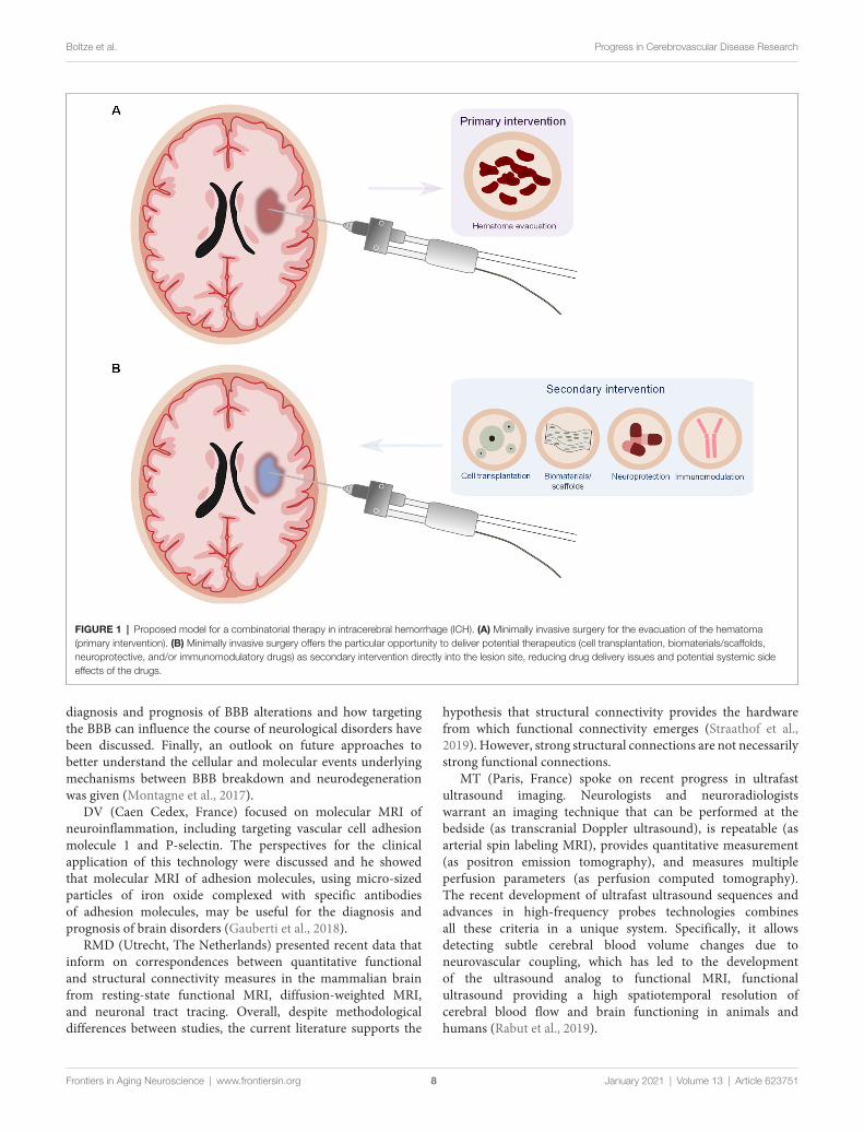
Taken together, the presented studies put forward the ideathat targeting the hematoma is an important strategy in ICHtherapy and that there should be a combinatorial approachconsisting of primary intervention (to evacuate the hematoma)together with a secondary intervention (immunomodulatory andneuroprotective; Figure 1).
Next-Generation Functional Testing inExperimental Brain ResearchThe reliable assessment of functional improvement aftercerebrovascular injury and potential treatment remainschallenging. The recent development of new assessmentstrategies in experimental brain research considers severalconceptual and methodological aspects.
GASM (Lethbridge, AB, Canada) showed that compensationafter brain injury represents the primary process of functionalimprovement, i.e., the development of alternate behavioralstrategies rather than the recovery of the original functions (Metzet al., 2005; Fouad et al., 2013). Compensatory behaviors areclearly recognizable and acquired through repetitive training ina task-specific manner (Girgis et al., 2007; Kirkland et al., 2012).
KF (Edmonton, AB, Canada) discussed recent advances thathave exploited automated test apparatus solutions to enhancethe efficacy of animal training and assessment (Fenrich et al.,2015; Bova et al., 2019) along with machine learning algorithmsto identify discrete aspects of compensatory movement patterns(Ryait et al., 2019).
EH (Montreal, QC, Canada) showed that exercise-basedrehabilitation and pharmacotherapy influence white matterinflammation and the degree of cognitive and cerebrovascularimprovement, including neurovascular coupling, in a mousemodel of vascular cognitive impairment and dementia (Trigianiand Hamel, 2017; Trigiani et al., 2019).
These observations open new avenues for the discoveryof biomarkers that aid in the stratification of patients topersonalized rehabilitation programs. The overarching goalof combined multi-level functional and molecular analysisis the improved translation of experimental innovations toclinical practice.
Cutting-Edge Brain Imaging in Disease andRegenerationMagnetic resonance imaging (MRI) has been decisive inunderstanding cerebrovascular disease over the past decades.Besides MRI, functional ultrasound has recently evolved as ananalog to functional MRI. Cutting-edge imaging-based findingswere reported at the 10th ISN&N.
AM (Los Angeles, CA, USA) reported on brain imagingof vascular dysfunction to understand BBB breakdown andhow this may contribute to cerebrovascular diseases, witha particular focus on dementia. Measuring BBB alterationsin vivo can be challenging. Examples of how brain imaging(i.e., dynamic contrast-enhancedMRI)may contribute to a better
Frontiers in Aging Neuroscience | www.frontiersin.org 7 January 2021 | Volume 13 | Article 623751
Boltze et al. Progress in Cerebrovascular Disease Research
FIGURE 1 | Proposed model for a combinatorial therapy in intracerebral hemorrhage (ICH). (A) Minimally invasive surgery for the evacuation of the hematoma(primary intervention). (B) Minimally invasive surgery offers the particular opportunity to deliver potential therapeutics (cell transplantation, biomaterials/scaffolds,neuroprotective, and/or immunomodulatory drugs) as secondary intervention directly into the lesion site, reducing drug delivery issues and potential systemic sideeffects of the drugs.
diagnosis and prognosis of BBB alterations and how targetingthe BBB can influence the course of neurological disorders havebeen discussed. Finally, an outlook on future approaches tobetter understand the cellular and molecular events underlyingmechanisms between BBB breakdown and neurodegenerationwas given (Montagne et al., 2017).
DV (Caen Cedex, France) focused on molecular MRI ofneuroinflammation, including targeting vascular cell adhesionmolecule 1 and P-selectin. The perspectives for the clinicalapplication of this technology were discussed and he showedthat molecular MRI of adhesion molecules, using micro-sizedparticles of iron oxide complexed with specific antibodiesof adhesion molecules, may be useful for the diagnosis andprognosis of brain disorders (Gauberti et al., 2018).
RMD (Utrecht, The Netherlands) presented recent data thatinform on correspondences between quantitative functionaland structural connectivity measures in the mammalian brainfrom resting-state functional MRI, diffusion-weighted MRI,and neuronal tract tracing. Overall, despite methodologicaldifferences between studies, the current literature supports the
hypothesis that structural connectivity provides the hardwarefrom which functional connectivity emerges (Straathof et al.,2019). However, strong structural connections are not necessarilystrong functional connections.
MT (Paris, France) spoke on recent progress in ultrafastultrasound imaging. Neurologists and neuroradiologistswarrant an imaging technique that can be performed at thebedside (as transcranial Doppler ultrasound), is repeatable (asarterial spin labeling MRI), provides quantitative measurement(as positron emission tomography), and measures multipleperfusion parameters (as perfusion computed tomography).The recent development of ultrafast ultrasound sequences andadvances in high-frequency probes technologies combinesall these criteria in a unique system. Specifically, it allowsdetecting subtle cerebral blood volume changes due toneurovascular coupling, which has led to the developmentof the ultrasound analog to functional MRI, functionalultrasound providing a high spatiotemporal resolution ofcerebral blood flow and brain functioning in animals andhumans (Rabut et al., 2019).
Frontiers in Aging Neuroscience | www.frontiersin.org 8 January 2021 | Volume 13 | Article 623751
Boltze et al. Progress in Cerebrovascular Disease Research
The presented imaging tools promise to further enhanceour understanding of cerebrovascular disease in the future thatwill form the basis for the development of novel therapeuticstrategies. Moreover, these new imaging techniques mightdevelop into invaluable diagnostic tools augmenting currentdiagnostic procedures and leading to better clinical treatmentby identifying patients with specific dysfunctions that wouldbenefit from targeted interventions. They may also provideopportunities to use imaging biomarkers of vascular dysfunctionin patients with TBI or cerebral small vessel disease.
Endovascular Approaches to Image andTreat Stroke: Beyond ThrombectomyModern mechanical thrombectomy techniques are highlyefficient and have revolutionized the treatment of acute strokepatients with large vessel occlusion. However, the restorationof blood flow is insufficient to completely reverse the ischemicinsult. The next phase of stroke intervention should thereforefocus on reparative mechanisms that can be delivered withimage-guidance (Chu et al., 2018) in the same setting afterthrombectomy to further improve patient outcomes, as outlinedby MP (Baltimore, MD, USA).
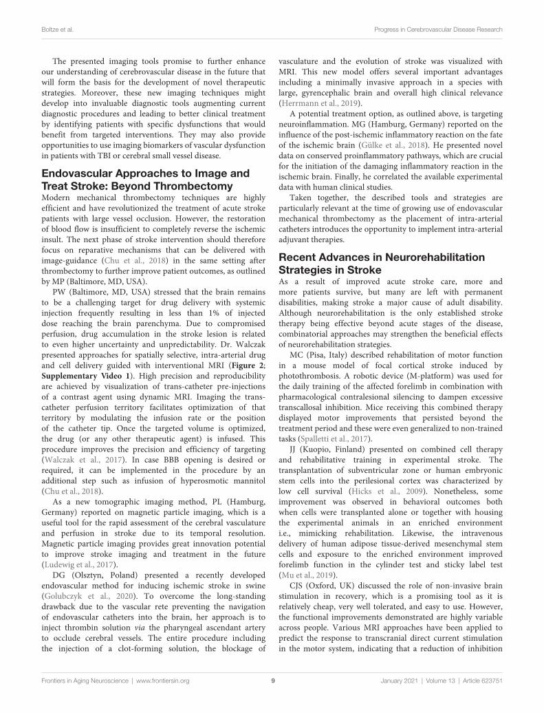
PW (Baltimore, MD, USA) stressed that the brain remainsto be a challenging target for drug delivery with systemicinjection frequently resulting in less than 1% of injecteddose reaching the brain parenchyma. Due to compromisedperfusion, drug accumulation in the stroke lesion is relatedto even higher uncertainty and unpredictability. Dr. Walczakpresented approaches for spatially selective, intra-arterial drugand cell delivery guided with interventional MRI (Figure 2;Supplementary Video 1). High precision and reproducibilityare achieved by visualization of trans-catheter pre-injectionsof a contrast agent using dynamic MRI. Imaging the trans-catheter perfusion territory facilitates optimization of thatterritory by modulating the infusion rate or the positionof the catheter tip. Once the targeted volume is optimized,the drug (or any other therapeutic agent) is infused. Thisprocedure improves the precision and efficiency of targeting(Walczak et al., 2017). In case BBB opening is desired orrequired, it can be implemented in the procedure by anadditional step such as infusion of hyperosmotic mannitol(Chu et al., 2018).
As a new tomographic imaging method, PL (Hamburg,Germany) reported on magnetic particle imaging, which is auseful tool for the rapid assessment of the cerebral vasculatureand perfusion in stroke due to its temporal resolution.Magnetic particle imaging provides great innovation potentialto improve stroke imaging and treatment in the future(Ludewig et al., 2017).
DG (Olsztyn, Poland) presented a recently developedendovascular method for inducing ischemic stroke in swine(Golubczyk et al., 2020). To overcome the long-standingdrawback due to the vascular rete preventing the navigationof endovascular catheters into the brain, her approach is toinject thrombin solution via the pharyngeal ascendant arteryto occlude cerebral vessels. The entire procedure includingthe injection of a clot-forming solution, the blockage of
vasculature and the evolution of stroke was visualized withMRI. This new model offers several important advantagesincluding a minimally invasive approach in a species withlarge, gyrencephalic brain and overall high clinical relevance(Herrmann et al., 2019).
A potential treatment option, as outlined above, is targetingneuroinflammation. MG (Hamburg, Germany) reported on theinfluence of the post-ischemic inflammatory reaction on the fateof the ischemic brain (Gülke et al., 2018). He presented noveldata on conserved proinflammatory pathways, which are crucialfor the initiation of the damaging inflammatory reaction in theischemic brain. Finally, he correlated the available experimentaldata with human clinical studies.
Taken together, the described tools and strategies areparticularly relevant at the time of growing use of endovascularmechanical thrombectomy as the placement of intra-arterialcatheters introduces the opportunity to implement intra-arterialadjuvant therapies.
Recent Advances in NeurorehabilitationStrategies in StrokeAs a result of improved acute stroke care, more andmore patients survive, but many are left with permanentdisabilities, making stroke a major cause of adult disability.Although neurorehabilitation is the only established stroketherapy being effective beyond acute stages of the disease,combinatorial approaches may strengthen the beneficial effectsof neurorehabilitation strategies.
MC (Pisa, Italy) described rehabilitation of motor functionin a mouse model of focal cortical stroke induced byphotothrombosis. A robotic device (M-platform) was used forthe daily training of the affected forelimb in combination withpharmacological contralesional silencing to dampen excessivetranscallosal inhibition. Mice receiving this combined therapydisplayed motor improvements that persisted beyond thetreatment period and these were even generalized to non-trainedtasks (Spalletti et al., 2017).
JJ (Kuopio, Finland) presented on combined cell therapyand rehabilitative training in experimental stroke. Thetransplantation of subventricular zone or human embryonicstem cells into the perilesional cortex was characterized bylow cell survival (Hicks et al., 2009). Nonetheless, someimprovement was observed in behavioral outcomes bothwhen cells were transplanted alone or together with housingthe experimental animals in an enriched environmenti.e., mimicking rehabilitation. Likewise, the intravenousdelivery of human adipose tissue-derived mesenchymal stemcells and exposure to the enriched environment improvedforelimb function in the cylinder test and sticky label test(Mu et al., 2019).
CJS (Oxford, UK) discussed the role of non-invasive brainstimulation in recovery, which is a promising tool as it isrelatively cheap, very well tolerated, and easy to use. However,the functional improvements demonstrated are highly variableacross people. Various MRI approaches have been applied topredict the response to transcranial direct current stimulationin the motor system, indicating that a reduction of inhibition
Frontiers in Aging Neuroscience | www.frontiersin.org 9 January 2021 | Volume 13 | Article 623751

Boltze et al. Progress in Cerebrovascular Disease Research
FIGURE 2 | Intraarterial magnetic resonance imaging (MRI)-guided injection of mesenchymal stem cells into the canine brain. (A) Raw T2*w MRI after intraarterialinjection of superparamagnetic iron oxide particle-labeled mesenchymal stem cells showing cell accumulation throughout the ipsilateral middle cerebral arteryterritory. (B) Segmentation of hypo-intense pixels and 3D visualization of stem cell biodistribution. Please see the Supplementary Video 1 for a dynamicvisualization of stem cell injection.
in the primary motor cortex (M1) is a central mechanism formotor plasticity in humans, both in health and in recovery afterstroke (Stagg et al., 2011; Blicher et al., 2015; Kolasinski et al.,2019). Indeed, GABA levels in the ipsilateral M1 cortex canpredict the response to transcranial direct current stimulation(O’Shea et al., 2014).
FCH (Geneva, Switzerland) summarized the reasons forheterogeneous results of neurorehabilitative interventions, suchas non-invasive brain stimulation, and discussed a paradigmshift from imprecision of ‘‘one treatment suits all’’ strategiestowards personalizedmedicine approaches delivered in amannertailored to the demands of the individual patient (Raffinand Hummel, 2018; Coscia et al., 2019). Structural MRIimaging and connectome analyses can be used to determinethe functional outcome of patients to stratify patients towardsspecific brain stimulation protocols (Koch and Hummel, 2017;Schulz et al., 2017).
Taken together, these studies support the use of rehabilitationand neuromodulatory approaches for promoting post-strokerecovery. Although combination therapy and early deliverymay be more effective, further studies with greater statisticalpower to cope with these complex experimental designs willbe needed to determine the optimal treatment protocol, theeffect of co-morbidities, and to explore underlying therapeuticmechanisms (Boltze et al., 2017). Moreover, novel imagingapproaches as discussed in the section ‘‘Cutting-Edge BrainImaging in Disease and Regeneration’’ (above) might be
helpful to identify patients that may benefit from brainstimulation interventions.
OUTLOOK AND CONCLUSIONS
Based on the setbacks seen in the past decades of cerebrovasculardisease research, the field has clearly consolidated and recognizedthe reasons for previous translational failure as exemplified inthe field of cell therapies (Cui et al., 2019). Recent guidelinesfor translational research in that field stress, for instance, theimportance of considering comorbidities, the use of highlypredictive animal models and experimental settings as well asa closer interaction between preclinical and clinical researchactivities for more integrated study designs being able to informeach other (Boltze et al., 2019). Moreover, with the refinement ofour research tools, some of which have been discussed herein,and increasing study quality (Ramirez et al., 2017), it seemsthat the translational roadblock is slowly overcome as newtherapeutic concepts start to arise (Liebeskind et al., 2018). Forinstance, new combinations of recanalization and neuroproteciveapproaches are believed to emerge for stroke (Savitz et al., 2019)and potentially ICH.
Encouraging progress is also witnessed in related fieldssuch as vascular dementia or even Alzheimer’s disease. Forinstance, significant progress has been made in understandingthe vascular contribution to various form of dementia (Iadecolaand Gottesman, 2018; Royea et al., 2020). While the role of
Frontiers in Aging Neuroscience | www.frontiersin.org 10 January 2021 | Volume 13 | Article 623751
Boltze et al. Progress in Cerebrovascular Disease Research
the frequent but treatable risk factors of dementia such ashypertension and hypercholesterinemia is better understood(Kaiser et al., 2014; Trigiani et al., 2019), new animal modelsof vascular cognitive impairment may facilitate translationalresearch programs (Hainsworth et al., 2017). In parallel,new targets for pharmacological interventions have beenidentified (Loera-Valencia et al., 2019) and are expectedto lead to future clinical trials (Howard et al., 2019).Although there will likely be setbacks on the way forwardand we shall avoid premature enthusiasm as seen in thepast, the ‘‘therapeutic nihilism’’ still observed in translationalcerebrovascular research just 5 years ago has turned intonew optimism.
The 11th ISN&N will continue to monitor the progress in thefields of basic translational and clinical cerebrovascular diseaseresearch. Moreover, it will extend its scope by partnering withthe 18th International Conference on Brain Edema and CellularInjury (BEM) for a single combined meeting in October 2022 inBerlin, Germany.
AUTHOR CONTRIBUTIONS
All authors listed have made a substantial, direct and intellectualcontribution to the work, and approved it for publication.
FUNDING
We wish to express our gratitude to all institutions and peoplewho supported the 10th ISN&N. First and foremost, we wantto thank all meeting participants for their outstanding academiccontributions. Moreover, we are particularly grateful to theDeutsche Forschungsgemeinschaft (grant number: BO 3518/2-1)and the International Society of Cerebral Blood Flow andMetabolism, both of which supported the meeting with generousgrants. We further acknowledge all industrial sponsors andexhibitors for their academic and invaluable financial support.
ACKNOWLEDGMENTS
The Eventlab congress organizing team deserves special thanksfor tireless and professional support that was not only crucialfor carefully preparing the meeting, but also for running itsuccessfully and smoothly.
SUPPLEMENTARY MATERIAL
The Supplementary Material for this article can be found onlineat: https://www.frontiersin.org/articles/10.3389/fnagi.2021.623751/full#supplementary-material.
REFERENCES
Alawieh, A., Langley, E. F., and Tomlinson, S. (2018). Targeted complementinhibition salvages stressed neurons and inhibits neuroinflammation afterstroke in mice. Sci. Transl. Med. 10:eaao6459. doi: 10.1126/scitranslmed.aao6459
Awad, I. A., Polster, S. P., Carrión-Penagos, J., Thompson, R. E., Cao, Y.,Stadnik, A., et al. (2019). Surgical performance determines functional outcomebenefit in the minimally invasive surgery plus recombinant tissue plasminogenactivator for intracerebral hemorrhage evacuation (MISTIE) procedure.Neurosurgery 84, 1157–1168. doi: 10.1093/neuros/nyz077
Balbi, M., Koide, M., Schwarzmaier, S. M., Wellman, G. C., and Plesnila, N.(2017). Acute changes in neurovascular reactivity after subarachnoidhemorrhage in vivo. J. Cereb. Blood Flow Metab. 37, 178–187.doi: 10.1177/0271678X15621253
Balbi, M., Vega, M. J., Lourbopoulos, A., Terpolilli, N. A., and Plesnila, N. (2020).Long-term impairment of neurovascular coupling following experimentalsubarachnoid hemorrhage. J. Cereb. Blood Flow Metab. 40, 1193–1202.doi: 10.1177/0271678X19863021
Blicher, J. U., Near, J., Næss-Schmidt, E., Stagg, C. J., Johansen-Berg, H.,Nielsen, J. F., et al. (2015). GABA levels are decreased after stroke andGABA changes during rehabilitation correlate with motor improvement.Neurorehabil. Neural Repair 29, 278–286. doi: 10.1177/1545968314543652
Boltze, J., Kleinschnitz, C., Reymann, K. G., Reiser, G., Wagner, D. C.,Kranz, A., et al. (2012). Neurovascular pathophysiology in cerebral ischemia,dementia and the ageing brain—current trends in basic, translationaland clinical research. Exp. Transl. Stroke Med. 4:14. doi: 10.1186/2040-7378-4-14
Boltze, J., Kranz, A., Wagner, D. C., Reymann, K., Reiser, G., and Hess, D. C.(2011). Recent advances in basic and translational stroke research. Expert Rev.Neurother. 11, 199–202. doi: 10.1586/ern.10.202
Boltze, J., Modo, M. M., Mays, R. W., Taguchi, A., Jolkkonen, J.,Savitz, S., et al. (2019). Stem cells as an emerging paradigm in stroke 4:advancing and accelerating preclinical research. Stroke 50, 3299–3306.doi: 10.1161/STROKEAHA.119.025436
Boltze, J., Nitzsche, F., Jolkkonen, J., Weise, G., Pösel, C., Nitzsche, B., et al. (2017).Concise review: increasing the validity of cerebrovascular disease models andexperimental methods for translational stem cell research. Stem Cells 35,1141–1153. doi: 10.1002/stem.2595
Bova, A., Kernodle, K., Mulligan, K., and Leventhal, D. (2019). Automated ratsingle-pellet reaching with 3-dimensional reconstruction of paw and digittrajectories. J. Vis. Exp. 149:e59979. doi: 10.3791/59979
Brochu-Gaudreau, K., Rehfeldt, C., Blouin, R., Bordignon, V., Murphy, B. D., andPalin, M. F. (2010). Adiponectin action from head to toe. Endocrine 37, 11–32.doi: 10.1007/s12020-009-9278-8
Busato, A., Bonafede, R., Bontempi, P., Scambi, I., Schiaffino, L., Benati, D., et al.(2017). Labeling and magnetic resonance imaging of exosomes isolated fromadipose stem cells. Curr. Protoc. Cell Biol. 75, 3.44.1-3.44.15. doi: 10.1002/cpcb.23
Chang, C. F., Goods, B. A., Askenase, M. H., Hammond, M. D., Renfroe, S. C.,Steinschneider, A. F., et al. (2018). Erythrocyte efferocytosis modulatesmacrophages towards recovery after intracerebral hemorrhage. J. Clin. Invest.128, 607–624. doi: 10.1172/JCI95612
Choi, H., and Lee, D. S. (2016). Illuminating the physiology ofextracellular vesicles. Stem Cell Res. Ther. 7:55. doi: 10.1186/s13287-016-0316-1
Chu, C., Liu, G., Janowski, M., Bulte, J. W. M., Li, S., Pearl, M., et al.(2018). Real-time MRI guidance for reproducible hyperosmolar opening ofthe blood-brain barrier in mice. Front. Neurol. 9:921. doi: 10.3389/fneur.2018.00921
Cohan, C. H., Stradecki-Cohan, H. M., Morris-Blanco, K. C., Khoury, N.,Koronowski, K. B., Youbi, M., et al. (2017). Protein kinase C epsilondelays latency until anoxic depolarization through arc expression andGluR2 internalization. J. Cereb. Blood Flow Metab. 37, 3774–3788.doi: 10.1177/0271678X17712178
Coscia, M., Wessel, M. J., Chaudary, U., Millán, J. D. R., Micera, S.,Guggisberg, A., et al. (2019). Neurotechnology-aided interventions for upperlimb motor rehabilitation in severe chronic stroke. Brain 142, 2182–2197.doi: 10.1093/brain/awz181
Cui, L. L., Golubczyk, D., Tolppanen, A. M., Boltze, J., and Jolkkonen, J. (2019).Cell therapy for ischemic stroke: are differences in preclinical and clinical study
Frontiers in Aging Neuroscience | www.frontiersin.org 11 January 2021 | Volume 13 | Article 623751
Boltze et al. Progress in Cerebrovascular Disease Research
design responsible for the translational loss of efficacy? Ann. Neurol. 86, 5–16.doi: 10.1002/ana.25493
Dabrowska, S., Del Fattore, A., Karnas, E., Frontczak-Baniewicz, M.,Kozlowska, H., Muraca, M., et al. (2018). Imaging of extracellular vesiclesderived from human bone marrow mesenchymal stem cells using fluorescentand magnetic labels. Int. J. Nanomedicine 13, 1653–1664. doi: 10.2147/IJN.S159404
D’Adesky, N. D., de Rivero Vaccari, J. P., Bhattacharya, P., Schatz, M., Perez-Pinzon, M. A., Bramlett, H. M., et al. (2018). Nicotine alters estrogenreceptor-β-regulated inflammasome activity and exacerbates ischemic braindamage in female rats. Int. J. Mol. Sci. 19:1330. doi: 10.3390/ijms19051330
DeFazio, R. A., Raval, A. P., Lin, H. W., Dave, K. R., Della-Morte, D., andPerez-Pinzon, M. A. (2009). GABA synapses mediate neuroprotectionafter ischemic and εPKC preconditioning in rat hippocampal slicecultures. J. Cereb. Blood Flow Metab. 29, 375–384. doi: 10.1038/jcbfm.2008.126
Demuth, H. U., Dijkhuizen, R. M., Farr, T. D., Gelderblom, M., Horsburgh, K.,Iadecola, C., et al. (2017). Recent progress in translational research onneurovascular and neurodegenerative disorders. Restor. Neurol. Neurosci. 35,87–103. doi: 10.3233/RNN-160690
de Rivero Vaccari, J. P., Patel, H. H., Brand, F. J. 3rd, Perez-Pinzon, M. A.,Bramlett, H. M., and Raval, A. P. (2016). Estrogen receptor beta signaling alterscellular inflammasomes activity after global cerebral ischemia in reproductivelysenescence female rats. J. Neurochem. 136, 492–496. doi: 10.1111/jnc.13404
Doeppner, T. R., Doehring, M., Kaltwasser, B., Majid, A., Lin, F., Bähr, M.,et al. (2017). Ischemic post-conditioning induces post-stroke neuroprotectionvia Hsp70-mediated proteasome inhibition and facilitates neural progenitorcell transplantation. Mol. Neurobiol. 54, 6061–6073. doi: 10.1007/s12035-016-0137-3
Doeppner, T. R., Herz, J., Görgens, A., Schlechter, J., Ludwig, A. K., Radtke, S.,et al. (2015). Extracellular vesicles improve post-stroke neuroregenerationand prevent postischemic immunosuppression. Stem Cells Transl. Med. 4,1131–1143. doi: 10.5966/sctm.2015-0078
Doyle, K. P., Quach, L. N., Solé,M., Axtell, R. C., Nguyen, T. V., Soler-Llavina, G. J.,et al. (2015). B-lymphocyte-mediated delayed cognitive impairment followingstroke. J. Neurosci. 35, 2133–2145. doi: 10.1523/JNEUROSCI.4098-14.2015
Dreier, J. P., Lemale, C. L., Kola, V., Friedman, A., and Schoknecht, K.(2018). Spreading depolarization is not an epiphenomenon but the principalmechanism of the cytotoxic edema in various graymatter structures of the brainduring stroke. Neuropharmacology 134, 189–207. doi: 10.1016/j.neuropharm.2017.09.027
Egashira, Y., Hua, Y., Keep, R. F., Iwama, T., and Xi, G. (2016). Lipocalin 2 andblood-brain barrier disruption in whitematter after experimental subarachnoidhemorrhage. Acta Neurochir. Suppl. 121, 131–134. doi: 10.1007/978-3-319-18497-5_23
Fenrich, K. K., May, Z., Hurd, C., Boychuk, C. E., Kowalczewski, J., Bennett, D. J.,et al. (2015). Improved single pellet grasping using automated ad libitumfull-time training robot. Behav. Brain Res. 281, 137–148. doi: 10.1016/j.bbr.2014.11.048
Fernández, G., Moraga, A., Cuartero, M. I., García-Culebras, A., Peña-Martínez, C., Pradillo, J. M., et al. (2018). TLR4-binding DNA aptamersshow a protective effect against acute stroke in animal models. Mol. Ther. 26,2047–2059. doi: 10.1016/j.ymthe.2018.05.019
Fouad, K., Hurd, C., and Magnuson, D. S. (2013). Functional testing in animalmodels of spinal cord injury: not as straight forward as one would think. Front.Integr. Neurosci. 7:85. doi: 10.3389/fnint.2013.00085
Fukuda, A. M., Adami, A., Pop, V., Bellone, J. A., Coats, J. S., Hartman, R. E., et al.(2013). Posttraumatic reduction of edema with aquaporin-4 RNA interferenceimproves acute and chronic functional recovery. J. Cereb. Blood Flow Metab.33, 1621–1632. doi: 10.1038/jcbfm.2013.118
García-Culebras, A., Durán-Laforet, V., Peña-Martínez, C., Moraga, A.,Ballesteros, I., Cuartero, M. I., et al. (2019). Role of TLR4 (toll-like receptor 4)in N1/N2 neutrophil programming after stroke. Stroke 50, 2922–2932.doi: 10.1161/STROKEAHA.119.025085
García-Culebras, A., Palma-Tortosa, S., Moraga, A., García-Yébenes, I., Durán-Laforet, V., Cuartero, M. I., et al. (2017). Toll-like receptor 4 mediateshemorrhagic transformation after delayed tissue plasminogen activator
administration in in situ thromboembolic stroke. Stroke 48, 1695–1699.doi: 10.1161/STROKEAHA.116.015956
Gauberti, M., Fournier, A. P., Docagne, F., Vivien, D., and Martinez deLizarrondo, S. (2018). Molecular magnetic resonance imaging of endothelialactivation in the central nervous system. Theranostics 8, 1195–1212.doi: 10.7150/thno.22662
Girgis, J., Merrett, D., Kirkland, S., Metz, G. A., Verge, V., and Fouad, K.(2007). Reaching training in rats with spinal cord injury promotes plasticityand task specific recovery. Brain 130, 2993–3003. doi: 10.1093/brain/awm245
Golubczyk, D., Kalkowski, L., Kwiatkowska, J., Zawadzki, M., Holak, P.,Glodek, J., et al. (2020). Endovascular model of ischemic stroke in swineguided by real-time MRI. Sci. Rep. 10:17318. doi: 10.1038/s41598-020-74411-3
Guarente, L. (2008). Mitochondria—a nexus for aging, calorie restriction andsirtuins? Cell 132, 171–176. doi: 10.1016/j.cell.2008.01.007
Gülke, E., Gelderblom, M., and Magnus, T. (2018). Danger signals in stroke andtheir role on microglia activation after ischemia. Ther. Adv. Neurol. Disord.11:1756286418774254. doi: 10.1177/1756286418774254
Guo, D.,Wilkinson, D. A., Thompson, B. G., Pandey, A. S., Keep, R. F., Xi, G., et al.(2017). MRI characterization in the acute phase of experimental subarachnoidhemorrhage. Transl. Stroke Res. 8, 234–243. doi: 10.1007/s12975-016-0511-5
Haigis, M. C., and Guarente, L. P. (2006). Mammalian sirtuins—emergingroles in physiology, aging and calorie restriction. Genes Dev. 20, 2913–2921.doi: 10.1101/gad.1467506
Hainsworth, A. H., Allan, S. M., Boltze, J., Cunningham, C., Farris, C., Head, E.,et al. (2017). Translational models for vascular cognitive impairment: areview including larger species. BMC Med. 15:16. doi: 10.1186/s12916-017-0793-9
Hanley, D. F., Thompson, R. E., Rosenblum, M., Yenokyan, G., Lane, K.,McBee, N., et al. (2019). Efficacy and safety of minimally invasive surgerywith thrombolysis in intracerebral haemorrhage evacuation (MISTIE III): arandomised, controlled, open-label, blinded endpoint phase 3 trial. Lancet 393,1021–1032. doi: 10.1016/S0140-6736(19)30195-3
Herrmann, A. M., Meckel, S., Gounis, M. J., Kringe, L., Motschall, E., Mülling, C.,et al. (2019). Large animals in neurointerventional research: a systematic reviewon models, techniques and their application in endovascular procedures forstroke, aneurysms and vascular malformations. J. Cereb. Blood Flow Metab. 39,375–394. doi: 10.1177/0271678X19827446
Hicks, A. U., Lappalainen, R. S., Narkilahti, S., Suuronen, R., Corbett, D.,Sivenius, J., et al. (2009). Transplantation of human embryonic stemcell-derived neural precursor cells and enriched environment after corticalstroke in rats: cell survival and functional recovery. Eur. J. Neurosci. 29,562–574. doi: 10.1111/j.1460-9568.2008.06599.x
Howard, R., Zubko, O., Bradley, R., Harper, E., Pank, L., O’Brien, J., et al.(2019). Minocycline at 2 different dosages vs. placebo for patients with mildAlzheimer disease: a randomized clinical trial. JAMA Neurol. 77, 164–174.doi: 10.1001/jamaneurol.2019.3762
Huang, H., Bhuiyan, M. I. H., Jiang, T., Song, S., Shankar, S., Taheri, T.,et al. (2019). A novel Na+−K+−Cl− cotransporter 1 inhibitor STS66*reduces brain damage in mice after ischemic stroke. Stroke 50, 1021–1025.doi: 10.1161/STROKEAHA.118.024287
Iadecola, C., and Gottesman, R. F. (2018). Cerebrovascular alterations inAlzheimer disease. Circ. Res. 123, 406–408. doi: 10.1161/CIRCRESAHA.118.313400
Jablonska, A., Drela, K., Wojcik-Stanaszek, L., Janowski, M., Zalewska, T., andLukomska, B. (2016). Short-lived human umbilical cord-blood-derived neuralstem cells influence the endogenous secretome and increase the number ofendogenous neural progenitors in a ratmodel of lacunar stroke.Mol. Neurobiol.53, 6413–6425. doi: 10.1007/s12035-015-9530-6
Ji, X., Zhao, W., Boltze, J., Li, S., Meng, R., Wang, Y., et al. (2019). Clinicalpractice guidelines of remote ischemic conditioning for the management ofcerebrovascular diseases. Cond. Med. 2, 225–241.
Jy, W., Rehni, A. K., Bidot, C. Jr., Navarro-Quero, H., Haase, C. R., Koch, S.,et al. (2018). Pharmacokinetics of human red blood cell microparticlesprepared using high-pressure extrusion method. Front. Pharmacol. 9:599.doi: 10.3389/fphar.2018.00599
Frontiers in Aging Neuroscience | www.frontiersin.org 12 January 2021 | Volume 13 | Article 623751

Boltze et al. Progress in Cerebrovascular Disease Research
Kaiser, D., Weise, G., Möller, K., Scheibe, J., Pösel, C., Baasch, S., et al. (2014).Spontaneous white matter damage, cognitive decline and neuroinflammationin middle-aged hypertensive rats: an animal model of early-stage cerebral smallvessel disease. Acta Neuropathol. Commun. 2:169. doi: 10.1186/s40478-014-0169-8
Karuppagounder, S. S., Alin, L., Chen, Y., Brand, D., Bourassa, M.W., Dietrich, K.,et al. (2018). N-acetylcysteine targets 5 lipoxygenase-derived, toxic lipidsand can synergize with prostaglandin E2 to inhibit ferroptosis and improveoutcomes following hemorrhagic stroke in mice. Ann. Neurol. 84, 854–872.doi: 10.1002/ana.25356
Keep, R. F., Hua, Y., and Xi, G. (2012). Intracerebral haemorrhage: mechanisms ofinjury and therapeutic targets. Lancet Neurol. 11, 720–731. doi: 10.1016/S1474-4422(12)70104-7
Khoury, N., Xu, J., Stegelmann, S. D., Jackson, C. W., Koronowski, K. B.,Dave, K. R., et al. (2019). Resveratrol preconditioning induces genomic andmetabolic adaptations within the long-term window of cerebral ischemictolerance leading to bioenergetic efficiency. Mol. Neurobiol. 56, 4549–4565.doi: 10.1007/s12035-018-1380-6
Kim, T. H., and Vemuganti, R. (2015). Effect of sex and age interactionson functional outcome after stroke. CNS Neurosci. Ther. 21, 327–336.doi: 10.1111/cns.12346
Kim, H., Walczak, P., Muja, N., Campanelli, J. T., and Bulte, J. W.(2012). ICV-transplanted human glial precursor cells are short-lived yetexert immunomodulatory effects in mice with EAE. Glia 60, 1117–1129.doi: 10.1002/glia.22339
Kirkland, S. W., Smith, L. K., and Metz, G. A. (2012). Task-specific compensationand recovery following focal motor cortex lesion in stressed rats. J. Integr.Neurosci. 11, 33–59. doi: 10.1142/S0219635212500033
Kleinschnitz, C., Schwab, N., Kraft, P., Hagedorn, I., Dreykluft, A., Schwarz, T.,et al. (2010). Early detrimental T-cell effects in experimental cerebral ischemiaare neither related to adaptive immunity nor thrombus formation. Blood 115,3835–3842. doi: 10.1182/blood-2009-10-249078
Koch, P. J., and Hummel, F. C. (2017). Toward precision medicine: tailoringinterventional strategies based on noninvasive brain stimulation for motorrecovery after stroke. Curr. Opin. Neurol. 30, 388–397. doi: 10.1097/WCO.0000000000000462
Kolasinski, J., Hinson, E. L., Divanbeighi Zand, A. P., Rizov, A., Emir, U. E., andStagg, C. J. (2019). The dynamics of cortical GABA in human motor learning.J. Physiol. 597, 271–282. doi: 10.1113/JP276626
Koronowski, K. B., Khoury, N., Saul, I., Loris, Z. B., Cohan, C. H., Stradecki-Cohan, H. M., et al. (2017). Neuronal SIRT1 (silent information regulator2 homologue 1) regulates glycolysis and mediates resveratrol-induced ischemictolerance. Stroke 48, 3117–3125. doi: 10.1161/STROKEAHA.117.018562
Kronenberg, G., Uhlemann, R., Richter, N., Klempin, F., Wegner, S., Staerck, L.,et al. (2018). Distinguishing features of microglia- and monocyte-derived macrophages after stroke. Acta Neuropathol. 135, 551–568.doi: 10.1007/s00401-017-1795-6
Lantigua, H., Ortega-Gutierrez, S., Schmidt, J. M., Lee, K., Badjatia, N., Agarwal, S.,et al. (2015). Subarachnoid hemorrhage: who dies and why? Crit. Care 19:309.doi: 10.1186/s13054-015-1036-0
Laterza, C., Wattananit, S., Uoshima, N., Ge, R., Pekny, R., Tornero, D., et al.(2017). Monocyte depletion early after stroke promotes neurogenesis fromendogenous neural stem cells in adult brain. Exp. Neurol. 297, 129–137.doi: 10.1016/j.expneurol.2017.07.012
Li, Q., Han, X., Lan, X., Gao, Y., Wan, J., Durham, F., et al. (2017). Inhibitionof neuronal ferroptosis protects hemorrhagic brain. JCI Insight 2:e90777.doi: 10.1172/jci.insight.90777
Li, Q., Weiland, A., Chen, X., Lan, X., Han, X., Durham, F., et al. (2018).Ultrastructural characteristics of neuronal death and white matter injury inmouse brain tissues after intracerebral hemorrhage: coexistence of ferroptosis,autophagy and necrosis. Front. Neurol. 9:581. doi: 10.3389/fneur.2018.00581
Liebeskind, D. S., Derdeyn, C. P., Wechsler, L. R., and STAIR X Consortium.(2018). STAIR X: emerging considerations in developing and evaluatingnew stroke therapies. Stroke 49, 2241–2247. doi: 10.1161/STROKEAHA.118.021424
Loera-Valencia, R., Cedazo-Minguez, A., Kenigsberg, P. A., Page, G., Duarte, A. I.,Giusti, P., et al. (2019). Current and emerging avenues for Alzheimer’s diseasedrug targets. J. Intern. Med. 286, 398–437. doi: 10.1111/joim.12959
Lückl, J., Lemale, C. L., Kola, V., Horst, V., Khojasteh, U., Oliveira-Ferreira, A. I.,et al. (2018). The negative ultraslow potential, electrophysiologicalcorrelate of infarction in the human cortex. Brain 141, 1734–1752.doi: 10.1093/brain/awy102
Ludewig, P., Gdaniec, N., Sedlacik, J., Forkert, N. D., Szwargulski, P., Graeser, M.,et al. (2017). Magnetic particle imaging for real-time perfusion imaging in acutestroke. ACS Nano 11, 10480–10488. doi: 10.1021/acsnano.7b05784
Mantovani, A. (2017). Wandering pathways in the regulation of innateimmunity and inflammation. J. Autoimmun. 85, 1–5. doi: 10.1016/j.jaut.2017.10.007
Mao, X., Terpolilli, N. A., When, A., Cheng, S., Hellal, F., Liu, B., et al. (2020).Progressive histopathological damage occurring up to 1 year after experimentaltraumatic brain injury is associated with cognitive decline and depression-likebehavior. J. Neurotrauma 37, 1331–1341. doi: 10.1089/neu.2019.6510
McCullough, L. D., Mirza, M. A., Xu, Y., Bentivegna, K., Steffens, E. B., Ritzel, R.,et al. (2016). Stroke sensitivity in the aged: sex chromosome complement vs.gonadal hormones. Aging 8, 1432–1441. doi: 10.18632/aging.100997
Mehta, S. L., Kim, T., and Vemuganti, R. (2015). Long noncodingRNA FosDT promotes ischemic brain injury by interacting withREST-associated chromatin-modifying proteins. J. Neurosci. 35, 16443–16449.doi: 10.1523/JNEUROSCI.2943-15.2015
Metz, G. A., Antonow-Schlorke, I., andWitte, O. W. (2005). Motor improvementsafter focal cortical ischemia in adult rats are mediated by compensatorymechanisms. Behav. Brain Res. 162, 71–82. doi: 10.1016/j.bbr.2005.03.002
Miró-Mur, F., Pérez-de-Puig, I., Ferrer-Ferrer, M., Urra, X., Justicia, C.,Chamorro, A., et al. (2016). Immature monocytes recruited to theischemic mouse brain differentiate into macrophages with features ofalternative activation. Brain Behav. Immun. 53, 18–33. doi: 10.1016/j.bbi.2015.08.010
Montagne, A., Zhao, Z., and Zlokovic, B. V. (2017). Alzheimer’s disease: amatter of blood-brain barrier dysfunction? J. Exp. Med. 214, 3151–3169.doi: 10.1084/jem.20171406
Morris-Blanco, K. C., Kim, T., Lopez, M. S., Bertogliat, M. J., Chelluboina, B.,and Vemuganti, R. (2019). Induction of DNA hydroxymethylation protectsthe brain after stroke. Stroke 50, 2513–2521. doi: 10.1161/STROKEAHA.119.025665
Mu, J., Bakreen, A., Juntunen, M., Korhonen, P., Oinonen, E., Cui, L., et al.(2019). Combined adipose tissue-derived mesenchymal stem cell therapy andrehabilitation in experimental stroke. Front. Neurol. 10:235. doi: 10.3389/fneur.2019.00235
Muhammad, S., Barakat, W., Stoyanov, S., Murikinati, S., Yang, H., Tracey, K. J.,et al. (2008). The HMGB1 receptor RAGE mediates ischemic brain damage.J. Neurosci. 28, 12023–12031. doi: 10.1523/JNEUROSCI.2435-08.2008
Narayanan, S. V., Dave, K. R., Saul, I., and Perez-Pinzon, M. A. (2015).Resveratrol preconditioning protects against cerebral ischemic injuryvia nuclear erythroid 2-related factor 2. Stroke 46, 1626–1632.doi: 10.1161/STROKEAHA.115.008921
Neumann, J. T., Thompson, J. W., Raval, A. P., Cohan, C. H., Koronowski, K. B.,and Perez-Pinzon, M. A. (2015). Increased BDNF protein expression afterischemic or PKC epsilon preconditioning promotes electrophysiologic changesthat lead to neuroprotection. J. Cereb. Blood Flow Metab. 35, 121–130.doi: 10.1038/jcbfm.2014.185
O’Shea, J., Boudrias, M. H., Stagg, C. J., Bachtiar, V., Kischka, U., Blicher, J. U.,et al. (2014). Predicting behavioural response to TDCS in chronicmotor stroke. Neuroimage 85, 924–933. doi: 10.1016/j.neuroimage.2013.05.096
Panta, A., Pandey, S., Duncan, I. N., Duhamel, S., and Sohrabji, F. (2019).Mir363-3p attenuates post-stroke depressive-like behaviors in middle-agedfemale rats. Brain Behav. Immun. 78, 31–40. doi: 10.1016/j.bbi.2019.01.003
Poinsatte, K., Betz, D., Torres, V. O., Ajay, A. D., Mirza, S., Selvaraj, U. M., et al.(2019). Visualization and quantification of post-stroke neural connectivity andneuroinflammation using serial two-photon tomography in the whole mousebrain. Front. Neurosci. 13:1055. doi: 10.3389/fnins.2019.01055
Poon, M. T., Fonville, A. F., and Al-Shahi Salman, R. (2014). Long-termprognosis after intracerebral haemorrhage: systematic review and meta-analysis. J. Neurol. Neurosurg. Psychiatry 85, 660–667. doi: 10.1136/jnnp-2013-306476
Frontiers in Aging Neuroscience | www.frontiersin.org 13 January 2021 | Volume 13 | Article 623751
Boltze et al. Progress in Cerebrovascular Disease Research
Pop, V., and Badaut, J. (2011). A neurovascular perspective for long-term changesafter brain trauma. Transl. Stroke Res. 2, 533–545. doi: 10.1007/s12975-011-0126-9
Potts, M. B., and Riina, H. A. (2014). Refining the role for evacuation ofspontaneous intracerebral hematomas: results of STICH II. World Neurosurg.82, 549–550. doi: 10.1016/j.wneu.2014.08.046
Pradillo, J. M., Fernández-López, D., García-Yébenes, I., Sobrado, M.,Hurtado, O., Moro, M. A., et al. (2009). Toll-like receptor 4 is involvedin neuroprotection afforded by ischemic preconditioning. J. Neurochem. 109,287–294. doi: 10.1111/j.1471-4159.2009.05972.x
Rabut, C., Correia, M., Finel, V., Pezet, S., Pernot, M., Deffieux, T., et al. (2019). 4Dfunctional ultrasound imaging of whole-brain activity in rodents.Nat. Methods16, 994–997. doi: 10.1038/s41592-019-0572-y
Raffin, E., and Hummel, F. C. (2018). Restoring motor functions afterstroke: multiple approaches and opportunities. Neuroscientist 24, 400–416.doi: 10.1177/1073858417737486
Ramirez, F. D., Motazedian, P., Jung, R. G., Di Santo, P., MacDonald, Z. D.,Moreland, R., et al. (2017). Methodological rigor in preclinical cardiovascularstudies: targets to enhance reproducibility and promote research translation.Circ. Res. 120, 1916–1926. doi: 10.1161/CIRCRESAHA.117.310628
Rehni, A. K., Shukla, V., Navarro Quero, H., Bidot, C. Jr., Haase, C. R.,Crane, E. A. A., et al. (2019). Preclinical evaluation of safety and biodistributionof red cell microparticles: a novel hemostatic agent. J. Cardiovasc. Pharmacol.Ther. 24, 474–483. doi: 10.1177/1074248419838512
Ritzel, R. M., Lai, Y. J., Crapser, J. D., Patel, A. R., Schrecengost, A., Grenier, J. M.,et al. (2018). Aging alters the immunological response to ischemic stroke. ActaNeuropathol. 136, 89–110. doi: 10.1007/s00401-018-1859-2
Royea, J., Martinot, P., and Hamel, E. (2020). Memory and cerebrovasculardeficits recovered following angiotensin IV intervention in a mouse modelof Alzheimer’s disease. Neurobiol. Dis. 134:104644. doi: 10.1016/j.nbd.2019.104644
Ryait, H., Bermudez-Contreras, E., Harvey, M., Faraji, J., Mirza Agha, B., Gomez-Palacio Schjetnan, A., et al. (2019). Data-driven analyses of motor impairmentsin animal models of neurological disorders. PLoS Biol. 17:e3000516.doi: 10.1371/journal.pbio.3000516
Salehi, A., Jullienne, A., Baghchechi, M., Hamer, M., Walsworth, M., Donovan, V.,et al. (2018). Up-regulation of Wnt/β-catenin expression is accompanied withvascular repair after traumatic brain injury. J. Cereb. Blood Flow Metab. 38,274–289. doi: 10.1177/0271678X17744124
Sandsmark, D. K., Bashir, A., Wellington, C. L., and Diaz-Arrastia, R. (2019).Cerebral microvascular injury: a potentially treatable endophenotype oftraumatic brain injury-induced neurodegeneration. Neuron 103, 367–379.doi: 10.1016/j.neuron.2019.06.002
Savitz, S. I., Baron, J. C., Fisher, M., and STAIR X Consortium. (2019). Stroketreatment academic industry roundtable X: brain cytoprotection therapies inthe reperfusion era. Stroke 50, 1026–1031. doi: 10.1161/STROKEAHA.118.023927
Schulz, R., Park, E., Lee, J., Chang, W. H., Lee, A., Kim, Y. H., et al.(2017). Interactions between the corticospinal tract and premotor-motorpathways for residual motor output after stroke. Stroke 48, 2805–2811.doi: 10.1161/STROKEAHA.117.016834
Selvamani, A., Sathyan, P., Miranda, R. C., and Sohrabji, F. (2012). An antagomirto microRNA Let7f promotes neuroprotection in an ischemic stroke model.PLoS One 7:e32662. doi: 10.1371/journal.pone.0032662
Selvamani, A., and Sohrabji, F. (2017). Mir363-3p improves ischemic strokeoutcomes in female but not male rats. Neurochem. Int. 107, 168–181.doi: 10.1016/j.neuint.2016.10.008
Selvaraj, U. M., and Stowe, A. M. (2017). Long-term T cell responses in the brainafter an ischemic stroke. Discov. Med. 24, 323–333.
Sohrabji, F., Okoreeh, A., and Panta, A. (2019). Sex hormones and stroke: beyondestrogens. Horm. Behav. 111, 87–95. doi: 10.1016/j.yhbeh.2018.10.010
Sohrabji, F., and Selvamani, A. (2019). Sex differences in miRNA as therapiesfor ischemic stroke. Neurochem. Int. 127, 56–63. doi: 10.1016/j.neuint.2018.10.021
Spalletti, C., Alia, C., Lai, S., Panarese, A., Conti, S., Micera, S., et al. (2017).Combining robotic training and inactivation of the healthy hemisphererestores pre-stroke motor patterns in mice. eLife 6:e28662. doi: 10.7554/eLife.28662
Stagg, C. J., Bachtiar, V., and Johansen-Berg, H. (2011). The role of GABA inhuman motor learning. Curr. Biol. 21, 480–484. doi: 10.1016/j.cub.2011.01.069
Stetler, R. A., Leak, R. K., Gan, Y., Li, P., Zhang, F., Hu, X., et al.(2014). Preconditioning provides neuroprotection in models of CNS disease:paradigms and clinical significance. Prog. Neurobiol. 114, 58–83. doi: 10.1016/j.pneurobio.2013.11.005
Straathof, M., Sinke, M. R., Dijkhuizen, R. M., and Otte, W. M. (2019). Asystematic review on the quantitative relationship between structural andfunctional network connectivity strength in mammalian brains. J. Cereb. BloodFlow Metab. 39, 189–209. doi: 10.1177/0271678X18809547
Struys, M. M., De Smet, T., Glen, J. I., Vereecke, H. E., Absalom, A. R., andSchnider, T. W. (2016). The history of target-controlled infusion. Anesth.Analg. 122, 56–69. doi: 10.1213/ANE.0000000000001008
Taylor, R. A., Chang, C. F., Goods, B. A., Hammond, M. D., Mac Grory, B.,Ai, Y., et al. (2017). TGF-β1 modulates microglial phenotype and promotesrecovery after intracerebral hemorrhage. J. Clin. Invest. 127, 280–292.doi: 10.1172/JCI88647
Trigiani, L. J., and Hamel, E. (2017). An endothelial link between the benefitsof physical exercise in dementia. J. Cereb. Blood Flow Metab. 37, 2649–2664.doi: 10.1177/0271678X17714655
Trigiani, L. J., Royea, J., Tong, X. K., and Hamel, E. (2019). Comparative benefitsof simvastatin and exercise in a mouse model of vascular cognitive impairmentand dementia. FASEB J. 33, 13280–13293. doi: 10.1096/fj.201901002R
van Asch, C. J., Luitse, M. J., Rinkel, G. J., van der Tweel, I., Algra, A.,and Klijn, C. J. (2010). Incidence, case fatality and functional outcomeof intracerebral haemorrhage over time, according to age, sex and ethnicorigin: a systematic review and meta-analysis. Lancet Neurol. 9, 167–176.doi: 10.1016/S1474-4422(09)70340-0
Vartanian, K. B., Stevens, S. L., Marsh, B. J., Williams-Karnesky, R., Lessov, N. S.,and Stenzel-Poore, M. P. (2011). LPS preconditioning redirects TLR signalingfollowing stroke: TRIF-IRF3 plays a seminal role in mediating tolerance toischemic injury. J. Neuroinflammation 8:140. doi: 10.1186/1742-2094-8-140
Vay, S. U., Flitsch, L. J., Rabenstein, M., Rogall, R., Blaschke, S., Kleinhaus, J., et al.(2018). The plasticity of primary microglia and their multifaceted effects onendogenous neural stem cells in vitro and in vivo. J. Neuroinflammation 15:226.doi: 10.1186/s12974-018-1261-y
Walczak, P., Wojtkiewicz, J., Nowakowski, A., Habich, A., Holak, P., Xu, J.,et al. (2017). Real-time MRI for precise and predictable intra-arterial stemcell delivery to the central nervous system. J. Cereb. Blood Flow Metab. 37,2346–2358. doi: 10.1177/0271678X16665853
Wan, Y., Hua, Y., Garton, H. J. L., Novakovic, N., Keep, R. F., and Xi, G.(2019). Activation of epiplexus macrophages in hydrocephalus caused bysubarachnoid hemorrhage and thrombin. CNS Neurosci. Ther. 25, 1134–1141.doi: 10.1111/cns.13203
Wang, Y., Reis, C., Applegate, R., Stier, G., Martin, R., and Zhang, J. H. (2015).Ischemic conditioning-induced endogenous brain protection: applicationspre-, per- or post-stroke. Exp. Neurol. 272, 26–40. doi: 10.1016/j.expneurol.2015.04.009
Wattananit, S., Tornero, D., Graubardt, N., Memanishvili, T., Monni, E.,Tatarishvili, J., et al. (2016). Monocyte-derived macrophages contribute tospontaneous long-term functional recovery after stroke in mice. J. Neurosci.36, 4182–4195. doi: 10.1523/JNEUROSCI.4317-15.2016
Xin, H., Katakowski, M., Wang, F., Qian, J. Y., Liu, X. S., Ali, M. M.,et al. (2017). MicroRNA cluster miR-17-92 cluster in exosomes enhanceneuroplasticity and functional recovery after stroke in rats. Stroke 48, 747–753.doi: 10.1161/STROKEAHA.116.015204
Xin, H., Li, Y., Buller, B., Katakowski, M., Zhang, Y., Wang, X., et al. (2012).Exosome-mediated transfer of miR-133b from multipotent mesenchymalstromal cells to neural cells contributes to neurite outgrowth. Stem Cells 30,1556–1564. doi: 10.1002/stem.1129
Xin, H., Li, Y., Cui, Y., Yang, J. J., Zhang, Z. G., and Chopp, M. (2013a).Systemic administration of exosomes released from mesenchymal stromalcells promote functional recovery and neurovascular plasticity after strokein rats. J. Cereb. Blood Flow Metab. 33, 1711–1715. doi: 10.1038/jcbfm.2013.152
Xin, H., Li, Y., Liu, Z., Wang, X., Shang, X., Cui, Y., et al. (2013b). MiR-133bpromotes neural plasticity and functional recovery after treatment of strokewith multipotent mesenchymal stromal cells in rats via transfer of exosome-
Frontiers in Aging Neuroscience | www.frontiersin.org 14 January 2021 | Volume 13 | Article 623751
Boltze et al. Progress in Cerebrovascular Disease Research
enriched extracellular particles. Stem Cells 31, 2737–2746. doi: 10.1002/stem.1409
Zaouter, C., Hemmerling, T. M., Lanchon, R., Valoti, E., Remy, A., Leuillet, S.,et al. (2016). The feasibility of a completely automated total iv anesthesiadrug delivery system for cardiac surgery. Anesth. Analg. 123, 885–893.doi: 10.1213/ANE.0000000000001152
Zhang, Z. G., Buller, B., and Chopp, M. (2019). Exosomes—beyond stem cellsfor restorative therapy in stroke and neurological injury. Nat. Rev. Neurol. 15,193–203. doi: 10.1038/s41582-018-0126-4
Zhang, Y., Chopp, M., Meng, Y., Katakowski, M., Xin, H., Mahmood, A.,et al. (2015). Effect of exosomes derived from multipluripotent mesenchymalstromal cells on functional recovery and neurovascular plasticity in rats aftertraumatic brain injury. J. Neurosurg. 122, 856–867. doi: 10.3171/2014.11.JNS14770
Zhang, J., Zhang, W., Gao, X., Zhao, Y., Chen, D., Xu, N., et al. (2018).Preconditioning with partial caloric restriction confers long-term protectionagainst gray and white matter injury after transient focal ischemia. J. Cereb.Blood Flow Metab. 39, 1394–1409. doi: 10.1177/0271678X18785480
Zhao, X., Ting, S. M., Liu, C. H., Sun, G., Kruzel, M., Roy-O’Reilly, M.,et al. (2017a). Neutrophil polarization by IL-27 as a therapeutic target forintracerebral hemorrhage. Nat. Commun. 8:602. doi: 10.1038/s41467-017-00770-7
Zhao, S. C., Wang, C., Xu, H., Wu,W. Q., Chu, Z. H., Ma, L. S., et al. (2017b). Age-related differences in interferon regulatory factor-4 and -5 signaling in ischemicbrains of mice. Acta Pharmacol. Sin. 38, 1425–1434. doi: 10.1038/aps.2017.122
Zheng, X., Zhang, L., Kuang, Y., Venkataramani, V., Jin, F., Hein, K., et al.(2021). Extracellular vesicles derived from neural progenitor cells–a preclinicalevaluation for stroke treatment in mice. Transl. Stroke Res. 12, 185–203.doi: 10.1007/s12975-020-00814-z
Zille, M., Ikhsan, M., Jiang, Y., Lampe, J., Wenzel, J., and Schwaninger, M.(2019). The Impact of endothelial cell death in the brain and its role afterstroke: a systematic review. Cell Stress 3, 330–347. doi: 10.15698/cst2019.11.203
Zille, M., Karuppagounder, S. S., Chen, Y., Gough, P. J., Bertin, J., Finger, J.,et al. (2017). Neuronal death after hemorrhagic stroke in vitro andin vivo shares features of ferroptosis and necroptosis. Stroke 48, 1033–1043.doi: 10.1161/STROKEAHA.116.015609
Conflict of Interest: The authors declare that the research was conducted in theabsence of any commercial or financial relationships that could be construed as apotential conflict of interest.
The reviewer AL declared a shared affiliation, though no other collaboration, withseveral of the authors (IK, NP) to the handling Editor.
Copyright © 2021 Boltze, Aronowski, Badaut, Buckwalter, Caleo, Chopp,Dave, Didwischus, Dijkhuizen, Doeppner, Dreier, Fouad, Gelderblom, Gertz,Golubczyk, Gregson, Hamel, Hanley, Härtig, Hummel, Ikhsan, Janowski, Jolkkonen,Karuppagounder, Keep, Koerte, Kokaia, Li, Liu, Lizasoain, Ludewig, Metz,Montagne, Obenaus, Palumbo, Pearl, Perez-Pinzon, Planas, Plesnila, Raval, Rueger,Sansing, Sohrabji, Stagg, Stetler, Stowe, Sun, Taguchi, Tanter, Vay, Vemuganti,Vivien, Walczak, Wang, Xiong and Zille. This is an open-access article distributedunder the terms of the Creative Commons Attribution License (CC BY). Theuse, distribution or reproduction in other forums is permitted, provided theoriginal author(s) and the copyright owner(s) are credited and that the originalpublication in this journal is cited, in accordance with accepted academic practice.No use, distribution or reproduction is permitted which does not comply withthese terms.
Frontiers in Aging Neuroscience | www.frontiersin.org 15 January 2021 | Volume 13 | Article 623751