Embed Size (px)
Citation preview
New Macrocyclic Terbium(III) Complex for Use in RNAFootprinting Experiments
Matthew J. Belousoff,†,‡ Phuc Ung,§ Craig M. Forsyth,† Yitzhak Tor,*,‡
Leone Spiccia,*,† and Bim Graham*,§
School of Chemistry, Monash UniVersity, Clayton, Vic 3800, Australia, Department of Chemistryand Biochemistry, UniVersity of California, San Diego, La Jolla, California 92093-0358, andMedicinal Chemistry and Drug Action, Monash Institute of Pharmaceutical Sciences, Monash
UniVersity, ParkVille, Vic 3052, Australia
Received September 22, 2008; E-mail: [email protected]; [email protected]; [email protected]
Abstract: Reaction of terbium triflate with a heptadentate ligand derivative of cyclen, L1 ) 2-[7-ethyl-4,10-bis(isopropylcarbamoylmethyl)-1,4,7,10-tetraazacyclododec-1-yl]-N-isopropyl-acetamide, produced a newsynthetic ribonuclease, [Tb(L1)(OTf)(OH2)](OTf)2 ·MeCN (C1). X-ray crystal structure analysis indicatesthat the terbium(III) center in C1 is 9-coordinate, with a capped square-antiprism geometry. While theterbium(III) center is tightly bound by the L1 ligand, two of the coordination sites are occupied by labilewater and triflate ligands. In water, the triflate ligand is likely to be displaced, forming [Tb(L1)(OH2)2]3+,which is able to effectively promote RNA cleavage. This complex greatly accelerates the rate ofintramolecular transesterification of an activated model RNA phosphodiester, uridine-3′-p-nitrophenylphos-phate (UpNP), with kobs ) 5.5(1) × 10-2 s-1 at 21 °C and pH 7.5, corresponding to an apparent second-order rate constant of 277(5) M-1s-1. By contrast, the analogous complex of an octadentate derivative ofcyclen featuring only a single labile coordination site, [Tb(L2)(OH2)](OTf)3 (C2), where L2 ) 2-[4,7,10-tris(isopropylcarbamoylmethyl)-1,4,7,10-tetraazacyclododec-1-yl]-N-isopropyl-acetamide, is inactive.[Tb(L1)(OH2)2]3+ is also capable of hydrolyzing short transcripts of the HIV-1 transactivation response (TAR)element, HIV-1 dimerization initiation site (DIS) and ribosomal A-site, as well as formyl methionine tRNA(tRNAfMet), albeit at a considerably slower rate than UpNP transesterification (kobs ) 2.78(8) × 10-5 s-1 forTAR cleavage at 37 °C, pH 6.5, corresponding to an apparent second-order rate constant of 0.56(2) M-1s-1).Cleavage is concentrated at the single-stranded “bulge” regions of these RNA motifs. Exploiting thisselectivity, [Tb(L1)(OH2)2]3+ was successfully employed in footprinting experiments, in which binding ofthe Tat peptide and neomycin B to the bulge region of the TAR stem-loop was confirmed.
Introduction
Phosphate ester bonds are an integral component of manyimportant biological molecules, most notably DNA and RNA,and many fundamental biological processes are underpinned byreactions involving their formation or cleavage, e.g., energystorage and transfer, and cellular signaling and regulation.1
Nature uses a complex series of enzymes (protein-based as wellas ribozymes) to mediate phosphate ester condensation andhydrolysis reactions.2 Many of these enzymes contain metalcenters that are crucial to their structural integrity, or whichplay pivotal mechanistic roles, for example, assisting withsubstrate binding and activation, provision of highly reactivemetal-bound nucleophiles, and stabilization of transition statesand leaving groups.2-8 By way of example, the group I intronribozyme features multiple magnesium centers that help to
neutralize the negative charge of the phosphodiester backboneand promote structure stabilization, but which also serve tocoordinate the phosphodiester backbone of the exon RNA strandand activate it toward scission.3,9
Inspired by the occurrence of metal-containing nucleases,ribonucleases, phosphatases and ribozymes in nature, manyresearch groups have sought to develop low-molecular weightmetal complexes that are able to cleave biologically importantphosphate esters.10-19 This research has been further stimulatedby the burgeoning growth in gene sequencing and genomics,
† School of Chemistry, Monash University.‡ University of California.§ Monash Institute of Pharmaceutical Sciences, Monash University.
(1) Lehninger, A. L.; Cox, M. M.; Nelson, D. L. Lehninger Principles ofBiochemistry, 4th ed.; W.H. Freeman: New York, 2005.
(2) Hegg, E. L.; Burstyn, J. N. Coord. Chem. ReV. 1998, 173, 133–165.(3) Adams, P. L.; Stahley, M. R.; Kosek, A. B.; Wang, J.; Strobel, S. A.
Nature 2004, 430, 45–50.(4) Kim, E. E.; Wyckoff, H. W. J. Mol. Biol. 1991, 218, 449–464.
(5) Strater, N.; Klabunde, T.; Tucker, P.; Witzel, H.; Krebs, B. Science1995, 268, 1489–1492.
(6) Strater, N.; Lipscomb, W. N.; Klabunde, T.; Krebs, B. Angew. Chem.,Int. Ed. 1996, 35, 2024–2055.
(7) Wilcox, D. E. Chem. ReV. 1996, 96, 2435–2458.(8) Beese, L. S.; Steitz, J. A. EMBO J. 1991, 10, 25–33.(9) Steitz, T. A.; Steitz, J. A. Proc. Natl. Acad. Sci. 1993, 90, 6498–
6502.(10) Sreedhara, A.; Cowan, J. A. J. Bio. Inorg. Chem. 2001, 6, 166–172.(11) Ait-Haddou, H.; Sumaoka, J.; Wiskur, S. J.; Folmer-Andersen, J. F.;
Anslyn, E. V. Angew. Chem., Int. Ed. 2002, 41, 4014–4016.(12) Fritsky, I. O.; Ott, R.; Kramer, R. Angew. Chem., Int. Ed. 2000, 39,
3255–3258.(13) Belousoff, M. J.; Duriska, M. B.; Graham, B.; Batten, S. R.;
Moubaraki, B.; Murray, K. S.; Spiccia, L. Inorg. Chem. 2006, 45,3746–3755.
Published on Web 01/02/2009
10.1021/ja807301r CCC: $40.75 2009 American Chemical Society1106 9 J. AM. CHEM. SOC. 2009, 131, 1106–1114

and the realization that small, hydrolytically active metalcomplexes (and their conjugates with various targeting agents)may potentially find utility as robust, versatile replacements forrestriction enzymes in molecular biology research, and as nucleicacid-targeting therapeutics. A number of metal complexes havealready found application as so-called “conformational probes”or “footprinting reagents”, used to map the structure of nucleicacids and their adducts with ligands, proteins or other nucleicacids in solution.20-22
Complexes of lanthanide ions have proved to be among themost effective synthetic hydrolases reported to date. The Lewisacidity of the trivalent and tetravalent lanthanide ions,23,24
together with their high coordination numbers, fast ligandexchange rates, and absence of accessible redox chemistry, makethem ideally suited to mimic the activity of the alkaline earthmetal ions, Ca2+ and Mg2+, employed by many hydrolases tobind and activate phosphate esters for cleavage. The “free”(hydrated) lanthanide ions themselves are capable of hydrolyzinga wide range of phosphate esters, including the sugar-phosphatebackbone of DNA,25-27 but are toxic and have a tendency toprecipitate out of solution as hydroxides under mildly basicconditions (pH 9). Consequently, the last two decades have seena wide variety of multidentate ligands developed to encapsulatelanthanide(III) ions in such a way as to tune and target theirreactivity for biochemical applications.28-30 Importantly, workby Husken and co-workers has shown that by conjugatingvarious lanthanide complexes to an antisense oligonucleotidean antisense gene shear could be developed.31,32
Among the various supporting ligands that have beenreported, those based on the cyclen macrocycle (1,4,7,10-tetrazacyclododecane) have featured prominently, due in largepart to their ease of synthesis. The majority of these ligandsare tetra-substituted derivatives featuring carboxylate, amide or
alcohol pendants,33-35 which present a total of eight coordina-tion donors, four provided by the macrocylic amines and fourby the pendant groups. While these ligands can be viewed asideal as they form complexes which are thermodynamicallystable and which exhibit greater kinetic inertness, only com-plexes of certain larger-sized lanthanide ions (featuring highercoordination numbers) are active as phosphate ester cleavageagents. This is because insufficient sites are left available aroundthe smaller-sized lanthanides to allow for substrate binding andsimultaneous provision of a metal-bound nucleophile.
With the above in mind, we have designed a new cyclenligand derivative, L1 (2-[7-ethyl-4,10-bis(isopropylcarbamoyl-methyl)-1,4,7,10-tetraazacyclododec-1-yl]-N-isopropyl-aceta-mide), with the specific objective of producing a highly active,lanthanide-based synthetic phosphate ester hydrolase. L1 isdistinguished by its three, as opposed to four, amide pendants,thus “freeing up” an additional coordination site to participatein hydrolytic reactions. Another notable feature is the presenceof isopropyl groups on the amide pendants. Previous workconducted with similar tetra-substituted cyclen ligands hasdemonstrated that, upon lanthanide complexation, such alkylgroups produce a hydrophobic cavity that reduces the pKa ofinner-sphere coordinated water ligands to values as low as 7.19,36
Given that metal-bound hydroxo ligands are superior nucleo-philes/bases to metal-bound waters, it was envisaged that L1might therefore afford lanthanide complexes exhibiting im-proved hydrolytic activity under near physiological conditions(pH ∼ 7.4). Herein, we report the synthesis of L1, and thecrystal structure of its terbium(III) complex, [Tb(L1)(OTf)(OH2)]-(OTf)2 ·MeCN (C1) (Figure 1), and examine the utility of C1as a synthetic ribonuclease (the triflate ligand readily dissociatesin aqueous solution, forming [Tb(L1)(OH2)2]3+). Initially, weinvestigated the ability of the complex to accelerate cleavageof the activated phosphodiester model compound, bis(p-nitro-phenyl)phosphate (BNPP), and the RNA model, uridine-3′-p-nitrophenylphosphate (UpNP). For comparative purposes, wealso prepared an analogue of C1 featuring an additional amidependant (C2), but found it to be unreactive toward thesesubstrates. The ability of [Tb(L1)(OH2)2]3+ to promote cleavageof unactivated substrates was then probed using a series of shortRNA olionucleotidessa construct of the HIV-1 transactivationresponse (TAR) element, a construct of the HIV-1 dimerizationinitiation site (DIS), and a model of the prokaryotic ribosomal
(14) Fry, F.; Fischmann, A.; Belousoff, M. J.; Spiccia, L.; Brugger, J. Inorg.Chem. 2005, 44, 941–950.
(15) Yamamoto, Y.; Uehara, A.; Tomita, T.; Komiyama, M. Nucleic Acid.Res. 2004, 32, e153/1–e153/7.
(16) Yamamoto, Y.; Uehara, A.; Watanabe, A.; Aburatani, H.; Komiyama,M. ChemBioChem 2006, 7, 673–677.
(17) Kimura, E.; Kodama, Y.; Koike, T.; Shiro, M. J. Am. Chem. Soc.1995, 117, 8304–8311.
(18) Iranzo, O.; Kovalevsky, A. Y.; Morrow, J. R.; Richard, J. P. J. Am.Chem. Soc. 2003, 125, 1988–1993.
(19) Amin, S.; Morrow, J. R.; Lake, C. H.; Churchill, M. R. Angew. Chem.,Int. Ed. 1994, 33, 773–775.
(20) Hampel, K. J.; Burke, J. M. Methods 2001, 23, 233–239.(21) Harris, D. A.; Walter, N. G. Curr. Protocols Nucleic Acid Chem. 2003,
6–8.(22) Shcherbakova, I.; Mitra, S.; Beer, R. H.; Brenowitz, M. Nucleic Acid.
Res. 2006, 34, e48.(23) Cotton, F. A. AdVanced Inorganic Chemistry; 6th ed.; John Wiley &
Sons: New York, 1999.(24) Lide, D. R. CRC Handbook of Chemistry and Physics: a Ready-
Reference Book of Chemical and Physical Data, 84th ed.; CRC: BocaRaton, FL, 2003.
(25) Rishavy, M. A.; Hengge, A. C.; Cleland, W. W. Bioorg. Chem. 2000,28, 283–292.
(26) Komiyama, M.; Takeda, N.; Shigekawa, H. Chem. Commun. 1999,1443–1451.
(27) Komiyama, M. J. Biochem. 1995, 118, 665–670.(28) Magda, D.; Wright, M.; Crofts, S.; Lin, A.; Sessler, J. L. J. Am. Chem.
Soc. 1997, 119, 6947–6948.(29) Williams, N. H.; Takasaki, B.; Wall, M.; Chin, J. Acc. Chem. Res.
1999, 32, 485–493.(30) Hurst, P.; Takasaki, B.; Chin, J. J. Am. Chem. Soc. 1996, 118, 9982–
9983.(31) Husken, D.; Goodall, G.; Blommers, M. J. J.; Jahnke, W.; Hall, J.;
Haner, R.; Moser, H. E. Biochemistry 1996, 35, 16591–16600.(32) Hall, J.; Husken, D.; Pieles, U.; Moser, H. E.; Haner, R. Chem. Biol.
1994, 1, 185–190.
(33) Gunnlaugsson, T.; Davies, R. J. H.; Nieuwenhuyzen, M.; O’Brian,J. E.; Stevenson, C. S.; Mulready, S. Polyhedron 2003, 22, 711–724.
(34) Gunnlaugsson, T.; O’Brian, J. E.; Mulready, S. Tetrahedron Lett. 2002,43, 8493–8497.
(35) Morrow, J. R.; Aures, K.; Epstein, D. Chem. Commun. 1995, 2431–2432.
(36) Chin, K. O. A.; Morrow, J. R. Inorg. Chem. 1994, 33, 5036–41.
Figure 1. Terbium(III) complexes investigated in this study.
J. AM. CHEM. SOC. 9 VOL. 131, NO. 3, 2009 1107
New Macrocyclic Terbium(III) Complex A R T I C L E S

A-site (A-Site), as well as the more complex, folded structureof formyl methionine tRNA (tRNAfMet) (Figure 2). We foundthat [Tb(L1)(OH2)2]3+ preferentially cleaves the exposed regionsof these RNA motifs. This ultimately led us to explore theapplication of this complex as a new footprinting agent, throughan examination of the changes in the metal complex-inducedTAR cleavage pattern brought about by addition of the well-known TAR binders, the Tat peptide and neomycin B.
Experimental Section
Materials. All chemicals were of reagent grade quality or better,obtained from commercial suppliers and used without furtherpurification. HPLC-grade CH3CN and distilled H2O were usedthroughout, and DMF was dried over 4 Å sieves. 1-Ethyl-1,4,7,10-tetraazacyclododecane was prepared according to a literaturemethod.37 Uridine-3′-p-nitrophenylphosphate was prepared accord-ing to a previously published procedure.25 Custom RNA oligo-nucleotides were procured from Dharmacon and were deprotectedusing the procedure supplied by the company, then purified byPAGE gels and desalted on a Waters Corporation C-18 Sep-Pakcolumn. Unlabeled tRNAfMet was kindly donated by Prof. SimpsonJoseph (Department of Chemistry and Biochemistry, University ofCalifornia, San Diego). The Tat peptide (YGRKKRRNRRRP) wasavailable from a previous study.38
Instrumentation. Microanalytical (C, H and N) analyses wereperformed by Campbell Microanalytical Service, Otago, NewZealand. 1H NMR Spectra were recorded in CDCl3 on a Varian300 MHz spectrometer. The chemical shifts, δ, are reported in ppm(parts per million). The abbreviations for the peak multiplicitiesare as follows: s (singlet), d (doublet), t (triplet), and m (multiplet).Electrospray Mass Spectrograms were recorded on MicromassPlatform II Quadrupole Mass Spectrometer fitted with an electro-spray source. A cone voltage of 20 V was used for the ligands andligand precursor, while 70 V was used for the complexes. UV-Visspectra were recorded using Varian Cary 3 or 5G spectrophotom-eters and quartz cuvettes.
Syntheses. 2-Chloro-N-isopropylacetamide. A solution ofchloroacetylchloride (5.48 mL, 68.9 mmol) in acetone (30 mL) wasadded in a dropwise fashion to a stirred, ice-cooled mixture of
isopropylamine (5.86 mL, 68.2 mmol) and Na2CO3 ·H2O (8.56 g,69.0 mmol) in H2O (30 mL) and acetone (30 mL). The mixturewas then filtered and the solvent removed under reduced pressure.The remaining residue was dissolved in CH2Cl2 (80 mL) andwashed with 1 M HCl (2 × 40 mL), followed by 2 M NaHCO3 (2× 40 mL). After drying over MgSO4, the CH2Cl2 was removedunder reduced pressure to yield the product as a white, crystallinesolid. Yield: 8.01 g, 78%. 1H NMR: δ 1.26 (d, J ) 6.6 Hz, 6H,CH(CH3)2), 4.07 (s, 2H, ClCH2), 4.15 (septet, J ) 6.6 Hz, 1H,CH(CH3)2), 6.41 (broad s, 1H, NH). ESI-MS (m/z): 136.1 [M+H]+
(100%).
2-[7-Ethyl-4,10-bis(isopropylcarbamoylmethyl)-1,4,7,10-tetra-azacyclododec-1-yl]-N-isopropylacetamide (L1). A stirred mixtureof 2-chloro-N-isopropylacetamide (0.416 g, 2.74 mmol), 1-ethyl-1,4,7,10-tetraazacyclododecane (0.168 g, 0.839 mmol), CsCO3 (0.967g, 2.97 mmol) and KI (0.492 g, 2.96 mmol) in DMF (50 mL) washeated at 70 °C for 72 h. After cooling to RT, the mixture was filteredto remove inorganic salts. Removal of the solvent under reducedpressure yielded an oily brown residue, which was taken up in H2O(30 mL) and then extracted into CHCl3 (3 × 30 mL). The combinedorganic layers were dried over anhydrous MgSO4 and the solventremoved under reduced pressure to yield a yellow oil, which wasdissolved in CH3CN (3 mL). Vapor diffusion of Et2O into this solutionyielded the product in the form of fine, white crystals. Yield: 0.171 g,41%. 1H NMR: δ 1.02 (t, J ) 7.0 Hz, 3H, NCH2CH3), 1.16 (d, J )6.6 Hz, 6H, CH(CH3)2), 1.20 (d, J ) 6.6 Hz, 12H, CH(CH3)2),2.03-2.65 (broad set of unresolved signals, 18H, cyclen CH2 andNCH2CH3), 2.95-3.39 (m, 6H, NCH2CO), 3.36 (s, 2H, NCH2CO),4.00 (septet, J ) 6.6 Hz, 1H, CH(CH3)2), 4.11 (septet, 2H, CH(CH3)2),6.61 (broad s, 2H, NH), 6.78 (d, J ) 7.2 Hz, 1H, NH). ESI-MS (m/z):250.1 [M + 2H]2+ (100%), 498.9 [M + H]+ (22%), 520.4 [M + Na]+
(5%).
2-[4,7,10-tris(Isopropylcarbamoylmethyl)-1,4,7,10-tetraaza-cyclododec-1-yl]-N-isopropylacetamide (L2). A stirred mixtureof 2-chloro-N-isopropylacetamide (1.70 g, 11.2 mmol), 1,4,7,10-tetraazacyclododecane (0.434 g, 2.52 mmol), Cs2CO3 (4.064 g, 12.5mmol) and KI (2.09 g, 12.5 mmol) in DMF (50 mL) was heated at70 °C for 48 h. After cooling to RT, the mixture was filtered toremove inorganic salts. Removal of the solvent under reducedpressure yielded an oily brown residue, which was taken up in H2O(30 mL) and then extracted into CHCl3 (3 × 30 mL). The combinedorganic layers were dried over anhydrous MgSO4 and the solventremoved under reduced pressure to yield a yellow oil, which was
(37) Baker, W. C.; Choi, M. J.; Hill, D. C.; Thomson, J. L.; Petillo, P. A.J. Org. Chem. 1999, 64, 2683–2689.
(38) Srivatsan, S. G.; Tor, Y. Tetrahedron 2007, 63, 3601–3607.
Figure 2. RNA oligonucleotides employed in this study.
1108 J. AM. CHEM. SOC. 9 VOL. 131, NO. 3, 2009
A R T I C L E S Belousoff et al.

dissolved in CH3CN (3 mL). Vapor diffusion of Et2O into thissolution yielded the product in the form of fine, white crystals.Yield: 0.394 g, 28%. 1H NMR: δ 1.17 (d, J ) 6.6 Hz, 24H,CH(CH3)2), 2.25-2.80 (broad set of unresolved signals, 16H, cyclenCH2), 3.09 (broad s, 8H, CH2CO), 3.99 (heptet, J ) 6.6 Hz, 4H,CH(CH3)2), 6.81 (d, J ) 7.2 Hz, 4H, NH). ESI-MS (m/z): 285.6[M + 2H]2+ (10 0%), 569.8 [M + H]+ (26%), 591.8 [M + Na]+
(4%).[Tb(L1)(OTf)(OH2)](OTf)2 ·MeCN (C1). Tb(OTf)3 (0.727 g,
1.20 mmol) and L1 (0.585 g, 1.18 mmol) were dissolved in CH3CN(50 mL) and refluxed for 72 h. The solvent was then removed underreduced pressure to yield a brown solid, which was redissolved inCH3CN (3 mL). Vapor diffusion of Et2O into this solution yieldedvery pale yellow crystals of the product, which were suitable forX-ray crystallography. Yield: 0.906 g, 66%. Micro: Calc forC30H56F9N8O13S3Tb: C 31.0 H 4.8 N 9.6%, Found: C 31.0 H 4.9N 9.7%. ESI-MS (m/z): 218.8 [Tb + L1]3+ (39%), 654.7 [Tb +L1 - 2H]+ (35%), 804.6 [Tb + L1 + OTf - H]+ (100%), 954.3[Tb + L1 + 2OTf]+ (4%).
[Tb(L2)(OH2)](OTf)3 (C2). Tb(OTf)3 (0.270 g, 0.445 mmol)and L2 (0.250 g, 0.440 mmol) were dissolved in CH3CN (50 mL)and refluxed for 24 h. The solvent was then removed under reducedpressure to yield a brown solid, which was redissolved in CH3CN(3 mL). Vapor diffusion of Et2O into this solution yielded verypale yellow crystals of the product. Yield: 0.174 g, 33%. Micro:Calc for C31H58F9N8O14S3Tb: C 31.2 H 4.9 N 9.6%, Found: C 31.5H 5.3 N 9.4%. ESI-MS (m/z): 242.7 [Tb + L2]3+ (100%), 725.7[Tb + L2 - 2H]+ (83%), 875.6 [Tb + L2 + OTf - H]+
(42%),1025.3 [Tb + L2 + 2OTf]+ (11%).BNPP Cleavage Kinetics. A solution (total reaction volume )
2.5 mL) containing 200 µM Tb(III) complex, 50 mM MOPS buffer(pH 7.5 @ 21 °C) and 100 mM NaClO4 was equilibrated at 50 °Cfor 20 min in a quartz cuvette (1 cm). After this time, 10 µL of a5 mM bis(p-nitrophenyl)phosphate (BNPP) solution was added(yielding [BNPP]0 ) 20 µM), and the release of p-nitrophenoxide(NP-) ion monitored by measuring the absorbance at 400 nm every0.5 s for 45 min, using a UV-vis spectrophotometer. No cleavagewas observed.
UpNP Cleavage Kinetics. A solution (total reaction volume )2.5 mL) containing 200 µM Tb(III) complex, 50 mM MOPS (pH7.5 @ 21 °C), and 150 mM NaClO4 was equilibrated at theappropriate temperature for 20 min in a quartz cuvette (1 cm). Afterthis time, 10 µL of 5 mM UpNP was added (yielding [UpNP]0 )20 µM), and the release of NP- measured and modeled as a firstorder process, by fitting the absorbance versus time data to theequation, Abs ) A + Be–kobst, where A and B are constants.
32P 5′-End-Labeling of RNA Oligonucleotides. Dephosphory-lated RNA (300 pmol) was reacted with Phage T4 polynucleotidekinase (80 U) in kinase buffer (70 mM Tris.HCl (pH 7.6 @ 25°C), 10 mM MgCl2, 5 mM DTT) containing 40 µCi of γ-32P-ATP(4500 Ci mmol-1) and 1 mg mL-1 bovine serum albumin. The totalvolume of the reaction was 30 µL. The solution was heated at 37°C for 1.5 h, quenched with 20 µL of loading buffer and loadeddirectly onto a 20% denaturing PAGE gel. The gel was imaged byautoradiography, allowing location of the product band, which wascut and extracted from the gel using 0.5 M NH4OAc (2 × 3 mL at55 °C for 30 min). The resulting solution was desalted using a WaterC-18 Sep-Pak column. The product was then resuspended in 100µL of H2O, yielding a 200k cpm µL-1 solution of end-labeled RNA.
Cleavage of RNA Oligonucleotides. The 32P end-labeled RNAoligonucleotides were refolded by slow thermal annealing, achievedby heating a 10 µM solution at 80 °C for 2 min, and then coolingthe solution to 4 °C over a period of 1 h. The annealed RNAsolutions were used to prepare a solution containing 1 µM RNA,50 mM MOPS buffer (pH 6.5 @ 21 °C), 100 mM NaClO4, and avarying concentration of C1 (which forms [Tb(L1)(OH2)2]3+ upondissolution), to a total reaction volume of 50 µL. Control reactionscontaining RNA in the reaction buffer, or 2.5 µL of DMSO andthe complex, were also prepared and incubated along with the other
reaction mixtures. The resulting solutions were incubated at 37 °C.Five µL aliquots were periodically taken from the reaction mixture(up to 48 h), quenched with 5 µL of loading buffer (7 M urea in10 mM Tris.HCl (pH 8.0), 100 mM EDTA and 0.05% w/vbromophenol blue), and cooled to -20 °C. The solutions werestored at this temperature until all samples had been collected.Aliquots of these stored solutions were then subjected to electro-phoretic separation on a 20% denaturing PAGE gel. The developedgel was visualized by phosphorimaging and analyzed using theImageQuant software package (Amersham Pharmacia Biotech,2000).
Cleavage of fMettRNA. A 10 µM solution of fMettRNA (spikedwith 32P end-labeled fMettRNA) was prepared in folding buffer (10mM MgCl2, 20 mM MOPS buffer (pH 7.0 @ 37 °C) and 100 mMNaClO4). This solution was incubated at 42 °C for 30 min, to foldthe tRNA, and then used directly to prepare a 50 µL solutioncontaining 1 µM RNA, 50 or 100 µM C1, 50 mM MOPS buffer(pH 6.5 @ 21 °C), 1 mM MgCl2 and 100 mM NaClO4, which wasincubated at 37 °C. Ten µL aliquots were periodically taken (up to8 h), quenched with 10 µL of loading buffer, and cooled im-mediately to -20 °C. After all samples had been collected, theywere subjected to electrophoretic separation on a 10% denaturingPAGE gel. The developed gel was visualized by phosphorimagingand analyzed using the ImageQuant software package.
Footprinting of TAR-Tat and TAR-Neomycin Interactionby C1. A 50 µL solution containing 1 µM TAR (spiked with 32Pend-labeled TAR), 100 mM NaClO4, 50 mM MOPS buffer (pH6.5 @ 21 °C) and either the Tat peptide (5-50 µM) or neomycinB (10-30 µM) was equilibrated at 37 °C for 20 min. C1 was thenadded to yield a 50 µM final concentration of [Tb(L1)(OH2)2]3+.The solution was incubated at 37 °C, with 10 µL aliquots beingremoved after 2-6 h, quenched with 10 µL loading buffer andcooled immediately to -20 °C. The aliquots were subjected toelectrophoretic separation on a 20% denaturing PAGE gel, visual-ized by phosphorimaging, and analyzed using the ImageQuantsoftware package.
X-Ray Crystallography. Intensity data were collected on acolorless crystal of C1 (0.31 × 0.21 × 0.10 mm3) at 123 K usinga Bruker Apex II CCD fitted with graphite-monochromated MoKR adiation (0.71073 Å). The data were collected to a maximum2θ value of 55° and processed using the Bruker Apex II softwarepackage. Crystal parameters and details of the data collection aresummarized in Table S1 (see Supporting Information).
The crystal structure of C1 was solved using the SHELX-97software package,39 and expanded using standard Fourier transformroutines. All hydrogen atoms were placed in idealized positions,except the water hydrogens, which were located on the Fourierdifference map. There was positional disorder for one of theisopropyl groups (containing C18, C19 and C20). The twodisordered positions had the occupancies refined against each other.
Results and Discussion
Synthesis of Ligands and Terbium(III) Complexes. The twoligands employed in this study, L1 and L2, were readilyaccessed through Finkelstein reactions between 2-chloro-N-isopropylacetamide and 1-ethylcylen or cyclen (Scheme 1).Formation was confirmed by 1H NMR spectroscopy andelectrospray ionization (ESI) mass spectrometry. Reaction ofthe ligands with Tb(OTf)3 in refluxing acetonitrile yielded thecorresponding terbium(III) complexes, which were crystallizedas [Tb(L1)(OTf)(OH2)](OTf)2 ·MeCN (C1) and [Tb(L2)(OH2)]-(OTf)3 (C2), following diffusion of diethyl ether into the reactionmixtures. ESI mass spectra showed peaks corresponding to [Tb
(39) Sheldrick, G. M. SHELXL-97; University of Gottingen: Germany,1997.
J. AM. CHEM. SOC. 9 VOL. 131, NO. 3, 2009 1109
New Macrocyclic Terbium(III) Complex A R T I C L E S

+ L]3+, [Tb + L - 2H]+, [Tb + L + OTf - H]+ and [Tb +L + 2OTf]+ (L ) L1, L2), confirming formation of thecomplexes.
X-Ray Structure Determination. The precise molecularstructure of C1 was established by X-ray crystallography. Theasymmetric unit of the triclinic (P1j) unit cell of C1 containsthe complex cation [Tb(L1)(OTf)(OH2)]2+ (Figure 3), twononcoordinated triflate anions and a cocrystallized acetonitrilemolecule. The terbium(III) center is nine-coordinate, residingin a distorted, capped square-antiprism coordination environment(Figure 4), with the four nitrogen donors of the cyclenmacrocycle occupying the basal positions of the square anti-prism, and three oxygens from the amide pendant arms and anO-coordinated triflate anion forming the “roof” of the antiprism.A coordinated water provides the apical cap. Importantly, thecrystal structure confirms that while the terbium center in C1is tightly bound by L1, two of the coordination sites are occupiedby labile ligands.
The Tb-N bonds at the base of the square vary by 0.1 Å,c.f. Tb(1)-N(1)/Tb(1)-N(3) (2.715(1) and 2.705(2) Å, respec-tively) and Tb(1)-N(2)/Tb(1)-N(4) (2.616(2) and 2.634(2) Å,respectively). This difference is also reflected in the N-Tb-Nangles, which are distorted from the ideal square angle of 90°(Table S2, see Supporting Information), due to the constraintsimposed by the cyclen macrocycle. The coordinated amideoxygens all share a Tb-O distance of ca. 2.35 Å, with theweaker coordinating triflate having a Tb-O length of 2.421(1)Å. Due to the affinity of terbium for oxygen donors, the Tb-Obond lengths are shorter on average than the Tb-N bonds byca. 0.3 Å. Again, due to the geometric constraints of the five-membered rings formed by the coordinating amide oxygens,the N-Tb-Oamide angles are all below the ideal 90°. Thecapping coordinated water has a Tb-O distance of 2.413(1) Å,and is symmetrically positioned atop the square antiprism, asevidenced by the fact that all of the O(1)-Tb(1)-OAmide/Triflate
angles are around 70°. The coordination geometry, bonddistances and angles found in C1 are very similar to those found
for other cyclen-based terbium(III) complexes with pendantsfeaturing oxygen donors.33,40-42
Cleavage of Model Phosphate Diesters. The capacity of thetwo new terbium(III) complexes to cleave phosphate esterlinkages was initially probed using the well-studied, activatedphosphate diesters, bis(p-nitrophenyl)phosphate and (BNPP) anduridine-3′-p-nitrophenylphosphate (UpNP), which on cleavageliberate p-nitrophenoxide (NP-), a chromophore that adsorbsat 400 nm, allowing the reaction progress to be readilymonitored by UV-visible spectrophotometry. The major dif-ference between these substrates is that UpNP incorporates aribose ring with a 2′-OH group, and is thus more representativeof the structure of RNA. Akin to RNA cleavage, release of NP-
from UpNP is able to proceed Via an intramolecular transes-terification mechanism, involving internal nucleophilic attackby the 2′-OH group (Scheme 2). Hydrolytic cleavage of BNPPoccurs through a mechanism more closely aligned to thatinvolved in DNA hydrolysis, for which an external nucleophileis required.
Test reactions conducted at pH 7.5, under first-order condi-tions (large excess of complex relative to substrate), revealedthat neither C1 nor C2 were reactive toward BNPP, even atelevated temperature (50 °C).43 C2 was also found to becompletely ineffectual in promoting the cleavage of UpNP abovebackground (hydroxide ion-mediated) levels. In stark contrast,C1 was found to greatly accelerate the rate of UpNP cleavage;200 µM complex yielded an observed rate constant, kobs, of5.5(1) × 10-2 s-1 at 21 °C, compared to 8.25(9) × 10-4 s-1
measured in the absence of complex. This corresponds to anapparent second-order rate constant of 277(5) M-1s-1 (a plotof kobs against C1 concentration was linear up to [complex] )800 µM, indicating a first-order dependence of the reaction rateon C1). Thus, under these conditions, 1 M of complex isexpected to accelerate the rate of UpNP cleavage by a factor of3.4 × 105 above the background rate of transesterification.
In the case of C2, the lack of reactivity toward both BNPPand UpNP can be rationalized in terms of the availability ofonly one labile coordination site on the terbium(III) center;substrate binding leaves no sites available for a water or hydroxoligand that may act as a nucleophile or general base catalyst.The coordination sphere in C1 is better suited to accommodateboth monodentate substrate binding and provision of a water/hydroxide nucleophile. The inability of this complex to promoteBNPP cleavage, however, suggests that the steric bulk of theL1 ligand and BNPP prevent the C1-BNPP adduct fromadopting a suitable conformation for direct nucleophilic attackto occur (Figure 5). Chang et al., who conducted a systematicstudy of the effects of various parameters on the hydrolyticactivity of macrocyclic lanthanide complexes, reported that aeuropium(III) complex with only two inner-sphere coordinatedwaters was also relatively inactive as a BNPP cleavage agent.44
UpNP cleavage by C1 therefore most likely proceeds througha mechanism involving formation of a C1-UpNP adduct,
(40) Thomson, M. K.; Lough, A. J.; White, A. J. P.; Williams, D. J.; Kahwa,I. A. Inorg. Chem. 2003, 42, 4828–4841.
(41) Vojtisek, P.; Cigler, P.; Kotek, J.; Rudovsky, J.; Hermann, P.; Lukes,I. Inorg. Chem. 2005, 44, 5591–5599.
(42) Zucchi, G.; Ferrand, A.-C.; Scopelliti, R.; Bunzli, J.-C. G. Inorg. Chem.2002, 41, 2459–2465.
(43) For simplicity we refer to the complexes present in solution as C1and C2 but it should be noted that on dissolution C1 forms[Tb(L1)(OH2)2]3+, while C2 forms [Tb(L2)(OH2)]3+.
(44) Chang, C. A.; Wu, B. H.; Kuan, B. Y. Inorg. Chem. 2005, 44, 6646–6654.
Scheme 1. Synthesis of Ligands L1 and L2
1110 J. AM. CHEM. SOC. 9 VOL. 131, NO. 3, 2009
A R T I C L E S Belousoff et al.

followed by internal nucleophilic attack by the 2′-OH group onthe electropositive P atom. The disparate reactivity of C1 andC2 toward UpNP indicates that the terbium(III) center in C1 isable to facilitate this transesterification step by virtue of itsadditional labile coordination site, conceivably through provisionof a hydroxide ligand that functions as a general base catalystin the mechanism (Figure 5). Our observations are consistentwith the findings of Morrow,19 who reported that [LaTCMC]3+,with two inner-sphere coordinated waters, promotes hydrolysisof RNA oligomers, while two complexes with only onecoordinated water, [EuTCMC]3+ and [DyTCMC]3+, are inactive
(TCMC ) 1,4,7,10-tetrakis(carbomylmethyl)-1,4,7,10-tetraaza-cyclododecane; this ligand is capable of octadentate coordina-tion).
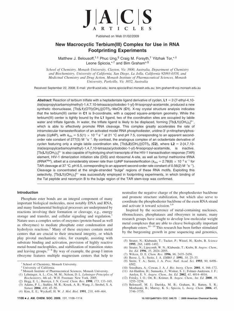
Cleavage of RNA. Encouraged by the finding that C1 is ableto greatly enhance the rate of cleavage of the RNA systemmodel, UpNP, we next examined whether this complex couldcleave the sugar-phosphate backbone of RNA. In the firstinstance, C1 was reacted with a short 32P 5′-end-labeledconstruct of the HIV TAR element (Figure 2) under pseudofirst-order conditions, and the reaction mixture analyzed bypolyacrylamide gel electrophoresis (PAGE) at increasing timeintervals (Figure 6). Cleavage activity was evident by theappearance of several bands of lower molecular weight thanthe original TAR construct. Concomitant PAGE analysis of thedegradation products obtained by RNase T1 digestion of theTAR construct (lane 2 of PAGE gel), located the initial scissionpoints to be almost exclusively within the triplet base “bulge”(U23C24U25) of the stem-loop structure of the construct, whichNMR studies have previously shown to coincide with theposition where the sugar-phosphate backbone is most exposed(Figure 6).45 Over time, the initial cleavage fragments were
(45) Aboul-ela, F.; Varani, G.; Karn, J. Nucleic Acid. Res. 1996, 24, 3974–3981.
Figure 3. Thermal ellipsoid plots of the complex cation unit and the asymmetric unit of C1 (ellipsoids drawn at 50% probability level; hydrogen atoms andthe disorder about the isopropyl group (attached to N6) have been omitted for clarity; hydrogen-bonds are indicated by dashed lines).
Figure 4. Thermal ellipsoid plots showing the distorted, capped squareantiprism coordination environment about the terbium(III) center in C1(least-squares planes through four oxygen donors and through four nitrogendonors shown in red and blue, respectively).
Scheme 2. Mechanism for Cleavage of UpNP by GeneralBase-Assisted Intramolecular Transesterification
Figure 5. Proposed rationale for the disparate reactivity of C1 toward BNPPand UpNP. UpNP features a potential internal nucleophile (2′-OH), whosereactivity may be enhanced through deprotonation by a Tb(III)-boundhydroxo group. Direct nucleophilic attack of a Tb(III)-bound hydroxo/watergroup on BNPP appears to be unfavorable.
J. AM. CHEM. SOC. 9 VOL. 131, NO. 3, 2009 1111
New Macrocyclic Terbium(III) Complex A R T I C L E S
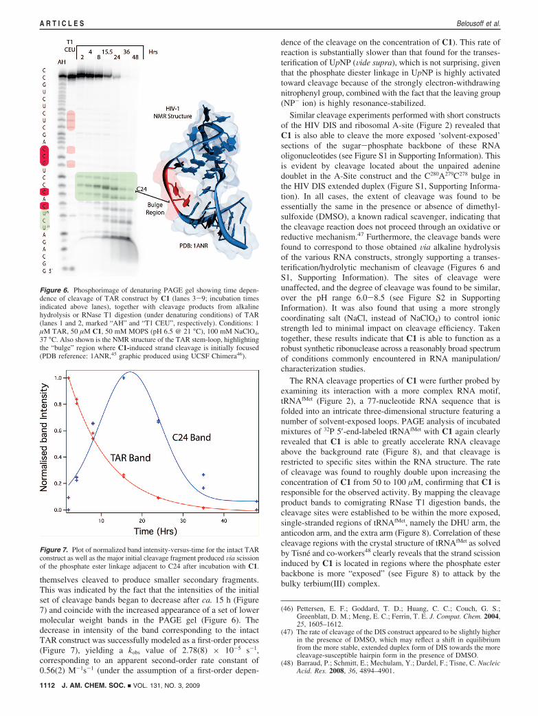
themselves cleaved to produce smaller secondary fragments.This was indicated by the fact that the intensities of the initialset of cleavage bands began to decrease after ca. 15 h (Figure7) and coincide with the increased appearance of a set of lowermolecular weight bands in the PAGE gel (Figure 6). Thedecrease in intensity of the band corresponding to the intactTAR construct was successfully modeled as a first-order process(Figure 7), yielding a kobs value of 2.78(8) × 10-5 s-1,corresponding to an apparent second-order rate constant of0.56(2) M-1s-1 (under the assumption of a first-order depen-
dence of the cleavage on the concentration of C1). This rate ofreaction is substantially slower than that found for the transes-terification of UpNP (Vide supra), which is not surprising, giventhat the phosphate diester linkage in UpNP is highly activatedtoward cleavage because of the strongly electron-withdrawingnitrophenyl group, combined with the fact that the leaving group(NP- ion) is highly resonance-stabilized.
Similar cleavage experiments performed with short constructsof the HIV DIS and ribosomal A-site (Figure 2) revealed thatC1 is also able to cleave the more exposed ‘solvent-exposed’sections of the sugar-phosphate backbone of these RNAoligonucleotides (see Figure S1 in Supporting Information). Thisis evident by cleavage located about the unpaired adeninedoublet in the A-Site construct and the C280A279C278 bulge inthe HIV DIS extended duplex (Figure S1, Supporting Informa-tion). In all cases, the extent of cleavage was found to beessentially the same in the presence or absence of dimethyl-sulfoxide (DMSO), a known radical scavenger, indicating thatthe cleavage reaction does not proceed through an oxidative orreductive mechanism.47 Furthermore, the cleavage bands werefound to correspond to those obtained Via alkaline hydrolysisof the various RNA constructs, strongly supporting a transes-terification/hydrolytic mechanism of cleavage (Figures 6 andS1, Supporting Information). The sites of cleavage wereunaffected, and the degree of cleavage was found to be similar,over the pH range 6.0-8.5 (see Figure S2 in SupportingInformation). It was also found that using a more stronglycoordinating salt (NaCl, instead of NaClO4) to control ionicstrength led to minimal impact on cleavage efficiency. Takentogether, these results indicate that C1 is able to function as arobust synthetic ribonuclease across a reasonably broad spectrumof conditions commonly encountered in RNA manipulation/characterization studies.
The RNA cleavage properties of C1 were further probed byexamining its interaction with a more complex RNA motif,tRNAfMet (Figure 2), a 77-nucleotide RNA sequence that isfolded into an intricate three-dimensional structure featuring anumber of solvent-exposed loops. PAGE analysis of incubatedmixtures of 32P 5′-end-labeled tRNAfMet with C1 again clearlyrevealed that C1 is able to greatly accelerate RNA cleavageabove the background rate (Figure 8), and that cleavage isrestricted to specific sites within the RNA structure. The rateof cleavage was found to roughly double upon increasing theconcentration of C1 from 50 to 100 µM, confirming that C1 isresponsible for the observed activity. By mapping the cleavageproduct bands to comigrating RNase T1 digestion bands, thecleavage sites were established to be within the more exposed,single-stranded regions of tRNAfMet, namely the DHU arm, theanticodon arm, and the extra arm (Figure 8). Correlation of thesecleavage regions with the crystal structure of tRNAfMet as solvedby Tisne and co-workers48 clearly reveals that the strand scissioninduced by C1 is located in regions where the phosphate esterbackbone is more “exposed” (see Figure 8) to attack by thebulky terbium(III) complex.
(46) Pettersen, E. F.; Goddard, T. D.; Huang, C. C.; Couch, G. S.;Greenblatt, D. M.; Meng, E. C.; Ferrin, T. E. J. Comput. Chem. 2004,25, 1605–1612.
(47) The rate of cleavage of the DIS construct appeared to be slightly higherin the presence of DMSO, which may reflect a shift in equilibriumfrom the more stable, extended duplex form of DIS towards the morecleavage-susceptible hairpin form in the presence of DMSO.
(48) Barraud, P.; Schmitt, E.; Mechulam, Y.; Dardel, F.; Tisne, C. NucleicAcid. Res. 2008, 36, 4894–4901.
Figure 6. Phosphorimage of denaturing PAGE gel showing time depen-dence of cleavage of TAR construct by C1 (lanes 3-9; incubation timesindicated above lanes), together with cleavage products from alkalinehydrolysis or RNase T1 digestion (under denaturing conditions) of TAR(lanes 1 and 2, marked “AH” and “T1 CEU”, respectively). Conditions: 1µM TAR, 50 µM C1, 50 mM MOPS (pH 6.5 @ 21 °C), 100 mM NaClO4,37 °C. Also shown is the NMR structure of the TAR stem-loop, highlightingthe “bulge” region where C1-induced strand cleavage is initially focused(PDB reference: 1ANR,45 graphic produced using UCSF Chimera46).
Figure 7. Plot of normalized band intensity-versus-time for the intact TARconstruct as well as the major initial cleavage fragment produced Via scissionof the phosphate ester linkage adjacent to C24 after incubation with C1.
1112 J. AM. CHEM. SOC. 9 VOL. 131, NO. 3, 2009
A R T I C L E S Belousoff et al.

Footprinting Experiments. Having established that C1 is ableto selectively cleave solvent-exposed, single-stranded (or strainedsections) of a folded RNA structure, we next explored whetherC1 could be employed as a new footprinting reagent, usefulfor examining the interaction between ligands and their RNAtargets. RNA-ligand footprinting is often carried out usingRNase enzymes, however, this can sometimes be a complexand time-consuming task (requiring case-by-case optimization),and the conditions required for enzyme activity may beincompatable with the conditions under which ligand bindingoccurs. Thus, robust synthetic reagents that are able to functionas artificial ribonucleases under near physiological conditionsrepresent valuable additions to the arsenal of tools used toexamine RNA structure and RNA-ligand interactions. TheFe(II)-EDTA/H2O2 system, which is utilized to generate Fentonbased hydroxide radicals, is often used for DNA and RNAfootprinting,22 but its lack of sequence specificity can beproblematic.49
We chose the TAR construct as a model RNA target, sincea great deal is known about its interactions with a range ofdifferent ligands. Figure 9 shows the cleavage “footprints”observed upon incubating C1 with the TAR construct, both inisolation and in the presence of two known TAR-binders, theTat peptide and neomycin B. The Tat peptide comprises theportion of the Tat protein that is involved in binding of thisprotein to the TAR element, an interaction that that is vital tothe HIV replication cycle. It is known that the Tat peptide bindsto the triplet base “bulge” region of TAR.50 Moreover, NMRstudies carried out by Crothers et al.50 suggest that upon Tatbinding, the phosphate ester groups associated with U23 and C24
become more exposed relative to that of U25. Consistent withthese observation/predictions, we found a change in the cleavagefootprint upon addition of Tat peptide to the C1/TAR mixture:
(49) Pogozelski, W. K.; D., T. T. Chem. ReV. 1998, 98, 1089–1107.(50) Long, K. S.; Crothers, D. M. Biochemistry 1999, 38, 10059–10069.
Figure 8. Phosphorimage of denaturing PAGE gel showing time dependence and concentration dependence of cleavage of fMettRNA by C1 (lanes 3-11),together with cleavage products from alkaline hydrolysis or RNase T1 digestion (under denaturing conditions) of TAR (lanes 1 and 2, marked “AH” and “T1CEU”, respectively). Lanes 2-4: 50 µM C1 with fMettRNA (2, 4, 8 h incubation time); lanes 5-7: 100 µM C1 with fMettRNA (2, 4, 8 h incubation time);lanes 8-10: fMettRNA only control (2, 4, 8 h incubation time); incubation times indicated above lanes. Conditions: 1 µM fMettRNA, 50 mM MOPS (pH 6.5@ 21 °C), 100 mM NaClO4, 1 mM MgCl2, 37 °C. Shown also is the crystal structure of tRNAfMet (as determined by Tisne et al.48 (PDB: 3cw5), graphicprepared using UCSF Chimera46) highlighting (in red) the areas of focused strand scission induced by C1.
J. AM. CHEM. SOC. 9 VOL. 131, NO. 3, 2009 1113
New Macrocyclic Terbium(III) Complex A R T I C L E S

cleavage adjacent to U25 was suppressed, while cleavageadjacent to U23 was enhanced.
It has been postulated that the naturally occurring aminogly-coside, neomycin B, also binds the triplet base “bulge” regionof TAR,51 but that this is a more dynamic on/off bindinginteraction than for Tat. Accordingly, we found that the cleavagepatterns obtained both in the absence and presence of neomycinB were very similar, but that the extent of cleavage was about4-fold lower when neomycin B was present (cf., lanes 3 and 4with lanes 7 and 8 in Figure 9). C1 is thus able to detect theinteraction of neomycin B with TAR, by virtue of the boundstate of neomycin B providing TAR with some protectionagainst C1-induced cleavage.
Conclusion
A new terbium(III) complex, C1, which is able to rapidlyaccelerate the cleavage of UpNP, as well as the solvent-exposed,single-stranded regions of folded RNA motifs, has beendeveloped. The complex does not require co-reactants to causeRNA scission, is able to function over a range of pHs, andexhibits cleavage activity that is sensitive to the presence ofRNA-binding ligands. C1 is thus a suitable candidate for usein footprinting assays directed at improving our understandingof RNA structure and RNA-ligands interactions.
Acknowledgment. M.J.B. is a recipient of an Australian PostGraduate Award, a Monash University travel grant, a MonashPostgraduate Publication Award and a Fulbright Fellowship. Weacknowledge the financial support of National Institutes of Health(grant numbers AI 47673 and GM 069773) (for Y.T.) and MonashUniversity.
Supporting Information Available: X-ray crystallographyrefinement data tables (Table S1) and selected bond lengths andangles for C1 (Table S2), phosphorimages of cleavage reactionof C1 with the TAR, DIS and A-Site RNA (Figure S1) andphosphorimage of the pH dependence of the reaction of C1 withTAR (Figure S2). This material is available free of charge viathe Internet at http://pubs.acs.org.
JA807301R
(51) Faber, C.; Sticht, H.; Schweimer, K.; Rosch, P. J. Biol. Chem. 2000,275, 20660–20666.
Figure 9. Phosphorimage of denaturing PAGE gel showing “footprints”for cleavage of TAR by C1, both in the absence and presence of Tat peptideor neomycin B. Lanes 1 and 9: alkaline hydrolysis ladder; lane 2: RNaseT1 digestion of TAR (under denaturing conditions); lanes 3 and 4: C1 withTAR (2, 6 h incubation time); lanes 4 and 5: C1 with TAR (6 h reactiontime); lanes 5 and 6: C1 with TAR and Tat (2, 6 h incubation time); lanes7 and 8: C1 with TAR and neomycin B (2, 6 h incubation time); incubationtimes indicated above lanes. Conditions: 1 µM TAR, 50 µM C1, 50 mMMOPS (pH 6.5 @ 21 °C), 100 mM NaClO4, 37 °C.
1114 J. AM. CHEM. SOC. 9 VOL. 131, NO. 3, 2009
A R T I C L E S Belousoff et al.