Embed Size (px)
Citation preview
New Broad-Spectrum Antibacterial Amphiphilic AminoglycosidesActive against Resistant Bacteria: From Neamine Derivatives toSmaller Neosamine AnaloguesLouis Zimmermann,† Indrajit Das,†,§ Jero me Desire,†,∥ Guillaume Sautrey,‡,⊥ Vinicius Barros R. S.,†
Micheline El Khoury,‡ Marie-Paule Mingeot-Leclercq,‡ and Jean-Luc Decout*,†
†Departement de Pharmacochimie Moleculaire, ICMG FR 2607, University Grenoble Alpes/CNRS, UMR 5063, 470 Rue de laChimie, BP 53, F-38041 Grenoble, France‡Unite de Pharmacologie Cellulaire et Moleculaire, Louvain Drug Research Institute, Universite Catholique de Louvain, Avenue E.Mounier 73, B1.73.05, B-1200 Brussels, Belgium
*S Supporting Information
ABSTRACT: Aminoglycosides (AGs) constitute a majorfamily of potent and broad-spectrum antibiotics disturbingprotein synthesis through binding to the A site of 16S rRNA.Decades of widespread clinical use of AGs strongly reducedtheir clinical efficacy through the selection of resistant bacteria.Recently, conjugation of lipophilic groups to AGs generated anovel class of potent antibacterial amphiphilic aminoglycosides(AAGs) with significant improved activities against varioussensitive and resistant bacterial strains. We have identifiedamphiphilic 3′,6-dialkyl derivatives of the small aminoglycosideneamine as broad spectrum antibacterial agents targeting bacterial membranes. Here, we report on the synthesis and the activityagainst sensitive and resistant Gram-negative and/or Gram-positive bacteria of new amphiphilic 3′,4′-dialkyl neamine derivativesand of their smaller analogues in the 6-aminoglucosamine (neosamine) series prepared from N-acetylglucosamine.
■ INTRODUCTION
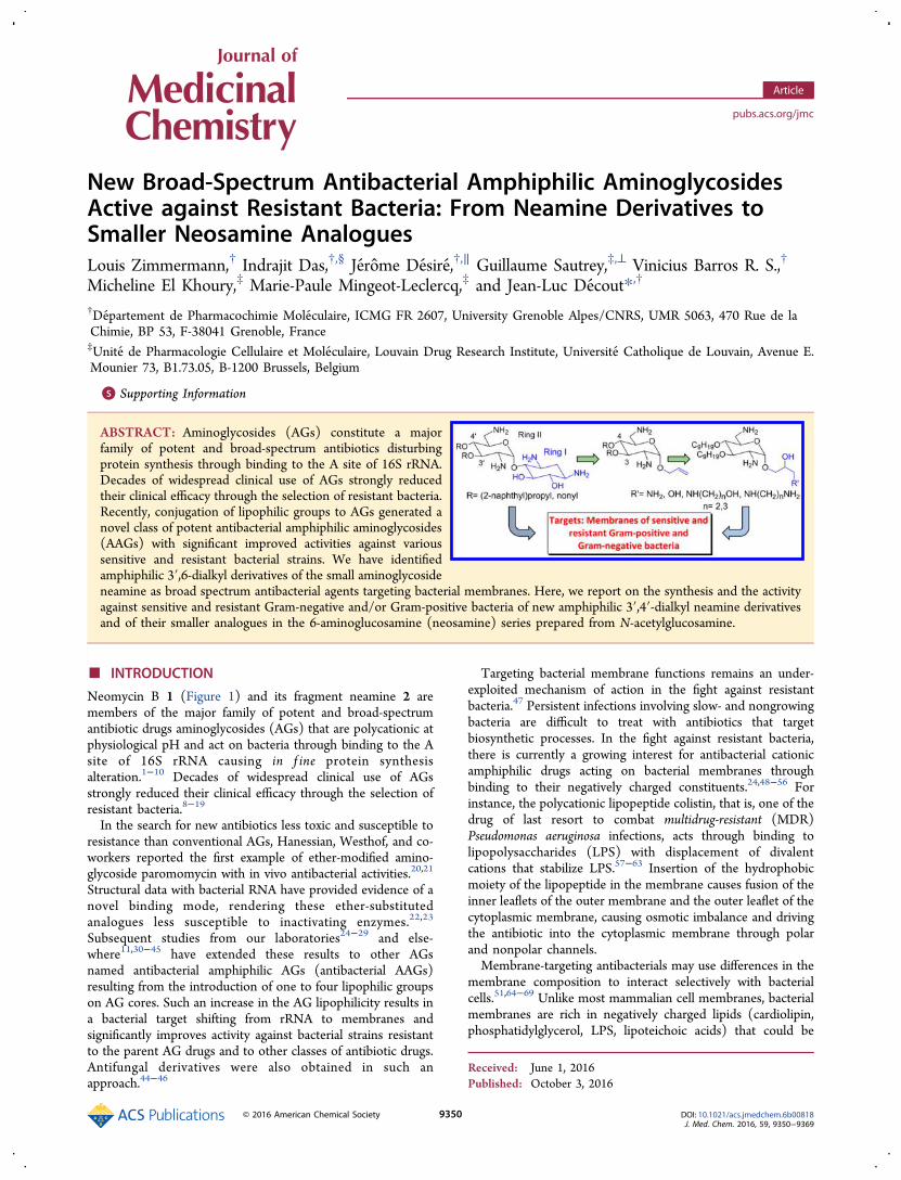
Neomycin B 1 (Figure 1) and its fragment neamine 2 aremembers of the major family of potent and broad-spectrumantibiotic drugs aminoglycosides (AGs) that are polycationic atphysiological pH and act on bacteria through binding to the Asite of 16S rRNA causing in f ine protein synthesisalteration.1−10 Decades of widespread clinical use of AGsstrongly reduced their clinical efficacy through the selection ofresistant bacteria.8−19
In the search for new antibiotics less toxic and susceptible toresistance than conventional AGs, Hanessian, Westhof, and co-workers reported the first example of ether-modified amino-glycoside paromomycin with in vivo antibacterial activities.20,21
Structural data with bacterial RNA have provided evidence of anovel binding mode, rendering these ether-substitutedanalogues less susceptible to inactivating enzymes.22,23
Subsequent studies from our laboratories24−29 and else-where11,30−45 have extended these results to other AGsnamed antibacterial amphiphilic AGs (antibacterial AAGs)resulting from the introduction of one to four lipophilic groupson AG cores. Such an increase in the AG lipophilicity results ina bacterial target shifting from rRNA to membranes andsignificantly improves activity against bacterial strains resistantto the parent AG drugs and to other classes of antibiotic drugs.Antifungal derivatives were also obtained in such anapproach.44−46
Targeting bacterial membrane functions remains an under-exploited mechanism of action in the fight against resistantbacteria.47 Persistent infections involving slow- and nongrowingbacteria are difficult to treat with antibiotics that targetbiosynthetic processes. In the fight against resistant bacteria,there is currently a growing interest for antibacterial cationicamphiphilic drugs acting on bacterial membranes throughbinding to their negatively charged constituents.24,48−56 Forinstance, the polycationic lipopeptide colistin, that is, one of thedrug of last resort to combat multidrug-resistant (MDR)Pseudomonas aeruginosa infections, acts through binding tolipopolysaccharides (LPS) with displacement of divalentcations that stabilize LPS.57−63 Insertion of the hydrophobicmoiety of the lipopeptide in the membrane causes fusion of theinner leaflets of the outer membrane and the outer leaflet of thecytoplasmic membrane, causing osmotic imbalance and drivingthe antibiotic into the cytoplasmic membrane through polarand nonpolar channels.Membrane-targeting antibacterials may use differences in the
membrane composition to interact selectively with bacterialcells.51,64−69 Unlike most mammalian cell membranes, bacterialmembranes are rich in negatively charged lipids (cardiolipin,phosphatidylglycerol, LPS, lipoteichoic acids) that could be
Received: June 1, 2016Published: October 3, 2016
Article
pubs.acs.org/jmc
© 2016 American Chemical Society 9350 DOI: 10.1021/acs.jmedchem.6b00818J. Med. Chem. 2016, 59, 9350−9369
selectively recognized by cationic amphiphiles (CAs) throughionic interactions and hydrophobic effects.Membrane-active agents can interact with many targets in the
bacterial membranes and inhibit the corresponding functions.Through binding to anionic lipids, they could modify theactivity of membrane proteins such as efflux pumps and/orproteins involved in cell division for which a dependence uponnegatively charged lipids has been demonstrated. For example,in the regulation of bacterial cell division, proteins MinD/MinEbind tightly to anionic lipids such as cardiolipin positioned atthe cell pole, and thus anionic lipids can be useful targets forantimicrobial development.29,70,71
Limited in vitro resistances to these amphiphiles have beenobserved due to their multiple modes of action.48,49,56
Membrane-active agents cannot be inactivated by intracellularbacterial enzymes and flushed out by efflux pumps. In addition,due to their interactions with many key membrane targetspresent in a great number of copies, biochemical modificationsof such multiple targets should have a high cost for the bacteriaand should result in a high sensitivity to other antibiotic drugs.However, the clinical use of CA drugs such as antimicrobial
or host defense peptides is limited due to protease susceptibilityand toxicity.48−50 Over the years, the therapeutic potential ofthese amphiphiles has been improved by (i) reducing theirability to lyse red blood cells, (ii) increasing selectivity towardbacteria,51−55 (iii) reducing nonspecific binding to humanserum proteins, and (iv) improving serum stability.72
AAGs are expected to possess improved metabolic stability inregard to peptide-based CA antibiotics and are more difficult tomodify by bacterial resistance-causing enzymes than AGs inregard to their expected mode of action. AAGs can also boostthe innate immune response, specifically the recruitment ofimmune cells such as neutrophils required for the resolution ofinfections and can selectively control inflammatory responsesinduced in the presence of endotoxins to prevent septicshock.41
Several strategies for obtaining antibacterial AAGs have beendeveloped including complete or partial conversion of the AGamine and hydroxyl functions into alkyl- or aryl-amide and-ether groups, respectively.24
In our approach in the field of antibacterial AAGs, weassumed that the presence of a large number of amine functions
in AG derivatives like in neomycin 1, which carries six aminefunctions, can be a source of toxicity through nonspecificbinding to the target.24−29 Neamine 2 carrying four aminefunctions is less toxic than neomycin,73−76 and the neaminecore corresponds to the minimum scaffold necessary forbinding to 16S rRNA.5,6 This core was used in the synthesis ofnew AGs in the search for antibacterial and antiviral agentstargeting RNA, gene therapy vectors, and for the treatment ofthe Meniere’s disease of the inner ear characterized by recurringattacks of disabling vertigo, hearing loss, and tinnitus.10,77−98
Therefore, for obtaining amphiphilic AGs targeting rRNA, wehave modified selectively the small AG neamine 2 on one, two,three, and the four hydroxyl functions in order to keepunchanged the four amine functions potentially protonated atphysiological pH, at least partially, in regard to their major rolein the binding to anionic targets in bacterial membranes andrRNA. We have identified a first antibacterial amphiphilicneamine derivative, namely 3′,4′,6-tri2-naphthylmethylene(3′,4′,6-tri2NM) neamine 3 (Figure 1), having a broadspectrum of activity25 and targeting LPS in the outer membraneof P. aeruginosa.26 Structure−activity and structure−cytotoxicityrelationships were delineated from various amphiphilic 3′,6-dialkyl neamine derivatives for obtaining compounds 4−7(Figure 1) more active than 3 against sensitive and resistantGram-positive and/or Gram-negative bacteria and strongly lesstoxic against eukaryotic cells.27 A critical window of lipophilicityappeared to be necessary for optimal antibacterial effects. Thestudy of the mode of action of compounds 5−7 confirmed astrong binding to LPS of P. aeruginosa as well as membranedepolarization.28 Compound 7 has been found the mostefficient neamine derivative against Gram-negative bacteria. Itappeared to also be able to inhibit growth of P. aeruginosabiofilms and be active against P. aeruginosa strains resistant tocolistin, suggesting a different mode of action from the one ofcolistin.28
In our first report on the identification of the tri-2NMneamine derivative 3, we have described the 3′,6- and 3′,4′-di2NM neamine derivatives 4 and 8 showing similarantibacterial effects against sensitive and resistant strains ofStaphylococcus aureus and weak activity against Gram-negativebacteria.25 Herein, we report on the synthesis and theantibacterial activities of new 3′,4′-dialkyl neamine derivatives
Figure 1. Structure of the natural antibiotic aminoglycoside neomycin B, of neamine and its amphiphilic derivatives previously prepared.24−29
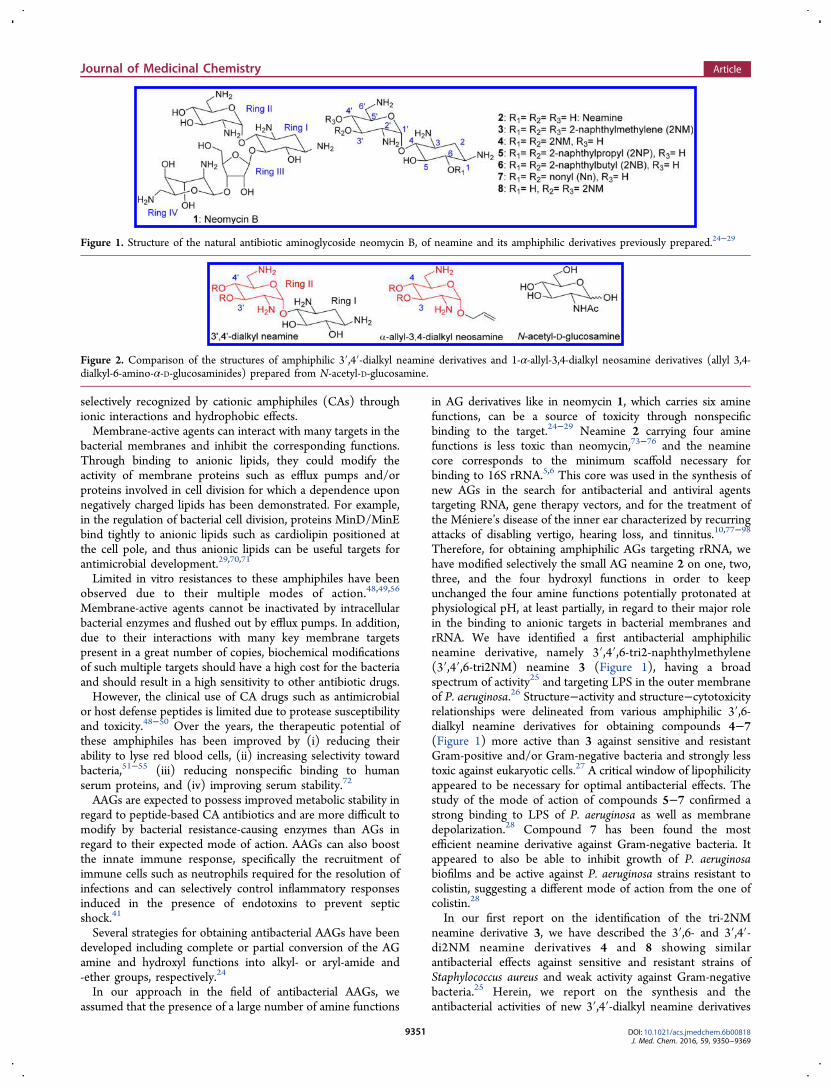
Figure 2. Comparison of the structures of amphiphilic 3′,4′-dialkyl neamine derivatives and 1-α-allyl-3,4-dialkyl neosamine derivatives (allyl 3,4-dialkyl-6-amino-α-D-glucosaminides) prepared from N-acetyl-D-glucosamine.
Journal of Medicinal Chemistry Article
DOI: 10.1021/acs.jmedchem.6b00818J. Med. Chem. 2016, 59, 9350−9369
9351
active against sensitive and resistant Gram-positive and Gram-negative bacteria in comparison to their active 3′,6-dialkylneamine isomers 5 and 7 previously described.In the previously identified antibacterial 3′,4′-di2NM ne-
amine derivative 8 (Figure 1), both lipophilic groups areattached on the glucosamine ring II. Therefore, small AAGs inwhich ring II carries at the 3- and 4-positions two lipophilicgroups and at the 1-position acyclic side chains generated froman allyl group (Figure 2) could have attractive antibacterialproperties. Such an approach has been previously developedfrom ring I or ring II of neamine in the search for antibioticaminosugars targeting 16S rRNA.97−100
We have also previously conjugated ring II of neamine to apeptide nucleic acid targeting transactivation response elementof HIV-1 RNA genome that shows a high bioavailability inhuman cells and strongly inhibits Tat-mediated transactivationof HIV-1 transcription.101
Amphiphilic 3,4-dialkyl derivatives of 6-amino-6-deoxyglu-cosamine named neosamine were synthesized from N-acetylglucosamine. First, derivatives carrying an allyl groupintroduced in α-configuration at the anomeric positioncorresponding to the location of ring I on ring II in neaminewere prepared (Figure 2). Second, the reactive allyl group ofthe 3,4-dinonyl neosamine derivative was chemically modifiedin order to adjust the lipophilicity/hydrophily balance of theresulting AAGs, balance previously identified in the neamineseries as a key parameter for obtaining a broad spectrum
antibacterial activity. This group was converted to an epoxidering (oxirane) in order to introduce by ring-opening hydroxyland/or amine functions like those found in ring I of thecorresponding 3′,4′-dialkyl neamine derivatives. The routeusing epoxides as intermediates was selected for the resultingobtention of diasteroisomers that extends the moleculardiversity in the antibacterial evaluation. Herein, we comparedthe antibacterial effects of the prepared 3,4-dialkyl neosaminederivatives to those of the corresponding novel 3′,4′-dialkylneamines described here and to those of the previouslydescribed 3′,6-dialkyl neamines. P. aeruginosa inner membranepermeabilization assays and MIC changes against P. aeruginosainduced upon exposure to some of the most active AAGsidentified are also reported.
■ SYNTHESIS
Synthesis of New 3′,4′-Dialkyl Neamine Derivatives.In regard to the structure of the 3′,6-dialkyl neamine derivativespreviously identified as interesting antibacterials, the 3′,4′-di-2-naphthylpropyl (2NP) and 3′,4′-dinonyl (Nn) neaminederivatives were prepared for evaluation of their antibacterialeffects (Scheme 1). The N-tetratrityl neamine derivative 990
selectively protected at the 6-position by the p-methoxybenzylgroup was first prepared in good yield under phase transferconditions with TBAF as a phase transfer agent from N-tetratritylneamine.96 Then, the 3′,4′-di2NP (10) and 3′,4′-diNn
Scheme 1. Synthesis of the 3′,4′-Dialkylneamine Derivatives from the p-Methoxybenzyl Neamine Derivative 9a96
aReagents and conditions: (a) R = 2NP: NaH/DMF, 3-(2′-naphthyl)propyl bromide (2NPBr),27 rt, 5 h, 44%; R = Nn: TBAF (2 equiv), 50% aqNaOH/toluene, 1-nonyl bromide (1NnBr), rt, 43%. (b) TFA/CH2Cl2, anisole, 0 °C; 10, 45%; 11, 55%.
Scheme 2. Preparation of the α-Allyl-3′,4′-di2NM Neosamine Derivative 17 and the Corresponding Reference Compound α-Allylneosamine 19a
aReagents and conditions: (a) 2NMBr, BEMP, DMF, rt, 48 h, 28%. (b) KOH, EtOH, reflux or Ba(OH)2·8H2O, H2O, reflux, 8 h, 78%. (c) TrCl,DMF, Et3N, rt, 8 h, 82%. (d) 2NMBr, NaH, DMF, rt, 10 h, 79%. (e) Ph3P, THF/H2O (19/1), 80 °C, 6 h, 91%. (f) TFA/anisole (1/1), 0 °C, 3 h.(g) Dowex resin (Cl− ion exchange); 17, 62%; 19, 84% (3 steps).
Journal of Medicinal Chemistry Article
DOI: 10.1021/acs.jmedchem.6b00818J. Med. Chem. 2016, 59, 9350−9369
9352
(11) neamine derivatives were obtained with moderate yields intwo steps: (i) alkylation of 9 with the correspondingbromoalkane RBr under phase transfer conditions (R = Nn)or in the presence of NaH in DMF (R = 2NP) and (ii)deprotection with TFA/anisole.Synthesis in the Neosamine Series. The neosamine core
corresponding to ring II in neamine has been previously used inthe search for antibacterial agents targeting 16S ribosomal A-site RNA.82,97−99 For instance, a library of compounds basedupon this core has been synthesized through a combinatorialapproach and screened for binding specifically to 16S rRNA bythe Wong group to lead to effective binders to a model of A-site16S RNA in the micromolar range.97 One of the key
intermediates used in the different reported approaches hasbeen compound 12, which was one of our key intermediates inthe preparation of amphiphilic neosamine derivatives.
Synthesis of α-Allyl-3,4-di(2′-naphthylmethylene) Neos-amine Derivative 17 and of the Corresponding ReferenceCompound α-Allylneosamine 19. N-Acetylglucosamine wasconverted to compound 12 (Scheme 2), possessing an allylgroup at the anomeric position in order to allow furthermodifications, using a three steps sequence adapted from themethod described by Wong et al.:97 (i) conversion of N-acetylglucosamine to the corresponding α-allyl glycoside, (ii)selective tosylation of the primary alcohol function, and (iii)displacement of the tosyl group with sodium azide. Then the
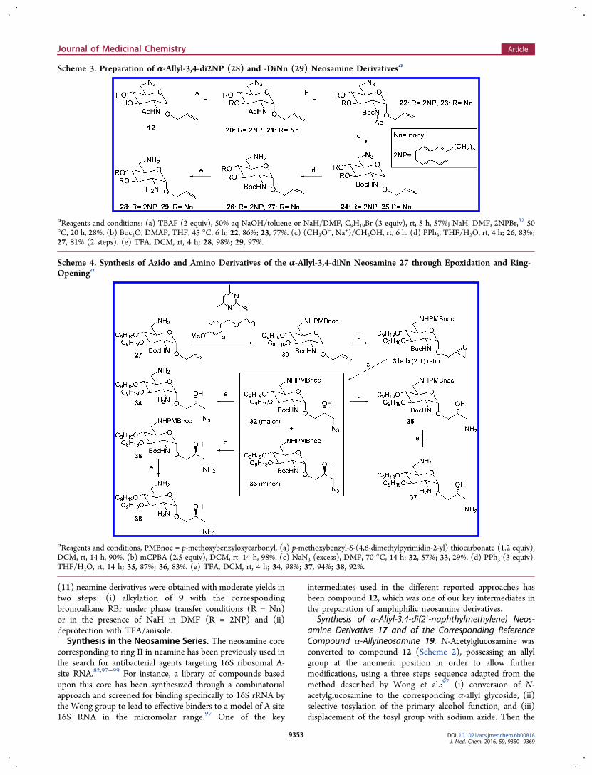
Scheme 3. Preparation of α-Allyl-3,4-di2NP (28) and -DiNn (29) Neosamine Derivativesa
aReagents and conditions: (a) TBAF (2 equiv), 50% aq NaOH/toluene or NaH/DMF, C9H19Br (3 equiv), rt, 5 h, 57%; NaH, DMF, 2NPBr,32 50°C, 20 h, 28%. (b) Boc2O, DMAP, THF, 45 °C, 6 h; 22, 86%; 23, 77%. (c) (CH3O
−, Na+)/CH3OH, rt, 6 h. (d) PPh3, THF/H2O, rt, 4 h; 26, 83%;27, 81% (2 steps). (e) TFA, DCM, rt, 4 h; 28, 98%; 29, 97%.
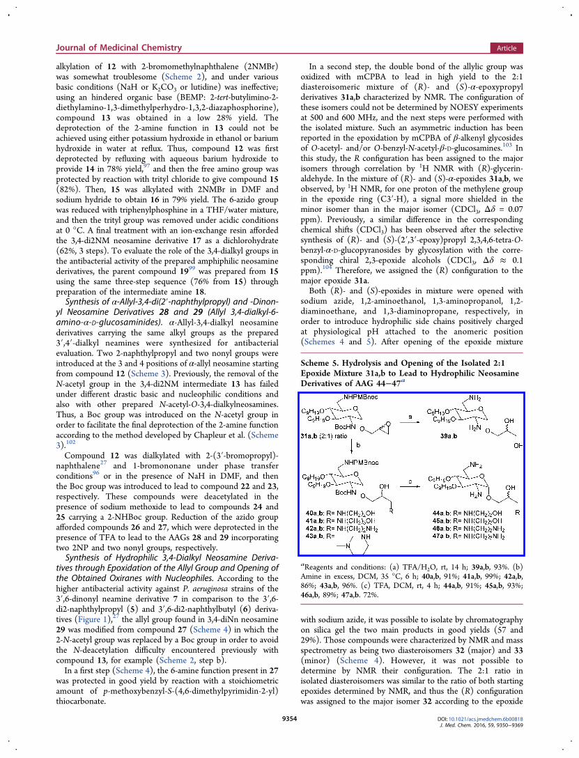
Scheme 4. Synthesis of Azido and Amino Derivatives of the α-Allyl-3,4-diNn Neosamine 27 through Epoxidation and Ring-Openinga
aReagents and conditions, PMBnoc = p-methoxybenzyloxycarbonyl. (a) p-methoxybenzyl-S-(4,6-dimethylpyrimidin-2-yl) thiocarbonate (1.2 equiv),DCM, rt, 14 h, 90%. (b) mCPBA (2.5 equiv), DCM, rt, 14 h, 98%. (c) NaN3 (excess), DMF, 70 °C, 14 h; 32, 57%; 33, 29%. (d) PPh3 (3 equiv),THF/H2O, rt, 14 h; 35, 87%; 36, 83%. (e) TFA, DCM, rt, 4 h; 34, 98%; 37, 94%; 38, 92%.
Journal of Medicinal Chemistry Article
DOI: 10.1021/acs.jmedchem.6b00818J. Med. Chem. 2016, 59, 9350−9369
9353
alkylation of 12 with 2-bromomethylnaphthalene (2NMBr)was somewhat troublesome (Scheme 2), and under variousbasic conditions (NaH or K2CO3 or lutidine) was ineffective;using an hindered organic base (BEMP: 2-tert-butylimino-2-diethylamino-1,3-dimethylperhydro-1,3,2-diazaphosphorine),compound 13 was obtained in a low 28% yield. Thedeprotection of the 2-amine function in 13 could not beachieved using either potassium hydroxide in ethanol or bariumhydroxide in water at reflux. Thus, compound 12 was firstdeprotected by refluxing with aqueous barium hydroxide toprovide 14 in 78% yield,97 and then the free amino group wasprotected by reaction with trityl chloride to give compound 15(82%). Then, 15 was alkylated with 2NMBr in DMF andsodium hydride to obtain 16 in 79% yield. The 6-azido groupwas reduced with triphenylphosphine in a THF/water mixture,and then the trityl group was removed under acidic conditionsat 0 °C. A final treatment with an ion-exchange resin affordedthe 3,4-di2NM neosamine derivative 17 as a dichlorohydrate(62%, 3 steps). To evaluate the role of the 3,4-dialkyl groups inthe antibacterial activity of the prepared amphiphilic neosaminederivatives, the parent compound 1999 was prepared from 15using the same three-step sequence (76% from 15) throughpreparation of the intermediate amine 18.Synthesis of α-Allyl-3,4-di(2′-naphthylpropyl) and -Dinon-
yl Neosamine Derivatives 28 and 29 (Allyl 3,4-dialkyl-6-amino-α-D-glucosaminides). α-Allyl-3,4-dialkyl neosaminederivatives carrying the same alkyl groups as the prepared3′,4′-dialkyl neamines were synthesized for antibacterialevaluation. Two 2-naphthylpropyl and two nonyl groups wereintroduced at the 3 and 4 positions of α-allyl neosamine startingfrom compound 12 (Scheme 3). Previously, the removal of theN-acetyl group in the 3,4-di2NM intermediate 13 has failedunder different drastic basic and nucleophilic conditions andalso with other prepared N-acetyl-O-3,4-dialkylneosamines.Thus, a Boc group was introduced on the N-acetyl group inorder to facilitate the final deprotection of the 2-amine functionaccording to the method developed by Chapleur et al. (Scheme3).102
Compound 12 was dialkylated with 2-(3′-bromopropyl)-naphthalene27 and 1-bromononane under phase transferconditions96 or in the presence of NaH in DMF, and thenthe Boc group was introduced to lead to compound 22 and 23,respectively. These compounds were deacetylated in thepresence of sodium methoxide to lead to compounds 24 and25 carrying a 2-NHBoc group. Reduction of the azido groupafforded compounds 26 and 27, which were deprotected in thepresence of TFA to lead to the AAGs 28 and 29 incorporatingtwo 2NP and two nonyl groups, respectively.Synthesis of Hydrophilic 3,4-Dialkyl Neosamine Deriva-
tives through Epoxidation of the Allyl Group and Opening ofthe Obtained Oxiranes with Nucleophiles. According to thehigher antibacterial activity against P. aeruginosa strains of the3′,6-dinonyl neamine derivative 7 in comparison to the 3′,6-di2-naphthylpropyl (5) and 3′,6-di2-naphthylbutyl (6) deriva-tives (Figure 1),27 the allyl group found in 3,4-diNn neosamine29 was modified from compound 27 (Scheme 4) in which the2-N-acetyl group was replaced by a Boc group in order to avoidthe N-deacetylation difficulty encountered previously withcompound 13, for example (Scheme 2, step b).In a first step (Scheme 4), the 6-amine function present in 27
was protected in good yield by reaction with a stoichiometricamount of p-methoxybenzyl-S-(4,6-dimethylpyrimidin-2-yl)thiocarbonate.
In a second step, the double bond of the allylic group wasoxidized with mCPBA to lead in high yield to the 2:1diasteroisomeric mixture of (R)- and (S)-α-epoxypropylderivatives 31a,b characterized by NMR. The configuration ofthese isomers could not be determined by NOESY experimentsat 500 and 600 MHz, and the next steps were performed withthe isolated mixture. Such an asymmetric induction has beenreported in the epoxidation by mCPBA of β-alkenyl glycosidesof O-acetyl- and/or O-benzyl-N-acetyl-β-D-glucosamines.103 Inthis study, the R configuration has been assigned to the majorisomers through correlation by 1H NMR with (R)-glycerin-aldehyde. In the mixture of (R)- and (S)-α-epoxides 31a,b, weobserved, by 1H NMR, for one proton of the methylene groupin the epoxide ring (C3′-H), a signal more shielded in theminor isomer than in the major isomer (CDCl3, Δδ = 0.07ppm). Previously, a similar difference in the correspondingchemical shifts (CDCl3) has been observed after the selectivesynthesis of (R)- and (S)-(2′,3′-epoxy)propyl 2,3,4,6-tetra-O-benzyl-α-D-glucopyranosides by glycosylation with the corre-sponding chiral 2,3-epoxide alcohols (CDCl3, Δδ ≈ 0.1ppm).104 Therefore, we assigned the (R) configuration to themajor epoxide 31a.Both (R)- and (S)-epoxides in mixture were opened with
sodium azide, 1,2-aminoethanol, 1,3-aminopropanol, 1,2-diaminoethane, and 1,3-diaminopropane, respectively, inorder to introduce hydrophilic side chains positively chargedat physiological pH attached to the anomeric position(Schemes 4 and 5). After opening of the epoxide mixture
with sodium azide, it was possible to isolate by chromatographyon silica gel the two main products in good yields (57 and29%). Those compounds were characterized by NMR and massspectrometry as being two diasteroisomers 32 (major) and 33(minor) (Scheme 4). However, it was not possible todetermine by NMR their configuration. The 2:1 ratio inisolated diasteroisomers was similar to the ratio of both startingepoxides determined by NMR, and thus the (R) configurationwas assigned to the major isomer 32 according to the epoxide
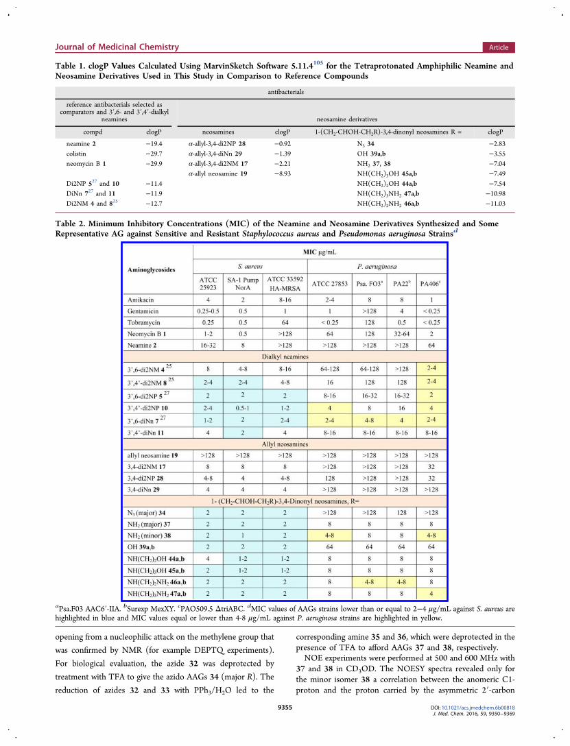
Scheme 5. Hydrolysis and Opening of the Isolated 2:1Epoxide Mixture 31a,b to Lead to Hydrophilic NeosamineDerivatives of AAG 44−47a
aReagents and conditions: (a) TFA/H2O, rt, 14 h; 39a,b, 93%. (b)Amine in excess, DCM, 35 °C, 6 h; 40a,b, 91%; 41a,b, 99%; 42a,b,86%; 43a,b, 96%. (c) TFA, DCM, rt, 4 h; 44a,b, 91%; 45a,b, 93%;46a,b, 89%; 47a,b. 72%.
Journal of Medicinal Chemistry Article
DOI: 10.1021/acs.jmedchem.6b00818J. Med. Chem. 2016, 59, 9350−9369
9354
opening from a nucleophilic attack on the methylene group that
was confirmed by NMR (for example DEPTQ experiments).
For biological evaluation, the azide 32 was deprotected by
treatment with TFA to give the azido AAGs 34 (major R). The
reduction of azides 32 and 33 with PPh3/H2O led to the
corresponding amine 35 and 36, which were deprotected in thepresence of TFA to afford AAGs 37 and 38, respectively.NOE experiments were performed at 500 and 600 MHz with
37 and 38 in CD3OD. The NOESY spectra revealed only forthe minor isomer 38 a correlation between the anomeric C1-proton and the proton carried by the asymmetric 2′-carbon
Table 1. clogP Values Calculated Using MarvinSketch Software 5.11.4105 for the Tetraprotonated Amphiphilic Neamine andNeosamine Derivatives Used in This Study in Comparison to Reference Compounds
antibacterials
reference antibacterials selected ascomparators and 3′,6- and 3′,4′-dialkyl
neamines neosamine derivatives
compd clogP neosamines clogP 1-(CH2-CHOH-CH2R)-3,4-dinonyl neosamines R = clogP
neamine 2 −19.4 α-allyl-3,4-di2NP 28 −0.92 N3 34 −2.83colistin −29.7 α-allyl-3,4-diNn 29 −1.39 OH 39a,b −3.55neomycin B 1 −29.9 α-allyl-3,4-di2NM 17 −2.21 NH2 37, 38 −7.04
α-allyl neosamine 19 −8.93 NH(CH2)3OH 45a,b −7.49Di2NP 527 and 10 −11.4 NH(CH2)2OH 44a,b −7.54DiNn 727 and 11 −11.9 NH(CH2)3NH2 47a,b −10.98Di2NM 4 and 825 −12.7 NH(CH2)2NH2 46a,b −11.03
Table 2. Minimum Inhibitory Concentrations (MIC) of the Neamine and Neosamine Derivatives Synthesized and SomeRepresentative AG against Sensitive and Resistant Staphylococcus aureus and Pseudomonas aeruginosa Strainsd
aPsa.F03 AAC6′-IIA. bSurexp MexXY. cPAO509.5 ΔtriABC. dMIC values of AAGs strains lower than or equal to 2−4 μg/mL against S. aureus arehighlighted in blue and MIC values equal or lower than 4-8 μg/mL against P. aeruginosa strains are highlighted in yellow.
Journal of Medicinal Chemistry Article
DOI: 10.1021/acs.jmedchem.6b00818J. Med. Chem. 2016, 59, 9350−9369
9355

atom in the side chain, whereas correlations between the H-1and H-1′ were detected for both isomers. Such an additionalcorrelation should be optimal in conformations in which theC1, H1, O1, C1′, C2′, and H2′ atoms are nearly coplanar. Insuch conformations, the sugar intracyclic oxygen atom and the2′-oxygen atom of the 2′-hydroxyl group are in a relative transposition in the (S)-isomer minimizing the electronic repulsiveeffects induced by the oxygen atoms, whereas their relative cisposition in the (R)-isomer should disadvantage a NOEcorrelation. Thus, the observed NOE effect confirmed the(S)-configuration assigned to the minor isomer from theproposed asymmetric induction observed in the epoxidation.Because 37 and 38 could be used to evaluate the role of the
stereochemistry of the side chain in the biological properties,the next synthesis were performed without separation of thediasteroisomers formed in the opening of the mixture ofepoxides (Scheme 5). Hydrolysis of the epoxide mixture 31a,band deprotection were performed in the presence of TFA tolead to the mixture of AAGs 39a,b. The opening of 31a,b withdifferent amines was performed by heating at 35 °C in DCM inthe presence of an excess of amine. The reactions with 1,2-
aminoethanol, 1,3-aminopropanol, and with 1,2-diaminoethaneled in excellent yields under mild conditions to the mixtures ofisomers 40a,b, 41a,b, and 42a,b, respectively, which cannot beseparated on TLC. They were deprotected in TFA to affordAAGs 44a,b, 45a,b, and 46a,b, respectively. The opening of theepoxides 31a,b with 1,3-diaminopropane in DCM ledsurprisingly to the isomers 43a,b in which an additionalmethylene group was observed by 1H and 13C NMR and massspectrometry. The presence of this methylene group resultsfrom cyclization of 1,3-diamino group with the solvent DCM(Scheme 5). It was removed concomitantly with the othercarbamate protecting groups by treatment with TFA to giveAAG 47a,b. In the mixtures 40a,b−47a,b, it was not possible todetect the presence of the diasteroisomers by NMR.
■ LIPOPHILICITY OF THE SYNTHESIZEDDERIVATIVES
In our previous report on the antibacterial activity ofamphiphilic neamine derivatives, their lipophilicity expressedby clogP values has been identified to be a key parameter in the
Table 3. Minimum Inhibitory Concentrations (MIC) of the Neamine and Neosamine Derivatives Synthesized and SomeRepresentative AG against Selected Bacterial Sensitive and Resistant Acinetobacter lwoffi, Escherichia coli, and Klebsiellapneumonia Strainsd
aAPH3′-VIA. bAAC6′-IB. cANT2″-IA. dMIC values of AAGs lower than or equal to 4 μg/mL are highlighted in green and MIC values equal to 4−8or 8 μg/mL highlighted in purple.
Journal of Medicinal Chemistry Article
DOI: 10.1021/acs.jmedchem.6b00818J. Med. Chem. 2016, 59, 9350−9369
9356
antibacterial activity as well as in their cytotoxicity. The clogPvalues of the completely protonated derivatives used in thisstudy were calculated using the MarvinSketch software 5.11.4and are compared in Table 1.105
■ ANTIBACTERIAL ACTIVITY, RESULTS ANDDISCUSSION
Three modes of bacterial resistance to AGs have beenidentified: (i) reduction in the intracellular concentration ofthe antibiotics by efflux pump proteins or through reducedmembrane permeability, (ii) deactivation by AG-modifyingenzymes, and (iii) structural modifications of the 16S rRNAbinding site that lead to reduced target affinity.8−19 Many AG-inactiving enzymes that modify the hydroxyl and/or aminefunctions have been identified and are classified in threefamilies: AG nucleotidyltransferases (ANTs), AG phospho-transferases (APHs), and AG acetyltransferases (AACs).8−16
Regarding the modifications of 16S rRNA, the methylation ofspecific nucleotides within the A-site hampers binding of AGsand appeared more and more to be a serious threat to theaminoglycoside antibiotics through the action of plasmid-mediated methyltransferases (r-methylases).16−19 These en-zymes that are spreading to different species confer high levelsof resistance to clinically useful AG such as amikacin,tobramycin, and gentamicin. In many cases, AG-resistantbacteria have selected combinations of resistance mechanismsthat render them very difficult to eradicate.The synthesized amphiphilic aminoglycosides were evaluated
against a large panel of Gram-positive and Gram-negativebacteria. In the former class of bacteria, the mimimuminhibitory concentrations (MIC) were measured againstsensitive Staphylococcus aureus ATCC 25923 and two resistantstrains, the SA-1 strain surexpressing resistance pump (NorA)and the methicillin-resistant strains ATCC 33592 HA-MRSA(Table 2). In the Gram-negative class of bacteria, the effectswere evaluated against sensitive and resistant strains of P.aeruginosa (Table 2), Acinetobacter lwoffi, and Escherichia colisurexpressing aminoglycoside-modifying enzymes or effluxpumps and against the sensitive ATCC 700603 Klebsiellapneumoniae strain (Table 3).Antibacterial Activities of the 3′,4-Dialkyl Neamine
Derivatives 8, 10, and 11. Against the selected S. aureusstrains (Table 2), the 3′,6-2NM neamine 4 showed the highestMIC values (4−16 μg/mL) and the 3′,4′-2NM neamine 8exhibited lower MIC values (2−8 μg/mL). The 3,4′-di2NP(10) and -diNn (11) neamine derivatives showed MIC valuesslightly lower than those measured with the 3′,4′-di2NMderivative 8 (MIC = 0.5−4 and 2−8 μg/mL, respectively). Nosignificant difference in the MIC values were observed between10 and 11 and their 3′,6-isomers 5 and 7, respectively. Amongthe six evaluated dialkyl neamine derivatives, the bestderivatives appeared to be the 3′,4′-di2NP derivative 10reported here.Against P. aeruginosa strains (Table 2), in contrast to the
3′,4′-di2NM (8) and 3′,6-di2NM (4) derivatives which areinactive, the 3,4′-di2NP 10 showed low MIC values slightlylower than those observed with the 3′,4′-diNn derivative 11(MIC= 4−16 and 8−16 μg/mL, respectively). As previouslyobserved in the 3′,6-dialkyl series, increases of the clogP valuesfrom the 2NM to the Nn and 2NP derivatives (Table 1)strongly enhance the antibacterial activity against Gram-negative bacteria. In the 3′,4′-series, the most active derivativeis the 2NP derivative (10), whereas, in the 3′,6-series, the diNn
derivative 7 appeared to be the best anti-P. aeruginosa agent.These Nn and 2NP derivatives have close lipophilicities (Table1). Interestingly, all derivatives showed an activity against P.aeruginosa PAO509.5 ΔtriABC (MIC = 2−16 μg/mL).As shown in Table 3, low MIC values against the sensitive
strain of A. lwoffi were obtained with the 3′,4′-dialkyl derivativesevaluated (MIC = 2−4 μg/mL) as well as with the 3′,6-dialkylderivatives. Higher MIC values against the resistant strainAI.88−483 (MIC = 32 to >128 μg/mL) were measured and thedi2NM derivatives 4 and 8 were found to be inactive (MIC =128 and >128 μg/mL, respectively). For the same lipophilicgroup introduced, the measured MIC values in the 3′,4′- and3′,6-series were close. The difference of activity against sensitiveand resistant A. lwoffi observed for the 3′,4′- and 3′,6-dialkylderivatives is surprising. This difference cannot be related to theoverexpression of the APH3′-VIA enzyme in the AI.88-483strain and the corresponding AAG 3′-modification because analkyl group is present at the 3′-position. It could be related todifference in the membrane structure of sensitive and resistantA. lwoffi.Against the sensitive and resistant E. coli strains (Table 3),
among the three 3′,4′-dialkyl neamines evaluated, 8, 10, and 11,the 3′,4′-diNn derivative 11 appeared to be the most active(MIC = 2−8 μg/mL). Similarly, in the 3′,6-series, the diNnderivative 7 showed the lowest MIC values (MIC = 2−4 μg/mL) as well as against the selected P. aeruginosa strains.Against sensitive K. pneumonia, the 3′,4′-di2NM compound
8 and its 3′,6-isomer 4 were inactive as well as against theresistant strains of A. lwoffi (MIC > 128 μg/mL). The MICvalues measured with the 3′,4′-di2NP (10), 3′,4′-diNn (11),and 3′,6-di2NP (5) neamines appeared to be high (MIC = 16−64 μg/mL for) in comparison to the MIC obtained with the3′,6-diNn derivative 7 showing low 2−4 μg/mL MIC.
Antibacterial Activities of α-Allyl-3,4-dialkyl Neos-amines 17, 28, and 29 and Hydrophilic 3,4-DialkylNeosamines 34, 37−39, and 44−47. The allyl derivatives17, 28, and 29 showed only a good activity against S. aureus andsensitive A. lwoffi strains (MIC = 4−8 μg/mL). The weak orlack of antibacterial activity against the other Gram-negativestrains of these compounds should be related to their highlipophilicity (Table 1; clogP = −0.9 to −2.2) as previouslyobserved in the 3′,6-dialkyl neamine series (in this series, clogPvalue have to be lower than −9 for obtaining a good and broad-spectrum activity).27 The lack of antibacterial activity of thesynthesized hydrophilic parent compound 19 (MIC > 128 μg/mL) confirmed the essential role of the lipophilic groups.As shown in Table 2, the neosamine derivatives 34, 37, 38,
39a,b, and 44a,b−47a,b, more hydrophilic than the allylderivatives 17, 28, and 29 (Table 1), showed close and lowMIC values against the S. aureus strains (MIC = 1−4 μg/mL,mainly 2 μg/mL). The most lipophilic derivatives in thishydrophilic series of neosamine derivatives, the azide 34 (Table1), appeared to be inactive against the selected Gram-negativebacteria (Tables 2 and 3) except against the sensitive A. lwoffiand E. coli strains (MIC = 4 and 32 μg/mL, respectively). Thisazide carries one or two amine function(s) protonated atphysiological pH less than the other derivatives in the seriesand its lipophilicity is probably too high (clogP = −2.8) aspreviously observed in the neamine series.27 Among the lesslipophilic neosamine derivatives than 34 (Table 3), the diols39a,b that are, after 34, the most lipophilic compounds in theseries (clogP = −3.6), showed high MIC values against P.aeruginosa and K. pneumonia strains (Tables 2 and 3; MIC = 64
Journal of Medicinal Chemistry Article
DOI: 10.1021/acs.jmedchem.6b00818J. Med. Chem. 2016, 59, 9350−9369
9357

μg/mL) and lower good MIC values against all A. lwoffi and E.coli sensitive and resistant strains (Table 3; MIC = 1−8 and 8−16 μg/mL, respectively).All other hydrophilic neosamine derivatives 37, 38 , and
44a,b−47a,b showed low to medium MIC values against P.aeruginosa, A. lwoffi, E. coli, and K. pneumonia strains (MIC =4−8, 1−16, 4−16, and 8−32 μg/mL, respectively). Clearly, thedecrease of the lipophilicity (Table 1; clogP = −7 to −11) andthe addition of at least one amine function protonated atphysiological pH in comparison to the allyl derivatives and theazide 34 strongly increase the antibacterial activity againstGram-negative bacteria. Indeed, at physiological pH, com-pounds 46 and 47 can bear only three positive charges due tothe proximity of the amine functions in their flexible 1-alkylchain.Concerning the role in the antibacterial effects observed of
the stereochemistry of the hydrophilic side chain attached tothe 1-anomeric position (R and S isomers), the evaluation ofthe isomeric α-aminoalcohols 37 and 38 led to close MICvalues showing weak effect on the antibacterial activity of theconfiguration of the asymmetric 2′-carbon atom found in the 1-hydrophilic side chain. Moreover, weak variations in theantibacterial activity were observed by replacement in thehydrophilic side chain of a terminal hydroxyl group by an aminefunction (from compounds 44a,b to 46a,b and from 45a,b to47a,b) and by addition a methylene group between theterminal hydroxyl or amine function and the central aminofunction (from compounds 44a,b to 45a,b and from 46a,b to47a,b).Overall, except for 39a,b, all derivatives retain antimicrobial
activity against all sensitive and resistant strains, even thoseagainst which conventional AGs are inactive (for examples,gentamicin against P. aeruginosa expressing AAC6′-IIA or E. coliexpressing ANT2″-IA).
■ CYTOTOXICITYUsing the MTT assay, the viability of murine J774 macrophageswere evaluated in the presence of 10 and 30 μM of the 3′,4′-dialkyl neamines (8, 10, and 11) and neosamines (34, 37−39,44−47) derivatives described here. The viability values werecompared in Table 4 to those measured under the sameconditions in the presence of the antibacterial 3′,6-dialkylneamines 4, 5, and 7 previously described, of conventional AGdrugs and the prepared reference AG in the neosamine series,compound 19, which does not carry lipophilic chains.At 10 μM, the lipophilic neosamine derivative 34 carrying an
azido group showed the lowest viability (62%) whereas allother evaluated derivatives exhibited viability higher than 78%.Among them, the less lipophilic neosamine derivatives having
good and broad-spectrum antibacterial activity 37−39 and 44−47 showed 85−100% viability.At 30 μM, the viability decreased for the majority of the
evaluated compounds including conventional AGs drugs withthe exception of amikacin (92% viability), the 3′,6-di2NPneamine derivative 5 (90% viability), and the neosaminereference compound 19 (82% viability) that has noantibacterial activity.In the neamine series, the viability strongly decreased from
10 to 30 μM in the presence of the 3′,4′- and 3′,6-di2Nnneamine derivatives 11 and 7 (from 83 to 49% and from 78 to39%, respectively). It decreased to a lower extent in thepresence of the other neamine derivatives, from 88 to 67% inthe presence of the active 3′,4′-di2NP neamine 10 whereas
unchanged in the presence of the 3′,6-di2NP isomer 5 (91 and90% from 10 to 30 μM).In the neosamine series, the viability strongly decreases for
the majority of the derivatives (17, 28, 29, 34, 37, 38, 39a,b,and 44a,b,45a,b; viability values from 85 to 100% at 10 μM to6−28% at 30 μM). For the tetra-amino derivatives 46a,b and47a,b, the viability decreased less strongly, from 88 to 53 in thepresence of 46a,b and from to 100 to 72% for 47a,b.Thus, the less cytotoxic AAGs at 30 μM are the 3′,6-di2NM
(4), 3′,6-di2NP (5), and 3′,4′-di2NP (10) neamines (76, 90,and 67% viability, respectively) and the diamino neosaminederivatives 47a,b (72%).
■ P. AERUGINOSA INNER MEMBRANEPERMEABILIZATION
To investigate if antibacterial activity could be related with amembrane permeabilizing effect, propidium iodide (PI)29,106,107
was used to investigate P. aeruginosa inner membranepermeabilization induced by 3′,6-diNn (7) and 3′,4′-diNn(11) neamines and 3,4-diNn neosamine 47a,b (Figure 3).When PI passes through the cell membrane and binds tonucleic acids, fluorescence intensity increases.The 3′,6-diNn neamine derivative (7) induced a dose-
dependent permeabilization of sensitive P. aeruginosa mem-branes, reaching a plateau value at 5 μM. This derivative (7)
Table 4. Viability (%) of Murine J774 MacrophagesDetermined Using the MTT Assay in the Presence of 10 and30 μM of the Prepared Neamine and NeosamineDerivativesa
aThe numbers of independent experiences are mentioned after theviability values in brackets.
Journal of Medicinal Chemistry Article
DOI: 10.1021/acs.jmedchem.6b00818J. Med. Chem. 2016, 59, 9350−9369
9358

induced a higher effect compared to that obtained with 3′,4′-diNn neamine (11) or 3,4-diNn neosamine 47a,b (75% and25% permeabilization both, respectively).Interestingly, these results demonstrated the critical role of
the position of the hydrophobic substituents (3′,6 versus 3′,4′),with a higher effect on inner membrane permeabilizationinduced when the substituents are more distant. When thehydrophobic substituent were adjacent (3′,4′-diNn neamine 11and neosamine derivatives 47a,b), no major difference wasobserved upon modification of ring I. The small effect on innermembrane permeabilization induced by neamine 11 andneosamines 47a,b is probably sufficient to provoke membranedepolarization and bacterial cell death. A similar behavior hasbeen described for some ceragenins,108 which showedmembrane depolarization and bactericidal effect without effecton inner membrane permeabilization to probes such as O-nitrophenyl-β-D-galactoside. Elucidation of the respective rolesof membrane permeabilization and depolarization is thereforecritical for understanding the mechanisms of action ofantimicrobial agents and for providing evidence of the role ofmembrane integrity in bacterial viability. The absence ofcorrelation between bacterial inner membrane permeabilisationand low values of MIC could be explained by differences inbinding to bacterial outer membranes or to other mechanismslike changes in lipid environment required for proper activitiesof proteins inserted within lipid bilayers.
■ MIC CHANGES AGAINST P. AERUGINOSA UPONLONG EXPOSURE TO AAGS
To study MIC changes upon exposure to 3′,6-di2NP (5) and3′,4′-di2NP (10) neamines in comparison to the fluoroquino-lone ciprofloxacin, P. aeruginosa ATCC 27853 were grown inthe continuous presence of a drug concentration correspondingto half of the MIC.109
The changes in MIC were observed during the 12 days ofexposure to half-MIC concentrations of 3′,6-di2NP (5) and3′,4′-di2NP (10) neamines in comparison to ciprofloxacin(Figure 4). Ciprofloxacin induced a marked increase of MIC(∼15-fold) at day 4. In comparison, the effect afforded by bothdi2NP neamines 5 and 10 appeared slower with a 15-foldincrease observed after days 7 and 9, respectively.These results show that exposure of P. aeruginosa to
subinhibitory concentrations of ciprofloxacin, 3′,6-di2NP (5),and 3′,4′-di2NP (10) neamines caused a decrease insusceptibility which appears later for new amphiphilic neamine
derivatives as compared to ciprofloxacin. The resistancemechanisms will be investigated in another study.
■ DISCUSSION AND CONCLUSIONComparison of the Antibacterial Activity along the
Bacteria Strains and Structure−Activity Relationships.Among the AAGs compared here for their antibacterial activity,the 3′,6-diNn neamine 7 previously described is the most activeagainst all selected bacteria strains excepted the resistant A.lwoffi strain (MIC = 1−8 μg/mL against sensitive and resistantS. aureus, P. aeruginosa, E. coli and sensitive A. lwoffi and K.pneumonia and MIC= 32 μg/mL against the resistant A. lwoffistrain). Its 3′,4′-diNn isomer 11 showed similar good activitiesagainst sensitive and resistant S. aureus and E. coli strains andsensitive A. lwoffi and lower activities against sensitive andresistant P. aeruginosa strains and sensitive K. pneumonia. The3′,4′-di2NP derivative 10 having close lipophilicity to 7 and 11has good activities similar to the ones of 7 against sensitive andresistant S. aureus and P. aeruginosa, resistant E. coli andsensitive A. lwoffi, activities stronger than those of its 3,′6-di2NP isomer 5.Concerning the neosamine derivatives prepared, the more
hydrophilic 37, 38, 39a,b, 44a,b−47a,b having clogP valuesbetween −7 to −11 (Table 1) showed similar goodantibacterial activity against all selected sensitive and resistantstrains (MIC= 1−16 μg/mL). Their antibacterial activities areweaker than the ones of 7 mainly against sensitive K. pneumonia(MIC= 8−16 and 2−4 μg/mL, respectively) but better againstresistant A. lwoffi (MIC= 8−16 and 32 μg/mL, respectively).They are the most active derivative against this resistant strain.These results point out the existence of different structure−
activity relationships against the selected Gram-negativebacteria strains in regard to the role of ring I and its acyclicscaffold mimics. On the contrary, good and close activities wereobtained with neamine and neosamine derivatives against S.aureus strains in the three series of compounds studied (3′,4′-and 3′,6-dialkyl neamines and 3,4-dialkyl neosamine deriva-tives).Regarding Gram-negative bacteria and compounds having
similar lipophilicity near to −11 (Table 1), the results obtainedare not very different in the neamine and neosamine seriesagainst P. aeruginosa and E. coli strains. Against A. lwoffi and K.pneumonia strains, flexibility appears to increase the anti-bacterial activity.
Figure 3. Inner membrane permeabilization of P. aeruginosa inducedby 3′,6-diNn neamine 7 (▼), 3′,4′-diNn neamine 11 (△), and 3,4-diNn neosamine 47a,b (●) as assessed by enhancing propidium iodidefluorescence.
Figure 4. Evolution of the MIC against P. aeruginosa of 3′,6-di2NPneamine 5 (dotted line) and 3′,4′-di2NP neamine 10 (dots) incomparison with MIC of ciprofloxacin (solid line) after exposure tohalf-MIC concentrations for the indicated times. The concentration ofthe antibiotic was readjusted each day to remain equivalent to half theMIC. Results are expressed in changes in MICs over initial value.
Journal of Medicinal Chemistry Article
DOI: 10.1021/acs.jmedchem.6b00818J. Med. Chem. 2016, 59, 9350−9369
9359

In the neamine series, the di2NP and diNn derivatives haveclose clogP values (Table 1). Among them, the 3′,4′-di2NP(10) and 3′,6-di2NP (5) derivatives show the best activityagainst S. aureus strains. Against P. aeruginosa strains, in 3′,4′-series, the 2NP and Nn substituents led to similar resultswhereas, in the 3′,6-series, the diNn substituent produces betterantibacterial effects than the di2NP group. Better activities ofthe 3′,4′-di2NP (10) and 3′,6-diNn (7) neamine derivatives arealso observed against E. coli strains. A comparative study of themodes of action of these derivatives on bacterial membranescould explain this result that merits attention for future leadselection.In the neamine and neosamine series, the viability of murine
J774 macrophages appeared to be mainly higher or close to80% at 10 μM. It decreases significantly at 30 μM in theneamine series in the presence of the strongly active 3′,4′- and3′,6-di2Nn neamine derivatives 11 and 7 and decreases evenmore drastically in the presence of the active neosaminederivatives 37, 38, 39a,b and 44a,b−47a,b. The viabilityremains close to 90% at 30 μM in the presence of the active3′,6-di2NP neamine 5 and decreased to be near to 70% in thepresence of the 3′,4′-di2NP 10 and the tetra-amino derivatives47a,b. These three compounds have strong antibacterial activityagainst the sensitive and resistant S. aureus strains and showedviability near or higher than 90% at 5 to 15-fold thecorresponding MIC, respectively.The amphiphilic hydrophilic neosamine derivatives synthe-
sized extend the spectrum of action of the antibacterial AAGs inthe class of Gram-negative bacteria to the resistant A. lwoffi.Focusing on A. lwoffi, all dialkyl neamine derivatives are weaklyactive or inactive against the selected resistant strain. Themolecular basis of this result probably related to thecomposition and structure of the membranes of the resistantstrain merits to be studied because multidrug-resistant A. lwofficlinical isolates are increasingly reported worldwide.Concerning the chemical modifications performed in the
acyclic scaffolds of the neosamine derivatives, the results pointout that (i) the lipophilicity of the derivatives and/or thepresence of at least three amine functions are parametersespecially critical for a good activity against P. aeruginosa strains,(ii) the clogP values of the antibacterial neosamine derivativeshave to be lower than −3.5 for exhibiting a large spectrum ofaction, and (iii) the modifications made in the acyclic scaffoldweakly affect the antibacterial activity in the −7.5 to −11.0clogP range for compounds having at least at physiological pHone protonated amine function in the side chain. Thus, we canconclude that ring I is the main pharmacophoric element in theantibacterial activity of amphiphilic neamine and neosaminederivatives and that the presence at physiological pH of at leastthree protonated amine functions and not more are necessaryfor a good antibacterial activity. This conclusion completes thestructure−activity relationships delineated in our previousreport, leading to the conclusion that the lipophilicy of dialkylneamine derivatives have to be included in the window −12.5to −9 for obtaining good antibacterial activities.27 We are notfar from this range in the neosamine series.In conclusion, we show here that the broad spectrum of
antibacterial activity observed previously in the 3′,6′-dialkyl and3′,4′,6-trialkyl neamine series can be extended to the 3′,4′-dialkyl neamine and 3,4-dialkyl neosamine series. Membranepermeabilization assays performed with the most active dinonylderivative in each series (7, 11, and 47a,b) showedpermeabilization of the P. aeruginosa inner membrane with
emphasis for the 3′,6-dinonyl neamine derivatives. Regardingthe development of resistance, first measurements of MICchanges against P. aeruginosa upon long exposure to the 3′,6-and 3′,4′-di2NP neamine derivatives showed a slower increaseof MICs in comparison to ciprofloxacin. The mechanisms ofaction and resistance will be studied further.
■ EXPERIMENTAL SECTIONCalculation of clogP Values. The lipophilicity character of the
neamine derivatives prepared was estimated through the calculation ofclogP values (octanol/water partition coefficients) using theMarvinSketch software [Marvin 5.11.4, 2012, ChemAxon (http://www.chemaxon.com)]. The clogP plug-in in this software calculatesthe octanol/water partition coefficient, which is used in QSAR analysisand rational drug design as a measure of molecular hydrophobicity.The calculation method used here is based on a modification of themethod published by Viswanadhan and Ghose et al. (VG method).105
The clogP of the substituents were determined through calculationwith the same method from the structure of the correspondingalkanes.
Synthesis. General Procedures. Procedure I. General procedurefor the deprotection of the alkylated tetra-N-tritylated neamine andBoc-N-neosamine derivatives. The protected compound was dissolvedat 0 °C or at room temperature in CH2Cl2/TFA (4/1, v/v). For thetetra-N-tritylated neamines, anisole (0.1 mL/mL) was added. After 2 hstirring at rt, the solvents were evaporated under reduced pressure.H2O and Et2O were added and the aqueous phase was washed twicewith Et2O before being evaporated to dryness and then the residue waschromatographed on C18 reversed phase eluting with a H2O/MeOHgradient and obtained pure as a TFA salt.
Procedure II. General procedure for opening of epoxides 31a,b byamine substrates. To a solution of epoxides 31a,b dissolved in CH2Cl2were added the amines in excess. After 16 h at 35 °C, the solvent wasevaporated under reduced pressure. The crude product was dilutedwith ethyl acetate and washed with water (×3). The dried organiclayer was evaporated to dryness. The residue was chromatographed onsilica gel with ethyl acetate/methanol (5−10%) to give the β-aminoalcohol derivatives.
Purification. The aminosugar purity of the evaluated compoundswas ≥95%. Before the final deprotection step under acidic conditions,careful purifications of tritylated or Boc derivatives allowing minorisomers or impurities removal were performed by chromatography onsilica gel.
The purities were measured by HPLC for the derivatives carryingchromophores and was controlled by 1H NMR spectrometry and TLCon silica gel (eluent, EtOH/H2O/(NH3, H2O) (20%) 80:10:10; TLCvisualizations, sulfuric acid spray (5 mL in 100 mL of EtOH) andninhydrin spray (0.3 g, 3 mL AcOH, 100 mL of EtOH)). For example,under these TLC conditions, the retardation factors of the 3′,6-dinonylderivative 7 and its 3′,4′-isomer 11 were 0.5 and 0.3, respectively. Itwas not possible to detect by NMR at 400 MHz the presence of twodiastereoisomers in compounds 39a,b and 44a,b−47a,b.
3′,4′-Di-O-alkylneamines. 3′,4′-Di-O-(3″-(2‴-naphthyl)propyl)-neamine (10). To a solution of compound 996 (0.75 g, 0.53 mmol) indry DMF under argon were added NaH (60%, 213 mg, 5.31 mmol),and then, after 30 min at rt, 2-(3′-bromopropyl)naphthalene (0.53 g,2.13 mmol). After 1 h stirring at rt, the solvent was evaporated underreduced pressure. The crude product was diluted with ethyl acetateand washed with water (×3). The organic layer was dried over MgSO4and evaporated to dryness. The residue was chromatographed on silicagel with toluene/ethyl acetate (95:5) to give the N-tetratrityl 3′,4′-O-di(2-naphthylpropyl)-6-O-para-methoxybenzyl neamine and 3′,4′,6-O-alkyl derivatives with 46% yield (0.41 g, 0.24 mmol). HRMS (ESI+)m/z: [M + K]+ calcd, 1785.8319; found, 1785.8327. HRMS (ESI+) m/z: [M + Na]+ calcd, 1769.85797; found, 1769.8570. The deprotectionof 0.30 g of this product was achieved following procedure II. 10: 97%yield (0.17g, 0.17 mmol white solid). 1H NMR (400 MHz, CD3OD) δ7.74−7.16 (m, 14H, H ar), 5.89 (d, 1H, J = 3.7 Hz, H-1′), 4.07 (td,1H, J = 2.5,6.2 Hz, H-5′), 3.95 (t, 1H, J = 6.5 Hz, H-4), 3.88 (dd, 1H, J
Journal of Medicinal Chemistry Article
DOI: 10.1021/acs.jmedchem.6b00818J. Med. Chem. 2016, 59, 9350−9369
9360

= 8.6, 10.4 Hz, H-3′), 3.81−3.75 (m, 1H, CH2O), 3.72−3.61 (m, 2H,CH2O), 3.56 (t, 1H, J = 9,1 Hz, H-5), 3.52−3.47 (m, 1H, CH2O),3.43−3.36 (m, 2H, H = 6.3), 3.31−3.26 (m, 2H, H-6′, H-2), 3.19−3.11 (m, 2H, H-4, H-1), 3.06 (dd, 1H, J = 9.5, 13.1 Hz, H-6′), 2.68 (t,2H, J = 7.4 Hz, CH2 np), 2.61 (t, 2H, J = 7.6 Hz, H-CH2 np), 2.40 (td,1H, J = 4.1, 12.5 Hz, H-2), 2.02−1.87 (m, 3H, H-2, CH2), 1.77−1.67(m, 2H, CH2).
13C NMR (100 MHz, CD3OD) δ 133.6−135.1 (6Cnp), 126.3−129.0 (14 CH np), 96.8 (C1′), 81.4 (C4′), 79.2 (C4), 77.8(C3′), 77.3 (C5), 74.4 (C6), 73.7, 74.1 (2CH2O), 70.9 (C5′), 54.7(C2′), 51.5 (C1), 50.1 (C3), 41.6 (C6′), 33.1−33.2 (2CH2 np), 32.3,32.6 (2CH2), 30.0 (C2). HRMS (ESI+) m/z: [M + H]+ calcd659.3803, found 659.3803. HRMS (ESI+) m/z: [M + Na]+ calcd681.3622, found 681.3631.3′,4′-Di-O-(1″-nonyl)neamine (11). To a solution of compound
996 (1.73 g, 1.23 mmol) in toluene (85 mL) were added TBAF·3H2O(1.55 g, 4.92 mmol), 1-bromononane (1.04 mL, 5.55 mmol), and anaqueous solution of NaOH (50% w/w, 42.5 mL). The resultingmixture was stirred vigorously for 24 h at rt. The organic solution wasdiluted with ethyl acetate and then washed twice with an aqueoussaturated ammonium chloride solution before being dried over MgSO4and evaporated to dryness. The residue was chromatographed on silicagel with toluene/ethyl acetate (95:5) to give the tetratrityl 3′,4′-O-dinonyl-6-O-para-methoxybenzyl neamine with 43% yield (0.88 g,0.53 mmol). HRMS (ESI+) m/z: [M + K]+calcd, 1701.9259; found,1701.9297. The deprotection of 0.50 g of this product was achievedfollowing procedure II. 11: 98% yield (0.30 g, 0.29 mmol, white solid).1H NMR (400 MHz, CD3OD) δ 5.88 (d, 1H, J = 3.7 Hz, H-1′), 4.06(td, 1H, J = 2.5, 6.2 Hz, H-5′), 3.96 (t, 1H, J = 6.5 Hz, H-4), 3.88 (dd,1H, J = 8.6, 10.4 Hz, H-3′), 3.80−3.73 (m, 1H, CH2O), 3.72−3.60 (m,2H, CH2O), 3.57 (t, 1H, J = 9.1 Hz, H-5), 3.53−3.47 (m, 1H, CH2O),3.44−3.35 (m, 2H, H-6, H-3), 3.32−3.25 (m, 2H, H-6a′, H-2′), 3.19−3.10 (m, 2H, H-4, H-1), 3.06 (dd, 1H, J = 9.5, 13.1 Hz, H-6′b) 2.41(td, 1H, J = 4.1, 12.5 Hz, H-2a), 2.04−1.86 (m, 1H, H-2b) 1.79−1.60(m, 4H, 2CH2), 1.47−1.28 (m, 24H, 12CH2), 0.94 (m, 6H, 2CH3).13C NMR (100 MHz, CD3OD) δ 96.7 (C1′), 82.3 (C4′), 79.0 (C4),77.7 (C5), 77.5 (C3′), 75.1 (CH2O), 74.7 (CH2O), 73.5 (C6), 71.7(C5′), 54.7 (C2′), 50.6 (C1), 50.3 (C3), 41.9 (C6′), 33.1 (2CH2),31.2 (CH2), 31.1 (CH2), 30.7 (4CH2), 30.4 (2CH2), 30.1 (C2), 27.1(CH2), 26.9 (CH2), 23.7 (2CH2), 14.5 (2CH3). HRMS (ESI+) m/z:[M + K]+ calcd 597.4562, found 597.4573. HRMS (ESI+) m/z: [M +H]+ calcd 575.4742, found, 575.4740.3,4-Di-O-alkylneosamines. Allyl 6-Azido-2,6-dideoxy-2-trityla-
mino-α-D-glucopyranoside (15). To a stirred solution of 1497 (0.3 g,1.23 mmol) in DMF (8 mL) and Et3N (0.5 mL) were added Et3N (0.5mL) and trityl chloride (1.03 g, 3.68 mmol, 3 equiv) in DMF (3 mL),and the mixture was stirred at room temperature for 8 h under argonatmosphere. A saturated NH4Cl solution (10 mL) was added, and themixture was extracted with EtOAc (3 × 10 mL). The combinedorganic layers were dried over MgSO4 and filtered, and the filtrate wasconcentrated under reduced pressure. The residue obtained waspurified by chromatography on silica gel in ethyl acetate/cyclohexane(1:3) with a few drops of Et3N to give compound 15 (0.49 g, 82%) asa white crystalline solid; mp 128−130 °C. 1H NMR (400 MHz,CDCl3) δ 7.54−7.56 (m, 5H ar), 7.23−7.32 (m, 10H ar), 5.81 (m, 1H,CH all), 5.21 (dd, 1H, J = 4.0, 16.0 Hz, CH2 all), 5.13 (dd, 1H, J = 4.0,12.0 Hz, CH2 all), 3.75−3.80 (m, 2H, H-3, OCH2), 3.60 (m, 1H, H-5), 3.43−3.48 (m, 2H, H-4, H-6), 3.27−3.38 (m, 2H, H-6′, OCH2),3.07 (d, 1H, J = 4.0 Hz, H-1), 2.96 (dd, 1H, J = 4.0, 12.0 Hz, H-2). 13CNMR (100 MHz, CDCl3) δ 146.7, 134.0 (CH all), 129.0, 128.2, 126.9,117.1 (CH2 all), 97.5 (C-1), 73.9 (C-3), 71.6 (C-4), 70.4 (C-5, CPh3),68.7 (OCH2), 57.6 (C-2), 51.7 (C-6). HRMS (ESI+) m/z: [M+K]+
calcd 525.1941, found 525.1931; [M + Na]+ calcd 509.2165, found509.2164.Allyl 6-Azido-2,6-dideoxy-3,4-di-O-[(2-naphthyl)methyl]-2-trityla-
mino-α-D-glucopyranoside (16). To a stirred solution of 15 (1.22 g,2.51 mmol) in DMF (10 mL) were added NaH (0.4 g, 10.04 mmol, 4equiv) and 2-bromomethylnaphthalene (2.22 g, 10.04 mmol, 4 equiv).The resulting mixture was stirred at ambient temperature for 10 hunder argon atmosphere. A saturated NH4Cl solution was added, andthe mixture was extracted with EtOAc (3 × 10 mL). The combined
organic layers were dried over MgSO4 and filtered, and the filtrate wasconcentrated under reduced pressure. The residue obtained waspurified by chromatography on silica gel in ethyl acetate/cyclohexane(1:45) with a few drops of Et3N to afford compound 16 (1.52 g, 79%)as a white crystalline solid; mp 118−120 °C. 1H NMR (400 MHz,CDCl3) δ 7.17−8.01 (m, 29H ar), 5.80 (m, 1H, CH all), 5.70 (m, 1H,CH2 np), 5.18−5.23 (m, 2H, CH2 all, CH2 np), 5.11−5.14 (m, 2H,CH2 all, CH2), 4.76 (m, 1H, CH2 np), 3.94 (t, 1H, J = 8.0 Hz, H-3),3.65−3.72 (m, 2H, H-5, OCH2 all), 3.50 (dd, 1H, J = 8.0, 12.0 Hz, H-4), 3.42 (m, 1H, H-6), 3.14−3.32 (m, 3H, H-2, H-6′, OCH2 all), 2.71(d, 1H, J = 4.0 Hz, H-1), 2.46 (d, J = 10.7 Hz, 1H). 13C NMR (100MHz, CDCl3) δ 147.4, 136.6, 135.7, 134.2 (CH all), 133.6, 133.4,133.1, 133.0, 129.2, 128.4, 128.3, 128.1, 127.9, 127.8, 126.7, 126.6,126.3, 126.2, 126.1, 126.0, 125.9, 117.0 (CH2 all), 97.9 (C-1), 83.4 (C-3), 79.7 (C-4), 77.2 (CH2 np), 75.5 (CH2 np), 70.8 (CPh3), 70.0 (C-5), 68.8 (OCH2 all), 58.3 (C-2), 51.6 (C-6). HRMS (ESI+) m/z: [M +Na]+ calcd 789.3417, found 789.3434; [M − N2 + Na]+ calculated761.3353, found 761.3339.
Allyl 2,6-Diamino-2,6-dideoxy-3,4-di-O-[(2-naphthyl)methyl]-α-D-glucopyranoside (17). To a stirred solution of 16 (0.41 g, 0.53mmol) in a 19:1 THF/H2O mixture was added Ph3P (0.21 g, 0.80mmol, 1.5 equiv), and the mixture was heated at 80 °C for 8 h. Afterevaporation to dryness, the resulting residue was purified bychromatography on silica gel in ethyl acetate/cyclohexane (1:4) witha few drops of Et3N to afford the detritylated compound. Thiscompound was treated with a 1:1 TFA/anisole mixture for 3 h at 0 °Cunder argon atmosphere. The mixture was co-evaporated twice withtoluene. The residue obtained was washed with dry Et2O (3 × 5 mL)to obtain a yellow compound. This compound was chromatographedon an ion-exchange resin (Dowex resin Cl− form) in methanol toafford the hydrochloride salt of compound 17 (0.19 g, 62% for the 3steps) as a yellowish white crystalline solid; mp 183−185 °C. 1H NMR(400 MHz, CD3OD) δ 7.34−7.81 (m, 14H ar), 6.03 (m, 1H, CH all),5.43 (m, 1H, CH2 all), 5.32 (m, 1H, CH2 all), 5.21 (d, 1H, J = 4.0 Hz,H-1), 5.07 (dd, 2H, CH2 np), 4.91 (m, 2H, CH2 np), 4.37 (dd, 1H, J =8.0, 12.0 Hz, OCH2 all), 4.20 (dd, 1H, J = 8.0, 12.0 Hz, OCH2 all),3.99−4.15 (m, 2H, H-3, H-5), 3.68 (t, 1H, J = 8.0 Hz, H-4), 3.53 (dd,1H, J = 4.0, 12.0 Hz, H-2), 3.35−3.40 (m, 1H, H-6), 3.04 (m, 1H, H-6′). 13C NMR (100 MHz, D2O) δ 136.7, 136.3, 134.8, 134.7, 134.6(CH all), 134.5, 129.4, 129.2, 129.1, 128.8, 127.7, 127.4, 127.3, 127.2,126.8, 126.7, 119.5 (CH2 all), 96.4 (C-1), 81.5 (C-4), 79.5 (C-3), 76.5(OCH2 np), 76.4 (OCH2 np), 70.8 (OCH2 all), 69.8 (C-5), 54.9 (C-2), 41.8 (C-6). HRMS (ESI+) m/z: [M + Na]+ calcd 521.2416, found521.2404; [M + H]+ calcd 499.2597, found 499.2594. ElementalAnalysis for C31H36N2O4 + 1.5H2O Calcd: C, 62.20; H, 6.57; N, 4.68.Found: C, 62.44; H, 6.30; N, 4.88.
Allyl 6-Amino-2,6-dideoxy-2-N-tritylamino-α-D-glucopyranoside(18). To a stirred solution of 15 (0.22 g, 0.45 mmol) in a 19:1THF/H2O mixture was added Ph3P (0.15 g, 0.59 mmol, 1.3 equiv),and the mixture was heated at 80 °C for 6 h. The solvent wasevaporated to dryness, and the resulting residue was purified bychromatography on silica gel in methanol/ethyl acetate (1:9 to 1:1)with a few drops of Et3N to afford compound 18 (0.19 g, 91%) as awhite crystalline solid; mp 139−141 °C. 1H NMR (400 MHz,CD3OD) δ 7.66−7.69 (m, 5H ar), 7.15−7.27 (m, 10H ar), 5.85 (m,1H, CH all), 5.25 (dd, 1H, J = 4.0, 16.0 Hz, CH2 all), 5.12 (m, 1H,CH2 all), 3.84 (dd, 1H, J = 4.0, 12.0 Hz, OCH2), 3.74 (t, 1H, J = 8.0,12.0 Hz, H-3), 3.58 (m, 1H, H-5), 3.24 (m, 2H, H-6, OCH2), 3.15 (m,1H, H-4), 2.90 (dd, 1H, J = 4.0, 12.0 Hz, H-6′), 2.87 (d, 1H, J = 4.0Hz, H-1), 2.75 (dd, 1H, J = 4.0, 12.0 Hz, H-2). 13C NMR (100 MHz,CD3OD) δ 148.7, 135.5 (CH all), 130.3, 129.0, 127.6, 117.0 (CH2 all),99.4 (C-1), 74.5 (C-3), 73.8 (C-4), 71.7 (CPh3), 69.8 (OCH2), 69.3(C-5), 59.2 (C-2), 42.4 (C-6). HRMS (ESI+) m/z: [M + Na]+ calcd483.2260, found 483.2260; [M + H]+ calcd 461.2440, found 461.2442.
Allyl 2,6-Diamino-2,6-dideoxy-α-D-glucopyranoside (19).99 Asolution of 18 (0.11 g, 0.24 mmol) in a 1:1 TFA/anisole mixturewas stirred for 6 h at 0 °C under argon atmosphere. The mixture wascoevaporated twice with toluene. The residue was washed with dryEt2O (3 × 5 mL) to obtain a gummy liquid, which waschromatographed on an ion-exchange resin (Dowex resin Cl− form)
Journal of Medicinal Chemistry Article
DOI: 10.1021/acs.jmedchem.6b00818J. Med. Chem. 2016, 59, 9350−9369
9361
in H2O to afford the hydrochloride salt of compound 19 (0.06 g, 84%for both steps) as a yellow gummy liquid. 1H NMR (400 MHz, D2O)δ 5.96 (m, 1H, CH all), 5.36 (dd, 1H, J = 4.0, 16.0 Hz, CH2 all), 5.28(dd, 1H, J = 4.0, 12.0 Hz, CH2 all), 5.21 (d, 1H, J = 4.0 Hz, H-1), 4.26(m, 1H, OCH2), 4.05 (m, 1H, OCH2), 3.85−3.93 (m, 2H, H-4, H-5),3.36−3.46 (m, 3H, H-2, H-3, H-6), 3.12 (dd, 1H, J = 4.0, 12.0 Hz, H-6′). 13C NMR (100 MHz, D2O) δ 133.0 (CH all), 118.7 (CH2 all),94.3 (C-1), 71.1 (C-3), 69.4 (C-4), 69.0 (OCH2 all), 68.0 (C-5), 53.6(C-2), 40.2 (C-6). HRMS (ESI+) m/z: [M + H]+ calcd for C9H18N2O4219.1345, found 219.1353.Allyl 2-Acetamido-6-azido-2,6-dideoxy-3,4-di-O-[(2-naphthyl)-
propyl]-α-D-glucopyranoside (20). To a solution of compound 12(1.0 g, 3.49 mmol) in dry DMF (20 mL) under argon were addedNaH (60%, 0.42 g, 10.5 mol) and, then, after 30 min at rt the 2-naphthylpropyl bromide (1.74 g, 6.99 mmol). After 6 h stirring at rt,the solvent was evaporated under reduced pressure. The crude productwas diluted with ethyl acetate and washed with water (x3). Theorganic layer was dried over MgSO4 and evaporated to dryness. Theresidue was chromatographed on silica gel with toluene/ethyl acetate(80:20) to give compound 20 with 29% yield (0.64 g, 1.02 mmol,white solid). 20: 1H NMR (400 MHz, CDCl3) δ 7.75−7.18 (m, 14H,H-ar), 5.93−5.78 (m, 1H, H-2′), 5.46 (d, 1H, J = 4.2 Hz, NH), 5.23(dd, 1H, J = 1.2, 17.5 Hz, H-3a′), 5.20 (dd, 1H, J = 1.1, 10.9 Hz, H-3b′), 4,70 (d, 1H, J = 6.5 Hz, H-1), 4.23 (dd, 1H, J = 5.3, 12.8 Hz, H-1a′), 4.03 (dd, 1H, J = 6.0, 12.8 Hz, H-1b′), 3.88 (dd, 1H, J = 3.5, 10.1Hz, H-2), 3.75−3.69 (m, 1H, H-5), 3.63−3.56 (m, 3H, CH2O, H-6),3.50−3.48 (m, 2H, CH2O), 3.44−3.23 (m, 3H, H-3, H-4, H-6), 2.70−2.55 (m, 4H, CH2 ar), 1.93−1.76 (m, 7H, 2CH2, CH3).
13C NMR (100MHz, CDCl3) δ 168.7 (CO), 139.5−135.0 (6C ar), 132.1 (C2′),126.1−128.7 (14CH ar), 118.3 (C3′), 96.8 (C1), 81.2 (C3), 79.02(C4), 72.5 (C5), 71.1, 68.4 (2CH2O), 62.3 (C1′), 52.4 (C2), 51.4(C6), 34.1, 32.5, 32.2, 31.8 (4CH2), 23.5 (CH3). HRMS (ESI+) m/z:[M + H]+ calcd, 623.3233; found, 623.3229.Allyl 2-Acetamido-6-azido-2,6-dideoxy-3,4-di-O-nonyl-α-D-gluco-
pyranoside (21). To a solution of compound 12 (1.0 g, 3.49 mmol) intoluene (50 mL) were added TBAF.3H2O (2.75 g, 8.72 mmol), the 1-bromononane (2.66 mL, 13.9 mmol) and an aqueous solution ofNaOH (50% w/w, 25 mL). The resulting mixture was stirredvigorously for 16 h at rt. The organic solution was diluted with ethylacetate and then washed twice with an aqueous saturated ammoniumchloride solution before being dried over MgSO4 and evaporated todryness. The residue was chromatographed on silica gel with toluene/ethyl acetate (80:20) to give compound 21 with 57% yield (1.069 g,1.98 mmol white solid). 21: 1H NMR (400 MHz, CDCl3) δ5.92−5.82(m, 1H, H-2′), 5.61 (d, 1H, NH), 5.28 (dd, 1H, J = 1.5, 17.2 Hz, H-3a′), 5.21 (dd, 1H, J = 1.4, 10.4 Hz, H3-b′), 4.78 (d, 1H, J = 3.7 Hz, H-1), 4.22−4.14 (m, 2H, H-2, H-1a′), 3.97 (ddt, 1H, J = 1.2, 6.3, 12,8Hz, H1b′), 3.81−3,67 (m, 3H, CH2), 3.54−3,33 (m, 5H, H-3, H-4, H-5, H-6, CH2), 3.27−3.23 (m, 1H, H-6), 2.00 (s, 3H, CH3), 1.57−1.46(m, 4H, CH2), 1.35−1.19 (m, 24H, CH2), 0.86 (dt, 6H, J = 3.3, 7.0Hz, CH3).
13C NMR (100 MHz, CDCl3) δ 169.8 (CO), 133.6(C2′), 118.3 (C3′), 97.0 (C1), 81.6 (C3), 79.1 (C4), 73.5, 73.4(2CH2O), 71.4 (C5), 68.6 (C1′), 52.7 (C2), 51.6 (C6), 32.1−23.7(CH3, 14CH2),22.9 (CH3), 14.3 (2CH3). HRMS (ESI+) m/z: [M +Na]+ calcd 561.3992, found 561.3990. HRMS (ESI+) m/z: [M + H]+
calcd 539.4167, found 539.4167.Compounds 22 and 23. To a solution of compound 20 or 21 (0.10
g, 0.159 or 0.186 mmol) in THF (6 mL) were added Boc2O (0.30 g,1.33 mmol) and DMAP (63 mg, 0.52 mmol). After 6 h at 70 °Cthesolvent was evaporated under reduced pressure.The crude product wasdiluted with ethyl acetate and washed with water (×3). The organiclayer was dried over MgSO4 and evaporated to dryness. The residuewas chromatographed on silica gel with cyclohexane/ethyl acetate(90:10) to give compound 22 with 86% yield (0.10 g, 0.14 mmolcolorless oil) or 23 with 88% yield (0.10 g, 0.16 mmol colorless oil).Allyl 2-[N-(tert-Butoxycarbonyl)acetamido]-6-azido-2,6-dideoxy-
3,4-di-O-[(2-naphthyl)propyl]-α-D-glucopyranoside (22). 1H NMR(400 MHz, CDCl3) δ 7.84−7.30 (m, 14H, H ar), 5.94−5.84 (m, 1H,H-2′), 5.34 (dd, 1H, J = 1.3, 17.2 Hz, H-3a′), 5.26 (dd, 1H, J = 1.0,10.4 Hz, H-3b′), 4,90 (d, 1H, J = 3.6 Hz, H-1), 5.70 (s, 1H, H-2), 4.52
(t, 1H, J = 9.3 Hz, H-3), 4.20 (dd, 1H, J = 4.9, 13.1 Hz, H-1a′), 4.07(dd, 1H, J = 6.8, 13.1 Hz, H-1b′), 3.92−3.78 (m, 4H, H-5, CH2),3.67−3.61 (m, 1H, CH2), 3.53 (dd, 1H, J = 2.2, 12.9 Hz, H-6), 3.45(dd, 1H, J = 6.0, 13.0 Hz, H-6), 3.31−3.27 (m, 1H, H-4), 2.86−2.71(m, 4H, CH2), 2.31 (s, 3H, CH3), 2.01−1.85 (m, 4H, CH2), 1.54 (s,9H, CH3).
13C NMR (100 MHz, CDCl3) δ 172.5 (CO), 154.9(COO), 139.3−131.9 (6C ar), 133.7 (C2′), 128.0−125.1 (14CH ar),118.3 (C3′), 97.3 (C1), 81.6 (C3), 79.6 (C−O), 79.3 (C4), 72.6 (C5),71.1, 68.5 (2CH2O), 54.2 (C1′), 54.1 (C2), 51.4 (C6), 32.7, 32.5,32.0, 31.7 (4CH2), 23.2 (CH3). HRMS (ESI+) m/z: [M + H]+ calcd723.3758, found 723.3760.
Allyl 2-[N-(tert-Butoxycarbonyl)acetamido]-6-azido-2,6-dideoxy-3,4-di-O-nonyl-α-D-glucopyranoside (23). 1H NMR (400 MHz,CDCl3)δ 5.92−5.82 (m, 1H, H-2′), 5.31 (dd, 1H, J = 1.5, 17.2 Hz,H-3a′), 5.23 (dd, 1H, J = 1.4, 10.4 Hz, H3-b′), 4.86 (d, 1H, J = 3.7 Hz,H-1), 4.63 (d, 1H, J = 9.2 Hz, H-2), 4.48−4.39 (m, 1H, H-3), 4.17(ddd, 1H, J = 3.2, 5.0, 7.3 Hz, H-1a′), 4.05 (dd, 1H, J = 6.8, 13.1 Hz,H-1b′), 3.87−3,69 (m, 4H, H-5, CH2), 3.58−3,49 (m, 2H, H-6, CH2),3.43 (dd, 1H, J = 6.1, 12.9 Hz, H-6), 3.22 (dd, 1H, J = 8.6, 9.9 Hz, H-4), 2.35 (s, 3H, CH3), 1.64−1.51 (m, 13H, CH2, CH3), 1.37−1.25 (m,24H, CH2), 0.93 (dt, 6H, J = 3.2, 6.8 Hz, CH3).
13C NMR (100 MHz,CDCl3) δ 172.9 (CO), 153.9 (COO), 133.5 (C2′), 118.3 (C3′),96.9 (C1), 83.6 (C−O), 81.0 (C3), 78.4 (C4), 73.2, 71.9 (2CH2O),71.0 (C5), 68.3 (C1′), 57.4 (C2), 51.5 (C6), 31,9−22.7 (4CH3,14CH2), 14.1 (2CH3). HRMS (ESI+) m/z: [M + H]+ calcd 639.4691,found 639.4687. HRMS (ESI+) m/z: [M + Na]+ calcd 661.4512, found661.4507.
Compounds 24 and 25. To a solution of 22 (138 mg, 0.19 mmol)or 23 (89 mg, 0.14 mmol) in anhydrous MeOH (14 or 9 mL) wasadded MeONa (15.6 mg, 0.29 mmol or 11.4 mg, 0.21 mmol). After 6h at rt, the solvent was evaporated under reduced pressure. The crudeproduct was diluted with ethyl acetate and washed with water (×3).The organic layer was dried over MgSO4 and evaporated to dryness.The residue was chromatographed on silica gel with cyclohexane/ethylacetate (90:10) to give compound 24 with 96% yield (125 mg, 0.18mmol, white solid) or 25 with 99% yield (82.3 mg, 0.14 mmol, whitesolid).
Allyl 6-Azido-2-(tert-butoxycarbonylamino)-2,6-dideoxy-3,4-di-O-[(2-naphthyl)propyl]-α-D-glucopyranoside (24). 1H NMR (400MHz, CDCl3) δ 7.85−7.31 (m, 14H, H-ar), 6.03−5.93 (m, 1H, H-2′),5.38 (dd, 1H, J = 1.3, 17.2 Hz, H-3a′), 5.30 (dd, 1H, J = 1.2, 10.4 Hz,H-3b′), 4,89 (d, 1H, J = 3.3 Hz, H-1), 4.83 (d, 1H, J = 10.1 Hz, NH),4.26 (dd, 1H, J = 5.3, 12.8 Hz, H-1a′), 4.07 (dd, 1H, J = 6.1, 12.8 Hz,H-1b′), 3.98−3.91 (m, 2H, H-6, CH2), 3.85−3.62 (m, 4H, CH2O, H-5), 3.58−3.40 (m, 3H, H-3, H-6), 3.35 (t, 1H, J = 9.3 Hz, H-4), 2.89−2.77 (m, 4H, CH2 ar), 2.04−1.94 (m, 4H, 2CH2), 1.48(s, 9H, CH3).13C NMR (100 MHz, CDCl3) δ 155.4 (COO), 139.6−132.0 (6C ar),133.7 (C2′), 128.0−125.2 (14CH ar), 118.3 (C3′), 97.3 (C1), 81.6(C3), 79.7 (C−O), 79.3 (C4), 72.6 (C5), 71.1, 68.4 (2CH2O), 54.2(C1′), 53.5 (C2), 51.4 (C6), 32.6, 32.5, 32.0, 31.8 (4CH2), 28.4(3CH3). HRMS (ESI+) m/z: [M + H]+ calcd 681.3652, found681.3649.
Allyl 6-Azido-2-(tert-butoxycarbonylamino)-2,6-dideoxy-3,4-di-O-nonyl-α-D-glucopyranoside (25). 1H NMR (400 MHz, CDCl3) δ5.99−5.89 (m, 1H, H-2′), 5.34 (dd, 1H, J = 1.5, 17.2 Hz, H-3a′), 5.26(dd, 1H, J = 1.2, 11.6 Hz, H3-b′), 4.84 (d, 1H, J = 3.4 Hz, H-1), 4.73(d, 1H, J = 10.8 Hz, NH), 4.21 (dd, 1H, J = 5.3, 12,8 Hz, H-1a′), 4.03(dd, 1H, J = 6.1, 12,8 Hz, H-1b′), 3.91−3,82 (m, 3H, H-2, CH2),3.80−3,70 (m, 2H, H-5, CH2), 3.63 (dd, 1H, J = 7.3, 15.1 Hz, CH2),3.57−3.51 (m, 2H, H-6, CH2), 3.45−3.38 (m, 2H, H-3, H-6),3.26 (t,1H, J = 9.4 Hz, H-4), 1.62−1.54 (m, 4H, CH2), 1.51 (s, 9H, CH3),1.38−1.25 (m, 24H, CH2), 0.93 (dd, 6H, J = 6.4, 7.1 Hz, CH3).
13CNMR (100 MHz, CDCl3) δ 155.3 (COO), 133.6 (C2′), 117.7 (C3′),97.3 (C1), 81.5 (C3), 79.5 (C−O), 79.1 (C4), 73.6, 73.4 (2CH2O),71.1 (C5), 68.4 (C1′), 54.2 (C2), 51.4 (C6), 31.9−26.1 (14CH2),22.7 (3CH3), 14.1 (2CH3). HRMS (ESI+) m/z: [M + H]+ calcd597.4586, found 597.4582. HRMS (ESI+) m/z: [M + Na]+ calcd619.4405, found 619.4400.
Compounds 26 and 27. To a solution of 24 (100 mg, 0.15 mmol)or 23 (93 mg, 0.16 mmol) in THF (10 mL) were added water (3.3
Journal of Medicinal Chemistry Article
DOI: 10.1021/acs.jmedchem.6b00818J. Med. Chem. 2016, 59, 9350−9369
9362

mL) and PPh3 (116 mg, 0.44 mmol or 123 mg, 0.47 mmol). After 4 hat rt, the solvent was evaporated under reduced pressure. The crudeproduct was diluted with ethyl acetate and washed with water (×3).The organic layer was dried over MgSO4 and evaporated to dryness.The residue was chromatographed on silica gel with ethyl acetate/MeOH (90:10) to give compound 26 with 86% yield (82.7 mg, 0.13mmol, white solid) or 27 with 96% yield (85.4 mg, 0.15 mmol, whitesolid).Allyl 6-Amino-2-(tert-butoxycarbonylamino)-2,6-dideoxy-3,4-di-
O-[(2-naphthyl)propyl]-α-D-glucopyranoside (26). 1H NMR (400MHz, CD3OD) δ 7.72−7.17 (m, 14H, H-ar), 5.97−5.87 (m, 1H, H-2′), 5.32 (dd, 1H, J = 1.5, 17.3 Hz, H-3a′), 5.17 (dd, 1H, J = 1.2, 10.4Hz, H-3b′), 4,72 (d, 1H, J = 3.4 Hz, H-1), 4.17 (dd, 1H, J = 5.1, 13.1Hz, H-1a′), 3.97 (dd, 1H, J = 6.1, 13.1 Hz, H-1b′), 3.75 (dt, 1H, J =6.7, 15.5 Hz, CH2O), 3.66−3.43 (m, 6H, H-2, H-3, H-5, CH2O),3.06−3.00 (m, 2H, H-4, H-6), 2.78−2.60 (m, 5H, H-6, CH2 ar), 1.86−1.71 (m, 4H, CH2), 1.34 (s, 9H, CH3).
13C NMR (100 MHz,CD3OD) δ 158.2 (COO), 141.0−133.5 (6C ar), 135.4 (C2′), 129.0−126.1 (14CH ar), 117.9 (C3′), 98.4 (C1), 82.1 (C3), 82.0 (C4), 80.45(C−O), 73.7, 73.4 (2CH2O), 72.2 (C5), 54.2 (C1′), 69.4 (C2), 43.4(C6), 33.6, 33.4, 33.3, 32.9 (4CH2), 28.9 (3CH3). HRMS (ESI+) m/z:[M + H]+ calcd 655.3747, found 655.3749.Allyl 6-Amino-2-(tert-butoxycarbonylamino)-2,6-dideoxy-3,4-di-
O-nonyl-α-D-glucopyranoside (27). 1H NMR (400 MHz, CDCl3) δ5.92−5.82 (m, 1H, H-2′), 5.25 (dd, 1H, J = 1.5, 17.2 Hz, H-3a′), 5.17(dd, 1H, J = 1.3, 10.4 Hz, H-3b′), 4.74 (d, 1H, J = 3.4 Hz, H-1), 4.70(d, 1H, J = 10.0 Hz, NH), 4.13 (dd, 1H, J = 5.3, 12,8 Hz, H-1a′), 3.92(dd, 1H, J = 6.0, 12,6 Hz, H-1b′), 3.80−3.45 (m, 6H, H-2, H-5,CH2O), 3.36 (t, 1H, J = 9.6 Hz, H-3), 3.09 (t, 1H, J = 9.3 Hz, H-4),3.00 (d, 1H, J = 12.4 Hz, H-6), 2.77 (dd, 6H, J = 6.6, 12.6 Hz, H-6),2.05−1.94 (m, 2H, NH), 1.59−1.48 (m, 4H, CH2), 1.43 (s, 9H, CH3),1.34−1.15 (m, 24H, CH2), 0.85 (t, 6H, J = 6.7 Hz, CH3).
13C NMR(100 MHz, CDCl3) δ 155.4 (COO), 133.9 (C2′), 117.4 (C3′), 97.3(C1), 81.6 (C3), 79.9 (C4), 79.4 (C−O), 73.5, 73.3 (2CH2O), 72.6(C5), 68.1 (C1′), 54.4 (C2), 42.9 (C6), 31.9−26.1 (14CH2), 22.7(3CH3), 14.1 (2CH3). HRMS (ESI+) m/z: [M + H]+ calcd 571.4686,found 571.4684.Compounds 28 and 29. The deprotection of 26 (73 mg, 0.11
mmol) or 27 (80 mg, 0.14 mmol) was achieved following procedure I.Allyl 2,6-Diamino-2,6-dideoxy-3,4-di-O-[(2-naphthyl)propyl]-α-D-
glucopyranoside (28). Yield 98% (85.6 mg, 0.11 mmol, white solid).1H NMR (400 MHz, CD3OD) δ 7.77−7.20 (m, 14H, H-ar), 6.06−5.96 (m, 1H, H-2′), 5.40 (dd, 1H, J = 1.4, 17.2 Hz, H-3a′), 5.30 (dd,1H, J = 1.3, 10.4 Hz, H-3b′), 5.15 (d, 1H, J = 3.2 Hz, H-1), 4.33 (dd,1H, J = 5.5, 12.5 Hz, H-1a′), 3.97 (dd, 1H, J = 6.0, 13.0 Hz, H-1b′),3.91−3.79 (m, 2H, H-5, CH2O), 3.75−3.64 (m, 3H, H-3, CH2O),3.61−3.52 (m, 1H, CH2O), 3.39−3.28 (m, 2H, H-2, H-6), 3.24 (t, 1H,J = 9.3 Hz, H-4), 3.10 (dd, 1H, J = 9.5, 13.0 Hz, H-6), 2.73 (t, 2H, J =7.4 Hz, CH2 ar), 2.65 (t, 2H, J = 7.6 Hz, CH2 ar), 2.02−1.93 (m, 2H,CH2), 1.85−1.71 (m, 2H, CH2).
13C NMR (100 MHz, CD3OD) δ140.4−133.6 (6C ar), 135.1 (C2′), 129.1−126.3 (14CH ar), 119.4(C3′), 96.0 (C1), 81.7 (C4), 79.4 (C3), 74.2, 73.8 (2CH2O), 70.4(C1′), 69.7 (C5), 54.6 (C2), 41.6 (C6), 33.1, 33.1, 32.6, 32.3 (4CH2).HRMS (ESI+) m/z: [M + H]+calcd 555.3223, found 555.3217.Allyl 2,6-Diamino-2,6-dideoxy-3,4-di-O-nonyl-α-D-glucopyrano-
side (29). Yield 97% (0.95 g, 0.14 mmol, white solid). 1H NMR(400 MHz, CD3OD) δ 5.93−5.83 (m, 1H, H-2′), 5.27 (dd, 1H, J =1.4, 17.2 Hz, H-3a′), 5.15 (dd, 1H, J = 1.1, 10.4 Hz, H-3b′), 5.03 (d,1H, J = 3.5 Hz, H-1), 4.20 (dd, 1H, J = 5.5, 12.5 Hz, H-1a′), 3.98 (dd,1H, J = 6.5, 12.5 Hz, H-1b′), 3.79−3.63 (m, 3H, H-5, CH2O), 3.53−3.44 (m, 3H, H-3, CH2O), 3.26−3.15 (m, 2H, H-2, H-6), 3.11 (t, 1H,J = 9.3 Hz, H-4), 3.00 (dd, 1H, J = 9.5, 12.9 Hz, H-6), 1.60−1.43 (m,4H, CH2), 1.33−1.10 (m, 24H, CH2), 0.79 (t, 6H, J = 6.8 Hz, CH3).13C NMR (100 MHz, CD3OD) δ 135.2 (C2′), 119.3 (C3′), 96.0(C1), 81.7 (C4), 79.6 (C3), 74.1, 73.8 (2CH2O), 70.3 (C1′), 69.5(C5), 54.7 (C2), 40.9 (C6), 32.1−26.0 (14CH2), 14.1 (2CH3). HRMS(ESI+) m/z: [M + Na]+ calcd 577.3941, found 577.3939.Allyl 2-(tert-Butoxycarbonylamino)-2,6-dideoxy-6-(p-methoxy-
benzyloxycarbonylamino)-3,4-di-O-nonyl-α-D-glucopyranoside(30). Compound 27 (2.5 g, 4.4 mmol) was dissolved in CH2Cl2 (15
mL). p-Methoxybenzyl-S-(4,6-dimethylpyrimidin-2-yl) thiocarbonate(1.47 g, 4.8 mmol) was added, and the reaction was stirred for 16 h atrt. The solvent was evaporated under reduced pressure, and the crudeproduct was chromatographed on silica gel with toluene/ethyl acetate(95:5) to give compound 30 with 90% yield (2.89 g, 3.95 mmol, whitepaste). 1H NMR (400 MHz, CDCl3) δ 7.29 (d, 2H, J = 8.5 Hz, H ar),6.87 (d, 2H, J = 8.6 Hz, H ar), 5.89−5.79 (m, 1H, H-2′), 5.23 (dd, 1H,J = 1.5, 17.2 Hz, H-3a′), 5.17 (dd, 1H, J = 1.3, 10.4 Hz, H-3b′), 5.09−4.95 (m, 3H, CH2 ar, NH), 4.71 (d, 1H, J = 3.0 Hz, H-1), 4.67 (d, 1H,J = 10.0 Hz, NH), 4.05 (dd, 1H, J = 5.4, 12,8 Hz, H-1a′), 3.90 (dd, 1H,J = 6.0, 12,7 Hz, H-1b′), 3.80 (s, 3H, CH3), 3.78−3,63 (m, 3H, H-2,CH2O), 3.61−3.45 (m, 4H, H-5, H-6, CH2O), 3.43−3.34 (m, 2 H, H-3, H-6), 3.07 (t, 1H, J = 9.2 Hz, H-4), 1.61−1.48 (m, 4H, CH2), 1.44(s, 9H, CH3), 1.34−1.20 (m, 24H, CH2), 0.87 (t, 6H, J = 6.8 Hz,CH3).
13C NMR (100 MHz, CDCl3) δ 159.6 (C ar), 156.4 (COO),155.4 (COO), 133.7(C2′),129.9 (CH ar), 128.8 (C ar), 117.7 (C3′),113.9 (CH ar), 97.4 (C1), 81.3 (C3), 79.5 (C4, C−O), 73.7, 73.5(2CH2O), 70.3 (C5), 68.4 (C1′), 66.7 (CH2 ar), 55.3 (CH3), 54.2(C2), 41.6 (C6), 31.9−22.7 (14CH2, 4CH3), 14.1 (2CH3). HRMS(ESI+) m/z: [M + H]+ calcd 735.5154, found 735.5153.
(2′R/S)-2′,3′-Epoxypropyl 2-(tert-Butoxycarbonylamino)-2,6-di-deoxy-6-(p-methoxybenzyloxycarbonylamino)-3,4-di-O-nonyl-α-D-glucopyranoside (31a,b). To a solution of 30 (2.88 g, 3.92 mmol) inCH2Cl2 (28 mL) was added mCPBA (2.33 g, 9.8 mmol). After onenight of stirring at 75 °C, CH2Cl2 (42 mL) was added and the solutionwas washed with NaOHaq (5%, 50 mL) and twice with water (50 mL).The organic layer was dried over MgSO4 and evaporated to dryness togive mixture 31a,b with 98% yield (2.89 g, 3.85 mmol). 1H NMR (400MHz, CDCl3) δ 7.29 (d, 2H, J = 8.5 Hz, H ar), 6.87 (d, 2H, J = 8.6 Hz,H ar), 5.13−4.99 (m, 3H, CH2 ar, NH), 4.73−4.67 (m, 2H, H-1, NH),3.80 (s, 3H, CH3), 3.78−3,48 (m, 8H, H-2, H-5, H-6, H-1′, CH2O)3.43−3.29 (m, 3H, H-3, H-6, H-1′), 3.12−3.05 (m, 2H, H-4, H-2′),2.89−2.81 (m, 1H, H-3′), 2.66 (dd, 0.66H, J = 2.7, 4.9 Hz, H-3a′),2.59 (dd, 0.33H, J = 2.6, 4.8 Hz, H-3b′), 1.59−1.49 (m, 4H, CH2),1.45 (s, 9H, CH3), 1.32−1.23 (m, 24H, CH2), 0.88 (dd, 6H, J = 6.3,7.0 Hz, CH3).
13C NMR (100 MHz, CDCl3) δ 159.6 (C ar), 156.5(COO), 155.4 (COO), 130.0 (CH ar), 128.8 (C ar), 113.9 (CH ar),98.6 (C1a), 98.3 (C1b), 81.2 (C3), 79.7, 79.5 (C4, C−O), 73.8, 73.5(2CH2O), 70.5 (C5), 69.0 (C2, C1′), 66.5 (CH2 ar), 55.3 (CH3), 50.5(C2′b), 50.3 (C2′a), 44.5 (C3′a), 44.4 (C3′b), 41.7 (C6), 31.9−22.7(14CH2, 4CH3), 14.1 (2CH3). HRMS (ESI+) m/z: [M + H]+ calcd751.5103, found 751.5101.
Compounds 32 and 33. To a solution of 31a,b (99 mg, 0.13mmol) in DMF (5 mL) was added NaN3 (260 mg, 3.98 mmol). Afterone night of stirring at 75 °C, the solvent was evaporated underreduced pressure.The crude product was diluted with ethyl acetate,filtered, and washed with water (×3). The organic layer was dried overMgSO4 and evaporated to dryness. The residue was chromatographedon silica gel with toluene/ethyl acetate (80:20) to give compound 32with 57% yield (60 mg, 75 μmol, white solid) and 33 with 29% yield(30 mg, 38 μmol, white solid).
3′-Azido-(2′R)-2′-hydroxypropyl 2-(tert-Butoxycarbonylamino)-2,6-dideoxy-6-(p-methoxybenzyloxycarbonylamino)-3,4-di-O-nonyl-α-D-glucopyranoside (32). 1H NMR (400 MHz, CDCl3) δ7.29 (d, 2H, J = 8.5 Hz, H ar), 6.87 (d, 2H, J = 8.6 Hz, H ar), 5.10−5.02 (m, 3H, CH2 ar, NH), 4.77−4.70 (m, 2H, H-1, NH), 3.93−3.85(m, 1H, H-2′), 3.80 (s, 3H, CH3), 3.77−3,65 (m, 3H, H-2, CH2O),3.62−3.26 (m, 10H, H-3, H-1′, H-3′, H-5, H-6, CH2O), 3.07 (t, 1H, J= 9.2 Hz, H-4), 1.60−1.49 (m, 4H, CH2), 1.44 (s, 9H, CH3), 1.35−1.20 (m, 24H, CH2), 0.87 (t, 6H, J = 6.8 Hz, CH3).
13C NMR (100MHz, CDCl3) δ 159.6 (C ar), 156.5 (COO), 155.5 (COO), 130.0(CH ar), 128.7 (C ar), 114.0 (CH ar), 98.5 (C1), 81.0 (C3), 79.8 (C−O), 79.5 (C4), 73.7, 73.5 (2CH2O), 70.5 (C5), 69.7 (C1′), 69.5(C2′), 66.6 (CH2 ar), 55.3 (CH3), 54.3 (C2), 53.6 (C3′), 41.6 (C6),31.9−22.7 (14CH2, 4CH3), 14.1 (2CH3). HRMS (ESI+) m/z: [M +H]+ calcd 794.5274, found 794.5269.
3′-Azido-(2′S)-2′-hydroxypropyl 2-(tert-Butoxycarbonylamino)-2,6-dideoxy-6-(p-methoxybenzyloxycarbonylamino)-3,4-di-O-nonyl-α-D-glucopyranoside (33). 1H NMR (400 MHz, CDCl3) δ7.30 (d, 2H, J = 8.5 Hz, H ar), 6.88 (d, 2H, J = 8.6 Hz, H ar), 5.06−
Journal of Medicinal Chemistry Article
DOI: 10.1021/acs.jmedchem.6b00818J. Med. Chem. 2016, 59, 9350−9369
9363

5.00 (m, 3H, CH2 ar, NH), 4.79−4.64 (m, 2H, H-1, NH), 3.96−3.87(m, 1H, H-2′), 3.80 (s, 3H, CH3), 3.77−3.67 (m, 3H, H-2, CH2O),3.63−3.27 (m, 10H, H-3, H-1′, H-3′, H-5, H-6, CH2O), 3.07 (t, 1H, J= 9.2 Hz, H-4), 1.60−1.50 (m, 4H, CH2), 1.45 (s, 9H, CH3), 1.35−1.25 (m, 24H, CH2), 0.87 (t, 6H, J = 6.8 Hz, CH3).
13C NMR (100MHz, CDCl3) δ 159.6 (C ar), 156.5 (COO), 155.5 (COO), 130.0(CH ar), 128.7 (C ar), 114.0 (CH ar), 98.8 (C1), 81.0 (C3), 79.8 (C−O), 79.5 (C4), 73.7, 73.5 (2CH2O), 70.5 (C5), 69.7 (C1′), 69.6(C2′), 66.6 (CH2 ar), 55.3 (CH3), 54.3 (C2), 53.5 (C3′), 41.7 (C6),31.9−22.7 (14CH2, 4CH3), 14.1 (2CH3). HRMS (ESI+) m/z: [M +H]+ calcd 794.5274, found 794.5272.3′-Azido-(2′R)-2′-hydroxypropyl 2,6-diamino-2,6-dideoxy-3,4-di-
O-nonyl-α-D-glucopyranoside (34). The deprotection of 32 (70 mg,88.2 μmol) was achieved following procedure II. 34: 71% yield (52mg, 68.6 μmol, white solid). 1H NMR (400 MHz, CD3OD) δ 5.12 (s,1H, H-1), 4.07−3.99 (m, 1H, H-2′), 3.96−3.76 (m, 4H, H-5, H-1′,CH2O), 3.75−3.67 (m, 2H, H-3, CH2O), 3.66−3.56 (m, H-1′,CH2O), 3.43 (d, 2H, J = 5.5 Hz, H-3′), 3.38−3.31 (m, 2H, H-2, H-6),3.25 (t, 1H, J = 9.3 Hz, H-4), 3.20−3.09 (m, 1H, H-6), 1.75−1.57 (m,4H, CH2), 1.44−1.24 (m, 24H, CH2), 0.92 (t, 6H, J = 6.9 Hz, CH3).13C NMR (100 MHz, CD3OD) δ 95.5 (C1), 80.0 (C4), 77.6 (C3),73.6, 73.2 (2CH2O), 69.6 (C1′), 68.9 (C2′), 68.4 (C5), 53.4 (C2),53.2 (C3′), 40.0 (C6), 31.6−22.3 (14CH2), 12.9 (2CH3). HRMS(ESI+) m/z: [M + H]+ calcd 530.4276, found 530.4277.Compounds 35 and 36. To a solution of 32 (33 mg, 41.6 μmol) or
33 (69 mg, 86.9 μmol) in THF (4 or 8 mL) were added water (1 or 2mL) and PPh3 (34 mg, 0.13 mmol or 70 mg, 0.27 mmol). After 4 h atrt, the solvent was evaporated under reduced pressure. The crudeproduct was diluted with ethyl acetate and washed with water (×3).The dried organic layer was evaporated to dryness. The residue waschromatographed on silica gel with ethyl acetate/MeOH (80:20) togive compound 35 with 75% yield (23.9 mg, 31.1 μmol, white paste)or 36 with 96% yield (35 mg, 45.6 μmol, white paste).3′-Amino-(2′R)-2′-hydroxypropyl 2-(tert-Butoxycarbonylamino)-
2,6-dideoxy-6-(p-methoxybenzyloxycarbonylamino)-3,4-di-O-nonyl-α-D-glucopyranoside (35). 1H NMR (400 MHz, CDCl3) δ7.29 (d, 2H, J = 8.5 Hz, H ar), 6.87 (d, 2H, J = 8.6 Hz, H ar), 5.27−4.56 (m, 7H, H-1, CH2 ar, NH, NH2), 4.06−3,20 (m, 15H, H-2, H-3,H-5, H-6, H-1′, H-2′, CH2O, CH3), 3.14−2.69 (m, 3H, H-4, H-3′),1.89 (s, NH2), 1.66−1.37 (m, 13H, CH2, CH3), 1.36−1.15 (m, 24H,CH2), 0.87 (t, 6H, J = 6.8 Hz, CH3).
13C NMR (100 MHz, CDCl3) δ159.6 (C ar), 156.5 (COO), 155.6 (COO), 130.0 (CH ar), 128.7 (Car), 114.0 (CH ar), 98.7 (C1), 81.1 (C3), 79.5 (C−O), 79.4 (C4),73.7, 73.5 (2CH2O), 70.3 (C5, C1′), 68.9 (C2′), 66.6 (CH2 ar), 55.3(CH3), 54.4 (C2), 43.3 (C3′), 41.6 (C6), 31.9−22.7 (14CH2, 4CH3),14.1 (2CH3). HRMS (ESI+) m/z: [M + H]+ calcd 768.5369, found768.5369.3′-Amino-(2′S)-2′-hydroxypropyl 2-(tert-Butoxycarbonylamino)-
2,6-dideoxy-6-(p-methoxybenzyloxycarbonylamino)-3,4-di-O-nonyl-α-D-glucopyranoside (36). 1H NMR (400 MHz, CDCl3) δ7.29 (d, 2H, J = 8.5 Hz, H ar), 6.87 (d, 2H, J = 8.6 Hz, H ar), 5.24−4.96 (m, 4H, CH2 ar, NH), 4.69 (s, 1H, H-1), 4.15−3,24 (m, 17H, H-2, H-3, H-5, H-6, H-1′, H-2′, CH2O, CH3, NH2), 3.10−2.64 (m, 3H,H-4, H-3′), 1.96 (s, NH2), 1.59−1.38 (m, 13H, CH2, CH3), 1.35−1.09(m, 24H, CH2), 0.87 (t, 6H, J = 6.8 Hz, CH3).
13C NMR (100 MHz,CDCl3) δ 159.6 (C ar), 156.5 (COO), 155.5 (COO), 130.0 (CH ar),128.7 (C ar), 114.0 (CH ar), 98.7 (C1), 81.1 (C3), 79.5 (C4, C−O),73.7, 73.5 (2CH2O), 70.3 (C5, C1′), 69.6 (C2′), 66.6 (CH2 ar), 55.3(CH3), 54.4 (C2), 43.5 (C3′), 41.7 (C6), 31.9−22.7 (14CH2, 4CH3),14.1 (2CH3). HRMS (ESI+) m/z: [M + H]+ calcd 768.5369, found768.5363.Compounds 37 and 38. The deprotection of 35 (21 mg, 27.3
μmol) or 36 (28 mg, 36.5 μmol) was achieved following procedure I.3′-Amino-(2′R)-2′-hydroxypropyl 2,6-Diamino-2,6-dideoxy-3,4-
di-O-nonyl-α-D-glucopyranoside (37). Yield 95% (21.9 mg, 25.9μmol, colorless paste). 1H NMR (400 MHz, CD3OD) δ 5.14 (d, 1H, J= 3.2 Hz, H-1), 4.14−4.05 (m, 1H, H-2′), 3.98−3.68 (m, 6H H-3, H-5, H-1′, CH2O), 3.66−3.51 (m, 2H, H-1′, CH2O), 3.36−3.29 (m, 2H,H-2, H-6), 3.27−3.09 (m, 3H, H-4, H-6, H-3′), 3.02 (dd, 1H, J = 9.5,12.8 Hz, H-3′), 1.70−1.55 (m, 4H, CH2), 1.42−1.24 (m, 24H, CH2),
0.91 (t, 6H, J = 6.8 Hz, CH3).13C NMR (100 MHz, CD3OD) δ 97.1
(C1), 81.4 (C4), 78.9 (C3), 75.1, 74.7 (2CH2O), 71.1 (C1′), 69.9(C5), 67.7 (C2′), 54.9(C2), 43.0 (C3′), 41.5 (C6), 33.1−23.8(14CH2), 14.4 (2CH3). HRMS (ESI+) m/z: [M + H]+ calcd 504.4371,found 504.4367.
3′-Amino-(2′S)-2′-hydroxypropyl 2,6-Diamino-2,6-dideoxy-3,4-di-O-nonyl-α-D-glucopyranoside (38). Yield 93% (28.7 mg, 33.9μmol, colorless paste). 1H NMR (400 MHz, CD3OD) δ 5.10 (d, 1H, J= 3.5 Hz, H-1), 4.10−4.01 (m, 1H, H-2′), 3.89 (td, 1H, J = 2.6, 9.6 Hz,H-5), 3.85−3.72 (m, 3H, H-1′, CH2O), 3.71−3.61 (m, 2H, H-3,CH2O), 3.60−3.47 (m, 2H, H-1′, CH2O), 3.34−3.25 (m, 2H, H-2, H-6), 3.20 (t, 1H, J = 9.3 Hz, H-4), 3.15−3.03 (m, 2H, H-6, H-3′), 2.96(dd, 1H, J = 9.5, 12.8 Hz, H3′), 1.67−1.51 (m, 4H, CH2), 1.36−1.21(m, 24H, CH2), 0.87 (t, 6H, J = 6.9 Hz, CH3).
13C NMR (100 MHz,CD3OD) δ 97.2 (C1), 81.4 (C4), 79.0 (C3), 75.0, 74.6 (2CH2O),71.3 (C1′), 69.8 (C5), 67.8 (C2′), 54.8 (C2), 42.8 (C3′), 41.5 (C6),33.1−23.8 (14CH2), 14.4 (2CH3). HRMS (ESI+) m/z: [M + H]+
calcd 504.4371, found 504.4365.(2′R/S)-2′,3′-Dihydroxypropyl 2,6-Diamino-2,6-dideoxy-3,4-di-O-
nonyl-α-D-glucopyranoside (39a,b). The mixture 31a,b (198 mg,0.26 mmol) was dissolved in CH2Cl2/TFA (4/1 mL) in the presenceof water (2 mL). After 4 h stirring, the solvents were evaporated underreduced pressure. MeOH (2 × 5 mL) and H2O (2 × 5 mL) wereadded and evaporated. Then the residue was chromatographed on C18reversed phase eluting with a H2O/MeOH (55:45). 39a,b wereobtained with 93% yield as a TFA salt (180 mg, 0.25 mmol, colorlesssolid). 39a,b: δ 5.05 (dd, 1H, J = 3.2, 7.4 Hz, H-1), 3.93−3.76 (m, 5H,H-5, H-1′, CH2O), 3.73−3.11 (m, 6H, H-3, H-2′, H-3′, CH2O), 3.36−3.28 (m, 1H, H-6), 3.23−3.02 (m, 3H, H-2, H-4, H-6), 1.72−1.75 (m,4H, CH2), 1.42−1.24 (m, 24H, CH2), 0.90 (t, 6H, J = 7.0 Hz, CH3).13C NMR (100 MHz, CD3OD) δ 97.4 (C1), 81.6 (C4), 80.0 (C3),75.0, 74.6 (2CH2O), 71.7 (C1′), 70.3 (C2′), 69.8 (C5), 63.5 (C3′),55.3 (C2), 41.5 (C6), 33.1−23.8 (14CH2), 14.4 (2CH3). HRMS(ESI+) m/z: [M + H]+ calcd 505.4211, found 505.4211.
3′-(2″-Hydroxyethylamino)-(2′R/S)-2′-hydroxypropyl 2-(tert-Bu-toxycarbonylamino)-2,6-dideoxy-6-(p-methoxybenzyloxycarbony-lamino)-3,4-di-O-nonyl-α-D-glucopyranoside (40a,b). The openingof 31a,b (200 mg, 0.27 mmol) by ethanolamine (2 mL) was achievedfollowing procedure II. 40a,b: 91% (197 mg, 0.24 mmol, colorlesspaste). 1H NMR (400 MHz, CDCl3) δ 7.25 (d, 2H, J = 8.5 Hz, H ar),6.83 (d, 2H, J = 8.6 Hz, H ar), 5.74−5.17 (m, NH), 5.07−4.92 (m, 2H,CH2 ar, NH), 4.67−4.60 (m, 1H, H-1, NH), 3.87−3.20 (m, 17H, H-2,H-3, H-5, H-6, H-1′, H-2′, H-5′, CH2O, CH3, NH), 3.06−2.98 (m,1H, H-4), 1.94 (N−H), 2.77−2.52 (m, 4H, H-3′, H-4′), 1.57−1.35(m, 13H, CH2, CH3), 1.33−1.17 (m, 24H, CH2), 0.84 (t, 6H, J = 6.8Hz, CH3).
13C NMR (100 MHz, CDCl3) δ 159.5 (C ar), 156.6(COO), 155.7 (COO), 129.9 (CH ar), 128.7 (C ar), 113.9 (CH ar),98.6, 98.5 (C1), 81.0 (C3), 79.5 (C−O), 79.4 (C4), 73.5 (2CH2O),70.6(C1′), 70.2 (C5), 68,6 (C2′), 66.5 (CH2 ar), 60.8 (C5′), 55.3(CH3), 54.4 (C2), 52.0 (C3′), 51.4 (C4′), 41.7 (C6), 31.9−22.7(14CH2, 4CH3), 14.1 (2CH3). HRMS (ESI+) m/z: [M + H]+ calcd812.5631, found 812.5638
3′-(3″-Hydroxypropylamino)-(2′R/S)-2′-hydroxypropyl 2-(tert-Bu-toxycarbonylamino)-2,6-dideoxy-6-(p-methoxybenzyloxycarbony-lamino)-3,4-di-O-nonyl-α-D-glucopyranoside (41a,b). The openingof 31a,b (200 mg, 0.27 mmol) by propanolamine (2 mL) was achievedfollowing procedure II. 41a,b: 99% yield (217 mg, 0.26 mmol,colorless paste). 1H NMR (400 MHz, CDCl3) δ 7.22 (d, 2H, J = 8.5Hz, H ar), 6.80 (d, 2H, J = 8.6 Hz, H ar), 5.99−5.24 (m, NH), 4.99−4.91 (m, 2H, CH2 ar, NH), 4.61−4.57 (m, 1H, H-1, NH), 4.06−3.95(m, 1H, H2′), 3.80−3.25 (m, 15H, H-2, H-3, H-5, H-6, H-1′, CH2O,CH3, NH), 3.10−2.72 (m, 5H, H-4, H-3′, H-4′), 1.89−1.73(m, 2H, H-5′),1.53−1.31 (m, 13H, CH2, CH3), 1.28−1.08 (m, 24H, CH2), 0.80(dd, 6H, J = 5.4, 6.9 Hz, CH3).
13C NMR (100 MHz, CDCl3) δ 159.5(C ar), 156.6 (COO), 156.0 (COO), 129.9 (CH ar), 128.7 (C ar),113.9 (CH ar), 98.9, 98.8 (C1), 81.0 (C3), 79.6 (C−O), 79.2 (C4),73.6, 73.5 (2CH2O), 70.2 (C5, C1′), 66.7 (C2′), 66.5 (CH2 ar), 61.1(C6′), 55.3 (CH3), 54.5 (C2), 51.1 (C3′), 47.9 (C4′), 41.7 (C6),32.1−22.7 (C5′, 14CH2, 4CH3), 14.1 (2CH3). HRMS (ESI+) m/z: [M+ H]+ calcd 826.5787, found 826.5794.
Journal of Medicinal Chemistry Article
DOI: 10.1021/acs.jmedchem.6b00818J. Med. Chem. 2016, 59, 9350−9369
9364

3′-(2″-Aminoethylamino)-(2′R/S)-2′-hydroxypropyl 2-(tert-Butox-ycarbonylamino)-2,6-dideoxy-6-(p-methoxybenzyloxycarbonylami-no)-3,4-di-O-nonyl-α-D-glucopyranoside (42a,b). The opening of31a,b (200 mg, 0.27 mmol) by ethylenediamine (2 mL) was achievedfollowing procedure II. 42a,b: 86% yield (186 mg, 0.23 mmol,colorless paste). 1H NMR (400 MHz, CDCl3) δ 7.26 (d, 2H, J = 8.5Hz, H ar), 6.85 (d, 2H, J = 8.6 Hz, H ar), 5.34−4.81 (m, 2H, CH2 ar,NH), 4.68 (s, 1H, H-1), 3.86−3.27 (m, 15H, H-2, H-3, H-5, H-6, H-1′, H-2′, CH2O, CH3, NH), 3.09−2.41 (m, 7H, H-4, H-3′, H-4′, H-5′), 1.58−1.38 (m, 13H, CH2, CH3), 1.32−1.15 (m, 24H, CH2), 0.85(dd, 6H, J = 6.0, 7.0 Hz, CH3).
13C NMR (100 MHz, CDCl3) δ 159.5(C ar), 156.6 (COO), 155.5 (COO), 130.0 (CH ar), 128.8 (C ar),113.9 (CH ar), 98.6, 98.5 (C1), 81.3 (C3), 81.2 (C−O), 79.5 (C4),73.6, 73.5 (2CH2O), 71.0 (C1′), 70.3 (C5), 68,5 (C2′), 66.5 (CH2ar), 56.4 (C4′),55.3 (CH3), 54.4 (C2), 52.0 (C3′), 41.7 (C6), 41.3(C5′), 31.9−22.7 (14CH2, 4CH3), 14.1 (2CH3). HRMS (ESI+) m/z:[M + H]+ calcd 811.5791, found 811.5788.3′-(Tetrahydropyrimidin-1(2H)-yl)-(2′R/S)-2′-hydroxypropyl 2-
(tert-Butoxycarbonylamino)-2,6-dideoxy-6-(p-methoxybenzyloxy-carbonylamino)-3,4-di-O-nonyl-α-D-glucopyranoside (43a,b). Theopening of 31a,b (195 mg, 0.26 mmol) by 1,3-diaminopropane (2mL) was achieved following procedure II. 43a,b: 82% yield (178 mg,0.21 mmol, colorless paste). 1H NMR (400 MHz, CDCl3) δ 7.26 (d,2H, J = 8.5 Hz, H ar), 6.84 (d, 2H, J = 8.6 Hz, H ar), 5.37−4.92 (m,2H, CH2 ar, NH), 4.66 (s, 1H, H-1), 4.27−3.85 (m, 1H, H2′, NH),3.80−3.20 (m, 17H, H-2, H-3, H-5, H-6, H-1′, H-7′, CH2O, CH3,NH), 3.13−2.49 (m, 7H, H-4, H-3′, H-4′, H-6′), 1.94−1.73 (m, 2H,H-5′), 1.57−1.35 (m, 13H, CH2, CH3), 1.33−1.03 (m, 24H, CH2),0.87−0.82 (m, 6H, CH3).
13C NMR (100 MHz, CDCl3) δ 159.5 (Car), 156.5 (COO), 155.6 (COO), 129.9 (CH ar), 128.8 (C ar), 113.9(CH ar), 98.7 (C1), 81.1 (C3), 79.5 (C−O), 79.4 (C4), 73.6, 73.5(2CH2O), 70.2 (C1′), 70.0 (C5), 66.9 (C2′), 66.5 (CH2 ar), 56.7(NCH2N), 56.6 (C3′), 56.3 (C7′), 55.3 (CH3), 54.4 (C2), 51.6 (C4′),43.3 (C6′), 41.7 (C6), 31.9−22.7 (C5′, 14CH2, 4CH3), 14.1 (2CH3).HRMS (ESI+) m/z: [M + H]+ calcd 825.5947, found 825.5943.3′-(2″-Hydroxyethylamino)-(2′R/S)-2′-hydroxypropyl 2,6-Diami-
no-2,6-dideoxy-3,4-di-O-nonyl-α-D-glucopyranoside (44a,b). Thedeprotection of 40a,b (195 mg, 0.24 mmol) was achieved followingprocedure I. 44a,b: 91% yield (194 mg, 0.22 mmol, colorless paste).1H NMR (400 MHz, CD3OD) δ 5.13 (d, 1H, J = 3.5 Hz, H-1), 4.23−4.14 (m, 1H, H-2′), 3.97−3.66 (m, 8H, H-3, H-5, H-1′, H-5′, CH2O),3.62−3.52 (m, 2H, H-1′, CH2O), 3.39−3.28 (m, 3H, H-2, H-6, H-3′),3.27−3.07 (m, 5H, H-4, H-6, H-3′, H-4′), 1.76−1.53 (m, 4H, CH2),1.41−1.23 (m, 24H, CH2), 0.90(t, 6H, J = 7.0 Hz, CH3).
13C NMR(100 MHz, CD3OD) δ 95.9, 95.8 (C1), 80.1, 80.0 (C4), 77.7, 77.6(C3), 73.7, 73.2 (CH2O), 70.0, 69.8 (C1′), 68.5 (C5), 65.5, 65.3(C2′), 56.3 (C5′), 53.5 (C2), 49.3, 49.2, 49.1 (C3′, C4′), 40.1 (C6),31.7−22.3 (14CH2), 13.0 (2CH3). HRMS (ESI+) m/z: [M + H]+
calcd 548.4633, found 548.4634.3′-(3″-Hydroxypropylamino)-(2′R/S)-2′-hydroxypropyl 2,6-Dia-
mino-2,6-dideoxy-3,4-di-O-nonyl-α-D-glucopyranoside (45a,b).The deprotection of 41a,b (215 mg, 0.29 mmol) was achievedfollowing procedure I. 45a,b: 93% yield (241 mg, 0.27 mmol, colorlesspaste). 1H NMR (400 MHz, CD3OD) δ 5.14 (d, 1H, J = 3.5 Hz, H-1),4.19−4.14 (m, 1H, H-2′), 3.97−3.69 (m, 8H, H-3, H-5, H-1′, H-6′,CH2O), 3.64−3.51 (m, 2H, H-1′, CH2O), 3.37−3.09 (m, 8H, H-2, H-4, H-6, H-3′, H-4′), 2.00−1.90 (m, 2H, H-5′), 1.73−1.56 (m, 4H,CH2), 1.42−1.25 (m, 24H, CH2), 0.94−0.87 (m, 6H, CH3).
13C NMR(100 MHz, CD3OD) δ 97.1 (C1), 81.5 (C4), 78.9 (C3), 75.1, 74.7(CH2O), 71.1 (C1′), 69.9 (C5), 66.8 (C2′), 60.7 (C6′), 54.9 (C2),50.9 (C3′), 47.8 (C4′), 41.5 (C6), 33.1−23.7 (C5′, 14CH2), 14.4(2CH3). HRMS (ESI+) m/z: [M + H]+ calcd 562.4790, found562.4791.3′-(2″-Aminoethylamino)-(2′R/S)-2′-hydroxypropyl 2,6-Fiamino-
2,6-dideoxy-3,4-di-O-nonyl-α-D-glucopyranoside (46a,b). The de-protection of 42a,b (181 mg, 0.22 mmol) was achieved followingprocedure I. 46a,b: 89% yield (199 mg, 0.20 mmol, colorless paste).1H NMR (400 MHz, CD3OD) δ 5.13 (d, 1H, J = 3.5 Hz, H-1), 4.23−4.14 (m, 1H, H-2′), 3.96−3.66 (m, 6H, H-3, H-5, H-1′, CH2O), 3.63−3.51 (m, 2H, H-1′, CH2O), 3.45−3.27 (m, 7H, H-2, H-6, H-3′, H-4′,
H-5′), 3.27−3.16 (m, 2H, H-4, H-3′), 3.11 (dd, J = 9.6, 13.0 Hz, H-6),1.70−1.56 (m, 4H, CH2), 1.41−1.22 (m, 24H, CH2), 0.90 (t, 6H, J =6.9 Hz, CH3).
13C NMR (100 MHz, CD3OD) δ 97.1 (C1), 81.5 (C4),78.9 (C3), 75.0, 74.6 (CH2O), 71.0 (C1′), 69.8 (C5), 67.1 (C2′), 54.8(C2), 51.5 (C3′), 46.0 (C4′), 41.5 (C6), 37.0 (C5′), 33.0−23.7(14CH2), 14.4 (2CH3). HRMS (ESI+) m/z: [M + H]+ calcd 547.4793,found 547.4793.
3′-(3″-Aminopropylamino)-(2′R/S)-2′-hydroxypropyl 2,6-diami-no-2,6-dideoxy-3,4-di-O-nonyl-α-D-glucopyranoside (47a,b). Thedeprotection of 43a,b (100 mg, 119.5 μmol) was achieved followingprocedure I. 47a,b: 72% yield (88 mg, 86.5 μmol, colorless paste). 1HNMR (400 MHz, CD3OD) δ 5.13 (d, 1H, J = 3.5 Hz, H-1), 4.21−4.14(m, 1H, H-2′), 3.97−3.66 (m, 6H, H-3, H-5, H-1′, CH2O), 3.64−3.48(m, 2H, H-1′, CH2O), 3.40−3.02 (m, 10H, H-2, H-4, H-6, H-3′, H-4′,H-6′), 2.19−1.05 (m, 2H, H-5′), 1.71−1.56 (m, 4H, CH2), 1.42−1.22(m, 24H, CH2), 0.90 (t, 6H, J = 6.9 Hz, CH3).
13C NMR (100 MHz,CD3OD) δ 97.2 (C1), 81.5 (C4), 79.0 (C3), 75.1, 74.6 (CH2O), 71.1(C1′), 69.8 (C5), 66.9 (C2′), 54.9 (C2), 51.2, 51.1 (C3′), 46.0 (C4′),41.5 (C6), 37.9 (C6′), 33.1−23.7 (C5′, 14CH2), 14.4 (2CH3). HRMS(ESI+) m/z: [M + H]+ calcd 562.4790, found 562.4791.
Biological and Biochemical Assays. MIC Determination. Allstrains were grown overnight at 37 °C on trypticase soy agar (TSA)petri dishes (BD Diagnostics, BD, Franklin Lakes, NJ). MICs weredetermined by microdilution using a fresh culture in cation-adjustedMueller−Hinton broth (CA-MHB) and a starting inoculum of 106
cells, according to the recommendations of the Clinical andLaboratory Standards Institute (CLSI).110
Assessment of Eukaryotic Cell Viability. Cell viability was assessedby evaluating their metabolic activity using the MTT assay (reductionin mitochondria of 3-[4,5-dimethylthiazol-2-yl]-2,5-diphenyl tetrazo-lium in purple formazan crystal).111,112 Briefly, cells exposed for 24 hto 10 or 30 μM of compounds were incubated for 1 h with 0.2 mg/mLMTT. The reaction was stopped by addition of dimethyl sulfoxide(DMSO). The OD was measured at 570 nm.
P. aeruginosa Inner Membrane Permeabilization. BacterialStrain and Growth Conditions. Trypticase soy agar (TSA) mediumwas used to grow P. aeruginosa ATCC 27853 overnight at 37 °C. Onecolony was suspended in cation-adjusted Muller−Hinton Broth(MHB-CA) and incubated overnight at 37 °C on a rotary shaker(130 rpm). The bacterial suspension was diluted 100-fold in MHB-CAand incubated (130 rpm; 37 °C; 4 h) until it reached the midlogarithmic (mid log) phase (OD620 ∼ 0.4−0.5).
Membrane Permeabilization.29 Permeabilization of the innerbacterial membrane was studied with a membrane-impermeablefluorescent dye (propidium iodide). A stock solution of PI (3 mMin pure water) was diluted 103-fold with the bacterial suspension(OD600: 0.05). 3′,6-DiNn (7) and 3′,4′-diNn (11) neamines and 3,4-diNn neosamine 47a,b in HEPES buffer, at final concentrationsranging from 1 to 10 μM, were added to the propidium iodide-containing bacterial suspension in 96-well microplates. Thefluorescence intensity was measured with a SpectraMax M3 microplatereader (Molecular Devices, Sunnyvale, CA, USA) at 25 °C after 15min of stabilization at excitation and emission wavelengths of 540 and610 nm, respectively. The data were normalized based on thefluorescence intensity measured in the presence of either quaternaryammonium hexadecyltrimethylammonium bromide (CTAB) 150 μM(positive control, 100%)107 or imipenem (negative control, 0%).
Resistance Selection Method.109 A serial passage method was usedin which P. aeruginosa ATCC 27853 were grown in the continuouspresence of a drug concentration corresponding to half of the MIC.The bacteria were examined daily for a change in MIC, followed by acorresponding increase of the drug concentration for up to 12 days.For this purpose, an initial inoculum of 2.5 × 10⁶ cfu/mL of each ofthe original strain was exposed in broth to a range of antibioticconcentrations from 0.1 to 5−10-fold their original MIC (usingarithmetic increases). After 24 h at 37 °C, the tubes were examined todetermine the minimal drug concentration preventing bacterial growth(this value was equal to the MIC determined on an agar plate for thecorresponding strain). Bacteria growing at a drug concentration of halfthis value were then readjusted at a density of 2.5 × 10⁶ cfu/mL and
Journal of Medicinal Chemistry Article
DOI: 10.1021/acs.jmedchem.6b00818J. Med. Chem. 2016, 59, 9350−9369
9365
again exposed for 24 h to drug concentrations from 0.1 to 5−10-foldthe MIC. This process was repeated each day, looking for growth atdrug concentrations larger than the original MIC. If this was observed,the new minimal drug concentration preventing bacterial growth wasdetermined based on visual inspection of the cultures (and samplestaken for confirmation of the MIC by agar dilution). Bacteria growingat a concentration corresponding to half of this new value were thenused for continuation of the experiment, for a total of 12 days.
■ ASSOCIATED CONTENT*S Supporting InformationThe Supporting Information is available free of charge on theACS Publications website at DOI: 10.1021/acs.jmed-chem.6b00818.
General information for the synthesis, 1H and 13C NMRspectra, and purities of the evaluated amphiphilicderivatives (PDF)Molecular formula strings (CSV)
■ AUTHOR INFORMATIONCorresponding Author*Phone: +33 4 76 63 53 17. Fax: +33 4 76 63 52 98. E-mail:[email protected] Addresses§For I.D.: Organic and Medicinal Chemistry Division, CSIRIndian Institute of Chemical Biology, 4 Raja S. C. Mullick Road,Jadavpur, Kolkata 700 032, India.∥For J.D.: University of Poitiers/CNRS UMR 7285, IC2MP, 2Rue Prosper BoissonadeTSA 51106, F-86073 Poitiers Cedex9, France.⊥For G.S.: SRSMC, Faculte des Sciences et Technologies,Universite de Lorraine/CNRS, UMR 7565, Vandœuvre-les-Nancy, France.NotesThe authors declare no competing financial interest.
■ ACKNOWLEDGMENTSThis work was supported by the Indo-French Centre for thePromotion of Advanced Research (IFCPAR, Project No. 3405-1, J.-L.D., postdoctoral fellowship for I.D.), by Region Rhone-Alpes (ARC 1, grant 12 00887201, postdoctoral fellowship forL.Z.), by the “Programme Labex” (ARCANE project ANR-11-LABX-003), by the Brazilian CAPES and the FrenchCOFECUB (Project 722) and by the Fonds de la RechercheScientifique Medicale (grants 3.4578.12 and PDR T 1003.14).We thank for providing us with strains R. Vanhoof (E. coliPAZ505H8101 and L58058.1, P. aeruginosa ATCC 27853,Psa.F03, A. lwof f i Al.88-483), P. Plesiat (P. aeruginosa PA22)and the Network on Antimicrobial Resistance in S. aureus(MRSA and VRSA; Eurofins Medinet Inc., Hendon, VA). M.-C. Molina provided technical assistance for NMR experimentsand M.-C. Cambier, J. Maron and V. Mohymont provideddedicated technical assistance for cell studies.
■ ABBREVIATIONS USEDAAC, aminoglycoside N-acetyltransferase; ar, aromatic (NMR);ax, axial (NMR); AG, aminoglycoside; ANT, aminoglycosideO-nucleotidyltransferase; APH, aminoglycoside O-phosphoryl-transferase; CA, cationic amphiphiles; eq, equatorial (NMR);MRSA, methicillin resistant S. aureus; 2NB, 2-naphthylbutyl;2NM, 2-naphthylmethylene; 2NP, 2-naphthylpropyl; np,naphthyl ring (NMR); Nn, nonyl; OM, outer membrane;
PMBnoc, para-methoxybenzyloxycarbonyl; PI, propidiumiodide
■ REFERENCES(1) Davies, J.; Gorini, L.; Davis, B. D. Misreading of RNA codewordsinduced by aminoglycoside antibiotics. Mol. Pharmacol. 1965, 1, 93−106.(2) Moazed, D.; Noller, H. F. Interaction of antibiotics withfunctional sites in 16S ribosomal RNA. Nature 1987, 327, 389−394.(3) Purohit, P.; Stern, S. Interaction of a small RNA with antibioticand RNA ligands of the 30S subunit. Nature 1994, 370, 659−662.(4) Recht, M. I.; Fourmy, D.; Blanchard, S. C.; Dahlquist, K. D.;Puglisi, J. D. RNA sequence determinants for aminoglycoside bindingto an A-site rRNA model oligonucleotide. J. Mol. Biol. 1996, 262, 421−436.(5) Fourmy, D.; Recht, M. L.; Puglisi, J. D. Binding of neomycin-classaminoglycoside antibiotics to the A-site of 16 S rRNA. J. Mol. Biol.1998, 277, 347−362.(6) Francois, B.; Russell, R. J. M.; Murray, J. B.; Aboul-ela, F.;Masquida, B.; Vicens, Q.; Westhof, E. Crystal structures of complexesbetween aminoglycosides and decoding site oligonucleotides: role ofthe number of rings and positive charges in the specific binding leadingto miscoding. Nucleic Acids Res. 2005, 33, 5677−5690.(7) Borovinskaya, M. A.; Pai, R. D.; Zhang, W.; Schuwirth, B. S.;Holton, J. M.; Hirokawa, G.; Kaji, H.; Kaji, A.; Cate, J. H. D. Structuralbasis for aminoglycoside inhibition of bacterial ribosome recycling.Nat. Struct. Mol. Biol. 2007, 14, 727−732.(8) Wright, G. D.; Berghuis, A. M.; Mobashery, S. Aminoglycosideantibiotics: structures, functions, and resistance. Adv. Exp. Med. Biol.1998, 456, 27−69.(9) Jana, S.; Deb, J. K. Molecular understanding of aminoglycosideaction and resistance. Appl. Microbiol. Biotechnol. 2006, 70, 140−150.(10) Fosso, Y.; Li, Y.; Garneau-Tsodikova, S. New trends in the useof aminoglycosides. MedChemComm 2014, 5, 1075−1091.(11) Chandrika, N. T.; Garneau-Tsodikova, S. A review of patents(2011−2015) towards combating resistance to and toxicity ofaminoglycosides. MedChemComm 2016, 7, 50−68.(12) Garneau-Tsodikova, S.; Labby, K. J. Mechanisms of resistance toaminoglycoside antibiotics: overview and perspectives. MedChem-Comm 2016, 7, 11−27.(13) Mingeot-Leclercq, M.-P.; Glupczynski, Y.; Tulkens, P. M.Aminoglycosides: activity and resistance. Antimicrob. Agents Chemother.1999, 43, 727−737.(14) Kotra, L.; Haddad, J.; Mobashery, S. Aminoglycosides:perspectives on mechanisms of action and resistance and strategiesto counter resistance. Antimicrob. Agents Chemother. 2000, 44, 3249−3256.(15) Magnet, S.; Blanchard, J. S. Insights into aminoglycoside actionand resistance. Chem. Rev. 2005, 105, 477−497.(16) Ramirez, M. S.; Tolmasky, M. E. Aminoglycoside modifyingenzymes. Drug Resist. Updates 2010, 13, 151−171.(17) Galimand, M.; Sabtcheva, S.; Courvalin, P.; Lambert, T.Worldwide disseminated armA aminoglycoside resistance methylasegene is borne by composite transposon Tn1548. Antimicrob. AgentsChemother. 2005, 49, 2949−2953.(18) Doi, Y.; Arakawa, Y. 16S Ribosomal RNA methylation:emerging resistance mechanism against aminoglycosides. Clin. Infect.Dis. 2007, 45, 88−94.(19) Schmitt, E.; Galimand, M.; Panvert, M.; Courvalin, P.;Mechulam, Y. Structural bases for 16 S rRNA methylation catalyzedby ArmA and RmtB methyltransferases. J. Mol. Biol. 2009, 388, 570−582.(20) Francois, B.; Szychowski, J.; Adhikari, S. S.; Pachamuthu, K.;Swayze, E. E.; Griffey, R. H.; Migawa, M. T.; Westhof, E.; Hanessian,S. Antibacterial aminoglycosides with a modified mode of binding tothe ribosomal-RNA decoding site. Angew. Chem., Int. Ed. 2004, 43,6735−6738.(21) Hanessian, S.; Szychowski, J.; Adhikari, S. S.; Vasquez, G.;Kandasamy, P.; Swayze, E. E.; Migawa, M. T.; Ranken, R.; Francois, B.;
Journal of Medicinal Chemistry Article
DOI: 10.1021/acs.jmedchem.6b00818J. Med. Chem. 2016, 59, 9350−9369
9366
Wirmer-Bartoschek, J.; Kondo, J.; Westhof, E. Structure-based design,synthesis, and A-site rRNA cocrystal complexes of functionally novelaminoglycoside antibiotics: C2″ ether analogues of paromomycin. J.Med. Chem. 2007, 50, 2352−2369.(22) Hanessian, S.; Pachamuthu, K.; Szychowski, J.; Giguere, A.;Swayze, E. E.; Migawa, M. T.; Francois, B.; Kondo, J.; Westhof, E.Structure-based design, synthesis and A-site rRNA co-crystalcomplexes of novel amphiphilic aminoglycoside antibiotics with newbinding modes: a synergistic hydrophobic effect against resistantbacteria. Bioorg. Med. Chem. Lett. 2010, 20, 7097−7101.(23) Szychowski, J.; Kondo, J.; Zahr, O.; Auclair, K.; Westhof, E.;Hanessian, S.; Keillor, J. W. Inhibition of aminoglycoside-deactivatingenzymes APH(3′)-IIIa and AAC(6′)-Ii by amphiphilic paromomycinO2″-ether analogues. ChemMedChem 2011, 6, 1961−1966.(24) For review see: Mingeot-Leclercq, M.-P.; Decout, J.-L. Bacteriallipid membranes as promising targets to fight antimicrobial resistance,molecular foundations and illustration through the renewal ofaminoglycoside antibiotics and emergence of amphiphilic aminoglyco-sides. MedChemComm 2016, 7, 586−611.(25) Baussanne, I.; Bussiere, A.; Halder, S.; Ganem-Elbaz, C.;Ouberai, M.; Riou, M.; Paris, J.-M.; Ennifar, E.; Mingeot-Leclercq, M.-P.; Decout, J.-L. Synthesis and antimicrobial evaluation of amphiphilicneamine derivatives. J. Med. Chem. 2010, 53, 119−127.(26) Ouberai, M.; El Garch, F.; Bussiere, A.; Riou, M.; Alsteens, D.;Lins, L.; Baussanne, I.; Dufrene, Y. F.; Brasseur, R.; Decout, J.-L.;Mingeot-Leclercq, M.-P. The Pseudomonas aeruginosa membranes: Atarget for a new amphiphilic aminoglycoside derivative? Biochim.Biophys. Acta, Biomembr. 2011, 1808, 1716−1727.(27) Zimmermann, L.; Bussiere, A.; Ouberai, M.; Baussanne, I.;Jolivalt, C.; Mingeot-Leclercq, M.-P.; Decout, J.-L. Tuning theantibacterial activity of amphiphilic neamine derivatives andcomparison to paromamine homologues. J. Med. Chem. 2013, 56,7691−7705.(28) Sautrey, G.; Zimmermann, L.; Delbar, A.; Deleu, M.; SouzaMachado, L.; Jeannot, K.; Van Bambeke, F.; Buyck, J.; Decout, J.-L.;Mingeot-Leclercq, M.-P. New amphiphilic neamine derivatives activeagainst resistant Pseudomonas aeruginosa: Interactions with lip-opolysaccharides. Antimicrob. Agents Chemother. 2014, 58, 4420−4430.(29) Sautrey, G.; El Khoury, M.; Giro dos Santos, A.; Zimmermann,L.; Deleu, M.; Lins, L.; Decout, J.-L.; Mingeot-Leclercq, M.-P.Negatively-charged lipids as potential target for new amphiphilicaminoglycoside antibiotics: a biophysical study. J. Biol. Chem. 2016,291, 13864−13874.(30) For review see: Gorityala, B. K.; Guchhait, G.; Schweizer, F.Amphiphilic aminoglycoside antimicrobials in antibacterial discovery.In Carbohydrates in Drug Design and Discovery (RSC Drug Discovery);Jimenez-Barbero, J., Canada, F. J., Martin-Santamaria, S., Eds.; RoyalSociety of Chemistry: Cambridge, 2015; pp 255−285.(31) For review see: Herzog, I. M.; Fridman, M. Design and synthesisof membrane-targeting antibiotics: from peptides- to aminosugar-based antimicrobial cationic amphiphiles. MedChemComm 2014, 5,1014−1026.(32) Zhang, J.; Chiang, F.-I.; Wu, L.; Czyryca, P. G.; Li, D.; Chang,C.-W. T. Surprising alteration of antibacterial activity of 5″-modifiedneomycin against resistant bacteria. J. Med. Chem. 2008, 51, 7563−7573.(33) Bera, S.; Zhanel, G. G.; Schweizer, F. Design, synthesis andantibacterial activities of neomycin-lipid conjugates: polycationic lipidswith potent gram-positive activity. J. Med. Chem. 2008, 51, 6160−6164.(34) Zhang, J.; Keller, K.; Takemoto, J. Y.; Bensaci, M.; Litke, A.;Czyryca, P. G.; Chang, C.-W. T. Synthesis and combinationalantibacterial study of 5″-modified neomycin. J. Antibiot. 2009, 62,539−544.(35) Bera, S.; Zhanel, G. G.; Schweizer, F. Antibacterial activities ofaminoglycoside antibiotics-derived cationic amphiphiles. Polyol-modified neomycin B-, kanamycin A-, amikacin- and neamine-basedamphiphiles with potent broad-spectrum antibacterial activity. J. Med.Chem. 2010, 53, 3626−3631.
(36) Dhondikubeer, R.; Bera, S.; Zhanel, G. G.; Schweizer, F.Antibacterial activity of amphiphilic tobramycin. J. Antibiot. 2012, 65,495−498.(37) Herzog, I. M.; Green, K. D.; Berkov-Zrihen, Y.; Feldman, M.;Vidavski, R. R.; Eldar-Boock, A.; Satchi-Fainaro, R.; Eldar, A.;Garneau-Tsodikova, S.; Fridman, M. 6″-Thioether tobramycinanalogues: towards selective targeting of bacterial membranes.Angew. Chem., Int. Ed. 2012, 51, 5652−5656.(38) Udumula, V.; Ham, Y. W.; Fosso, M. Y.; Chan, K. Y.; Rai, R.;Zhang, J.; Li, J.; Chang, C.-W. T. Investigation of antibacterial mode ofaction for traditional and amphiphilic aminoglycosides. Bioorg. Med.Chem. Lett. 2013, 23, 1671−1675.(39) Berkov-Zrihen, Y.; Herzog, I. M.; Benhamou, R. I.; Feldman,M.; Steinbuch, K. B.; Shaul, P.; Lerer, S.; Eldar, A.; Fridman, M.Tobramycin and nebramine as pseudo-oligosaccharide scaffolds for thedevelopment of antimicrobial cationic amphiphiles. Chem. - Eur. J.2015, 21, 4340−4349.(40) Benhamou, R. I.; Shaul, P.; Herzog, I. M.; Fridman, M. Di-N-methylation of anti-gram-positive aminoglycoside-derived membranedisruptors improves antimicrobial potency and broadens spectrum togram-negative bacteria. Angew. Chem., Int. Ed. 2015, 54, 13617−13621.(41) Guchhait, G.; Altieri, A.; Gorityala, B.; Yang, X.; Findlay, B.;Zhanel, G. G.; Mookherjee, N.; Schweizer, F. Amphiphilic tobramycinswith immunomodulatory properties. Angew. Chem., Int. Ed. 2015, 54,6278−6282.(42) Gorityala, B. K.; Guchhait, G.; Fernando, D. M.; Deo, S.;McKenna, S. A.; Zhanel, G. G.; Kumar, A.; Schweizer, F. Adjuvantsbased on hybrid antibiotics overcome resistance in Pseudomonasaeruginosa and enhance fluoroquinolone efficacy. Angew. Chem., Int.Ed. 2016, 55, 555−559.(43) Shaul, P.; Benhamou, R. I.; Herzog, I. M.; Louzoun Zada, S.;Ebenstein, Y.; Fridman, M. Synthesis and evaluation of membranepermeabilizing properties of cationic amphiphiles derived from thedisaccharide trehalose. Org. Biomol. Chem. 2016, 14, 3012−3015.(44) Fosso, M. Y.; Shrestha, S. K.; Green, K. D.; Garneau-Tsodikova,S. Synthesis and bioactivities of kanamycin B-derived cationicamphiphiles. J. Med. Chem. 2015, 58, 9124−9132.(45) Chang, C.-W. T.; Takemoto, J. Y. Antifungal amphiphilicaminoglycosides. MedChemComm 2014, 5, 1048−1057.(46) Fosso, M.; AlFindee, M. N.; Zhang, Q.; Nziko, V. d. P. N.;Kawasaki, Y.; Shrestha, S. K.; Bearss, J.; Gregory, R.; Takemoto, J. Y.;Chang, C.-W. T. Structure-Activity relationships for antibacterial toantifungal conversion of kanamycin to amphiphilic analogues. J. Org.Chem. 2015, 80, 4398−4411.(47) Hurdle, J. G.; O’Neill, A. J.; Chopra, I.; Lee, R. E. Targetingbacterial membrane function: an underexploited mechanism fortreating persistent infections. Nat. Rev. Microbiol. 2011, 9, 62−75.(48) Findlay, B.; Zhanel, G. G.; Schweizer, F. Cationic amphiphiles, anew generation of antimicrobials inspired by the natural antimicrobialpeptide scaffold. Antimicrob. Agents Chemother. 2010, 54, 4049−4058.(49) Domalaon, R.; Zhanel, G. G.; Schweizer, F. Short antimicrobialpeptides and peptide scaffolds as promising antibacterial agents. Curr.Top. Med. Chem. 2016, 16, 1217−1230.(50) Marr, A. K.; Gooderham, W. J.; Hancock, R. E. Antibacterialpeptides for therapeutic use: Obstacles and realistic outlook. Curr.Opin. Pharmacol. 2006, 6, 468−472.(51) Van Bambeke, F.; Mingeot-Leclercq, M.-P.; Struelens, M. J.;Tulkens, P. M. The bacterial envelope as a target for novel anti-MRSAantibiotics. Trends Pharmacol. Sci. 2008, 29, 124−134.(52) Rotem, S.; Mor, A. Antimicrobial peptide mimics for improvedtherapeutic properties. Biochim. Biophys. Acta, Biomembr. 2008, 1788,1582−1592.(53) Lohner, K. New strategies for novel antibiotics: peptidestargeting bacterial cell membranes. Gen. Physiol. Biophys. 2009, 28,105−116.(54) Chen, C.; Pan, F.; Zhang, S.; Hu, J.; Cao, M.; Wang, J.; Xu, H.;Zhao, X.; Lu, J. R. Antibacterial activities of short designer peptides: Alink between propensity for nanostructuring and capacity formembrane destabilization. Biomacromolecules 2010, 11, 402−411.
Journal of Medicinal Chemistry Article
DOI: 10.1021/acs.jmedchem.6b00818J. Med. Chem. 2016, 59, 9350−9369
9367

(55) Giuliani, A.; Rinaldi, A. C. Beyond natural antimicrobialpeptides: multimeric peptides and other peptidomimetic approaches.Cell. Mol. Life Sci. 2011, 68, 2255−2266.(56) Steinbuch, K. B.; Fridman, M. Mechanisms of resistance tomembrane-disrupting antibiotics in Gram-positive and Gram-negativebacteria. MedChemComm 2016, 7, 86−102.(57) Gurjar, M. Colistin for lung infection: an update. J. Intensive Care2015, 3, 3−12.(58) Velkov, T.; Roberts, K. D.; Nation, R. L.; Wang, J.; Thompson,P. E.; Li, J. Teaching ‘old’ polymyxins new tricks: New-generationlipopeptides targeting Gram-negative ‘superbugs’. ACS Chem. Biol.2014, 9, 1172−1177.(59) Li, J.; Nation, R. L.; Turnidge, J. D.; Milne, R. W.; Coulthard, K.;Rayner, C. R.; Paterson, D. L. Colistin: The re-emerging antibiotic formultidrug-resistant gram-negative bacterial infections. Lancet Infect.Dis. 2006, 6, 589−601.(60) Molina, J.; Cordero, E.; Pachon, J. New information about thepolymyxin/colistin class of antibiotics. Expert Opin. Pharmacother.2009, 10, 2811−2828.(61) Michalopoulos, A. S.; Karatza, D. C. Multidrug-resistant Gramnegative infections: The use of colistin. Expert Rev. Anti-Infect. Ther.2010, 8, 1009−1017.(62) Velkov, T.; Roberts, K. D.; Nation, R. L.; Thompson, P. E.; Li, J.Pharmacology of polymyxins: new insights into an old’ class ofantibiotics. Future Microbiol. 2013, 8, 711−724.(63) Velkov, T.; Thompson, P. E.; Nation, R. L.; Li, J. Structure-activity relationships of polymyxin antibiotics. J. Med. Chem. 2010, 53,1898−1916.(64) Ma, Z.; Wei, D.; Yan, P.; Zhu, X.; Shan, A.; Bi, Z.Characterization of cell selectivity, physiological stability andendotoxin neutralization capabilities of alpha-helix-based peptideamphiphiles. Biomaterials 2015, 52, 517−530.(65) Hevener, K. E.; Cao, S.; Zhu, T.; Su, P.-C.; Mehboob, S.;Johnson, M. E. Chapter Eighteen − Special challenges to the rationaldesign of antibacterial agents. Annu. Rep. Med. Chem. 2013, 48, 283−298.(66) Yin, L. M.; Edwards, M. A.; Li, J.; Yip, C. M.; Deber, C. M. Rolesof hydrophobicity and charge distribution of cationic antimicrobialpeptides in peptide-membrane interactions. J. Biol. Chem. 2012, 287,7738−7745.(67) Su, Y.; Waring, A. J.; Ruchala, P.; Hong, M. Structures of β-hairpin antimicrobial protegrin peptides in lipopolysaccharidemembranes: mechanism of Gram selectivity obtained from solid-state NMR. Biochemistry 2011, 50, 2072−2083.(68) Epand, R. F.; Savage, P. B.; Epand, R. M. Bacterial lipidcomposition and the antimicrobial efficacy of cationic steroidcompounds (Ceragenins). Biochim. Biophys. Acta, Biomembr. 2007,1768, 2500−2509.(69) Glukhov, E.; Stark, M.; Burrows, L. L.; Deber, C. M. Basis forselectivity of cationic antimicrobial peptides for bacterial versusmammalian membranes. J. Biol. Chem. 2005, 280, 33960−33967.(70) Renner, L. D.; Weibel, D. B. Cardiolipin microdomains localizeto negatively curved regions of Escherichia coli membranes. Proc. Natl.Acad. Sci. U. S. A. 2011, 108, 6264−6269.(71) Renner, L. D.; Weibel, D. B. MinD and MinE interact withanionic phospholipids and regulate division plane formation inEscherichia coli. J. Biol. Chem. 2012, 287, 38835−38844.(72) Svenson, J.; Brandsdal, B. O.; Stensen, W.; Svendsen, J. S.Albumin binding of short cationic antimicrobial micropeptides and itsinfluence on the in vitro bactericidal effect. J. Med. Chem. 2007, 50,3334−3339.(73) Perigolo de Oliveira, M.; Constant, J.-F.; Peuchmaur, M.; Pitta,I.; Decout, J.-L. Antibiotic drugs aminoglycosides cleave DNA at abasicsites: Shedding new light on their toxicity? Chem. Res. Toxicol. 2013,26, 1710−1719.(74) Denamur, S.; Tyteca, D.; Marchand-Brynaert, J.; Van Bambeke,F.; Tulkens, P. M.; Courtoy, P. J.; Mingeot-Leclercq, M.-P. Role ofoxidative stress in lysosomal membrane permeabilization and
apoptosis induced by gentamicin, an aminoglycoside antibiotic. FreeRadical Biol. Med. 2011, 51, 1656−1665.(75) Tabuchi, K.; Nishimura, B.; Nakamagoe, M.; Hayashi, K.;Nakayama, M.; Hara, A. Ototoxicity: mechanisms of cochlearimpairment and its prevention. Curr. Med. Chem. 2011, 18, 4866−4871.(76) Mingeot-Leclercq, M.-P.; Tulkens, P. Aminoglycosides:nephrotoxicity. Antimicrob. Agents Chemother. 1999, 43, 1003−1012.(77) Kotra, L. P.; Mobashery, S. A renaissance of interest inaminoglycoside antibiotics. Curr. Org. Chem. 2001, 5, 193−205.(78) Haddad, J.; Kotra, L. P.; Llano-Sotelo, B.; Kim, C.; Azucena, E.F.; Liu, M.; Vakulenko, S. B.; Chow, C. S.; Mobashery, S. Design ofnovel antibiotics that bind to the ribosomal acyltransfer site. J. Am.Chem. Soc. 2002, 124, 3229−3237.(79) Agnelli, F.; Sucheck, S. J.; Marby, K. A.; Rabuka, D.; Yao, S.-L.;Sears, P. S.; Liang, F.-S; Wong, C.-H. Dimeric aminoglycosides asantibiotics. Angew. Chem., Int. Ed. 2004, 43, 1562−1566.(80) Hermann, T. Aminoglycoside antibiotics: old drugs and newtherapeutic approaches. Cell. Mol. Life Sci. 2007, 64, 1841−1852.(81) Silva, J. G.; Carvalho, I. New insights into aminoglycosideantibiotics and derivatives. Curr. Med. Chem. 2007, 14, 1101−1119.(82) Thomas, J. R.; Hergenrother, P. J. Targeting RNA with smallmolecules. Chem. Rev. 2008, 108, 1171−1224.(83) Zhou, J.; Wang, G.; Zhang, L.-H.; Ye, X.-S. Modifications ofaminoglycoside antibiotics targeting RNA. Med. Res. Rev. 2007, 27,279−316.(84) Dozzo, P.; Moser, H. E. New aminoglycoside antibiotics. ExpertOpin. Ther. Pat. 2010, 20, 1321−1341.(85) Houghton, J. L.; Green, K. D.; Chen, W.; Garneau-Tsodikova, S.The future of aminoglycosides: The end or renaissance. ChemBioChem2010, 11, 880−902.(86) Guo, L. N.; Wan, Y.; Wang, X.; Wang, P. G.; Zhao, W.Development of aminoglycoside antibiotics by carbohydrate chemistry.Mini-Rev. Med. Chem. 2012, 12, 1533−1541.(87) Hevener, K. E.; Cao, S. Y.; Zhu, T.; Su, P. C.; Mehboob, S.;Johnson, M. E. Special challenges to the rational design of antibacterialagents. Annu. Rep. Med. Chem. 2013, 48, 283−298.(88) Labby, K. J.; Garneau-Tsodikova, S. Strategies to overcome theaction of aminoglycoside-modifying enzymes for treating resistantbacterial infections. Future Med. Chem. 2013, 5, 1285−1309.(89) Pinsetta, F. R.; Kawano, D. F.; de Carvalho, M. R.; de Oliveira, J.A. A.; Corrado, A. P.; Hyppolito, M. A.; Carvalho, I. Synthesis ofneamine-based pseudodisaccharides as potential vestibulotoxic agentsto treat vertigo in Meniere’s disease. Carbohydr. Res. 2013, 373, 97−102.(90) Riguet, E.; Desire, J.; Bailly, C.; Decout, J.-L. A route forpreparing new neamine derivatives targeting HIV-1 TAR RNA.Tetrahedron 2004, 60, 8053−8064.(91) Riguet, E.; Tripathi, S.; Chaubey, B.; Desire, J.; Pandey, V. N.;Decout, J.-L. A Peptide nucleic acid-neamine conjugate that targetsand cleaves HIV-1 TAR RNA inhibits viral replication. J. Med. Chem.2004, 47, 4806−4809.(92) Riguet, E.; Desire, J.; Boden, O.; Ludwig, V.; Gobel, M.; Bailly,C.; Decout, J.-L. Neamine dimers targeting the HIV-1 TAR RNA.Bioorg. Med. Chem. Lett. 2005, 15, 4651−4655.(93) Chaubey, B.; Tripathi, S.; Desire, J.; Baussanne, I.; Decout, J.-L.;Pandey, V. N. Mechanism of RNA cleavage catalyzed by sequencespecific polyamide nucleic acid-neamine conjugate. Oligonucleotides2007, 17, 302−313.(94) Ennifar, E.; Paillart, J.-C.; Bernacchi, S.; Walter, P.; Pale, P.;Decout, J.-L.; Marquet, R.; Dumas, P. A structure-based approach fortargeting the HIV-1 genomic RNA dimerization initiation site.Biochimie 2007, 89, 1195−1203.(95) Le Gall, T.; Baussanne, I.; Halder, S.; Carmoy, N.; Montier, T.;Lehn, P.; Decout, J.-L. Synthesis and transfection properties of a seriesof lipidic neamine derivatives. Bioconjugate Chem. 2009, 20, 2032−2046.(96) Jackowski, O.; Bussiere, A.; Vanhaverbeke, C.; Baussanne, I.;Peyrin, E.; Mingeot-Leclercq, M.-P.; Decout, J.-L. Major increases of
Journal of Medicinal Chemistry Article
DOI: 10.1021/acs.jmedchem.6b00818J. Med. Chem. 2016, 59, 9350−9369
9368
the reactivity and selectivity in aminoglycoside O-alkylation due to thepresence of fluoride ions. Tetrahedron 2012, 68, 737−746.(97) Wong, C.-H.; Hendrix, M.; Manning, D. D.; Rosenbohm, C.;Greenberg, W. A. A library approach to the discovery of smallmolecules that recognize RNA: use of a 1,3-hydroxyamine motif ascore. J. Am. Chem. Soc. 1998, 120, 8319−8327.(98) Sucheck, S. J.; Wong, A. L.; Koeller, K. M.; Boehr, D. D.;Draker, K.; Sears, P. S.; Wright, G. D.; Wong, C.-H. Design ofbifunctional antibiotics that target bacterial rRNA and inhibitresistance-causing enzymes. J. Am. Chem. Soc. 2000, 122, 5230−5231.(99) Wu, B.; Yang, J.; Robinson, D.; Hofstadler, S.; Griffey, R.;Swayze, E. E.; He, Y. Synthesis of linked carbohydrates and evaluationof their binding for 16S RNA by mass spectrometry. Bioorg. Med.Chem. Lett. 2003, 13, 3915−3918.(100) Vourloumis, D.; Winters, G. C.; Takahashi, M.; Simonsen, K.B.; Ayida, B. K.; Shandrick, S.; Zhao, Q.; Hermann, T. Novel acyclicdeoxystreptamine mimetics targeting the ribosomal decoding site.ChemBioChem 2003, 4, 879−885.(101) Das, I.; Desire, J.; Manvar, D.; Baussanne, I.; Pandey, V. N.;Decout, J.-L. A peptide nucleic acid-aminosugar conjugate targetingtransactivation response element of HIV-1 RNA genome shows a highbioavailability in human cells and strongly inhibits Tat-mediatedtransactivation of HIV-1 transcription. J. Med. Chem. 2012, 55, 6021−6032.(102) Henry, C.; Joly, J.-P.; Chapleur, Y. Efficient syntheses of methyl2-amino-2-deoxy-3,4,6-tri-O-benzyl-α-D-glucopyranoside and its 2-tert-butoxycarbonylamino- and 2-methylamino derivatives from N-acetyl-D-glucosamine. J. Carbohydr. Chem. 1999, 18, 689−695.(103) Peter, M. G.; Boldt, P.-C.; Petersen, S. Asymmetric inductionin the epoxidation of alkenyl glycosides of tri-O-acetyl-N-acetyl-β-D-glucosamine. Liebigs Ann. Chem. 1992, 1992, 1275−1279.(104) Kasprzycka, A.; Szeja, W.; Grynkiewicz, G. Glycosylation ofacid sensitive acceptors. Synthesis of (2,3-epoxy-1-propyl) glycosides.Carbohydr. Res. 2005, 340, 2443−2446.(105) Viswanadhan, V. N.; Ghose, A. K.; Revankar, G. R.; Robins, R.K. Atomic physicochemical parameters for three dimensional structuredirected quantitative structure-activity relationships. 4. Additionalparameters for hydrophobic and dispersive interactions and theirapplication for an automated superposition of certain naturallyoccurring nucleoside antibiotics. J. Chem. Inf. Model. 1989, 29, 163−172.(106) Niven, G. W.; Mulholland, F. Cell membrane integrity andlysis in Lactococcus lactis: the detection of a population of permeablecells in post-logarithmic phase cultures. J. Appl. Microbiol. 1998, 84,90−96.(107) Di Pasquale, E.; Salmi-Smail, C.; Brunel, J. M.; Sanchez, P.;Fantini, J.; Maresca, M. Biophysical studies of the interaction ofsqualamine and other cationic amphiphilic molecules with bacterialand eukaryotic membranes: importance of the distribution coefficientin membrane selectivity. Chem. Phys. Lipids 2010, 163, 131−140.(108) Epand, R. F.; Pollard, J. E.; Wright, J. O.; Savage, P. B.; Epand,R. M. Depolarization, bacterial membrane composition, and theantimicrobial action of ceragenins. Antimicrob. Agents Chemother. 2010,54, 3708−3713.(109) Avrain, L.; Garvey, M.; Mesaros, N.; Glupczynski, Y.; Mingeot-Leclercq, M.-P.; Piddock, L. J.; Tulkens, P. M.; Vanhoof, R.; VanBambeke, F. Selection of quinolone resistance in Streptococcuspneumoniae exposed in vitro to subinhibitory drug concentrations. J.Antimicrob. Chemother. 2007, 60, 965−972.(110) Performance Standards for Antimicrobial Susceptibility Testing:225 Informational Supplement; Document M100-S25; Clinical andLaboratory Standards Institute: Wayne, PA, 2015; http://clsi.org/blog/2015/01/08/clsi-publishes-new-antimicrobial-susceptibility-testing-standards/.(111) Berridge, M. V.; Herst, P. M.; Tan, A. S. Tetrazolium dyes astools in cell biology: new insights into their cellular reduction.Biotechnol. Annu. Rev. 2005, 11, 127−152.(112) Berridge, M. V.; Tan, A. S. Characterization of the cellularreduction of 3-(4,5-dimethylthiazol-2-yl)-2,5-diphenyltetrazolium bro-
mide (MTT): subcellular localization, substrate dependence, andinvolvement of mitochondrial electron transport in MTT reduction.Arch. Biochem. Biophys. 1993, 303, 474−482.
Journal of Medicinal Chemistry Article
DOI: 10.1021/acs.jmedchem.6b00818J. Med. Chem. 2016, 59, 9350−9369
9369
S1
Supporting information
New Broad-spectrum Antibacterial Amphiphilic Aminoglycosides
Active Against Resistant Bacteria: From Neamine Derivatives to
Smaller Neosamine Analogues
Louis Zimmermann,† Indrajit Das,†ǂ Jérôme Désiré, †¥ Guillaume
Sautrey,§ Vinicius Barros R. S.,† Micheline El Khoury,§ Marie-Paule
Mingeot-Leclercq,§ Jean-Luc Décout†*
†University Grenoble Alpes / CNRS, UMR 5063, Département de Pharmacochimie
Moléculaire, ICMG FR 2607, 470 rue de la Chimie, BP 53, F-38041 Grenoble, France
§ Université catholique de Louvain, Louvain Drug Research Institute, Unité de
Pharmacologie Cellulaire et Moléculaire, avenue E. Mounier 73, B1.73.05, Brussels,
Belgium
ǂ Organic and Medicinal Chemistry Division, CSIR-Indian Institute of Chemical
Biology, 4, Raja S. C. Mullick Road, Jadavpur, Kolkata 700 032, India
¥ Present address: University of Poitiers / CNRS UMR 7285, IC2MP, 2 Rue Prosper
Boissonade – TSA 51106, F-86073 Poitiers Cedex 9, France
Table of Content
I. Purity determination of the evaluated derivatives page S2-S3
II. 1H, 13C NMR spectra and HPLC profiles for the evaluated
compounds prepared
pages S4-S39
S2
Purity determination of the evaluated derivatives prepared
The purity was determined by HPLC for the compounds carrying chromophores (also by 1H
NMR spectrometry and TLC) and by 1H NMR spectrometry and TLC for the other
derivatives (compounds 12, 37, 39, 40, 43 described in the manuscript and compounds 27, 38
previously described).
The AAGs carrying chromophores were analyzed by HPLC on an Agilent 1100 series HPLC
machine using C18 reversed phase column (Macherey-Nagel, Nucleodur C18 ISIS, 5 µm
particle size, 100 Å pore size, 250 x 4.6 mm) and a diode array detector eluting with mixtures
of different solutions:
X: Aqueous solution of trifluroroacetic acid (pH 2.5 at 20°C)
Y: Aqueous solution of ammonium acetate 10 mM (pH 2.9 at 20 °C)
Z: Acetonitrile
Method A: Gradient 100% X to 95:5 Z:X over 30 min, 1 mL/min, detection at 281 nm.
Method B: Gradient 100% X to 95:5 Z:Y over 30 min, 1 mL/min, detection at 281 nm.
Method C: Isocratic 95:5 Z:Y over 30 min, 1 mL/min, detection at 281 nm.
The purity was not determined by elemental analysis since (i) the main difficulty in the
purification was the removal of isomers (for example, the 3’,6-isomers of the 3’,4’-neamine
derivatives), (ii) the final compounds are very hygroscopic and (iii) it was not possible to
detect by NMR at 400 MHz the presence of two diastereoisomers in compounds 39a,b, 44a,b-
47a,b (expected diastereoisomeric ratio 2:1 from the corresponding epoxides mixture 31a,b
detected in the NMR spectrum).
TLC and 1H NMR spectrometry allowed to detect the presence of isomers, for instance, the
retardation factors of the 3′,6-dinonyl derivative 7 and its 3′,4′-isomer 11 were 0.5 and 0.3,
respectively (eluent: EtOH/H2O/(NH3, H2O) (20%) 80:10:10; TLC visualizations: sulfuric
acid spray (5 mL in 100 mL EtOH) and ninhydrin spray (0.3 g, 3 mL AcOH, 100 mL EtOH).
Therefore, the absence or the presence of aminoglycoside isomers and other aminosugars as
minor impurities was checked by 1H NMR spectrometry (integration of the peaks
corresponding to anomeric protons) and by TLC on silica gel.

S3
Compounds
Purity (%) Method
4, 5, 7, 8 > 95 HPLC, NMRa (previously described)
10 98 HPLC
11 > 98 NMRa
17 96 HPLC, elemental analysis
28 96 HPLC
29 > 95 NMRa
34 > 95 NMRa,b
37 > 95 NMRa,b
38 > 95 NMRa,b
39a,b > 95 NMRa,b
44a,b > 95 NMRa,b
45a,b > 95 NMRa,b
46a,b > 95 NMRa,b
47a,b > 95 NMRa,b
a: determined by 1H NMR integration of the observed peaks corresponding to anomeric protons
b: one diastereoisomer
c: 2:1 mixture of two diastereoisomers, the presence of two diastereoisomers cannot be detected
by 1H NMR at 400 MHz.
Compound 11 (1H NMR):

S4
Compound 29 (1H NMR):
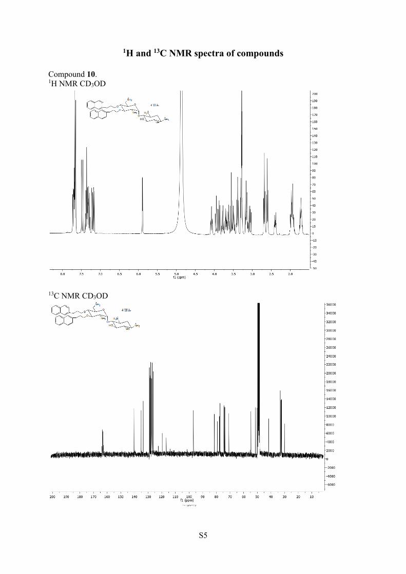
S5
1H and 13C NMR spectra of compounds
Compound 10. 1H NMR CD3OD
13C NMR CD3OD
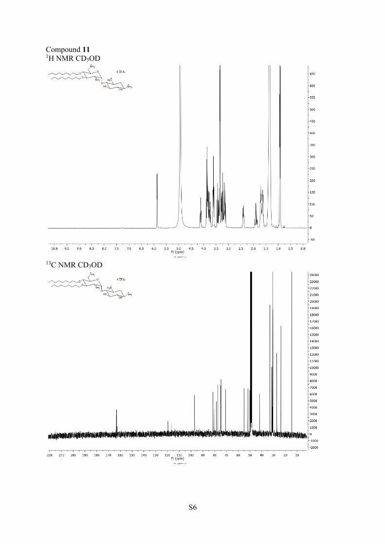
S6
Compound 11 1H NMR CD3OD
13C NMR CD3OD

S7
Allyl 6-azido-2,6-dideoxy-2-tritylamino--D-glucopyranoside (15) 1H NMR CDCl3
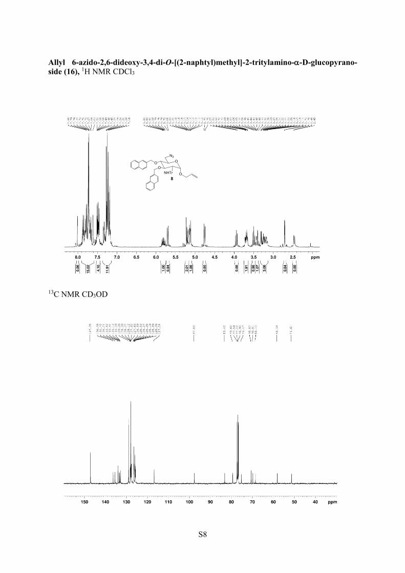
S8
Allyl 6-azido-2,6-dideoxy-3,4-di-O-[(2-naphtyl)methyl]-2-tritylamino--D-glucopyrano-
side (16), 1H NMR CDCl3
13C NMR CD3OD
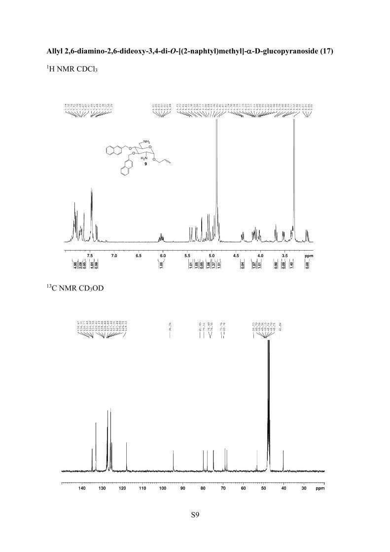
S9
Allyl 2,6-diamino-2,6-dideoxy-3,4-di-O-[(2-naphtyl)methyl]--D-glucopyranoside (17)
1H NMR CDCl3
13C NMR CD3OD
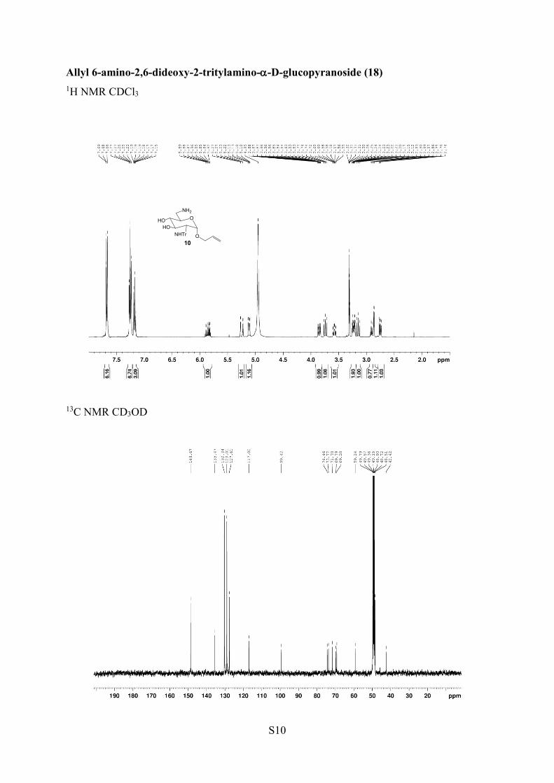
S10
Allyl 6-amino-2,6-dideoxy-2-tritylamino--D-glucopyranoside (18)
1H NMR CDCl3
13C NMR CD3OD
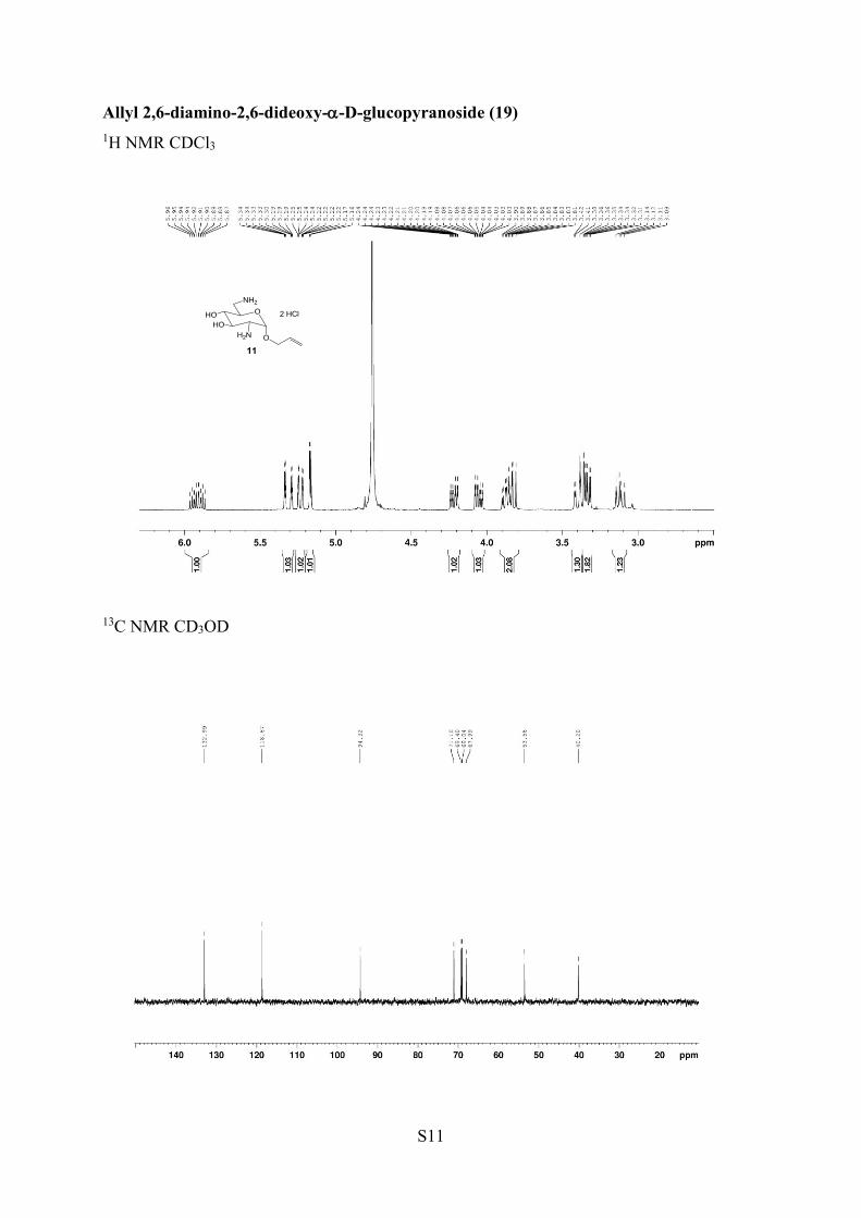
S11
Allyl 2,6-diamino-2,6-dideoxy--D-glucopyranoside (19)
1H NMR CDCl3
13C NMR CD3OD
S12
Compound 20
1H NMR CDCl3
13C NMR CDCl3
Compound 21

S13
1H NMR CDCl3
13C NMR CDCl3
S14
Compound 22
1H NMR CDCl3
13C NMR CDCl3

S15
Compound 23
1H NMR CDCl3
13C NMR CDCl3

S16
Compound 24
1H NMR CDCl3
13C NMR CDCl3

S17
Compound 25
1H NMR CDCl3
13C NMR CDCl3
S18
Compound 26
1H NMR CD3OD
13C NMR CD3OD
S19
Compound 27
1H NMR CDCl3
13C NMR CDCl3
S20
Compound 28
1H NMR CD3OD
13C NMR CD3OD
S21
Compound 29
1H NMR CD3OD
13C NMR CD3OD

S22
Compound 30
1H NMR CDCl3
13C NMR CDCl3
S23
Compound 31a,b
1H NMR CDCl3
13C NMR CDCl3
S24
Compound 32
1H NMR CDCl3
13C NMR CDCl3
S25
Compound 33
1H NMR CDCl3
13C NMR CDCl3
S26
Compound 34
1H NMR CD3OD
13C NMR CD3OD
S27
Compound 35
1H NMR CDCl3
13C NMR CDCl3
S28
Compound 36
1H NMR CDCl3
13C NMR CDCl3
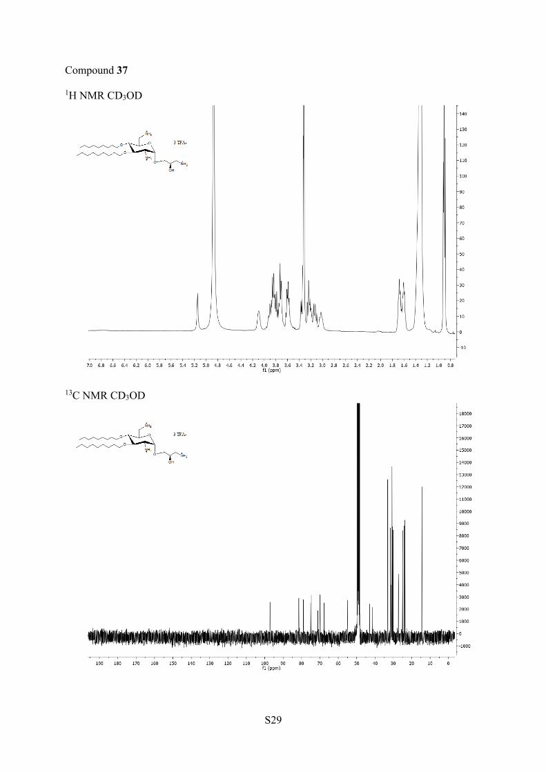
S29
Compound 37
1H NMR CD3OD
13C NMR CD3OD

S30
Compound 38
1H NMR CD3OD
13C NMR CD3OD

S31
Compound 39a,b
1H NMR CD3OD
13C NMR CD3OD
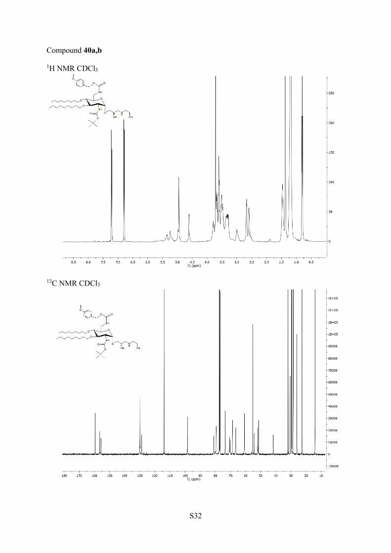
S32
Compound 40a,b
1H NMR CDCl3
13C NMR CDCl3

S33
Compound 41a,b
1H NMR CDCl3
13C NMR CDCl3
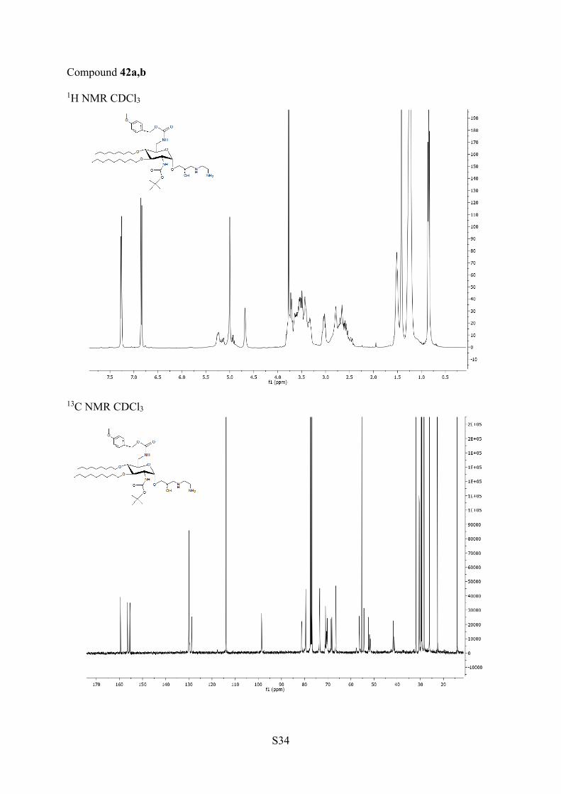
S34
Compound 42a,b
1H NMR CDCl3
13C NMR CDCl3

S35
Compound 43a,b
1H NMR CDCl3
13C NMR CDCl3

S36
Compound 44a,b
1H NMR CD3OD
13C NMR CD3OD
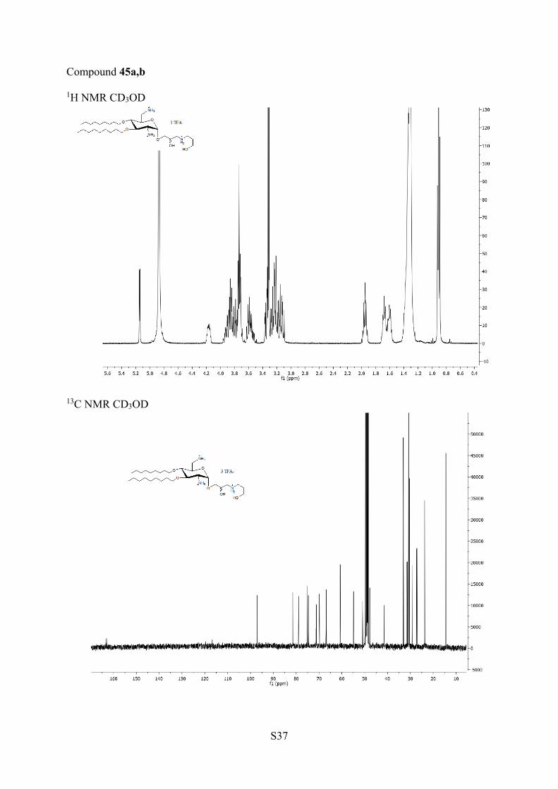
S37
Compound 45a,b
1H NMR CD3OD
13C NMR CD3OD

S38
Compound 46a,b
1H NMR CD3OD
13C NMR CD3OD
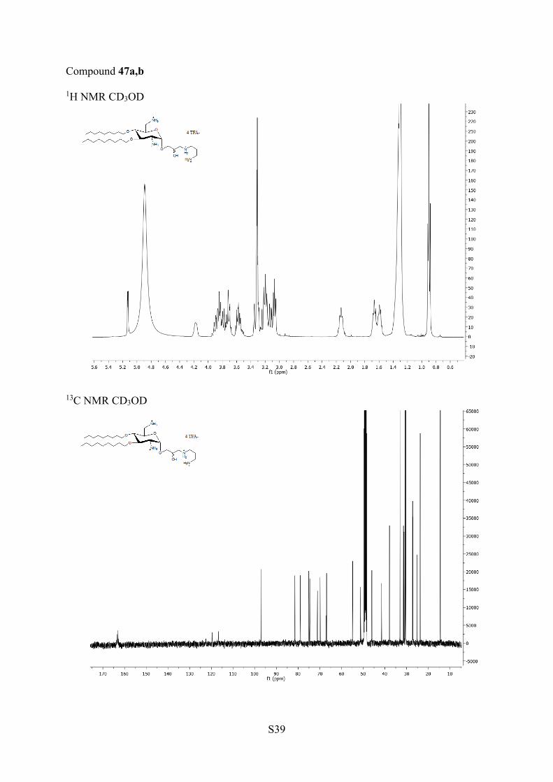
S39
Compound 47a,b
1H NMR CD3OD
13C NMR CD3OD