Embed Size (px)
Citation preview
AJR:197, September 2011 W423
FOCU
S O
N:
CMESAM
icle plate (Fig. 1) is a new hardware option for fracture fixation. Unlike dynamic com-pression plates and intramedullary rods, the blade hook clavicle plate allows normal bio-mechanical rotation between the clavicle and the acromion [2]. The plate is anatomically precontoured with a distal hook inserted un-derneath the acromion and posterior to the ac-romioclavicular joint. Generally three or four screws are used to fix the plate to the clavicle. Interfragmentary screws are often concomi-tantly used to improve apposition of fracture fragments; these screws should be perpen-dicular to the fracture plane. The end of the hook should be within 1–2 mm of the acromi-on. The main complication of the blade hook clavicle plate is unhooking from the acromion (Fig. 2). Other complications include osteoly-sis around the hook, medial clavicle fracture, rotator cuff damage, skin sloughing, and acro-mioclavicular joint osteoarthritis.
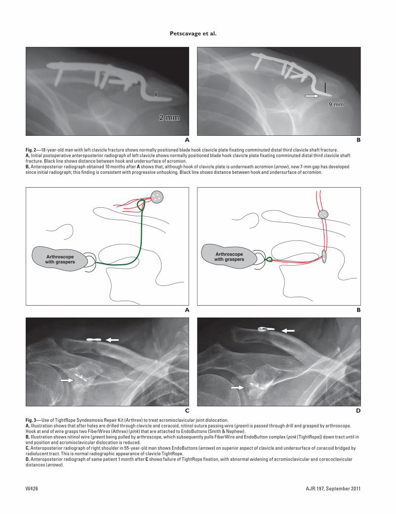
New hardware is also available for the treatment of acromioclavicular joint disloca-tion. The TightRope technique (TightRope Syndesmosis Repair Kit, Arthrex) uses ra-diolucent FiberWire (Athrex) sutures fixed by EndoButtons (Smith & Nephew) between the coracoid and clavicle [3] (Fig. 3A). The advantage of this technique is that it is mini-mally invasive and a second surgery is not required. On postoperative imaging, the En-doButtons should be flush against the cora-coid and clavicle, and the acromioclavicu-lar and coracoclavicular distances should be stable. Possible complications include loos-ening of the sutures, seen as increased cor-acoclavicular or acromioclavicular distance
New and Improved Orthopedic Hardware for the 21st Century: Part 1, Upper Extremity
Jonelle M. Petscavage1,2
Alice S. Ha1
Leila Khorashadi1,3
Kiley Perrich1,4
Felix S. Chew1
Petscavage JM, Ha AS, Khorashadi L, Perrich K, Chew FS
1Department of Radiology, University of Washington Medical Center, Seattle, WA.
2Present address: Department of Radiology, Penn State Hershey Medical Center, 500 University Dr, Hershey, PA 17033. Address correspondence to J. M. Petscavage ([email protected]).
3Present address: Schatzki Associates, Cambridge, MA.
4Present address: Radiology Associates of Hawaii, Honolulu, HI.
Musculoskeleta l Imaging • Pictor ia l Essay
WEB This is a Web exclusive article.
CME/SAM This article is available for CME/SAM credit. See www.arrs.org for more information.
AJR 2011; 197:W423–W433
0361–803X/11/1973–W423
© American Roentgen Ray Society
New orthopedic products are con-stantly being developed for frac-ture fixation, arthrodesis, and ar-throplasty. Eloquent description
of orthopedic hardware and of related compli-cations is an essential part of interpreting musculoskeletal radiology studies and is more important than merely reporting the manufac-turer name for a particular construct. Keeping up with the latest hardware technology is an important task for radiologists. This article will provide a survey of new orthopedic de-vices for use in the upper extremity.
Hardware ComplicationsA solid understanding of common types
of hardware complications is necessary be-fore starting this review of newer orthope-dic hardware. The hardware can fracture, disengage if there are multiple components, or loosen. Because of differences in stress distribution after hardware fixation, peri-prosthetic fractures are also possible. Some hardware materials introduce specific com-plications; for example, polyethylene is asso-ciated with asymmetric wear and small-par-ticle disease. Thus, knowledge of component design and material composition of the hard-ware is important for predicting and under-standing complications.
Clavicle, Acromioclavicular Joint, and Shoulder
Traditionally, clavicle fractures have been treated nonoperatively. However, recent stud-ies have shown better functional outcomes with surgical fixation [1]. A blade hook clav-
Keywords: arthroplasty, musculoskeletal imaging, orthopedic hardware, radiography
DOI:10.2214/AJR.10.5347
Received July 17, 2010; accepted after revision January 30, 2011.
FOCU
S O
N:
New and Improved Orthopedic Hardware
OBJECTIVE. The purpose of this article is to provide a survey of new orthopedic prod-ucts for use in the upper extremity.
CONCLUSION. Knowledge of the physiologic purpose, orthopedic trends, imaging findings, and complications is important in assessing new orthopedic devices.
Petscavage et al.Orthopedic Hardware for the 21st Century
Musculoskeletal ImagingPictorial Essay
CMESAM
W424 AJR:197, September 2011
Petscavage et al.
(Fig. 3D); EndoButton displacement; frac-ture; or a soft-tissue mass adjacent to the En-doButtons from granulomatous reaction [4].
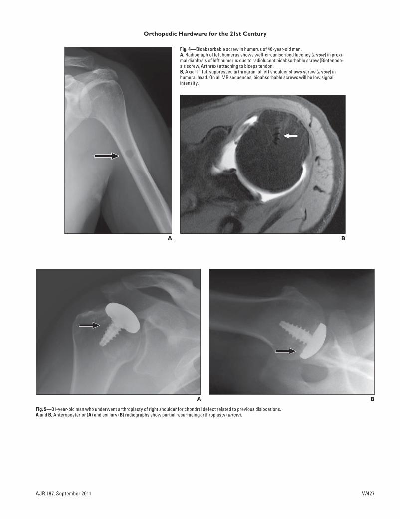
Biotenodesis screws (Arthrex), composed of bioabsorbable, radiolucent poly-L–lactide acid, are hardware used for tendon transfers or acromioclavicular joint dislocations. Al-though radiographic findings may be small in these patients, the underlying procedure may have been quite extensive, including transfer of large amounts of muscle or ten-don from other sites of the body. Without a clinical history, these screws could raise con-cern for metastatic or myelomatous lesions because they appear on radiographs as new round, radiolucent defects in previously nor-mal cancellous bone (Fig. 4A). On MRI, the bioabsorbable screws are low signal intensity on all sequences (Fig. 4B).
Partial resurfacing of the humeral head is a new hardware design introduced in the past few years to treat avascular necrosis or asymmetric chondral defects [5] (Fig. 5). The hardware consists of a titanium screw post attached to a cobalt-chromium alloy surface component. Long-term outcome studies at 48 months have shown increased patient range of motion, improved functional and mental scores, and decreased pain [5]. The only reported complication is the pres-ence of an intraarticular loose body.
ElbowRadial head replacement is performed to
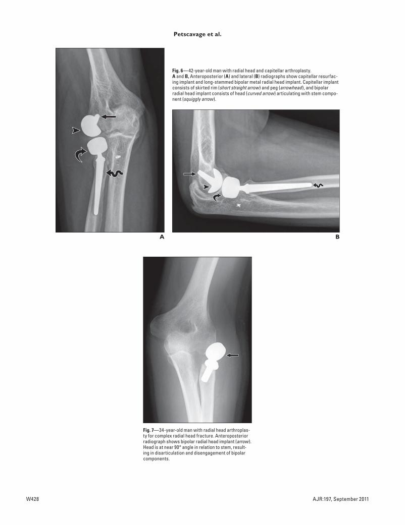
treat complex radial head fractures. Tradition-ally, unipolar radial head replacements have been used. Recently, a bipolar radial head re-placement was developed (Fig. 6). The replace-ment head can appear to have mild angulation on radiographs because it has a 15–35° range of motion in all planes, mimicking native anat-omy, whereas the stem will remain parallel to the radial shaft. There is a unique risk of dis-engagement of the snap-on head-to-stem com-ponents of the bipolar prosthesis (Fig. 7) com-pared with the unipolar type, which is one single unit of hardware. Other possible compli-cations include loosening, periprosthetic frac-ture, and asymmetric joint alignment from too thick of a radial head, termed “overstuffing.” Overstuffing is seen on anteroposterior radio-graphs as an asymmetrically narrowed medi-al aspect of the ulnohumeral joint space with gapping of the lateral aspect of the ulnohumer-al joint space [6]. Overstuffing can result in os-teoarthritis of the radiocapitellar joint space, capitellar erosions, capitellar osteopenia, and decreased range of motion at the elbow.
Capitellar resurfacing has recently been described to treat radiocapitellar osteoar-thritis in patients with radial head arthro-plasty [7]. The implant consists of a skirted rim attached to a cobalt-chrome peg (Fig. 6). The capitellar implant should be flush against the bone and normally articulating with the radial head. Possible complications include loosening and overstuffing of the radiocapitellar joint.
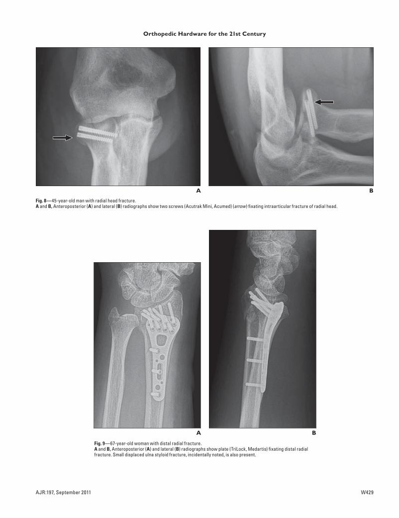
Headless titanium screws (Acutrak Mini screws, Acumed) (Fig. 8) are an alternative to radial head arthroplasty for complex radi-al head and capitellar fractures because they do not pose the risks of arthroplasty loosen-ing or overstuffing. These screws are head-less and therefore can be inserted through and beneath articular surfaces without blocking or damaging joint function. These titanium screws are cannulated and tapered with a thread pitch that gets narrower to al-low compressive function and to improve in-ternal holding power. Additionally, they are self-tapping—meaning that the screw cuts its own threads as it is inserted into the bone over a guidewire [8]. Reported complica-tions include avascular necrosis of the head and neck and fracture nonunion.
WristFor many years, fracture fixation has relied
on anatomic reduction using a dynamic com-pression system. Recently, the locking plate technique has been shown to be superior for the treatment of comminuted, unstable, or os-teoporotic fractures [9]. In locked plates, the locking screws mate with the threaded plate holds, creating a fixed-angle construct, negat-ing the need for double-cortex fixation (Fig. 9). Although compression plating requires absolute stability for bone healing, locking plates function as internal fixators with mul-tiple anchor points. The locking plates are bet-ter for osteoporotic fractures because they de-crease the potential for toggling of screws in the cortex and loss of purchase of small bone fragments, thus providing stable fixation re-quired for healing.
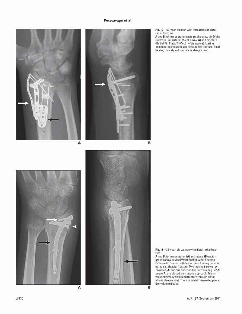
Another notable new locking hardware design is the Volar Buttress Pin (TriMed) and Radial Pin Plate (TriMed) (Fig. 10); they are unique in that they act as a three-axis correction of radial length, volar tilt, and radial inclination [10, 11]. There is also decreased soft-tissue irritation. The buttress plate clip allows elevation of impacted frac-tures for correction of radial length by bend-ing of the pins and movement of the pins
along the length of the plate. Unique com-plications include pin protrusion through the dorsal surface resulting in damage to exten-sor tendons and nerves.
An even less invasive new hardware for distal radius fracture fixation is a flexible stainless-steel curved intramedullary im-plant (Wrist Rocket WRx, Sonoma Orthope-dic Products) (Fig. 11). It has locking corti-cal screws and distal and proximal grippers to engage the intramedullary cortex for rig-id fixation while a buttress peg supports the subchondral bone. The main advantage of the hardware is less volar soft-tissue disruption, allowing earlier range of motion and less soft tissue being affected by complications [12].
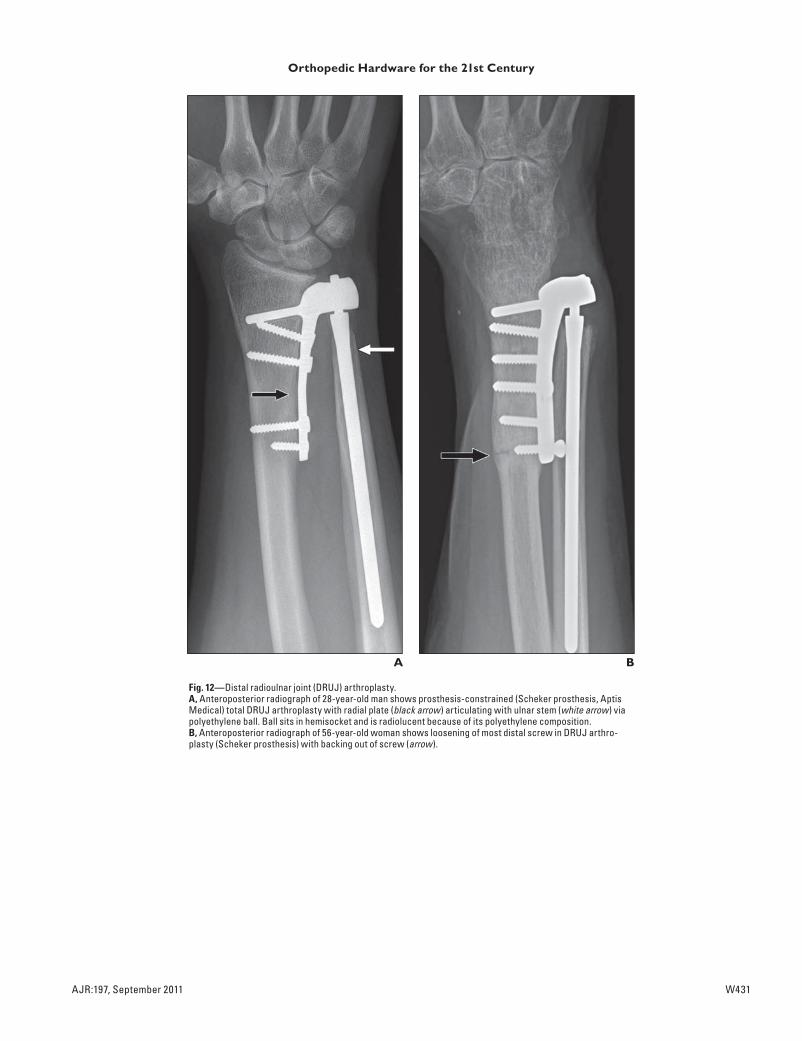
New hardware for arthroplasty and ar-throdesis of the wrist is available. At the dis-tal radioulnar joint (DRUJ), a new prosthesis (Scheker prosthesis, Aptis Medical) that has a constrained design allows full supination and pronation at the joint (Fig. 12A). The pros-thesis consists of a cobalt-chromium plate at-tached to the distal radius by a peg and five screws. An ulnar polyethylene polymer ball attaches to the plate through a hemisocket [13]. Possible complications include hardware loosening (Fig. 12B), disarticulation between the radial and ulnar components, particle dis-ease, and periprosthetic fracture.
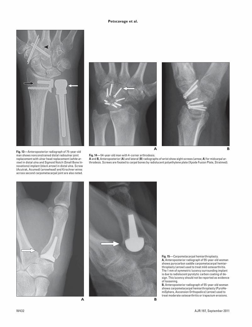
Alternatively, the DRUJ can be replaced in a nonconstrained design: The Sigmoid Notch (Small Bone Innovations) implant is a polyethylene insert that slides on a metal radial plate and is secured by lock and screw [14] (Fig. 13). With no link between the com-ponents, malarticulation and dislocation are additional potential complications.
Established options for midcarpal fusion or arthroplasty include the Spider Plate (Ki-netikos Medical), universal total wrist arthro-plasty, and arthrodesis plate. A new device is the Xpode Fusion Plate (Stratmed) (Fig. 14), modeled after the Spider Plate. The Xpode Fusion Plate differs from the Spider Plate in that the Xpode plate is composed of polyeth-ylene, thus allowing full visualization of the degree of fusion between the carpal bones.
Hand and FingersMultiple methods for first carpometacar-
pal joint arthroplasty are available, including hematoma interposition, tendon transfer, and cartilage interposition. Among these meth-ods, new hardware currently available include the PyroHemiSphere (Ascension Orthope-dics) and saddle carpometacarpal pyrocarbon implant [15] (Fig. 15). Both designs preserve
AJR:197, September 2011 W425
Orthopedic Hardware for the 21st Century
the trapezium as a bridge to future carpometa-carpal joint arthroplasty. These prostheses are composed of pyrolytic carbon coating over a graphite core. Pyrocarbon is a material with an elastic modulus similar to that of cortical bone, thus providing bone-stress transfer.
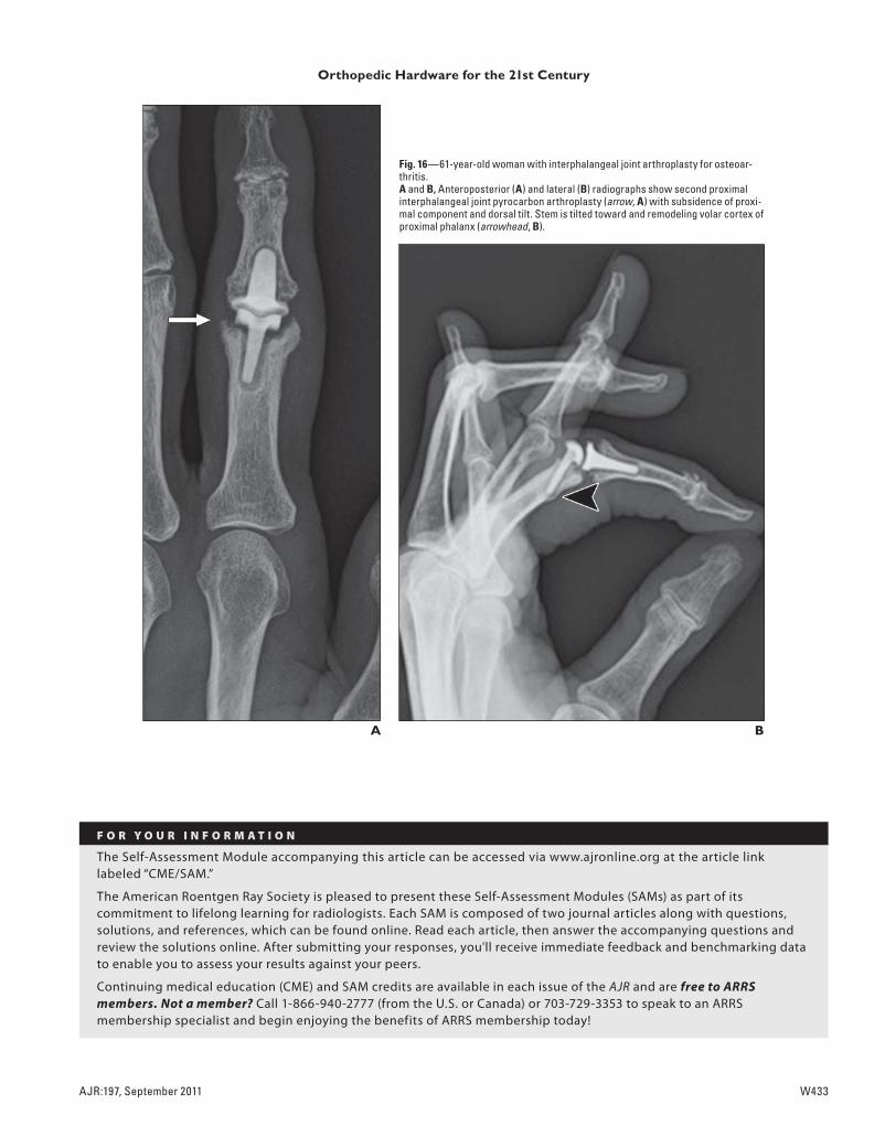
Pyrocarbon implants are also being used instead of silicone for metacarpophalange-al and proximal interphalangeal joint arthro-plasty because of the high complication rates of silicone synovitis and silicone implant frac-ture. Pyrocarbon implants rely on the overly-ing soft tissues for movement at the joints.
In postoperative evaluation of pyrocarbon implants, visualization of greater than 2 mm of periprosthetic lucency suggests loosening of the implant and can result in changes in range of motion at the joint and subsequent volar or dorsal tilt of the implant (Fig. 16). No complications unique to the pyrocarbon material have been reported to date.
ConclusionWith continued evolution of orthopedic
hardware, the radiologist must keep pace with these advancements and understand the indications for, functions of, and potential complications of these devices.
References 1. Canadian Orthopaedic Trauma Society. Nonop-
erative treatment compared with plate fixation of
displaced mid clavicular fractures: a multicenter,
randomized clinical trial. J Bone Joint Surg Am
2007; 89:1–10
2. Melenevsky Y, Yablon CM, Ramappa A, Hochman
MG. Clavicle and acromioclavicular joint injuries: a
review of imaging, treatment, and complications.
Skeletal Radiol 2010 June 6 [Epub ahead of print]
3. Hernegger GS, Kadletz R. TightRope: the revolu-
tionary anatomical fixation in acromioclavicular
joint dislocation—a case report. Tech Shoulder
Elbow Surg 2006; 7:86–88
4. McMurray D, Hornung B, Venkateswaran B, Ali
Z. Walking on a tightrope: our experience in the
treatment of traumatic ankle syndesmosis rup-
ture. Injury Extra 2008; 39:182
5. Uribe JW, Botto-van Bemden A. Partial humeral
head resurfacing for osteonecrosis. J Shoulder El-
bow Surg 2009; 18:711–716
6. Frank SG, Grewal R, Johnson J, et al. Determina-
tion of correct implant size in radial head arthro-
plasty to avoid overlengthening. J Bone Joint Surg
Am 2009; 91:1738–1746
7. Heijink A, Morrey BF, Cooney WP 3rd. Radio-
capitellar hemiarthroplasty for radiocapitellar ar-
thritis: a report of three cases. J Shoulder Elbow
Surg 2008; 17:e12–e15
8. Loving VA, Richardson ML. Scaphoid fracture
fixation with an Acutrak screw. Radiology Case
Reports [Online] 2006; 1:13
9. Greiwe RM, Archdeacon MT. Locking plate technol-
ogy: current concepts. J Knee Surg 2007; 20:50–55
10. Chung KC, Watt AJ, Kotsis SV, et al. Treatment of
unstable distal radial fractures with the volar
locking plating system. J Bone Joint Surg Am
2006; 88:2687–2694
11. Perry J, Medoff R. Mini-invasive, independent 3 axis
correction of radial malunions: a new technique.
www.ota.org/eposters/OTA%20E-Posters/53Perry.
pdf. Accessed July 17, 2010
12. Sonoma Orthopedic Products, Inc., Website.
Fracture solutions: Sonoma WRx. www.so-
nomaorthopedics.com/index.php?page=wrist-
fracture-solutions. Accessed July 17, 2010
13. Scheker LR, Babb BA, Killion PE. Distal ulnar
prosthetic replacement. Orthop Clin North Am
2001; 32:365–376
14. Small Bone Innovations, Inc., Website. Stability:
Sigmoid Notch. www.totalsmallbone.com/us/prod-
ucts/wrist/sigmoid_notch.php4. Accessed July
17, 2010
15. Ascension Orthopedics Website. Hand & wrist.
www.ascensionortho.com/content/surgeons.
php?p=1&seg=2. Accessed July 17, 2010
A
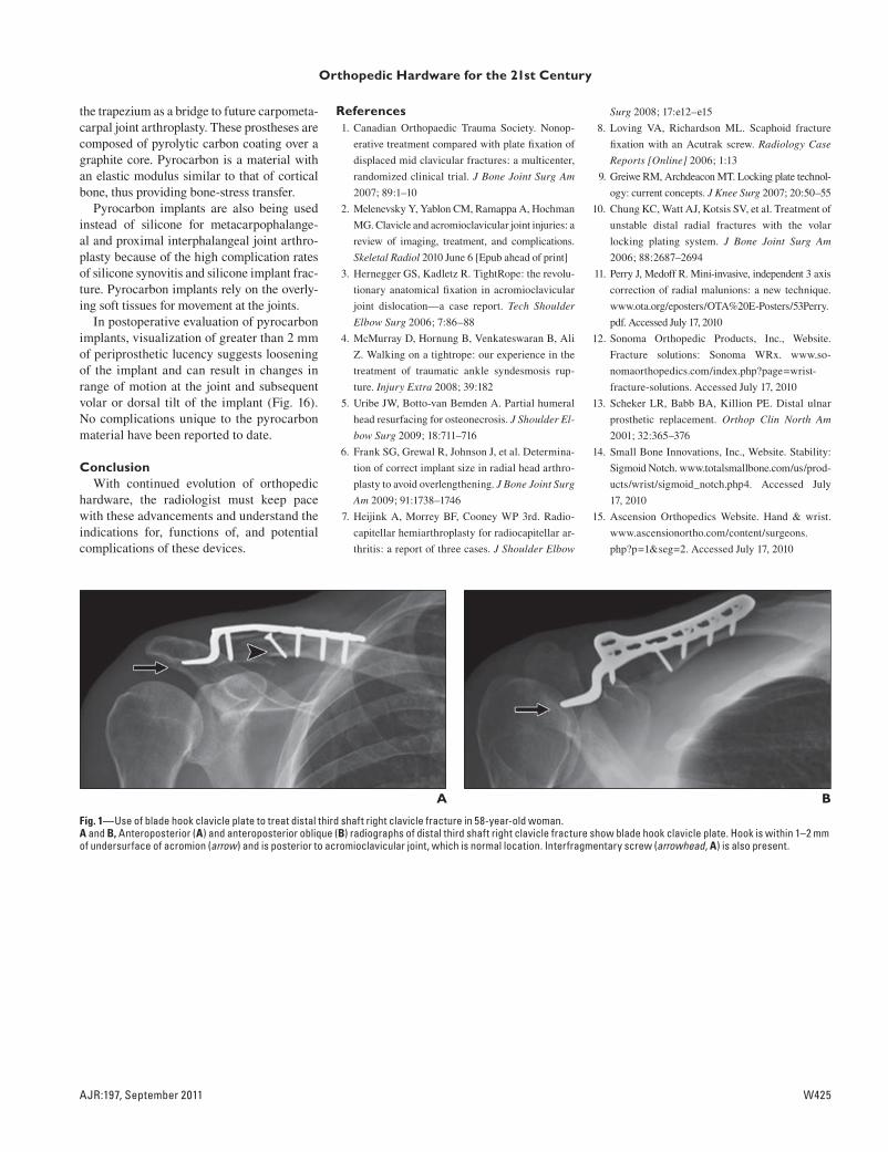
Fig. 1—Use of blade hook clavicle plate to treat distal third shaft right clavicle fracture in 58-year-old woman.A and B, Anteroposterior (A) and anteroposterior oblique (B) radiographs of distal third shaft right clavicle fracture show blade hook clavicle plate. Hook is within 1–2 mm of undersurface of acromion (arrow) and is posterior to acromioclavicular joint, which is normal location. Interfragmentary screw (arrowhead, A) is also present.
B
W426 AJR:197, September 2011
Petscavage et al.
Arthroscopewith graspers
Arthroscopewith graspers
A
A
C
Fig. 2—18-year-old man with left clavicle fracture shows normally positioned blade hook clavicle plate fixating comminuted distal third clavicle shaft fracture. A, Initial postoperative anteroposterior radiograph of left clavicle shows normally positioned blade hook clavicle plate fixating comminuted distal third clavicle shaft fracture. Black line shows distance between hook and undersurface of acromion.B, Anteroposterior radiograph obtained 10 months after A shows that, although hook of clavicle plate is underneath acromion (arrow), new 7-mm gap has developed since initial radiograph; this finding is consistent with progressive unhooking. Black line shows distance between hook and undersurface of acromion.
Fig. 3—Use of TightRope Syndesmosis Repair Kit (Arthrex) to treat acromioclavicular joint dislocation. A, Illustration shows that after holes are drilled through clavicle and coracoid, nitinol suture passing wire (green) is passed through drill and grasped by arthroscope. Hook at end of wire grasps two FiberWires (Athrex) (pink) that are attached to EndoButtons (Smith & Nephew).B, Illustration shows nitinol wire (green) being pulled by arthroscope, which subsequently pulls FiberWire and EndoButton complex (pink [TightRope]) down tract until in end position and acromioclavicular dislocation is reduced. C, Anteroposterior radiograph of right shoulder in 55-year-old man shows EndoButtons (arrows) on superior aspect of clavicle and undersurface of coracoid bridged by radiolucent tract. This is normal radiographic appearance of clavicle TightRope. D, Anteroposterior radiograph of same patient 1 month after C shows failure of TightRope fixation, with abnormal widening of acromioclavicular and coracoclavicular distances (arrows).
B
B
D
AJR:197, September 2011 W427
Orthopedic Hardware for the 21st Century
Fig. 4—Bioabsorbable screw in humerus of 46-year-old man.A, Radiograph of left humerus shows well-circumscribed lucency (arrow) in proxi-mal diaphysis of left humerus due to radiolucent bioabsorbable screw (Biotenode-sis screw, Arthrex) attaching to biceps tendon. B, Axial T1 fat-suppressed arthrogram of left shoulder shows screw (arrow) in humeral head. On all MR sequences, bioabsorbable screws will be low signal intensity.
A B
A
Fig. 5—31-year-old man who underwent arthroplasty of right shoulder for chondral defect related to previous dislocations.A and B, Anteroposterior (A) and axillary (B) radiographs show partial resurfacing arthroplasty (arrow).
B
W428 AJR:197, September 2011
Petscavage et al.
A
Fig. 6—42-year-old man with radial head and capitellar arthroplasty. A and B, Anteroposterior (A) and lateral (B) radiographs show capitellar resurfac-ing implant and long-stemmed bipolar metal radial head implant. Capitellar implant consists of skirted rim (short straight arrow) and peg (arrowhead), and bipolar radial head implant consists of head (curved arrow) articulating with stem compo-nent (squiggly arrow).
B
Fig. 7—34-year-old man with radial head arthroplas-ty for complex radial head fracture. Anteroposterior radiograph shows bipolar radial head implant (arrow). Head is at near 90° angle in relation to stem, result-ing in disarticulation and disengagement of bipolar components.
AJR:197, September 2011 W429
Orthopedic Hardware for the 21st Century
A B
A
Fig. 8—45-year-old man with radial head fracture.A and B, Anteroposterior (A) and lateral (B) radiographs show two screws (Acutrak Mini, Acumed) (arrow) fixating intraarticular fracture of radial head.
B
Fig. 9—67-year-old woman with distal radial fracture.A and B, Anteroposterior (A) and lateral (B) radiographs show plate (TriLock, Medartis) fixating distal radial fracture. Small displaced ulna styloid fracture, incidentally noted, is also present.
W430 AJR:197, September 2011
Petscavage et al.
A
A
B
B
Fig. 10—60-year-old man with intraarticular distal radial fracture.A and B, Anteroposterior radiographs show pin (Volar Buttress Pin, TriMed) (black arrow, A) and pin plate (Radial Pin Plate, TriMed) (white arrows) fixating comminuted intraarticular distal radial fracture. Small healing ulna styloid fracture is also present.
Fig. 11—36-year-old woman with distal radial frac-ture.A and B, Anteroposterior (A) and lateral (B) radio-graphs show device (Wrist Rocket WRx, Sonoma Orthopedic Products) (black arrows) fixating commi-nuted distal radial fracture. Two locking screws (ar-rowhead, A) and one subchondral buttress peg (white arrow, A) are placed from lateral approach. Trans-verse minimally displaced fracture through distal ulna is also present. There is mild diffuse osteopenia, likely due to disuse.
AJR:197, September 2011 W431
Orthopedic Hardware for the 21st Century
A B
Fig. 12—Distal radioulnar joint (DRUJ) arthroplasty. A, Anteroposterior radiograph of 28-year-old man shows prosthesis-constrained (Scheker prosthesis, Aptis Medical) total DRUJ arthroplasty with radial plate (black arrow) articulating with ulnar stem (white arrow) via polyethylene ball. Ball sits in hemisocket and is radiolucent because of its polyethylene composition. B, Anteroposterior radiograph of 56-year-old woman shows loosening of most distal screw in DRUJ arthro-plasty (Scheker prosthesis) with backing out of screw (arrow).
W432 AJR:197, September 2011
Petscavage et al.
A B
Fig. 14—54-year-old man with 4-corner arthrodesis.A and B, Anteroposterior (A) and lateral (B) radiographs of wrist show eight screws (arrow, A) for midcarpal ar-throdesis. Screws are fixated to carpal bones by radiolucent polyethylene plate (Xpode Fusion Plate, Stratmed).
Fig. 13—Anteroposterior radiograph of 75-year-old man shows nonconstrained distal radioulnar joint replacement with ulnar head replacement (white ar-row) in distal ulna and Sigmoid Notch (Small Bone In-novations) implant (black arrow) in distal ulna. Screw (Acutrak, Acumed) (arrowhead) and Kirschner wires across second carpometacarpal joint are also noted.
A B
Fig. 15—Carpometacarpal hemiarthroplasty. A, Anteroposterior radiograph of 55-year-old woman shows pyrocarbon saddle carpometacarpal hemiar-throplasty (arrow) used to treat mild osteoarthritis. The 1 mm of symmetric lucency surrounding implant is due to radiolucent pyrolytic carbon coating of de-sign. This lucency should not be reported as evidence of loosening.B, Anteroposterior radiograph of 55-year-old woman shows carpometacarpal hemiarthroplasty (PyroHe-miSphere, Ascension Orthopedics) (arrow) used to treat moderate osteoarthritis or trapezium erosions.
AJR:197, September 2011 W433
Orthopedic Hardware for the 21st Century
A B
Fig. 16—61-year-old woman with interphalangeal joint arthroplasty for osteoar-thritis.A and B, Anteroposterior (A) and lateral (B) radiographs show second proximal interphalangeal joint pyrocarbon arthroplasty (arrow, A) with subsidence of proxi-mal component and dorsal tilt. Stem is tilted toward and remodeling volar cortex of proximal phalanx (arrowhead, B).
F O R Y O U R I N F O R M A T I O N
The Self-Assessment Module accompanying this article can be accessed via www.ajronline.org at the article link labeled “CME/SAM.”
The American Roentgen Ray Society is pleased to present these Self-Assessment Modules (SAMs) as part of its commitment to lifelong learning for radiologists. Each SAM is composed of two journal articles along with questions, solutions, and references, which can be found online. Read each article, then answer the accompanying questions and review the solutions online. After submitting your responses, you'll receive immediate feedback and benchmarking data to enable you to assess your results against your peers.
Continuing medical education (CME) and SAM credits are available in each issue of the AJR and are free to ARRS members. Not a member? Call 1-866-940-2777 (from the U.S. or Canada) or 703-729-3353 to speak to an ARRS membership specialist and begin enjoying the benefits of ARRS membership today!