Embed Size (px)
Citation preview

Radiation Measurements 34 (2001) 567–569www.elsevier.com/locate/radmeas
Neutron autoradiography: working-out method and applicationin investigations of test paintings
A. Kalickia, E. Panczyka, L. Rowinskaa, B. Sartowskaa ; ∗, L. Walisa, K. Pytelb, B. Pytelb,A. Kozielb, L. Dabkowskib, M. Wierzchnickab, L. Strzalkowskib, T. Ostrowskib
aInstitute of Nuclear Chemistry and Technology, 16 Dorodna St., 03-195 Warsaw, PolandbInstitute of Atomic Energy, 05-400 Otwock-Swierk, Poland
Received 28 August 2000; received in revised form 2 January 2001; accepted 8 March 2001
Abstract
Neutron-induced autoradiography was carried out at MARIA research reactor in Poland. The paintings were exposed to thethermal neutrons. As a result, the radionuclides emitting beta particles and gamma rays were created from some of the elementsexisting in the painting. Beta particles were detected during successive exposure to a series of X-ray medical-sensitive =lms.The obtained images—blackening of the =lms depends mainly on the nuclear characteristic of recorded radionuclides andexposure parameters.The main purpose of this work was to work out a method, build a special stand and test sample paintings using neutron
autoradiography.Samples of paintings were investigated and according to the obtained results, optimum test parameters have been selected:
neutron irradiation conditions and autoradiographs exposure conditions. c© 2001 Elsevier Science Ltd. All rights reserved.
Keywords: Neutron autoradiography; Beta particles; Thermal neutrons
1. Neutron autoradiography
This kind of painting investigations was used for the=rst time in the 1960s in Brookhaven National Laboratory,Brookhaven, USA (Ainsworth, 1982). In a conventionalneutron activation autoradiography technique, a painting isexposed to a beam of neutrons from the thermal column orneutron guide tube from a research reactor (method appliedin Hahn-Meitner Institute, Berlin, Germany) (Pytel et al.,1998). The radionuclides emitting beta particles and gammarays were formed from some elements existing mainly inthe pigments present in the layers of the painting. Possibleevents are: gamma rays escape without absorption in the =lm
∗ Corresponding author. Tel.: +48-22-811-15-02; fax:+48-22-811-15-32.E-mail address: [email protected] (B. Sartowska).
emulsion, electrons emitted from the part of the painting inthe decay of radioactive isotope do not reach =lm emulsionand electrons interact with the =lm. Only those last betaparticles held close in contact to the surface of painting arerecorded on the sensitive =lm. Even though the paint layeris overpainted, its image would be visible on the =lm. Theobtained blackening depends on the nuclear characteristicof the radionuclide, the amount of used pigments and ex-posure conditions. Beta particles from short-lived isotopescan be recorded on the =lm only during a short time afterthe end of neutron irradiation. Long-lived isotopes can berecorded during the next exposures as well. It means thatafter one neutron irradiation of the painting, we can obtainsome of its diGerent “images”—autoradiographs. For exam-ple: 54Mn (half-life—2:6 h) present in umber can producethe best image 0–24 h after irradiation; 203 Hg (half-life—48 d) present in vermilion can produce the best image morethen 25 days after irradiation.
1350-4487/01/$ - see front matter c© 2001 Elsevier Science Ltd. All rights reserved.PII: S1350 -4487(01)00229 -3
568 A. Kalicki et al. / Radiation Measurements 34 (2001) 567–569
2. Stand for painting irradiation
The mentioned neutron sources are not available atMARIA reactor in Swierk-Otwock. So, a novel techniquehas been proposed and developed (Pytel et al., 1998).Thermal neutrons were scattered from the main beamusing a scattering block with a polyethylene foil as a scat-terer. Undesirable gamma and fast neutron radiation werestrongly reduced. The most important point was to obtaina proper distribution of scattering medium density alongthe neutron beam, allowing to obtain the homogeneousintensity of scattered neutrons. The special stand has beendesigned, manufactured and installed on the beam hole H-8of MARIA reactor. The eGective area of the cross-sectionof scattered neutron =eld was 2000 × 280 mm. The standwas equipped with a vertical direction scanning device.
3. Investigations of test paintings
The main purpose of those examinations was to =nd outthe optimal irradiation and exposure conditions and providesuLciently low radiation hazard to the painting and operat-ing personnel.Two samples painted using equivalents to historical
pigments produced nowadays by KREMER were tested:“stripes”—linen, 56 × 74 cm and “check”—linen, 35×50 cm. The test paintings are shown in Fig. 1 and
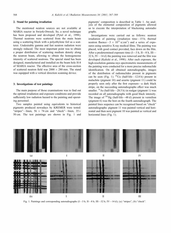
Fig. 1. Paintings and corresponding autoradiographs (I—3 h; II—8 h; III—32 h; IV—14 d); (a) “stripes”, (b) “check”.
pigments’ composition is described in Table 1. An anal-ysis of the elemental composition of pigments allowedus to execute the interpretation of blackening autoradiographs.Investigations were carried out as follows: neutron
irradiation of painting (irradiation time—15 h; thermalneutron Ouence—3 × 10¹² n=cm2) and a series of expo-sures using sensitive X-ray medical =lms. The painting wasplaced, with good contact provided, face down on the =lm.After a predetermined exposure time (I—3 h, II—8 h, III—32 h, IV—14 d) the painting was removed and the =lm wasdeveloped (Kalicki et al., 1998). After each exposure, thehigh-resolution gamma rays spectrometric measurements ofthe painting were conducted for a more precise radionuclideidenti=cation. On all obtained autoradiographs, imagesof the distribution of radionuclides present in pigmentscan be seen (Fig. 1). 64Cu (half-life—12:8 h) present inmalachite (pigment 10) and azurite (pigment 11) could beproperly seen only after the =rst exposure—a dark blackstripe, on the succeeding autoradiographs eGect was muchsmaller. 76As (half-life—28:5 h) in realgar (pigment 1) wasrecorded on all autoradiographs with good black intensity.The image of 203Hg (half-life—48 d) present in vermilion(pigment 6) was the best on the fourth autoradiograph. Thepainted lines sequence can be recognised based on “check”autoradiographs: pigment 11 was painted vertical and hori-zontal and then next pigment 10 was painted as vertical andhorizontal lines (Fig. 1).

A. Kalicki et al. / Radiation Measurements 34 (2001) 567–569 569
Table 1Elements present in pigments used in “ stripes” and “check” test paintings
No. Pigment Main elements Trace elements
1. Realgar As, Fe, Si, Sb, Ca, Mg Al, Cu, Ti, Ca, Ni2. Brown from Elba Fe, Si, Al, Ca, Mg, Mn Ti, Ba, Ni3. Minium-red lead Pb, Si, Mg Ca, Fe, Cu4. Umber Fe, Si, Al, Ca, Mg, Mn, Ti Cu, Cr5. Tin-lead yellow Sn, Pb6. Vermilion Hg, Fe, Al, Si, Ti, Ca, Mg, Ni, Mn Cu, Ba7. Ultra-marine Si, Al, Na, Fe, Ca, Mg, Ti Cu, Pb8. Copper resinate Fe, Cu9. Smalt Si, Co, Mg Ca, Pb, Cu, Fe10. Malachite Cu, Si, Al, Mg, Fe Ca11. Azurite Cu, Si, Al, Mg, Ca, Fe Ti12. Bone black Ca, Mg, P, Si, Fe Al, Cu, Ni
4. Final remarks
• Neutron autoradiography helps us to disclose newfacts about painted layers, used pigments and method ofpainting.
• A stand for painting irradiation gives us proper conditionsfor obtaining autoradiographs with suLcient resolution.
• The experiments carried out according to the describedprocedure, do not involve radiological hazard to the paint-ings and personnel carrying out the investigations andto the people who will later come into contact with thepaintings (Ainsworth, 1982; Pytel et al., 1998).
• Built special stand—an activation facility was used tocarry out investigations of the original painting using theneutron autoradiography method.
Acknowledgements
The State Committee for Scienti=c Research supportedthis work No 1H01E 001 96C=3009.
References
Ainsworth, M.W. (Ed.), 1982. Art. & Radiography: Insights intoGenesis of Paintings by Rembrandt, Van Dyck and Vermeer.The Metropolitan Museum of Art, New York.
Kalicki, A., Ligeza, M., Panczyk, E., Rowinska, L., Sartowska,B., Walis, L., 1998. Neutron Autoradiography of Paintings—prepare stand for exposure and photochemical treatment ofautoradiographs. INCT Report, Warsaw (unpublished).
Pytel, K., Pytel, B., Dabkowski, L., Koziel, A., Wierzchnicka, M.,Strzalkowski, L., Ostrowski, T., 1998. Neutron autoradiographyof paintings in Maria reactor. Report of experimental standinvestigation and irradiation of paintings, IEA Report No.B.20=98 (in Polish).