Embed Size (px)
Citation preview

Review Acta Neurobiol Exp 2010, 70: 454–467
© 2010 by Polish Neuroscience Society - PTBUN, Nencki Institute of Experimental Biology
NEUROTROPHINS AND THEIR RECEPTORS
Neurotrophins
In the early 1950s Levi-Montalcini and Cohen (Levi-Montalcini and Hamburger 1951, Cohen et al. 1954) were first to describe the nerve growth factor (NGF) and for that discovery they won the Nobel Prize in Physiology or Medicine in 1986. Afterwards, several novel structurally homologous neurotrophic factors belonging to the nerve growth factor family termed neurotrophins were discovered in verte-brates. These were BDNF, NT-3, NT-4/5 (Barde et al. 1982, Phillips et al. 1990, Ibáñez et al. 1993). Other members of neurotrophins neurotrophin-6 (NT-6) and neurotrophin-7 (NT-7) were only cloned from some teleost species (Götz et al. 1994, Lai et al.
1998) and are not expressed in other vertebrate than teleost fishes.
Structure of all neurotrophins is highly conserved with the exception of NT-4/5 (Hallböök 1999) that shares only about 50% amino acid identity with other neurotrophins (Shooter 2001). An important common feature of all neurotrophins is the presence of six cysteine residues that enable formation of disulfide bridges. Neurotrophins are synthesized as pre-pro-proteins by both neuronal and non-neuronal cell types (Thoenen 1995, Seidah et al. 1996). Protein products of all genes encoding neurotrophin contain a signal pep-tide for protein secretion (pre-protein) and the precur-sor protein (pro-protein). When the hydrophobic region of the signal peptide is removed from pre-proneurotro-phin at the N-terminal, the proneurotrophin is gener-ated (Fig. 1). The proneurotrophin is either cleaved of the signalling peptide in the endoplasmic reticulum and converted to the mature neurotrophin or is trans-ported to the plasma membrane and released in an unprocessed form (Seidah et al. 1996). In that case plasmin or another extracellular protease converts the
Neurotrophins and their receptors in early development of the mammalian nervous system
Katarzyna Bartkowska*, Kris Turlejski and Rouzanna L. Djavadian
Department of Molecular and Cellular Neurobiology, Nencki Institute of Experimental Biology, Warsaw, Poland; *Email: [email protected]
Neurotrophins belonging to the class of growth factors and including nerve growth factor (NGF), brain-derived neurotrophic factor (BDNF), neurotrophin-3 (NT-3) and neurotrophin-4/5 (NT-4/5) are widely recognized as essential factors in the developing central nervous system (CNS). Neurotrophins are synthesized as precursor forms (proneurotrophins). Mature forms of neurotrophins exert their effect by binding to specific tyrosine kinases receptors (TrkA, TrkB and TrkC) as well as via the p75 receptor, a member of the tumor necrosis factor receptor superfamily while proneurotrophins interact with the receptor p75 or co-receptor complex of p75 and sortilin, that is a Vps10p domain-containing transmembrane protein.Expression of neurotrophins corresponds with the onset of neurogenesis in developing mammalian species. BDNF is low in early embryonic stages of development, while NT-3 highly expresses in the developing CNS. Expression of neurotrophins receptors mainly overlaps at early development. Data concerning early distribution of neurotrophins and their receptors in the nervous system and results in mice with targeted disruptions of neurotrophin or receptor genes show that neurotrophins and their receptors play distinct roles in control and regulation of the most crucial developmental processes such as proliferation, migration, differentiation, survival, apoptosis and synaptic plasticity.
Key words: BDNF, developing brain, NGF, NT-3, NT-4/5, p75, sortilin, TrkA, TrkB, TrkC
Corresponding should be addressed to K. Bartkowska E-mail: [email protected]
Received 15 October, accepted 02 December 2010
Neurotrophins and their receptors in development 455
precursor proneurotrophins to mature neurotrophins through proteolytic cleavage (Pang et al. 2004). Mature neurotrophins are secreted as homodimeric proteins into the extracellular space and act in a paracrine and/or autocrine way (Lu et al. 2005), controlling many crucial processes in development of the nervous sys-tem such as proliferation, migration, differentiation, survival, apoptosis and synaptic plasticity. All these processes lead to control neuronal numbers and den-dritic growth.
Neurotrophin receptors
Neurotrophins interact with three distinct classes of receptors: three members of the tropomyosin receptor kinase (Trk) family of receptor tyrosine kinases (TrkA, TrkB and TrkC), the p75 neurotrophin receptor belong-ing to the tumor necrosis factor receptor (TNFR) superfamily and sortilin, a Vps10p domain-containg transmembrane protein. All neurotrophins mediate their effects via activation of one or more Trk recep-
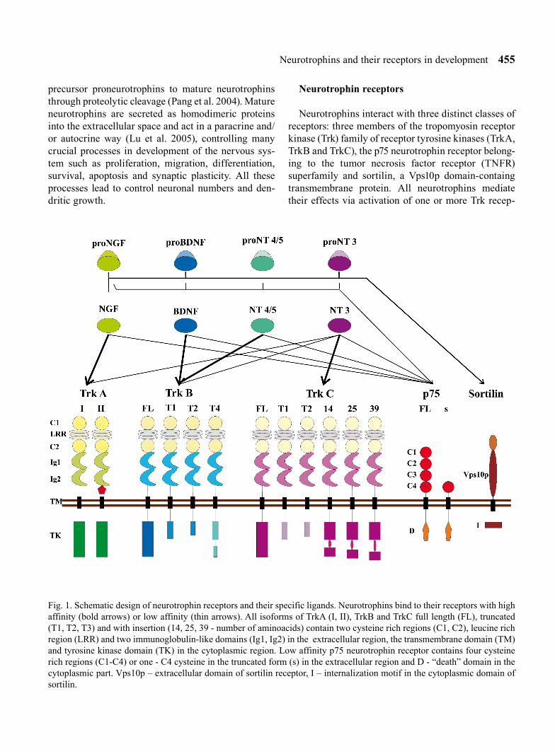
Fig. 1. Schematic design of neurotrophin receptors and their specific ligands. Neurotrophins bind to their receptors with high affinity (bold arrows) or low affinity (thin arrows). All isoforms of TrkA (I, II), TrkB and TrkC full length (FL), truncated (T1, T2, T3) and with insertion (14, 25, 39 - number of aminoacids) contain two cysteine rich regions (C1, C2), leucine rich region (LRR) and two immunoglobulin-like domains (Ig1, Ig2) in the extracellular region, the transmembrane domain (TM) and tyrosine kinase domain (TK) in the cytoplasmic region. Low affinity p75 neurotrophin receptor contains four cysteine rich regions (C1-C4) or one - C4 cysteine in the truncated form (s) in the extracellular region and D - “death” domain in the cytoplasmic part. Vps10p – extracellular domain of sortilin receptor, I – internalization motif in the cytoplasmic domain of sortilin.

456 Bartkowska K. et al.
tors. NGF activates the TrkA receptor, BDNF and NT-4/5 bind to the TrkB receptor and NT-3 activates predominantly the TrkC receptor but NT-3 can also bind to other Trk receptors. All these neurotrophins bind to the p75 receptor.
Proneurotrophins (precursor proteins) are also active as ligands of Trk receptors, but their binding elicits functional effects opposite to those elicited by binding of mature neurotrophins. All proneurotrophins interact with p75 or co-receptor complex of p75 and sortilin receptors inducing cell death or survival (Lee et al. 2001, Nykjaer et al. 2004, Teng et al. 2005).
Trk receptors
TrkA, TrkB and TrkC receptors belong to the family of receptor tyrosine kinases and neurotrophins are their common ligands. All Trk receptors consist of three structural regions: an extracellular ligand bind-ing region that contains two cysteine-rich clusters, one of which is followed by three leucine-rich repeats and two immunoglobulin-like domains, a transmembrane region and the cytoplasmic region, where a tyrosine kinase catalytic domain is present (Fig. 1). The second immunoglobulin-like domain enables each of Trk receptors TrkA, TrkB and TrkC to bind selectively to their specific neurotrophins. However, other extracel-lular domains also regulate Trk catalytic activity (Arevalo et al. 2000). It has been shown that inhibition of N-glycosalytion of the extracellular domain which contains consensus sites for N-glycosylation induces activation of the Trk tyrosine kinase without binding of a ligand (Watson et al. 1999). Binding of a neurotro-phin dimerizes the Trk receptor (Ohira et al. 2001), which activates the tyrosine kinase catalytic domain of the cytoplasmic region through its auto trans-phospho-rylation for adaptor proteins (Huang and Reichardt 2003). Phosphorylation of tyrosine undergoes also out-side of the tyrosine kinase domain at the C-terminus of the receptor.
Many isoforms of Trk receptors have been described, among them four isoforms of TrkA, eigth isoforms of TrkB and six isoforms of TrkC. The trkA locus encodes two isoforms, TrkA-I and TrkA-II (Fig. 1). TrkA-II contains an additional 6 amino acids-long insertion between the second immunoglobulin-like domain and the transmembrane region of the extracellular domain, while TrkA-I lacks that insertion (Barker et al. 1993). Both of them are biologically active receptors that rec-
ognize their specific ligand NGF and transduce func-tional signals. Two other isoforms are distinguished from the full-length isoforms by presence of only one leucine-rich region in the extracellular domain instead of three in the full-length TrkA or absent at all. These isoforms are expressed only in the thymus (Dubus et al. 2000). Distribution of the two TrkA splice variants has been investigated with in situ hybridization tech-nique (Barker et al. 1993). High level of TrkA-II tran-scripts has been found in the sympathetic and dorsal root ganglia of the rat and human, the human trigemi-nal ganglia and rat brain whereas expression of TrkA-I was high in the non-neuronal tissue like kidney, lung. However in a neuronal cell line TrkA-II displays sig-nificantly higher activation by NT-3 (Clary and Reichardt 1994).
Various isoforms may be generated from the trkB and trkC genes by alternative splicing of transcripts of their exons (Fig. 1). Alternative mRNA splicing of trkB exons creates eight receptor isoforms. These isoforms that have truncated cytoplasmic domains lack the tyrosine kinase motif. The full length TrkB receptor is named gp145trkB. Truncated forms lack most of the cyto-plasmic domain of the full-length receptor but contain a short C-terminal sequences (Eide et al. 1996). The TrkB-T1 and TrkB-T2 trunctated isoforms differ from the full-length TrkB receptor by lacking the intracel-lular kinase domain. They have short intracellular tails of 23 and 21 aminoacids respectively whereas TrkB-T4, which has also been described as TrkB-T-ShC (Stoilov et al. 2002), is much longer than the other two truncated domains and contains a putative internalization sequence (Forooghian et al. 2001) as well as an Shc binding domain (Stoilov et al. 2002). Additional TrkB isoforms are distinguished from the full length TrkB receptor or TrkB-T1 that contain one (L1) or none (L0) of leucine-rich regions in the extracellular domain. The L1 and L0 variants are not biologically active and do not bind TrkB specific ligands such as BDNF, NT-3 and NT-4/5 (Armanini et al. 1995). High levels of expres-sion of the TrkB-T1 and TrkB-T2 receptors has been found in neurons of the adult CNS (Carim-Todd et al. 2009) although expression of TrkB-T1 is also observed in glial cells such as astrocytes, oligodendrocytes and Schwann cells (Silhol et al. 2005). Truncated forms of TrkB receptors are also expressed in the choroid plexus and ependyma (Fryer et al. 1996).
Six TrkC isoforms have been identified so far (Fig. 1). They differ from the full length TrkC by trun-

Neurotrophins and their receptors in development 457
cations or insertions in the intracellular domain. Two TrkC isoforms, TrkC-T1 and TrkC-T2 are character-ized by truncated intracellular kinase domain that is replaced by distinct short C-terminal sequences (Valenzuela et al. 1993, Hapner et al. 1998). They were also named the TrkC-NC1 and TrkC-NC2 noncatalytic forms (Menn et al. 1998). Three other isoforms, TrkC-14, TrkC-25 and TrkC-39 are characterized by differ-ent length of insertions (14, 25 or 39 aminoacids) in the intracellular domain (Valenzuela et al. 1993, Tsoulfas et al. 1996, Menn et al. 1998). It has been shown that only the truncated isoforms are expressed in periph-eral nerves and astrocytes, whereas TrkC insert iso-forms are expressed in the CNS during the postnatal period and in the adult life (Tsoulfas et al. 1996). The full-length Trk receptors are the major forms early in development, whereas truncated forms predominate later.
TrkB and TrkC isoforms may modulate signal trans-duction either by formation of heterodimers with full-length receptors or by competitive binding of the avail-able ligand. The truncated Trk receptors can inhibit the full-length Trk receptors either by acting as the domi-nant negative receptor or by forming non-functional heterodimers (Eide et al. 1996, Carim-Todd et al. 2009). Co-expression of the truncated and full-length isoforms has been shown for both TrkB and TrkC receptors (Eide et al. 1996, Palko et al. 1999). Truncated TrkC receptors may function as inhibitors of the TrkA or TrkB receptors, even though heterodimerization of different Trk receptors has not been demonstrated in vivo. The high degree of conservation of the intracel-lular domains of truncated receptors in evolution may suggest presence of other functions of these receptors (Hapner et al. 1998, Cheng et al. 2007, Islam et al. 2009). For example, truncated TrkB receptors may sequester ligands and limit their diffusion (Fryer et al. 1997). What more, after binding neurotrophins they may induce an increase in the rate release of acidic metabolites from cells (Baxter et al. 1997). Therefore, they may autonomously activate signalling cascades in a neurotrophin-dependent manner.
p75
p75 belongs to the tumor necrosis factor family of receptors. It consists of an extracellular region that contains of four cystein-rich domains, a single trans-membrane domain and the intracellular domain named
death domain which is characteristic for all members of the tumour necrosis factor family receptors (Fig. 1). Activation of the cytoplasmic region leads to activa-tion of NFkB which induces apoptosis (Liepinsh et al. 1997). The intracellular domain of this receptor can be phosphorylated. It can bind a number of death-signal-ling ligands and the PDZ domain containing proteins known for protein trafficking and receptor complex association (Roux and Barker 2002, Coulson et al. 2004). Therefore it was shown that p75 interacts with several proteins that transmit signals important for regulating survival, differentiation and synaptic plas-ticity (Underwood and Coulson 2008).
One truncated isoform of p75 has been identified. This isoform that was termed s-p75 has only one cysteine-rich repeat in the extracellular domain instead of four.
Sortilin
A few years ago a novel neurotrophin receptor called sortilin has been described. Sortilin is a member of the family of Vps10p domain-containing transmem-brane proteins and binds mature NGF, proNGF, proBD-NF and proNT-3 (Nykjaer et al. 2004, Teng et al. 2005, Yano et al. 2009). NGF binds to sortilin with moderate affinity, as compared to its high affinity binding to TrkA and p75 (Hempstead et al. 1991). If sortilin is co-expressed with p75 and associates with it, then the affinity of this receptor complex to proNGF is increased. Nothing is known about the signalling path-ways triggered by sortilin. It is not clear whether sorti-lin acts only as a co-receptor of p75 facilitating its binding to proNGF or if it can independently trigger a signalling cascade. If such independent function of sortilin exists, it has most probably a proapoptotic character (Schweigreiter 2006).
EXPRESSION OF NEUROTROPHINS AND THEIR RECEPTORS DURING EARLY DEVELOPMENT OF THE NERVOUS SYSTEM
The level of expression of neurotrophins and their receptors is generally high throughout development of the mammalian CNS (Knusel et al. 1994, Kordower et al. 1994, Fryer et al. 1996, Tessarollo 1998, Quartu et al. 2003a, b, Beltaifa et al. 2005, Numan et al. 2005, Tang et al. 2010). During development neurotrophins

458 Bartkowska K. et al.
are expressed selectively at different stages in various structures of the nervous system (Table I), regulating different processes in various brain areas. It is worth to note, that almost all data relating to timing and local-ization of expression of neurotrophins and their recep-tors at early stages of development are derived from studies on rodents, carnivores and primates including human. Very few data are available on other species of mammals.
Expression of NGF and TrkA during development
During development of the nervous system various processes such as neurogenesis, migration, growth of neuritis and forming connections, apoptosis, develop-ment of the neuronal dendritic field and their pruning occur in a sequential order and at specific developmen-tal stages. Therefore timing of expression of neurotro-phins and specific forms of their receptors defines the scope of developmental events they influence. It has been shown that NGF and its TrkA receptor are expressed during early or mid stages of development (Table I). In the rat PNS, e.g., in the trigeminal gan-glion, the peak of NGF expression occurs at E12 (Arumäe et al. 1993), while in the human it takes place during 23rd week of gestation (Quartu et al. 1997). In the rat spinal cord NGF mRNA starts to be expressed at the stage E12 and it is detectable until E17.5 (Ayer-Lelievre et al. 1983, Elkabes et al. 1994). In primates expression of NGF in the CNS appears at compara-tively later stages. In the monkey neocortex the NGF mRNA starts to be present at E120 till birth at E165 (Mori et al. 2006) and in the human neocortex at the 15-16th week of gestation (E105-112). Its expression in the hippocampus occurs even later, between 23rd and 28th weeks of gestation (Pizzuti et al. 1990, Quartu et al. 2003a).
Expression of TrkA receptors also occurs at early stages of brain development. Martin-Zanca and coau-thors (1990) cloned the mouse trk proto-onconge (pres-ently known as TrkA) and selected two of its putative exons for generating probes that were used in the Northern analysis and in situ hybridization. They found that in the mouse the TrkA mRNA could be first detect-ed in the brain at the stage E8.5. The trk-specific band was first observed in Northern blots from brains of E9.5 embryos and its intensity increased until E13.5. Afterwards, its expression decreased to the level found
in the adult. Analysis of expression of trk receptors with in situ hybridisation in E12.5-E14.5 mouse embryos showed the highest expression of trk mRNA in the sen-sory cranial and spinal dorsal root ganglia. Also in the rat expression of TrkA in the dorsal root sensory gan-glia was first observed at E12.5 (Elkabes et al. 1994).
Development of expression of TrkA receptor was also investigated in mammalian species other than rodents (Table I). No data is available regarding pres-ence the TrkA receptor in the monkey at earlier stages. In E133-135 fetuses of the macaque monkeys (Macaca fascicularis) NGF receptor immunoreactivity labeled with the monoclonal antibody against the human NGF receptor was visible in axons of the retinal ganglion cells, in Mueller glial cells of the retina and in the cer-ebellum (Schatteman et al. 1988). In the cerebellum the level of NGF receptor declined starting from E164 (Schatteman et al. 1988). In the rat cerebellum the NGF receptor was present during the first 20 days of post-natal development which is equivalent to the late pre-natal period of the primate cerebellum (Quartu et al. 2003b). It has been shown that during late phases of the CNS development NGF is also involved in neuronal plasticity (Macias 2008, Badowska-Szalewska et al. 2009).
The role of NGF and TrkA receptors in the CNS development were also investigated in mice in which the ngf gene or trkA gene were knocked out (Crowley et al. 1994, Smeyne et al. 1994). Pups of the ngf (-/-) knockout mice were born alive but had a short life span (about 4 weeks) because of massive cell loss in the sensory and sympathetic ganglia (Crowley et al. 1994). These data provide direct information regarding the role of NGF in promoting cell survival of embryonic sensory neurons. NGF acts via the TrkA receptor and therefore the Trk-A null mice also die shortly after birth (Smeyne et al. 1994). TrkA knockout mice con-tained significantly fewer and smaller cholinergic neurons in the basal forebrain and striatum which indi-cates that the NGF/TrkA signalling plays an important role in maturation of neurons (Fagan et al. 1997).
Expression of BDNF, NT-4/5 and TrkB during development
Expression of BDNF, NT-4/5 and TrkB receptors also occurs at early stages of development of the mam-malian nervous system (Table I). At E10-E12 BDNF is present in the rat trigeminal ganglia (Arumäe et al.

Neurotrophins and their receptors in development 459
1993). There are no data concerning expression of BDNF in the PNS of other mammalian species. Beginning from E13 BDNF immunoreactivity was also present in the CNS, especially in the neocortical subplate and developing cortical plate neuroblasts of the rat (Fukumitsu et al. 1998). The number of labeled cells increased till E18 when all cells of the cortical plate were labelled. In the macaque monkey expres-sion of BDNF mRNA in the cerebral cortex was first detected at E121 (Huntley et al. 1992). The level of BDNF protein expression was low at that age and gradually increased afterwards (Mori et al. 2004).
Hayashi and coworkers (1999, 2000) investigated localization of TrkB-FL immunoreactivity in the developing hippocampal formation and cerebral cortex of the macaque monkey but only at two developmental stages (E140 and P7). At both stages the TrkB-FL pro-tein was detected in the dentate gyrus, Ammon’s horn, subiculum and the entorhinal cortex. In the prefrontal and visual cortices the number of cells immunoposi-tive for the full-length TrkB was high at E140 and its expression has been maintained till the postnatal day 7. The truncated TrkB-T1 was also observed at late stages of the macaque pregnancy (Ohira et al. 1999). At E140 the level of truncated TrkB in the prefrontal cortex was about 7% of that in the adult macaque but in the hippocampus and cerebellum the level of expres-sion of the truncated TrkB receptor was already high (Ohira et al. 1999). In the rat the truncated form of TrkB has not been detected before E15 and its expres-sion was low until birth. Afterwards it increased gradually during neonatal development (Fryer et al. 1996). In human infants at the postnatal age 2 to 9 months BDNF and its TrkB receptor are expressed in the brainstem nuclei and hippocampus (Tang et al. 2010), but the time this expression begins is not known.
Mouse trkB cDNA has been cloned (Klein et al. 1989, 1990) and expression of its products during mouse embryogenesis was studied. Expression of trkB tran-scripts has been first visible at E9.5 in the neuroepithe-lium and neural crest cells that formed the dorsal root ganglia. At E13.5 trkB mRNA has been shown with the in situ hybridization in the lateral wall of the telenceph-alon, trigeminal nerve and the PNS (from sensory gan-glia of the spinal cord to the visceral plexus). At that age and also at E16.5 particularly high levels of trkB expres-sion in the CNS were visible in the olfactory lobe and ependymal layer of the fourth ventricle.
The full-length TrkB protein (TrkB-FL) is expressed in the brain during early embryonic development: in the rat at E13-14 (Knűsel et al. 1994, Freyer et al. 1996, Fukumitsu et al. 1998), in the mouse at E12.5 (Klein et al. 1989, 1990, Barnabé-Heider and Miller 2003, Bartkowska et al. 2007, Islam et al. 2009) and in the pre-term human newborns (Quartu et al. 2003a, b). In the rat E13 embryos TrkB immunoreactivity was strong in the cortical primordial plexiform layer and ventricular zone cells. Transient expression of TrkB that was observed at E18 in the cortical plate and sub-plate neurons disappeared at E20 (Fukumitsu et al. 1998). In the cingulate and entorhinal cortex expres-sion of TrkB also appeared at E18 and increased till birth (Fryer et al. 1996) while expression of BDNF mRNA in the occipital cortex appeared only during postnatal development. At P10 the level of BDNF mRNA was still low and gradually increased until P30 (Schoups et al. 1995). Expression of NT-4/5 mRNA was found only in the rat trigeminal ganglia at E10-E11 (Arumäe et al. 1993, Ibáñez et al. 1993).
BDNF knockout mice were born alive but most of them died before the second postnatal week (Ernfors et al. 1994, Jones et al. 1994). They had poor motor coor-dination and body balance. Knockout of the bdnf gene resulted in cell loss of neurons in the sensory ganglia, including the vestibular ganglion (Bianchi et al. 1996), dorsal root ganglia, trigeminal ganglia (Ernfors et al. 1994), geniculate ganglia of the facial nerve (Patel and Krimm 2010) and the cranial and spinal sensory gan-glia (Jones et al. 1994). In the bdnf (-/-) knockout mice the most affected structures in the brain were thalamus (Lotto et al. 2001), substantia nigra (Baker et al. 2005) and cerebellum (Schwartz et al. 1997).
trkB (-/-) knockout mice were born alive but majority of them died within 48 h after birth. These mutant mice had abnormalities in the facial motor nucleus and trigem-inal ganglion hampering their suckling, which caused their death by starvation but abnormalities were also vis-ible in other sensory ganglia of the head, like the vestibu-lar and cochlear ganglia (Klein et al. 1993, Piñon et al. 1996, Silos-Santiago et al. 1997) and also in the dorsal root ganglia (Perez-Pinera et al. 2008). During early postnatal stage apoptotic cell death was significant in various brain regions of the TrkB mutant mice (Alcañtara et al. 1997, Holm et al. 2003). Knockout mice lacking both truncated and full-length isoforms of the TrkB receptor experienced less pronounced neuronal losses compared to animals with full-length TrkB knockout

460 Bartkowska K. et al.
Tabl
e I
The
time
of a
ppea
ranc
e of
neu
rotro
phin
s an
d th
eir r
ecep
tors
exp
ress
ion
in th
e de
velo
ping
CN
S an
d PN
S of
mam
mal
ian
spec
ies
NG
FTr
kAB
DN
FN
T-4/
5Tr
kBN
T-3
TrkC
p75
Sort
ilin
Neu
roep
ithel
ium
and
neu
ral t
ube
E9.5
m 21
E9.5
m 17
E9.5
m 30
E9.5
m 13
Fore
brai
nE1
1-15
r 8
14w
g h
5E9
.5 m
17E1
3.5
m 30
E13
r 18
E56-
64 m
k 20
14w
g h
5
Subp
late
E13
r 10E1
3-18
r 8,
10
E15
r 10E1
8 r 10
E30
c 1 ,
P2 f
1
E54
mk
20
14-2
6wg
h 5, 19
Neo
corte
xE1
20 m
k 23
15-1
6wg
h 24
E13-
18 r
10
E120
mk
14, 2
3E1
40 m
k 22
E13-
18 r
8, 9
, 10
E140
m 12
E18
r 10
E120
mk
23
E13-
18 r
8, 1
0
E11.
5 m
30E1
1.5
m 13
Stria
tum
and
bas
al g
angl
iaE1
3-18
r 8
E15
r 9
E15
rat 32
P0 ra
t 4
14w
g h
5
Hip
poca
mpu
s23
-28w
g h
26
E18
r 8
E120
mk
23
23-2
8wg
h 26
23-2
8wg
h 26
E13-
18 r
8, 9
E140
mk
11
E18
r 8
E120
mk
23
23-2
8wg
h 26
E13-
18 r
8
E13.
5 m
3016
-26w
g h
5, 1
9E1
1.5-
13.5
m 13
Olfa
ctor
y lo
beE1
5 r 3
E15
r 9
E16
m 16
E15
rat 32
Mid
brai
n14
wg
h 5
E9.5
m 17
E11.
5 m
30E1
5 r 32
14w
g h
5
Pons
and
med
ulla
E15
r 310
wg
h 5
E9.5
m 17
E11.
5 m
30E1
5 r 32
10w
g h
5
Cer
ebel
lum
24w
g h
2714
-24w
g h
5, 2
8
E133
mk
2924
wg
h 27
24w
g h
2724
wg
h 28
E9.5
m 17
E16
r 8
24w
g h
27
E13-
18 r
8
E11.
5 m
30
24w
g h
28
E7 r
7 , E1
5 r 32
14w
g h
5E1
1.5-
13.5
m 13
Spin
al c
ord
E12.
5 r 6
E13-
18 r 8
E9.5
m 17
E13-
18 r
6, 8
E13-
18 r
8
E11.
5 m
30
E7 r 7
E15
r 32
Sym
path
etic
gan
glia
E10.
5 m
17E1
5.5
m 30
E7 r
7
E11
r 32
Cra
nial
gan
glia
E12
r 2
23w
g h
25
E10-
18 r
2, 6
, 8
E9.5
m 21
, 31
E12-
18 r
2, 8
23w
g h
25
E11-
20 r
2, 15
23w
g h
25
E11-
18 r
2, 8
E9.5
m 17
E12-
18 r 2
, 6, 8
23w
g h
25E1
1-18
r 2,
6, 8
E7 r
7
E9.5
m 31
E11.
5 m
13
Spin
al g
angl
ia
E12.
5 r 6
E13-
18 r
8
E9.5
m 21
E13-
18 r
8E1
3-20
r 15
E13-
18 r
8
E9.5
m 17
E13-
18 r
8
E12
.5 r
6
E13-
18 r
8
E11.
5 m
30E7
r 7
E11.
5 m
13
c –
cat,
E –
embr
yona
l day
, f –
ferr
et, h
– h
uman
, m –
mou
se, m
k –
mon
key,
P –
pos
tnat
al d
ay, w
g –
wee
k ge
stat
ion.
1.A
llend
oerf
er e
t al.
1990
, 2.A
rum
ae e
t al.
1993
, 3.A
yer-L
elie
vre
et a
l. 19
83, 4
.Buc
k et
al.
1987
, 5.C
hen
et a
l. 19
96, 6
.Elk
abes
et a
l. 19
94, 7
.Ern
fors
et a
l. 19
88, 8
.Ern
fors
et a
l. 19
92, 9
.Fry
er e
t al.1
996,
10.
Fuku
mits
u et
al.
1998
, 11.
Hay
ashi
et a
l. 19
99, 1
2.H
ayas
hi e
t al.
2000
, 13.
Her
man
s-B
orgm
eyer
et a
l. 19
99, 1
4.H
untle
y et
al.
1992
, 15.
Iban
ez e
t al.
1993
, 16.
Kle
in e
t al.
1989
, 17.
Kle
in e
t al.
1990
, 18.
Koh
and
Loy
, 198
9, 1
9.K
ordo
wer
and
Muf
son
1992
, 20.
Mei
neck
e an
d R
akic
199
3, 2
1.M
artin
-Zan
ca e
t al.
1990
, 22.
Mor
i et a
l. 20
04, 2
3.M
ori e
t al.
2006
, 24.
Pizz
uti e
t al.
1990
, 25.
Qua
rtu e
t al.
1997
, 26.
Qua
rtu e
t al.
1999
, 27.
Qua
rtu e
t al.
2003
a, 2
8.Q
uartu
et a
l. 20
03b,
29
.Sch
atte
man
et a
l. 19
88, 3
0.Te
saro
llo e
r al.
1993
, 31.
Wya
tt an
d D
avie
s 19
93, 3
2.Ya
n an
d Jo
hnso
n, 1
988.

Neurotrophins and their receptors in development 461
(Luikart et al. 2003). Lack of the truncated TrkB caused also neurite abnormalities and reduced the length of den-drites in amygdalar neurons (Carim-Todd et al. 2009).
Expression of NT-3 and TrkC during development
Pattern of developmental distribution of NT-3 and its TrkC receptor has been investigated in the rat embryo (Table I). In the cortex NT-3 immunoreactive cells were first present at E13 in the ventricular zone cells and primordial plexiform layer and at E15-E18 they were located in the subplate (Fukumitsu et al. 1998). Expression of TrkC was first observed in the rat at E13 in the primordial plexiform layer. At E18 it was visible in the subplate and in the deepest neuronal layer of the cortical plate (Fukumitsu et al. 1998).
In the mouse embryo expression of the TrkC mRNA was present from the earliest stages of neural tube for-mation, i.e. about E10.5 (Tessarollo et al. 1993). By E11.5 trkC expression was increased throughout CNS and trkC transcrips were present in the neocortex, striatum, pons, medulla, cerebellum and the mantle (postmitotic) layer of the spinal cord. In the human embryo TrkC was discovered in the cerebellum at the 24th week of gestation (Quartu et al. 2003b). The full-length form of TrkC was highly expressed during embryogenesis and at low level throughout postnatal development while expression of the truncated forms was low during early stages of development (Table I) and afterwards gradually increased to reach the mature levels by adolescence (Beltaifa et al. 2005).
NT-3-deficient mice displayed severe movement defects and most of them died shortly after birth (Ernfors et al. 1994). Mice lacking the nt-3 gene expe-rienced severe loss of the cranial and spinal sensory and sympathetic neurons (Fariñas et al. 1994, Ernfors et al. 1995, ElShamy et al. 1996, Liebl et al. 1997). The number of oligoprogenitors in these knockouts was lower (Kahn et al. 1999) and they had lower numbers of glial cells in the CNS. In the nt-3 (-/-) mice proteins content in the myelin and myelin thickness were reduced (Woolley et al. 2008).
Mice lacking TrkC were born alive but died within 3 weeks after birth. trkC (-/-) mice showed abnormal behavior (Klein 1994) and they had reduced numbers of sensory neurons (Piñon et al. 1996, Liebl et al. 1997, Silos-Santiago et al. 1997). Mice lacking TrkC had also deficiencies in glial cells (Kahn et al. 1999).
Expression of the p75 receptor during development
Expression of p75 has been shown at early develop-mental stages of the CNS and PNS (Table I). In the rat it was shown at E7 (Ernfors et al. 1988), particularly in the forming dorsal root ganglia. During subsequent days of development p75 was selectively expressed in the sym-pathetic and sensory ganglia and also in the forebrain (Buck et al. 1987). In the ventrolateral telencephalic wall the p75 receptor (described by the authors as the NGF receptor) immunoreactivity has been first found at E13 and its expression increased during following days (Koh and Loy 1989). In the monkey immunoreactivity for p75 was first visible at E56 in the embryonic cerebral wall, especially in the subplate which disappeared by birth (Meinecke and Rakic 1993).
In carnivores data about timing of expression of the p75 receptor in the nervous system during fetal period are known only for the cerebral cortex. Development of the cerebral cortex has been investigated in two carnivore species, the cat and ferret (Allendoerfer et al. 1990, 1994). NGF receptors (p75) on the subplate neu-rons were first labeled at E30 of the cat fetuses. They were then expressed there for about three weeks. Expression of the p75 receptor decreased at around E52 and disappeared at E60, when subplate neurons were starting to die out. Immunostaining for NGF receptors in the subplate neurons of the cerebral cortex in the ferret was established at the postnatal (P) day 2 that is developmentally equivalent to the developmen-tal stage E43 in the cat. Kittens are born at the 65th
gestational day, whereas ferret pups are born at the 41st gestational day, at much earlier stage of development (Luskin and Shatz 1985).
Mice lacking the p75 gene had deficits in the PNS (Lee et al. 1992, Jahed and Kawaja 2005). They dis-played behavioral impairment and loss of neurons in the basal forebrain (Peterson et al. 1999). On the con-trary, Yeo and others (1997) showed that the size of neurons in the basal forebrain increased in the p75 (-/-) knockout mice. These mice displayed also reduced apoptosis in the retina at E15.5.
Expression of sortilin receptor during development
Expression of the sortilin receptor was observed in the developing nervous system (Table I). Transcripts of
462 Bartkowska K. et al.
sortilin were first detected at E7.5 in the ectodermal cell layer of the mouse embryo (Hermans-Borgmeyer et al. 1999). At E9.5 the hybridization signal was found in the neural tube. Later the sortilin gene was expressed in all areas of the CNS. Between E14.5 and E16.5 intensity of the signal decreased in proliferative zones but was still strong in the cerebral cortex and retina. During embry-onal development of the retina sortilin coexpressed with p75 but at the postnatal day 6 only sortilin was expressed there. In the retinal neurons at E15 substantial amounts of sortilin receptors were localized in the intracellular membranes of the Golgi apparatus while at the postna-tal period sortilin changed its localization and was placed on the cell surface (Nakamura et al. 2007). Beginning from E11.5 sortilin was also present in the peripheral nervous system, i.e. dorsal root ganglia and trigeminal ganglion (Hermans-Borgmeyer et al. 1999).
Sortilin knockout mice showed reduced neuronal apoptosis in the developing retina and in retinal cell culture (Nykjaer et al. 2004). Although sortilin defi-ciency did not affect developmentally regulated apop-tosis in sympathetic neurons, it did prevent their age-dependent degeneration (Jansen et al. 2007).
CONCLUSIONS
Onset of expression of neurotrophins (NGF, BDNF, NT-3 and NT-4/5) corresponds with the onset of neu-rogenesis in the neural tube during brain development of investigated mammalian species and is differen-tially regulated in later development. In spite of the fact that structure of neurotrophins and their receptors is very conservative, their functions are variable and complex, depending on cells they are expressed in and stage of development. What more, all neurotrophins are synthesized as proneurotrophins and all proneu-rotrophins are active ligands binding to the p75 recep-tor and activating either the apoptosis pathway or sig-nal cascades that lead to cell survival.
ACKNOWLEDGEMENT
This paper was supported by the Ministry of Science and Higher Education, grant 3089/B/P01/2009/37.
REFERENCES
Alcántara S, Frisén J, del Río JA, Soriano E, Barbacid M, Silos-Santiago I (1997) TrkB signaling is required for
postnatal survival of CNS neurons and protects hip-pocampal and motor neurons from axotomy-induced cell death. J Neurosci 17: 3623–3633.
Allendoerfer KL, Shelton DL, Shooter EM, Shatz CJ (1990) Nerve growth factor receptor immunoreactivity is tran-siently associated with the subplate neurons of the mam-malian cerebral cortex. Proc Natl Acad Sci 87: 187–190.
Allendoerfer KL, Cabelli RJ, Escandón E, Kaplan DR, Nikolics K, Shatz CJ (1994) Regulation of neurotrophin receptors during the maturation of the mammalian visual system. J Neurosci 14: 1795–1811.
Armanini MP, McMahon SB, Sutherland J, Shelton DL, Phillips HS (1995) Truncated and catalytic isoforms of trkB are co-expressed in neurons of rat and mouse CNS. Eur J Neurosci 7: 1403–1409.
Arevalo JC, Conde B, Hempstead BL, Chao MV, Martin-Zanca D, Perez P (2000) TrkA immunoglobulin-like ligand binding domains inhibit spontaneous activation of the receptor. Mol Cell Biol 20: 5908–5916.
Arumäe U, Pirvola U, Palgi J, Kiema TR, Palm K, Moshnyakov M, Ylikoski J, Saarma M (1993) Neurotrophins and their receptors in rat peripheral trigem-inal system during maxillary nerve growth. J Cell Biol 122: 1053–1065.
Ayer-Lelievre CS, Ebendal T, Olson L, Seiger A (1983) Localization of nerve growth factor-like immunoreactiv-ity in rat nervous tissue. Med Biol 61: 296–304.
Badowska-Szalewska E, Klejbor I, Cecot T, Spodnik JH, Moryś J (2009) Changes in NGF/c-Fos double staining in the structures of the limbic system in juvenile and aged rats exposed to forced swim test. Acta Neurobiol Exp (Wars) 69: 448–458.
Baker SA, Stanford LE, Brown RE, Hagg T (2005) Maturation but not survival of dopaminergic nigrostriatal neurons is affected in developing and aging BDNF-deficient mice. Brain Res 1039: 177–188.
Barde YA, Edgar D, Thoenen H (1982) Purification of a new neurotrophic factor from mammalian brain. EMBO J 1: 549–553.
Barnabé-Heider F and Miller FD (2003) Endogenously pro-duced neurotrophins regulate survival and differentiation of cortical progenitors via distinct signaling pathways. J Neurosci 23: 5149–5160.
Barker PA, Lomen-Hoerth C, Gensch EM, Meakin SO, Glass DJ, Shooter EM (1993) Tissue-specific alternative splicing generates two isoforms of the trkA receptor. J Biol Chem 268: 15150–15157.
Bartkowska K, Paquin A, Gauthier AS, Kaplan DR, Miller FD (2007) Trk signaling regulates neural precursor cell

Neurotrophins and their receptors in development 463
proliferation and differentiation during cortical develop-ment. Development 134: 4369–4380.
Baxter GT, Radeke MJ, Kuo RC, Makrides V, Hinkle B, Hoang R, Medina-Selby A, Coit D, Valenzuela P, Feinstein SC (1997) Signal transduction mediated by the truncated trkB receptor isoforms, trkB.T1 and trkB.T2. J Neurosci 17: 2683–2690.
Beltaifa S, Webster MJ, Ligons DL, Fatula RJ, Herman MM, Kleinman JE, Weickert CS (2005) Discordant changes in cortical TrkC mRNA and protein during the human lifespan. Eur J Neurosci 21: 2433–2444.
Bianchi LM, Conover JC, Fritzsch B, DeChiara T, Lindsay RM, Yancopoulos GD (1996) Degeneration of vestibular neurons in late embryogenesis of both heterozygous and homozygous BDNF null mutant mice. Development. 122: 1965–1973.
Buck CR, Martinez HJ, Black IB, Chao MV (1987) Developmentally regulated expression of the nerve growth factor receptor gene in the periphery and brain. Proc Natl Acad Sci 84: 3060–3063.
Carim-Todd L, Bath KG, Fulgenzi G, Yanpallewar S, Jing D, Barrick CA, Becker J, Buckley H, Dorsey SG, Lee FS, Tessarollo L (2009) Endogenous truncated TrkB.T1 receptor regulates neuronal complexity and TrkB kinase receptor function in vivo. J Neurosci 29: 678–685.
Chen EY, Mufson EJ, Kordower JH (1996) TRK and p75 neurotrophin receptor systems in the developing human brain. J Comp Neurol 369: 591–618.
Cheng A, Coksaygan T, Tang H, Khatri R, Balice-Gordon RJ, Rao MS, Mattson MP (2007) Truncated tyrosine kinase B brain-derived neurotrophic factor receptor directs cortical neural stem cells to a glial cell fate by a novel signaling mechanism. J Neurochem 100: 1515–1530.
Clary DO, Reichardt LF (1994) An alternatively spliced form of the nerve growth factor receptor TrkA confers an enhanced response to neurotrophin 3. Proc Natl Acad Sci 91: 11133–11137.
Cohen S, Levi-Montalcini R, Hamburger V (1954) A nerve growth-stimulating factor isolated from sarcomas 37 and 180. Proc Natl Acad Sci 40: 1014–1018.
Coulson EJ, Reid K, Shipham KM, Morley S, Kilpatrick TJ, Bartlett PF (2004) The role of neurotransmission and the Chopper domain in p75 neurotrophin receptor death sig-naling. Prog Brain Res 146: 41–62.
Crowley C, Spencer SD, Nishimura MC, Chen KS, Pitts-Meek S, Armanini MP, Ling LH, McMahon SB, Shelton DL, Levinson AD, et al. (1994) Mice lacking nerve growth factor display perinatal loss of sensory and sym-
pathetic neurons yet develop basal forebrain cholinergic neurons. Cell 76: 1001–1011.
Dubus P, Parrens M, El-Mokhtari Y, Ferrer J, Groppi A, Merlio JP (2000) Identification of novel trkA variants with deletions in leucine-rich motifs of the extracellular domain. J Neuroimmunol 107: 42–49.
Eide FF, Vining ER, Eide BL, Zang K, Wang XY, Reichardt LF (1996) Naturally occurring truncated trkB receptors have dominant inhibitory effects on brain-derived neu-rotrophic factor signaling. J Neurosci 16: 3123–3129.
Elkabes S, Dreyfus CF, Schaar DG, Black IB (1994) Embryonic sensory development: local expression of neurotrophin-3 and target expression of nerve growth fac-tor J Comp Neurol 341: 204–213.
ElShamy WM, Linnarsson S, Lee KF, Jaenisch R, Ernfors P (1996) Prenatal and postnatal requirements of NT-3 for sympathetic neuroblast survival and innervation of spe-cific targets. Development 122: 491–500.
Ernfors P, Hallböök F, Ebendal T, Shooter EM, Radeke MJ, Misko TP, Persson H (1988) Developmental and regional expression of beta-nerve growth factor receptor mRNA in the chick and rat. Neuron 1: 983–996.
Ernfors P, Merlio JP, Persson H (1992) Cells expressing mRNA for neurotrophins and their receptors during embry-onic rat development. Eur J Neurosci 4: 1140–1158.
Ernfors P, Lee KF, Jaenisch R (1994) Mice lacking brain-derived neurotrophic factor develop with sensory deficits. Nature 368: 147–150.
Ernfors P, Kucera J, Lee KF, Loring J, Jaenisch R (1995) Studies on the physiological role of brain-derived neu-rotrophic factor and neurotrophin-3 in knockout mice. Int J Dev Biol 39: 799–807.
Fagan AM, Garber M, Barbacid M, Silos-Santiago I, Holtzman DM (1997) A role for TrkA during maturation of striatal and basal forebrain cholinergic neurons in vivo J Neurosci 17: 7644–7654.
Fariñas I, Jones KR, Backus C, Wang XY, Reichardt LF (1994) Severe sensory and sympathetic deficits in mice lacking neurotrophin-3. Nature 369: 658–661
Forooghian F, Kojic L, Gu Q, Prasad SS (2001) Identification of a novel truncated isoform of trkB in the kitten primary visual cortex. J Mol Neurosci 17: 81-88.
Fryer RH, Kaplan DR, Feinstein SC, Radeke MJ, Grayson DR, Kromer LF (1996) Developmental and mature expression of full-length and truncated TrkB receptors in the rat forebrain. J Comp Neurol 374: 21–40.
Fryer RH, Kaplan DR, Kromer LF (1997) Truncated trkB receptors on nonneuronal cells inhibit BDNF-induced neurite outgrowth in vitro. Exp Neurol 148: 616–627.

464 Bartkowska K. et al.
Fukumitsu H, Furukawa Y, Tsusaka M, Kinukawa H, Nitta A, Nomoto H, Mima T, Furukawa S, (1998) Simultaneous expression of brain-derived neurotrophic factor and neu-rotrophin-3 in Cajal-Retzius, subplate and ventricular progenitor cells during early development stages of the rat cerebral cortex. Neuroscience 84 :115–127.
Götz R, Köster R, Winkler C, Raulf F, Lottspeich F, Schartl M, Thoenen H (1994) Neurotrophin-6 is a new member of the nerve growth factor family. Nature 372: 266–269.
Hallböök F (1999) Evolution of the vertebrate neurotrophin and Trk receptor gene families. Curr Opin Neurobiol 9: 616–621.
Hapner SJ, Boeshore KL, Large TH, Lefcort F (1998) Neural differentiation promoted by truncated trkC recep-tors in collaboration with p75(NTR). Dev Biol 201: 90–100.
Hayashi M, Mitsunaga F, Ohira K, Shimizu K, Yamashita A (1999) Development of full-length Trk B-immunoreactive structures in the hippocampal formation of the macaque monkey. Anat Embryol 199: 529–537.
Hayashi M, Mitsunaga F, Itoh M, Shimizu K, Yamashita A (2000) Development of full-length Trk B-immunoreactive structures in the prefrontal and visual cortices of the macaque monkey. Anat Embryol 201: 139–147.
Hempstead BL, Martin-Zanca D, Kaplan DR, Parada LF, Chao MV (1991) High-affinity NGF binding requires coexpression of the trk proto-oncogene and the low-affin-ity NGF receptor. Nature 350: 678–683.
Hermans-Borgmeyer I, Hermey G, Nykjaer A, Schaller C (1999) Expression of the 100-kDa neurotensin receptor sortilin during mouse embryonal development. Brain Res Mol Brain Res 65: 216–219.
Holm PC, Rodríguez FJ, Kresse A, Canals JM, Silos-Santiago I, Arenas E (2003) Crucial role of TrkB ligands in the survival and phenotypic differentiation of develop-ing locus coeruleus noradrenergic neurons. Development 130: 3535–3545.
Huang EJ, Reichardt LF (2003) Trk receptors: roles in neuronal signal transduction. Annu Rev Biochem 72: 609–642.
Huntley GW, Benson DL, Jones EG, Isackson PJ (1992) Developmental expression of brain derived neurotrophic factor mRNA by neurons of fetal and adult monkey pre-frontal cortex. Brain Res Dev 70: 53–63.
Ibáñez CF, Ernfors P, Timmusk T, Ip NY, Arenas E, Yancopoulos GD, Persson H (1993) Neurotrophin-4 is a target-derived neurotrophic factor for neurons of the trigeminal ganglion. Development 117: 1345–1353.
Islam O, Loo TX, Heese K (2009) Brain-derived neu-rotrophic factor (BDNF) has proliferative effects on neu-
ral stem cells through the truncated TRK-B receptor, MAP kinase, AKT, and STAT-3 signaling pathways. Curr Neurovasc Res 6: 42–53.
Jahed A, Kawaja MD (2005) The influences of p75 neu-rotrophin receptor and brain-derived neurotrophic factor in the sympathetic innervation of target tissues during murine postnatal development. Auton Neurosci 118: 32–42.
Jansen P, Giehl K, Nyengaard JR, Teng K, Lioubinski O, Sjoegaard SS, Breiderhoff T, Gotthardt M, Lin F, Eilers A, Petersen CM, Lewin GR, Hempstead BL, Willnow TE, Nykjaer A (2007) Roles for the pro-neurotrophin receptor sortilin in neuronal development, aging and brain injury. Nat Neurosci 10: 1449–1457.
Jones KR, Fariñas I, Backus C, Reichardt LF (1994) Targeted disruption of the BDNF gene perturbs brain and sensory neuron development but not motor neuron devel-opment. Cell 76: 989–999.
Kahn MA, Kumar S, Liebl D, Chang R, Parada LF, De Vellis J (1999) Mice lacking NT-3, and its receptor TrkC, exhibit profound deficiencies in CNS glial cells. Glia 26: 153–165.
Klein R (1994) Role of neurotrophins in mouse neuronal development. FASEB J 8: 738–744.
Klein R, Parada LF, Coulier F, Barbacid M (1989) TrkB, a novel tyrosine protein kinase receptor expressed during mouse neural development. EMBO J 8: 3701–3709.
Klein R, Martin-Zanca D, Barbacid M, Parada LF (1990) Expression of the tyrosine kinase receptor gene trkB is confined to the murine embryonic and adult nervous sys-tem. Development 109: 845–850.
Klein R, Smeyne RJ, Wurst W, Long LK, Auerbach BA, Joyner AL, Barbacid M (1993) Targeted disruption of the trkB neurotrophin receptor gene results in nervous system lesions and neonatal death. Cell 75: 113–122.
Knüsel B, Rabin SJ, Hefti F, Kaplan DR (1994) Regulated neurotrophin receptor responsiveness during neuronal migrationand early differentiation. J Neurosci 14: 1542–1554.
Koh S, Loy R (1989) Localization and development of nerve growth factor-sensitive rat basal forebrain neurons and their afferent projections to hippocampus and neocortex. J Neurosci 9: 2999–3018.
Kordower JH, Mufson EJ (1992) Nerve growth factor receptor-immunoreactive neurons within the developing human cortex. J Comp Neurol 323: 25–41.
Kordower JH, Chen EY, Sladek JR Jr, Mufson EJ (1994) Trk-immunoreactivity in the monkey central nervous system: forebrain. J Comp Neurol 349: 20–35.

Neurotrophins and their receptors in development 465
Lai KO, Fu WY, Ip FC, Ip NY (1998) Cloning and expres-sion of a novel neurotrophin, NT-7, from carp. Mol Cell Neurosci 11: 64–76.
Lee KF, Li E, Huber LJ, Landis SC, Sharpe AH, Chao MV, Jaenisch R (1992) Targeted mutation of the gene encod-ing the low affinity NGF receptor p75 leads to deficits in the peripheral sensory nervous system. Cell 69: 737–749.
Lee R, Kermani P, Teng KK, Hempstead BL (2001) Regulation of cell survival by secreted proneurotrophins. Science 294: 1945–1948.
Levi-Montalcini R, Hamburger V (1951) Selective growth stimulating effects of mouse sarcoma on the sensory and sympathetic nervous system of the chick embryo. J Exp Zool 116: 321–361.
Liebl DJ, Tessarollo L, Palko ME, Parada LF (1997) Absence of sensory neurons before target innervation in brain-derived neurotrophic factor-, neurotrophin 3-, and TrkC-deficient embryonic mice. J Neurosci 17: 9113–9121.
Liepinsh E, Ilag LL, Otting G, Ibáñez CF (1997) NMR structure of the death domain of the p75 neurotrophin receptor. EMBO J 16: 4999–5005.
Lotto RB, Asavaritikrai P, Vali L, Price DJ (2001) Target-derived neurotrophic factors regulate the death of devel-oping forebrain neurons after a change in their trophic requirements. J Neurosci 21: 3904–3910.
Lu J, Wu Y, Sousa N, Almeida OF (2005) SMAD pathway mediation of BDNF and TGF beta 2 regulation of prolif-eration and differentiation of hippocampal granule neu-rons. Development 132: 3231–3242.
Luikart BW, Nef S, Shipman T, Parada LF (2003) In vivo role of truncated trkb receptors during sensory ganglion neurogenesis. Neuroscience 117: 847–858.
Luskin MB, Shatz CJ (1985) Neurogenesis of the cat’s pri-mary visual cortex. J Comp Neurol 242: 611–631.
Macias M (2008) Injury induced dendritic plasticity in the mature central nervous system. Acta Neurobiol Exp 68: 334–346.
Martin-Zanca D, Barbacid M, Parada LF (1990) Expression of the trk proto-oncogene is restricted to the sensory cra-nial and spinal ganglia of neural crest origin in mouse development. Genes Dev 4: 683–694.
Meinecke DL, Rakic P (1993) Low-affinity p75 nerve growth factor receptor expression in the embryonic mon-key telencephalon: timing and localization in diverse cel-lular elements. Neuroscience 54: 105–116.
Menn B, Timsit S, Calothy G, Lamballe F (1998) Differential expression of TrkC catalytic and noncatalytic isoforms
suggests that they act independently or in association J Comp Neurol 401: 47–64.
Mori T, Shimizu K, Hayashi M (2004) Differential expres-sion patterns of TrkB ligands in the macaque monkey brain. Neuroreport 15: 2507–2511.
Mori T, Takumi K, Shimizu K, Oishi T, Hayashi M (2006) Heterogeneity of the developmental patterns of neurotro-phin protein levels among neocortical areas of macaque monkeys. Exp Brain Res 171: 129–138.
Nakamura K, Namekata K, Harada C, Harada T (2007) Intracellular sortilin expression pattern regulates proNGF-induced naturally occurring cell death during develop-ment. Cell Death Differ 14: 1552–1554.
Numan S, Gall CM, Seroogy KB (2005) Developmental expression of neurotrophins and their receptors in postna-tal rat ventral midbrain. J Mol Neurosci 27: 245–260.
Nykjaer A, Lee R, Teng KK, Jansen P, Madsen P, Nielsen MS, Jacobsen C, Kliemannel M, Schwarz E, Willnow TE, Hempstead BL, Petersen CM (2004) Sortilin is essential for proNGF-induced neuronal cell death. Nature 427: 843–848.
Ohira K, Shimizu K, Hayashi M (1999) Change of expression of full-length and truncated TrkBs in the developing mon-key central nervous system. Brain Res Dev 112: 21–29.
Ohira K, Shimizu K, Hayashi M (2001) TrkB dimerization during development of the prefrontal cortex of the macaque. J Neurosci Res 65: 463–469.
Palko ME, Coppola V, Tessarollo L (1999) Evidence for a role of truncated trkC receptor isoforms in mouse devel-opment. J Neurosci 19: 775–782.
Pang PT, Teng HK, Zaitsev E, Woo NT, Sakata K, Zhen S, Teng KK, Yung WH, Hempstead BL, Lu B (2004) Cleavage of proBDNF by tPA/plasmin is essential for long-term hippocampal plasticity. Science 306: 487–491.
Patel AV, Krimm RF (2010) BDNF is required for the sur-vival of differentiated geniculate ganglion neurons. Dev Biol 340: 419–429.
Perez-Pinera P, García-Suarez O, Germanà A, Díaz-Esnal B, de Carlos F, Silos-Santiago I, del Valle ME, Cobo J, Vega JA (2008) Characterization of sensory deficits in TrkB knockout mice. Neurosci Lett 433: 43–47.
Peterson DA, Dickinson-Anson HA, Leppert JT, Lee KF, Gage FH (1999) Central neuronal loss and behavioral impairment in mice lacking neurotrophin receptor p75. J Comp Neurol 404: 1–20.
Phillips HS, Hains JM, Laramee GR, Rosenthal A, Winslow JW (1990) Widespread expression of BDNF but not NT3 by target areas of basal forebrain cholinergic neurons. Science 250: 290–294.

466 Bartkowska K. et al.
Piñon LG, Minichiello L, Klein R, Davies AM (1996) Timing of neuronal death in trkA, trkB and trkC mutant embryos reveals developmental changes in sensory neuron depen-dence on Trk signalling. Development 122: 3255–3261.
Pizzuti A, Borsani G, Falini A, Rugarli EI, Sidoli A, Baralle FE, Scarlato G, Silani V (1990) Detection of beta-nerve growth factor mRNA in the human fetal brain. Brain Res 518: 337–341.
Quartu M, Geic M, Del Fiacco M (1997) Neurotrophin-like immunoreactivity in the human trigeminal ganglion. Neuroreport 8: 3611–3617.
Quartu M, Lai ML, Del Fiacco M (1999) Neurotrophin-like immunoreactivity in the human hippocampal formation. Brain Res Bull 48: 375–382.
Quartu M, Serra MP, Manca A, Follesa P, Lai ML, Del Fiacco M (2003a) Neurotrophin-like immunoreactivity in the human pre-term newborn, infant, and adult cerebel-lum. Int J Dev Neurosci 21: 23–33.
Quartu M, Serra MP, Manca A, Follesa P, Ambu R, Del Fiacco M (2003b) High affinity neurotrophin receptors in the human pre-term newborn, infant, and adult cerebel-lum. Int J Dev Neurosci 21: 309–320.
Roux PP, Barker PA (2002) Neurotrophin signaling through the p75 neurotrophin receptor. Prog Neurobiol 67:203–233.
Schatteman GC, Gibbs L, Lanahan AA, Claude P, Bothwell M (1988) Expression of NGF receptor in the developing and adult primate central nervous system. J Neurosci 8: 860–873.
Schoups AA, Elliott RC, Friedman WJ, Black IB (1995) NGF and BDNF are differentially modulated by visual experience in the developing geniculocortical pathway. Brain Res Dev Brain Res 86: 326–334.
Schwartz PM, Borghesani PR, Levy RL, Pomeroy SL, Segal RA (1997) Abnormal cerebellar development and folia-tion in BDNF-/- mice reveals a role for neurotrophins in CNS patterning. Neuron 19: 269–281.
Schweigreiter R (2006) The dual nature of neurotrophins. Bioessays 28: 583–594.
Seidah NG, Benjannet S, Pareek S, Savaria D, Hamelin J, Goulet B, Laliberte J, Lazure C, Chrétien M, Murphy RA (1996) Cellular processing of the nerve growth factor precursor by the mammalian pro-protein convertases. Biochem J 314: 951–960.
Shooter EM (2001) Early days of the nerve growth factor proteins. Annu Rev Neurosci 24: 601–629.
Silhol M, Bonnichon V, Rage F, Tapia-Arancibia L (2005) Age-related changes in brain-derived neurotrophic factor and tyrosine kinase receptor isoforms in the hippocampus and hypothalamus in male rats. Neuroscience 132: 613–624.
Silos-Santiago I, Fagan AM, Garber M, Fritzsch B, Barbacid M (1997) Severe sensory deficits but normal CNS devel-opment in newborn mice lacking TrkB and TrkC tyrosine protein kinase receptors. Eur J Neurosci 9: 2045–2056.
Smeyne RJ, Klein R, Schnapp A, Long LK, Bryant S, Lewin A, Lira SA, Barbacid M (1994) Severe sensory and sym-pathetic neuropathies in mice carrying a disrupted Trk/NGF receptor gene. Nature 368: 246–249.
Stoilov P, Castren E, Stamm S (2002) Analysis of the human TrkB gene genomic organization reveals novel TrkB iso-forms, unusual gene length, and splicing mechanism. Biochem Biophys Res Commun 290: 1054–1065.
Tang S, Machaalani R, Waters KA (2010) Immunolocalization of pro- and mature-brain derived neurotrophic factor (BDNF) and receptor TrkB in the human brainstem and hippocampus. Brain Res 1354: 1–14.
Teng HK, Teng KK, Lee R, Wright S, Tevar S, Almeida RD, Kermani P, Torkin R, Chen ZY, Lee FS, Kraemer RT, Nykjaer A, Hempstead BL (2005) ProBDNF induces neuronal apoptosis via activation of a receptor complex of p75NTR and sortilin. J Neurosci 25: 5455–5463.
Tessarollo L, Tsoulfas P, Martin-Zanca D, Gilbert DJ, Jenkins NA, Copeland NG, Parada LF (1993) TrkC, a receptor for neurotrophin-3, is widely expressed in the developing nervous system and in non-neuronal tissues. Development 118: 463–475.
Tessarollo L (1998) Pleiotropic functions of neurotrophins in development. Cytokine Growth Factor Rev 9: 125–137.
Thoenen H (1995) Neurotrophins and neuronal plasticity. Science 270: 593–598.
Tsoulfas P, Stephens RM, Kaplan DR, Parada LF (1996) TrkC isoforms with inserts in the kinase domain show impaired signaling responses. J Biol Chem 271: 5691–5697.
Underwood CK, Coulson EJ (2008) The p75 neurotrophin receptor. Int J Biochem Cell Biol 40: 1664–1668.
Watson FL, Porcionatto MA, Bhattacharyya A, Stiles CD, Segal RA (1999) TrkA glycosylation regulates receptor localization and activity. J Neurobiol 39: 323–336.
Woolley AG, Tait KJ, Hurren BJ, Fisher L, Sheard PW, Duxson MJ (2008) Developmental loss of NT-3 in vivo results in reduced levels of myelin-specific proteins, a reduced extent of myelination and increased apoptosis of Schwann cells. Glia 56: 306–317.
Wyatt S, Davies AM (1993). Regulation of expression of mRNAs encoding the nerve growth factor receptors p75 and trkA in developing sensory neurons. Development 119: 635–648.
Valenzuela DM, Maisonpierre PC, Glass DJ, Rojas E, Nuñez L, Kong Y, Gies DR, Stitt TN, Ip NY, Yancopoulos GD

Neurotrophins and their receptors in development 467
(1993) Alternative forms of rat TrkC with different func-tional capabilities. Neuron 10: 963–974.
Yan Q, Johnson EM Jr (1988) An immunohistochemical study of the nerve growth factor receptor in developing rats. J Neurosci 8: 3481–398.
Yano H, Torkin R, Martin LA, Chao MV, Teng KK (2009) Proneurotrophin-3 is a neuronal apoptotic ligand: evi-
dence for retrograde-directed cell killing. J Neurosci 29: 14790–14802.
Yeo TT, Chua-Couzens J, Butcher LL, Bredesen DE, Cooper JD, Valletta JS, Mobley WC, Longo FM (1997) Absence of p75NTR causes increased basal forebrain cholinergic neuron size, choline acetyltransferase activity, and target innervation. J Neurosci 17: 7594–7605.


![Neuroimag Clin N Am 16 (2006) 529–552 Opioid Imaging · tiple receptors [4]. In the early 1980s, there was ev-idence for the existence of at least three types of opiate receptors:](https://img.dokumen.tips/doc/110x75/5f63111ec9a74e2dd84c2697/neuroimag-clin-n-am-16-2006-529a552-opioid-imaging-tiple-receptors-4-in-the.jpg)
![Characterization of Prejunctional Serotonin Receptorsjpet.aspetjournals.org/content/jpet/early/2005/09/... · Characterization of Prejunctional Serotonin Receptors Modulating [3H]-Acetylcholine](https://img.dokumen.tips/doc/110x75/5f349070f7d6224d5a109173/characterization-of-prejunctional-serotonin-characterization-of-prejunctional-serotonin.jpg)