Embed Size (px)
Citation preview

NEUROTROPHINS AND OXIDATIVE STRESS
IN PREECLAMPSIA
A thesis submitted to the
Bharati Vidyapeeth Deemed University, Pune
For award of degree of
DOCTOR OF PHILOSOPHY
In
Biotechnology
Under the
Faculty of Science
By
Ms. Vandita A. D’Souza
Under the guidance of
Dr. Sadhana R. Joshi
Bharati Vidyapeeth Deemed University (BVDU),
Interactive Research School for Health Affairs (IRSHA),
Pune Satara Road, Katraj, Pune-411043
Maharashtra, India.
April 2015

Certificate
This is to certify that the work incorporated in the thesis entitled
‘Neurotrophins and Oxidative Stress in Preeclampsia’ for the degree
of Doctor of Philosophy in the Subject of Biotechnology under the
Faculty of Science has been carried out by Ms. Vandita A. D’Souza in
the Department of Nutritional Medicine, at Bharati Vidyapeeth Deemed
University (BVDU), Interactive Research School of Health Affairs
(IRSHA), Pune, during the period 4/8/2010 to 10/4/2015 under the
guidance of Dr. Sadhana R. Joshi.
Place: Pune Dr. A. C. Mishra
Date: (Director)

Certificate
This is to certify that the work incorporated in the thesis entitled
“Neurotrophins and Oxidative Stress in Preeclampsia” submitted by
Ms. Vandita A. D’Souza for the degree of Doctor of Philosophy in the
Subject of Biotechnology under the Faculty of Science has been carried
out in the Department of Nutritional Medicine, Bharati Vidyapeeth
Deemed University (BVDU), Interactive Research School for Health
Affairs (IRSHA), Pune, during the period 4/8/2010 to 10/4/2015, under
my direct guidance.
Place: Pune Dr. Sadhana R. Joshi
Date: (Research Guide)

Certificate
This is to certify that the thesis entitled “Neurotrophins and Oxidative
Stress in Preeclampsia” submitted by Ms. Vandita A. D’Souza for the
degree of Doctor of Philosophy (Ph.D.) in the Subject of Biotechnology
under the Faculty of Science is a record of original bonafide research work
carried out in the Department of Nutritional Medicine, Bharati Vidyapeeth
Deemed University, Interactive Research School for Health Affairs, Pune,
under the guidance of Dr. Sadhana R. Joshi. I was closely associated with
this work as a collaborating clinician at the Department of Obstetrics and
Gynaecology, Bharati Hospital, Pune.
Place: Pune Dr. Girija Wagh
Date: (Collaborating Clinician)

Certificate
This is to certify that the thesis entitled “Neurotrophins and Oxidative
Stress in Preeclampsia” submitted by Ms. Vandita A. D’Souza for the
degree of Doctor of Philosophy (Ph.D.) in the Subject of Biotechnology
under the Faculty of Science is a record of original bonafide research work
carried out in the Department of Nutritional Medicine, Bharati Vidyapeeth
Deemed University, Interactive Research School for Health Affairs, Pune,
under the guidance of Dr. Sadhana R. Joshi. I was closely associated with
this work as a collaborating clinician at the Department of Obstetrics and
Gynaecology, Bharati Hospital, Pune.
Place: Pune Dr. Savita Mehendale
Date: (Collaborating Clinician)

Certificate
This is to certify that the thesis entitled “Neurotrophins and Oxidative
Stress in Preeclampsia” submitted by Ms. Vandita A. D’Souza for the
degree of Doctor of Philosophy (Ph.D.) in the Subject of Biotechnology
under the Faculty of Science is a record of original bonafide research work
carried out in the Department of Nutritional Medicine, Bharati Vidyapeeth
Deemed University, Interactive Research School for Health Affairs, Pune,
under the guidance of Dr. Sadhana R. Joshi. I was closely associated with
this work as a collaborating clinician at the Department of Obstetrics and
Gynaecology, Bharati Hospital, Pune.
Place: Pune Dr. Sanjay Gupte
Date: (Collaborating Clinician)

Declaration by the Candidate
I hereby declare that the thesis entitled “Neurotrophins and
Oxidative Stress in Preeclampsia” submitted by me to the Bharati
Vidyapeeth University, Pune, for the degree of Doctor of Philosophy
(Ph.D.) in Biotechnology under the Faculty of Science is an original
piece of work carried out by me under the supervision of Dr. Sadhana R.
Joshi. I further declare that it has not been submitted to this or any other
university or institution for the award of any degree or diploma.
I also confirm that all the material which I have borrowed from
other sources and incorporated in this thesis is duly acknowledged. If
any material is not duly acknowledged and found incorporated in this
thesis, it is entirely my responsibility. I am fully aware of the
implications of any such act which might have been committed by me
advertently or inadvertently.
Place: Pune Ms. Vandita A. D’Souza
Date: Research Student

Acknowledgement
Pursuing a Ph.D. has been a big challenge for me. It has included long
working hours testing one’s physical, mental and emotional capacity. Looking
back to where I started, I have gained immensely from all the experiences
obtained in the last few years. None of this would be possible without those who
have helped, advised, opinionated, motivated and supported me. I would like to
thank each and everyone who has made this thesis a reality.
Foremost, I would like to express gratitude to my guide Dr. Sadhana R.
Joshi for her guidance, support and encouragement at all times during this
Ph.D. study. She has always set the sky as the limit and that has helped me excel.
I am greatly indebted to her for always having time to guide me, despite
numerous other projects that she is handling.
I gratefully acknowledge our collaborating clinicians Dr. Savita Mehendale
and Dr. Girija Wagh, Department of Obstetrics and Gynaecology, Bharati
Hospital, Pune and Dr. Sanjay Gupte, Gupte Hospital and Research Centre,
Pune. Their role in diagnosis of patients has played a major role in developing
this cohort and has made this study possible.
My sincere thanks to Prof. Emeritus Dr. S. P. Mahadik, Department of
Psychiatry and Health Behaviour, Georgia Regents University, Augusta, USA,
for his support throughout this research work. His vision, comments and
suggestions have been valuable in shaping this thesis.
I would like to thank Prof. Prabhakar Ranjekar, former Director, IRSHA,
Bharati Vidyapeeth Deemed University, Pune, who provided an ideal
environment to conduct my research work. It has been a great privilege to spend
several years in the Department of Nutritional Medicine.
I am grateful to Late Prof. Ramanlal Kothari, former Principal, Rajiv
Gandhi Institute of Information Technology and Biotechnology (RGITBT),
Bharati Vidyapeeth Deemed University, Pune, for initiating my interest in
research.
I take this opportunity to thank Department of Biotechnology for partially
funding the work discussed in this thesis.

I thank my senior colleagues Dr. Preeti Chavan-Gautam, Dr. Anvita Kale,
Dr. Anitha Kilari, Dr. Asmita Joshi, Dr. Madhavi Dhobale, Kamini Dangat,
Hemlata Pisal and Vidya Patil for their valuable inputs and help throughout this
work.
Heartfelt thanks to my Ph.D. batchmates Dr. Pratiksha Sable, Nisha
Wadhwani, Dr. Deepali Sundrani and Harsha Chopra for creating memories
that will last forever. I thank my fellow labmates Karuna, Vrushali, Vinita,
Richa, Dipali P., Shruti J, Gopika, Akshaya, Amrita, Nisha K, Alka, Akriti,
Shruti K, Priyanka, Anubha, Surabhi, Ankita, Snehal and Vinayak who created a
friendly working environment.
Special thanks to Mr.Gavade, Mrs. Kadam, Mrs. Gajre, Mr. Patil, Nitin,
Tushar, Deshmukh, Ajay, Deepali and Maushi for all their help which facilitated
the day-to-day laboratory work. Further, my acknowledgements to all the
subjects who participated in this study and made it a success. I would like to
thank the staff of the Department of Obstetrics and Gynaecology, Bharati
Hospital and Gupte Hospital and Research Centre, Pune, for their cooperation
in collecting samples.
The love, support and encouragement of my family and friends has enabled
me to complete this project. Heartfelt gratitude to my parents, my sister and her
family for being a cornerstone through these years of Ph.D. I’ll forever be
indebted to them for this accomplishment. I thank my husband and his family for
their motivation and tolerance that helped me complete the study. I am grateful
to all my extended family members and friends for their blessings and patience
that saw me through this study.
Last, but not the least, I thank Almighty God for giving me inner strength
throughout my work.
.....Vandita

List of Abbreviations
AA Arachidonic Acid
ACOG American Congress of Obstetrics and Gynaecology
ADHD Attention Deficit Hyperactivity Disorder
ALA Alpha Linolenic Acid
ANOVA Analysis Of Variance
BDNF Brain Derived Neurotrophic Factor
BMI Body Mass Index
COMT Catechol-O-Methyltransferase
COPD Chronic Obstructive Pulmonary Disease
CREB cAMP Response Element-Binding Protein
CTB Cytotrophoblast
CVD Cardiovascular Disease
DHA Docosahexaenoic Acid
DOHaD Developmental Origins of Health and Disease
eNOS Endothelial Nitric Oxide Synthase
EPA Eicosapentaenoic Acid
FLT-1 Fms-like Tyrosine Kinase-1
FOAD Foetal Origins of Adult Disease
GPx Glutathione Peroxidase
GR Glutathione Reductase
GSH Glutathione
GST Glutathione Transferase
HELLP Haemolysis, Elevated Liver Enzymes and Low Platelet
HIF Hypoxia Inducible Factor
H2O2 Hydrogen Peroxide
IUGR Intrauterine Growth Restriction
LA Linoleic Acid
LBW Low Birth Weight
LCPUFA Long Chain Polyunsaturated Fatty Acids
MDA Malondialdehyde
MS Methionine Synthase
MTHFR Methylene Tetrahydrofolate Reductase
NCD Non-Communicable Disease
NGF Nerve Growth Factor
NT-3 Neurotrophin-3
NT-4 Neurotrophin-4
PlGF Placental Growth Factor
p75NTR p75 Neurotrophin Receptor
Pro-NTs Proneurotrophins
ROS Reactive Oxygen Species
RT-qPCR Real Time Quantitative Polymerase Chain Reaction
SAH S-Adenosylhomocysteine
SAH-H SAH Hydrolase
SAM S-Adenosylmethionine
SOD Superoxide Dismutase
SPSS Statistical Package of Social Sciences
STB Syncytiotrophoblast
Trk Tropomyosin-related Receptor Kinase
VEGF Vascular Endothelial Growth Factor

Index
Sr. No. Particulars Pg. No.
Chapter: 1 Introduction and Genesis of the Thesis
1.1 Link between Adverse Pregnancy Outcomes and Non-Communicable
Diseases (NCDs) .................................................................................................1
1.1.1 Global Scenario of NCDs .......................................................................1
1.1.2 Indian Scenario of NCDs ........................................................................2
1.1.3 Early Life Insults and Risk for NCDs .....................................................2
1.2 Developmental Origins of Health and Disease ................................................3
1.2.1 The Placenta: A Programming Agent .....................................................5
1.2.2 Nutrient Transfer to Foetus via Placenta ................................................6
1.2.3 Oxygen Transfer to Foetus via Placenta .................................................8
1.3 Development of Placenta ...................................................................................9
1.3.1 Abnormal Placental Development ........................................................11
1.4 Preeclampsia .....................................................................................................11
1.4.1 Risk Factors for Preeclampsia ..............................................................12
1.4.2 Epidemiology of Preeclampsia .............................................................12
1.4.3 Pathophysiology of Preeclampsia .........................................................13
1.4.4 Maternal Endothelial Dysfunction ........................................................16
1.5 Oxidative Stress ................................................................................................18
1.5.1 Oxidative Stress in Pregnancy ..............................................................19
1.6 Neurotrophins ..................................................................................................20
1.7 Maternal Nutrition, Oxidative Stress and Neurotrophins ...........................22
1.7.1 Folate (Vitamin B9) ...............................................................................22
1.7.2 Vitamin B12 ...........................................................................................23

1.7.3 Long Chain Polyunsaturated Fatty Acids .............................................23
1.7.4 Antioxidants ..........................................................................................25
1.7.5 One Carbon Metabolism .......................................................................25
1.8 Genesis of the Thesis ........................................................................................26
1.9 Hypothesis .........................................................................................................28
Chapter 2: Brain Derived Neurotrophic Factor Levels in
Preeclampsia: A Cross Sectional Study
2.1 Introduction ......................................................................................................30
2.1.1 Neurotrophins .......................................................................................30
2.1.2 Neurotrophins in Pregnancy .................................................................31
2.1.3 Cross Talk between Growth Factors .....................................................33
2.1.4 Factors Influncing Neurotrophins .........................................................34
2.1.5 Brain Derived Neurotrophic Factor (BDNF) .........................................37
2.1.6 BDNF Signalling Pathways ..................................................................38
2.1.7 Functions of BDNF in the Nervous System .........................................39
2.1.8 BDNF and Neurodevelopment .............................................................40
2.1.9 BDNF Levels in Preeclampsia ..............................................................41
2.2 Materials and Methods ....................................................................................42
2.2.1 Participants ............................................................................................42
2.2.2 Sample Collection, Processing and Storage..........................................44
2.2.3 BDNF Assay .........................................................................................44
2.2.4 Power of the Study ................................................................................45
2.2.5 Statistical Analysis ................................................................................45
2.3 Results ...............................................................................................................46

2.3.1 Maternal and Neonatal Characteristics .................................................46
2.3.2 Maternal BDNF Levels .........................................................................48
2.3.3 Cord BDNF Levels ...............................................................................48
2.3.4 Correlations between Maternal BDNF Levels and Maternal
Blood Pressure ......................................................................................49
2.4 Discussion..........................................................................................................49
2.4.1 Lower Maternal BDNF Levels in Preeclampsia ...................................50
2.4.2 Differential Regulation of Cord BDNF in Term and Preterm
Preeclampsia .........................................................................................51
2.4.3 Negative Association of Maternal BDNF Levels with Maternal
Blood Pressure ......................................................................................53
Chapter 3: Gestation Dependent Changes in Brain Derived
Neurotrophic Factor Levels in Preeclampsia
3.1 Introduction ......................................................................................................55
3.1.1 Placental Development across Gestation ..............................................55
3.1.2 BDNF Levels in Pregnancy ..................................................................58
3.1.3 Longitudinal Changes of BDNF in Pregnancy .....................................59
3.2 Materials and Methods ....................................................................................60
3.2.1 Participants ............................................................................................60
3.2.2 Power of the Study ................................................................................61
3.2.3 Sample Collection, Processing and Storage..........................................62
3.2.4 BDNF Assay .........................................................................................63
3.2.5 Extraction of Total RNA, cDNA Synthesis and Quantitative
Real-Time (RT)-PCR Assays ...............................................................63
3.2.6 Statistical Analysis ................................................................................64
3.3 Results ...............................................................................................................65
3.3.1 Maternal and Neonatal Characteristics .................................................65
3.3.2 Maternal BDNF Levels .........................................................................67
3.3.3 Cord BDNF Levels ...............................................................................67
3.3.4 Placental BDNF Gene Expression ........................................................68
3.3.5 Association between Maternal BDNF Levels and Maternal Blood
Pressure at T3 ........................................................................................69
3.3.6 Association between Maternal BDNF Levels and Neonatal Growth
Measures ...............................................................................................69
3.4 Discussion..........................................................................................................69
3.4.1 Lower Maternal BDNF Levels at T1 and T3 ........................................69
3.4.2 Lower Placental BDNF Gene Expression and Cord BDNF Levels ......71
Chapter 4: Gestation Dependent Changes in Nerve Growth Factor
Levels in Preeclampsia
4.1 Introduction ......................................................................................................73
4.1.1 Nerve Growth Factor (NGF) .................................................................73
4.1.2 Functions of NGF in the Nervous System ............................................75
4.1.3 NGF and Neurodevelopment ................................................................76
4.1.4 NGF and Placenta .................................................................................77
4.1.5 NGF and Angiogenesis .........................................................................77
4.1.6 NGF and Preeclampsia .........................................................................78

4.2 Materials and Methods ....................................................................................80
4.2.1 Participants ............................................................................................80
4.2.2 Power of the Study ................................................................................80
4.2.3 Sample Collection, Processing and Storage..........................................81
4.2.4 NGF Assay ............................................................................................81
4.2.5 Extraction of Total RNA, cDNA Synthesis and Quantitative
Real-Time (RT)- PCR Assays ..............................................................82
4.2.6 Statistical Analysis ................................................................................82
4.3 Results ...............................................................................................................83
4.3.1 Maternal and Neonatal Characteristics ..................................................83
4.3.2 Maternal NGF Levels ...........................................................................85
4.3.3 Cord NGF Levels ..................................................................................85
4.3.4 Placental NGF Gene Expression ...........................................................86
4.3.5 Association between Maternal NGF and Maternal Blood Pressure
at T1 and T2 ..........................................................................................87
4.3.6 Association between Cord NGF Levels and Neonatal Growth
Measures ...............................................................................................87
4.4 Discussion..........................................................................................................87
4.4.1 Higher Cord NGF Levels ......................................................................87
4.4.2 Negative Association between Maternal NGF and Maternal
Diastolic Blood Pressure at T1 .............................................................89
4.4.3 Positive Association between Cord NGF and Baby Head
Circumference ........................................................................................89

Chapter 5: Gestation Dependent Changes in Oxidative Stress
Markers and their Association with Neurotrophins in
Preeclampsia
5.1 Introduction .....................................................................................................92
5.1.1 Free Radicals and Reactive Oxygen Species .........................................92
5.1.2 Biological Generation of ROS ...............................................................93
5.1.3 ROS and Cell Damage ...........................................................................95
5.1.4 Products of Oxidative Damage .............................................................96
5.1.5 Antioxidants...........................................................................................97
5.1.6 Factors Affecting Oxidative Stress in Pregnancy ...............................101
5.1.7 Oxidative Stress and Antioxidants in Preeclampsia ...........................107
5.2 Materials and Methods ..................................................................................109
5.2.1 Study Subjects .....................................................................................109
5.2.2 Power of the Study ..............................................................................109
5.2.3 Sample Collection, Processing and Storage........................................110
5.2.4 Biochemical Estimations ....................................................................110
5.2.5 Extraction of Total RNA, cDNA Synthesis and Quantitative
Real-Time (RT)- PCR Assays ............................................................111
5.2.6 Statistical Analysis ..............................................................................111
5.3 Results .............................................................................................................112
5.3.1 Maternal and Neonatal Characteristics ................................................112
5.3.2 Maternal and Cord Malondialdehyde (MDA) Levels .........................114
5.3.3 Maternal and Cord Glutathione Peroxidase (GPx) Levels .................115
5.3.4 Maternal and Cord Superoxide Dismutase (SOD) Levels ..................116
5.3.5 Maternal and Cord Glutathione (GSH) Levels ...................................117

5.3.6 Placental SOD and GPx Gene Expression ..........................................118
5.3.7 Placental Endothelial Nitric Oxide Synthase Levels ...........................119
5.3.8 Association of MDA with Blood Pressure..........................................120
5.3.9 Association of SOD with Blood Pressure ...........................................120
5.3.10 Association of GPx with Blood Pressure and Birth Outcomes...........120
5.3.11 Association between Oxidative Stress Markers and Neurotrophins ...120
5.4 Discussion........................................................................................................120
5.4.1 Higher MDA Levels in Women with Preeclampsia ...........................121
5.4.2 Higher GPx Levels in Women with Preeclampsia .............................122
5.4.3 Lower SOD Levels in Women with Preeclampsia .............................123
5.4.4 Lower GSH Levels in Women with Preeclampsia ..............................123
5.4.5 Lower Placental Expression of GPx Gene in Women with
Preeclampsia .......................................................................................124
5.4.6 Lower Placental eNOS Levels in Women with Preeclampsia ............124
5.4.7 Negative Association between MDA and BDNF in Women with
Preeclampsia ........................................................................................124
Summary .............................................................................................. 127
References ............................................................................................. 133
Publications .......................................................................................... 179
Award .................................................................................................... 180
International Conferences .................................................................. 181
National Conferences........................................................................... 182
Workshop / Course / Seminar ............................................................ 183

List of Tables
Sr. No. Particulars Pg. No.
1. Maternal and Neonatal Characteristics (Cross Sectional BDNF Study) ...47
2. Maternal and Cord BDNF Levels (Cross Sectional BDNF Study) ...........49
3. Maternal Characteristics (Longitudinal BDNF Study) ..............................66
4. Neonatal Characteristics at Birth (Longitudinal BDNF Study) .................67
5. Maternal Characteristics (Longitudinal NGF Study) ................................84
6. Neonatal Characteristics at Birth (Longitudinal NGF Study) ...................85
7. Maternal and Neonatal Characteristics (Longitudinal Oxidative Stress
Study).......................................................................................................113

List of Figures
Sr. No. Particulars Pg. No.
1. Worldwide Deaths due to Non-Communicable Diseases............................1
2. India’s Disease Profile .................................................................................2
3. Early Life Insults and Risk of Adult Diseases .............................................3
4. Developmental Origins of Health and Disease by Late Prof. David
Barker ..........................................................................................................4
5. Transfer of Metabolites from Mother to Fetus via the Placenta ..................6
6. Transfer of Glucose, Amino Acids and Fatty Acids across the Placenta ....7
7. Passive Diffusion in the Placenta ................................................................8
8. Formation of Placenta and Foetal Membranes ..........................................10
9. Trophoblast Invasion .................................................................................14
10. Pathophysiology of Preeclampsia ..............................................................18
11. Neurotrophins and their Receptors ............................................................21
12. One Carbon Metabolism and Oxidative Stress in Pregnancy ....................29
13. Pro-Neurotrophins, Neurotrophins and their Receptors ............................31
14. Cross Talk of Neurotrophins and VEGF in Endothelial Cells ..................33
15. BDNF and its Trk B Receptor ...................................................................38
16. BDNF- Trk B Signalling Pathways ...........................................................39
17. Stages of Development of the Placenta and Fetus .....................................56
18. Flowchart of Number of Samples Analyzed for BDNF ............................62
19. Maternal and Cord BDNF Levels across Gestation in Women with
Preeclampsia as Compared to Normotensive Control Women ................ 68
20. Placental BDNF Gene Expression .............................................................68
21. NGF and its Trk A Receptor ......................................................................74

22. NGF- Trk A Signalling Pathways .............................................................75
23. Flowchart of Number of Samples Analyzed for NGF ...............................81
24. Maternal and Cord NGF Levels in Women with Preeclampsia as
Compared to Normotensive Control Women ...........................................86
25. Placental NGF Gene Expression ...............................................................86
26. Dual Role of Reactive Oxygen Species .....................................................93
27. Mechanism of Oxidative Stress Leading to Pregnancy Complications ....95
28. Levels of Maternal and Cord Plasma MDA at Different Time Points in
Normotensive Control Women and Women with Preeclampsia .............115
29. Levels of Maternal and Cord Erythrocyte GPx at Different Time Points in
Normotensive Control Women and Women with Preeclampsia .............116
30. Levels of Maternal and Cord Erythrocyte SOD at Different Time Points in
Normotensive Control Women and Women with Preeclampsia .............117
31. Levels of Maternal and Cord Erythrocyte GSH at Different Time Points in
Normotensive Control Women and Women with Preeclampsia .............118
32. Placental SOD and GPx Gene Expressions .............................................119
33. Placental Levels of eNOS ........................................................................119
34. Altered One Carbon Metabolism Leads to Epigenetic Modification of
Vital Genes (Antioxidant Enzymes and Neurotrophins) Resulting in
Preeclampsia ...........................................................................................128


1
1.1 Link between Adverse Pregnancy Outcomes and
Non-Communicable Diseases (NCDs)
1.1.1 Global Scenario of NCDs
Non-Communicable Diseases (NCDs) include cardiovascular diseases
(CVDs), diabetes, cancer and mental disorders (WHO, 2013). Reports indicate that
they are responsible for about two third of deaths worldwide (Ezzati and Riboli,
2012). According to the assessment of the World Health Organization (WHO),
current global mortality from NCDs remains unacceptably high and is continuously
increasing (WHO, 2014; Hanson and Gluckman, 2011) (Fig.1). Among NCDs, CVDs
are the most prevalent and leading cause of death (Turk-Adawi et al., 2014) while
cerebrovascular diseases are the second most common cause of mortality and are
responsible for 6.15 million deaths (10.8% of all deaths) worldwide (Mehndiratta et
al., 2013). Morbidity associated with NCDs threatens the health of populations and
economies (Matheson et al., 2013) and hence there is an urgent need for preventive
strategies.
Fig. 1: Worldwide Deaths due to Non-Communicable Diseases
Source: WHO Map Production; Health Statistics and Information Systems (HSI); WHO
2014. http://gamapserver.who.int/mapLibrary/Files/Maps/Global_NCD_deaths_2012.png

2
1.1.2 Indian Scenario of NCDs
NCDs are reported to be responsible for 80% of deaths occurring in low and
middle income countries like India (Roura and Arulkumaran, 2014) (Fig. 2). Among
the emerging economies, India with the world's second largest population has the
highest number of people with CVDs (Madan et al., 2014). The population in India is
projected to have over 1 million strokes per year (Mehndiratta et al., 2013).
Fig. 2: India’s Disease Profile
Source: WHO – NCD Country Profiles 2014
http://www.who.int/nmh/countries/ind_en.pdf?ua=1
1.1.3 Early Life Insults and Risk for NCDs
Risk factors for NCDs broadly include genetics, adult lifestyle, behavioral
factors and early life exposures (Sears and Genuis, 2011; Burdge and Lillycrop,
2010). Research indicates that early development (in utero and early postnatal life) is
susceptible to disruption by nutritional and environmental chemical exposures, with
potentially adverse consequences for health later in life (Barouki et al., 2012; Hallows
et al., 2012) (Fig. 3). Cues for plasticity operate particularly during early development

3
and via epigenetic mechanisms induced by environmental insults may affect a single
organ or system and induce changes in the mature phenotype (Gluckman et al., 2007).
Fig. 3: Early Life Insults and Risk of Adult Diseases
Source: Hallows et al., J Pregnancy, 2012; 2012:638476.
Therefore, WHO suggests a need to focus efforts on improving maternal
health to combat the growing burden of NCDs (WHO, 2013). The concept that the
risk of developing NCDs in later life may be related to environmental exposures
during the developmental period is known as ‘Developmental Origins of Health and
Disease’ (DOHaD) (Langley-Evans, 2006).
1.2 Developmental Origins of Health and Disease
Studies by Late Prof. David Barker, Southampton University, UK and his
colleagues during the late 1980s suggest that the incidence of adult diseases such as

4
stroke, type 2 diabetes and dyslipidaemia may be linked to in utero development and
was initially referred to as the foetal origins of adult disease (FOAD) (Sinclair et al.,
2007) (Fig. 4). This concept also referred to as ‘Barker's hypothesis’ emerged 25
years ago from epidemiological studies of birth and death records that revealed an
association between birth weight and rates of adult death from ischemic heart disease
(Wadhwa et al., 2009). Later the FOAD theory was referred to as the DOHaD to also
include early postnatal exposures (Smith, 2012).
Fig. 4: Developmental Origins of Health and Disease by Late Prof. David Barker
Source: Journal of Developmental Origins of Health and Disease
http://journals.cambridge.org/action/displayJournal?jid=DOH
http://www.southampton.ac.uk/medicine/research/dohad.page
http://www.southampton.ac.uk/mediacentre/news/2013/sep/13_160.shtml
Research till date also shows that low birth weight, indicative of poor foetal
growth is found to be associated with an increased risk of NCDs in later life
(Alexander et al., 2014). The DOHaD hypothesis highlights the relation between the
poor nutritional state during the periconceptional, embryonic, foetal and early infant
phases and the subsequent metabolic disorders in later life (Fukuoka et al., 2014; Yan
and Yang, 2014; Wadhwa et al, 2009; Sinclair and Singh, 2007). The DOHaD theory
provides new insights into the molecular pathogenesis of NCDs.
5
Nutritional stress, multiple pregnancy, environmental stress, gynaecological
immaturity and maternal or foetal genotype are the factors influencing developmental
programming (Reynold et al., 2010). Among them quality or quantity of nutrient
provision in pregnancy and/or lactation has a major impact on tissue development and
function and can promote both disease susceptibility and resistance (Langley-Evans et
al., 2006).
It is now widely accepted that early-life nutritional exposures affect disease
development through epigenetic processes (Fall, 2013). Epigenetics is defined as the
study of the inheritance (between cells and/or organisms) of traits (gene expression or
phenotypes) without changes to the underlying deoxyribonucleic acid (DNA)
sequence (Loi et al., 2013). Recent studies have now indicated that there are
important links between pregnancy complications and epigenetic regulation in the
placenta (Lesseur et al., 2014; Maccani and Marsit, 2009). A disturbance in epigenetic
regulation of genes can lead to abnormal placental development and function with
possible consequences for maternal morbidity, foetal development and disease
susceptibility in later life (Nelissen et al., 2011).
1.2.1 The Placenta: A Programming Agent
The placenta is a specialized pregnancy specific organ that regulates metabolic
processes between the mother and her developing foetus (Herrera et al., 2014; Gude
et al., 2004) (Fig. 5). Placental development is influenced by several growth factors,
cytokines and transcription factors (Murthi, 2014). The placenta is involved in
embryo implantation, transport of nutrients, elimination of metabolic waste products
and endocrine activity (Giaginis et al., 2012). Several mechanisms account for
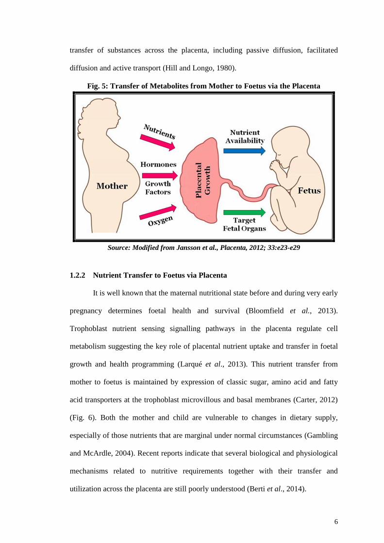
6
transfer of substances across the placenta, including passive diffusion, facilitated
diffusion and active transport (Hill and Longo, 1980).
Fig. 5: Transfer of Metabolites from Mother to Foetus via the Placenta
Source: Modified from Jansson et al., Placenta, 2012; 33:e23-e29
1.2.2 Nutrient Transfer to Foetus via Placenta
It is well known that the maternal nutritional state before and during very early
pregnancy determines foetal health and survival (Bloomfield et al., 2013).
Trophoblast nutrient sensing signalling pathways in the placenta regulate cell
metabolism suggesting the key role of placental nutrient uptake and transfer in foetal
growth and health programming (Larqué et al., 2013). This nutrient transfer from
mother to foetus is maintained by expression of classic sugar, amino acid and fatty
acid transporters at the trophoblast microvillous and basal membranes (Carter, 2012)
(Fig. 6). Both the mother and child are vulnerable to changes in dietary supply,
especially of those nutrients that are marginal under normal circumstances (Gambling
and McArdle, 2004). Recent reports indicate that several biological and physiological
mechanisms related to nutritive requirements together with their transfer and
utilization across the placenta are still poorly understood (Berti et al., 2014).

7
Fig. 6: Transfer of Glucose, Amino Acids and Fatty Acids across the Placenta
BM - basal membrane; EL - endothelial lipase; FA - fatty acid; FABP - fatty acid binding protein;
FABPpm - plasma membrane fatty acid binding protein; FAT/CD36 - fatty acid translocase; FATP -
fatty acid transport protein; FFA - free fatty acid; GLUT - glucose transporter; LAT - large neutral
amino acid transport; LPL - lipoprotein lipase; MVM - microvillous membrane; TG - triglycerides; X
- exchangers
Source: Modified from Brett et al., Int. J. Mol. Sci., 2014; 15, 16153-16185
During the first trimester, foetal development takes place in a low oxygen
environment and is supported by histiotrophic nutrition (in which local
macromolecules are chiefly responsible for the maintenance of the embryo) from the
endometrial glands (Burton et al., 2002). Proper trophoblast invasion results in the
opening of spiral and uterine glands towards the intervillous space enabling
hemotrophic nutrition (which results from a transfer of material between the maternal
and foetal circulations) in the second and third trimesters of pregnancy (Huppertz et
al., 2014). In addition to nutrition the foetus also depends on the mother for supply of
oxygen.

8
1.2.3 Oxygen Transfer to the Foetus via Placenta
During pregnancy both the placenta and foetus require high amounts of
oxygen (Mutinati et al., 2013). The placenta allows passage of oxygen from mother to
foetus by passive diffusion (Mayhew, 2014) (Fig. 7). The placenta lacks neuronal
innervation suggesting that local physical (e.g., oxygenation; flow rate), paracrine
(e.g., endothelial cell nitric oxide) and circulating (e.g., angiotensin II) factors
contribute to blood flow regulation in small foeto-placental vessels (Wareing et al.,
2014). The placenta not only transfers oxygen to the foetal circulation but also
consumes oxygen in order to support its own energy demands for the processes of
nutrient transport, protein synthesis and growth (Mayhew, 2014). The reactive oxygen
species (ROS) that are generated as a result of metabolism participate in cell survival,
proliferation and apoptosis (Bevilacqua et al., 2012).
Fig. 7: Passive Diffusion in the Placenta
Source: © Boardworks Ltd. 2007, http://www.boardworks.co.uk/
In the first trimester the extravillous cytotrophoblast (CTB) cells occlude the
uterine spiral arterioles creating a low oxygen environment early in pregnancy, which
is essential for the trophoblast invasion and the embryo development (Schneider,
2011). Hypoxia is a key signal for normal placental development in pregnancy and
has a crucial role in the control of trophoblast differentiation into invasive or
9
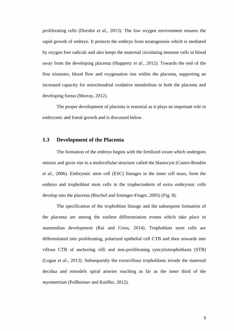
proliferating cells (Doridot et al., 2013). The low oxygen environment ensures the
rapid growth of embryo. It protects the embryo from teratogenesis which is mediated
by oxygen free radicals and also keeps the maternal circulating immune cells in blood
away from the developing placenta (Huppertz et al., 2012). Towards the end of the
first trimester, blood flow and oxygenation rise within the placenta, supporting an
increased capacity for mitochondrial oxidative metabolism in both the placenta and
developing foetus (Murray, 2012).
The proper development of placenta is essential as it plays an important role in
embryonic and foetal growth and is discussed below.
1.3 Development of the Placenta
The formation of the embryo begins with the fertilized ovum which undergoes
mitosis and gives rise to a multicellular structure called the blastocyst (Castro-Rendón
et al., 2006). Embryonic stem cell (ESC) lineages in the inner cell mass, form the
embryo and trophoblast stem cells in the trophectoderm of extra embryonic cells
develop into the placenta (Bischof and Irminger-Finger, 2005) (Fig. 8).
The specification of the trophoblast lineage and the subsequent formation of
the placenta are among the earliest differentiation events which take place in
mammalian development (Rai and Cross, 2014). Trophoblast stem cells are
differentiated into proliferating, polarized epithelial cell CTB and then onwards into
villous CTB of anchoring villi and non-proliferating syncytiotrophoblasts (STB)
(Logan et al., 2013). Subsequently the extravillous trophoblasts invade the maternal
decidua and remodels spiral arteries reaching as far as the inner third of the
myometrium (Pollheimer and Knöfler, 2012).

10
Fig. 8: Formation of Placenta and Foetal Membranes
Source: Pacific Research Centre for Early Human Development, Hawaii, USA.
http://www2.jabsom.hawaii.edu/PRCEHD/placenta_development.html
The trophoblast invasion leads to increased uterine and umbilical blood flow
required for vascularization of the placenta (Kliman et al., 2000). This process
provides stability to the placenta and also results in efficient utero-placental blood
flow (Sharp et al., 2010). After the trophoblast invasion and widening of the maternal
spiral arterioles in the first trimester the maternal blood replaces plasma flowing
through intervillous space of the placenta (Huppertz et al., 2012). After the blood flow
is established, placental villi are developed and matured by angiogenesis and
vascularization in the placenta (Huppertz and Peeters, 2005).
Reports suggest that any perturbations in the processes of placental
development lead to variations in the supply of nutrients and oxygen to the foetus and
program key systems (Kadyrov et al., 2013). These programming effects influence the
risk of diseases in later life (Barker and Thornburg, 2013).

11
1.3.1 Abnormal Placental Development
It is well established that many pregnancy related problems such as
preeclampsia and preterm labour appear to have their origins early in pregnancy. Most
pregnancy complications arise from abnormalities in implantation and placental
development (Norwitz, 2007). Shallow trophoblast invasion and defective vascular
remodelling of the uterine spiral arteries in the first trimester may result in impaired
placental perfusion and chronic placental ischemia and hypoxia later in gestation
leading to adverse pregnancy outcomes (Pringle et al., 2010). The premature
loosening of the trophoblast plugs and consequent premature and disorganized flow of
maternal blood into the intervillous space are known to be associated with recurrent
miscarriage (Hempstock et al. 2003). Preeclampsia and intra uterine growth
restriction (IUGR) are known to be associated with inadequate spiral artery
remodelling (Pijnenborg et al. 1991).
1.4 Preeclampsia
Preeclampsia is defined as a pregnancy disorder characterized by high blood
pressure (≥140/≥90 mm Hg) and an abnormal amount of protein in the urine (>1 + on
a dipstick test) in a previously normotensive pregnant women (ACOG, 2014). The
condition is associated with a reduced plasma volume, hemoconcentration and
increased vascular resistance (Haram et al., 2014). The clinical findings of
preeclampsia can manifest as a maternal syndrome with hypertension and proteinuria
with or without other multisystem abnormalities or foetal syndrome that includes
IUGR, reduced amniotic fluid and abnormal oxygenation (Sibai et al., 2005). If
preeclampsia remains untreated, it moves towards a more serious condition known
as eclampsia that includes systemic endothelial dysfunction, microangiopathy and

12
HELLP syndrome (haemolysis, elevated liver enzymes, and low platelet) (Al-Jameil
et al., 2014). Early onset preeclampsia (≤34 weeks) is known to be associated with
greater perinatal and maternal mortality and morbidity as compared to the late onset
disease (≥34 weeks) (Soto et al., 2012).
The underlying mechanisms leading to preeclampsia remain elusive (Sones et
al., 2014) and the primary treatment for the disorder is delivery of the placenta. In
view of this, there is a pressing need to better understand the mechanisms of the
disease, with the ultimate goal of preventing the disorder (Ilekis et al., 2007)
especially since the incidence of preeclampsia and rates of adverse outcomes are
increasing (Gilbert et al., 2014).
1.4.1 Risk Factors for Preeclampsia
The risk factors of preeclampsia include maternal smoking, pre-existing
medical conditions such as hypertension, diabetes mellitus, older maternal age and
obesity (Hutcheon et al., 2011). Other risk factors of preeclampsia include nulliparity,
first pregnancy after age of 35 years, thrombotic vascular disease, multiple gestations,
proteinuria and a prior history of preeclampsia (Al-Jameil et al., 2014). Further
ethnicity also is a risk factor as African-American women are reported to have a
higher risk of mortality from hypertensive disorders of pregnancy compared with
Hispanic, American Indian/Alaska Native, Asian/Pacific Islander, and Caucasian
women (Lo et al., 2013).
1.4.2 Epidemiology of Preeclampsia
Preeclampsia is a leading cause of maternal and perinatal morbidity/mortality
(Zuniga et al., 2014). It affects about 5-10% of all pregnancies, affecting a total of
13
about 8.5 million women worldwide (Telang et al., 2013; Soto et al., 2012). The
prevalence of preeclampsia in developing countries ranges from 1.8% to 16.7%
(Osungbade and Ige, 2011). WHO estimates the incidence of preeclampsia to be
seven times higher in developing countries (2.8% of live births) than in developed
countries (0.4%) (WHO, 2013). The HELLP syndrome occurs in 0.5% to 0.9% of all
pregnancies and in 10% to 20% of women with severe preeclampsia (WHO, 2013;
Haram et al., 2009).
In addition to mortality, maternal morbidity is associated with preeclampsia
(Kane et al., 2013). Women with preeclampsia are at an increased risk of later
cardiovascular disease and neurological manifestations such as blindness, persistent
neurological deficits secondary to stroke and later cognitive impairment (Brown et al.,
2013; Aukes et al., 2007). Further, babies born to women with preeclampsia are at a
twofold increased risk of neonatal mortality, or are born preterm and/or growth
restricted and are susceptible to long term neurological disability as well as
cardiovascular and metabolic diseases in later life (Davis et al., 2012; Basso et al.,
2006; Barker et al., 2002). The neurological complications of preeclampsia and
eclampsia are responsible for a major proportion of the morbidity and mortality both
for women and their infants (Kane et al., 2014).
1.4.3 Pathophysiology of Preeclampsia
The two stage model of preeclampsia proposes that a poorly perfused placenta
(stage 1) produces factor(s) leading to the clinical manifestations of preeclampsia
(stage 2) (Redman et al., 2014). In preeclampsia the maternal spiral arteries fail to be
invaded or remodelled by foetal cytotrophoblasts, resulting in constricted, high-
resistance vessels altering the blood flow to the placenta (Powe et al., 2011).

14
Inadequate spiral artery remodelling leading to poor placental development and
disturbed angiogenesis are currently regarded as the underlying pathophysiology of
preeclampsia (Rosser and Katz, 2013; Moffett-King, 2002) (Fig. 9).
Fig. 9: Trophoblast Invasion
Source: Moffett-King, Nature Reviews Immunology, 2002; 2, 656-663
1.4.3.1 Placental Ischemia/ Hypoxia
Placental ischemia is a condition in which blood flow to the tissues is
restricted while in hypoxia tissues are deprived of adequate oxygen supply. Placental
ischemia induces the release of growth factor inhibitors, anti-angiogenic factors,
inflammatory cytokines, ROS and hypoxia-inducible factors (HIF) which in turn
generate widespread dysfunction of the maternal vascular endothelium (Reslan and
Khalil, 2010). Chronic hypoxia during gestation may adversely affect the normal
adaptation of uterine vascular tone and increase the risk of preeclampsia (Zhu et al.,
2013). Hypoxia in the placenta is associated with vascular remodelling, hypertension,
metabolic changes, oxidative stress, mitochondrial dysfunction and endoplasmic
reticular stress (van Patot et al., 2012).
15
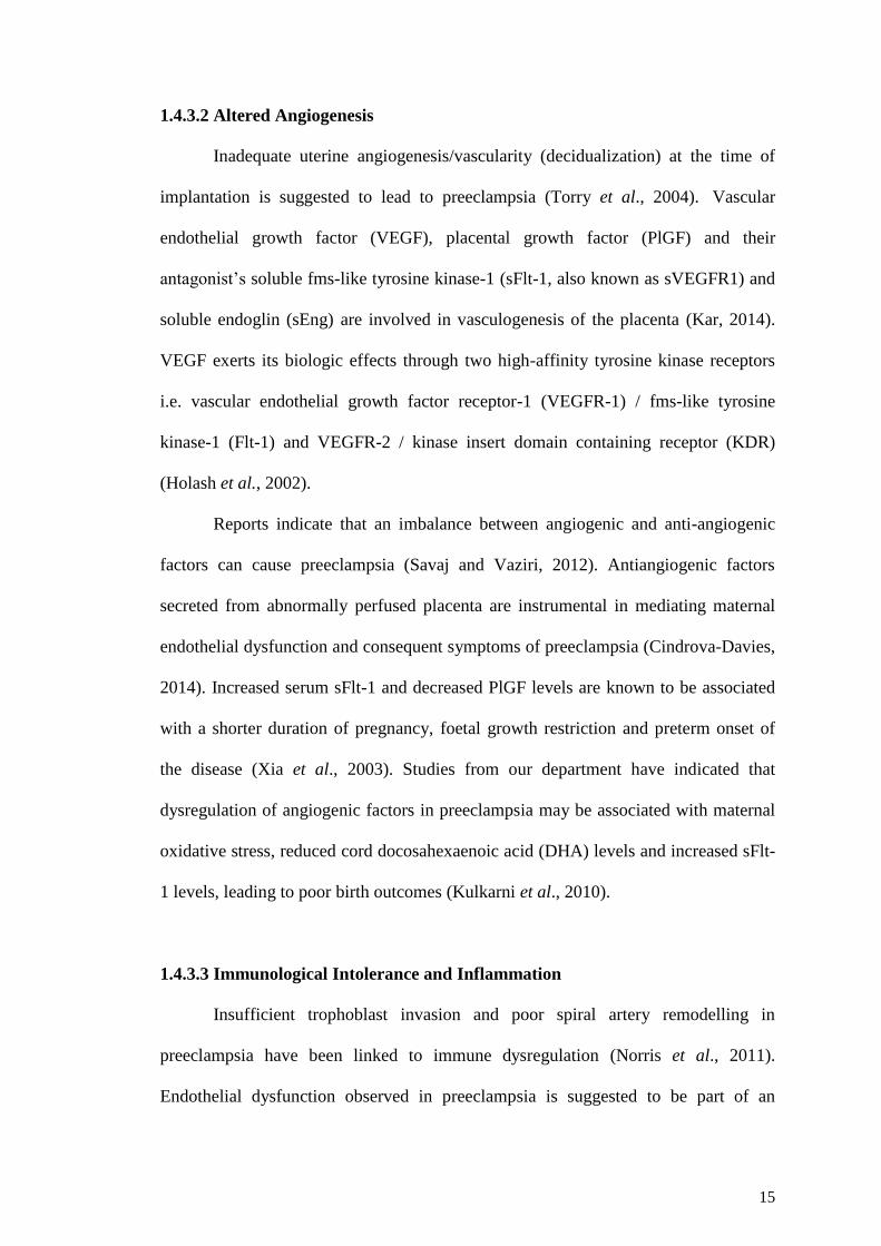
1.4.3.2 Altered Angiogenesis
Inadequate uterine angiogenesis/vascularity (decidualization) at the time of
implantation is suggested to lead to preeclampsia (Torry et al., 2004). Vascular
endothelial growth factor (VEGF), placental growth factor (PlGF) and their
antagonist’s soluble fms-like tyrosine kinase-1 (sFlt-1, also known as sVEGFR1) and
soluble endoglin (sEng) are involved in vasculogenesis of the placenta (Kar, 2014).
VEGF exerts its biologic effects through two high-affinity tyrosine kinase receptors
i.e. vascular endothelial growth factor receptor-1 (VEGFR-1) / fms-like tyrosine
kinase-1 (Flt-1) and VEGFR-2 / kinase insert domain containing receptor (KDR)
(Holash et al., 2002).
Reports indicate that an imbalance between angiogenic and anti-angiogenic
factors can cause preeclampsia (Savaj and Vaziri, 2012). Antiangiogenic factors
secreted from abnormally perfused placenta are instrumental in mediating maternal
endothelial dysfunction and consequent symptoms of preeclampsia (Cindrova-Davies,
2014). Increased serum sFlt-1 and decreased PlGF levels are known to be associated
with a shorter duration of pregnancy, foetal growth restriction and preterm onset of
the disease (Xia et al., 2003). Studies from our department have indicated that
dysregulation of angiogenic factors in preeclampsia may be associated with maternal
oxidative stress, reduced cord docosahexaenoic acid (DHA) levels and increased sFlt-
1 levels, leading to poor birth outcomes (Kulkarni et al., 2010).
1.4.3.3 Immunological Intolerance and Inflammation
Insufficient trophoblast invasion and poor spiral artery remodelling in
preeclampsia have been linked to immune dysregulation (Norris et al., 2011).
Endothelial dysfunction observed in preeclampsia is suggested to be part of an
16
exaggerated systemic inflammatory response that involves maternal leukocytes and
proinflammatory cytokines (Faas et al., 2014; Raghupathy, 2013). Various signalling
molecules responsible for the propagation of an inflammatory response have been
identified in placental cells which have to be explored in pregnancy complications
(Weiss et al., 2009).
1.4.3.4 Genetics
Although most cases of preeclampsia occur in women without a family
history, the presence of preeclampsia in a first-degree relative increases a woman’s
risk of severe preeclampsia two to fourfold (Carr et al., 2005). Two preeclampsia
susceptibility genes such as activin receptor type 2 gene (ACVR2A) and storkhead
box 1 (STOX1) gene have been identified within confirmed regions with significant
genome-wide linkage (van Dijk and Oudejans, 2011). Catechol-O-methyltransferase
(COMT) is an enzyme involved in catecholamine and estrogen degradation and is
suggested to have a role in development of preeclampsia (Roten et al., 2011). Allelic
variations in the fms-like tyrosine kinase 1 and VEGF C genes are associated with
preeclampsia in black women and white women (Srinivas et al., 2010).
1.4.4 Maternal Endothelial Dysfunction
Oxidative stress on the endothelium leading to endothelial dysfunction is
suggested to be the root cause of preeclampsia (Yelumalai et al., 2010). Reactive ROS
are implicated in the pathogenesis of vascular dysfunction in preeclampsia (Steinert et
al., 2006). It has been hypothesized that ROS or their metabolites ultimately
compromise the vasodilatory, anti-aggregatory and barrier functioning of the vascular
endothelium in preeclampsia (Hubel, 1998). Both the placenta and maternal
17
vasculatures are major sources of reactive oxygen and nitrogen species which can
produce powerful pro-oxidants that covalently modify proteins and alter vascular
function in preeclampsia (Lorquet et al., 2010).
During pregnancy, a critical balance exists between endothelium derived
relaxing and contracting factors that maintain vascular homeostasis (Gilbert et al.,
2008). When this delicate balance is disrupted, the vasculature is predisposed to
vasoconstriction, leukocyte adherence, mitogenesis, prooxidation and vascular
inflammation (LaMarca et al., 2008). A hypoxic placenta accelerates shedding of STB
basement membrane fragments and other factors like leukocyte and platelet
membrane particles, ROS, activated neutrophils, cytokines, growth factors,
angiogenic factors and hormones into the maternal circulation causing endothelial
dysfunction (Parikh and Karumanchi, 2008; Myatt, 2006).
Endothelial cell dysfunction is suggested to be the main cause of multiorgan
failure (Brozović et al., 2012) (Fig. 10). Generalized damage to the endothelium of
the maternal kidneys, liver and brain at the cellular level probably occurs following
the release of vasopressive factors from the diseased placenta (Redman et al., 1991).
Kidney function is particularly susceptible to the endothelial changes in preeclampsia
that manifest functionally as vascular constriction, decline in renal blood flow,
glomerular filtration rate and proteinuria (Moran et al., 2004). Cerebral edema and
intracerebral parenchymal haemorrhage are commonly seen in women who die from
eclampsia (Sibai, 2005). Liver and adrenals show infarction, necrosis and
intraparenchymal hemorrhage in women with preeclampsia (Young et al., 2010).
Further, endothelial dysfunction in preeclampsia has been attributed to placental
oxidative stress (Redman and Sargent, 2005).

18
Fig. 10: Pathophysiology of Preeclampsia
AT1-AA’s – angiotensin receptor agonistic autoantibodies; NK cells – natural killer
cells; O2- - superoxide anion; sENG – soluble endoglin; sFlt1 – soluble fms-like
tyrosine kinase-1
Source: Parikh and Karumanchi, Nat Med, 2008 14:810-2.
1.5 Oxidative Stress
Stress that arises when the production of ROS overwhelms the intrinsic anti-
oxidant defences is known as oxidative stress (Tadesse et al., 2014). Reactive species
and free radicals are unstable molecules that oxidize other molecules in order to
become stable (Gomes et al., 2012). It is known that although oxygen supports life it
acts as a double edged sword (Greabu et al., 2008). Reactivity allows oxygen to
participate in high-energy electron transfers during metabolism and hence supports
the generation of large amounts of adenosine-5-triphosphate (ATP) through oxidative

19
phosphorylation (Burton and Jauniaux, 2011). Oxidative stress and oxidants play a
critical role in defence against infection, tissue repair and signalling (Wahlqvist,
2013). However, oxygen can also react rapidly with most other radicals, forming ROS
that cause selective oxidation of lipid, protein or DNA molecules (Loh et al., 2006;
Lewén et al., 2000; Halliwell and Cross, 1994).
A continuing balance between oxidation and antioxidation is necessary for
good health (Wahlqvist, 2013). Free radicals produced during metabolism are
balanced by the body’s endogenous antioxidant systems and by the ingestion of
exogenous antioxidants (Rahman, 2007). The enzymatic detoxification mechanisms
involve antioxidant enzymes [superoxide dismutase (SOD), catalase (CAT), and
glutathione peroxidase (GPx)], small molecular-weight antioxidants [Vitamin E,
Vitamin C, glutathione (GSH), ubiquinone, beta-carotene] and adaptive mechanisms
leading to antioxidant gene expression (Kalyanaraman, 2013). In the absence of
antioxidants there is an accumulation of prooxidants which leads to oxidative stress
(Poljsak, 2011).
1.5.1 Oxidative Stress in Pregnancy
Oxidative stress is required for the normal progression of embryonic,
placental, foetal growth and development (Lappas et al., 2011). However, increased
oxidative stress is implicated in the pathophysiology of many adverse pregnancy
outcomes such as miscarriage, diabetes related congenital malformations, spontaneous
abortions, preterm birth, preeclampsia, foetal growth restriction and low birth weight
(Burton and Jauniaux, 2011; Poston et al., 2011; Al-Gubory et al., 2010). Oxidative
stress induced placental dysfunction (Dennery, 2010), suppression of placental
angiogenesis, endothelial damage, altered vascular function (Lorquet et al., 2010;

20
Burton et al., 2009), immune malfunction (Hannan et al., 2011), myometrium damage
(Khan et al., 2010) are suggested to underlie pregnancy complications.
Increased oxidative stress is suggested to influence growth factors such as
neurotrophins.
1.6 Neurotrophins
Neurotrophins are a family of growth factors that are required for the
proliferation, differentiation, survival and death of neuronal cells in the nervous
system (Li and Zhou, 2013). The four neurotrophins – nerve growth factor (NGF),
brain derived neurotrophic factor (BDNF), neurotrophin -3 (NT-3) and neurotrophin-4
(NT - 4) each bind and activate one or more of the tropomyosin-related receptor
kinase (Trk) family of receptor tyrosine kinases (Trk A, Trk B and Trk C) (Reichardt,
2006; Chao, 2003) (Fig. 11). In addition, all neurotrophins activate the p75
neurotrophin receptor, a member of the tumour necrosis factor receptor family
(Skaper, 2012). Through these receptors, neurotrophins activate many intracellular
signalling pathways, thereby affecting both the development and function of the
nervous system (Patapoutian and Reichardt, 2001). Expression of neuropeptides,
small protein-like molecules (peptides) used by neurons to communicate with each
other, depends on neurotrophins (Ernsberger, 2009; Petit et al., 2002; Nawa et al.,
1993).
Apart from their role in the nervous system, studies have shown that
neurotrophins promote angiogenesis, control the survival of adult endothelial cells,
protect against oxidative stress and may have an important role in early vascular
development in mother, placenta and foetus (Fujita et al., 2011; Caporali and
Emanueli, 2009; Wu et al., 2004).

21
Fig. 11: Neurotrophins and their Receptors
BDNF - brain derived neurotrophic factor; NGF - nerve growth factor;
NT3 - neurotrophin 3; NT4 - neurotrophin 4; Trk - tropomyosin-related
receptor kinase
Source: Chao, 2003; Nat Rev Neurosci. 4:299-309.
Neurotrophins are naturally occurring molecules that regulate development of
the placenta and brain. They play a crucial role in neurodevelopment (Shoval and
Weizman, 2005). Further, neurotrophins can influence other growth factors and are
involved in the vascular development and angiogenesis (Lazarovici et al., 2006).
Growth factors such as neurotrophins have been demonstrated to act in a synergistic
way in angiogenesis and neurogenesis contributing to self-healing powers of the adult
human brain (Sopova et al., 2014). Variations in neurotrophins are suggested to have
a role in the neurodevelopmental alterations and molecular mechanisms of cognitive
dysfunction (Nieto et al., 2013). Differential patterns of cord blood neurotrophins
have been observed in preterm pregnancies (Matoba et al., 2009)
The capacity of various neurotrophic factors to affect neurons is reported to be
regulated during development (Knüsel et al., 1994). Studies suggest that
neurotrophins form a link between maternal nutrient supply and foetal demand and

22
alterations in diet may lead to alterations in these growth factor levels through
epigenesis (Dhobale and Joshi, 2012; Mayeur et al., 2011). The BDNF gene is
reported to be under extensive epigenetic regulation (Schanker, 2012). Accumulating
evidence suggests that epigenetic modifications of neurotrophins are associated with
the pathophysiology of psychiatric disorders, such as schizophrenia and mood
disorders (Ikegame et al., 2013). Reports indicate that oxidative stress and omega-3
fatty acids such as DHA affect the levels of neurotrophins (Bhatia et al., 2011;
Kapczinski et al., 2008, Wu et al., 2008).
1.7 Maternal Nutrition, Oxidative Stress and Neurotrophins
Pregnancy is a state of increased requirement of macro- and micronutrients
and malnourishment before and during pregnancy can lead to adverse perinatal
outcomes (Imdad et al., 2011). Studies have adequately demonstrated the importance
of maternal nutrition, particularly, micronutrients (folic acid, vitamin B12) and long
chain polyunsaturated fatty acids (LCPUFA) in determining pregnancy outcome
(Khot et al., 2014). Various studies in our department have extensively discussed the
link between folic acid, vitamin B12 and LCPUFA in the one carbon cycle (Dhobale
and Joshi, 2012; Sundrani et al., 2011; Kulkarni et al., 2011)
1.7.1 Folate (Vitamin B9)
Folates are a group of water-soluble B vitamins naturally existing in food such
as legumes, green leafy vegetables and fruits (Mantovani et al., 2014). Folate in foods
is naturally present in the form of reduced folate polyglutamate conjugates, while
folic acid refers to the fully oxidized and most stable form of folate (Lucock, 2000).
Folic acid is comprised of pteridine ring, p-aminobenzoic acid and glutamic acid
23
(Tamura and Picciano, 2006). Folic acid is required for DNA replication and as a
substrate for a range of enzymatic reactions involved in amino acid synthesis and
vitamin metabolism (Greenberg et al., 2011). Folate is critical for DNA, RNA and
protein methylation as well as DNA synthesis and maintenance (Crider et al., 2012).
Folate metabolism is known to affect ovarian function, implantation, embryogenesis
and the entire process of pregnancy (Thaler, 2014). Folate deficiency is associated
with hyperhomocysteinemia, megaloblastic anemia, embryonic and neural tube
defects (NTD) (Guilland and Aimone-Gastin, 2013).
1.7.2 Vitamin B12
Vitamin B12 is a crystallisable cobalt-complex, which belongs to a group of
unique corrinoids, named cobalamins (Kräutler, 2012). The dietary sources of vitamin
B12 are animal-source based foods, including meat, milk, eggs, fish, and shellfish
(Watanabe, 2013). Vitamin B12 is a cofactor of methionine synthase in the synthesis
of methionine, the precursor of the universal methyl donor S-Adenosylmethionine
(SAM), (Gröber, 2013). It is required for the optimal functioning of the central and
peripheral nervous system (Kumar, 2014). Low levels of vitamin B12 have been
associated with neurocognitive disorders (Health Quality Ontario, 2013). Low
maternal vitamin B12 status is known to be associated with increased risk of NTD, low
lean mass, excess adiposity, increased insulin resistance and impaired
neurodevelopment in the offspring (Rush et al., 2014).
1.7.3 Long Chain Polyunsaturated Fatty Acids
A fatty acid is a carboxylic acid with a long aliphatic tail (chain), which is
either saturated or unsaturated. LCPUFA are the major components of brain and

24
retina and are the essential fatty acids with important physiologically active functions
(Lee, 2013). There are two series of LCPUFA i.e. omega-3 and the omega-6 series. A
complex series of desaturation and elongation reactions acting in concert transform
the precursors linoleic acid (LA) of the omega-6 series and alpha linolenic acid
(ALA) of the omega-3 series to their higher unsaturated derivatives: arachidonic acid
(AA) and DHA respectively (Russo, 2009).
The demand for LCPUFA is increased during pregnancy because of the
increasing needs of the foetus, mother and placenta (Makridis et al., 2011). Since their
synthesis in the foetus and placenta is low, both the maternal LCPUFA status and
placental function are critical for their supply to the foetus (Larqué et al., 2012). The
LCPUFA, DHA is incorporated in large amounts in foetal brain and other tissues
during the second half of pregnancy (Koletzko et al., 2007). Studies have claimed a
beneficial effect of DHA supplementation on visual, neural or developmental
outcomes (Campoy et al., 2012)
DHA, a major component of membrane phospholipids, plays an important role
as an antioxidant agent (Suganuma et al., 2010). Recent reports indicate that omega-3
fatty acid supplementation reduced lipid peroxidation, nucleic acid and protein
oxidation and promoted neuronal and glial cell survival in brain tissue of traumatic
brain injury models (Kumar et al., 2014). Maternal dietary omega-3 fatty acid
supplementation during pregnancy in rats is known to reduce placental oxidative
damage and increase placental levels of pro-resolving mediators, which are associated
with enhanced foetal and placental growth (Jones et al., 2014).
The ability of DHA to alter placental prooxidant/antioxidant balance is
dependent on the DHA concentration used and the gestational age of the placental
tissue (Stark et al., 2013).
25
1.7.4 Antioxidants
Supplementation with antioxidant micronutrients during pregnancy can be an
early and innovative alternative to strengthen the prevention of chronic diseases in the
population (Ramírez-Vélez et al., 2011). Antioxidants including vitamin E, vitamin C,
vitamin A, vitamin B6, beta-carotene, zinc and selenium are known to have a
beneficial role in reducing oxidative stress (Hsu and Guo, 2002) however their effect
on oxidative stress markers has been inconsistent. Excessive vitamin A intake during
gestation and lactation might be toxic for mothers with adverse effects for the
developing offspring (Schnorr et al., 2011). Early observations suggest that vitamins
C and E supplementation can prevent or ameliorate preeclampsia, but most large
randomized clinical trials have failed to show any benefit (Talaulikar and Manyonda,
2011).
1.7.5 One Carbon Metabolism
B-vitamins like folate, vitamin B12 and vitamin B6 are important components
of the one carbon metabolism. Vitamin B12 and folic acid mediate the remethylation
of homocysteine, which affects the production of the universal methyl donor, SAM
(Herrmann and Obeid, 2007). S-adenosylmethionine (SAM) maintains methyl group
supply for various macromolecules like DNA, neurotransmitters, proteins and
membrane phospholipids (Umhau et al., 2006). Inadequate enzyme activities and
imbalances of substrates and cofactors in one carbon metabolism may cause
homocysteine and S-adenosylhomocysteine (SAH) accumulation (Muskiet, 2005).
Hyperhomocysteinemia has been associated with oxidative stress (Kolling et
al., 2011; Forges et al., 2007) and is proposed to play a role in the pathogenesis of
preeclampsia, IUGR, preterm birth, spontaneous abortions and intrauterine foetal
26
death (Micle et al., 2012). The thiol group of homocysteine can auto-oxidize in
circulation at physiological pH in the presence of oxygen leading to the production of
hydrogen peroxide (H2O2), thereby generating oxidative stress (Jacobson, 2000).
Further, the synthesis of the cellular antioxidant, GSH is linked to the one
carbon metabolic pathway. Homocysteine can indirectly result in oxidative stress by
decreasing the transcription, translation and catalytic activity of GPx (Lubos et al.,
2007). Any disruption in the one carbon cycle or chronic methyl group deficiency can
also result in cell death as a result of an imbalance in the cellular antioxidant defence
systems and increased oxidative stress (Bagnyukova et al., 2008).
Although a lot of importance has been given to the methyl donors, the methyl
acceptors also play an important role in the one carbon metabolism. Membrane
phospholipids are major methyl group acceptors and earlier reports from our
department have elaborately discussed that reduced DHA levels may result in
diversion of methyl groups towards DNA ultimately resulting in altered DNA and
histone methylation which may lead to altered gene expression patterns (Dhobale and
Joshi, 2012; Sundrani et al., 2011). Such alterations in gene expression during critical
periods of development may result in complications during pregnancy and lead to
adverse birth outcomes.
1.8 Genesis of the Thesis
As discussed in the above section changes in maternal micronutrients such as
folate and vitamin B12 could lead to increased homocysteine, thereby increasing the
oxidative stress and altering DNA methylation patterns. This may further lead to
increased lipid peroxidation, altered expression of antioxidant enzymes and
neurotrophins ultimately leading to endothelial dysfunction associated with

27
preeclampsia. Studies from our laboratory have shown changes in global DNA
methylation in response to altered one carbon metabolism in animals (Kulkarni et al.,
2011). Several other animal studies have also suggested that disturbances of the folate
and methionine-homocysteine cycles have an impact upon the epigenetic regulation of
gene expression and hence may program long-term physiology and metabolism of the
offspring (Engeham et al., 2010; Kim et al., 2009). Moreover, it has been proposed
that maternal factors such as endothelial function and oxidative stress are key
mechanisms of both foetal metabolic alterations and subsequent development of non-
transmissible chronic diseases (Ramírez-Vélez et al., 2011; Donkena et al., 2010).
Increased oxidative stress in pathologic pregnancies such as preeclampsia has
important implications for placental function and foetal well-being (Lappas et al.,
2011). This is of significance as adverse influences during foetal life are known to
alter the structure and function of various cells, organ systems or homoeostatic
pathways, resulting in increased risk for diseases in adult life (Jansson and Powell,
2007). Studies indicate that children born to mothers with preeclampsia are at
increased risk for neurodevelopmental and metabolic disorders in later life (Herrera-
Garcia and Contag, 2014; Silveira et al., 2007). Thus, there may be a link between
maternal nutritional status, oxidative stress, foetal metabolic alterations and epigenetic
programming of adult diseases in children born to mothers with preeclampsia.
A series of cross sectional studies carried out by our department earlier have
demonstrated altered levels of folate and vitamin B12, reduced DHA levels, increased
homocysteine and oxidative stress in pregnancy complications such as preeclampsia
(Kulkarni et al., 2011; Mehendale et al., 2008). The department has further shown that
DHA is negatively associated with homocysteine concentrations in preeclampsia
(Kulkarni et al., 2011). Studies from our department also indicate increased oxidative
28
stress and lower levels of neurotrophins at the time of delivery in women with
preeclampsia (Kilari et al., 2011; Mehendale et al., 2008). The increased oxidative
stress has also been shown to be associated with altered angiogenesis leading to poor
birth outcomes in preeclampsia (Kulkarni et al., 2010). It is however likely that these
changes observed at delivery may be a result of secondary effects where the
etiopathology has progressed for several weeks and months.
Further, it is known that the degree of vulnerability to changes in epigenetic
patterns is high during early embryonic development (Gomes and Pelosi, 2013). The
timing of environmental insults generating oxidative stress at critical periods of
neurodevelopment may also play a decisive role in inducing neurodevelopmental
disorders in the offspring (Do et al., 2009). A recent review indicates a need for
prospective cohort studies to collect data on relevant clinical, environmental and
lifestyle risk factors and longitudinal measurement of several factors such as
angiogenic factors, neurotrophins and genetic variations in candidate gene pathways
to establish interactions which predispose to pregnancy complications (Andraweera
et al., 2012). Therefore, in order to better understand the role of oxidative stress and
neurotrophins in preeclampsia it is important to prospectively examine their levels
across gestation.
1.9 Hypothesis
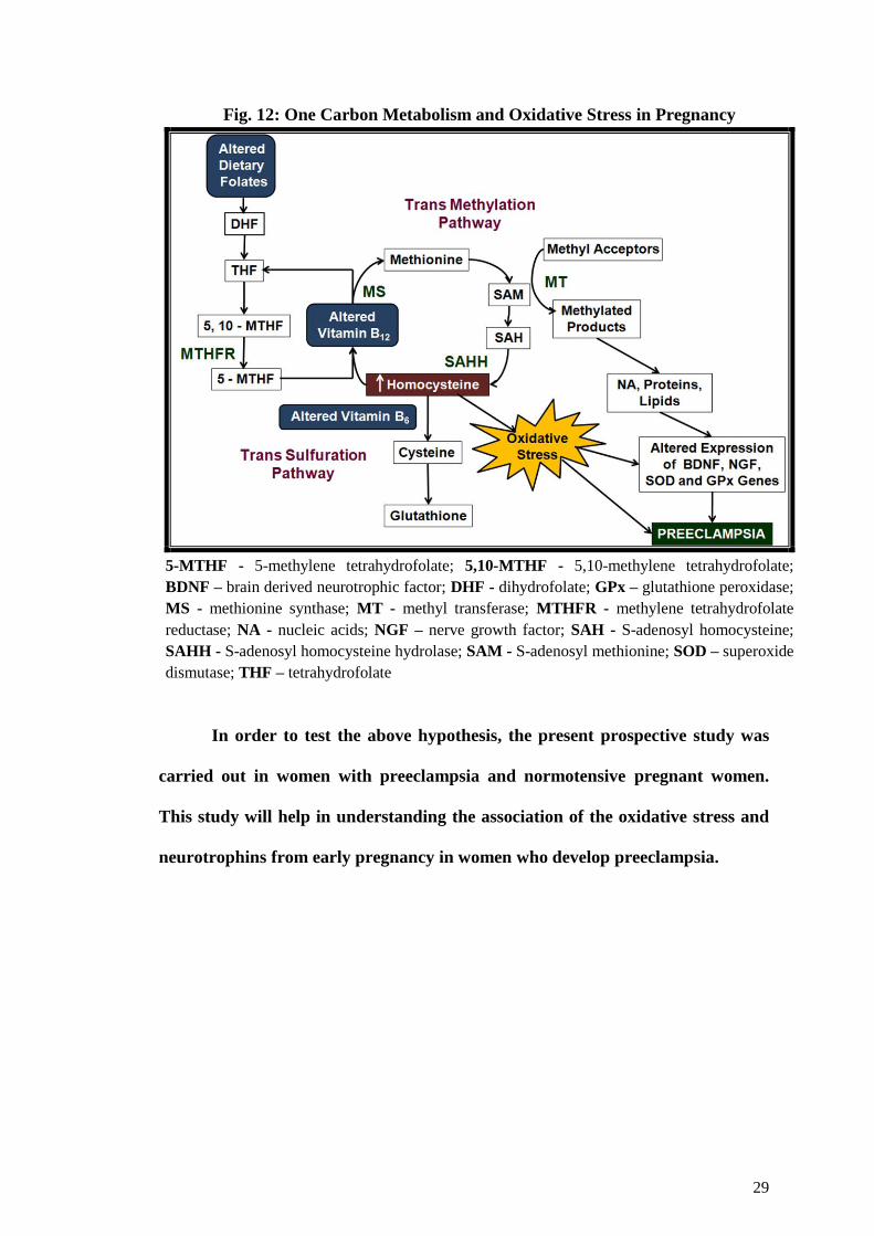
With the above background we hypothesize that, “Oxidative stress, through
the dysregulation of the one carbon cycle leads to altered levels and expression of
antioxidant enzymes and neurotrophins in preeclampsia” (Fig. 12)
29
Fig. 12: One Carbon Metabolism and Oxidative Stress in Pregnancy
5-MTHF - 5-methylene tetrahydrofolate; 5,10-MTHF - 5,10-methylene tetrahydrofolate;
BDNF – brain derived neurotrophic factor; DHF - dihydrofolate; GPx – glutathione peroxidase;
MS - methionine synthase; MT - methyl transferase; MTHFR - methylene tetrahydrofolate
reductase; NA - nucleic acids; NGF – nerve growth factor; SAH - S-adenosyl homocysteine;
SAHH - S-adenosyl homocysteine hydrolase; SAM - S-adenosyl methionine; SOD – superoxide
dismutase; THF – tetrahydrofolate
In order to test the above hypothesis, the present prospective study was
carried out in women with preeclampsia and normotensive pregnant women.
This study will help in understanding the association of the oxidative stress and
neurotrophins from early pregnancy in women who develop preeclampsia.
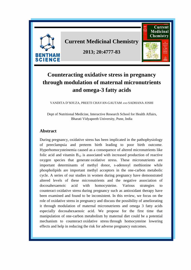
{
Counteracting oxidative stress in pregnancy
through modulation of maternal micronutrients
and omega-3 fatty acids
VANDITA D’SOUZA, PREETI CHAVAN-GAUTAM AND SADHANA JOSHI
Dept of Nutritional Medicine, Interactive Research School for Health Affairs,
Bharati Vidyapeeth University, Pune, India
Abstract
During pregnancy, oxidative stress has been implicated in the pathophysiology
of preeclampsia and preterm birth leading to poor birth outcome.
Hyperhomocysteinemia caused as a consequence of altered micronutrients like
folic acid and vitamin B12 is associated with increased production of reactive
oxygen species that generate oxidative stress. These micronutrients are
important determinants of methyl donor, s-adenosyl methionine while
phospholipids are important methyl acceptors in the one-carbon metabolic
cycle. A series of our studies in women during pregnancy have demonstrated
altered levels of these micronutrients and the negative association of
docosahexaenoic acid with homocysteine. Various strategies to
counteract oxidative stress during pregnancy such as antioxidant therapy have
been examined and found to be inconsistent. In this review, we focus on the
role of oxidative stress in pregnancy and discuss the possibility of ameliorating
it through modulation of maternal micronutrients and omega 3 fatty acids
especially docosahexaenoic acid. We propose for the first time that
manipulation of one-carbon metabolism by maternal diet could be a potential
mechanism to counteract oxidative stress through homocysteine lowering
effects and help in reducing the risk for adverse pregnancy outcomes.
Current Medicinal Chemistry
2013; 20:4777-83

30
2.1 Introduction
Neurotrophins are growth factors which regulate the development and
function of the nervous system. Apart from their role in the nervous system
neurotrophins are involved in initiation of vascular development, maintaining
vessel stability and cardiovascular stability. Preeclampsia is a complex pathology
that involves placental endothelial dysfunction. The brain derived neurotrophic
factor (BDNF) is a neurotrophin implicated in pathologies related to placental
and foetal growth disturbances and hence changes in its levels may contribute to
the pathophysiology of preeclampsia. This chapter therefore examines the levels
of BDNF at delivery in the mother and cord blood to better understand its role in
preeclampsia.
2.1.1 Neurotrophins
The members of the neurotrophin family include nerve growth factor (NGF),
BDNF, neurotrophin-3 (NT-3) and neurotrophin-4 (NT-4) (Fargali et al., 2012). Each
neurotrophin consists of a noncovalently-1 linked homodimer and contains (1) a
signal peptide following the initiation codon; and (2) a pro-region containing an N-
linked glycosylation site (Binder et al., 2004). Neurotrophins are translated from
single coding exons and are synthesized as larger precursors (proneurotrophins) of
~30–34 kDa which are then proteolytically processed to generate small mature
proteins (Teng et al., 2010). They exert their action by the activation of two distinct
classes of transmembrane receptors; p75NTR and tropomyosin-related receptor kinase
(Trk) family. Pro-forms preferentially activate p75NTR receptor that mediates
apoptosis and the mature forms activate Trk family of receptors promoting survival of
neuronal cells (Carvalho et al., 2008; Lee et al., 2001) (Fig. 13). Neurotrophins are

31
important regulators of neural survival, development, function and plasticity (Huang
and Reichardt, 2001).
Fig. 13: Pro-Neurotrophins, Neurotrophins and their Receptors
BDNF - brain derived neurotrophic factor; Ig – immunoglobulin; NGF - nerve
growth factor; NT3 -neurotrophin 3; NT4 - neurotrophin 4, p75 -NTR – p75
neurotrophin receptor; Trk - tropomyosin-receptor kinase
Source: Modified from Arévalo and Wu, 2006; Cell Mol Life Sci. 63:1523-37.
2.1.2 Neurotrophins in Pregnancy
Neurotrophins play important roles in influencing brain function from early
age to adulthood (Molteni et al., 2001). By exerting neuroprotection, neurotrophins
are critical for pre- and postnatal brain development (Malamitzi-Puchner et al., 2006).
Apart from the nervous system, neurotrophins play numerous roles in angiogenesis,
energy homoeostasis and regulation of other growth factors in the materno-feto-
placental unit (Mayeur et al., 2011). Animal and cell culture studies have also

32
demonstrated that BDNF/Trk B signalling system plays a critical role in implantation,
placental development and foetal growth (Kawamura et al., 2011). Reports indicate
that the cytotrophoblast’s differentiation, proliferation and survival of the placenta are
regulated by neurotrophins (Mayeur et al., 2010; Kawamura et al., 2009; Kermani and
Hempstead, 2007; Donovan et al., 2000), suggesting a role for BDNF in angiogenesis
and placental development. Neurotrophins are important in the survival, maintenance
and differentiation of neuronal tissue and functions in follicle maturation, tumour
growth, angiogenesis and immunomodulation; however its role in the placenta
remains unexplored (Fujita et al., 2011).
Studies from our department indicate dysregulation of neurotrophins like
BDNF and NGF in normotensive preterm pregnancies (Dhobale et al., 2012a,b).
Higher maternal BDNF and placental Trk B in women delivering preterm as
compared to control indicated that BDNF protein may not be utilized for placental
growth and development which leads to the increased levels in preterm deliveries. In
another study from the department the NGF levels in women with preeclampsia were
lower as compared to NC women indicating altered angiogenesis in these women
(Kilari et al., 2011). Further, it is also reported that a reduction in cord BDNF and
NGF levels may have implications for altered neurodevelopment in childhood and
later life in children born preterm (Dhobale et al., 2012a, b). Abnormal neurotrophic
factor gene activity is suggested to be a leading mechanism by which early life
adverse experiences may persistently modify brain and behavioural plasticity (Roth
and Sweatt, 2011).
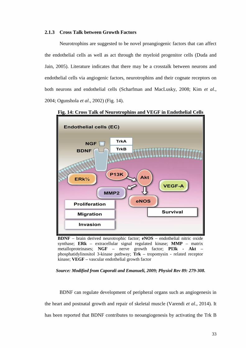
33
2.1.3 Cross Talk between Growth Factors
Neurotrophins are suggested to be novel proangiogenic factors that can affect
the endothelial cells as well as act through the myeloid progenitor cells (Duda and
Jain, 2005). Literature indicates that there may be a crosstalk between neurons and
endothelial cells via angiogenic factors, neurotrophins and their cognate receptors on
both neurons and endothelial cells (Scharfman and MacLusky, 2008; Kim et al.,
2004; Ogunshola et al., 2002) (Fig. 14).
Fig. 14: Cross Talk of Neurotrophins and VEGF in Endothelial Cells
BDNF – brain derived neurotrophic factor; eNOS – endothelial nitric oxide
synthase; ERk – extracellular signal regulated kinase; MMP – matrix
metalloproteinases; NGF – nerve growth factor; PI3k - Akt –
phosphatidylinositol 3-kinase pathway; Trk – tropomysin - related receptor
kinase; VEGF – vascular endothelial growth factor
Source: Modified from Caporali and Emanueli, 2009; Physiol Rev 89: 279-308.
BDNF can regulate development of peripheral organs such as angiogenesis in
the heart and postnatal growth and repair of skeletal muscle (Varendi et al., 2014). It
has been reported that BDNF contributes to neoangiogenesis by activating the Trk B

34
receptor expression in endothelial cells and by additionally recruiting bone marrow-
derived cells (Kermani and Hempstead, 2007). BDNF is suggested to induce VEGF
expression via HIF-1 alpha in neuroblastoma cells (Nakamura et al., 2006).
BDNF has also been shown to play a role in the etiology of some cardiovascular
diseases: induction of angiogenesis in ischemic tissues (Lorgis et al., 2009). Recent
reports suggest that the two complementary processes, proangiogenic stimulation of
new vessel growth and anti-angiogenic inhibition of vessel overgrowth, should be
balanced to restore the vascular network in the placenta in any pregnancy
complication (Chen and Zheng, 2014).
2.1.4 Factors Influencing Neurotrophins
There are various factors which influence neurotrophin levels and are
discussed below
2.1.4.1 Nutrition
Undernutrition and overnutrition are implicated in many disorders of mental
health and neurology and these actions are mediated by changes in energy metabolism
and multiple signalling molecules such as neurotrophins (Dauncey, 2012). Nutrients
like vitamins, minerals, polyunsaturated fatty acids (PUFA) and trace elements are
needed for the normal metabolism of neurotrophic factors. However, the exact
relationship among these factors and their interaction with genes and proteins
concerned with brain development and growth is not well understood (Das, 2013).
High saturated fat and high sugar has been shown to reduce BDNF levels in
schizophrenia (Peet, 2004). Animal studies in our laboratory have shown that
maternal diets imbalanced in micronutrients like folic acid and vitamin B12 reduced

35
brain DHA and BDNF levels (Sable et al., 2013). Reports from our department
further indicate that a combination of omega 3 fatty acids and vitamin B12 enriched
diet influence neurotrophin levels and improved cognition (Rathod et al., 2014).
Reports indicate that DHA and AA are essential nervous system components
that increase in concentration throughout gestation and may influence neurotrophin
mediated foetal brain development (Benn et al., 2014). LCPUFA upregulate the
expression of neurotrophins and their target receptors (Balogun and Cheema, 2014)
and have an influence on brain structure and function, including cognition (Bourre,
2006). Neuroprotectin D1 (NPD1), a bioactive anti-inflammatory and anti-apoptotic
molecule formed from free DHA synthesis, is upregulated by neurotrophins and
downregulated by oxidative stress (Bazan et al., 2010; Das, 2008).
Omega-3 fatty acids regulate neurotrophins that are important for the proper
functioning of the central nervous system (CNS) (Wu et al., 2004) and are required
for normal neurological development (Pandya and Yakel, 2013). Neurotrophins are
very susceptible to the effects of dietary manipulations and diets rich in omega-3 fatty
acids are known to upregulate BDNF while high energy diets do the opposite (Tyagi
et al., 2013). Further, neurotrophins play a role in neuronal metabolism and synaptic
plasticity and can be epigenetically regulated by diets rich in omega-3 fatty acids
(Gomez-Pinilla and Tyagi, 2014).
2.1.4.2 Lifestyle factors
Stress-induced remodelling of the hippocampus, prefrontal cortex and
amygdala in the brain is reported to be associated with changes in the levels of
neurotrophins (Gray et al., 2013). Further stress-induced alterations in growth factors
such as neurotrophins have been identified in modulating stress associated pathologies

36
(Bath et al., 2013; Nowacka and Obuchowicz, 2013). Studies suggest that
neurotrophins plays a role in the development of stress vulnerability and resilience in
the pathophysiology of major depression (Dwivedi, 2013; Masi and Brovedani, 2011).
Alcohol consumption during pregnancy is known to result in impairments of
the CNS that result in cognitive deficits (Boehme et al., 2011). Inherited variation in
the BDNF gene due to alcohol dependence may promote early developmental
differences in neuronal proliferation in the brain (Hill et al., 2011). Dietary factors
influence the basic mechanisms underlying ethanol-induced functional alterations and
the related neuropathology in the brain (Jaatinen and Rintala, 2008).
BDNF signalling is well known to be involved in adaptation to stress and
stress-related disorders (Pardon, 2010). Exposure to drugs of abuse such as cocaine is
known to modulate epigenetic regulation of BDNF gene expression (McCarthy et al.,
2012).
Prenatal exposure to maternal cigarette smoking is reported to affect brain
development and behaviour in the adolescent offspring (Lotfipour et al., 2009). This
may be due to significant reduction in BDNF mRNA and BDNF protein levels in the
brain of offspring due to epigenetic changes induced by cigarette smoking (Yochum
et al., 2014).
Exercise is a beneficial lifestyle factor with public health implications. It has a
positive influence on mood, cognition and memory retrieval as it is known to increase
neurotrophin levels (Meeusen, 2014; Vega et al., 2011). BDNF levels in humans are
significantly elevated in response to exercise and the magnitude of increase is exercise
intensity dependent (Ferris et al., 2007). Exercise during pregnancy is suggested to
have long-lasting effects on the offspring health (Rosa et al., 2013).

37
2.1.5 Brain Derived Neurotrophic Factor (BDNF)
BDNF, a secreted glycoprotein, shares about 50% amino acid identity with
other neurotrophins. It is a noncovalently-1 linked homodimer having a signal peptide
following the initiation codon and a pro-region containing an N-linked glycosylation
site (Binder and Scharfman, 2004). It was first purified from the pig brain in 1982
(Barde et al., 1982). BDNF also shares a distinctive three dimensional structure
containing two pairs of antiparallel β-strands and cysteine residues in a cystine knot
motif with other growth factors (Binder and Scharfman, 2004).
BDNF is found in different areas of the CNS such as cortex, hypothalamus,
hippocampus and amygdala (Andero and Ressler, 2012). It is also found in platelets,
which bind, store and release BDNF upon activation (Fujimura et al., 2002).
It is well established that the synaptic effects of BDNF are attributed to Trk B
activation (Lu and Nagappan, 2014) (Fig. 15). Activation of Trk B by BDNF mediates
the survival and differentiation of neurons during development and is also required
throughout adulthood to sustain the growth and function of neuronal synapses
(Tessarollo, 1998). BDNF activation of Trk B is the key for the enhancement of
activity-stimulated excitation of neurons that is needed for memory development and
maintenance (Thiele et al., 2009).

38
Fig. 15: BDNF and its Trk B Receptor
BDNF – brain derived neurotrophic factor; Trk B – tropomyosin-
related receptor kinase B
Source: Modified from Deinhardt and Jeanneteau, 2012 Protein
Phosphorylation in Human Health 7: 217-232
2.1.6 BDNF Signalling Pathways
BDNF-induced dimerization of Trk B receptors leads to activation of the
tyrosine kinase domain that initiates three major cascades of signalling pathways
(Longo and Massa, 2013; Reichardt, 2006) (Fig. 16). They are as follows:
1) Mitogen-activated protein kinases (MAPKs),
2) Phosphoinositide 3-kinase (PI3K)
3) Phospholipase Cγ1 (PLCγ1)
Each of these signalling pathways also regulates several elements of gene
transcription such as cAMP response-element binding protein (CREB) (Kim et al.,
2013). CREB is a key component of diverse physiological processes, including
nervous system development, cell survival, plasticity, as well as learning and memory
(Lonze and Ginty, 2002). Several neurodegenerative disorders are a result of

39
dysregulation of the CREB transcriptional cascade possibly due to its effect on cell
viability and cognitive function (Sakamoto et al., 2011).
Fig. 16: BDNF- Trk B Signalling Pathways
Akt - protein kinase B; BDNF - brain derived neurotrophic factor; CAMK - calmodulin-
dependent protein kinase; DAG - diacylglycerol; ERK - extracellular signal regulated
kinase; GAB - Grb2-associated binding protein 1; IP3 - inositol trisphosphate; MEK –
mitogen activated protein kinase/ERK kinase; MKP1 - mitogen activated protein kinase
phosphatase 1; PDK1 - 3-phosphoinositide-dependent protein kinase 1; PI3K –
phosphotidylinositol 3-kinase; PKC - protein kinase C; PLCγ – phospholipase Cγ;
PTEN - phosphatase and tensin homologue; Ras, Raf - rho-associated kinases; Shc - Src
homology and collagen related molecules; Sos - son of sevenless proteins; Trk B -
tropomyosin-related kinase B
Source: Duman and Voleti, 2012; Trends Neurosci. 35:47-56
2.1.7 Functions of BDNF in the Nervous System
BDNF is an important regulator of synaptogenesis and synaptic plasticity and
is involved in mechanisms underlying learning and memory in the adult CNS (Cunha
et al., 2010; Minichiello, 2009). BDNF is capable of mediating many activity-
dependent processes in the mammalian brain, including neuronal differentiation and

40
growth, synapse formation and plasticity, and higher cognitive functions (Park and
Poo, 2013). It is also known to regulate the fate of various cells axon growth, dendrite
growth and pruning (Reichardt, 2006). It is known to support the survival of retinal
ganglion cells and peripheral sensory neurons (Thoenen et al., 1987). Apart from
these functions BDNF is involved in regulation of energy homeostasis, metabolic
functions, eating behaviour and body weight (Rios, 2014; Noble et al., 2011; Rosas-
Vargas et al., 2011).
2.1.8 BDNF and Neurodevelopment
BDNF influences cell proliferation, differentiation, survival and death of both
neuronal and non-neuronal cells, thereby making it critical in the health and well
being of the nervous system (Balaratnasingam and Janca, 2012). Developmental
changes alter the neuronal responsiveness to certain neurotrophins, which
subsequently are variously expressed, to properly balance their action and ensure
protection to foetus (Ciccolini and Svendsen, 2001). BDNF is suggested to mediate
the interactions between gene and environment, synaptic plasticity and apoptosis and
influence transgenerational transmission of disease vulnerability (Balaratnasingam
and Janca, 2012). Abnormal brain development in a compromised prenatal and/or
early postnatal environment is thought to be a risk factor for several neurological
disorders (Rehn and Rees, 2005). Changes in the levels of neurotrophins (growth
factors) can produce long-lasting effects on neurotrophic processes (neuron number
and synapse) that alter neuronal maturation and plasticity in later life (Vicario-
Abejo´n et al., 2002). Reports indicate that low levels of BDNF are associated with
mental disorders (Passaro et al., 2014; Duman and Monteggia, 2006).

41
Studies have shown that obstetric complications, particularly foetal hypoxia,
leads to long-term alterations in brain structure and function that was in turn
associated with increased risk of behavioural problems in children in later life
(Cannon et al., 2008; Rehn et al., 2004). Manipulating the various pathways
associated with BDNF may be a viable treatment approach to a variety of
neurological and psychiatric disorders (Lu et al., 2014).
2.1.9 BDNF Levels in Preeclampsia
Preeclampsia is characterized by abnormal trophoblast invasion of the spiral
arteries of the decidua and myometrium leading to a failure to establish an adequate
uteroplacental blood flow giving rise to relatively hypoxic trophoblast tissue (Myatt,
2002). Recent reports indicate that abnormal development and dysfunction of the
placenta due to changes in cell proliferation, differentiation and death induce
preeclampsia (Gong and Kim, 2014). Growth factors like neurotrophins are critical in
placental development (Malamitsi-Puchner et al., 2007; Quinn, 2005). BDNF and its
receptor Trk B are expressed in rodent and human placenta supporting a role for
BDNF in the regulation of several metabolic functions during pregnancy (Garcés et
al., 2014). Alteration in the levels of BDNF during pregnancy may be associated with
an abnormal development of the placenta resulting in preeclampsia. Children born to
mothers with preeclampsia have consistently been suggested to be at risk for cognitive
and behavioural disorders in later life (Kajantie and Räikkönen, 2010; Cannon et al.,
2008). They also have a lower intelligence quotient and reduced cognitive
performance at later ages (Ehrenstein et al., 2009; Many et al., 2003).
Further, preeclampsia is suggested to have two or more phenotypes of
differing etiologies and manifestations (Phillips et al., 2010). The placentas of women
42
with preeclampsia onset before 34 weeks of gestation demonstrated placental findings
predominantly consistent with insufficiency because of vascular abnormalities and
were significantly different from those with preeclampsia at term (Nelson et al.,
2014). Different subsets of preeclampsia having different pathophysiological
mechanisms may contribute differently to the development of preterm versus term
preeclampsia (Roberts and Catov, 2008). It is likely that the levels of neurotrophins in
the cord may influence the neurodevelopment of the child. It is likely that BDNF is
differentially regulated in term and preterm preeclampsia.
Therefore, the present chapter for the first time simultaneously compares
the levels of BDNF both in maternal and in cord plasma samples in women with
preeclampsia (delivering at term and preterm) and normotensive control (NC)
women.
2.2 Materials and Methods
2.2.1 Participants
This study was conducted at the Department of Obstetrics and Gynaecology,
Bharati Hospital, Pune and was approved by the Institutional Ethical Committee. A
written informed consent was taken from each subject. A total number of 201
singleton pregnant women were recruited for this cross sectional study. This included
NC women (n=95) and women with preeclampsia (n=106). Women with
preeclampsia were further divided into term-preeclampsia (≥37 weeks gestation)
(term-PE) (n=60) and preterm-preeclampsia (<37 weeks gestation) (preterm-PE)
(n=46) groups.
Women were excluded from the study if there was evidence of other
pregnancy complications, such as multiple gestation, chronic hypertension, type I or

43
type II diabetes mellitus, seizure disorder, renal or liver disease. Pregnant women with
alcohol or drug abuse were also excluded from the study. The NC group consisted of
pregnant women with no medical or obstetrical complications. Preeclampsia was
diagnosed as per the ACOG standard criteria and was defined by systolic and diastolic
blood pressure (BP) greater than 140/90 mmHg respectively, with the presence of
proteinuria (>1+ or 300mg) on a dipstick test and recorded at 2 time points > 6 hour
apart. Edema was present in some cases. This diagnosis of preeclampsia has been
reported by our department in a series of earlier studies (Kulkarni et al., 2011;
Kulkarni et al., 2010; Mehendale et al., 2008). Gestational age was based on day of
last menstrual period and then confirmed by ultrasound. All women were from a
lower socioeconomic group and had similar levels of education and lifestyle.
Education was considered as the class/grade unto which the women had studied. None
of the women smoked.
Mode of delivery was recorded for all the women. In the nomotensive control
group, 70.7% deliveries were normal (spontaneous vaginal), 21.2% deliveries were by
caesarean section and 5.1% were assisted deliveries. In the term-PE group, 58.2%
deliveries were normal (spontaneous vaginal), 35.8% deliveries were by caesarean
section and 6% were assisted deliveries. In the preterm-PE group 48.1% deliveries
were normal, 50% deliveries were by caesarean section and 1.9% were assisted
deliveries.
All biochemical measurements were carried out by the candidate and the intra
assay coefficient of variability was determined by multiple aliquots of same sample
and was less than 10%. The inter variability is routinely established with other
investigators in the lab.
44
2.2.2 Sample Collection, Processing and Storage
Ten millilitres of maternal venous blood was collected at in ethylene diamine
tetraacetic acid (EDTA) vials. In addition, 10 ml cord blood was also collected from
the umbilical cord just after delivery. The blood was immediately layered on
Histopaque (Sigma-Aldrich, St Louis, MO, USA) and centrifuged at 1800 rpm for 35
minutes to separate the plasma and erythrocytes. The erythrocyte fraction was
washed 3 times with normal saline. Then, the plasma and erythrocyte aliquots were
stored at -80°C until further analysis.
2.2.3 BDNF Assay
The BDNF assay was performed at a laboratory separate from subject
recruitment sites. Investigators were blinded to subject identity which was indicated
by a code number maintained by the clinical staff until analysis was completed.
BDNF levels were measured in both maternal and cord plasma using the
BDNF Emax Immuno Assay System (Promega) and the method has been reported by
our department earlier (Dhobale et al., 2012a; Pillai et al., 2010). Briefly, the BDNF
Emax Immunoassay system is designed for the sensitive and specific detection of
BDNF in an antibody sandwich format. In this format, flat bottom 96-well plates are
coated with anti-BDNF monoclonal antibody (mAb) which binds soluble BDNF. The
captured BDNF is bound by a second, specific polyclonal antibody (pAb). After
washing, the amount of specifically bound pAb is then detected using a species-
specific anti-immunoglobulin Y antibody conjugated to horse radish peroxidase
(HRP) as a tertiary reactant. The unbound conjugate is removed by washing and
following incubation with a chromogenic substrate. The colour change is measured
spectrophotometrically using enzyme-linked immunosorbent assay (ELISA) reader.
45
The amount of BDNF in the test solutions is proportional to the colour generated in
the oxidation–reduction reaction. BDNF concentrations are expressed as pg/mL.
2.2.4 Power of the Study
There are no studies which have reported BDNF concentrations in
preeclampsia. The statistical power was calculated using plasma BDNF values from
our earlier study where we have reported significant group differences (p<0.05) in
BDNF concentrations in plasma from 31 drug-naive first-episode psychotic subjects
and 36 controls (Pillai et al., 2010). This indicates that the difference in BDNF
concentrations between patients and controls is indeed large. Thus, with such an effect
size we proposed the sample size of 60 women with term-PE, 46 women with
preterm-PE and 95 NC women which would give greater than 90 % probability of
detecting a difference at an alpha of 0.05.
2.2.5 Statistical Analysis
Values are mean ± SD. The data was analyzed using SPSS/PC + statistical
package (Version 20, Chicago IL). The data was checked for normal distribution by
testing for skewness and kurtosis. Skewed variable i.e. cord BDNF was transformed
to normality using the following transformations: log to the base 10. Mean values of
the various parameters were compared using one way analysis of variance (ANOVA)
and the post-hoc least significant difference (LSD) test. Contribution of covariates to
differences in 3 populations was assessed by analysis of covariance. The actual
difference between BDNF levels in preeclamptic and normotensive women after
adjusting for gestational age was carried out using a linear regression model. Pooled

46
data from the whole cohort was used to test the association between maternal BDNF
and blood pressure after adjusting for body mass index and gestation.
2.3 Results
2.3.1 Maternal and Neonatal Characteristics
Table 1 shows the maternal and neonatal characteristics. Both systolic and
diastolic BP in the term and preterm-PE groups were significantly higher (p<0.01)
than the NC group. Gestation and placental weights were reduced (p<0.01 for both) in
the preterm-PE group as compared to the NC and term-PE groups. The neonatal
characteristics like the baby weight, head circumference and chest circumference were
significantly lower (p<0.01) in the term and preterm-PE groups as compared to the NC
group.

47
Table 1- Maternal and Neonatal Characteristics
Values are expressed as mean ± S.D.; **p<0.01 (compared to NC); ##
p<0.01 (compared to Term-PE),
BMI - basic metabolic index; BP - blood pressure; CC - chest circumference; HC - head circumference;
NC - normotensive control; PE - preeclampsia whole cohort; Preterm-PE - preterm preeclampsia; Term-
PE - term preeclampsia
NC
(n=95)
Term-PE
(n=60)
Preterm-PE
(n=46)
PE
(n=106)
Maternal Characteristics
Age (years) 22.8 ± 3.4 23.3 ± 3.8 23.8 ± 4.5 23.3 ± 3.9
Income (INR) 5210 ± 2984 5402 ± 2674 5194 ± 2990 5280 ± 2835
Education (grade) 9.7 ± 3.4 10.2 ± 2.8 9.4 ± 3.6 9.8 ± 3.4
BMI (kg/m2) 22.3 ± 3.0 22.0 ± 3.5 21.8 ± 4.2 21.9 ± 3.8
Systolic BP
(mmHg) 123.4 ± 6.6 147.9 ± 12.5** 157.8 ± 17.6**## 151.9 ± 15.7**
Diastolic BP
(mmHg) 78.4 ± 5.3 97.8 ± 9.4** 104.6 ± 13.7**## 100.8 ± 12.1**
Total gestation
(weeks) 39.1 ± 1.1 38.77 ± 1.3 34.2 ± 2**## 36.75 ± 2.0**
Placental wt (gm) 513.4 ± 86.0 510.5 ± 96.0 422.5 ± 114.6**## 476.2 ± 128**
Neonatal Characteristics
Baby Weight (kg) 2.9 ± 0.3 2.7 ± 0.4** 1.9 ± 0.5** ##
2.3 ± 0.6**
Baby Length (cm) 48.6 ± 2.4 47.9 ± 3.0 44.6 ± 3.9** ##
44.4 ± 4.0**
HC (cm) 34.2 ± 1.4 33.2 ± 1.8** 30.8 ± 3.0** ##
32.13 ± 2.8**
CC (cm) 32.6 ± 1.5 31.5 ± 2.1** 27.8 ± 2.9** ##
29.8 ± 3.2**

48
2.3.2 Maternal BDNF Levels
The mean maternal plasma BDNF levels were lower (p<0.01) in women with
preeclampsia (term and preterm) than in the NC group. The data on control women
(n=95) has been reported by us recently (Dhobale et al., 2012a). These differences in
BDNF levels remained significant (p<0.01) even after adjusting for gestational age
(Table 2). Further dividing the women with preeclampsia into term and preterm-PE
and adjusting for gestation indicated that the mean maternal plasma BDNF levels
were similar in the term and preterm-PE group.
2.3.3 Cord BDNF Levels
The mean cord plasma BDNF levels were higher (p<0.05) in the preeclampsia
group (term and preterm) as compared to the NC group. After adjusting for gestational
age these differences in BDNF levels remained higher (p<0.05) in preeclampsia as
compared to the NC (Table 2). Further dividing the preeclampsia group indicated that
cord plasma BDNF levels were higher (p<0.01) in the term-PE group as compared to
the preterm-PE group. After adjusting for gestation, cord BDNF levels in the term-PE
group continued to remain higher (p<0.01) as compared to the preterm-PE group.

49
Table 2- Maternal and Cord BDNF Levels
Values are expressed as mean ± S.D.; * p<0.05; **p<0.01 (compared to NC); ## p<0.01 (compared to
Term-PE); NC - normotensive control; PE - preeclampsia whole cohort; Preterm-PE - preterm
preeclampsia; Term-PE - term preeclampsia
2.3.4 Correlations between Maternal BDNF Levels and Maternal Blood Pressure
Maternal BDNF levels were negatively associated with the systolic blood
pressure (r = - 0.176, p = 0.034, df = 143) and diastolic blood pressure (r = - 0.261, p
= 0.002, df = 143) in the whole cohort.
2.4 Discussion
This study is novel, since we report for the first time both maternal and cord
plasma BDNF levels in preeclampsia (both term and preterm) and compare them with
NC women. Our results indicate (1) women with preeclampsia (term and preterm) had
lower BDNF levels when compared to NC women. (2) Among the preeclampsia
groups, maternal BDNF levels were similar in both term-PE and preterm-PE. (3) Cord
Maternal BDNF
(pg/ml)
NC
(n=95)
Term-PE
(n=60)
Preterm-PE
(n=46)
PE
(n=106)
Without adjusting
for gestation
458.5 ± 151.6 384.9 ± 97.7** 372.5 ± 86.8** 379.6 ± 92.9**
Adjusting for
gestation
450.3 ± 152.3 379.6 ± 99.7** 396.6 ± 88.1* 386.9 ± 94.8**
Cord BDNF
(pg/ml)
NC
(n=90)
Term-PE
(n=54)
Preterm-PE
(n=44)
PE
(n=98)
Without adjusting
for gestation
609.2 ± 342.7 799.6 ± 391.3** 453.5 ± 126.8**## 644.2 ± 347.5*
Adjusting for
gestation
573.4 ± 343.8 775.4 ± 396.3** 557.5 ± 136.3## 677.6 ± 325.5**
50
BDNF levels were higher in women with preeclampsia (term and preterm) when
compared to NC group. (4) Among the preeclampsia groups, cord BDNF levels in the
preterm-PE group were lower as compared to the term-PE group. The differences in
BDNF levels remained significant even after adjusting for gestation.
2.4.1 Lower Maternal BDNF Levels in Preeclampsia
Our data indicates that mothers with preeclampsia had lower BDNF levels
when compared to normotensive women. These findings are in contrast to earlier
reports on a small sample size, which suggest higher maternal plasma BDNF levels in
preeclampsia (Fujita et al., 2011). Other reports indicate that the maternal BDNF
levels were similar in mothers delivering intrauterine growth retarded (IUGR) babies
as compared to mothers delivering appropriate for gestational age babies (Malamitsi-
Puchner et al., 2007). However, the above mentioned study included IUGR cases
caused by preeclampsia, gestational hypertension, chronic diseases such as severe
anemia, type I diabetes mellitus, hepatitis B, rheumatoid arthritis, renal insufficiency,
asthma and psoriasis. Furthermore, the mothers in their cohort smoked cigarettes and
it is well established that smoking affects BDNF levels (Kim et al., 2007). Since none
of the women in our cohort smoked, our result mainly indicates the impact of
preeclampsia on BDNF levels. Further, our study subjects were matched for age and
dietary patterns all of which are known to confound the levels of neurotrophins
(Lommatzsch et al., 2005).
We have earlier reported lower maternal VEGF levels in preeclampsia as
compared to NC women (Kulkarni et al., 2010). It has been reported that there may be
a cross-talk between the vascular and nervous systems (Li et al., 2006). We have
recently reported higher maternal plasma BDNF levels (n=96, 505.26 ± 174.97 pg/ml,

51
p<0.05) and lower cord plasma BDNF levels in preterm deliveries (n= 96, 466.81 ±
227.63 pg/ml, p<0.01) as compared to term deliveries (Dhobale et al., 2012a). The
levels of maternal BDNF in preterm-PE in the current study were lower as compared
to those who delivered preterm without preeclampsia suggesting that the reduction of
BDNF levels in maternal samples may be attributed to the altered placental pathology
of preeclampsia. BDNF has been reported to play a role in embryo implantation,
placental development and foetal growth in mice (Kawamura et al., 2009).
2.4.2 Differential Regulation of Cord BDNF in Term and Preterm Preeclampsia
The current data for the first time reports differential levels of cord BDNF in
preeclampsia. Our findings highlight elevated cord BDNF levels despite reduced
maternal BDNF levels in women with preeclampsia delivering at term. This may be
due to either a preferential transport for brain-sparing in babies born to women with
preeclampsia or impaired BDNF receptor signalling. Studies suggest that BDNF/Trk
B signalling may play a protective role in the placenta whenever in utero conditions
are unfavourable (Fujita et al., 2011).
The levels of cord BDNF in the current study were lower in women with
preeclampsia delivering preterm. Similarly lower levels of cord BDNF has been
reported by us earlier in women delivering preterm without preeclampsia. Infact, it
has been suggested that there is a need to investigate the levels of BDNF in infants
presenting various pathological conditions in the perinatal period (Nikolaou et al.,
2006). One possible explanation for the findings in the current study may be that
these women had lower placental weights which may lead to reduced placental flow.
Chronic placental insufficiency has been shown to alter neurotrophin expression in
the developing brain (Dieni and Rees, 2005).

52
Reduced BDNF levels observed in the current study may have implications for
neonates born to women with preeclampsia delivering preterm since they may be at
increased risk for neurodevelopmental disorders in later life. Earlier reports suggest
similar levels of cord BDNF in both small for gestational age babies and appropriate
for gestational age babies (Malamitsi-Puchner et al., 2006). SGA was caused by
different clinical conditions which may confound the findings. It is also reported that
lower BDNF concentrations in preterm neonates are indicative of prematurity
(Malamitsi-Puchner et al., 2007). A study examined levels of neurotrophins in human
umbilical cord blood from infants at different gestational ages and clinical conditions
and suggested that cord plasma levels of BDNF may reflect the degree of neural
maturity in premature infants (Chouthai et al., 2003). There is abundance of evidence
in literature which shows that neurotrophic genes like BDNF and NGF may play a
vital role in mediating processes linking early life environment and adult brain health
(Roth and Sweatt, 2011; Branchi et al., 2006). In view of this, studies have been
initiated in the department to follow-up infants born to women with preeclampsia for
childhood or adult cognitive impairment.
Studies from our department have earlier reported increased oxidative stress
and a consequential reduction in omega-3 fatty acids in preeclampsia (Mehendale et
al., 2008). It is known that omega-3 fatty acids regulate levels of neurotrophins (Wu
et al., 2004a). In addition, increased oxidative stress decreases serum BDNF
concentrations (Kapczinski et al., 2008; Wu et al., 2004b). Oxidative stress increases
BP which is suggested to increase sympathetic nerve activity (Gilbert et al., 2008). A
review by Hirooka et al. also highlights the role of oxidative stress in the pathogenesis
of hypertension (Hirooka et al., 2010). Increased oxidative stress and decreased
BDNF levels are known to impair hippocampal neurogenesis in rats (Park et al.,

53
2010). The reduction in BDNF levels observed in the current study may be a
consequence of increased oxidative stress and reduced omega-3 fatty acids in women
with preeclampsia.
2.4.3 Negative Association of Maternal BDNF with Maternal Blood Pressure
In this study, maternal BDNF levels showed a negative association with blood
pressure. BDNF has been suggested to work as a protective factor for endothelial cells
in vasculature (Malamitsi-Puchner et al., 2006). Consequences of abnormal vascular
development have been associated with preeclampsia which causes high BP
(Malamitsi-Puchner et al., 2004). Preeclampsia involves endothelial dysfunction,
platelet dysfunction/activation and sympathetic over-activity similar to cardiovascular
disorders (Qiu et al., 2007). Epidemiological studies have demonstrated a relation
between preeclampsia and an increased risk of maternal coronary heart disease and
metabolic syndrome in later life (Craici et al., 2008; Ram and Santoro, 2005). BDNF
has been shown to play a role in the etiology of some cardiovascular diseases and
induction of angiogenesis in ischemic tissues. Reduced BDNF serum levels in patients
with acute myocardial infarction or under cardiopulmonary bypass are related to
oxidative stress and inflammatory response (Lorgis et al., 2010). Reduced maternal
BDNF levels observed in this study may also be a predisposing factor for postpartum
depression (Pinheiro et al., 2012) or cardiovascular disorders since reduced BDNF
levels are also reported in metabolic syndrome patients (Chaldakov et al., 2004).
Our data highlights a role for BDNF in the pathology of preeclampsia and its
differential effects on cord BDNF levels in women delivering preterm and term. The
study suggests 1) reduced maternal BDNF levels in preeclampsia 2) differential
regulation of cord plasma BDNF in women with preeclampsia delivering either term

54
or preterm. Based on our earlier reported data on angiogenesis reported earlier from
our department and the reduced levels of BDNF in preeclampsia in the present study
highlights the need to understand possible mechanisms underlying the association
between angiogenesis and neurotrophins like BDNF in preeclampsia. Infants born to
these mothers may present different pathologies in the neonatal period and it is
important to follow up babies born to mothers with preeclampsia for cardiovascular
risk or cognitive impairment in later life since they may be at risk for
neurodevelopmental disorders.
This chapter examined the levels of BDNF at delivery in women with
preeclampsia when the etiopathology has progressed for several weeks and
months. Therefore, the next chapter examines the changes in BDNF levels in a
prospective study at various time points across gestation both in normotensive
women as well as in women who develop preeclampsia.
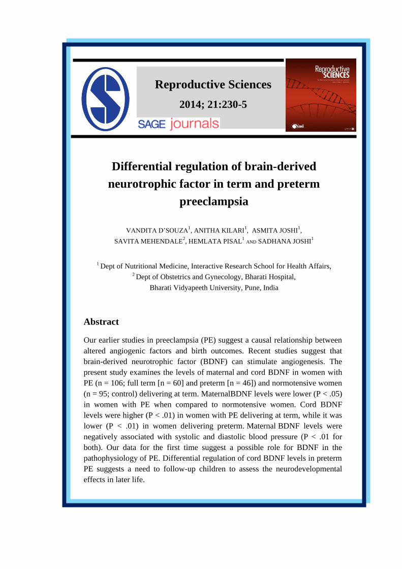
{
Differential regulation of brain-derived
neurotrophic factor in term and preterm
preeclampsia
VANDITA D’SOUZA1, ANITHA KILARI
1, ASMITA JOSHI
1,
SAVITA MEHENDALE2, HEMLATA PISAL
1 AND SADHANA JOSHI
1
1 Dept of Nutritional Medicine, Interactive Research School for Health Affairs,
2 Dept of Obstetrics and Gynecology, Bharati Hospital,
Bharati Vidyapeeth University, Pune, India
Abstract
Our earlier studies in preeclampsia (PE) suggest a causal relationship between
altered angiogenic factors and birth outcomes. Recent studies suggest that
brain-derived neurotrophic factor (BDNF) can stimulate angiogenesis. The
present study examines the levels of maternal and cord BDNF in women with
PE (n = 106; full term [n = 60] and preterm [n = 46]) and normotensive women
(n = 95; control) delivering at term. MaternalBDNF levels were lower (P < .05)
in women with PE when compared to normotensive women. Cord BDNF
levels were higher (P < .01) in women with PE delivering at term, while it was
lower (P < .01) in women delivering preterm. Maternal BDNF levels were
negatively associated with systolic and diastolic blood pressure (P < .01 for
both). Our data for the first time suggest a possible role for BDNF in the
pathophysiology of PE. Differential regulation of cord BDNF levels in preterm
PE suggests a need to follow-up children to assess the neurodevelopmental
effects in later life.
Reproductive Sciences
2014; 21:230-5

55
3.1 Introduction
The last chapter compared the levels of BDNF in maternal and cord
plasma samples of women with preeclampsia (delivering at term and preterm)
and normotensive control women at the end of pregnancy. Studies indicate that
neurotrophins play a critical role in implantation, placental development and
foetal growth from early pregnancy. This chapter is a prospective study which
examines the levels of maternal plasma BDNF from early pregnancy both in
normotensive women and women with preeclampsia.
3.1.1 Placental Development across Gestation
In order to understand the role of BDNF in early placentation this section
describes the development of the placenta across gestation. Normal placental
development and function are essential for a successful pregnancy (John and
Hemberger, 2012). Several steps result in the formation of the placenta: (1) The
blastocyst attaches to the uterine epithelium and differentiates into an inner cell mass,
an outer sphere of cells and the trophoblast (2) The trophoblast invades uterine tissues
and spiral arterioles (3) Vascularization and branching angiogenesis in the placenta
(Mess and Carter, 2007; Gude et al., 2004).
Early in mammalian development, the origins of trophoblast and embryonic
cell lineages are established as the trophectoderm and the inner cell mass (ICM) in the
blastocyst (Oda et al., 2006). The inner cell mass gives rise to the foetus and an outer
layer of trophectoderm (TE) is the precursor of the placenta (Kimber, 2000) (Fig. 17).

56
Fig. 17: Stages of Development of the Placenta and Foetus
Source: Modified from Carlson BM: Patten’s Foundations of Embryology, Ed 6, New
York, 1996, McGraw-Hill, pg. 180.
The main functional units of the placenta are the chorionic villi within which
foetal blood is separated by only three or four cell layers (placental membrane) from
maternal blood in the surrounding intervillous space (Gude et al., 2004). Trophoblasts

57
that form the placenta provide the epithelial cover of the villous trees of the placenta
and are in direct contact with maternal blood (Huppertz and Herrler, 2005).
During early pregnancy the placenta-derived trophoblasts start to invade the
maternal uterus in order to regulate adequate blood flow and nutrient supply to the
growing foetus (Pollheimer and Knöfler, 2012). The early invasive
syncytiotrophoblast (STB) invades into uterine tissues (Zhang et al., 2013).
Extravillous trophoblasts (EVT) first invade into the uterine decidua and then reach
the myometrium (Lash et al., 2006). Subsets of the EVT invade into uterine spiral
arteries (endovascular trophoblast) or uterine glands (endoglandular trophoblast)
(Huppertz et al., 2014). Initially only maternal plasma flows through the intervillous
space of the placenta in the first trimester which is replaced by maternal blood after
the proper invasion of spiral arterioles (Huppertz et al., 2012).
Transplacental exchange, that depends on rates of uterine (maternal placental)
and umbilical (foetal placental) blood flow, provides for all the metabolic demands of
foetal growth and development (Reynolds and Redmer, 2001). This is mediated by
two key processes, vascularization and angiogenesis, in the developing placenta
(Huppertz and Peeters, 2005). Vasculogenesis is the process of formation of the
earliest primitive capillaries and is achieved by differentiation of hemangiogenic stem
cells that are derived from pluripotent mesenchymal cells (Burton et al., 2009). The
subsequent process, angiogenesis, is characterized by the development of new vessels
from already existing vessels and is a well coordinated process initiated by
stimulation of various growth factors (Demir et al., 2010). Additionally, the
developing placenta undergoes a process of vascular mimicry (referred to as
pseudovasculogenesis) as cytotrophoblasts convert from an epithelial to an
endothelial phenotype (Cerdeira and Karumanchi, 2012).

58
Placental development is well regulated by various autocrine/paracrine factors
at the maternal-foetal interface (Chen, 2014). Every endothelial cell destined for
vessel formation is equipped with receptors for several angiogenic factors such as
vascular endothelial growth factor (VEGF), fibroblast growth factor (FGF), epidermal
growth factor (EGF), hypoxia inducible factor-1 (HIF-1) and transforming growth
factor (TGF) that regulates the process of neoangiogenesis (Gacche and Meshram,
2013). Patterns of placental angiogenesis are suggested to vary in pregnancy
pathologies (Mayhew et al., 2004).
3.1.2 BDNF Levels in Pregnancy
It is well known that the neurotrophins receptors are mainly expressed in the
nervous system. However, their mRNAs have also been detected in several utero
foeto-placental tissues (Mayeur et al., 2011). BDNF has been shown to increase in the
uterus before implantation and the expression of TrkB in trophectoderm cells of
blastocysts suggests a potential paracrine role of BDNF in the blastocyst during
implantation (Kawamura et al., 2007). After implantation, BDNF suppresses
apoptosis in the embryo and promotes early embryonic development (Kawamura et
al., 2007). It has also been reported that BDNF levels significantly decrease in the
maternal serum before and after giving birth (Lommatzsch et al., 2006).
Altered BDNF levels have been observed in pregnancy complications. BDNF
levels have been shown to be significantly elevated in the umbilical cord and is
reported to be associated with increasing gestational age, reflecting the degree of
neural maturity in premature infants (Chouthai et al., 2003). Furthermore, studies
from our department have shown that the ratio of maternal/foetal BDNF levels are
altered in preterm deliveries with the maternal levels being higher compared to levels
in the umbilical cord (Dhobale et al., 2012a). Further, placental levels of the Trk B
59
receptor were significantly higher in the case of preterm deliveries (Dhobale et al.,
2012a).
3.1.3 Longitudinal Changes of BDNF in Pregnancy
A recent longitudinal study in normotensive pregnant women demonstrates
that serum BDNF levels in the first trimester are lower than those in the second and
third trimester (Garcés et al., 2013). Alterations in BDNF levels may contribute to the
early abnormal placentation in women with preeclampsia. However there are no
longitudinal studies that have examined the levels of BDNF in women who develop
preeclampsia.
As discussed in the earlier chapter, the cross sectional study demonstrated
altered plasma BDNF levels at delivery in women with preeclampsia as compared to
normotensive control (NC) women. It is likely that the changes at delivery may be
secondary effects where the etiopathology has progressed for several weeks and
months. Therefore, in order to better understand the role of BDNF in the pathology of
preeclampsia, it is important to examine its levels in early gestation.
This study for the first time examines maternal BDNF levels across
gestation, cord BDNF levels and placental BDNF gene expression in the complex
pathology of preeclampsia. Their associations with the blood pressure (BP) of the
mother and pregnancy outcome are also examined.

60
3.2 Materials and Methods
3.2.1 Participants
Pregnant women were enrolled for this longitudinal study from 2 major
hospitals, Bharati hospital and Gupte hospital, in Pune, India. The research protocols
and consent forms were approved by the Bharati Vidyapeeth Medical College
Institutional Ethical Committee. Written consent was obtained from all the study
subjects. This study is part of a large ongoing departmental study which recruits all
consecutive pregnant women at 16-20 weeks of gestation who are willing to
participate and give a written consent. The incidence of preeclampsia in this
population is about 8-10% of total pregnancies. The first blood sample was taken
between 16 - 20 weeks of gestation (T1), the second between 26 - 30 weeks of
gestation (T2) and the third sample was taken at delivery (T3). Umbilical cord blood
sample was also collected just after delivery. All women were followed up till
delivery and categorized as preeclampsia if there was a presence of high BP (systolic
BP > 140 mmHg and/or diastolic BP > 90 mmHg) and proteinuria (>1+ or 300mg) on
a dipstick test. BP was measured with a mercury sphygmomanometer, and
preeclampsia was confirmed by repeated recording of the BP with an interval of 6 h at
the time of diagnosis.
The pregnancies were dated by ultrasound as well as last menstrual period
(LMP). The cases in which there were discrepancies of more than a week in
gestational age between LMP and ultrasound were excluded from the study. Neonatal
growth measures like birth weight, length, head circumference and chest
circumference were recorded. Birth weight was recorded using a digital weighing
scale (Zeal medical private limited, India) with an accuracy of 10 g. The length was
measured to the nearest 0.1 cm using a portable Infantometer. The head circumference
was measured using a measuring fiber glass measuring tape which was placed around
61
the head, just above the eyebrows anteriorly, and around the most prominent bulge
posteriorly. The chest circumference was measured using fiber glass measuring tape
which was placed around the lower chest. These procedures have been described in
our earlier departmental study (Sundrani et al., 2013).
3.2.2 Power of the Study
Till date there are no studies examining plasma BDNF levels at various time
points across gestation. This is the first longitudinal study of BDNF in women with
preeclampsia. Therefore, we calculated the power of the study based on our earlier
cross sectional study on BDNF levels in preeclampsia. Based on results from this
study the Power and Sample Size Calculation version 3.0.43 software was used to
calculate sample size of the current study. In the earlier chapter BDNF levels in NC
(458.5 ± 151.6 pg/ml) and preeclampsia (379.6 ± 92.9 pg/ml) group were normally
distributed having a population standard deviation = 122.25. If the true difference in
the means of BDNF levels between preeclampsia and NC is 78.9, we would need to
study 39 preeclampsia subjects and 78 NC subjects to be able to reject the null
hypothesis that the population means of the preeclampsia and NC groups are equal
with probability (power) 0.9. The Type I error probability associated with this test of
this null hypothesis is 0.05. In view of this, this study was undertaken on 89 NC and
61 preeclampsia women. (Fig. 18) Out of the 61 women with preeclampsia, 9 were
severe, 2 showed IUGR and 10 delivered preterm.

62
Fig. 18: Flowchart of Number of Samples Analyzed for BDNF
BDNF – brain derived neurotrophic factor; n –
number; T1 - 16-20 weeks of gestation; T2 - 26-30
weeks of gestation; T3 - at delivery
3.2.3 Sample Collection, Processing and Storage
10 ml of maternal venous blood was collected into the EDTA vials at all the
time points while 10 ml of cord blood was collected at the time of delivery and was
processed as described by us earlier in Chapter 2 Section 2.2.2.
Placental samples were collected on a random sub-sample. Thus, 21
preeclampsia and 22 control placental samples were collected and analyzed in this
study. Fresh placental tissues were obtained on a sub sample from normal (n=22) and
preeclamptic pregnancies (n=21) immediately after delivery. Briefly, the foetal
membranes were trimmed off and small pieces were randomly cut out from the
placental cotyledons. Tissue were rinsed in phosphate-buffered saline to wash off
maternal and foetal blood, snap frozen in liquid nitrogen and stored at −80°C until
assayed.

63
3.2.4 BDNF Assay
BDNF levels were measured in both maternal and cord plasma using the
BDNF Emax Immuno Assay System (Promega). The details of this assay have been
explained in Chapter 2, Section 2.2.3.
3.2.5 Extraction of Total RNA, cDNA Synthesis and Quantitative Real-Time
(RT)-PCR Assays
Extraction of total RNA, cDNA synthesis and mRNA expression of BDNF
and GAPDH genes from 22 NC and 21 preeclampsia placental samples was
performed. The detailed method has been described by our department earlier (Meher
et al., 2013). Total RNA from placenta samples was isolated using Trizol method and
quantified by the Eppendorf Biophotometer plus (Hamburg, Germany). For each
placental sample, 2 μg of RNA was reverse transcribed to cDNA using the High-
Capacity cDNA reverse transcription kit (Cat No. 4368814, Applied Biosystems).
RT-qPCR for BDNF and GAPDH were performed with the TaqMan Universal
PCR Master Mix (Cat No. 4324018, Applied Biosystems, California, USA) using the
Applied Biosystems 7500 Standard Real Time PCR system. The relative expression
level of the gene of interest was examined with respect to GAPDH to normalize for
variation in the quality of mRNA and the amount of input cDNA. The real-time
quantitative PCR reactions for each gene were performed in duplicate. To analyze the
real-time quantitative PCR results, the average cycle number (Ct) of the reaction
when it crossed a threshold value was determined for each reaction. Differences in Ct
(ΔCt) between GAPDH and targeted gene BDNF were calculated by subtracting the
Ct of the targeted gene BDNF from Ct of GAPDH. Relative expression levels of
genes were calculated and expressed as 2ΔCt
. The following TaqMan® Gene

64
Expression Assays (Applied Biosystems) were used in this study: GAPDH
(Hs99999905_m1); BDNF (Hs00542425_s1).
3.2.6 Statistical Analysis
Continuous variables are reported as mean ± S.D. and discrete variables as
percent (%). The data were checked for normal distribution by testing for skewness
and kurtosis and BDNF values were found normally distributed. Differences in
maternal and neonatal characteristics between preeclampsia and normotensive control
(NC) were assessed using Student's t test for continuous variables and chi-square tests
for categorical data. Pearson’s correlation was used to examine the associations
between BDNF levels and maternal age, body mass index (BMI), gestational age,
parity, socioeconomic status (SES), education and income as they have been shown to
affect BDNF levels (Pillai et al., 2012; Sher and La Bode, 2011; Rao et al., 2009;
Zdanys et al., 2009; Macbeth et al., 2008; Lasky-Su et al., 2007; Lommatzsch et al.,
2005). BDNF was correlated with maternal age, BMI and SES and hence BDNF
levels were adjusted for these variables using multiple linear regression.
Associations of BDNF with BP and neonatal growth measures were analyzed
after adjusting for maternal age, BMI and SES. The variable sample number (n) in
different measures was either due to loss of follow up at various time points across
gestation or insufficient sample volume available. Results corresponding to p-values
lower than 0.05 are described as significant. The data were analyzed using the
SPSS/PC+ package (Version 20.0, Chicago, IL, USA).

65
3.3 Results
3.3.1 Maternal and Neonatal Characteristics
The maternal body mass index (BMI) at all time points in women with
preeclampsia (T1, p<0.01; T2, p<0.01; T3, p<0.01) was higher than NC women.
Maternal systolic (T1, p<0.01; T2, p<0.01; T3, p<0.01) and diastolic BP (T2, p<0.01;
T3, p<0.01) of women with preeclampsia was higher than NC women. The
percentage of nulliparous women was higher in the preeclampsia (n=61) group
compared to the NC (n=89) group (72% vs. 46%, p<0.01). There was no difference in
education between groups (Table 3). Neonatal characteristics show that birth weight,
head circumference and chest circumference was lower (p<0.05) in preeclampsia than
in the NC group (Table 4).
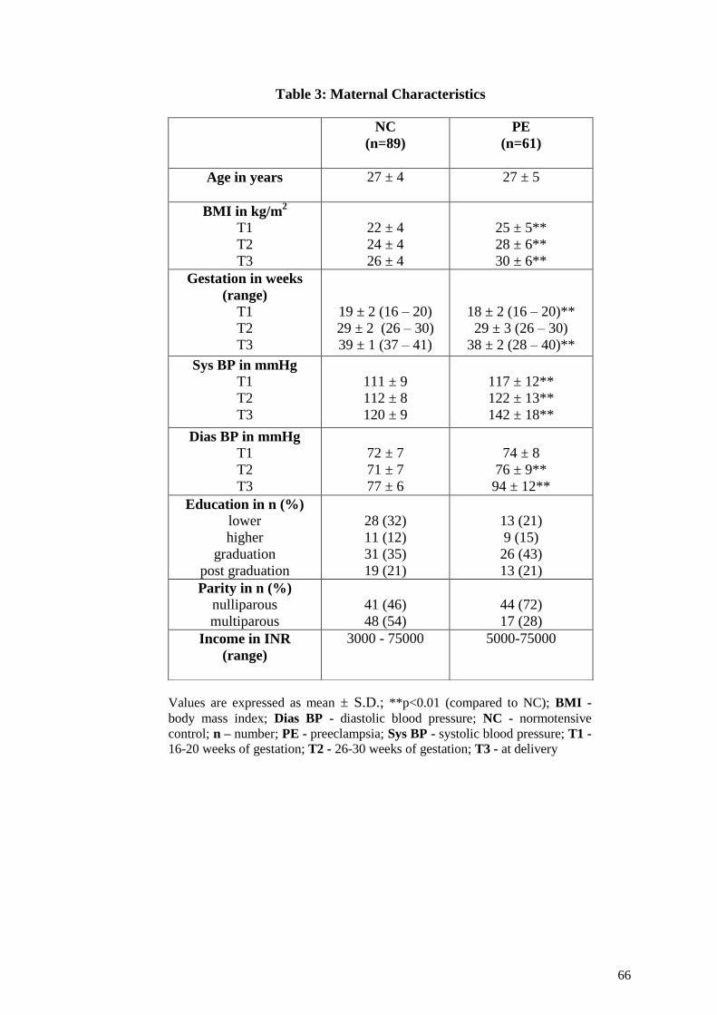
66
Table 3: Maternal Characteristics
Values are expressed as mean ± S.D.; **p<0.01 (compared to NC); BMI -
body mass index; Dias BP - diastolic blood pressure; NC - normotensive
control; n – number; PE - preeclampsia; Sys BP - systolic blood pressure; T1 -
16-20 weeks of gestation; T2 - 26-30 weeks of gestation; T3 - at delivery
NC
(n=89)
PE
(n=61)
Age in years
27 ± 4 27 ± 5
BMI in kg/m2
T1
T2
T3
22 ± 4
24 ± 4
26 ± 4
25 ± 5**
28 ± 6**
30 ± 6**
Gestation in weeks
(range)
T1
T2
T3
19 ± 2 (16 – 20)
29 ± 2 (26 – 30)
39 ± 1 (37 – 41)
18 ± 2 (16 – 20)**
29 ± 3 (26 – 30)
38 ± 2 (28 – 40)**
Sys BP in mmHg
T1
T2
T3
111 ± 9
112 ± 8
120 ± 9
117 ± 12**
122 ± 13**
142 ± 18**
Dias BP in mmHg
T1
T2
T3
72 ± 7
71 ± 7
77 ± 6
74 ± 8
76 ± 9**
94 ± 12**
Education in n (%)
lower
higher
graduation
post graduation
28 (32)
11 (12)
31 (35)
19 (21)
13 (21)
9 (15)
26 (43)
13 (21)
Parity in n (%)
nulliparous
multiparous
41 (46)
48 (54)
44 (72)
17 (28)
Income in INR
(range)
3000 - 75000 5000-75000

67
Table 4: Neonatal Characteristics at Birth
Values are expressed as mean ± S.D.; *p<0.05 (compared to NC);
NC - normotensive control; PE – preeclampsia
3.3.2 Maternal BDNF Levels
Maternal BDNF levels at T1 were lower (p<0.05) in women with
preeclampsia (455 ± 128 pg/mL) as compared to NC women (486 ± 148 pg/mL).
Maternal BDNF levels at T2 were comparable in women with preeclampsia (462 ±
151 pg/mL) and NC women (467 ±117 pg/mL). Maternal BDNF levels at T3 were
lower (p<0.05) in women with preeclampsia (426 ± 118 pg/mL) as compared to NC
women (478 ± 135 pg/mL) (Fig. 19).
3.3.3 Cord BDNF Levels
Cord BDNF levels were lower in preeclampsia (381 ± 76 pg/mL) (p<0.05) as
compared to the NC group (429 ± 92 pg/mL) (Fig. 19).
NC
(n=89)
PE
(n=61)
Weight (kg)
2.9 ± 0.3 2.8 ± 0.4*
Length (cm)
48 ± 2.9 47.5 ± 2.3
Head Circumference (cm)
33.9 ± 1.3 33.2 ± 1.3*
Chest Circumference (cm)
32.2 ± 1.6 31.5 ± 2.5*

68
Fig. 19: Maternal and Cord BDNF Levels across Gestation in Women with Preeclampsia
as Compared to Normotensive Control Women
Values are expressed as Mean ± S.D.; *p<0.05, **p<0.01 as compared to NC. A - all samples; B - all
timepoints; BDNF - brain derived neurotrophic factor; NC - normotensive control; PE - preeclampsia;
T1 - 16 to 20 weeks of gestation; T2 - 26 to 30 weeks of gestation; T3 - at delivery
3.3.4 Placental BDNF Gene Expression
Placental BDNF gene expression was lower (p<0.01) in women with
preeclampsia than in NC women (Fig. 20).
Fig. 20: Placental BDNF Gene Expression
Values are expressed as Mean ± S.E.;
**p<0.01 as compared to NC; NC -
Normotensive control; PE - Preeclampsia
69
3.3.5 Association between Maternal BDNF Levels and Maternal Blood
Pressure at T3
Maternal systolic BP was negatively associated with BDNF levels at T3 (r = -
304, p = 0.0050, n=82) in NC women. However, there was no association between
maternal BP and BDNF in women with preeclampsia.
3.3.6 Association between Maternal BDNF Levels and Neonatal Growth Measures
There was no association between maternal BDNF levels at any time point and
neonatal growth measures in both NC women and women with preeclampsia.
3.3 Discussion
This study for the first time reports levels of maternal plasma BDNF across
gestation, cord plasma BDNF and placental BDNF gene expression in women with
preeclampsia compared with NC women. Our results indicate that 1) Maternal BDNF
levels were lower at T1 and T3 in women with preeclampsia than those in NC women
2) Cord BDNF levels were lower in the preeclampsia group as compared to NC group
3) Placental BDNF gene expression levels were also lower in women with
preeclampsia than in the NC women.
3.4.1 Lower Maternal BDNF Levels at T1 and T3
This study reports lower maternal BDNF levels in women with preeclampsia
at T1 and T3 as compared to NC women. As mentioned in chapter 2, we observed
lower maternal BDNF and cord BDNF levels in preterm preeclampsia as compared to
NC group at delivery in a cross sectional study. BDNF levels are known to be
influenced by number of factors such as urbanicity, age, food intake and SES (Bus et
al., 2011). Lower BDNF levels observed in this study may be a result of increased
70
oxidative stress and reduced omega-3 fatty acids like docosahexaenoic acid (DHA)
both of which are known to influence the levels of BDNF (Bhatia et al., 2011;
Kapczinski et al., 2008; Wu et al., 2008). Further, the studies in our department have
reported increased oxidative stress at delivery in women with preeclampsia as
compared with NC women (Mehendale et al., 2008). Similarly these studies have
also demonstrated a negative association between DHA and BDNF in preterm
pregnancy (Dhobale et al., 2012a) and lower levels of DHA in preeclampsia as
compared to control in a cross sectional cohort (Kulkarni et al., 2011).
This data is a part of a large multi-investigator study where detailed
information on nutritional intakes has been recorded and is reported separately where
the frequency of consumption of omega 3 fatty acid (ALA and DHA) rich foods was
similar in both groups at all time points (Wadhwani et al., 2014). Studies have shown
that omega-3 fatty acids influence levels of neurotrophins (Sharma et al., 2012; Bhatia
et al., 2011; Wu et al., 2004). Our earlier cross sectional studies indicate that there is
no difference in the intake of omega-3 fatty acids between NC women and women
with preeclampsia (Kulkarni et al., 2011). In addition, studies have shown that serum
BDNF concentrations are season dependent (Molendijk et al., 2012; Comasco et al.,
2011). However, in contrast others do not report seasonal changes in BDNF levels
(Katoh-Semba et al., 2007). Three seasons define the Indian subtropical climate,
summer (from February to May), the monsoon (from June to September) and winter
(from October to January) and climatic variations in Pune, Western India are not
drastic. In our study women from both groups have been recruited all around the year
and the BDNF values at each time point are an average of women recruited across all
the seasons. In view of this, it is unlikely that seasonality would affect the levels of
BDNF. Further, it has been reported that in the tropical climate of western India,

71
seasonal variation is not associated with incidence of preeclampsia (Subramaniam,
2007).
It is known that the implantation of the blastocyst and early development of
the placenta are crucial for a successful pregnancy (James et al., 2012) and
neurotrophic factors like BDNF potentiate placental development (Kawamura et al.,
2009). Recent reports suggest that as pregnancy progresses, the placenta and foetus
may have a role in the production of BDNF (Garcés et al., 2013). Maternal BDNF is
reported to reach the foetal brain through the utero-placental barrier and might
contribute to its development (Kodomari et al., 2009). BDNF is known to stimulate
angiogenesis in the endothelial cells in the placenta (Caporali and Emanueli, 2009).
Further, reports indicate that during embryonic development BDNF expresses its
TrkB receptor resulting in a direct angiogenic action on endothelial cells involved in
cardiac vessel wall stability (Kermani et al., 2007). This may be due to a cross talk
between neurons and endothelial cells via VEGF, neurotrophins and their cognate
receptors on both neurons and endothelial cells (Li et al., 2006). Our findings suggest
that reduced BDNF levels in early pregnancy may be partly responsible for reported
abnormal foeto-placental development in preeclampsia.
3.4.2 Lower Placental BDNF Gene Expression and Cord BDNF Levels
In the current study, placental BDNF gene expression in women with
preeclampsia was lower as compared to NC women. Studies indicate that BDNF is
expressed in the human placenta, immunolocalized in the membranous chorion,
trophoblast layer and endothelium, supporting its role in the regulation of several
metabolic functions during pregnancy (Garcés et al., 2013; Fujita et al., 2011). It has
been reported that the BDNF/Trk B signaling system is localized in the utero-foetal
tissue in the placenta, suggesting a transient action of the ligand in these tissues

72
throughout pregnancy (Mayeur et al., 2011). Our data suggests that the lower BDNF
gene expression may be associated with impaired placental development in
preeclampsia.
Our findings suggest that low levels of BDNF exist in women with
preeclampsia from the 16th
week of pregnancy and may be implicated in the
improper placental development observed in these women. Further, lower levels
of cord BDNF was also observed in preeclampsia which may have long-lasting
consequences in their babies.
Like BDNF, NGF too is an important neurotrophin. It was the first
neurotrophin to be discovered playing an important role in the nervous system.
NGF also plays a role in angiogenesis and may regulate the development of the
placenta. The next chapter therefore examines the levels of NGF across gestation
in women with preeclampsia. Further, the associations of NGF with BP and birth
outcome are also examined.

{
Levels of brain derived neurotrophic factors
across gestation in women with preeclampsia
VANDITA D’SOUZA1, VIDYA PATIL
1, HEMLATA PISAL
1, KARUNA
RANDHIR1, ASMITA JOSHI
1, SAVITA MEHENDALE
2, GIRIJA WAGH
2,
SANJAY GUPTE3 AND SADHANA JOSHI
1
1 Dept of Nutritional Medicine, Interactive Research School for Health Affairs,
2 Dept of Obstetrics and Gynecology, Bharati Hospital,
Bharati Vidyapeeth University, 3 Gupte Hospital and Research Centre, Pune, India
Abstract
Preeclampsia (PE) is a major pregnancy complication of placental origin which
leads to adverse pregnancy outcome. Brain derived neurotrophic factor
(BDNF) is suggested to promote trophoblast growth and regulate placental and
fetal development. This study for the first time examines the levels of maternal
plasma BDNF at various time points during gestation, cord plasma and
placental BDNF levels and their association with birth outcome in women with
PE. Normotensive control (NC) women (n=89) and women with PE (n=61)
were followed at three different time points [16-20 weeks (T1), 26-30 weeks
(T2) and at delivery (T3)]. Maternal blood at all time points and cord blood
was collected. Results indicate that maternal BDNF levels at T1 (p=0.050) and
T3 (p=0.025) were lower in women with PE than in NC women. Cord BDNF
levels at delivery in women with PE were lower (p=0.032) than those in NC
women. Placental BDNF gene expression was also lower (p=0.0082) in women
with PE than in NC women. Our data suggests that BDNF plays an important
role in the development of the materno-fetal-placental unit during pregnancy.
Alteration in the levels of BDNF during pregnancy may be associated with an
abnormal development of the placenta resulting in PE.
International Journal of
Developmental Neuroscience
2014; 21:230-5


73
4.1 Introduction
In Chapter 3 we have demonstrated changes in the levels of BDNF from
early pregnancy. Studies have also shown that nerve growth factor (NGF) is
essential for a successful pregnancy as it constitutes a functional link between the
maternal nervous, endocrine and immune system. There are very few studies,
which have examined the levels of NGF in preeclampsia. This study for the first
time examines the levels of NGF in women with preeclampsia throughout
gestation and compares them with normotensive pregnant women.
4.1.1 Nerve Growth Factor
NGF is a member of the neurotrophin family of proteins that was discovered
in the 1950’s (McKelvey et al., 2013) (Fig. 21). NGF has been characterized first in
the sensory and autonomic nervous system, then in central nervous, endocrine and
immune systems (Aloe et al., 2012). The molecular weights of NGFs from human
serum, saliva and urine have been found to be 36 kDa (Lipps, 2000). It has a tertiary
structure based on a cluster of 3 cystine disulfides and 2 very extended, but distorted
beta-hairpins (Bradshaw et al., 1994).

74
Fig. 21: NGF and its Trk A Receptor
NGF – nerve growth factor; Trk A – tropomyosin-related
receptor kinase A
Source: Modified from Deinhardt and Jeanneteau, 2012
Protein Phosphorylation in Human Health 7:217-232
NGF is a signaling protein that interacts with specific receptors in autocrine,
paracrine and endocrine modes (Rachaneni et al., 2013). Its biological activity is
mediated by two classes of receptors: (i) p75 neurotrophin receptor (p75NTR), a 75
kDa glycoprotein, belonging to a superfamily of cytokine receptors including TNF
receptors, and (ii) Trk A, a transmembrane tyrosine kinase of 140 kDa (Fiore et al.,
2009) (Fig. 22). Different intracytoplasmic signaling pathways that may be activated
by binding of NGF to Trk A receptor include phosphatidylinositol-3-kinase (PI3K)
and mitogenic activated protein kinase (MAPK) to promote cell survival and
proliferation and activation of the phospholipase type Cγ (PLCγ) pathway. This
results in the production of diacylglycerol (DAG) and inositol triphosphate (IP3),
culminates in the release of calcium from the intracytoplasmic cellular stocks (Chaves
et al., 2013).

75
Fig. 22: NGF- Trk A Signalling Pathways
Akt - serine/threonine kinase; CREB - cAMP response-element binding protein; DAG - diacylglycerol; ELK
1- E twenty-six (ETS)-like transcription factor 1; ERK - extracellular signal regulated kinase; IKK - NF-κB-
binding cofactor kinase; IP3 - inositol trisphosphate; IRAK - interleukin-1β receptor-associated kinase; JNK -
c-Jun N-terminal kinase; MEK – mitogen activated protein kinase/ERK kinase; NF-κB - nuclear factor-κB;
NGF – nerve growth factor; NRAGE - neurotrophin receptor-mediated melanoma-associated antigens
homolog; NRIF - neurotrophin receptor interacting factor; p75NTR – p75 neurotrophin receptor; PDK1 - 3-
phosphoinositide-dependent protein kinase 1; PI3K – phosphotidylinositol 3-kinase; PKC - protein kinase C;
PIP - phosphatidylinositol-bis-phosphate; PLCγ – phospholipase Cγ; Rac, Raf, Ras - rho-associated kinases;
RIP2 - receptor interacting protein 2; RSK - ribosomal S6 kinase; Shc - Src homology and collagen related
molecules; SOS - son of sevenless proteins; TNAF 6 - tumour necrosis factor receptor-associated factor 6; Trk
A - tropomyosin-related receptor kinase A
Source: Modified from Pollack and Harper, 2002 Drug News Perspect, 15:268
4.1.2 Functions of NGF in the Nervous System
NGF was discovered as a molecule that stimulates the survival and maturation
of developing sensory neurons in the peripheral nervous system (PNS) and has later

76
been shown to protect adult neurons in the degenerating mammalian brain (Roberti et
al., 2014; Manni et al., 2013). Additionally, NGF is known to have trophic and
differentiating activity on several populations of cholinergic neurons of the CNS
(Roberti et al., 2014). NGF ensures the maintenance of phenotypic and functional
characteristic of several populations of neurons as well as immune cells (Aloe et al.,
2012). It has important functions in the maintenance of viability and proliferation of
neuronal and non-neuronal cells (Chaves et al., 2013). Recent studies have recognized
the role of NGF beyond nerve cells and the peripheral PNS and CNS and that topical
application of NGF give a protective effect on corneal ulcer and glaucoma (Manni et
al., 2013).
4.1.3 NGF and Neurodevelopment
Neurodevelopment is coordinated by a series of growth factors including NGF
which are involved in neuritogenesis (Li et al., 2014). NGF is related to the growth
and differentiation of nerve cells, as well as to a decline in cognitive function (Bae et
al., 2014). Enhancement in NGF levels after brain injury is a part of neuronal
recovery process (Dinçel et al., 2013). NGF concentrations in serum and
cerebrospinal fluid (CSF) may be enhanced in various cerebral pathologies such as
hypoxia, ischaemia, trauma, seizures, neuroimmunological diseases and intracranial
hypertension syndrome (Gioiosa et al., 2009; Sofroniew et al., 2001; Dixon et al.,
1997). A recent study has shown that maternal psychological and biological factors
are associated with infant motor development and are mediated by changes in NGF
levels (Pinheiro et al., 2014).

77
4.1.4 NGF and Placenta
Human placental tissue contains a significant amount of NGF (Purcell and
Atterwill, 1994). Reports indicate that NGF is expressed, localized and active in the
trophoblast, amnion/chorion and maternal decidua of the placenta both in early
gestation and at term (Toti et al., 2006; Marvin et al., 2002). Growth factors such as
NGF, VEGF, produced by nerves and blood vessels may contribute to the process of
normal placentation and may be compromised in pregnancy complications (Quinn,
2005).
4.1.5 NGF and Angiogenesis
NGF has historically been implicated in several functions of the nervous
system, however, it can also regulate hypertension (Hennigan et al., 2009; Supowit et
al., 2001) and elicit cardiovascular actions, including angiogenesis during foetal and
neonatal stages (Meloni et al., 2010; Cabrera-Vásquez et al., 2009). Blood vessel and
nerve growth are linked by common pathways that involve the release of
proangiogenic factors, such as vascular endothelial growth factor, β-NGF and
neuropeptides (Mapp and Walsh, 2012). Every endothelial cell destined for vessel
formation is equipped with receptors for angiogenic peptides such as NGF (Gacche
and Meshram, 2013). NGF has been shown to have an influence on the functional
activity of the cardiovascular system and can regulate the functional state of
endothelial and vascular smooth muscle cells and cardiomyocytes (Kryzhanovskiĭ and
Vititnova, 2011).

78
4.1.6 NGF and Preeclampsia
Recent studies have revealed that preeclampsia appears to originate in placenta
and is characterized by widespread maternal endothelial dysfunction (Petla et al.,
2013). Abnormal placentation and reduced blood flow in preeclampsia is a result of
impaired invasion of maternal spiral arteries (Steegers et al., 2010; Myatt and
Webster, 2009). In a normal pregnancy, development of the placenta is controlled by
a plethora of growth factors, such as cytokines, neurotrophins like NGF and
angiogenic molecules controlling trophoblast motility, which are secreted from
numerous trophoblast cells that regulates trophoblast invasiveness (Knöfler, 2010).
Defective expression and activity of these molecules are potential candidates
for triggering preeclampsia (Ong, 2004). NGF is expressed and is active in different
parts of the placenta in early gestation and at term (Toti et al., 2006; Marvin et al.,
2002).This expression may be differentially regulated in preeclampsia due to
improper development of placenta.
NGF plays an important role in the regulation of growth, survival and
differentiation of neurons in both the CNS and PNS (Bibel and Barde, 2000).
Although NGF has historically been implicated in several functions of the nervous
system, it can also regulate hypertension (Hennigan et al., 2009; Supowit et al., 2001)
and elicit cardiovascular actions, including angiogenesis during foetal and neonatal
stages (Meloni et al., 2010; Cabrera-Vásquez et al., 2009). It has been reported that
NGF is mandatory for the success of pregnancy as it constitutes a functional link
between the nervous, endocrine and immune system translating environmental or
endocrine signals (Tomettan et al., 2005). It has been reported that NGF levels
increase as the trimester progresses in monkeys (Neubert et al., 2014). A recent study
suggests that a local threshold of NGF expression is necessary to ensure maternal
79
tolerance in healthy pregnancies, but when surpassed may result in foetal rejection
due to exacerbated inflammation (Frank et al., 2014).
There are very few studies, which have studied the levels of NGF in different
pregnancy complications like IUGR (Malamitsi-Puchner et al., 2007) but not in
women with preeclampsia. Xia et al. (2011) have shown reduced NGF levels in
amniotic fluid specimens were obtained during amniocentesis or caesarean section
(Xia et al., 2011).
Our earlier cross sectional study on women with preeclampsia has shown
altered NGF levels as compared to normotensive women (Kilari et al., 2011).
Although preeclampsia is characteristically diagnosed in the last third of pregnancy, it
is evident that many of these pathophysiological changes can be detected long before
clinically evident disease (Roberts and Bell, 2013). Further, infants born to women
with preeclampsia are at increased risk for cardiovascular and neurodevelopmental
disorders (Akcakus et al., 2010; Silveira et al., 2007). Hence there is a need to study
the maternal and cord NGF levels across the pregnancy to study the mechanism and
severity of preeclampsia.
Therefore, in order to better understand the role of NGF in
preeclampsia, the present study for the first time examines maternal NGF levels
across gestation along with cord NGF levels in women with preeclampsia and
their associations with mother’s blood pressure (BP) and foetal outcome.
80
4.2 Materials and Methods
4.2.1 Participants
The recruitment of study population and the inclusion and exclusion criteria
are as mentioned in Chapter 3 (Section 3.2.1). Neonatal growth measures were
recorded as mentioned in Chapter 3 (Section 3.2.1).
4.2.2 Power of the Study
Till date there are no studies examining plasma NGF levels at various time
points across gestation. This is the first longitudinal study of NGF in women with
preeclampsia. Therefore we calculated the power of the study based on our earlier
cross sectional study on NGF levels in preeclampsia (Kilari et al., 2011). Based on
results from this study the Power and Sample Size Calculation version 3.0.43 software
was used to calculate sample size of the current study. In our earlier study, NGF
levels in NC (316.3 ± 109.7 pg/ml) and preeclampsia (257.6 ± 122.85 pg/ml) group
were normally distributed having a population standard deviation = 116.275. If the
true difference in the means of NGF levels between preeclampsia and NC is 58.7, we
would need to study 47 PE subjects and 94 NC subjects to be able to reject the null
hypothesis that the population means of the preeclampsia and NC groups are equal
with probability (power) 0.8. The Type I error probability associated with this test of
this null hypothesis is 0.05. In view of this, this study was undertaken on 88 NC who
had complete data for all three time-points and the cord-blood sample. In case of
preeclampsia, a total of 48 women were recruited out of which 25 women had
complete data at all time points (Fig. 23).

81
Fig. 23: Flowchart of Number of Samples Analyzed for NGF
NGF – nerve growth factor; n – number; T1 - 16-20
weeks of gestation; T2 - 26-30 weeks of gestation; T3
- at delivery
4.2.3 Sample Collection, Processing and Storage
Blood sample collection was as described in Chapter 2 (Section 2.2.2) while
placental sample collection was as described in Chapter 3 (Section 3.2.2)
4.2.4 NGF Assay
NGF levels were measured in maternal and cord blood plasma samples using
the NGF Emax Immuno Assay System Promega kit (Zettler et al., 1996). Briefly, the
NGF Emax® Immunoassay system is designed for the sensitive and specific detection
of NGF in the antibody sandwich format. In this format, flat bottom 96-well plates are
coated with anti-NGF polyclonal antibody (pAb) which binds soluble NGF. The
captured NGF is bound by a second monoclonal antibody (mAb). After washing, the
amount of specifically bound mAb is then detected using a species-specific antibody
conjugated to horse radish peroxidase (HRP) as a tertiary reactant.

82
The unbound conjugate is removed by washing and following incubation with
a chromogenic substrate. The colour change is measured by a spectrophotometer
using an ELISA plate reader. The amount of NGF in the test solutions is proportional
to the colour generated in the oxidation–reduction reactions. NGF concentrations
were expressed as pg/ml. We have reported this method in our earlier studies
(Dhobale et al., 2012b; Kilari et al., 2011).
4.2.5 Extraction of Total RNA, cDNA Synthesis and Quantitative Real-Time
(RT)-PCR Assays
Extraction of total RNA, cDNA synthesis and mRNA expression of NGF and
GAPDH genes from 22 NC and 24 preeclamptic placental samples was performed.
The detailed method has been described by us earlier in Chapter 3 (Section 3.2.4)
(D’Souza et al., 2014). The following TaqMan® Gene Expression Assays (Applied
Biosystems) were used in this study: GAPDH (Hs99999905_m1); NGF
(Hs01113193_m1). The NGF mRNA levels are expressed as mean ± SE.
4.2.6 Statistical Analysis
Continuous variables are reported as mean ± S.D. and discrete variables as
percent (%). The data were checked for normal distribution by testing for skewness
and kurtosis and NGF values were found normally distributed. Differences in
maternal and neonatal characteristics between preeclampsia and NC women were
assessed using Student's t test for continuous variables and chi-square tests for
categorical data. Pearson’s correlation was used to examine the associations between
NGF levels and maternal age, body mass index (BMI), gestational age, parity,
socioeconomic status (SES), education and income as they have been shown to affect

83
NGF levels (Kumar et al., 2012; Hackman et al., 2010; López-Valladares et al., 2010;
Bulló et al., 2007; Marx et al., 1999). NGF was correlated with gestational age, BMI
and SES and hence NGF levels were adjusted for these variables using multiple linear
regression.
Associations of NGF with BP and neonatal growth measures were analyzed
after adjusting for gestational age, BMI and SES. Results corresponding to p-values
lower than 0.05 (5%) are described as significant and reported. The data were
analyzed using the SPSS/PC+ package (Version 20.0, Chicago, IL, USA).
4.3 Results
4.3.1 Maternal and Neonatal Characteristics
The maternal BMI at all time points in women with preeclampsia (T1, p<0.05;
T2, p<0.05; T3, p<0.01) was higher than NC women. Maternal systolic at T1, T2 and
T3 and diastolic BP at T1, T2 and T3 (p<0.01 for all) in women with preeclampsia
was higher than NC women. The percentage of nulliparous women was higher in the
preeclampsia group as compared to the NC group (65% vs. 35%, p = 0.038). Parity
was found to be different between groups. There was no difference in education and
income between the groups (Table 5).

84
Table 5: Maternal Characteristics
Values are expressed as Mean ± S.D.; *p<0.05, **p<0.01 as compared to
NC; BMI - body mass index; Dias BP - diastolic blood pressure; n –
number; NC - normotensive control; PE - preeclampsia; Sys BP - systolic
blood pressure; T1 - 16-20 weeks of gestation; T2 - 26-30 weeks of
gestation; T3 - at delivery
NC
(n=88)
PE
(n=48)
Age in years 27 ± 4 26 ± 5
BMI in kg/m2
T1
T2
T3
22 ± 4
24 ± 4
26 ± 4
24 ± 5*
27 ± 5*
29 ± 5**
Gestation in weeks
(range)
T1
T2
T3
18 ± 2
29 ± 2
39 ± 1
17 ± 2
30 ± 2*
37 ± 3**
Sys BP in mmHg
T1
T2
T3
110 ± 9
112 ± 8
120 ± 9
118 ± 11**
122 ± 13**
145 ± 15**
Dias BP in mmHg
T1
T2
T3
72 ± 7
70 ± 7
77 ± 6
75 ± 8*
77 ± 8**
95 ± 12**
Education in n (%)
lower
higher
graduation
post graduation
29
10
36
25
24
15
42
19
Parity in n (%)
nulliparous
multiparous
47
53
65
35
Income in INR (range)
3000 - 75000 5000-75000

85
Table 6: Neonatal Characteristics at Birth
Values are expressed as Mean ± S.D.; *p<0.05, as compared to NC.
NC – normotensive control; PE - preeclampsia
Neonatal characteristics show that birth weight (p<0.05) and head
circumference (p<0.05) was lower in preeclampsia than in the NC group (Table 6).
4.3.2 Maternal NGF levels
Maternal NGF levels at T1 (NC: 96 ± 53 pg/mL; PE: 103 ± 44 pg/mL), T2
(NC: 90 ± 51pg/mL; PE: 86 ± 40 pg/mL) and T3 (NC: 84 ± 44 pg/mL; PE: 76 ± 32
pg/mL) were comparable in women with preeclampsia as compared to NC women
(Fig. 24).
4.3.3 Cord NGF levels
Cord NGF levels were higher in preeclampsia (70 ± 53 pg/mL) (p<0.05) as
compared to the NC group (53 ± 44 pg/mL) when compared between all samples (Fig
24A). However, cord NGF levels showed a trend but were not significant in samples
obtained at all time points (Fig 24B).
NC
(n=88)
PE
(n=48)
Weight (kg) 2.9 ± 0.3 2.8 ± 0.6*
Length (cm) 48.1 ± 2.9 47.6 ± 2.4
Head Circumference
(cm) 33.9 ± 1.3 33.3 ± 1.3*
Chest Circumference
(cm) 32.2 ± 1.7 31.5 ± 1.8*

86
Fig. 24: Maternal and Cord NGF Levels in Women with Preeclampsia as Compared to Normotensive Control Women
Values are expressed as Mean ± SD; *p<0.05 as compared to NC. A - all samples; B - all timepoints;
NC - normotensive control; NGF – nerve growth factor; PE - preeclampsia; T1 - 16 to 20 weeks of
gestation; T2 - 26 to 30 weeks of gestation; T3 - at delivery
4.3.4 Placental NGF Gene Expression
Placental NGF gene expression was higher although not significant in women
with preeclampsia as compared to NC women (Fig. 25).
Fig. 25: Placental NGF Gene Expression
Values are expressed as Mean ± S.E.; NC -
normotensive control; PE – preeclampsia

87
4.3.5 Association between Maternal NGF Levels and Maternal Blood Pressure
at T1 and T2
There was a negative association between maternal NGF levels and maternal
diastolic BP at T1 (r = -0.471, p = 0.027, n = 25) and T2 (r = -0.412, p = 0.063, n =
25) in women with preeclampsia.
4.3.6 Association between Cord NGF Levels and Neonatal Growth Measures
There was a positive association between cord NGF levels and baby head
circumference (r = 0.218, p = 0.041, n = 92) and chest circumference (r = 0.218, p =
0.041, n = 92) in the whole cohort.
4.4 Discussion
This study reports novel findings 1) Maternal NGF levels in women with
preeclampsia were comparable to NC women at all time points 2) Cord NGF levels at
delivery were higher in women with preeclampsia as compared to NC women 3) A
negative association was observed between maternal diastolic BP and NGF levels at
T1 in women with preeclampsia 4) There was a positive association between cord
NGF levels and baby head and chest circumference in the whole cohort.
4.4.1 Higher Cord NGF Levels
In this study, maternal NGF levels in women who develop preeclampsia were
comparable to NC women. In contrast, there was an increase in cord levels in women
with preeclampsia. NGF is of major importance in prenatal and postnatal brain
development, due to its neuroprotective action. One study has shown lower
circulating levels of NGF in mothers delivering intrauterine growth restricted babies

88
(IUGR) as compared to mothers delivering appropriate for gestational age (AGA)
babies (Malamitsi- Puchner et al., 2007). Studies have shown that loss of neural
connections through the process of partial uterine denervation may cause reduced
foetal growth without the maternal circulatory changes of preeclampsia (Quinn,
2005).
Studies suggest that NGF is involved in angiogenesis (Park et al., 2007) and
acts in a pleiotropic manner at both neuronal and vascular levels (Nico et al., 2008).
Our earlier study in women with preeclampsia has shown altered levels of angiogenic
factors (Kulkarni et al., 2010). Studies suggest that there is a cross talk between
cardiovascular and nervous systems. Growth factors like VEGF and NGF have been
implicated in both neurodegenerative and vascular diseases (Lazarovici et al., 2006).
It has been reported that physiological actions of NGF and VEGF are mediated by
receptors (Trk A for NGF and VEGFR-2 for VEGF) which activate a complex and
integrated network of signaling pathways in neurons and endothelial cells (Lazarovici
et al., 2006).
In the current study, although there was no change in maternal NGF levels in
women with preeclampsia there was an increase in cord NGF levels. Further,
placental NGF mRNA levels were higher though not statistically significant possibly
due to the small sample size in women with preeclampsia as compared to NC women.
Studies from our department have earlier reported increased oxidative stress in
women with preeclampsia (Mehendale et al., 2008). This increase in the expression of
NGF may possibly lead to an increase in cord NGF levels. Our findings suggest that
this may be a compensatory mechanism to protect the developing foetus from adverse
outcomes.

89
Studies from our department have earlier reported that maternal NGF levels
are lower in women with preeclampsia delivering low birth weight babies as
compared to normotensive women delivering low birth weight babies (Kilari et al.,
2011). Our departmental study has also reported lower cord NGF levels in preterm
pregnancy (Dhobale et al., 2012b). Future studies need to understand the mechanisms
influencing NGF levels in pregnancy.
4.4.2 Negative Association between Maternal NGF and Maternal Diastolic
Blood Pressure at T1
A negative association was observed between maternal diastolic BP and NGF
levels at T1 in women with preeclampsia. Neurotrophins are involved in sympathetic
hyperinnervation and progression of systemic hypertension (Ricci et al., 2004).
Recent studies indicate that a reduction in maternal/adult NGF level plays an
important role in reducing/increasing metabolic syndrome (Histova, 2013). Reports
indicate that an enhanced excitation of the sympathetic nervous system by NGF may
be a common mechanism underlying hypertension (Chen et al., 2011). NGF released
during and following stress may serve to prevent possible deficits and/or damage
linked to sympathetic and cardiovascular activation (Manni et al., 2008). However the
exact mechanisms associated with the NGF and BP need additional research.
4.4.3 Positive Association between Cord NGF and Baby Head Circumference
There was a positive association between cord NGF levels and baby head
circumference in the whole cohort. NGF plays a role in neurodevelopment and is
reported to be altered in several neurodevelopmental disorders such as schizophrenia
and attention deficit hyperactivity disorder (ADHD) (Syed et al., 2007; Shoval and

90
Weizman, 2005). Our results are similar to an earlier study that reports a positive
correlation between maternal NGF levels and birth weight in IUGR infants
(Malamitsi-Puchner et al., 2007). NGF levels are reported to correlate with
customized centiles and birth weights of infants (Malamitsi and Puchner et al., 2007).
Reports indicate that Beta-NGF levels show developmental changes and may be used
to assess NGF utilization under normal and pathologic conditions (Haddad et al.,
1994).
Studies suggest that maladaptive or repeated activation of NGF, early during
postnatal life, may influence stress sensitivity at adulthood and increase vulnerability
for stress-related psychopathology (Cirulli et al., 2009). During early developmental
phases, experiences such as maternal deprivation or exposure to an enriched
environment are known to markedly affect NGF levels (Branchi et al., 2004). Further,
studies in rats have suggested that the offspring that were exposed to increased
maternal care had reduced methylation of transcription factor NGF-inducible A,
increased expression of hippocampal glucocorticoid receptor and a modest HPA
responses to stress (Champagne, 2008; Weaver, 2007). Environment enrichment is
reported to upregulate the expression of cellular signals that are involved in activity
dependent synapse formation involving neurotrophins such as NGF (van Praag et al.,
2000). Preeclampsia is known to be a stressful condition and increased oxidative
stress in women with preeclampsia has been reported by us earlier (Mehendale et al.,
2008).
Thus, our data suggests that neurotrophins may influence pregnancy outcome
possibly by regulating angiogenesis. Studies from our department have earlier
reported lower placental LCPUFA levels and altered fatty acid metabolism in
preeclampsia (Wadhwani et al, 2014; Kulkarni et al, 2010). Our department has also

91
demonstrated that DHA influences global methylation levels in the placenta (Kulkarni
et al, 2011). It is therefore likely that the expression of neurotrophins may be
influenced by prenatal LCPUFA metabolism. Future studies need to examine the role
of DHA in epigenetically regulating neurotrophins on a larger sample size.
The current longitudinal study for the first time reports maternal and
cord NGF levels in women with preeclampsia and compares them with NC
women. Studies have shown that oxidative stress can regulate neurotrophins
such as NGF and BDNF. Our data suggests that higher cord NGF levels may
possibly be a response to higher oxidative stress observed in preeclampsia.
The next chapter therefore examines oxidative stress markers
throughout gestation in women with preeclampsia and also examines their
association with neurotrophins such as BDNF and NGF.

92
5.1 Introduction
Oxidative stress is defined as a process resulting from an imbalance
between production of reactive oxygen species (ROS) and antioxidant capacity
that damages cell structures, including lipids, proteins and DNA. Reports
indicate that oxidative stress can influence neurotrophin levels. This study
therefore examines oxidative stress levels across gestation in women with
preeclampsia and compares it with normotensive women. Their association with
neurotrophins (BDNF and NGF), blood pressure and birth outcome is also
examined.
5.1.1 Free Radicals and Reactive Oxygen Species
A molecule with one or more unpaired electron in its outer shell is called a
free radical. Once formed free radicals participate in several reactions yielding ROS
(Ortiz et al., 2008). Free radicals such as superoxide, nitric oxide and hydroxyl
radicals and ROS such as hydrogen peroxide, peroxynitrite and hypochlorous acid are
produced in the body, primarily as a result of aerobic metabolism (Fang et al., 2002).
They play a dual role as both toxic and beneficial compounds, since they can be either
harmful or helpful to the body (Pham-Huy et al., 2008) (Fig. 26). A cross-talk of
molecular oxygen, nitric oxide and superoxide radicals regulates the circulation,
energy metabolism, apoptosis and functions as a major defense system against
pathogens (Inoue et al., 2003). In contrast, imbalance between ROS production and
antioxidant systems induces oxidative stress (Al-Gubory et al., 2010). Oxidative
stress may be detrimental in acquired immunity by activation of nuclear factor kappa
B, which governs gene expression involving various cytokines, chemokines and cell
adhesion molecules (Knight, 2000).

93
Fig. 26: Dual Role of Reactive Oxygen Species
CAT - catalase; GPx - glutathione peroxidase; H2O2 – hydrogen peroxide; O2 - oxygen; O2- -
superoxide anion, OH- - hydroxy ion; ONOO
- - peroxy nitrite NO - nitric oxide; ROS - reactive
oxygen species; SOD - superoxide dismutase
Source: Modified from Finkel and Holbrook, 2000 Nature 408:239-47.
5.1.2 Biological Generation of ROS
ROS are a family of molecules that are continuously generated, transformed
and consumed in all living organisms as a consequence of aerobic life (Dickinson and
Chang, 2011). The main routes of their formation in living organisms include
interaction of physical agents with cells and organisms, auto-oxidation of biochemical
intermediates, xenobiotics and biochemical synthesis for defence (Bartosz, 2003).
Components of tobacco smoke, air pollutants, the metabolism of certain chemicals
and exposure to radiation lead to the origin of exogenous free radicals (Machlin

94
and Bendich, 1987). Free radicals can originate endogenously from normal metabolic
enzymatic reactions involved in respiratory chain, phagocytosis, prostaglandin
synthesis and in the cytochrome P-450 system (Lobo et al., 2010). ROS such as
superoxide, hydrogen peroxide, single oxygen and lipid peroxides are derived from
many sources including mitochondria, xanthine oxidase, uncoupled nitric oxide
synthases and NADPH oxidase (Mueller et al., 2005).
5.1.2.1 Homocysteine
Homocysteine, which is biosynthesized from methionine in the one carbon
cycle is also known to be associated with oxidative stress. It has been suggested that
thiol group of homocysteine can rapidly auto-oxidize in circulation in the presence of
ceruloplasmin, the major copper binding protein in plasma, to form homocysteine and
hydrogen peroxide (H2O2), thereby generating oxidative stress (Starkbaum and
Harlan, 1986) (Fig. 27).
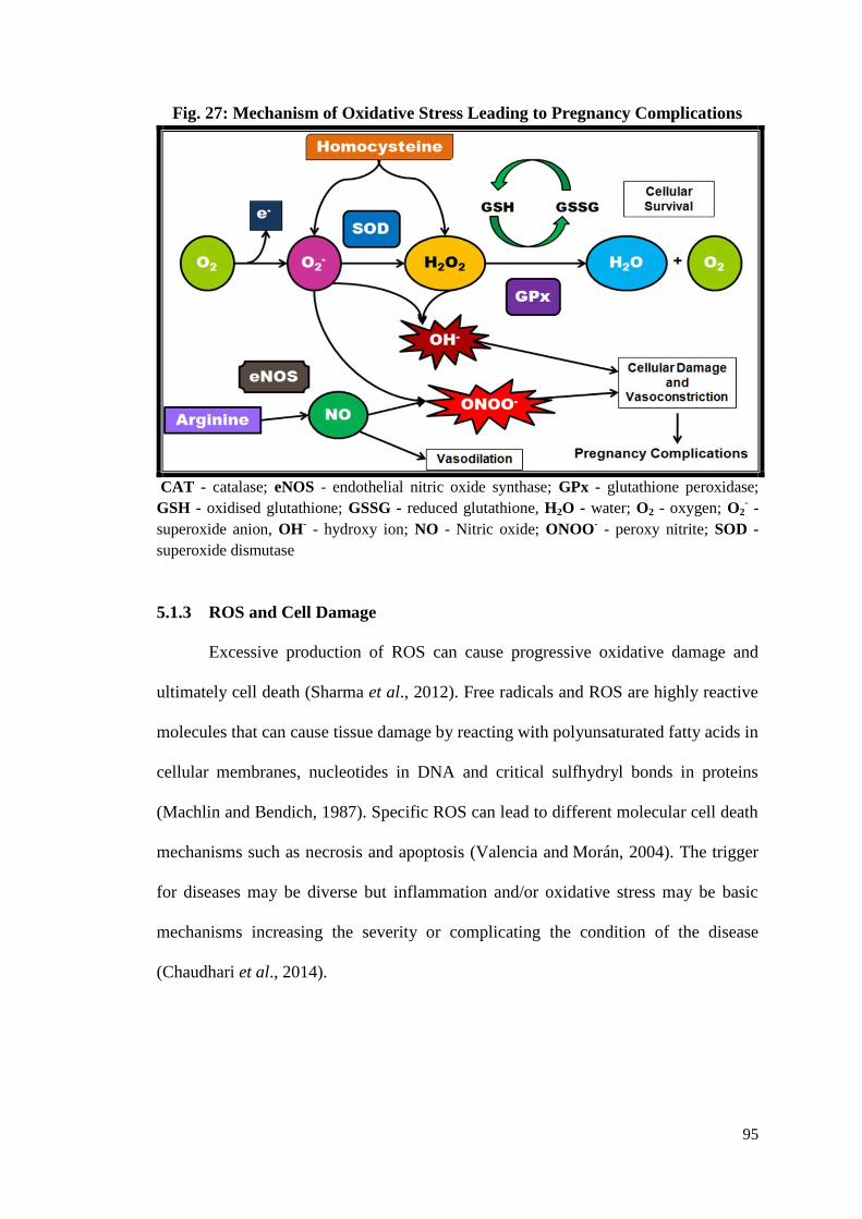
95
Fig. 27: Mechanism of Oxidative Stress Leading to Pregnancy Complications
CAT - catalase; eNOS - endothelial nitric oxide synthase; GPx - glutathione peroxidase;
GSH - oxidised glutathione; GSSG - reduced glutathione, H2O - water; O2 - oxygen; O2- -
superoxide anion, OH- - hydroxy ion; NO - Nitric oxide; ONOO
- - peroxy nitrite; SOD -
superoxide dismutase
5.1.3 ROS and Cell Damage
Excessive production of ROS can cause progressive oxidative damage and
ultimately cell death (Sharma et al., 2012). Free radicals and ROS are highly reactive
molecules that can cause tissue damage by reacting with polyunsaturated fatty acids in
cellular membranes, nucleotides in DNA and critical sulfhydryl bonds in proteins
(Machlin and Bendich, 1987). Specific ROS can lead to different molecular cell death
mechanisms such as necrosis and apoptosis (Valencia and Morán, 2004). The trigger
for diseases may be diverse but inflammation and/or oxidative stress may be basic
mechanisms increasing the severity or complicating the condition of the disease
(Chaudhari et al., 2014).

96
5.1.4 Products of Oxidative Damage
Oxidative stress and oxidative modification of biomolecules are involved in a
number of physiological and pathophysiological processes such as aging,
artheroscleosis, inflammation, carcinogenesis and drug toxicity (Lobo et al., 2010).
Overproduction of ROS results in oxidative stress and is an important mediator of
damage to cell structures, including lipids and membranes, proteins and DNA (Valko
et al., 2007).
5.1.4.1 Lipid Peroxidation
ROS can react with the PUFA of lipid membranes and induce lipid
peroxidation (Barrera, 2012). Lipid peroxidation is known to be involved in
endothelial cell dysfunction in preeclampsia (Gupta et al., 2005). Phosphatidylcholine
hydroperoxide is the primary product of lipid peroxidation, which undergoes non-
enzymatic reactions, leading to the formation of 4-hydroxynonenal (HNE) and
malondialdehyde (MDA), secondary products of lipid peroxidation (Uchida, 2003;
Esterbauer et al., 1991). Lipid peroxidation products are known to regulate gene
expression, signalling and activate receptors (Noguchi et al., 2008; Niki, 2008).
5.1.4.2 Protein Oxidation
Proteins can be oxidatively modified in three ways: oxidative modification of
specific amino acid, free radical mediated peptide cleavage and formation of protein
cross-linkage due to reaction with lipid peroxidation products (Lobo et al., 2010).
Protein containing amino acids such as methionine, cysteine, arginine, and histidine
are the most vulnerable to oxidation (Freeman and Crapo, 1982). One of the
hallmarks of severe oxidative stress is the accumulation of oxidized proteins, which
97
tend to form high molecular weight aggregates (Bader and Grune, 2006). Protein
aggregation occurs if protein repair and degradative systems are unable to act upon
oxidized proteins and restore cellular function (Squier, 2001).
5.1.4.3 DNA Oxidation
ROS can cause severe damage to DNA which can determine cell fate through
cell cycle arrest and DNA repair or the activation of apoptotic pathways (Barzilai and
Yamamoto, 2004). DNA bases are vulnerable to oxidative stress damage involving
hydroxylation, protein carbonylation and nitration (Lovell and Markesbery, 2007).
The main oxidation product in cellular DNA is 8-oxo-7, 8-dihydroguanine (Cadet et
al., 2014). Hydroxymethylcytosine, formylcytosine, and carboxylcytosine are
products generated by oxidation of methylated DNA (Delatte et al., 2014).
Oxidatively damaged DNA has been associated with aging, cancer and some
degenerative diseases (Chao et al., 2013).
5.1.5 Antioxidants
The human body has several mechanisms to counteract oxidative stress one of
which is by producing antioxidants, which are either naturally produced in situ
(endogenous), or externally supplied through foods and/or supplements (exogenous)
(Pham-Huy et al., 2008). Antioxidants including antioxidant enzymes and antioxidant
metabolites exert synergistic actions in scavenging free radicals and ROS (Fang et al.,
2002). Antioxidants act as safeguard against the accumulation of ROS and their
elimination from the system (Rajendran et al., 2014). However their target selectivity
and antioxidant effectiveness vary for different oxidants (Winterbourn, 2008).

98
5.1.5.1 Antioxidant Enzymes
Antioxidant enzymes play a major role in ROS scavenging and changes of
their expression or/and activity are reported to be associated with several disorders
(Tokarz et al., 2013; Fujita et al., 2012). In order to overcome the oxidative damage,
there are some protective signalling pathways related to transcriptional upregulation
of antioxidant enzymes (Fujita et al., 2012). Antioxidant enzymes include superoxide
dismutase (SOD), catalase (CAT), glutathione transferase (GST), glutathione
reductase (GR) and glutathione peroxidases (GPx) (Fang et al., 2002).
Superoxide Dismutase
SODs were discovered by McCord and Fridovich between 1968–1969
(Buettner, 2011). They are major antioxidant enzymes for the removal of superoxide
radicals in different subcellular compartments (Huang et al., 2012). SOD is the first
line of defence against oxidative stress under physiological and pathological
conditions (Batinić-Haberle et al., 2010). They catalyze the breakdown of the
superoxide anion into oxygen and hydrogen peroxide (Lobo et al., 2010). SODs
consist of three isoforms in mammals: the cytoplasmic Cu/ZnSOD (SOD1), the
mitochondrial MnSOD (SOD2), and the extracellular Cu/ZnSOD (SOD3) (Fukai and
Ushio-Fukai, 2011). The human gene for SOD1 has been localized to the 21q22.1
region of chromosome 21 that encodes the 32kDa homodimer (Levanon et al., 1985).
Glutathione Peroxidase
Cellular GPx was the first enzyme recognized as a selenoprotein. In GPx
selenium (Se) was found in the form of selenocysteine. The enzyme is a tetrameric
protein and is composed of four apparently identical subunits (Zachara, 1992). GPx

99
protects the organism from oxidative damage by catalyzing the reduction of harmful
hydroperoxides with thiol cofactors (Pieczyńska and Grajeta, 2015; Bhabak and
Mugesh, 2010). The selenocysteine residue at the active site of this enzyme forms a
"catalytic triad" with tryptophan and glutamine, which activates the selenium moiety
for an efficient reduction of peroxides (Bhabak and Mugesh, 2010). GPx removes
H2O2 by using it to oxidize reduced glutathione (GSH) into oxidized GSH (Lobo et
al., 2010). A total of eight mammalian GPx are classified according to their functions
in the system (Ramming and Appenzeller-Herzog, 2013). GPx are involved in
signalling cascades, e.g. in the insulin signalling pathway by GPx1; GPx2 plays a dual
role in carcinogenesis depending on the mode of initiation and cancer stage; GPx3 is
membrane associated possibly explaining a peroxidatic function despite low plasma
concentrations of GSH; GPx4 has novel roles in the regulation of apoptosis and,
together with GPx5, in male fertility. Functions of GPx6 are still unknown, and the
proposed involvement of GPx7 and GPx8 in protein folding awaits elucidation
(Brigelius-Flohé and Maiorino, 2013).
Endothelial Nitric Oxide Synthase
Nitric oxide (NO·) is an important protective molecule in the vasculature, and
endothelial NO· synthase (eNOS) is responsible for most of the vascular NO·
produced (Förstermann and Münzel, 2006). eNOS couples with essential cofactor
(6R)-5,6,7,8-tetrahydro-L-biopterin (BH4) and catalyzes the conversion of arginine,
oxygen and NADPH to NO· and citrulline (Solomonson et al., 2003). Cardiovascular
risk factors such as hypertension, hypercholesterolemia, diabetes mellitus, or chronic
smoking stimulate the production of reactive oxygen species in the vascular wall
(Tousoulis et al., 2011). Superoxide reacts avidly with vascular NO· to form

100
peroxynitrite which is known to accumulate in placental tissues in pregnancy
complications such as preeclampsia (Matsubara et al., 2015). Peroxynitrite oxidizes
BH4 promoting superoxide production by eNOS (Förstermann and Münzel, 2006). It
is believed that new information about redox regulation of eNOS may point to ways
of controlling oxidative stress in the vasculature (Hoang et al., 2013).
5.1.5.2 Antioxidant Metabolites
Antioxidant metabolites or non-enzymatic antioxidants include GSH, arginine,
citrulline, taurine, creatine, selenium, zinc, vitamin E, C, A and tea polyphenols (Fang
et al., 2002). The section below discusses the role of GSH as it plays a role in the one
carbon cycle.
Glutathione
GSH was discovered in yeast cells in 1888 (Aoyama and Nakaki, 2013). It is
the main non-protein thiol in cells whose functions are dependent on the redox-active
thiol of its cysteine moiety that serves as a cofactor for a number of antioxidant and
detoxifying enzymes (Ribas et al., 2014). GSH is a small redox-active molecule
existing in two main stable forms: the thiol and the disulphide. Challenging
environmental conditions are known to alter this ratio, notably by inducing the
accumulation of GSSG (Rahantaniaina et al., 2013). GSH is responsible for supplying
reducing equivalents to neutralize hydrogen peroxide, a toxic oxidizing agent that is
produced during normal metabolism. Without GSH, hydrogen peroxide can rise to
toxic levels in tissues and blood where it can cause severe oxidative injury to organs
and to the microvasculature (Pravda, 2014). It is involved in nutrient metabolism,
antioxidant defence and regulation of cellular metabolic functions ranging from gene

101
expression, DNA and protein synthesis to signal transduction, cell proliferation and
apoptosis (Aquilano et al., 2014). GSH is the major antioxidant in the brain, and as
such plays a key role in the detoxification of reactive oxidants (Carvalho et al., 2014;
Aoyama and Nakaki, 2013). Several antioxidant systems depend on GSH (Mailloux et
al., 2013).
5.1.6 Factors Affecting Oxidative Stress in Pregnancy
Pregnancy, placental abnormality and preexisting maternal constitutional
conditions are the major factors influencing placental oxidative stress (Yang et al.,
2012). Maternal factors like age, smoking, socioeconomic status (SES), infections,
and nutritional status influence oxidative stress (Langie et al., 2012). Modern lifestyle
including exposure to pollutants, poor diet and lack of exercise cause excess
inflammation, oxidative stress and ultimately DNA damage which increases the risk
of infertility, miscarriage and pregnancy complications (Furness et al., 2011).
5.1.6.1 Maternal Nutrition
Maternal diet, specifically the intakes of antioxidants and polyunsaturated fats
are implicated in the development of oxidative stress with subsequent effects on the
offspring health (Thornburg et al., 2010; Turpeinen et al., 1998).
Antioxidants
Antioxidant nutrients including vitamin E, vitamin C, vitamin B6, beta-
carotene, zinc and selenium have a beneficial role in oxidative stress (Hsu and Guo,
2002). The reduced levels of trace elements associated with inadequate amount of
antioxidant enzymes are suggested to be important factors associated with oxidative

102
stress leading to endothelial dysfunction in preeclamptic mothers (Negi et al., 2012).
Further, antioxidants have a beneficial role in early status of brain development in rats
against oxidative stress (Antonio-García and Massó-Gonzalez, 2008).
Complementation with micronutrients during pregnancy is an early and innovative
alternative to strengthen the prevention of chronic diseases in the population
(Ramírez-Vélez et al., 2011).
Oxidative stress resulting from folic acid deficiency is reported to be
responsible for neural tube defects (Wegrzyn et al., 2013). Low folate supply during
early life may leave an epigenetic mark that can predispose the offspring to further
dietary insults, causing adverse effects during adult life (Langie et al., 2013).
Supplementation with high-dose folic acid during pregnancy in mice is known to
protect against lipopolysaccharide-induced neural tube defects through its anti-
inflammatory and anti-oxidative effects (Zhao et al., 2014). Fortification of flour with
folic acid has a major impact on neural tube defects in many countries (Castillo-
Lancellotti et al., 2013).
Vitamin D supplementation for 9 weeks among pregnant women has been
reported to have beneficial effects on oxidative stress (Asemi et al., 2009). The
neuroprotective effect of vitamin D is associated with its influence on neurotrophin
production and release, neuromediator synthesis, intracellular calcium homeostasis,
and prevention of oxidative damage to nervous tissue (Wrzosek et al., 2013).
Vitamins A, C, E are potent antioxidants (Poston et al., 2011) but their effect
on oxidative stress markers has been inconsistent (Schnorr et al., 2011; Pasquali et al.,
2010). Supplementation of these vitamins during pregnancy to reduce the risk of
preeclampsia and preterm birth has not been consistent (Talaulikar and Manyonda,
2011; Bastani et al., 2011; Conde-Agudelo et al., 2011; Basaran et al., 2010; Pandey

103
et al., 2000). Studies suggest a need to investigate the optimum dose and timing of
antioxidant administration (Woods et al., 2001; Buettner, 1993)
Selenium supplementation can up-regulate endogenous antioxidant systems
and protect trophoblast cells from oxidative stress (Watson et al., 2012). Studies have
reported that selenium could play an important role in adverse outcomes such as
miscarriages, neural tube defects, diaphragmatic hernia, premature birth, low birth
weight, preeclampsia, glucose intolerance and gestational diabetes (Mariath et al.,
2011). Antioxidant activity of selenium is via its incorporation into selenoproteins
such as glutathione peroxidase enzymes, levels of which have been shown to be
reduced in miscarriage and preeclampsia (Mistry and Williams, 2011). It is important
to maintain an adequate selenium supply in pregnancy complications (Khera et al.,
2013).
Zinc deficiency during foetal life and lactation induces an early decrease in
renal functional units, associated with a decrease in nitric oxide activity and an
increase in oxidative stress (Tomat et al., 2013). Zinc is a widely studied
microelement in infant feeding because it is a component of several enzymes involved
in intermediary metabolism ranging from growth to cell differentiation and
metabolism of proteins, carbohydrates, and lipids (Tomat et al., 2011).
Oxidative stress is a characteristic of copper (Cu) deficiency (Uriu-Adams et
al., 2005). In dietary-induced foetal growth restriction, Cu deficiency plays a major
etiologic role in the decrease of foetal growth and anti-oxidant capacity (Ergaz et al.,
2012). Premature and low birth weight infants can be born with low Cu stores which
may be due to increased oxidative stress (Uriu-Adams et al., 2010)
Iron, deficiency and excess, can result in free radical mitochondrial damage
(Casanueva and Viteri, 2003). Supplementation with Fe and particularly in

104
combination with vitamins (riboflavin, folic acid) has been shown to improve the
haematological status as well as oxidative stress in anaemic pregnant women (Ma et
al., 2010).
Long Chain Polyunsaturated Fatty Acids
Cell membranes rich in LCPUFA are particularly susceptible to oxidative
stress (Shichiri, 2014). Oxidative stress promotes damage to plasma membrane
omega-3 fatty acids such as DHA thereby, disrupting neuronal signalling (Gomez-
Pinilla and Tyagi, 2013). Primary prevention of cardiovascular disease and mental ill
health starts, crucially, with maternal nutrition that includes consuming more omega-3
fatty acids to lower transient short-lived meal-induced oxidative stress (Robson,
2009).
Supplementation of omega-3 fatty acids have shown significantly reduced
lipid peroxidation, nucleic acid and protein oxidation, thereby promoting neuronal and
glial cell survival on brain tissue analysis of traumatic brain injury models (Kumar et
al., 2014).
Omega-3 fatty acid supplementation is reported to have beneficial effects on
oxidative stress supporting their role as antioxidants (Chaudhary et al., 2014;
Fernández-Iglesias et al., 2013; Patten et al., 2013). Maternal dietary omega-3 fatty
acid supplementation during pregnancy is reported to reduce placental oxidative
damage and increase placental levels of pro-resolving mediators in rats (Jones et al.,
2014). However, omega 3 fatty acids are also known to be highly susceptible to lipid
peroxidation (Jones et al., 2013). During pregnancy the ability of DHA to alter
placental pro-oxidant/antioxidant balance is dependent on the DHA concentration
used and the gestational age of the placental tissue (Stark et al., 2013).
105
5.1.6.2 Maternal Characteristics influencing Oxidative Stress
Age
Increase in the risk of preeclampsia with maternal age could be due to an
increase in oxidative stress associated with age (Hague et al., 2010, Tikhonova et al.,
2014). Studies in animals have shown that delayed childbearing results in shortening
of oviduct and somatic ovarian telomere length possibly due to increased oxidative
stress (Aiken et al., 2013) and this may lead to several complications in pregnancy.
Weight
Inadequate or excessive pregnancy weight gain can complicate both maternal
and foetal health (Agarwal et al., 2012). Maternal obesity (BMI, 30-43 kg/m2) is
known to be associated with increased markers of inflammation and oxidative stress
(Saben et al., 2014). Excess maternal weight gain during pregnancy elevates oxidative
stress indicating a possible increased risk of CVD development in later life
(Stefanović et al., 2012).
Increased oxidative stress is thought to be a very early response to poor
maternal nutrition and suboptimal environment in utero, and these effects are
exacerbated by accelerated postnatal growth (Tarry Adkins et al., 2013). Intrauterine
malnutrition is associated with significant oxidative stress in small for gestational age
neonates born at term to malnourished mothers (Gupta et al., 2004).
Socioeconomic Status
Lower SES is associated with higher oxidative stress and lower antioxidant
levels (Janicki-Deverts et al., 2009). Poor people age faster than rich people due to the
unhealthy environments and high oxidative stress to which they are exposed and

106
results in socioeconomic differences in health (Adams and White, 2004). The
response of a person to resource scarcity depends on the harshness of their early-life
environment and increase in oxidative stress (Griskevicius et al., 2013).
Exercise
A well-planned program of exercise is an important component of a healthy
lifestyle and is also recommended during pregnancy (Golbidi and Laher, 2013).
Exercise training can promote the synthesis and release of nitric oxide, improve
angiogenesis and decrease oxidative stress (Gąsiorowski and Dutkiewicz, 2013).
However excessive maternal activity during pregnancy is associated with smaller
foetal size in rural India (Rao et al., 2003).
Alcohol/ Smoking/ Drug Addiction
The adverse effects of smoking and alcohol on birth outcomes have been well
documented, and growing evidence indicates that exposure to either can delay the
time to conception possibly through increases in oxidative stress (Ruder et al., 2009).
Ethanol and nicotine, which harm the development of foetus, may induce both
oxidative and endoplasmic reticulum stress responses in human placental
trophoblastic cells (Repo et al., 2014). The ingestion of alcohol and/or cigarette
smoke during pregnancy can lead to the generation of ROS and produce an imbalance
in the intracellular redox state, leading to an overall increase in oxidative stress
resulting in abnormal foetal development (Dittrich et al., 2012). Smoking mothers are
reported to have more oxidative stress than non-smoking mothers (Aydogan et al.,
2013). Environmental tobacco smoke in the early postnatal period adversely affects

107
pro-oxidative/antioxidative status within first weeks of life in very early infancy
(Noakes et al., 2007).
5.1.7 Oxidative Stress and Antioxidants in Preeclampsia
Reports indicate that decreased levels of antioxidants and increased lipid
peroxidation play an important role in the pathogenesis of preeclampsia (Genc et al.,
2011; Karacay et al., 2010; Rani et al., 2010; Gupta et al., 2009; Patil et al., 2007;
Rudra et al., 2006). Oxidative stress associated with preeclampsia may be a
consequence of reduced antioxidant defence pathways. It has been suggested that
GSH, GPx and antioxidant vitamin status contribute to the endothelial dysfunction
and hypertension in preeclampsia (Mistry et al., 2008; Krishna Mohan and
Venkataramana, 2007). Reduced levels of enzymatic antioxidants like SOD, catalase,
GPx and glutathione reductase (GSR) have also been observed (Dordević et al., 2008;
Vanderlie et al., 2008).
Several mechanisms have been proposed for the protection of developing
embryo from ROS. An internal protection system exists which is comprised of both
enzymatic and nonenzymatic antioxidant defences such as SOD, GPx and GSH
(Finkel 2000). In addition there also exists an association between ROS and the
enzyme eNOS which produces nitric oxide in endothelial cells which works as
vasodilator and has been linked with preeclampsia (Kim et al., 2006).
Studies from our department and other studies have reported an imbalance
between oxidative stress and antioxidants in women which is associated with poor
birth outcome in preeclampsia (Hilali et al., 2013; Mehendale et al., 2008; Kulkarni et
al., 2010). However, these results were observed when the pathology had progressed
and may have been secondary to the effects of the disease. Further, it is suggested that
108
early pregnancy is susceptible to oxidative stress, and thus characterization of
antioxidant systems would improve the understanding of placental development and
function (Garrel et al., 2010).
Hypoxic conditions that prevail in the placenta after the first trimester increase
the production of ROS and leads to oxidative stress associated with preeclampsia
(Murray, 2012; Tal, 2012; van Patot et al., 2012). Oxidative damage leads to breaks in
DNA providing access to sites for DNA methyltransferases, which promote DNA
methylation (O'Hagan et al., 2011). Studies in humans and animal models have
reported that oxidative stress plays an important pathophysiological role in the foetal
programming of neurodevelopmental and metabolic diseases (Sartori et al., 2012,
Estringer et al., 2012). A compromised pregnancy such as preeclampsia may expose
the foetus to increased oxidative stress and through epigenetic changes may alter its
development in later life (Chen et al., 2013).
Several longitudinal studies have examined changes in oxidative stress
parameters across gestation in women with preeclampsia. However, these are on a
small sample size (Genc et al., 2011; Loverro et al., 1996). They have mainly
examined parameters like thiols, uric acid, thiobarbituric acid, vitamin E, vitamin C
and selenium (Anastasakis et al., 2008; Roes et al., 2006). They are on a
heterogeneous population and from varying ethnicities (Medrano Rodríguez et al.,
2008; Ademuyiwa et al., 2007; Jahn et al., 1997) and had women that smoked (Rudra
et al., 2006). These studies may be confounded by a heterogeneous population and
smoking that may result in altered oxidative stress levels (Szanton et al., 2012;
Noakes et al., 2007; Janicki-Deverts et al., 2009, Bizoń et al., 2011). Further, there
are limited studies examining the association of maternal levels of oxidative stress
109
markers with those in the cord blood (Mihailović et al., 2000) or birth outcome (Min
et al., 2009).
A departmental study in an animal model suggests that antioxidant defence
system of the brain may be programmed during the critical developmental windows
and may be the key mechanism leading to impaired cognition in later life (Roy et al.,
2013). We hypothesize that an abnormal antioxidant defence mechanism exists early
in pregnancy in mothers who develop preeclampsia. This may increase the risk for
adverse foetal programming of metabolic and neurodevelopmental disorders in later
life. Thus, there is a need to understand the levels of oxidative stress and antioxidant
enzymes from early gestation until delivery in women with preeclampsia and examine
their association with blood pressure (BP) and birth outcome.
This prospective study reports the maternal and cord MDA, SOD, GPx
and GSH levels at different gestational time-points in women who developed
preeclampsia as compared to normotensive controls (NC), associating it with the
birth weight and maternal systolic and diastolic BP.
5.2 Materials and Methods
5.2.1 Study Subjects
The recruitment of study population and the inclusion and exclusion criteria
are as mentioned in Chapter 3 (Section 3.2.1). Neonatal growth measures were
recorded as mentioned in Chapter 3 (Section 3.2.1).
5.2.2 Power of the Study
The power of the study based on our earlier cross sectional study on MDA
levels in mothers with preeclampsia using Power and Sample Size Calculation

110
software (version 3.0.43) (Mehendale et al., 2008). For 3 controls per preeclampsia
subject we would need to study 46 preeclampsia subjects and 138 NC subjects to be
able to reject the null hypothesis. The incidence of preeclampsia in this population is
about 8-10% of total pregnancies. In view of this, during the period of our sample
collection 148 women for NC group were selected randomly among which 123 had
complete data for all three gestational time-points with cord blood samples. In case
of preeclampsia, a total of 63 women were recruited out of which 34 women had
complete data at all gestational time-points including cord samples.
5.2.3 Sample Collection, Processing and Storage
Blood sample collection was as described in Chapter 2 (Section 2.2.2) while
placental sample collection was as described in Chapter 3 (Section 3.2.2)
5.2.4 Biochemical Estimations
MDA levels were measured from maternal and cord plasma using the Oxis
Research TM Bioxytech® (New York, USA) MDA-586TM Spectrophotometric
Assay and are expressed as µM/mL. Levels of GPx were measured from maternal
and cord erythrocytes using Oxis Research TM Bioxytech® (New York, U.S.) GPx-
340TM Colorimetric Assay for Cellular Glutathione Peroxidase and are expressed as
mU/ml. SOD levels were measured from maternal and cord erythrocytes using
Cayman’s Superoxide Dismutase Assay Kit (Michigan, USA). SOD activity was
expressed as U/ml. GSH levels were measured in maternal and cord erythrocytes
using Cayman’s (Michigan, USA) Glutathione Assay Kit and was expressed as
µM/mL. eNOS levels were measured in placental homogenates using Quantikine
Human eNOS Quantikine ELISA Kit by R&D Systems and was expressed as pg/ml.
111
5.2.5 Extraction of Total RNA, cDNA Synthesis and Quantitative Real Time
(RT)- PCR Assays
Isolation of total RNA, cDNA synthesis and mRNA levels of SOD, GPx,
GAPDH genes from NC and preeclampsia placental samples was performed. Total
RNA from placenta samples was isolated using Trizol method and quantified by the
Eppendorf Biophotometer Plus (Hamburg, Germany) by the method described by us
earlier (Meher et al., 2014). For each placental sample, 2 μg of RNA was reverse
transcribed to cDNA using the High-Capacity cDNA reverse transcription Kit from
Applied Biosystems (California, USA). RT-PCR for SOD, GPx and GAPDH were
erformed with the TaqMan Universal PCR Master Mix (California, USA) using 7500
Standard Real Time PCR system from Applied Biosystems (California, USA). The
relative expression level of the gene of interest was examined with respect
to GAPDH. Relative expression levels of genes were calculated and expressed as 2ΔCt
.
The following TaqMan® primers were used in this study: GAPDH
(Hs99999905_m1); SOD (Hs00829989_gH); GPx (Hs00166575_m1).
5.2.6 Statistical Analysis
The data were analyzed using the SPSS/PC+ package (Version 20.0, Chicago,
IL, USA). Continuous variables are reported as mean ± S.D and discrete variables as
percent (%). Log10 transformation was used for skewed distributions. Mean values of
the estimates of various parameters were compared at conventional levels of
significance (p<0.05 and p<0.01) using the independent ‘t’ test and one-way analysis
of variance (ANOVA). The data was also analyzed after adjusting for gestational age
and SES. Association between different parameters with birth outcome and BP were
analyzed after adjusting for maternal age, BMI, gestational age and SES. In the

112
current study, the variable sample numbers for different time-points and different
parameters are due to either loss in the follow up of subjects at various time-points or
insufficient sample volume available. This study reports results before and after
adjusting for confounders such as SES and gestation suggesting the importance of
confounders and appropriate statistical tools while analyzing the data. However, most
of the studies reported in literature have not defined confounders while comparing
means of the parameters.
5.3 Results
5.3.1 Maternal and Neonatal Characteristics
The maternal body mass index (BMI) at all gestational time-points in women
with preeclampsia was higher (p<0.01 for all) than NC women. Systolic BP of women
with preeclampsia were higher (p<0.01 for all) at all time-points than NC women.
Diastolic BP were higher at T2 and T3 (p<0.01 for both) in women with preeclampsia
than NC women. The percentage of nulliparous women was higher in the
preeclampsia group compared to the NC group (70% vs. 45%, p<0.01) (Table 7).
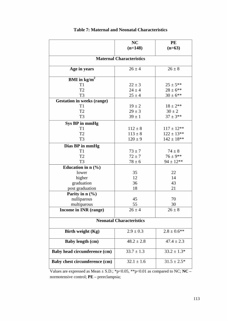
113
Table 7: Maternal and Neonatal Characteristics
Values are expressed as Mean ± S.D.; *p<0.05, **p<0.01 as compared to NC; NC –
normotensive control; PE – preeclampsia;
NC
(n=148)
PE
(n=63)
Maternal Characteristics
Age in years
26 ± 4 26 ± 8
BMI in kg/m2
T1
T2
T3
22 ± 3
24 ± 4
25 ± 4
25 ± 5**
28 ± 6**
30 ± 6**
Gestation in weeks (range)
T1
T2
T3
19 ± 2
29 ± 3
39 ± 1
18 ± 2**
30 ± 2
37 ± 3**
Sys BP in mmHg
T1
T2
T3
112 ± 8
113 ± 8
120 ± 9
117 ± 12**
122 ± 13**
142 ± 18**
Dias BP in mmHg
T1
T2
T3
73 ± 7
72 ± 7
78 ± 6
74 ± 8
76 ± 9**
94 ± 12**
Education in n (%)
lower
higher
graduation
post graduation
35
12
36
18
22
14
43
21
Parity in n (%)
nulliparous
multiparous
45
55
70
30
Income in INR (range)
26 ± 4 26 ± 8
Neonatal Characteristics
Birth weight (Kg) 2.9 ± 0.3 2.8 ± 0.6**
Baby length (cm) 48.2 ± 2.8 47.4 ± 2.3
Baby head circumference (cm) 33.7 ± 1.3 33.2 ± 1.3*
Baby chest circumference (cm) 32.1 ± 1.6 31.5 ± 2.5*

114
Neonatal characteristics show that birth weight (p<0.01), baby head and chest
circumference (p<0.05) were lower in preeclampsia group as compared to the NC
group (Table 7).
5.3.2 Maternal and Cord Malondialdehyde Levels
Maternal plasma MDA levels at T1 (p<0.01), T2 (p<0.01) and T3 (p<0.05)
were higher in women with preeclampsia than those in NC women. No difference in
cord plasma MDA levels were observed in preeclampsia group as compared to NC
group. Levels of MDA were lower at only T1 (p<0.01) and T2 (p<0.01) after
adjusting for gestational age and SES (Fig. 28 A & B).
In the NC group the MDA levels at T3 were significantly higher (p<0.01 for
both) than T1 and T2. Similarly cord levels were also higher (p<0.01 for both) than
T1 and T2. However in women with preeclampsia the levels of MDA did not vary
across gestation.
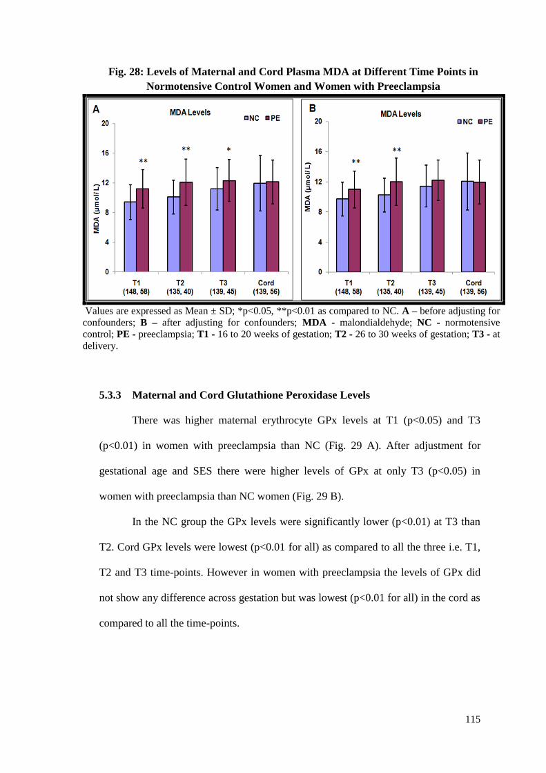
115
Fig. 28: Levels of Maternal and Cord Plasma MDA at Different Time Points in
Normotensive Control Women and Women with Preeclampsia
Values are expressed as Mean ± SD; *p<0.05, **p<0.01 as compared to NC. A – before adjusting for
confounders; B – after adjusting for confounders; MDA - malondialdehyde; NC - normotensive
control; PE - preeclampsia; T1 - 16 to 20 weeks of gestation; T2 - 26 to 30 weeks of gestation; T3 - at
delivery.
5.3.3 Maternal and Cord Glutathione Peroxidase Levels
There was higher maternal erythrocyte GPx levels at T1 (p<0.05) and T3
(p<0.01) in women with preeclampsia than NC (Fig. 29 A). After adjustment for
gestational age and SES there were higher levels of GPx at only T3 (p<0.05) in
women with preeclampsia than NC women (Fig. 29 B).
In the NC group the GPx levels were significantly lower (p<0.01) at T3 than
T2. Cord GPx levels were lowest (p<0.01 for all) as compared to all the three i.e. T1,
T2 and T3 time-points. However in women with preeclampsia the levels of GPx did
not show any difference across gestation but was lowest (p<0.01 for all) in the cord as
compared to all the time-points.

116
Fig. 29: Levels of Maternal and Cord Erythrocyte GPx at Different Time Points in
Normotensive Women and Women with Preeclampsia
Values are expressed as Mean ± SD; *p<0.05, **p<0.01 as compared to NC; A – before adjusting for
confounders; B – after adjusting for confounders; GPx – glutathione peroxidase; NC - normotensive
control; PE - preeclampsia; T1 - 16 to 20 weeks of gestation; T2 - 26 to 30 weeks of gestation; T3 - at
delivery
5.3.4 Maternal and Cord Superoxide Dismutase Levels
The levels of maternal erythrocyte SOD were lower at T2 (p<0.05), T3
(p<0.05) and in cord (p<0.05) in the preeclampsia group than NC group (Fig. 30 A).
After adjusting for gestational age and SES, SOD levels were lower at T2 (p<0.05),
T3 (p<0.01) and in cord (p<0.05) in preeclampsia group than NC group (Fig. 30 B).
There was no longitudinal difference within the NC and preeclampsia groups.

117
Fig. 30: Levels of Maternal and Cord Erythrocyte SOD at Different Time Points in
Normotensive Control Women and Women with Preeclampsia
Values are expressed as Mean ± S.D.; *p<0.05, **p<0.01 as compared to NC. A – before adjusting for
confounders; B – after adjusting for confounders; NC - normotensive control; PE - preeclampsia; SOD –
superoxide dismutase; T1 - 16 to 20 weeks of gestation; T2 - 26 to 30 weeks of gestation; T3 - at delivery
5.3.5 Maternal and Cord Glutathione Levels
The maternal erythrocyte GSH levels at T1 (p<0.05) and T2 (p<0.05) were
lower in women with preeclampsia than NC women (Fig. 31 A). However, after
adjusting for gestational age and SES the GSH levels were lower only at T1 (p<0.05)
in women with preeclampsia than NC (Fig. 31 B).
In the NC group the maternal GSH levels showed reducing trend towards term
where the levels at T3 was lower (p=0.05) than T2 although it was not statistically
significant. However in women with preeclampsia the levels of GSH did not show
any difference across gestation.

118
Fig. 31: Levels of Maternal and Cord Erythrocyte GSH at Different Time-Points in
Normotensive Control Women and Women with Preeclampsia
Values are expressed as Mean ± S.D.; *p<0.05. A – before adjusting for confounders; B – after
adjusting for confounders; GSH - glutathione; NC - normotensive control; PE - preeclampsia; T1 -
16 to 20 weeks of gestation; T2 - 26 to 30 weeks of gestation; T3 - at delivery
5.3.6 Placental SOD and GPx Gene Expression
Placental GPx mRNA levels were lower (p<0.05) in women with
preeclampsia than in NC women (Fig. 32 A). However placental SOD gene mRNA
levels were comparatively higher in preeclampsia than NC group although it was not
statistically significant (Fig. 32 B).

119
Fig. 32: Placental SOD and GPx Gene Expression
Values are expressed as Mean ± S.E.; *p<0.05 as compared to NC. A – expression of
SOD; B – expression of GPx; GPx – glutathione peroxidase; NC - normotensive control;
PE - preeclampsia; SOD – superoxide dismutase
5.3.7 Placental Endothelial Nitric Oxide Synthase Levels
Placental levels of eNOS were comparatively lower in preeclampsia than NC
group although it was not statistically significant (Fig. 33).
Fig. 33: Placental Levels of eNOS
Values are expressed as Mean ± S.D.;
eNOS – endothelial nitric oxide synthase
NC - normotensive control; PE -
preeclampsia;

120
5.3.8 Association of MDA with Blood Pressure
Maternal MDA was positively (r = 0.176, p = 0.049, n = 129) associated with
diastolic BP at T2 in the NC group but not in the preeclampsia group.
5.3.9 Association of SOD with Blood Pressure
Maternal SOD was positively (r = 0.267, p = 0.043, n = 62) associated with
diastolic BP at T1 in preeclampsia group.
5.3.10 Association of GPx with Blood Pressure and Birth Outcomes
Maternal GPx was negatively (r = -0.22, p = 0.02, n = 115) associated with
diastolic BP at T2 in NC group. Maternal GPx of T3 was negatively (r = -0.195, p =
0.035, n = 122) associated with birth weight in NC group while at T2 it was positively
(r = 0.386, p = 0.026, n = 37) associated with birth weight in preeclampsia group.
5.3.11 Association between Oxidative Stress Markers and Neurotrophins
Maternal MDA levels were negatively associated with BDNF at T2 (r = -
0.198, p = 0.028, n = 124) in the whole cohort and at T2 (r = -0.368, p = 0.020, n =
40) in women with preeclampsia.
5.4 Discussion
This study demonstrates some important findings in the preeclampsia group
(1) Higher plasma MDA levels at T1 and T2 (2) Higher erythrocyte GPx levels at T3
(3) Lower erythrocyte SOD at T2, T3 and in cord (3) Lower erythrocyte GSH levels
at T1 (4) Lower expression of GPx in the placenta (5) The cord GPx was lowest as
compared to maternal levels at all the three time-points (6) There was a positive

121
association of maternal SOD with diastolic BP at T1 and positive association of
maternal GPx at T2 with birth weight. In addition in the NC group (1) MDA levels
were highest at term while GPx levels were lowest in cord (2) Positive association
between MDA and diastolic BP but a negative association between GPx and diastolic
BP at T2. (3) Negative association between GPx of T3 with birth weight.
5.4.1 Higher MDA Levels in Women with Preeclampsia
In the current study, maternal plasma MDA levels were higher in women with
preeclampsia at T1, and T2 as compared to NC women. However at delivery, the
difference between preeclampsia and NC women MDA levels were significant only
without adjusting for gestational age and SES. This is similar to studies which have
shown higher MDA levels in serum, plasma and placental tissue samples in women
with preeclampsia at delivery (Can et al., 2014; Siddiqui et al., 2013; Suhail et al.,
2009). Further, higher MDA levels during 20–23 weeks of gestation have been
reported in preeclampsia (Rudra et al. 2006, Anastasakis et al., 2008). Lipid
peroxidation has been shown to induce endothelial dysfunction in experimental
models, endothelial cell swelling, and damage cell membranes directly and reduced
endothelium-dependent vasorelaxation (Molyneux et al., 2002). It has been suggested
that higher MDA levels parallel the severity of preeclampsia (Wiktor and Kankofer,
2001). Reports indicate that oxidative stress is a cause of apoptosis in feto-placental
unit which predates abnormalities in the foetus and foetal growth restriction
(Nakatsukasa et al., 2013).
In the NC group MDA levels were highest at term and in cord blood. MDA
level in umbilical cord blood is suggested to serve as an indication of perinatal
oxidative stress (Gulbayzar et al., 2011). However, in women with preeclampsia the

122
levels of MDA were high right from T1 and remained similar throughout suggesting
that this may be affecting the early developmental processes. MDA levels at T2 were
positively associated with diastolic BP in a normotensive pregnancy and support
studies which indicate that oxidative stress increases mid gestation possibly
elevating blood pressure in normal pregnancy (Casanueva and Viteri, 2003).
5.4.2 Higher GPx Levels in Women with Preeclampsia
In this study maternal erythrocyte GPx levels were higher at delivery in
women with preeclampsia as compared to NC women. There are several studies
which report higher GPx levels in mothers with preeclampsia at term (Krishna Mohan
and Venkataramana, 2007; Sharma et al., 2006; Llurba et al., 2004). In contrast lower
maternal serum levels of GPx have also been reported (Negi et al., 2012).
In the NC group, the GPx levels were lower at term and lowest in cord.
Similar trends were also observed in women with preeclampsia. This supports other
studies which report lower levels of antioxidants like GPx, SOD and GSH at term
(Patil et al., 2007; Davidge et al., 1992). In this study, the levels of GPx at T3 were
negatively associated with birth weight in normal pregnancy while on contrary at
delivery GPx levels were positively associated with birth weight in preeclampsia.
Further, higher GPx levels were associated with reduced diastolic BP in normal
pregnancy. Both the levels of GPx and MDA during T2 time-point and their
associations with blood pressure indicate that the level of oxidative stress during this
period of pregnancy affects endothelial functioning of the mothers. However further
studies are required to understand these associations.

123
5.4.3 Lower SOD Levels in Women with Preeclampsia
In this study, lower levels of maternal erythrocyte SOD at T2, at delivery and
in cord was observed in preeclampsia as compared to control. It has been reported that
SOD levels are lower in women who develop preeclampsia at 10–14 and 20–24
weeks of gestation (Genc et al., 2011). Reports from cross-sectional studies indicate
lower maternal and cord SOD levels in women with preeclampsia (Negi et al., 2012;
Patil et al., 2009).
A positive association between maternal SOD and diastolic BP at T1 was
observed in the preeclampsia group. SOD is responsible for production of H2O2 which
if not converted to H2O results in production of harmful radicals. It is reported that
accumulation of H2O2 in kidney results in sustained hypertension and arterial BP
(Makino et al., 2003). However, treatment with SOD mimetics has been reported to
reduce blood pressure in rat models (Rodriguez-Iturbe et al., 2003).
5.4.4 Lower GSH Levels in Women with Preeclampsia
Maternal GSH levels were lower in women who develop preeclampsia as
compared to NC women at T1. Reports indicate that GSH and GSH-related enzymes
are involved in the metabolism and detoxification of cytotoxic compounds (Knapen et
al., 1999). However, no change in placental or serum GSH levels in women with
preeclampsia has also been reported (Das et al., 2012). A study in animals reports that
reduced GSH causes significant increase in blood pressure (Vaziri et al., 2000).
In the present study, higher levels of GSH were observed during early
pregnancy only in NC but not in preeclampsia women. Early embryos are sensitive to
oxidative stress and mitochondria are known to provide ATP for GSH production to
participate in the regeneration of NADPH (Dumollard et al., 2009).

124
5.4.5 Lower Placental Expression of GPx Gene in Women with Preeclampsia
In this study, there was a lower expression of GPx gene in the placenta in the
preeclampsia group. This is in accordance with other studies that show highly
significant reductions in expression of GPx in placentae from women with
preeclampsia (Roland-Zejly et al., 2011; Mistry et al., 2010). The placenta is known
to synthesize extracellular GPx (Avissar et al., 1994). Reduced expression of GPx and
increased levels of GPx in maternal blood observed in this study may be due to the
placental pathology of preeclampsia.
5.4.6 Lower Placental eNOS Levels in Women with Preeclampsia
In our study, the levels of eNOS were comparatively lower in preeclampsia
than NC group but it was not statistically significant possibly due to small sample
size. The altered production of nitric oxide may influence the reduced placental blood
flow associated with preeclampsia. Our findings support earlier studies which report
lower placental tissue of nitric oxide synthase in preeclampsia (Brennecke et al.,
1997; Morris et al., 1995).
5.4.7 Negative Association between MDA and BDNF in Women with
Preeclampsia
In this study maternal MDA levels were negatively associated with BDNF at
T2 in the whole cohort and in women with preeclampsia. Oxidative stress is a factor
that has been identified as important in the pathogenesis of neurological diseases
(Crane, 2014). Antioxidant enzymes are associated with, and in some cases, prevent
neuronal death in animal models of neurodegenerative diseases (Fujita et al., 2012).
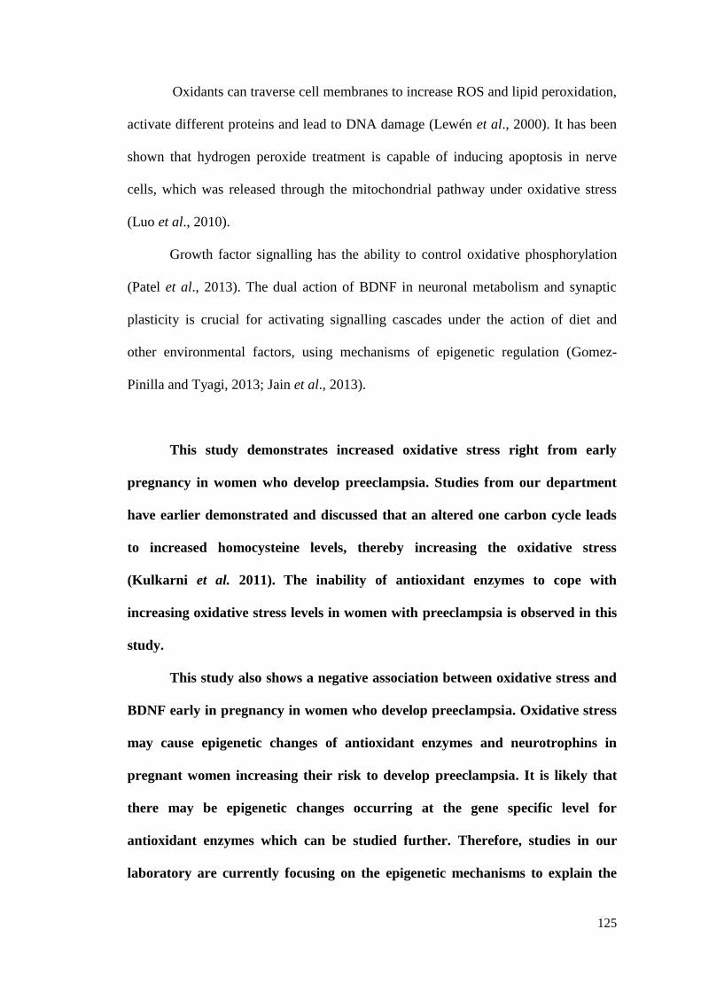
125
Oxidants can traverse cell membranes to increase ROS and lipid peroxidation,
activate different proteins and lead to DNA damage (Lewén et al., 2000). It has been
shown that hydrogen peroxide treatment is capable of inducing apoptosis in nerve
cells, which was released through the mitochondrial pathway under oxidative stress
(Luo et al., 2010).
Growth factor signalling has the ability to control oxidative phosphorylation
(Patel et al., 2013). The dual action of BDNF in neuronal metabolism and synaptic
plasticity is crucial for activating signalling cascades under the action of diet and
other environmental factors, using mechanisms of epigenetic regulation (Gomez-
Pinilla and Tyagi, 2013; Jain et al., 2013).
This study demonstrates increased oxidative stress right from early
pregnancy in women who develop preeclampsia. Studies from our department
have earlier demonstrated and discussed that an altered one carbon cycle leads
to increased homocysteine levels, thereby increasing the oxidative stress
(Kulkarni et al. 2011). The inability of antioxidant enzymes to cope with
increasing oxidative stress levels in women with preeclampsia is observed in this
study.
This study also shows a negative association between oxidative stress and
BDNF early in pregnancy in women who develop preeclampsia. Oxidative stress
may cause epigenetic changes of antioxidant enzymes and neurotrophins in
pregnant women increasing their risk to develop preeclampsia. It is likely that
there may be epigenetic changes occurring at the gene specific level for
antioxidant enzymes which can be studied further. Therefore, studies in our
laboratory are currently focusing on the epigenetic mechanisms to explain the

126
increased oxidative stress in women with preeclampsia. These changes can link
maternal nutrition, oxidative stress, altered placental development and foetal
programming of adult NCDs.


127
The present thesis entitled “Neurotrophins and Oxidative Stress in
Preeclampsia” examines the levels of neurotrophins and oxidative stress markers in a
cross sectional study on women recruited at delivery as well as in a prospective study
at various time points across gestation. The cross sectional study was carried out on
95 normotensive control women and 106 women with preeclampsia (those delivering
at term [n = 60] and those delivering preterm [n = 46]). The prospective study
followed pregnant women at three time points i.e. 16 - 20 weeks of gestation (T1), at
26 - 30 weeks of gestation (T2) and the third at delivery (T3) and included 148
normotensive women women and 63 women with preeclampsia. The hypothesis of
the study is “Oxidative stress, through the dysregulation of the one carbon cycle leads
to altered levels and expression of antioxidant enzymes and neurotrophins in
preeclampsia.”
Preeclampsia is a pregnancy related disorder characterized by hypertension
and proteinuria that develops after 20 weeks of gestation and the precise origins are
unknown. Maternal nutrition is an important factor that contributes to adverse
pregnancy outcomes such as preeclampsia. Studies carried out earlier in our
department have shown altered levels of micronutrients (folic acid and vitamin B12),
lower docosahexaenoic acid (DHA), increased homocysteine and altered angiogenic
balance at the end of pregnancy in women with preeclampsia. However, these results
were observed when the pathology had progressed and may have been secondary to
the effects of the disease.
Aberrations in the one carbon cycle leading to increased homocysteine have
been suggested to induce oxidative stress. Oxidative stress is defined as an imbalance
between the generation of reactive oxygen species and the antioxidants that prevent
oxidative damages. Antioxidants like superoxide dismutase (SOD), catalase and
128
glutathione peroxidase (GPx) are known to play a key role in combating oxidative
stress. Studies have shown that oxidative stress regulates neurotrophins such as nerve
growth factor (NGF) and brain derived neurotrophic factor (BDNF). These
neurotrophins play an important role in the implantation, placental development and
foetal growth. It is likely that an imbalanced one carbon cycle epigenetically
influences the expression of various growth factors like neurotrophins and antioxidant
enzymes. This will lead to pregnancy complications like preeclampsia and increase
risk for diseases in children at later life. (Fig. 34)
Fig. 34: Altered One Carbon Metabolism Leads to Epigenetic Modification of
Vital Genes (Antioxidant Enzymes and Neurotrophins) Resulting in Preeclampsia
DNA – deoxyribonucleic acid; LCPUFA - long chain polyunsaturated fatty acids

129
This is of significance since it is known that children born to mothers with
preeclampsia are said to be at increased risk for developing behavioral problems
during childhood and schizophrenia in later life.
The current study examines the levels of oxidative stress markers and
neurotrophins from early pregnancy in women who develop preeclampsia. Further,
the association of these parameters with blood pressure (BP) and birth outcome are
also examined.
The Novel Findings from the Cross Sectional Study
This study for the first time demonstrates the following interesting findings
1. Women with preeclampsia (both delivering at term and preterm) had lower
BDNF levels as compared to NC women.
2. Cord BDNF levels were higher in women with preeclampsia (term and
preterm) as compared to the normotensive group.
3. Among the preeclampsia groups it was lower in the preterm-PE group as
compared to the term-PE group. This may be due to either a preferential
transport for brain-sparing in babies born to women with preeclampsia or
impaired BDNF receptor signalling. Reduced cord BDNF levels observed in
the current study may have implications for neonates born to women with
preeclampsia delivering preterm, since they may be at increased risk of
neurodevelopmental disorders in later life.
4. Maternal BDNF levels showed a negative correlation with blood pressure.
130
Our data based on altered angiogenesis reported earlier from our department
and the reduced levels of BDNF in preeclampsia in the present study
highlights the need to understand possible mechanisms underlying the
association between angiogenesis and neurotrophins like BDNF in
preeclampsia.
The Novel Findings from the Prospective Study:
Women with preeclampsia demonstrated the following
1. Higher plasma malondialdehyde (MDA) levels at T1 and T2 as compared to
NC women indicating presence of increased oxidative stress in women with
preeclampsia from early pregnancy.
2. Higher erythrocyte GPx levels at T3 as compared to normotensive control
women.
3. Lower erythrocyte SOD at T2, T3 and in cord as compared to NC women.
4. Lower erythrocyte GSH levels at T1 as compared to NC women.
5. Lower expression of GPx in the placenta of women with preeclampsia as
compared to NC women. The placenta is known to synthesize extracellular
glutathione peroxidase and reduced expression observed in this study may be
due to the placental pathology of preeclampsia.
6. Lower maternal BDNF levels at T1 and T3 in women with preeclampsia than
those in NC women may be partly responsible for abnormal foeto-placental
development in preeclampsia.
7. Lower levels of cord BDNF was also observed in preeclampsia which may
have long-lasting consequences in their babies.

131
8. The lower placental BDNF gene expression levels observed in women with
preeclampsia may be associated with impaired placental development in
preeclampsia.
9. Maternal NGF levels in women with preeclampsia were comparable to NC
women at all time points.
10. Cord NGF levels at delivery were higher in women with preeclampsia as
compared to NC women
11. A negative association was observed between maternal diastolic BP and NGF
levels at T1 in women with preeclampsia.
12. There was a positive association between cord NGF levels and baby head and
chest circumference in the whole cohort.
The current study demonstrates increased oxidative stress from early
pregnancy in women who develop preeclampsia. This study also shows the inability
of antioxidant enzymes to cope with increasing oxidative stress levels in women with
preeclampsia. Lower maternal BDNF levels in women with preeclampsia during early
pregnancy may be implicated in the improper placental development. This study also
shows a negative association between oxidative stress and BDNF early in pregnancy
in women who develop preeclampsia. It is likely that oxidative stress may be
associated with epigenetic changes of antioxidant enzymes and neurotrophins in
preeclampsia.
IMPLICATIONS OF THE STUDY
This study for the first time examines the levels of oxidative stress markers
and neurotrophins from early pregnancy in women who develop preeclampsia. The

132
current study provides a plausible molecular mechanism leading to preeclampsia. Our
findings suggest that altered levels of neurotrophins and oxidative stress may affect
the placental development in preeclampsia. Lower levels of BDNF in the cord may
have implications for altered neurodevelopment in children born with preeclampsia.
SOCIETAL RELEVANCE OF THE STUDY
The incidence of preeclampsia is increasing in developing countries like India
making it an important health issue. Therefore, there is an urgent need to understand
the mechanisms of preeclampsia in order to develop strategies for prevention and
treatment. The current study highlights the role of oxidative stress and neurotrophins
in the pathophysiology of preeclampsia from early pregnancy.
FUTURE DIRECTIONS
Future studies need to be undertaken to examine gene specific methylation of
the antioxidant enzymes and neurotrophin genes. This will help in understanding the
epigenetic mechanism involved in the pathogenesis of preeclampsia. There is also a
need to follow-up children born to mothers with preeclampsia to identify whether they
are at risk of developing neurodevelopmental disorders in later life.

133
Akcakus M, Altunay L, Yikilmaz A, Yazici C, Koklu E. The relationship between
abdominal aortic intima-media thickness and lipid profile in neonates born to
mothers with preeclampsia. J Pediatr Endocrinol Metab. 2010; 23:1143-9.
Alexander BT, Henry Dasinger J, Intapad S. Effect of Low Birth Weight on Women's
Health. Clin Ther. 2014; 36:1913-23.
Al-Gubory KH, Fowler PA, Garrel C. The roles of cellular reactive oxygen species,
oxidative stress and antioxidants in pregnancy outcomes. Int J Biochem Cell
Biol. 2010; 42:1634-50.
Al-Jameil N, Aziz Khan F, Fareed Khan M, Tabassum H. A brief overview of
preeclampsia. J Clin Med Res. 2014; 6:1-7.
Aloe L, Rocco ML, Bianchi P, Manni L. Nerve growth factor: from the early
discoveries to the potential clinical use. J Transl Med. 2012; 10:239.
Andero R, Ressler KJ. Fear extinction and BDNF: translating animal models of PTSD
to the clinic. Genes Brain Behav. 2012; 11:503-12.
Andraweera PH, Dekker GA, Dissanayake VH, Bianco-Miotto T, Jayasekara RW,
Roberts CT. Vascular endothelial growth factor family gene polymorphisms in
preeclampsia in Sinhalese women in Sri-Lanka. J Matern Fetal Neonatal Med.
2013; 26:532-6.
Arévalo JC, Wu SH. Neurotrophin signaling: many exciting surprises! Cell Mol Life
Sci. 2006; 63:1523-37.
Aukes AM, Wessel I, Dubois AM, Aarnoudse JG, Zeeman GG. Self-reported
cognitive functioning in formerly eclamptic women. Am J Obstet Gynecol.
2007; 197: 365.e1-6.

134
Bae H, Ra Y, Han C, Kim DJ. Decreased serum level of NGF in alcohol-dependent
patients with declined executive function. Neuropsychiatr Dis Treat. 2014;
10:2153-2157.
Bagnyukova TV, Powell CL, Pavliv O, Tryndyak VP, Pogribny IP. Induction of
oxidative stress and DNA damage in rat brain by a folate/methyl-deficient
diet. Brain Res. 2008; 1237:44-51.
Balaratnasingam S, Janca A. Brain Derived Neurotrophic Factor: a novel
neurotrophin involved in psychiatric and neurological disorders. Pharmacol
Ther. 2012; 134:116-24.
Balogun KA, Cheema SK. The expression of neurotrophins is differentially regulated
by ω-3 polyunsaturated fatty acids at weaning and postweaning in C57BL/6
mice cerebral cortex. Neurochem Int. 2014; 66:33-42.
Barde YA, Edgar D, Thoenen H. Purification of a new neurotrophic factor from
mammalian brain. EMBO J. 1982; 1:549-53.
Barker DJ, Eriksson JG, Forsén T, Osmond C. Fetal origins of adult disease: strength
of effects and biological basis. Int J Epidemiol. 2002; 31:1235-9.
Barker DJ, Thornburg KL. Placental programming of chronic diseases, cancer and
lifespan: a review. Placenta. 2013; 34:841-5.
Barouki R, Gluckman PD, Grandjean P, Hanson M, Heindel JJ. Developmental
origins of non-communicable disease: implications for research and public
health. Environ Health. 2012; 11:42.
Basso O, Rasmussen S, Weinberg CR, Wilcox AJ, Irgens LM, Skjaerven R. Trends in
fetal and infant survival following preeclampsia. JAMA. 2006; 296:1357-62.
Bath KG, Schilit A, Lee FS. Stress effects on BDNF expression: effects of age, sex,
and form of stress. Neuroscience. 2013; 239:149-56.

135
Bazan NG, Calandria JM, Serhan CN. Rescue and repair during photoreceptor cell
renewal mediated by docosahexaenoic acid-derived neuroprotectin D1. J Lipid
Res. 2010; 51:2018-31.
Benn K, Passos M, Jayaram A, Harris M, Bongiovanni AM, Skupski D, Witkin SS.
Association between neurotrophin 4 and long-chain polyunsaturated fatty acid
levels in mid-trimester amniotic fluid. Reprod Sci. 2014; 21:1395-400.
Berti C, Cetin I, Agostoni C, Desoye G, Devlieger R, Emmett PM, Ensenauer R,
Hauner H, Herrera E, Hoesli I, Krauss-Etschmann S, Olsen SF, Schaefer-Graf
U, Schiessl B, Symonds ME, Koletzko B. Pregnancy and infants' outcome:
nutritional and metabolic implications. Crit Rev Food Sci Nutr. 2014 (In
press)
Bevilacqua E, Gomes SZ, Lorenzon AR, Hoshida MS, Amarante-Paffaro AM.
NADPH oxidase as an important source of reactive oxygen species at the
mouse maternal-fetal interface: putative biological roles. Reprod Biomed
Online. 2012; 25:31-43.
Bhatia HS, Agrawal R, Sharma S, Huo YX, Ying Z, Gomez-Pinilla F. Omega-3 fatty
acid deficiency during brain maturation reduces neuronal and behavioral
plasticity in adulthood. PLoS One. 2011; 6:e28451.
Bibel M, Barde YA. Neurotrophins: key regulators of cell fate and cell shape in the
vertebrate nervous system. Genes Dev. 2000; 14:2919-37.
Binder DK, Scharfman HE. Brain-derived neurotrophic factor. Growth Factors. 2004;
22:123-31.
Biondi C, Pavan B, Lunghi L, Fiorini S, Vesce F. The role and modulation of the
oxidative balance in pregnancy. Curr Pharm Des. 2005; 11:2075-89.
136
Bisai S, Mahalanabis D, Sen A, Bose K, Datta N. Maternal early second trimester
pregnancy weight in relation to birth outcome among Bengalee Hindus of
Kolkata, India. Ann Hum Biol. 2007; 34:91-101.
Bischof P, Irminger-Finger I. The human cytotrophoblastic cell, a mononuclear
chameleon. Int J Biochem Cell Biol. 2005; 37:1-16.
Bloomfield FH, Jaquiery AL, Oliver MH. Nutritional regulation of fetal growth.
Nestle Nutr Inst Workshop Ser. 2013; 74:79-89.
Boehme F, Gil-Mohapel J, Cox A, Patten A, Giles E, Brocardo PS, Christie BR.
Voluntary exercise induces adult hippocampal neurogenesis and BDNF
expression in a rodent model of fetal alcohol spectrum disorders. Eur J
Neurosci. 2011; 33:1799-811.
Bourre JM. Effects of nutrients (in food) on the structure and function of the nervous
system: update on dietary requirements for brain. Part 1: micronutrients. J
Nutr Health Aging. 2006; 10:377-85.
Bradshaw RA, Murray-Rust J, Ibáñez CF, McDonald NQ, Lapatto R, Blundell TL.
Nerve growth factor: structure/function relationships. Protein Sci. 1994;
3:1901-13.
Branchi I, D'Andrea I, Fiore M, Di Fausto V, Aloe L, Alleva E. Early social
enrichment shapes social behavior and nerve growth factor and brain-derived
neurotrophic factorlevels in the adult mouse brain. Biol Psychiatry. 2006;
60:690-6.
Branchi I, Francia N, Alleva E. Epigenetic control of neurobehavioural plasticity: the
role of neurotrophins. Behav Pharmacol. 2004; 15:353-62.

137
Brett KE, Ferraro ZM, Yockell-Lelievre J, Gruslin A, Adamo KB. Maternal-fetal
nutrient transport in pregnancy pathologies: the role of the placenta. Placenta.
2012; 33 S Int J Mol Sci. 2014; 15:16153-85.
Brown MC, Best KE, Pearce MS, Waugh J, Robson SC, Bell R. Cardiovascular
disease risk in women with pre-eclampsia: systematic review and meta-
analysis. Eur J Epidemiol. 2013; 28:1-19.
Brozović G, Sklebar I, Sunko BM, Blagaić V. Neurological disorders in pregnancy.
Acta Med Croatica. 2012; 66:81-4.
Bulló M, Peeraully MR, Trayhurn P, Folch J, Salas-Salvadó J. Circulating nerve
growth factor levels in relation to obesity and the metabolic syndrome in
women. Eur J Endocrinol. 2007; 157:303-10.
Burdge GC, Lillycrop KA. Nutrition, epigenetics, and developmental plasticity:
implications for understanding human disease. Annu Rev Nutr. 2010; 30:315-
39.
Burton GJ, Charnock-Jones DS, Jauniaux E. Regulation of vascular growth and
function in the human placenta. Reproduction. 2009; 138:895-902
Burton GJ, Jauniaux E, Charnock-Jones DS. The influence of the intrauterine
environment on human placental development. Int J Dev Biol. 2010; 54:303-
12.
Burton GJ, Jauniaux E. Oxidative stress. Best Pract Res Clin Obstet Gynaecol. 2011;
25:287-99.
Burton GJ, Watson AL, Hempstock J, Skepper JN, Jauniaux E. Uterine glands
provide histiotrophic nutrition for the human fetus during the first trimester of
pregnancy. J Clin Endocrinol Metab. 2002; 87:2954-9.

138
Bus BA, Molendijk ML, Penninx BJ, Buitelaar JK, Kenis G, Prickaerts J, Elzinga
BM, Voshaar RC. Determinants of serum brain-derived neurotrophic factor.
Psychoneuroendocrinology. 2011; 36:228-39.
Cabrera-Vásquez S, Navarro-Tableros V, Sánchez-Soto C, Gutiérrez-Ospina G,
Hiriart M. Remodelling sympathetic innervation in rat pancreatic islets
ontogeny. BMC Dev Biol. 2009; 9:34.
Campoy C, Escolano-Margarit MV, Anjos T, Szajewska H, Uauy R. Omega 3 fatty
acids on child growth, visual acuity and neurodevelopment. Br J Nutr. 2012;
107:S85-106.
Cannon TD, Yolken R, Buka S, Torrey EF; Collaborative Study Group on the
Perinatal Origins of Severe Psychiatric Disorders. Decreased neurotrophic
response to birth hypoxia in the etiology of schizophrenia. Biol Psychiatry.
2008; 64:797-802.
Caporali A, Emanueli C. Cardiovascular actions of neurotrophins. Physiol Rev. 2009;
89:279-308.
Carlson BM. Patten’s Foundations of Embryology, Ed 6, New York, McGraw-Hill,
1996, 180.
Carr DB, Epplein M, Johnson CO, Easterling TR, Critchlow CW. A sister's risk:
family history as a predictor of preeclampsia. Am J Obstet Gynecol. 2005;
193:965-72.
Carter AM. Evolution of placental function in mammals: the molecular basis of gas
and nutrient transfer, hormone secretion, and immune responses. Physiol Rev.
2012; 92:1543-76.

139
Carvalho AL, Caldeira MV, Santos SD, Duarte CB. Role of the brain-derived
neurotrophic factor at glutamatergic synapses. Br J Pharmacol. 2008; 153
Suppl 1: S310-24.
Castro-Rendón WA, Castro-Alvarez JF, Guzmán-Martinez C, Bueno-Sanchez JC.
Blastocyst-endometrium interaction: intertwining a cytokine network. Braz J
Med Biol Res. 2006; 39:1373-85.
Cerdeira AS, Karumanchi SA. Angiogenic factors in preeclampsia and related
disorders. Cold Spring Harb Perspect Med. 2012; 2: a006585.
Chaldakov GN, Fiore M, Stankulov IS, Manni L, Hristova MG, Antonelli A, Ghenev
PI, Aloe L. Neurotrophin presence in human coronary atherosclerosis and
metabolic syndrome: a role for NGF and BDNF incardiovascular disease?
Prog Brain Res. 2004; 146:279-89.
Champagne FA. Epigenetic mechanisms and the transgenerational effects of maternal
care. Front Neuroendocrinol. 2008; 29:386-97.
Chao MV. Neurotrophins and their receptors: a convergence point for many signalling
pathways. Nat Rev Neurosci. 2003; 4:299-309.
Chaves RN, Alves AM, Lima LF, Matos HM, Rodrigues AP, Figueiredo JR. Role of
nerve growth factor (NGF) and its receptors in folliculogenesis. Zygote. 2013;
21:187-97.
Chen CP. Placental villous mesenchymal cells trigger trophoblast invasion. Cell Adh
Migr. 2014; 8:94-7.
Chen DB, Zheng J. Regulation of placental angiogenesis. Microcirculation. 2014;
21:15-25.

140
Chen ZP, Zhou HL, Cao LS, Gao R. Significance of nerve growth factor expression in
the prostate tissue of spontaneously hypertensive rats. Zhonghua Nan Ke Xue.
2011; 17:781-4.
Chouthai NS, Sampers J, Desai N, Smith GM. Changes in neurotrophin levels in
umbilical cord blood from infants with different gestational ages and clinical
conditions. Pediatr Res. 2003; 53:965-9.
Ciccolini F, Svendsen CN. Neurotrophin responsiveness is differentially regulated in
neurons and precursors isolated from the developing striatum. J Mol Neurosci.
2001; 17:25-33.
Cindrova-Davies T. The therapeutic potential of antioxidants, ER chaperones, NO and
H2S donors, and statins for treatment of preeclampsia. Front Pharmacol. 2014;
5:119.
Cirulli F, Alleva E. The NGF saga: from animal models of psychosocial stress to
stress-related psychopathology. Front Neuroendocrinol. 2009; 30:379-95.
Comasco E, Sylvén SM, Papadopoulos FC, Oreland L, Sundström-Poromaa I,
Skalkidou A. Postpartum depressive symptoms and the BDNF Val66Met
functional polymorphism: effect of season of delivery. Arch Womens Ment
Health. 2011; 14:453-63.
Conde-Agudelo A, Romero R, Kusanovic JP, Hassan SS. Supplementation with
vitamins C and E during pregnancy for the prevention of preeclampsia and
other adverse maternal and perinatal outcomes: a systematic review and
metaanalysis. Am J Obstet Gynecol. 2011; 204:503.e1-12.
Craici I, Wagner S, Garovic VD. Preeclampsia and future cardiovascular risk: formal
risk factor or failed stress test? Ther Adv Cardiovasc Dis. 2008; 2:249-59.

141
Crider KS, Yang TP, Berry RJ, Bailey LB. Folate and DNA methylation: a review of
molecular mechanisms and the evidence for folate's role. Adv Nutr. 2012;
3:21-38.
Cunha C, Brambilla R, Thomas KL. A simple role for BDNF in learning and
memory? Front Mol Neurosci. 2010; 3:1.
Das UN. Folic acid and polyunsaturated fatty acids improve cognitive function and
prevent depression, dementia, and Alzheimer's disease--but how and why?
Prostaglandins Leukot Essent Fatty Acids. 2008; 78:11-9.
Dauncey MJ. Recent advances in nutrition, genes and brain health. Proc Nutr Soc.
2012; 71:581-91.
Davis EF, Newton L, Lewandowski AJ, Lazdam M, Kelly BA, Kyriakou T, Leeson P.
Pre-eclampsia and offspring cardiovascular health: mechanistic insights from
experimental studies. Clin Sci (Lond). 2012; 123:53-72.
Deinhardt K. and Jeanneteau F. Protein Phosphorylation in Human Health 2012; 7:
217-232.
Demir R, Yaba A, Huppertz B. Vasculogenesis and angiogenesis in the endometrium
during menstrual cycle and implantation. Acta Histochem. 2010; 112:203-14.
Dennery PA. Oxidative stress in development: nature or nurture? Free Radic Biol
Med. 2010; 49:1147-51.
Dey SK, Lim H, Das SK, Reese J, Paria BC, Daikoku T, Wang H. Molecular cues to
implantation. Endocr Rev. 2004; 25:341–373.
Dhobale M, Joshi S. Altered maternal micronutrients (folic acid, vitamin B(12)) and
omega 3 fatty acids through oxidative stress may reduce neurotrophic factors
in preterm pregnancy. J Matern Fetal Neonatal Med. 2012; 25:317-23.
142
Dhobale M, Mehendale S, Pisal H, D'Souza V, Joshi S. Association of brain-derived
neurotrophic factor and tyrosine kinase B receptor in pregnancy.
Neuroscience. 2012a; 216:31-7.
Dhobale M, Mehendale S, Pisal H, Nimbargi V, Joshi S. Reduced maternal and cord
nerve growth factor levels in preterm deliveries. Int J Dev Neurosci. 2012b;
30:99-103.
Dieni S, Rees S. BDNF and TrkB protein expression is altered in the fetal
hippocampus but not cerebellum after chronic prenatal compromise. Exp
Neurol. 2005; 192:265-73.
Dinçel N, Ünalp A, Kutlu A, Öztürk A, Uran N, Ulusoy S. Serum nerve growth factor
levels in autistic children in Turkish population: a preliminary study. Indian J
Med Res. 2013; 138:900-3.
Dixon CE, Flinn P, Bao J, Venya R, Hayes RL. Nerve growth factor attenuates
cholinergic deficits following traumatic brain injury in rats. Exp Neurol. 1997;
146:479-90.
Do KQ, Cabungcal JH, Frank A, Steullet P, Cuenod M. Redox dysregulation,
neurodevelopment, and schizophrenia. Curr Opin Neurobiol. 2009; 19:220-30.
Donkena KV, Young CY, Tindall DJ. Oxidative stress and DNA methylation in
prostate cancer. Obstet Gynecol Int. 2010; 2010:302051.
Donovan MJ, Lin MI, Wiegn P, Ringstedt T, Kraemer R, Hahn R, Wang S, Ibañez
CF, Rafii S, Hempstead BL. Brain derived neurotrophic factor is an
endothelial cell survival factor required for intramyocardial vessel
stabilization. Development. 2000; 127:4531-40.
Doridot L, Miralles F, Barbaux S, Vaiman D. Trophoblasts, invasion, and microRNA.
Front Genet. 2013; 4:248.

143
D'Souza VA, Kilari AS, Joshi AA, Mehendale SS, Pisal HM, Joshi SR. Differential
regulation of brain-derived neurotrophic factor in term and preterm
preeclampsia. Reprod Sci. 2014; 21:230-5.
Duda DG, Jain RK. Pleiotropy of tissue-specific growth factors: from neurons to
vessels via the bone marrow. J Clin Invest. 2005; 115:596-8.
Duman RS, Monteggia LM. A neurotrophic model for stress-related mood disorders.
Biol Psychiatry. 2006; 59:1116-27.
Duman RS, Voleti B. Signaling pathways underlying the pathophysiology and
treatment of depression: novel mechanisms for rapid-acting agents. Trends
Neurosci. 2012; 35:47-56.
Dwivedi Y. Involvement of brain-derived neurotrophic factor in late-life depression.
Am J Geriatr Psychiatry. 2013; 21:433-49.
Ehrenstein V, Rothman KJ, Pedersen L, Hatch EE, Sørensen HT. Pregnancy-
associated hypertensive disorders and adult cognitive function among Danish
conscripts. Am J Epidemiol. 2009; 170:1025-31.
Engeham SF1, Haase A, Langley-Evans SC. Supplementation of a maternal low-
protein diet in rat pregnancy with folic acid ameliorates programming
effectsupon feeding behaviour in the absence of disturbances to the
methionine-homocysteine cycle. Br J Nutr. 2010; 103:996-1007.
Ernsberger U. Role of neurotrophin signalling in the differentiation of neurons from
dorsal root ganglia and sympathetic ganglia. Cell Tissue Res. 2009; 336:349-
84.
Ezzati M, Riboli E. Can noncommunicable diseases be prevented? Lessons from
studies of populations and individuals. Science. 2012; 337:1482-7.

144
Faas MM, Spaans F, De Vos P. Monocytes and macrophages in pregnancy and pre-
eclampsia. Front Immunol. 2014; 5:298.
Fall CH. Fetal programming and the risk of noncommunicable disease. Indian J
Pediatr. 2013; 80:S13-20.
Fargali S, Sadahiro M, Jiang C, Frick AL, Indall T, Cogliani V, Welagen J, Lin WJ,
Salton SR. Role of neurotrophins in the development and function of neural
circuits that regulate energy homeostasis. J Mol Neurosci. 2012; 48:654-9.
Ferris LT1, Williams JS, Shen CL. The effect of acute exercise on serum brain-
derived neurotrophic factor levels and cognitive function. Med Sci Sports
Exerc. 2007; 39:728-34.
Finkel T, Holbrook NJ. Oxidants, oxidative stress and the biology of ageing. Nature.
2000; 408:239-47.
Fiore M, Chaldakov GN, Aloe L. Nerve growth factor as a signaling molecule for
nerve cells and also for the neuroendocrine-immune systems. Rev Neurosci.
2009; 20:133-45.
Forges T, Monnier-Barbarino P, Alberto JM, Guéant-Rodriguez RM, Daval JL,
Guéant JL. Impact of folate and homocysteine metabolism on human
reproductive health. Hum Reprod Update. 2007; 13:225-38.
Förstermann U, Münzel T. Endothelial nitric oxide synthase in vascular disease: from
marvel to menace. Circulation. 2006; 113:1708-14.
Friel S, Bowen K, Campbell-Lendrum D, Frumkin H, McMichael AJ, Rasanathan K.
Climate change, noncommunicable diseases, and development: the
relationships and common policy opportunities. Annu Rev Public Health.
2011; 32:133-47.

145
Fujita K, Tatsumi K, Kondoh E, Chigusa Y, Mogami H, Fujii T, Yura S, Kakui K,
Konishi I. Differential expression and the anti-apoptotic effect of human
placental neurotrophins and their receptors. Placenta. 2011; 32:737-44.
Fukuoka H, Mukai S, Taniguchi T. Nutritional environment in utero and development
of obesity. Nihon Rinsho. 2013; 71:237-43.
Gacche RN, Meshram RJ. Targeting tumor micro-environment for design and
development of novel anti-angiogenic agents arresting tumor growth. Prog
Biophys Mol Biol. 2013; 113:333-54.
Gambling L, McArdle HJ. Iron, copper and fetal development. Proc Nutr Soc. 2004;
63:553-62.
Garcés MF, Sanchez E, Torres-Sierra AL, Ruíz-Parra AI, Angel-Müller E, Alzate JP,
Sánchez ÁY, Gomez MA, Romero XC, Castañeda ZE, Sanchez-Rebordelo E,
Diéguez C, Nogueiras R, Caminos JE. Brain-derived neurotrophic factor is
expressed in rat and human placenta and its serum levels are similarly
regulated throughout pregnancy in both species. Clin Endocrinol (Oxf). 2014;
81:141-51.
Giaginis C, Theocharis S, Tsantili-Kakoulidou A. Current toxicological aspects on
drug and chemical transport and metabolism across the human placental
barrier. Expert Opin Drug Metab Toxicol. 2012; 8:1263-75.
Gilbert JS, Ryan MJ, LaMarca BB, Sedeek M, Murphy SR, Granger JP.
Pathophysiology of hypertension during preeclampsia: linking placental
ischemia with endothelial dysfunction. Am J Physiol Heart Circ Physiol. 2008;
294:H541-50.

146
Gioiosa L, Iannitelli A, Aloe L. Stress, anxiety schizophrenia and neurotrophic
factors: the pioneer studies with nerve growth factor. Riv Psichiatr. 2009;
44:88-94.
Gluckman PD, Hanson MA, Beedle AS. Early life events and their consequences for
later disease: a life history and evolutionary perspective. Am J Hum Biol.
2007; 19:1-19.
Gomes EC, Silva AN, de Oliveira MR. Oxidants, antioxidants, and the beneficial
roles of exercise-induced production of reactive species. Oxid Med Cell
Longev. 2012; 2012:756132.
Gomes MV, Pelosi GG. Epigenetic vulnerability and the environmental influence on
health. Exp Biol Med (Maywood). 2013; 238:859-65.
Gomez-Pinilla F, Tyagi E. Diet and cognition: interplay between cell metabolism and
neuronal plasticity. Curr Opin Clin Nutr Metab Care. 2013; 16:726-33.
Gong JS, Kim GJ. The role of autophagy in the placenta as a regulator of cell death.
Clin Exp Reprod Med. 2014; 41:97-107.
Gray JD, Milner TA, McEwen BS. Dynamic plasticity: the role of glucocorticoids,
brain-derived neurotrophic factor and other trophic factors. Neuroscience.
2013; 239:214-27.
Greabu M, Battino M, Mohora M, Olinescu R, Totan A, Didilescu A. Oxygen, a
paradoxical element? Rom J Intern Med. 2008; 46:125-35.
Greenberg JA, Bell SJ, Guan Y, Yu YH. Folic Acid supplementation and pregnancy:
more than just neural tube defect prevention. Rev Obstet Gynecol. 2011; 4:52-
9.
Gröber U, Kisters K, Schmidt J. Neuroenhancement with vitamin B12-underestimated
neurological significance. Nutrients. 2013; 5:5031-45.

147
Gude NM, Roberts CT, Kalionis B, King RG. Growth and function of the normal
human placenta. Thromb Res. 2004; 114:397-407.
Guilland JC, Aimone-Gastin I. Vitamin B9. Rev Prat. 2013; 63:1079, 1081-4.
Hackman DA, Farah MJ, Meaney MJ. Socioeconomic status and the brain:
mechanistic insights from human and animal research. Nat Rev Neurosci.
2010; 11: 651-659.
Haddad J, Vilge V, Juif JG, Maitre M, Donato L, Messer J, Mark J. Beta-nerve
growth factor levels in newborn cord sera. Pediatr Res. 1994; 35:637-9.
Halliwell B, Cross CE. Oxygen-derived species: their relation to human disease and
environmental stress. Environ Health Perspect. 1994; 102 Suppl 10:5-12.
Hallows SE, Regnault TR, Betts DH. The long and short of it: the role of telomeres in
fetal origins of adult disease. J Pregnancy. 2012; 2012:638476.
Hannan NJ, Evans J, Salamonsen LA. Alternate roles for immune regulators:
establishing endometrial receptivity for implantation. Expert Rev Clin
Immunol. 2011; 7:789-802.
Hanson M, Gluckman P. Developmental origins of noncommunicable disease:
population and public health implications. Am J Clin Nutr. 2011; 94:1754S-8S
Hanson M, Godfrey KM, Lillycrop KA, Burdge GC, Gluckman PD. Developmental
plasticity and developmental origins of non-communicable disease: theoretical
considerations and epigenetic mechanisms. Prog Biophys Mol Biol. 2011;
106:272-80.
Haram K, Mortensen JH, Nagy B. Genetic aspects of preeclampsia and the HELLP
syndrome. J Pregnancy. 2014; 2014:910751.
Haram K, Svendsen E, Abildgaard U. The HELLP syndrome: clinical issues and
management. A Review. BMC Pregnancy Childbirth. 2009; 9:8.

148
Health Quality Ontario. Vitamin B12 and cognitive function: an evidence-based
analysis. Ont Health Technol Assess Ser. 2013; 13:1-45.
Hempstock J, Jauniaux E, Greenwold N, Burton GJ. The contribution of placental
oxidative stress to early pregnancy failure. Hum Pathol. 2003; 34:1265-75.
Hennigan A, Callaghan CK, Kealy J, Rouine J, Kelly AM. Deficits in LTP and
recognition memory in the genetically hypertensive rat are associated with
decreased expression of neurotrophic factors and their receptors in the dentate
gyrus. Behav Brain Res. 2009; 197:371-7.
Herrera EA, Krause B, Ebensperger G, Reyes RV, Casanello P, Parra-Cordero M,
Llanos AJ. The placental pursuit for an adequate oxidant balance between the
mother and the fetus. Front Pharmacol. 2014; 24;5:149.
Herrera-Garcia G, Contag S. Maternal preeclampsia and risk for cardiovascular
disease in offspring. Curr Hypertens Rep. 2014; 16:475.
Herrmann W, Obeid R. Biomarkers of folate and vitamin B(12) status in
cerebrospinal fluid. Clin Chem Lab Med. 2007; 45:1614-20.
Hill EP, Longo LD. Dynamics of maternal-fetal nutrient transfer. Fed Proc. 1980;
39:239-44.
Hill SY, Wang S, Carter H, Tessner K, Holmes B, McDermott M, Zezza N, Stiffler S.
Cerebellum volume in high-risk offspring from multiplex alcohol dependence
families: association with allelic variation in GABRA2 and BDNF. Psychiatry
Res. 2011; 194:304-13.
Hirooka Y, Sagara Y, Kishi T, Sunagawa K. Oxidative stress and central
cardiovascular regulation – Pathogenesis of hypertension and therapeutic
aspects. Circ J. 2010; 74:827-35.

149
Holash J, Davis S, Papadopoulos N, Croll SD, Ho L, Russell M, Boland P, Leidich R,
Hylton D, Burova E, Ioffe E, Huang T, Radziejewski C, Bailey K, Fandl
JP,Daly T, Wiegand SJ, Yancopoulos GD, Rudge JS. VEGF-Trap: a VEGF
blocker with potent antitumor effects. Proc Natl Acad Sci U S A. 2002;
99:11393-8.
Hoang HH, Padgham SV, Meininger CJ. L-arginine, tetrahydrobiopterin, nitric oxide
and diabetes. Curr Opin Clin Nutr Metab Care. 2013; 16:76-82.
Hristova MG. Metabolic syndrome--from the neurotrophic hypothesis to a theory.
Med Hypotheses. 2013; 81:627-34.
Hsu PC, Guo YL. Antioxidant nutrients and lead toxicity. Toxicology. 2002; 180:33-
44.
Huang EJ, Reichardt LF. Neurotrophins: roles in neuronal development and function.
Annu Rev Neurosci. 2001; 24:677-736.
Hubel CA. Dyslipidemia, iron, and oxidative stress in preeclampsia: assessment of
maternal and feto-placental interactions. Semin Reprod Endocrinol. 1998;
16:75-92.
Huppertz B, Berghold VM, Kawaguchi R, Gauster M. A variety of opportunities for
immune interactions during trophoblast development and invasion. Am J
Reprod Immunol. 2012; 67:349-57.
Huppertz B, Herrler A. Regulation of proliferation and apoptosis during development
of the preimplantation embryo and the placenta. Birth Defects Res C Embryo
Today. 2005; 75:249-61.
Huppertz B, Peeters LL. Vascular biology in implantation and placentation.
Angiogenesis. 2005; 8:157-67.

150
Huppertz B, Weiss G, Moser G. Trophoblast invasion and oxygenation of the
placenta: measurements versus presumptions. J Reprod Immunol. 2014; 101-
102:74-9.
Hutcheon JA, Lisonkova S, Joseph KS. Epidemiology of pre-eclampsia and the other
hypertensive disorders of pregnancy. Best Pract Res Clin Obstet Gynaecol.
2011; 25:391-403.
Ikegame T, Bundo M, Murata Y, Kasai K, Kato T, Iwamoto K. DNA methylation of
the BDNF gene and its relevance to psychiatric disorders. J Hum Genet. 2013;
58:434-8.
Ilekis JV, Reddy UM, Roberts JM. Preeclampsia--a pressing problem: an executive
summary of a National Institute of Child Health and Human Development
workshop. Reprod Sci. 2007; 14:508-23.
Imdad A, Yakoob MY, Bhutta ZA. The effect of folic acid, protein energy and
multiple micronutrient supplements in pregnancy on stillbirths. BMC Public
Health. 2011; 11:S4.
Jaatinen P, Rintala J. Mechanisms of ethanol-induced degeneration in the developing,
mature, and aging cerebellum. Cerebellum. 2008; 7:332-47.
Jacobsen DW. Hyperhomocysteinemia and oxidative stress: time for a reality check?
Arterioscler Thromb Vasc Biol. 2000; 20:1182-4.
James JL, Carter AM, Chamley LW. Human placentation from nidation to 5 weeks of
gestation. Part I: What do we know about formative placental development
following implantation? Placenta. 2012; 33:327-34.
Jansson T, Aye IL, Goberdhan DC. The emerging role of mTORC1 signaling in
placental nutrient-sensing. Placenta. 2012; 33:e23-9.
151
Jansson T, Powell TL. Role of the placenta in fetal programming: underlying
mechanisms and potential interventional approaches. Clin Sci (Lond). 2007;
113:1-13.
John R, Hemberger M. A placenta for life. Reprod Biomed Online. 2012; 25:5-11.
Jones ML, Mark PJ, Waddell BJ. Maternal dietary omega-3 fatty acids and placental
function. Reproduction. 2014; 147:R143-52.
Kadyrov M, Moser G, Rath W, Kweider N, Wruck CJ, Pufe T, Huppertz BZ.
Maternal-placental interactions and fetal programming. Geburtshilfe Neonatol.
2013; 217:88-94.
Kajantie E, Räikkönen K. Early life predictors of the physiological stress response
later in life. Neurosci Biobehav Rev. 2010; 35:23-32.
Kalyanaraman B. Teaching the basics of redox biology to medical and graduate
students: Oxidants, antioxidants and disease mechanisms. Redox Biol. 2013;
1:244-57.
Kane SC, Dennis A, da Silva Costa F, Kornman L, Brennecke S. Contemporary
clinical management of the cerebral complications of preeclampsia. Obstet
Gynecol Int. 2013; 2013:985606.
Kapczinski F, Frey BN, Andreazza AC, Kauer-Sant'Anna M, Cunha AB, Post RM.
Increased oxidative stress as a mechanism for decreased BDNF levels in acute
manic episodes. Rev Bras Psiquiatr. 2008; 30:243-5.
Kar M. Role of biomarkers in early detection of preeclampsia. J Clin Diagn Res.
2014; 8:BE01-4.
Katoh-Semba R, Wakako R, Komori T, Shigemi H, Miyazaki N, Ito H, Kumagai T,
Tsuzuki M, Shigemi K, Yoshida F, Nakayama A. Age-related changes in

152
BDNF protein levels in human serum: differences between autism cases and
normal controls. Int J Dev Neurosci. 2007; 25:367-72.
Kawamura K, Kawamura N, Fukuda J, Kumagai J, Hsueh AJ, Tanaka T. Regulation
of preimplantation embryo development by brain-derived neurotrophic factor.
Dev Biol. 2007; 311:147-58.
Kawamura K, Kawamura N, Kumazawa Y, Kumagai J, Fujimoto T, Tanaka T. Brain-
derived neurotrophic factor/tyrosine kinase B signaling regulates human
trophoblast growth in an in vivo animal model of ectopic pregnancy.
Endocrinology. 2011; 152:1090-100.
Kawamura K, Kawamura N, Sato W, Fukuda J, Kumagai J, Tanaka T. Brain-derived
neurotrophic factor promotes implantation and subsequent placental
development by stimulating trophoblast cell growth and survival.
Endocrinology. 2009; 150:3774-82.
Kermani P, Hempstead B. Brain-derived neurotrophic factor: a newly described
mediator of angiogenesis. Trends Cardiovasc Med. 2007; 17:140-3.
Kermani P, Rafii D, Jin DK, Whitlock P, Schaffer W, Chiang A, Vincent L, Friedrich
M, Shido K, Hackett NR, Crystal RG, Rafii S, Hempstead BL. Neurotrophins
promote revascularization by local recruitment of TrkB+ endothelial cells and
systemic mobilization of hematopoietic progenitors. J Clin Invest. 2005;
115:653-63.
Khan RN, Matharoo-Ball B, Shaw RW. Antioxidant enzyme expression, lipid
peroxidation, and protein oxidation in human myometrium with parturition.
Reprod Sci. 2010; 17:78-84.
Khot V, Chavan-Gautam P, Joshi S. Proposing interactions between maternal
phospholipids and the one carbon cycle: A novel mechanism influencing the

153
risk for cardiovascular diseases in the offspring in later life. Life Sci. 2014 (In
press)
Kilari A, Mehendale S, Pisal H, Panchanadikar T, Kale A, Joshi S. Nerve growth
factor, birth outcome and pre-eclampsia. Int J Dev Neurosci. 2011; 29:71-5.
Kim H, Li Q, Hempstead BL, Madri JA. Paracrine and autocrine functions of brain-
derived neurotrophic factor (BDNF) and nerve growth factor (NGF) in brain-
derived endothelial cells. J Biol Chem. 2004; 279:33538-46.
Kim JM, Hong K, Lee JH, Lee S, Chang N. Effect of folate deficiency on placental
DNA methylation in hyperhomocysteinemic rats. J Nutr Biochem. 2009;
20:172-6.
Kim TS, Kim DJ, Lee H, Kim YK. Increased plasma brain-derived neurotrophic
factor levels in chronic smokers following unaided smokingcessation.
Neurosci Lett. 2007; 423:53-7.
Kimber SJ. Molecular interactions at the maternal-embryonic interface during the
early phase of implantation. Semin Reprod Med. 2000;18(3):237-53.
Knöfler M. Critical growth factors and signalling pathways controlling human
trophoblast invasion. Int J Dev Biol. 2010; 54:269-80.
Knüsel B, Rabin SJ, Hefti F, Kaplan DR. Regulated neurotrophin receptor
responsiveness during neuronal migration and early differentiation.J Neurosci.
1994; 14:1542-54.
Kodomari I, Wada E, Nakamura S, Wada K. Maternal supply of BDNF to mouse fetal
brain through the placenta. Neurochem Int. 2009; 54:95-8.
Koletzko B, Larqué E, Demmelmair H. Placental transfer of long-chain
polyunsaturated fatty acids (LC-PUFA). J Perinat Med. 2007; 35:S5-11.

154
Kolling J, Scherer EB, da Cunha AA, da Cunha MJ, Wyse AT. Homocysteine induces
oxidative-nitrative stress in heart of rats: prevention by folic acid. Cardiovasc
Toxicol. 2011; 11:67-73.
Kräutler B. Biochemistry of B12-cofactors in human metabolism. Subcell Biochem.
2012; 56:323-46.
Kryzhanovskiĭ SA, Vititnova MB. Cardiovascular effects of nerve growth factor
(analytical literature review) Part I. NGF-indirect intracellular signal
pathways. Fiziol Cheloveka. 2011; 37:104-16.
Kulkarni A, Mehendale S, Pisal H, Kilari A, Dangat K, Salunkhe S, Taralekar V,
Joshi S. Association of omega-3 fatty acids and homocysteine concentrations
in pre-eclampsia. Clin Nutr. 2011; 30:60-4.
Kulkarni A, Dangat K, Kale A, Sable P, Chavan-Gautam P, Joshi S. Effects of altered
maternal folic acid, vitamin B12 and docosahexaenoic acid on placental global
DNA methylation patterns in Wistar rats. PLoS One. 2011; 6:e17706.
Kulkarni AV, Mehendale SS, Yadav HR, Joshi SR. Reduced placental
docosahexaenoic acid levels associated with increased levels of sFlt-1 in
preeclampsia. Prostaglandins Leukot Essent Fatty Acids. 2011; 84:51-5.
Kulkarni AV, Mehendale SS, Yadav HR, Kilari AS, Taralekar VS, Joshi SR.
Circulating angiogenic factors and their association with birth outcomes in
preeclampsia. Hypertens Res. 2010; 33:561-7.
Kumar N, Gupta N, Kishore J. Kuppuswamy's socioeconomic scale: updating income
ranges for the year 2012. Indian J Public Health. 2012; 56: 103-104.
Kumar N. Neurologic aspects of cobalamin (B12) deficiency. Handb Clin Neurol.
2014; 120:915-26.

155
Kumar PR, Essa MM, Al-Adawi S, Dradekh G, Memon MA, Akbar M, Manivasagam
T. Omega-3 Fatty acids could alleviate the risks of traumatic brain injury - a
mini review. J Tradit Complement Med. 2014; 4:89-92.
LaMarca BD, Gilbert J, Granger JP. Recent progress toward the understanding of the
pathophysiology of hypertension during preeclampsia. Hypertension. 2008;
51:982-8.
Langley-Evans SC. Developmental programming of health and disease. Proc Nutr
Soc. 2006; 65:97-105.
Lappas M, Hiden U, Froehlich J, Desoye G, Haugel-de Mouzon S, Jawerbaum A. The
Role of Oxidative Stress in the Pathophysiology of Gestational Diabetes
Mellitus. Antioxid Redox Signal. Antioxid Redox Signal. 2011; 15:3061-100.
Larqué E, Gil-Sánchez A, Prieto-Sánchez MT, Koletzko B. Omega 3 fatty acids,
gestation and pregnancy outcomes. Br J Nutr. 2012; 107:S77-84.
Larqué E, Ruiz-Palacios M, Koletzko B. Placental regulation of fetal nutrient supply.
Curr Opin Clin Nutr Metab Care. 2013; 16:292-7.
Lash GE, Otun HA, Innes BA, Kirkley M, De Oliveira L, Searle RF, Robson SC,
Bulmer JN. Interferon-gamma inhibits extravillous trophoblast cell invasion
by a mechanism that involves both changes inapoptosis and protease levels.
FASEB J. 2006; 20:2512-8.
Lasky-Su J, Faraone SV, Lange C, Tsuang MT, Doyle AE, Smoller JW, Laird NM,
Biederman J. A study of how socioeconomic status moderates the relationship
between SNPs encompassing BDNF and ADHD symptom counts in ADHD
families. Behav Genet. 2007; 37:487-97.
156
Lazarovici P, Marcinkiewicz C, Lelkes PI. Cross talk between the cardiovascular and
nervous systems: neurotrophic effects of vascular endothelial growth factor
(VEGF) and angiogenic effects of nerve growth factor (NGF)-implications in
drug development. Curr Pharm Des. 2006; 12:2609-22.
Lee JH. Polyunsaturated Fatty acids in children. Pediatr Gastroenterol Hepatol Nutr.
2013; 16:153-61.
Lee R, Kermani P, Teng KK, Hempstead BL. Regulation of cell survival by secreted
proneurotrophins. Science. 2001; 294:1945-8.
Lesseur C, Paquette AG, Marsit CJ. Epigenetic Regulation of Infant Neurobehavioral
Outcomes. Med Epigenet. 2014; 2:71-79.
Li C, Zhou X. The potential roles of neurotrophins in male reproduction.
Reproduction. 2013; 145:R89-95.
Li Q, Ford MC, Lavik EB, Madri JA. Modeling the neurovascular niche: VEGF- and
BDNF-mediated cross-talk between neural stem cells andendothelial cells: an
in vitro study. J Neurosci Res. 2006; 84:1656-68.
Lipps BV. Isolation of nerve growth factor (NGF) from human body fluids; saliva,
serum and urine: comparison betweencobra venom and cobra serum NGF. J
Nat Toxins. 2000; 9:349-56.
Lo JO, Mission JF, Caughey AB. Hypertensive disease of pregnancy and maternal
mortality. Curr Opin Obstet Gynecol. 2013; 25:124-32.
Logan PC, Mitchell MD, Lobie PE. DNA methyltransferases and TETs in the
regulation of differentiation and invasiveness of extra-villous trophoblasts.
Front Genet. 2013; 4:265.
Loi M, Del Savio L, Stupka E. Social Epigenetics and Equality of Opportunity. Public
Health Ethics. 2013; 6:142-153.
157
Lommatzsch M, Hornych K, Zingler C, Schuff-Werner P, Höppner J, Virchow JC.
Maternal serum concentrations of BDNF and depression in the perinatal
period. Psychoneuroendocrinology. 2006; 31:388-94.
Lommatzsch M, Zingler D, Schuhbaeck K, Schloetcke K, Zingler C, Schuff-Werner
P, Virchow JC. The impact of age, weight and gender on BDNF levels
in human platelets and plasma. Neurobiol Aging. 2005; 26:115-23.
Longo FM, Massa SM. Small-molecule modulation of neurotrophin receptors: a
strategy for the treatment of neurological disease. Nat Rev Drug Discov. 2013;
12:507-25.
Lonze BE, Ginty DD. Function and regulation of CREB family transcription factors
in the nervous system. Neuron. 2002; 35:605-23.
López-Valladares MJ, Teresa Rodríguez-Ares M, Touriño R, Gude F, Teresa Silva M,
Couceiro J. Donor age and gestational age influence on growth factor levels in
human amniotic membrane. Acta Ophthalmol. 2010; 88:e211-6.
Lorgis L, Amoureux S, Vergely C, Zeller M, Cottin Y, Rochette L. Brain-Derived
Neurotrophic Factor (BDNF): role of this neurotrophin in cardiovascular
physiopathology. Ann Cardiol Angeiol (Paris). 2009; 58:99-103.
Lorquet S, Pequeux C, Munaut C, Foidart JM. Aetiology and physiopathology of
preeclampsia and related forms. Acta Clin Belg. 2010; 65:237-41.
Lotfipour S, Ferguson E, Leonard G, Perron M, Pike B, Richer L, Séguin JR, Toro R,
Veillette S, Pausova Z, Paus T. Orbitofrontal cortex and drug use during
adolescence: role of prenatal exposure to maternal smoking and BDNF
genotype. Arch Gen Psychiatry. 2009; 66:1244-52.
Lu B, Nagappan G, Lu Y. BDNF and synaptic plasticity, cognitive function, and
dysfunction. Handb Exp Pharmacol. 2014; 220:223-50.

158
Lubos E, Loscalzo J, Handy DE. Homocysteine and glutathione peroxidase-1.
Antioxid Redox Signal. 2007; 9:1923-40.
Lucock M. Folic acid: nutritional biochemistry, molecular biology, and role in disease
processes. Mol Genet Metab. 2000; 71:121-38.
Macbeth AH, Scharfman HE, Maclusky NJ, Gautreaux C, Luine VN. Effects of
multiparity on recognition memory, monoaminergic neurotransmitters, and
brain-derived neurotrophic factor (BDNF). Horm Behav. 2008; 54:7-17.
Maccani MA, Marsit CJ. Epigenetics in the placenta.Am J Reprod Immunol. 2009;
62:78-89.
Madan K, Babu AS, Contractor A, Sawhney JP, Prabhakaran D, Gupta R. Cardiac
rehabilitation in India. Prog Cardiovasc Dis. 2014; 56:543-50.
Makrides M. The role of n-3 LCPUFA in pregnancy OCL 2011; 18: 255–8
Malamitsi-Puchner A, Economou E, Rigopoulou O, Boutsikou T. Perinatal changes of
brain-derived neurotrophic factor in pre and fullterm neonates. Early Hum
Dev. 2004; 76:17-22.
Malamitsi-Puchner A, Nikolaou KE, Economou E, Boutsikou M, Boutsikou T,
Kyriakakou M, Puchner KP, Hassiakos D. Intrauterine growth restriction and
circulating neurotrophin levels at term. Early Hum Dev. 2007; 83:465-9.
Malamitsi-Puchner A, Nikolaou KE, Puchner KP. Intrauterine growth restriction,
brain-sparing effect, and neurotrophins. Ann N Y Acad Sci. 2006; 1092:293-6.
Manni L, Di Fausto V, Fiore M, Aloe L. Repeated restraint and nerve growth factor
administration in male and female mice: effect on sympathetic and
cardiovascular mediators of the stress response. Curr Neurovasc Res. 2008;
5:1-12.

159
Manni L, Rocco ML, Bianchi P, Soligo M, Guaragna M, Barbaro SP, Aloe L. Nerve
growth factor: basic studies and possible therapeutic applications. Growth
Factors. 2013; 31:115-22.
Mantovani E, Filippini F, Bortolus R, Franchi M. Folic acid supplementation and
preterm birth: results from observational studies. Biomed Res Int. 2014;
2014:481914.
Many A, Fattal A, Leitner Y, Kupferminc MJ, Harel S, Jaffa A. Neurodevelopmental
and cognitive assessment of children born growth restricted to mothers with
and without preeclampsia. Hypertens Pregnancy. 2003; 22:25-9.
Mapp PI, Walsh DA. Mechanisms and targets of angiogenesis and nerve growth in
osteoarthritis. Nat Rev Rheumatol. 2012; 8:390-8.
Marvin KW, Keelan JA, Eykholt RL, Sato TA, Mitchell MD. Expression of
angiogenic and neurotrophic factors in the human amnion and choriodecidua.
Am J Obstet Gynecol. 2002; 187:728-34.
Matsubara K,Higaki T, Matsubara Y, Nawa A. Nitric Oxide and Reactive Oxygen
Species in the Pathogenesis of Preeclampsia. Int J Mol Sci. 2015; 16:4600-
4614.
Marx CE, Vance BJ, Jarskog LF, Chescheir NC, Gilmore JH. Nerve growth factor,
brain-derived neurotrophic factor, and neurotrophin-3 levels in human
amniotic fluid. Am J Obstet Gynecol. 1999; 181:1225-30.
Masi G, Brovedani P. The hippocampus, neurotrophic factors and depression:
possible implications for the pharmacotherapy of depression. CNS Drugs.
2011; 25:913-31.
Matheson GO, Klügl M, Engebretsen L, Bendiksen F, Blair SN, Börjesson M,
Budgett R, Derman W, Erdener U, Ioannidis JP, Khan KM, Martinez R, van

160
Mechelen W, Mountjoy M, Sallis RE, Schwellnus M, Shultz R, Soligard T,
Steffen K, Sundberg CJ, Weiler R, Ljungqvist A. Prevention and management
of noncommunicable disease: the IOC Consensus Statement, Lausanne 2013.
Clin J Sport Med. 2013; 23:419-29.
Matoba N, Yu Y, Mestan K, Pearson C, Ortiz K, Porta N, Thorsen P, Skogstrand K,
Hougaard DM, Zuckerman B, Wang X. Differential patterns of 27 cord blood
immune biomarkers across gestational age. Pediatrics. 2009; 123:1320-8.
Mayeur S, Lukaszewski MA, Breton C, Storme L, Vieau D, Lesage J. Do
neurotrophins regulate the feto-placental development? Med Hypotheses.
2011; 76:726-8.
Mayeur S, Silhol M, Moitrot E, Barbaux S, Breton C, Gabory A, Vaiman D, Dutriez-
Casteloot I, Fajardy I, Vambergue A, Tapia-Arancibia L, Bastide B, Storme
L,Junien C, Vieau D, Lesage J. Placental BDNF/TrkB signaling system is
modulated by fetal growth disturbances in rat and human. Placenta. 2010;
31:785-91.
Mayhew TM, Charnock-Jones DS, Kaufmann P. Aspects of human fetoplacental
vasculogenesis and angiogenesis. III. Changes in complicated pregnancies.
Placenta. 2004; 25:127-39.
Mayhew TM. Estimating oxygen diffusive conductances of gas-exchange systems: A
stereological approach illustrated with the human placenta. Ann Anat. 2014;
196:34-40.
McCarthy DM, Brown AN, Bhide PG. Regulation of BDNF expression by cocaine.
Yale J Biol Med. 2012; 85:437-46.

161
McKelvey L, Shorten GD, O'Keeffe GW. Nerve growth factor-mediated regulation of
pain signalling and proposed new intervention strategies in clinical pain
management. J Neurochem. 2013; 124:276-89.
Meeusen R. Exercise, nutrition and the brain. Sports Med. 2014; 44:S47-56.
Mehendale S, Kilari A, Dangat K, Taralekar V, Mahadik S, Joshi S. Fatty acids,
antioxidants, and oxidative stress in pre-eclampsia. Int J Gynaecol Obstet.
2008; 100:234-8.
Meher AP, Joshi AA, Joshi SR. Preconceptional omega-3 fatty acid supplementation
on a micronutrient-deficient diet improves the reproductive cycle in Wistar
rats. Reprod Fertil Dev. 2013; 25:1085-94.
Mehndiratta MM, Singhal AB, Chaturvedi S, Sivakumar MR, Moonis M. Meeting the
challenges of stroke in India. Neurology. 2013; 80:2246-7.
Meloni M, Caporali A, Graiani G, Lagrasta C, Katare R, Van Linthout S, Spillmann
F, Campesi I, Madeddu P, Quaini F, Emanueli C. Nerve growth factor
promotes cardiac repair following myocardial infarction. Circ Res. 2010;
106:1275-84.
Mess A, Carter AM. Evolution of the placenta during the early radiation of placental
mammals. Comp Biochem Physiol A Mol Integr Physiol. 2007; 148:769-79.
Micle O, Muresan M, Antal L, Bodog F, Bodog A. The influence of homocysteine
and oxidative stress on pregnancy outcome. J Med Life. 2012; 5:68-73.
Moffett-King. Natural killer cells and pregnancy. Nat Rev Immunol. 2002; 2:656-63.
Molendijk ML, Haffmans JP, Bus BA, Spinhoven P, Penninx BW, Prickaerts J, Oude
Voshaar RC, Elzinga BM. Serum BDNF concentrations show strong seasonal
variation and correlations with the amount of ambient sunlight. PLoS One.
2012; 7(11):e48046.
162
Molteni R, Lipska BK, Weinberger DR, Racagni G, Riva MA. Developmental and
stress-related changes of neurotrophic factor gene expression in an animal
model of schizophrenia. Mol Psychiatry. 2001; 6:285-92.
Moran P, Lindheimer MD, Davison JM. The renal response to preeclampsia. Semin
Nephrol. 2004; 24:588-95.
Murray AJ. Oxygen delivery and fetal-placental growth: beyond a question of supply
and demand? Placenta. 2012; 33:e16-22.
Murthi P. Review: placental homeobox genes and their role in regulating human fetal
growth. Placenta. 2014; 35:S46-50.
Muskiet FA. The importance of (early) folate status to primary and secondary
coronary artery disease prevention. Reprod Toxicol. 2005; 20:403-10.
Mutinati M, Piccinno M, Roncetti M, Campanile D, Rizzo A, Sciorsci R. Oxidative
stress during pregnancy in the sheep. Reprod Domest Anim. 2013; 48:353-7.
Myatt L. Placental adaptive responses and fetal programming. J Physiol. 2006;
572:25-30.
Myatt L. Role of placenta in preeclampsia. Endocrine. 2002; 19:103-11.
Myatt L, Webster RP. Vascular biology of preeclampsia. J Throm Haemost. 2009;
7:375–84.
Nakamura K, Martin KC, Jackson JK, Beppu K, Woo CW, Thiele CJ. Brain-derived
neurotrophic factor activation of TrkB induces vascular endothelial growth
factor expression via hypoxia-inducible factor-1alpha in neuroblastoma cells.
Cancer Res. 2006; 66:4249-55.
Nawa H, Bessho Y, Carnahan J, Nakanishi S, Mizuno K. Regulation of neuropeptide
expression in cultured cerebral cortical neurons by brain-derived neurotrophic
factor. J Neurochem. 1993; 60:772-5.

163
Nelissen EC, van Montfoort AP, Dumoulin JC, Evers JL. Epigenetics and the
placenta. Hum Reprod Update. 2011; 17:397-417.
Nelson DB, Ziadie MS, McIntire DD, Rogers BB, Leveno KJ. Placental pathology
suggesting that preeclampsia is more than one disease. Am J Obstet Gynecol.
2014; 210: 66.e1-7.
Neubert H, Clark TH, Deng S, Hurst SI. Serum β-nerve growth factor concentrations
in pregnant female, nonpregnant female, and male cynomolgus monkeys.
Neuroreport. 2014. (In press)
Nico B, Mangieri D, Benagiano V, Crivellato E, Ribatti D. Nerve growth factor as an
angiogenic factor. Microvasc Res. 2008; 75:135-41.
Nieto R, Kukuljan M, Silva H. BDNF and schizophrenia: from neurodevelopment to
neuronal plasticity, learning, and memory. Front Psychiatry. 2013; 4:45.
Nikolaou KE, Malamitsi-Puchner A, Boutsikou T, Economou E, Boutsikou M,
Puchner KP, Baka S, Hassiakos D. The varying patterns of neurotrophin
changes in the perinatal period. Ann N Y Acad Sci. 2006; 1092:426-33.
Noble EE, Billington CJ, Kotz CM, Wang C. The lighter side of BDNF. Am J Physiol
Regul Integr Comp Physiol. 2011; 300:R1053-69.
Norris W, Nevers T, Sharma S, Kalkunte S. Review: hCG, preeclampsia and
regulatory T cells. Placenta. 2011; 32 Suppl 2:S182-5.
Norwitz ER. Defective implantation and placentation: laying the blueprint for
pregnancy complications. Reprod Biomed Online. 2007; 1:101-9.
Nowacka M, Obuchowicz E. BDNF and VEGF in the pathogenesis of stress-induced
affective diseases: an insight from experimental studies. Pharmacol Rep. 2013;
65:535-46.

164
Oda M, Shiota K, Tanaka S. Trophoblast stem cells. Methods Enzymol. 2006;
419:387-400.
Ogunshola OO, Antic A, Donoghue MJ, Fan SY, Kim H, Stewart WB, Madri JA,
Ment LR. Paracrine and autocrine functions of neuronal vascular endothelial
growth factor (VEGF) in the central nervous system. J Biol Chem. 2002;
277:11410-5.
Ong S. In: Baker PN, Kingdom JCP, editors. Pr-eclampsia: Current perspectives on
management. NY: The Parthenon Publishing Group. Pre-eclampsia: A
historical perspective. 2004; 15-24.
Osungbade KO, Ige OK. Public health perspectives of preeclampsia in developing
countries: implication for health system strengthening. J Pregnancy. 2011;
2011:481095.
Pardon MC. Role of neurotrophic factors in behavioral processes: implications for the
treatment of psychiatric and neurodegenerative disorders. Vitam Horm. 2010;
82:185-200.
Parikh SM, Karumanchi SA. Putting pressure on pre-eclampsia. Nat Med. 2008;
14:810-2.
Park H, Poo MM. Neurotrophin regulation of neural circuit development and
function. Nat Rev Neurosci. 2013; 14:7-23.
Park HR, Park M, Choi J, Park KY, Chung HY, Lee J. A high-fat diet impairs
neurogenesis: involvement of lipid peroxidation and brain-derived
neurotrophic factor. Neurosci Lett. 2010; 482:235-9.
Park MJ, Kwak HJ, Lee HC, Yoo DH, Park IC, Kim MS, Lee SH, Rhee CH, Hong SI.
Nerve growth factor induces endothelial cell invasion and cord formation by
promoting matrix metalloproteinase-2 expression through the

165
phosphatidylinositol 3-kinase/Akt signaling pathway and AP-2 transcription
factor. J Biol Chem. 2007; 282:30485-96.
Passaro A, Dalla Nora E, Morieri ML, Soavi C, Sanz JM, Zurlo A, Fellin R, Zuliani
G. Brain-Derived Neurotrophic Factor Plasma Levels: Relationship with
Dementia and Diabetes in the Elderly Population. J Gerontol A Biol Sci Med
Sci. 2014 (in press).
Patapoutian A, Reichardt LF. Trk receptors: mediators of neurotrophin action. Curr
Opin Neurobiol. 2001; 11:272-80.
Peet M. Nutrition and schizophrenia: beyond omega-3 fatty acids. Prostaglandins
Leukot Essent Fatty Acids. 2004; 70:417-22.
Petit F, Huicq S, Gardette R, Epelbaum J, Loudes C, Kordon C, Faivre-Bauman A.
The neurotrophins NT3 and BDNF induce selective specification of
neuropeptide coexpression and neuronal connectivity in arcuate and
periventricular hypothalamic neurons in vitro. Neuroendocrinology. 2002;
75:55-69.
Petla LT, Chikkala R, Ratnakar KS, Kodati V, Sritharan V. Biomarkers for the
management of pre-eclampsia in pregnant women. Indian J Med Res. 2013;
138:60-7.
Pijnenborg R, Anthony J, Davey DA, Rees A, Tiltman A, Vercruysse L, van Assche
A. Placental bed spiral arteries in the hypertensive disorders of pregnancy. Br
J Obstet Gynaecol. 1991; 98:648-55.
Pillai A, Bruno D, Sarreal AS, Hernando RT, Saint-Louis LA, Nierenberg J, Ginsberg
SD, Pomara N, Mehta PD, Zetterberg H, Blennow K, Buckley PF. Plasma
BDNF levels vary in relation to body weight in females. PLoS One. 2012;
7:e39358.
166
Pillai A, Kale A, Joshi S, Naphade N, Raju MS, Nasrallah H, Mahadik SP. Decreased
BDNF levels in CSF of drug-naive first-episode psychotic subjects:
correlation with plasma BDNF and psychopathology. Int J
Neuropsychopharmacol. 2010; 13:535-9.
Pinheiro KA, Pinheiro RT, Coelho FM, da Silva RA, Quevedo LÁ, Schwanz CC,
Wiener CD, Manfro GG, Giovenardi M, Lucion AB, de Souza DO, Portela
LV, Oses JP. Serum NGF, BDNF and IL-6 levels in postpartum mothers as
predictors of infant development: the influence of affective disorders. PLoS
One. 2014; 9:e94581.
Pinheiro RT, Pinheiro KA, da Cunha Coelho FM, de Ávila Quevedo L, Gazal M, da
Silva RA, Giovenardi M, Lucion AB, de Souza DO, Portela LV, Oses JP.
Brain-derived neurotrophic factor levels in women with postpartum affective
disorder and suicidality. Neurochem Res. 2012; 37:2229-34.
Poljsak B. Strategies for reducing or preventing the generation of oxidative stress.
Oxid Med Cell Longev. 2011; 2011:194586.
Pollack SJ, Harper SJ. Trk Neurotrophin Receptor Activators. Drug News Perspect.
2002; 15:268-277.
Pollheimer J, Knöfler M. The role of the invasive, placental trophoblast in human
pregnancy. Wien Med Wochenschr. 2012; 162:187-90.
Poston L, Igosheva N, Mistry HD, Seed PT, Shennan AH, Rana S, Karumanchi SA,
Chappell LC. Role of oxidative stress and antioxidant supplementation in
pregnancy disorders. Am J Clin Nutr. 2011 (In press)
Powe CE, Levine RJ, Karumanchi SA. Preeclampsia, a disease of the maternal
endothelium: the role of antiangiogenic factors and implications for later
cardiovascular disease. Circulation. 2011; 123:2856-69.
167
Pringle KG, Kind KL, Sferruzzi-Perri AN, Thompson JG, Roberts CT. Beyond
oxygen: complex regulation and activity of hypoxia inducible factors in
pregnancy. Hum Reprod Update. 2010; 16:415-31.
Purcell WM, Atterwill CK. Human placental mast cells as an in vitro model system in
aspects of neuro-immunotoxicity testing. Hum Exp Toxicol. 1994; 13:429-33.
Qiu C, Sanchez SE, Lam N, Garcia P, Williams MA. Associations of depression and
depressive symptoms with preeclampsia: results from a Peruvian case-control
study. BMC Womens Health. 2007; 7:15.
Quinn M. Pre-eclampsia and partial uterine denervation. Med Hypotheses. 2005;
64:449-54.
Rachaneni S, Arya P, Latthe P. Urinary nerve growth factor: a biomarker of detrusor
overactivity? A systematic review. Int Urogynecol J. 2013; 24:1603-9.
Raghupathy R. Cytokines as key players in the pathophysiology of preeclampsia. Med
Princ Pract. 2013; 22:8-19.
Rahman K. Studies on free radicals, antioxidants, and co-factors. Clin Interv Aging.
2007; 2:219-36.
Rai A, Cross JC. Development of the hemochorial maternal vascular spaces in the
placenta through endothelial and vasculogenic mimicry. Dev Biol. 2014;
387:131-41.
Ram K, Santoro N. Does pregnancy-induced hypertension increase the risk of
developing metabolic syndrome? Nat Clin Pract Endocrinol Metab. 2005;
1:76-7.
Ramírez-Vélez R, Romero M, Echeverri I, Ortega JG, Mosquera M, Salazar B, Girón
SL, Saldarriaga W, Aguilar de Plata AC, Mateus JC. A factorial randomized
controlled trial to evaluate the effect of micronutrients supplementation and

168
regular aerobicexercise on maternal endothelium-dependent vasodilatation and
oxidative stress of the newborn. Trials. 2011; 12:60.
Rao R, Mashburn CB, Mao J, Wadhwa N, Smith GM, Desai NS. Brain-derived
neurotrophic factor in infants <32 weeks gestational age: correlation with
antenatal factors and postnatal outcomes. Pediatr Res. 2009; 65:548-52.
Rathod R, Khaire A, Kemse N, Kale A, Joshi S. Maternal omega-3 fatty acid
supplementation on vitamin B12 rich diet improves brain omega-3 fatty acids,
neurotrophins and cognition in the Wistar rat offspring. Brain Dev. 2014;
36:853-63.
Redman CW, Sargent IL, Staff AC. IFPA Senior Award Lecture: making sense of
pre-eclampsia - two placental causes of preeclampsia? Placenta. 2014; 35:S20-
5.
Redman CW, Sargent IL. Latest advances in understanding preeclampsia. Science.
2005; 308:1592-4.
Redman CW. Current topic: pre-eclampsia and the placenta. Placenta. 1991; 12:301-
8.
Rehn AE, Rees SM. Investigating the neurodevelopmental hypothesis of
schizophrenia. Clin Exp Pharmacol Physiol. 2005; 32:687-96.
Rehn AE, Van Den Buuse M, Copolov D, Briscoe T, Lambert G, Rees S. An animal
model of chronic placental insufficiency: relevance to neurodevelopmental
disorders including schizophrenia. Neuroscience. 2004; 129:381-91.
Reichardt LF. Neurotrophin-regulated signalling pathways. Philos Trans R Soc Lond
B Biol Sci. 2006; 361:1545-64.
Reynolds LP, Borowicz PP, Caton JS, Vonnahme KA, Luther JS, Hammer CJ,
Maddock Carlin KR, Grazul-Bilska AT, Redmer DA. Developmental

169
programming: the concept, large animal models, and the key role of
uteroplacental vascular development. J Anim Sci. 2010; 88:E61-72.
Reynolds LP, Redmer DA. Angiogenesis in the placenta. Biol Reprod. 2001;
64:1033-40.
Ricci A, Bronzetti E, Mannino F, Felici L, Terzano C, Mariotta S. Elevated
neurotrophin and neurotrophin receptor expression in spontaneously
hypertensive rat lungs. Growth Factors. 2004; 22:195-205.
Rios M. Neurotrophins and the regulation of energy balance and body weight. Handb
Exp Pharmacol. 2014; 220:283-307.
Roberti G, Mantelli F, Macchi I, Massaro-Giordano M, Centofanti M. Nerve growth
factor modulation of retinal ganglion cell physiology. J Cell Physiol. 2014;
229:1130-3.
Roberts JM, Bell MJ. If we know so much about preeclampsia, why haven't we cured
the disease? J Reprod Immunol. 2013; 99:1-9.
Roberts JM, Catov JM. Preeclampsia more than 1 disease: or is it? Hypertension.
2008; 51:989-90.
Rosa BV, Blair HT, Vickers MH, Dittmer KE, Morel PC, Knight CG, Firth EC.
Moderate exercise during pregnancy in Wistar rats alters bone and body
composition of the adult offspring in a sex-dependent manner. PLoS One.
2013; 8:e82378.
Rosas-Vargas H, Martínez-Ezquerro JD, Bienvenu T. Brain-derived neurotrophic
factor, food intake regulation, and obesity. Arch Med Res. 2011; 42:482-94.
Rosser ML, Katz NT. Preeclampsia: an obstetrician's perspective. Adv Chronic
Kidney Dis. 2013; 20:287-96.

170
Roten LT, Fenstad MH, Forsmo S, Johnson MP, Moses EK, Austgulen R, Skorpen F.
A low COMT activity haplotype is associated with recurrent preeclampsia in a
Norwegian population cohort (HUNT2). Mol Hum Reprod. 2011; 17:439-46.
Roth TL, Sweatt JD. Annual Research Review: Epigenetic mechanisms and
environmental shaping of the brain during sensitiveperiods of development. J
Child Psychol Psychiatry. 2011; 52:398-408.
Roura LC, Arulkumaran SS. Facing the noncommunicable disease (NCD) global
epidemic - The battle of prevention starts in utero - The FIGO challenge. Best
Pract Res Clin Obstet Gynaecol. 2014 (In press)
Rush EC, Katre P, Yajnik CS. Vitamin B12: one carbon metabolism, fetal growth and
programming for chronic disease. Eur J Clin Nutr. 2014; 68:2-7.
Russo GL. Dietary n-6 and n-3 polyunsaturated fatty acids: from biochemistry to
clinical implications in cardiovascular prevention. Biochem Pharmacol. 2009 ;
77:937-46.
Sable PS, Kale AA, Joshi SR. Prenatal omega 3 fatty acid supplementation to a
micronutrient imbalanced diet protects brain neurotrophins in both the cortex
and hippocampus in the adult rat offspring. Metabolism. 2013; 62:1607-22.
Sado T, Naruse K, Noguchi T, Haruta S, Yoshida S, Tanase Y, Kitanaka T, Oi H,
Kobayashi H. Inflammatory pattern recognition receptors and their ligands:
factors contributing to the pathogenesis of preeclampsia. Inflamm Res. 2011;
60:509-20.
Sakamoto K, Karelina K, Obrietan K. CREB: a multifaceted regulator of neuronal
plasticity and protection. J Neurochem. 2011; 116:1-9.
Savaj S, Vaziri N. An overview of recent advances in pathogenesis and diagnosis of
preeclampsia. Iran J Kidney Dis. 2012; 6:334-8.
171
Schanker BD. Epigenetic regulation of brain-derived neurotrophic factor: implications
in neurodevelopment and behavior. Behav Brain Sci. 2012; 35:377-8.
Scharfman HE, MacLusky NJ. Estrogen-growth factor interactions and their
contributions to neurological disorders. Headache. 2008; 48 Suppl 2:S77-89.
Schneider H. Oxygenation of the placental-fetal unit in humans. Respir Physiol
Neurobiol. 2011; 178:51-8.
Schnorr CE1, Morrone Mda S, Weber MH, Lorenzi R, Behr GA, Moreira JC. The
effects of vitamin A supplementation to rats during gestation and lactation
upon redox parameters: increased oxidative stress and redox modulation in
mothers and their offspring. Food Chem Toxicol. 2011; 49:2645-54.
Sears ME, Genuis SJ. Environmental determinants of chronic disease and medical
approaches: recognition, avoidance, supportive therapy, and detoxification. J
Environ Public Health. 2012; 2012:356798.
Sharma S, Zhuang Y, Gomez-Pinilla F. High-fat diet transition reduces brain DHA
levels associated with altered brain plasticity and behaviour. Sci Rep. 2012;
2:431.
Sharp AN, Heazell AE, Crocker IP, Mor G. Placental apoptosis in health and disease.
Am J Reprod Immunol. 2010; 64:159-69.
Sher L, La Bode V. Childhood abuse, brain-derived neurotrophic factor and
psychiatric disorders. Prog Neuropsychopharmacol Biol Psychiatry. 2011;
13:19–20.
Shoval G, Weizman A. The possible role of neurotrophins in the pathogenesis and
therapy of schizophrenia. Eur Neuropsychopharmacol. 2005; 15:319-29.
Sibai BM. Thrombophilia and severe preeclampsia: time to screen and treat in future
pregnancies? Hypertension. 2005; 46:1252-3.

172
Silveira PP, Portella AK, Goldani MZ, Barbieri MA. Developmental origins of health
and disease (DOHaD). J Pediatr (Rio J). 2007; 83:494-504.
Sinclair KD, Singh R. Modelling the developmental origins of health and disease in
the early embryo. Theriogenology. 2007; 67:43-53.
Skaper SD. The neurotrophin family of neurotrophic factors: an overview. Methods
Mol Biol. 2012; 846:1-12.
Sofroniew MV, Howe CL, Mobley WC. Nerve growth factor signaling,
neuroprotection, and neural repair. Annu Rev Neurosci. 2001; 24:1217-81.
Solomonson LP, Flam BR, Pendleton LC, Goodwin BL, Eichler DC. The caveolar
nitric oxide synthase/arginine regeneration system for NO production in
endothelial cells. J Exp Biol. 2003; 206:2083-7.
Sones JL, Lob HE, Isroff CE, Davisson RL. Role of decidual natural killer cells,
interleukin-15, and interferon-γ in placental development and preeclampsia.
Am J Physiol Regul Integr Comp Physiol. 2014; 307:R490-R492.
Sopova K, Gatsiou K, Stellos K, Laske C. Dysregulation of neurotrophic and
haematopoietic growth factors in Alzheimer's disease: from pathophysiology
to novel treatment strategies. Curr Alzheimer Res. 2014; 11:27-39.
Soto E, Romero R, Kusanovic JP, Ogge G, Hussein Y, Yeo L, Hassan SS, Kim CJ,
Chaiworapongsa TJ. Late-onset preeclampsia is associated with an imbalance
of angiogenic and anti-angiogenic factors in patients with and without
placental lesions consistent with maternal underperfusion. Matern Fetal
Neonatal Med. 2012; 25:498-507.
Srinivas SK, Morrison AC, Andrela CM, Elovitz MA. Allelic variations in angiogenic
pathway genes are associated with preeclampsia. Am J Obstet Gynecol. 2010;
202:445.e1-11.
173
Stark MJ, Clifton VL, Hodyl NA. Differential effects of docosahexaenoic acid on
preterm and term placental pro-oxidant/antioxidant balance. Reproduction.
2013 146:243-51.
Starkebaum G, Harlan JM. Endothelial cell injury due to copper-catalyzed hydrogen
peroxide generation from homocysteine. J Clin Invest. 1986; 77:1370-6.
Steinert JR, Wyatt AW, Jacob R, Mann GE. Redox modulation of Ca2+ signaling in
human endothelial and smooth muscle cells in pre-eclampsia. Antioxid Redox
Signal. 2009; 11:1149-63.
Subramaniam V. Seasonal variation in the incidence of preeclampsia and eclampsia in
tropical climatic conditions. BMC Womens Health. 2007; 7:18.
Suganuma H, Arai Y, Kitamura Y, Hayashi M, Okumura A, Shimizu T. Maternal
docosahexaenoic acid-enriched diet prevents neonatal brain injury.
Neuropathology. 2010; 30:597-605.
Sundrani D, Khot V, Pisal H, Mehendale S, Wagh G, Joshi A, Joshi S. Gestation
dependant changes in angiogenic factors and their associations with fetal
growth measures innormotensive pregnancy. PLoS One. 2013; 8:e54153.
Sundrani DP, Chavan Gautam PM, Mehendale SS, Joshi SR. Altered metabolism of
maternal micronutrients and omega 3 fatty acids epigenetically regulate matrix
metalloproteinases in preterm pregnancy: a novel hypothesis. Med
Hypotheses. 2011; 77:878-83.
Supowit SC, Zhao H, DiPette DJ. Nerve growth factor enhances calcitonin gene-
related peptide expression in the spontaneously hypertensive rat.
Hypertension. 2001; 37:728-32.

174
Syed Z, Dudbridge F, Kent L. An investigation of the neurotrophic factor genes
GDNF, NGF, and NT3 in susceptibility to ADHD. Am J Med Genet B
Neuropsychiatr Genet. 2007; 144B:375-8.
Tadesse S, Kidane D, Guller S, Luo T, Norwitz NG, Arcuri F, Toti P, Norwitz ER. In
vivo and in vitro evidence for placental DNA damage in preeclampsia. PLoS
One. 2014; 9:e86791.
Talaulikar VS, Manyonda IT. Vitamin C as an antioxidant supplement in women's
health: a myth in need of urgent burial. Eur J Obstet Gynecol Reprod Biol.
2011; 157:10-3.
Tamura T, Picciano MF. Folate and human reproduction.Am J Clin Nutr. 2006;
83:993-1016.
Telang MA, Bhutkar SP, Hirwani RR. Analysis of patents on preeclampsia detection
and diagnosis: a perspective. Placenta. 2013; 34:2-8.
Teng KK, Felice S, Kim T, Hempstead BL. Understanding proneurotrophin actions:
Recent advances and challenges. Dev Neurobiol. 2010; 70:350-9.
Tessarollo L. Pleiotropic functions of neurotrophins in development. Cytokine
Growth Factor Rev. 1998; 9:125-37.
Thaler CJ. Folate Metabolism and Human Reproduction. Geburtshilfe Frauenheilkd.
2014; 74:845-851.
The American College of Obstetricians and Gynecologists (ACOG) 2014. Annual
Clinical Meeting. Chicago, Illinois, USA.
Thiele CJ, Li Z, McKee AE. On Trk--the TrkB signal transduction pathway is an
increasingly important target in cancer biology. Clin Cancer Res. 2009;
15:5962-7.

175
Thoenen H, Barde YA, Davies AM, Johnson JE. Neurotrophic factors and neuronal
death. Ciba Found Symp. 1987; 126:82-95.
Torry DS, Hinrichs M, Torry RJ. Determinants of placental vascularity. Am J Reprod
Immunol. 2004; 51:257-68.
Toti P, Ciarmela P, Florio P, Volpi N, Occhini R, Petraglia F. Human placenta and
fetal membranes express nerve growth factor mRNA and protein. J Endocrinol
Invest. 2006; 29:337-41.
Tousoulis D, Briasoulis A, Papageorgiou N, Tsioufis C, Tsiamis E, Toutouzas
K, Stefanadis C. Oxidative stress and endothelial function: therapeutic
interventions. Recent Pat Cardiovasc Drug Discov. 2011; 6:103-14.
Turk-Adawi K, Sarrafzadegan N, Grace SL. Global availability of cardiac
rehabilitation. Nat Rev Cardiol. 2014 (In press)
Tyagi E, Agrawal R, Zhuang Y, Abad C, Waschek JA, Gomez-Pinilla F.
Vulnerability imposed by diet and brain trauma for anxiety-like phenotype:
implications for post-traumatic stress disorders. PLoS One. 2013; 8:e57945.
Umhau JC, Dauphinais KM, Patel SH, Nahrwold DA, Hibbeln JR, Rawlings RR,
George DT. The relationship between folate and docosahexaenoic acid in men.
Eur J Clin Nutr. 2006; 60:352-7.
van Dijk M, Oudejans CB. STOX1: Key player in trophoblast dysfunction underlying
early onset preeclampsia with growth retardation. J Pregnancy. 2011;
2011:521826.
van Patot MC, Ebensperger G, Gassmann M, Llanos AJ. The hypoxic placenta. High
Alt Med Biol. 2012;13:176-84.
van Praag H, Kempermann G, Gage FH. Neural consequences of environmental
enrichment. Nat Rev Neurosci. 2000; 1:191-8.

176
Varendi K, Kumar A, Härma MA, Andressoo JO. miR-1, miR-10b, miR-155, and
miR-191 are novel regulators of BDNF. Cell Mol Life Sci. 2014. (In press)
Vega SR1, Kleinert J, Sulprizio M, Hollmann W, Bloch W, Strüder HK. Responses of
serum neurotrophic factors to exercise in pregnant and postpartum women.
Psychoneuroendocrinology. 2011; 36:220-7.
Vicario-Abejón C, Owens D, McKay R, Segal M. Role of neurotrophins in central
synapse formation and stabilization. Nat Rev Neurosci. 2002; 3:965-74.
Wadhwa PD, Buss C, Entringer S, Swanson JM. Developmental origins of health and
disease: brief history of the approach and current focus on epigenetic
mechanisms. Semin Reprod Med. 2009; 27:358-68.
Wadhwani N, Patil V, Pisal H, Joshi A, Mehendale S, Gupte S, Wagh G, Joshi S.
Altered maternal proportions of long chain polyunsaturated fatty acids and
their transport leads to disturbed fetal stores in preeclampsia. Prostaglandins
Leukot Essent Fatty Acids. 2014; 91:21-30.
Wahlqvist ML. Antioxidant relevance to human health. Asia Pac J Clin Nutr. 2013;
22:171-6.
Wareing M. Oxygen sensitivity, potassium channels, and regulation of placental
vascular tone. Microcirculation. 2014; 21:58-66.
Watanabe F, Yabuta Y, Tanioka Y, Bito T. Biologically active vitamin B12
compounds in foods for preventing deficiency among vegetarians and elderly
subjects. J Agric Food Chem. 2013; 61:6769-75.
Weaver IC. Epigenetic programming by maternal behavior and pharmacological
intervention. Nature versus nurture: let's call the whole thing off. Epigenetics.
2007; 2:22-8.

177
Weiss G, Goldsmith LT, Taylor RN, Bellet D, Taylor HS. Inflammation in
reproductive disorders. Reprod Sci. 2009; 16:216-29.
Wu A, Ying Z, Gomez-Pinilla F. Dietary omega-3 fatty acids normalize BDNF levels,
reduce oxidative damage, and counteract learning disabilityafter traumatic
brain injury in rats. J Neurotrauma. 2004a; 21:1457-67.
Wu A, Ying Z, Gomez-Pinilla F. Docosahexaenoic acid dietary supplementation
enhances the effects of exercise on synaptic plasticity and cognition.
Neuroscience. 2008; 55:751-9.
Wu A, Ying Z, Gomez-Pinilla F. The interplay between oxidative stress and brain-
derived neurotrophic factor modulates the outcome of saturated fat diet on
synaptic plasticity and cognition. Eur J Neurosci. 2004b; 19:1699-707.
Xia Y, Wen H, Bobst S, Day MC, Kellems RE. Maternal autoantibodies from
preeclamptic patients activate angiotensin receptors on human trophoblast
cells. J Soc Gynecol Investig. 2003; 10:82-93.
Yan J, Yang H. Gestational diabetes mellitus, programing and epigenetics. J Matern
Fetal Neonatal Med. 2014; 27:1266-9.
Yang X, Guo L, Li H, Chen X, Tong X. Analysis of the original causes of placental
oxidative stress in normal pregnancy and pre-eclampsia: a hypothesis. J
Matern Fetal Neonatal Med. 2012; 25:884-8.
Xia XY, Huang XH, Xia YX, Zhang WH. Changes of nerve growth factor in amniotic
fluid and correlation with ventriculomegaly. Chin Med Sci J. 2011; 26:109-12.
Yelumalai S, Muniandy S, Zawiah Omar S, Qvist R. Pregnancy-induced hypertension
and preeclampsia: levels of angiogenic factors in malaysian women. J Clin
Biochem Nutr. 2010; 47:191-7.

178
Yochum C, Doherty-Lyon S, Hoffman C, Hossain MM, Zelikoff JT, Richardson JR.
Prenatal cigarette smoke exposure causes hyperactivity and aggressive
behavior: role of altered catecholamines and BDNF. Exp Neurol. 2014;
254:145-52.
Young BC, Levine RJ, Karumanchi SA. Pathogenesis of preeclampsia. Annu Rev
Pathol. 2010; 5:173-92.
Zdanys KF, Kleiman TG, Zhang H, Ozbay F, MacAvoy MG, Gelernter J, van Dyck
CH. BDNF variants, premorbid educational attainment, and disease
characteristics in Alzheimer's disease: an exploratory study. J Alzheimers Dis.
2009; 17:887-98.
Zettler C, Bridges DC, Zhou XF, Rush RA. Detection of increased tissue
concentrations of nerve growth factor with an improved extraction procedure.
J Neurosci Res. 1996; 46:581-94.
Zhang Q, Chen Q, Lu X, Zhou Z, Zhang H, Lin HY, Duan E, Zhu C, Tan Y, Wang H.
CUL1 promotes trophoblast cell invasion at the maternal-fetal interface. Cell
Death Dis. 2013; 4:e502.
Zuniga, Ormazabal V, Gutierrez N, Aguilera V, Radojkovic C, Veas C, Escudero C,
Lamperti L, Aguayo C. Role of lectin-like oxidized low density lipoprotein-1
in fetoplacental vascular dysfunction in preeclampsia. Biomed Res Int. 2014;
2014:353616.

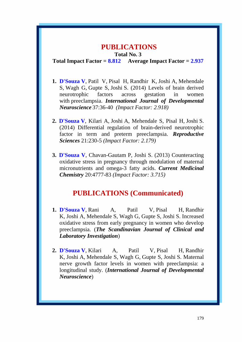
179
PUBLICATIONS Total No. 3
Total Impact Factor = 8.812 Average Impact Factor = 2.937
1. D'Souza V, Patil V, Pisal H, Randhir K, Joshi A, Mehendale
S, Wagh G, Gupte S, Joshi S. (2014) Levels of brain derived
neurotrophic factors across gestation in women
with preeclampsia. International Journal of Developmental
Neuroscience 37:36-40 (Impact Factor: 2.918)
2. D'Souza V, Kilari A, Joshi A, Mehendale S, Pisal H, Joshi S.
(2014) Differential regulation of brain-derived neurotrophic
factor in term and preterm preeclampsia. Reproductive
Sciences 21:230-5 (Impact Factor: 2.179)
3. D'Souza V, Chavan-Gautam P, Joshi S. (2013) Counteracting
oxidative stress in pregnancy through modulation of maternal
micronutrients and omega-3 fatty acids. Current Medicinal
Chemistry 20:4777-83 (Impact Factor: 3.715)
PUBLICATIONS (Communicated)
1. D'Souza V, Rani A, Patil V, Pisal H, Randhir
K, Joshi A, Mehendale S, Wagh G, Gupte S, Joshi S. Increased
oxidative stress from early pregnancy in women who develop
preeclampsia. (The Scandinavian Journal of Clinical and
Laboratory Investigation)
2. D'Souza V, Kilari A, Patil V, Pisal H, Randhir
K, Joshi A, Mehendale S, Wagh G, Gupte S, Joshi S. Maternal
nerve growth factor levels in women with preeclampsia: a
longitudinal study. (International Journal of Developmental
Neuroscience)

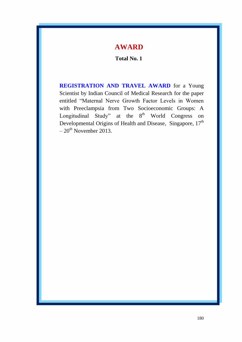
180
AWARD
Total No. 1
REGISTRATION AND TRAVEL AWARD for a Young
Scientist by Indian Council of Medical Research for the paper
entitled “Maternal Nerve Growth Factor Levels in Women
with Preeclampsia from Two Socioeconomic Groups: A
Longitudinal Study” at the 8th World Congress on
Developmental Origins of Health and Disease, Singapore, 17th
– 20th
November 2013.

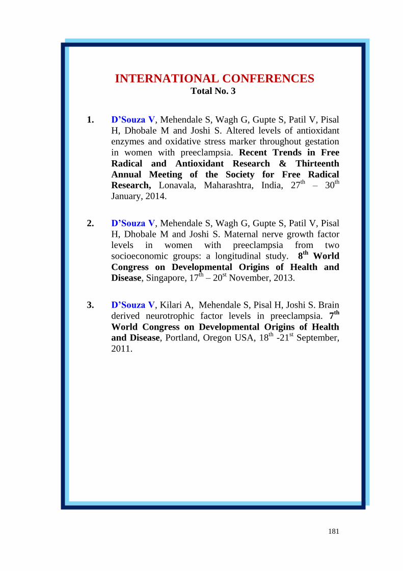
181
INTERNATIONAL CONFERENCES Total No. 3
1. D’Souza V, Mehendale S, Wagh G, Gupte S, Patil V, Pisal
H, Dhobale M and Joshi S. Altered levels of antioxidant
enzymes and oxidative stress marker throughout gestation
in women with preeclampsia. Recent Trends in Free
Radical and Antioxidant Research & Thirteenth
Annual Meeting of the Society for Free Radical
Research, Lonavala, Maharashtra, India, 27th
– 30th
January, 2014.
2. D’Souza V, Mehendale S, Wagh G, Gupte S, Patil V, Pisal
H, Dhobale M and Joshi S. Maternal nerve growth factor
levels in women with preeclampsia from two
socioeconomic groups: a longitudinal study. 8th
World
Congress on Developmental Origins of Health and
Disease, Singapore, 17th
– 20st November, 2013.
3. D’Souza V, Kilari A, Mehendale S, Pisal H, Joshi S. Brain
derived neurotrophic factor levels in preeclampsia. 7th
World Congress on Developmental Origins of Health
and Disease, Portland, Oregon USA, 18th -21
st September,
2011.


182
NATIONAL CONFERENCES Total No. 3
1. D’Souza V, Mehendale S, Wagh G, Gupte S, Patil V, Pisal H,
Joshi S. Brain Derived Neurotrophic Factor Levels in
Preeclampsia (Oral Presentation – Free Communication), at
the 44th
National Conference of the Nutrition Society of
India, Tirupati, 16th- 17
th November, 2012. OPEN-3.
2. D’Souza V, Kilari A, Joshi A, Pisal H. and Joshi S.
Differential Regulation of Cord Brain Derived Neurotrophic
Factor Levels in Term and Preterm Preeclampsia (Young
Scientist – Junior Award), at the 43rd
National Conference of
the Nutrition Society of India, Hyderabad, 11th- 12
th
November, 2011. JAEN-09.
3. Kulkarni A , D’Souza V, Mehendale S, Yadav H, Joshi S.
Reduced Placental Docosahexaenoic Acid Levels Associated
with Increased Plasma Soluble Flt-1 Levels in Preeclampsia
(Oral Presentation – Free Communication), at the 42nd
National Conference of the Nutrition Society of India,
Mumbai, 19th - 20
th November, 2010. FC/CN/E.


183
WORKSHOP / COURSE / SEMINAR
Total No. 3
1. 1st David Barker Memorial Symposium on the
Developmental Origins of Health and Disease at the 18th
Annual SNEHA-India International Workshop, Fariyas
Resort, Lonavala, India, 26th
-28th
September 2014.
2. International Course in Nutrition Research Methods
organized and sponsored by the Bangalore Boston Nutrition
Collaborative (BBNC) at St. John’s Research Institute in
Bangalore (SJRI) from 21st January –1st February, 2013.
3. ICMR centenary celebrations National Institute of Nutrition
organized Seminar (one day) / brain storming session (half
a day) Maternal under-nutrition and developmental
origins of health and disease held at NIN, Hyderabad, 25th -
26th June, 2011.