Embed Size (px)
Citation preview

MQP-BIO-DSA-9160
MQP-BC-DSA-0805
Neurotransmitter Signal Transduction and its Role in Pulmonary Arterial
Hypertension Vasodilatation and Vasoconstriction
A Major Qualifying Project Report
Submitted to the Faculty of the
WORCESTER POLYTECHNIC INSTITUTE
in partial fulfillment of the requirements for the
Degrees of Bachelor of Science
in
Biology and Biotechnology
and Biochemistry
by
_________________________ _________________________
Felipe Strefling Jenny Strefling
April 29, 2010
APPROVED:
_________________________ _________________________
Alice Gardner, Ph.D. David Adams, Ph.D.
Department of Pharmaceutical Science Biology and Biotechnology
Massachusetts College of Pharmacy WPI Project Advisor
Major Advisor

2
ABSTRACT
Pulmonary Arterial Hypertension (PAH) is a devastating disease characterized by a
persistent increase in pulmonary arterial resistance. Endothelin is a hormone involved in
maintaining vasoconstriction, but the enzymes and proteins involved in its signaling pathway are
not fully known. Understanding the endothelin-B (ETB) signaling pathway is crucial for the
development of a more specific endothelin-targeted therapy for PAH. Therefore, the objective of
this project was to identify mediators utilized by the ETB receptor and help determine their
function in the regulation of ATP neurotransmitter release. In rat PC12 cells differentiated by
neuronal growth factor, the inhibition of PLC was shown to reduce ETB-mediated ATP release.

3
TABLE OF CONTENTS
Signature Page ………………………………………………………………………. 1
Abstract ……………………………………………………………………………… 2
Table of Contents ……………………………………………………………….…… 3
Acknowledgements ………………………………………………………………….. 4
Background ………………………………………………………………………….. 5
Project Purpose ………………………………………………………………………. 20
Methods ……………………………………………………………………………… 21
Results ……………………………………………………………………………….. 28
Discussion …………………………………………………………………………… 33
Bibliography …………………………………………………………………………. 36

4
ACKNOWLEDGEMENTS
This project would not have been possible without the support of our advisor Professor
Alice Gardner at the Massachusetts College of Pharmacy and Life Science (Worcester campus).
Dr. Gardner provided us with the necessary background and skills to complete this project. We
would also like to extend thanks to the faculty and staff of the pharmaceutical sciences
department for allowing us to use laboratory resource and space. We would like to additionally
thank Professor David Adams, our WPI advisor, for helping write this report and for technical
advice.

5
BACKGROUND
Pulmonary Arterial Hypertension
Pulmonary Arterial Hypertension (PAH) is a progressive, symptomatic, and ultimately a
fatal disorder that elevates the blood pressure in the pulmonary arteries, putting the patients’
median life expectancy at only 2.8 years (McGoon & Garvan, 2009). Although treatment for the
disease has improved in the last decade, our limited understanding of the disease pathogenesis
impedes the achievement of optimal outcomes. The pathobiology of PAH is composed of many
factors, including increased pressure in the arteries that connect the lung to the heart. As a result,
the right side of the heart has to work harder to pump blood through the lungs. As the disease
progresses, morphological changes to the pulmonary vessel wall and coagulation increase
pulmonary vascular resistance (McGoon & Garvan, 2009).
PAH was first described over 100 years ago in a patient with right-heart failure that was
diagnosed with syphilitic pulmonary arteritis. Clinically PAH is defined simply as raised blood
pressure in the pulmonary arteries. Normal pressure ranges from 15 to 25 mm Hg systolic, and 8
to 15 mm Hg diastolic, with mean pressure between 10 and 20 mm Hg. When mean pulmonary
artery pressure increases to 25 mm Hg or higher, with a pulmonary capillary wedge pressure of
15 mm Hg or less, then the patient is diagnosed with PAH (Gaine & Rubin, 1998).
Pathophysiology of PAH
PAH, regardless of the cause, leads to the enlargement of the right ventricle of the heart
as it attempts to compensate for abnormally high pressure in the pulmonary arteries. If the right
ventricle cannot enlarge, then the pressure will increase in the right atrium and systemic venous
system. Eventually symptoms of right-sided heart failure, or cor pulmonale, are manifested in

6
the patient. Patients often complain about fatigue, this is because the right ventricle cannot pump
enough blood, which is deoxygenated, into the lungs to be oxygenated. The bone marrow
attempts to compensate for the lack of oxygen by stepping up red blood cell production.
Unfortunately this leads to polycythemia, a thickening of the blood. This puts even more stress
on the heart since viscous blood is harder to pump, and clotting is more likely (Holcomb, 2005).
Signs and symptoms associated with PAH include reduced oxygenation, decreased
cardiac output, and an inability to increase cardiac output, which leads to symptoms mimicking
heart failure, such as shortness of breath, fatigue, and syncope (fainting). During increased
activity these symptoms may increase and lead to angina-like chest pains. Direct signs of right
ventricular failure include peripheral edema, hepatomegaly, tricuspid regurgitation, an S3 heart
sound, prominent right ventricular impulse and jugular vein distension. Diagnosis is usually
reached by analyzing a patient's medical history for congenital heart disease or the use of banned
weight-reduction medication (Holcomb, 2005).
PAH Classification
Patients with PAH are divided into 4 classes of degree of severity. Class I patients have
no limitation of physical activity, and ordinary activity does not cause fatigue, chest pain, or near
syncope. Class II patients have slight limitation of physical activity. Patients are comfortable at
rest, but ordinary physical activity causes fatigue, chest pain, or near syncope. Class III patients
have limitations to physical activity. These patients are comfortable at rest, but less-than-
ordinary physical activity causes fatigue, chest pain, and near syncope. Class IV patients cannot
do any physical activity without symptoms. When at rest, these patients may be fatigued, and any

7
physical activity leads to discomfort. These patients show signs of right-sided heart failure
(Cipla Doc, 2000).
In July of 2004, the American College of Chest Physicians reclassified PAH on the basis
of etiology. Primary PAH became idiopathic PAH, or familial PAH if the cause is supported
genetically. Idiopathic PAH occurs predominantly among young adults who are probably
predisposed to the disorder. Other classifications are related to specific etiologies. PAH cases
caused by underlying disease such as HIV or toxicity are much more common. Prior to
widespread use of the weight-reduction drugs fenfluramine and dexfenfluramine in 1967, PAH
was a relatively rare disease afflicting only 1-2 cases per million people annually. The rate of
prevalence rose to 25-50 per million annually following the widespread use of these appetite
suppressants. Although these drugs were banned from the market in 1997, PAH caused by these
toxic drugs did not go away because of an illegal black-market. Also because of the prevalence
of HIV the incidence rate of PAH has not been reduced (Holcomb, 2005).
Cellular Pathways of PAH
Various cellular pathway abnormalities are associated with the development and
progression of PAH. Regardless of the cause all PAH patients have abnormal pulmonary
endothelium function. The pulmonary endothelium's purpose is to maintain low pulmonary
vascular resistance. In order to perform its job correctly, a balanced production of vasodilators
(in this case prostacyclin and nitric oxide) and vasoconstrictors (such as endothelin-1,
thromboxan A2, and serotonin) must be maintained. Therefore, patients with PAH are
characterized with a decreased concentration of prostacyclin and nitric oxide, while the

8
production of thromboxan A2 and endothelin-1 increases (Yildiz, 2009, p. 9). An increasing
concentration of endothelin is associated with the progression of PAH.
However, endothelial cell dysfunction is just one of the causes of the disease. PAH can
also be caused by other factors that lead to dysfunction of pulmonary circulation, such as
activation of adventitial fibroblasts, or the alteration of extracellular matrix components. Factors
that trigger the initiation of the disease are unknown, but once the disease is triggered the
consequences include vasoconstriction, vascular smooth muscle cell and endothelial cell
proliferation, and remodeling and thrombosis that cause the blood vessel to become narrower. As
the disease progress, more collagen and smooth muscle cells are synthesized in the pulmonary
artery in response to pulmonary artery pressure experienced by endothelial cells (Yildiz, 2009, p.
10). These changes in the vascular structure of the pulmonary artery lead to loss of function and
eventually right ventricular dysfunction.
In 2000, a gene on chromosome 2 that encodes for bone morphogenetic protein receptor
II (BMPR2) was found to be associated with familial pulmonary arterial hypertension (FPAH).
Mutations in BMPR2 occur in 50% of patients with familial PAH, while 25% of patients with
idiopathic PAH display this mutation (Holcomb, 2005). However, fewer than 20% of the
individuals carrying the mutated version of this gene develop FPAH. About 65% of families
with familial PAH have exonic or intronic allelic variants in the BMPR2 gene, and they are
transmitted in an autosomal dominant manner. Also some 10% of sporadic cases of IPAH
involve isolated exonic allelic variants of the BMPR2 gene (McGoon & Garvan, 2009, p. 1). The
reason why BMPR2 mutates is still unknown, but researchers believe environmental stimuli or
other genes interfering with BMPR2 may lead to its mutation (Gaine & Rubin, 1998, p. 2). The
mutated allelic variants of BMPR2 cause amino acid changes in the BMPR2 protein that

9
interrupt signal transduction during the process of pulmonary vascular smooth muscle cell
apoptosis, thus promoting cellular proliferation (McGoon & Garvan, 2009).
PAH Treatments
Treating PAH starts with removing any stressors or drugs that give rise to or exacerbates
the disease. For example, the weight loss drugs fenfluramine and dexfenfluramine are known to
cause PAH, so if a patient was taking one of these they are discontinued. PAH is not curable, so
treatment is aimed at reducing pressure, removing excess fluid, and reducing the risk of clotting.
The usual drug cocktail consists of vasodilators, anticoagulants, and careful use of diuretics.
Supplemental oxygen is also advisable if blood oxygen saturation reaches below 90%. Patients
with sleep apnea and PAH should use a Continuous Positive Airway Pressure device, as this
treatment decreases pulmonary artery pressure. Modern pharmaceutical treatment for PAH
includes calcium channel blockers, prostacyclin analogues, endothelin receptor antagonists,
phosphodiesterase inhibitors, and thromboxane inhibitors, all of which dilate the pulmonary
artery, and thus may stop the progression of PAH by maintaining endothelial integrity (Holcomb,
2005).
The first vasodilators used to treat PAH were calcium channel blockers. This approach
only works with about 20% of patients with idiopathic PAH, and does not seem to help other
types of PAH. Also patients with cor pulmonale must avoid calcium channel blockers because
they decrease myocardial contractility and may thus induce heart failure. High doses of
dihydropyridine calcium channel blockers are required for adequate dilation of the pulmonary
arteries (Robbins, 2006). Initial administration must be done in a clinical setting to monitor for
adverse reactions. An acute vasoreactivity test is recommended for calcium channel blocker

10
candidates. This test uses known short-acting vasodilators such as intravenous adenosine or
epoprostenol or inhaled nitric oxide. About 25% of patients with PAH have a positive response
to these short acting drugs, which is defined as a decrease in pulmonary arterial pressure of 10 to
40 mm Hg, and an increased or unchanged cardiac output (Mehta, 2003). Those with a negative
response usually have a worse prognosis. Another side affect of calcium channel blockers is
edema in the lower extremities, although this is usually controlled by diuretics (Rich, 2000, p. 3).
Only 30% of patients lived past 3 years after diagnosis before the development of
vasodilators and endothelin receptor antagonists. These new drugs dilate the pulmonary arteries,
reducing pressure and thus increasing life expectancy. Epoprostenol, a prostacyclin approved by
the FDA in 1995, is administered via continuous I.V. infusion through a long-term central venous
access device by a portable battery operated pump. This drug is a candidate for Class III and IV
patients who are not candidates for calcium channel blocker therapy. Treprostinil, an analogue
of prostacyclin, is given as a continuous subcutaneous infusion. It was approved in 2002 and has
boosted the survival rates to over 65%. Again the guidelines stipulate that this drug should only
be given to Class III and Class IV patients that are not candidates for calcium channel blockers.
Iloprost is a newer drug similar to prostacyclin that is inhaled, avoiding all the risks associated
with a continuously administered injectable. It is only given along the same guidelines as
Epoprostenol (Holcomb, 2005).
In 2001, Bosentan, an endothelin receptor antagonist, became the first oral drug for PAH
approved by the FDA. Only used for Class III and IV patients, this drug may stop progression of
PAH or may even reverse it. Future research focuses on finding better pulmonary vasodilators
that have a convenient admission route while also not causing systemic hypotension (Holcomb,
2005).

11
Endothelin
An increasing concentration of endothelin is associated with the progression of PAH. As
PAH progresses, changes in the vascular structure become apparent, such as inflammation,
vasoconstriction, cell proliferation, hypertrophy, and the formation of plexiform lesions. These
changes in the vascular structure of the pulmonary artery lead to loss of function and eventually
right ventricular dysfunction (Goraca, 2002). Hickey discovered an unknown factor produced in
the endothelium that causes smooth muscles to contract (Galie et al., 2004). By 1988,
Yanagisawa isolated and identified this factor from pig arterial endothelial cells, and called it
endothelin-1 (ET-1) (Galie et al., 2004).
Endothelin-1 is a 21 amino acid peptide with a molecular weight of 2492 daltons
(Figure-1). There are currently 3 known isomers (ET-1, ET-2, and ET-3). Endothelin has two
disulfide bonds, one between cysteine amino acids 1 and 15, and another between 3 and 11. The
presence of these disulfide bonds is critical because it contributes to the biological activity of ET
(Goraca, 2002). All three isoforms share a common structure that is characterized by the
existence of two disulfide bridges, a loop configuration, and an active site at the C terminus
according to figure 1 (Bouallegue & Srivastava, 2007).
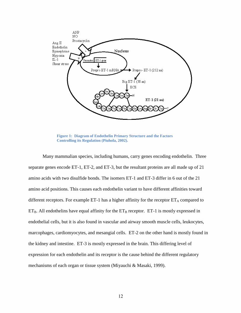
12
Figure 1: Diagram of Endothelin Primary Structure and the Factors
Controlling its Regulation (Piuhola, 2002).
Many mammalian species, including humans, carry genes encoding endothelin. Three
separate genes encode ET-1, ET-2, and ET-3, but the resultant proteins are all made up of 21
amino acids with two disulfide bonds. The isomers ET-1 and ET-3 differ in 6 out of the 21
amino acid positions. This causes each endothelin variant to have different affinities toward
different receptors. For example ET-1 has a higher affinity for the receptor ETA compared to
ETB. All endothelins have equal affinity for the ETB receptor. ET-1 is mostly expressed in
endothelial cells, but it is also found in vascular and airway smooth muscle cells, leukocytes,
marcophages, cardiomyocytes, and mesangial cells. ET-2 on the other hand is mostly found in
the kidney and intestine. ET-3 is mostly expressed in the brain. This differing level of
expression for each endothelin and its receptor is the cause behind the different regulatory
mechanisms of each organ or tissue system (Miyauchi & Masaki, 1999).

13
There are many factors involved in the up regulation or down regulation of endothelin,
among them, shear stress, extreme pH, oxidized low-density lipoprotein, glucose, insulin,
angiotensin II, catecholamines, growth factors, nitric oxide, and prostacyclin (Ergul, 2002).
An elevated level of ET-1 is seen in patients with acute myocardial infarction,
hypertension, and heart failure. Because of these observations, ET-1 has become associated with
the pathophysiology of these disease states. ET-1 mediates its effects through two distinct
heptahelical G-protein-coupled receptors, the endothelin ETA and endothelin ETB receptors
(Kanai & Hasegawa, 2004). On the smooth muscles of the vascular bed, both receptors are found
and both are responsible for mediating contraction. On the other hand, the endothelin ETB
receptors on endothelial cells are responsible for mediating vasodilatative factors such as nitric
oxide (NO) and prostacyclin (Masaki et al., 1999).
Developmental studies of endothelin-deficient and endothelin-receptor-deficient mice
have shown the important role of endothelin during embryonic development. ET-1-deficient
mice soon die after birth because of abnormal development of craniofacial and cardiac muscles
(Miyauchi & Masaki, 1999).
Endothelin Signal Transduction
Three signaling pathways are disturbed in patients with PAH: the nitric oxide pathway,
the prostacyclin pathway, and the endothelin pathway (see figure 2). We will mainly focus on
the endothelin pathway (Figure-2), since it is the major participant in the pathogenesis of PAH
(McGoon & Garvan, 2009, p. 192).

14
Figure 2: Endothelin-1 Signal Transduction. Shown are the molecular mechanisms for
vasoconstriction mediated by endothelin-1 binding to endothelin ETA and ETB receptors. NSCC:
non-selective cation channel, Kch: calcium sensitive potassium channel, PLC: phospholipase C,
VOC: voltage operated calcium channel, IP3: inositol tris-phosphate, SR: sarcoplasmic reticulum,
+ potentiate, - inhibition (Masaki et al., 1999).
Both endothelin ETA and ETB receptors bind to G-proteins on the cytoplasmic side of the
membrane. These G-proteins are then linked to adenylyl cyclase which dephophorylates ATP
into cyclic AMP (cAMP). According to figure 2 Gs-protein (stimulatory G-protein) binds to the
ETA receptor, and Gi-protein (inhibitory G-protein) binds to the ETB receptor. The activation of
ETA thus releases cAMP, which then triggers the formation of PK-A (cAMP stimulated protein
kinase), and eventually leads to the influx of calcium ion by phosphorylation of L-type calcium
channels. If contraction persists, calcium ions from inside the cells are also released (Klabunde,
2007).

15
In figure 2, both endothelin ETA and ETB receptors are also linked to Gq. This pathway
triggers the release of intracellular and extracellular Ca++
by the activation of phospholipase C
(PLC). Activation of PLC stimulates the formation of inositol triphosphate (IP3) from
phosphatidylinositol biphosphate (PIP2). This increase in IP3 triggers the sacroplasmic reticulum
in the heart to release calcium ions. However, under normal physiological conditions, voltage-
operating calcium channels do not contribute much to cause muscle contraction. Instead,
contraction is mediated by the activation of non-selective cation channels. When ET-1 level is
high, this causes the intercellular free calcium ions to increase. In order to physiologically meet
the high vasoconstriction necessary of PAH, extracellular calcium ions are released to enter the
cell (Masaki et al., 1999).
Nitric oxide inhibits contraction by decreasing calcium entry through the non-selective
cation channel. It is also used to inhibit the intracellular free calcium ion concentration triggered
by ET-1 binding to its receptors. In guinea pig tracheal smooth muscle, the use of nifedipine (a
voltage-operated calcium channel inhibitor) effectively inhibits ETB mediated contraction but not
ETA mediated contraction. This suggests that there may be another molecular mechanism that
induces contraction via the ETB receptor (Masaki et al., 1999).
Co-Transmission in the Sympathetic Nervous System
The sympathetic nervous system innervates many organs and tissues, one of them being
the vasculature. Larger pulmonary arteries appear to be more richly innervated compared to
smaller ones (Downing & Lee, 1980). These sympathetic innervations normally control the tone
and thus pressure of the pulmonary arteries. For example, externally stimulating the upper
thoracic sympathetic chain, middle cervical ganglia or thoracic vagosympathetic branches with a
16
current consistently raises pulmonary arterial pressure (10-15% over resting pressure) while
maintaining constant blood flow. However, when the pulmonary arterial pressure is kept
constant and flow is allowed to vary, stimulating the same nervous structures elicits a drop in
blood flow by as much as 30% (Downing & Lee, 1980). Pulmonary arterial pressure is
controlled by sympathetic neuronal release of neurotransmitters at the vascular neuroeffector
junction. Specifically, the tone of the arteries is controlled by the noradrenergic neurons, which
release neurotransmitters towards vascular smooth muscles. For example, arterial tone is
maintained upon continuous stimulation of noradrenergic neurons releasing norepinephrine
(NE). Increasing sympathetic stimulation of noradrenergic neurons results in arterial constriction.
On the other hand, decreasing sympathetic stimulation of noradrenergic neurons results in
arterial dilation (Craig, 2004). Increased arterial constriction, and thus resistance, is a sign of
pulmonary arterial hypertension (Pulmonary Arterial Hypertension, 2005).
In the past, it was thought that postganglionic neurons used only one neurotransmitter.
However, this has been proven incorrect by evidence obtained from anatomical and
pharmacological studies. It is has now been demonstrated that multiple neurotransmitters are
released by postganglionic neurons at the neuroeffector junctions. Neuropeptides Y, NE, or ATP
were found upon sympathetic nerve stimulation. Histochemical studies have shown that
immunoreactive neuropeptides are co-localized with NE, indicating that multiple
neurotransmittes are released by the sympathetic postganglionic neurons. Additionally,
neurotransmitters released as a result of sympathetic nervous system stimulation can function as
co-transmitters, modulators, or co-mediators along with NE (Loewy & Spyer, 1990).
NE, ATP and NPY are the three main neurotransmitters/ neuromodulators released by
postganglionic sympathetic nerves (Figure-3) (Sneddon & Burnstock, 1984; Stjarne & Astrand,

17
1986). Depending on cellular conditions or stimulation frequency, NE, ATP and NPY are co-
released at different proportions in order to better control smooth muscle tone (Kasakov et al.,
1988).
Figure 3: Role of ATP and NE as Neuromodulators. ATP and NE are
coreleased from the same vesicle prejunctionally, and they can act as
neuromodulators. At the postjunction, ATP acts on P2- purinoceptor receptor to
caused blood vessel contraction, and NE binds to α1- adrenoceptor to prolong
the contraction. These neurotransmitters can modulate its own release by
binding to the α2- adrenoceptors and P1 purinoceptors located at the prejunctions
(Burnstock, 1984)
These neurotransmitters can modulate the release of themselves and each other (Figure-
3). For example, ATP can postjunctionally modulate and enhance the responses of the other co-
transmitters. Prejunctionally, adenosine (ATP's metabolic breakdown product) was shown to
modulate the release of noradrenaline from peripheral sympathetic nerves in a variety of tissues
(Burnstock, 2009). NPY is usually co-stored with ATP and NE, and acts as a neuromodulator by
prejunctionally reducing the release of NE and ATP, and/or by postjunctionally increasing the

18
response to adrenergic and purinergic components of the sympathetic nervous response (Ellis &
Burnstock, 1990; Lundberg, 1996).
Varying stimulation frequency can also modulate neurotransmitters released at the
neuroeffector junction. Analyzing the kinetics of neuronally released co-transmitters from guinea
pig vas deferens has shown that ATP and NE are released at different proportions depending on
electrical frequency. ATP was released at low frequencies (8 HZ) of nerve stimulation. However,
when stimulated at higher frequencies (16HZ or more), NE was released along with ATP after
10s of stimulation. It was noted that arterial smooth muscle contracted more at the frequencies in
which NE was released. These results suggest that ATP and NE could be stored in different
vesicles (Todorov et al., 1999).
Additional mechanisms have now been identified that control constriction and
vasodilatation of arteries; for example, neuromodulators have the ability to alter the release and
actions of other neurotransmitters. Known neuromodulators include circulating neurohormones,
prostanoids, bradykinin, histamine, endothelin, in addition to neurotransmitters themselves
(Milner et al., 1999). Neurotransmitters released at the neuroeffector junction can be modulated
by non-neuromediators, such as the vasoactive peptide endothelin ET-1. Prejunctionally,
endothelin-1 modulates the release of NE to decrease sympathetic neurotransmission. Numerous
experiments have shown the modulation of sympathetic neurotransmission by ET-1, 6 such
experiments were performed in the guinea pig pulmonary and femoral arteries (Wiklund &
Cederqvist, 1989), rat and guinea pig vas deferens (Wiklund et al., 1990), dog coronary artery
(Aarnio et al., 1993) and rat mesenteric artery (Aarnio et al., 1993). In the rat mesenteric bed,
ET-1 negatively modulates the release of NPY instead of NE at the neuroeffector junction
(Hoang et al., 2002). ET-1 regulates sympathetic neurotransmission postjucntionally by

19
enhancing vasoconstriction caused by both nerve stimulation and a variety of vasoactive agents
(Henrion & Laher, 1993; Hoang et al., 2002). As ET-1 binds to the ETA receptors, it potentiates
the postjunctional contractile effects of ATP (Mutafova-Yambolieva & Radomirov, 1993; Hoang
et al., 2002).
Hypertension has no single cause, and has been linked to various factors. Some of these
factors are structural and functional changes in the vasculature. Medial smooth muscle
hypertrophy and hyperplasia are all symptoms of hypertension. The three major
neurotransmitter/ neuromodulators NE, NPY, and ATP in the sympathetic nervous system have
the ability to trigger a mitogenic response in human vascular smooth muscle cells (Thulin &
Erlinge, 1995). In the spontaneously hypertensive rat (SHR) model, ATP was shown to regulate
vascular smooth muscle cell proliferation (Harper et al., 1998). Other factors include the
sympathetic nervous system, the rennin-angiotensin system and other endothelial irregularities
(Milner et al., 1999). Animal models have become particularly useful in mapping the vascular
changes that lead to the onset of hypertension. The SHR model has become very useful for
understanding genetically role of hypertension. Using this model it has become possible to look
at the involvement of the sympathetic nervous system in the origin and continuation of
hypertension in SHR. An increase in the density of cerebral artery innervations by sympathetic
nerves containing NE and NPY was shown to precede the onset of hypertension and related
medial hypertrophy. After looking at sympathetic neurotransmission in the tail and mesenteric
arteries of SHRs it has been suggested that ATP plays a greater role in co-transmission than NE.
So much so, that ATP is the main component of the sympathetic response (Vidal et al., 1986).
Thus, enhanced sympathetic activity has been implicated in the pathogenesis of hypertension in
humans.

20
PROJECT PURPOSE
Endothelin has been shown to modulate the release of the neurotransmitters NE and NPY
at the neuroeffector junction. Previous studies on NGF differentiated PC12 cells suggest that
ATP levels (a key modulator of vasoconstriction) were modulated by ET-1 (Gardner et al.,
2005). It was shown that the ETB receptor was able to inhibit the release of ATP by “a specific
associated G-protein belonging to the Gi/o family, and attenuation of calcium levels” (Gardner et
al., 2005). Notably, ATP release is the factor that causes contraction of vascular smooth muscle
cells, and as a result it also regulates vascular resistance (Mutafova-Yambolieva & Radomirov,
1993). Therefore, Gardner’s hypothesis states that activation of the ETB receptor by ET-1
recruits specific receptor associated G-proteins belonging to the Gi/o family, which also
attenuates calcium levels. The enzymes and proteins involved in the signaling pathway that
causes the release of ATP are not fully known. Therefore, the objective of this project is to
identify some of the mediators utilized by the ETB receptor and demonstrate their function in the
regulation of neurotransmitter release. The knowledge gained from the ETB signaling pathway is
crucial for the development of a more specific endothelin-targeted therapy for PAH (Gardner,
2008).

21
METHODS
Cell Culture
Undifferentiated rat pheochromocytoma (PC12) cells were obtained from Professor Alice
Gardner’s lab at the Massachusetts College of Pharmacy and Life Science. After being thawed,
cells were transferred into T75 flask (polystyrene, Corning, New York) containing growth
medium [Dulbecco’s Modified Eagle’s Medium (DMEM with 4.5 g/L glucose and 4.0 mM L-
glutamine, Lonza) supplemented with 10% (v/v) horse serum (heat-inactivated at 55°C, 30 min),
5% (v/v) fetal bovine serum, and a penicillin streptomycin solution (100 U penicillin and 100 g
streptomycin/mL, Sigma)]. Cells were grown in a NAPCO 5400 series incubator at 37°C, 5.0%
CO2. After 24 hours, the cells were differentiated via the addition of mouse nerve growth factor
(2.5 S NGF, BD Bioscience, 50 ng/mL). Medium was replaced every 2-3 days with growth
medium containing NGF, as before. At 80% confluency, cells were subcultured and transferred
into six well plates (polystyrene, Corning, New York).
Cell Stimulations
When cells reached confluence (70%), the growth media were replaced with DMEM
media (4.5 g/L glucose & 4.0 mM L-glutamine, Lonza) supplemented with mouse NGF (BD
Bioscience, 50 ng/mL). Cells were serum starved for 24 hours to achieve base line activity. The
agents used to stimulated cells were BQ 3020 (10 mM; ETB agonist), U73122 (5 µM; PLC
inhibitor), and KCl (50 mM). The control set was not treated with any of the agents. The first set
of wells was basal and not stimulated. The second set was stimulated for 5 minutes with KCl.
The third set was incubated for 5 minutes with BQ3020, and then stimulated for 5 minutes with

22
KCl. And finally the last set was initially pretreated for 20 minutes with U73122 then incubated
for 5 minutes with BQ3020, and finally stimulated for 5 minutes with KCl. All cells were
incubated in a NAPCO 5400 series incubator.
Preparation of Cell Lysates
Following stimulation, all media were aspirated, and the cells were washed with
phosphate-buffered saline (ice-cold 1X PBS, without calcium & magnesium, Cellgrow). Lysis
buffer containing 1X RIPA buffer (Pierce, Prod. # 89900) supplemented with 1x Halt Protease
and phosphatase inhibitor cocktail (Pierce, 100x) was used to lyse cells. All cells were kept on
ice, and incubated for 15 minutes to facilitate cell lysis. Using a cell scraper and pipetter the
lysate was removed from each well and placed in a microcentrifuge tube. Inhibitor cocktail (40
µL) was used to wash each well. The wash from each well was added to the corresponding
microcentrifuge tube containing cell lysate. This was incubated for 30 minutes. Cell lysate was
centrifuged at 10,000xg for 10 minutes at 4°C. The supernatant was collected from each tube.
The total protein concentration in the supernatant was detrermined by the manufacturer’s
microplate procedure obtained from the Pierce BCATM
Protein Assay Kit. Readings were
obtained from a Synergy HT microplate reader. The BSA standard curve was made according to
table 1, in the BCATM
Protein Assay Kit procedure pamphlet from Pierce thermo scientific. A
modification to the table was made to extend the range of BSA standard concentration curve
from 2,000 µg/mL – 25 µg/mL, to 2,000 µg/mL - 12.5 µg/mL.

23
Electrophoresis
Samples were added to Lane Marker Sample Reducing Buffer (Pierce, cat# 3900) at a
ratio of 1 to 4 to make a 1X solution. The solution was boiled for 5 minutes. The total protein
from each sample (20 µg), TrichromrangerTM
Prestain Protein (10 µL), Prosieve®Color Protein
Marker (Lonza, Cat. No 50550), and Prosieve®Color Protein Marker (10 µL; Cambrex, Cat. No
50552) were loaded into PAGE® Gold Precast Gel (Lonza, 4-20% Tris-Glycine polyacrylamide
Gel). Initially the gels were run in electrophoresis buffer (Pierce, 25 mM Tris, 192 mM glycine,
0.1% SDS, pH 8.3) at 125V for 30 minutes, then changed to 200V for the remainder of the run.
Immunoblots
Proteins were transferred onto PVDF membrane (BioTraceTM
PVDF Polyvinylidene
Floride Transfer membrane 0.45 µM, Pall Corporation Life Science) at 100 V for 1 hour or 30V
overnight in transfer buffer (25 mM Tris, 192 mM glycine, pH 8.0, Pierce) at 4°C. MemcodeTM
Reversible Protein Stain Kit (Pierce) was used to confirm the efficiency of protein transfer.
Membrane was blocked with Starting Block TM
T20 Blocking buffer (10 mL; Tris buffer saline
containing 0.05% Tween-20, Pierce) for 30 minutes at 37°C, and then washed with 1X PBS
(0.1% Tween) for 15 minutes.
The antibodies were titrated under different concentrations to determine the optimal band
contrast. The primary antibody (PLC-β1 rabbit polyclonal IgG, Santa Cruz, 200 µg/mL) was
diluted 100 and 500 fold with blocking buffer (T20, pierce) up to 10 mL. The membrane was
then incubated at room temperature for 1 hour with the primary antibody. The secondary
antibody (Bovine anti–rabbit IgG-HRP, Santa Cruz, 200 µg/0.5 mL) was diluted 500 and 2000
fold with T20 blocking buffer (Tris buffer saline containing 0.05%Tween-20, Pierce) up to 10

24
mL. The membrane was then incubated at room temperature for 1 hour with the secondary
antibody following a 15 minute 1X PBS wash (0.1% Tween). Bands of interest were detected
using chemiluminescence (Pierce® ECL Western Blotting Substrate, Pierce) and analyzed as a
high-resolution scanned image of the film with the computer NIH program Scion Image.
Immunoprecipitation of Phospholipase C
Rat PC12 cells (passage number 22) were used for immunoprecipitation. Cells were
treated with the same agents as before. Proteins were quantified using a BSA protein assay kit as
before. The following steps were completed at 4°C.
Following cell lysis, the supernatant (containing 100 µg of protein) was pre-cleared
before being used for immunoprecipitation. First, the volume of the lysate was brought up to 1
mL per vial of solution using 1x RIPA and HaltTM
Protease & Phosphatase Inhibitor Cocktail 1X
(Pierce). To the supernatant control IgG (0.25 µg; normal rabbit IgG, 200 µg/0.5mL, Santa Cruz)
and suspended agarose conjugate (20 µL; Protein A/G PLUS- Agarose, Santa Cruz) were added
to each vial. The vials were tumbled using a shaker (model 55 rocking shaker, Midwest
Scientific) for 30 minutes at 4°C. The supernatant was then collected by centrifuging at 1,000xg
for 30 seconds. The primary antibody (5 µL; PLC-β1 rabbit polyclonal IgG, Santa Cruz) was
added to the supernatant and tumbled using the rocking shaker for two hours at 4°C. Afterwards,
20 µL of the same protein A/G-agarose was added to the solution and tumbled overnight at 4°C
under the same conditions as the previous step. The pellet was collected by centrifuging at
1,000xg for 30 seconds. The pellet was washed and centrifuged (1,000xg for 30 seconds) three
times using RIPA buffer (300 µL; Pierce) each wash. On the final wash, the resuspended pellet
solution was centrifuged under the same conditions as before. The pellet was collected. The

25
beads were resuspended in electrophoresis sample buffer (2x, 40 µL; ProTrackTM
Loading buffer
Lonza; DTT 0.1 M Invitrogen, and 50 mM Tris-HCl 1% SDS). The samples were boiled for 3
minutes, and centrifuged at 1000xg for 30 seconds. The supernatant (40 µL) was collected from
each tube, subdivided into two 20 µL aliquots, and stored at -80°C.
Western-blot of Immunoprecipitated Phospholipase C
The samples (20 µL) and marker (Prosieve® Color Protein Marker, Lonza, Cat. No.
50550) were loaded into 8-16% Precise TM
Protein SDS-PAGE gels (Prod. No. 25203). The gel
was run at 125 V in BupHTM
Tris-HEPES-SDS running buffer (100 mM Tris, 100 mM HEPES
and 3 mM SDS at pH 3±0.5, Pierce) at 4°C. The proteins were transferred onto PVDF membrane
(BioTraceTM
PVDF Polyvinylidene Floride Transfer membrane 0.45 µM, Pall Corporation Life
Science) at 30V overnight in transfer buffer (25 mM Tris, 192 mM glycine, pH 8.0, Pierce) at
4°C.
The PVDF membrane was blocked with Starting Block TM
T20 Blocking buffer (10 mL;
Tris buffer saline containing 0.05%Tween-20, Pierce) for 30 minutes at room temperature on a
rocking machine (Jahre Garentie, Heidolph Rotamax 120). Anti-Phosphotyrosine (4G10®) HRP
Conjugated monoclonal antibody (Millipore, Cat# 16105) was diluted 1:1000 using the Starting
Block TM
T20 Blocking buffer. The PVDF membrane was incubated in the antibody at 4°C
overnight on the rocking machine. The PVDF membrane was washed twice with water, then
T20 blocking buffer for 5 minutes and finally with water 5 times. The bands were detected
according to the previous method.

26
Quantifying ATP Release
Cells were grown and differentiated as before. On the day of the experiment the media
was replaced with Krebs’ buffer (1.5 mL per well) composed of NaCl (119 mM), KCl (2.5 mM),
MgSO4 (1.3 mM), CaCl2 (2.5 mM), NaH2PO4 (1.0 mM), NaHCO3 (26.2 mM), and HEPES (10
mM) at pH 7.4. The plates were allowed to equilibrate in a shaking water bath at 37°C for 15
minutes. Cells were stimulated with BQ 3020 (10 mM; ETB agonist), ET-1 (10-10
M), U73122 (5
µM; PLC inhibitor), and KCl (50 mM) in the same manner as before. ET-1 (10-10
M) was
stimulated in a similar way as BQ 3020 (10 mM; ETB agonist). After stimulation, plates were
placed on ice. The buffer and cells were removed from plates as before, and centrifuged at 1000
rpm for 2 min. The supernatant was used for quantifying ATP (Gardner et al., 2005).
Analysis of Purines
The following method used to analyze purines was described by (Levitt et al., 1984).
Chloroacetaldehyde was synthesized according to the methods described by (JA et al., 1972).
This compound was used to form fluorescent 1, N6-ethenopurne analogs (E-purine). E-purines
can be “simultaneously separated from the sample by reverse-phase HPLC and quantified by
fluorescent detection “ (Gardner et al., 2005). Chloroacetaldehyde (50 uL) was incubated with
the supernatant (from quantifying ATP release) at 80°C for 40 minutes in a dry bath. Placing the
samples on ice stopped the reaction. Afterwards, the samples were analyzed by HPLC-
fluorometric detection. The different purine analogs formed were separated by a reverse-phase
C-18 column. Two different buffers were used to create a dual buffer gradient system which
could be used to separate and elute the purines from the column. Buffer A was composed of
phosphate buffer (0.1 M, pH 6.0) and buffer B was composed of 75% 0.1M phosphate buffer and

27
25% methanol at pH 6.0. The dual buffer gradient system was created by gradually increasing
the concentration of buffer B while decreasing the concentration of buffer A. A Varian 9070
Fluorescence Detector was set at an excitation wavelength of 300nm, and an emission
wavelength of 420 nm, in order to detect the fluorescent purine derivatives. Carrying out a
comparison against purine standard retention times identified the purine peaks. The purine
content of the sample was quantified using the Varian Star Workstation Software to integrate the
purine peaks. “ATP analysis was performed on perfusates and normalized against protein
concentrations” (Gardner et al., 2005).

28
RESULTS
NGF-differentiated PC12 cells were used in this study of the signaling mechanism
utilized by the endothelin-1 (ET-1) modulation of sympathetic neurotransmission. Since ET-1 is
an important vasomodulating hormone, understanding its mechanism of action is important for
designing drugs to treat hypertension diseases. Analyzing the signaling pathway of the ETB
receptor has given insight into its role in pulmonary arterial hypertension (PAH). It was
previously shown that ET-1 binding to the ETA receptor resulted in phosphorylation of PLC,
which caused the levels of 1,4,5-triphosphate (IP3) and diacyglycerol to increase (Senogles,
1994). This increased level of IP3 in turn caused the intracellular calcium concentration to
increase. In smooth muscle, studying the ET-1 pathway revealed that phosphorylation of PLC
caused intracellular calcium to increase transiently (Takuwa, 1990). In this project, treatment of
PC12 cells with PLC inhibitors, or ET-1, or other ETB agonists were used to test whether PLC
kinase plays a role in ETB-mediated inhibition of ATP release.
Phospholipase C Modulates Endothelin-Mediated ATP Release Via the ETB Receptor
In order to determine whether phospholipase C (PLC) modulates the release of ATP from
PC12 cells, the effects of a PLC antagonist on the K+ evoked release of ATP from NGF-
differentiated PC12 cells were studied to show whether this agent could reverse the ET-1
induced inhibition on the K+ evoked release of ATP (Figure-4). Stimulation of NGF
differentiated PC12 cells with KCl (50 mM) (first histobar) caused ATP to release over basal
levels. Adding ET-1 (second histobar) reduced ATP levels. The addition of the PLC inhibitor
(U73122, shown as U7 in the figure) (third histobar) reversed the ET-1-induced inhibition on the

29
K+ evoked release of ATP; however, the changes in ATP levels were not significant. Thus, this
data shows that in PC12 cells, ET-1 may act to lower KCl-induced ATP release, and this release
appears to require PLC.
Figure 4: The Effect of ETA/B Agonist ET-1 and Phospholipase C Inhibition
on Stimulated KCl Induced ATP Release. Undifferentiated rat pheochromo-
cytoma (PC12) cells were grown to 70-80% confluency and were differentiated
by mouse NGF (50 ng/mL). Cells were serum starved for 24 hours and were
stimulated with U73122 (5µM), ET-1 (10-10 M), or KCl (50 mM) accordingly.
Treatment with the ETA/B agonist ET-1 reduced ATP levels compared to KCL-
stimulated ATP release. While in the presence of U73122, ET-1 modulated
KCL-stimulation increased ATP release. Both of these changes were non-
significant. Values are shown relative to untreated samples. Histobars denote
the mean of 3 experiments. Error bars denote standard error.
Because ET-1 binds with equal affinity to the ETA and the ETB receptors, the effect of the
ETB receptor by itself on PLC activation could not be clearly delineated. Therefore, the ETB
agonist (BQ3020) was tested in conjunction with the U7 PLC inhibitor to further elucidate the
role of PLC in ATP release mediated by the ETB receptor (Figure-5). NGF-differentiated PC 12
cells were stimulated with KCl (50 mM) to evoke ATP release above basal level (first histobar).
The ATP levels decreased 30.7% under the presence of the ETB receptor agonist BQ3020
(second histobar). The PLC inhibitor U73122 reversed this inhibition by evoking a 73.5%

30
increased in ATP release (third histobar). Thus, receptor ETB indeed appears to participate in the
lowering of KCL-induced ATP release.
Figure 5: The Effect of ETB Agonist and Phospholipase C Inhibition on KCl
Induced ATP Release. Undifferentiated rat pheochromocytoma (PC12) cells
were grown to 70-80% confluency and were differentiated by mouse NGF (50
ng/mL). Cells were serum starved for 24 hours and were stimulated with
U73122 (5 µM), BQ 3020 (10 mM), or KCl (50 mM) accordingly. After
stimulation with KCl, ATP levels decreased (30.7%, p<0.05) under the
presence of the ETB receptor agonist, BQ-3020, compared to KCl stimulation
only. The inhibitory effects of BQ-3020 on ATP release was reversed after
pretreating with the non-specific PLC inhibitor U73122 (73.5% increase,
p<0.05). Each bar represents the mean ±S.E.M of 12 to 15 from six individual
experiments. *, P< 0.05 KCl vs. KCl+ BQ3020 and KCl+ BQ3020 vs. KCl
+BQ3020 +U73122. Error bars denote standard error.
In order to determine whether PLC plays a significant role in modulating basal ATP
release, BQ 3020 or U73122 were incubated with control cells (Figure-6). The BQ3020 (shown
as BQ in the figure) ETB agonist (second histobar) had no significant impact on basal ATP
levels. However, the addition of the U73122 PLC-inhibitor (third histobar) resulted in a
significant increase in the basal level of ATP release, suggesting that PLC plays a role in
modulating basal neurotransmitter release.

31
Figure 6: The Effect of ETB Agonist and Phospholipase C Inhibitor on
Basal ATP Release. Undifferentiated rat pheochromocytoma (PC12) cells were
grown to 70-80% confluency and were differentiated by mouse NGF (50
ng/mL). Cells were serum starved for 24 hours and were incubated in either BQ
3020 (10 mM; 5 min) or U73122 (5 µM; 20 min). The BQ ETB agonist (second
histobar) did not have a significant impact on basal ATP level, but inhibiting
PLC with U7 significantly increased ATP release (third histobar). Therefore,
PLC significantly modulates basal ATP release. Each bar represents the mean
±S.E.M of 12 to 15 from six individual experiments. *, P< 0.05 Basal + BQ3020
vs. Basal + U73122. Error bars denote standard error.
Quantifying PLC Phosphorylation Via Western Blotting
After determining that PLC plays a role in ATP release, it became important to figure out
which PLC family and isotype takes part in the ETB signal transduction pathway. U73122 has
been shown to only inhibit PLC- and PLC- (Heemskerk et al., 1997). PLC- was chosen over
other PLC family members because it is involved in the IP3 pathway. Cellular levels of PLC-β
were monitored by immunoblot (Figure-7). For each lane there was only one PLC-1 band
instead of a doublet, as one would expect from a phosphorylated enzyme in a western blot. The
primary antibody (in the immunoblots section under methods) is capable of detecting both
phosphorylated and unphosphorylated PLC-1. The phosphorylated PLC-1 should appear as a
slightly heavier band above the unphosphorylated band. Because this assay was not sensitive
enough, a doublet was not seen.

32
Figure 7: The Effect of ETB Agonist (BQ3020) on PLC Phosphorylation.
Undifferentiated rat pheochromocytoma (PC12) cells were grown to 70-80%
confluency and were differentiated by mouse NGF (50 ng/mL). Cells were
serum starved for 24 hours and were stimulated with BQ 3020 (10 mM), and
KCl (50 mM) as listed. Primary antibody detection (5 µL; PLC-β1 rabbit
polyclonal IgG, Santa Cruz diluted 1:500 in Tris buffer saline containing
0.05%Tween-20, Pierce) and secondary antibody (Bovine anti –rabbit IgG-HRP,
Santa Cruz, 200µg/0.5 mL: diluted 1:2000 in Tris buffer saline containing
0.05%Tween-20, Pierce).

33
DISCUSSION
In the sympathetic nervous system, endothelin-1 (ET-1) was previously shown to act as a
neuromodulator in experiments done on the guinea pig pulmonary and femoral arteries (Wiklund
& Cederqvist, 1989), rat and guinea pig vas deferens (Wiklund et al., 1990), dog coronary artery
(Aarnio et al., 1993) and rat mesenteric artery (Aarnio et al., 1993). Specifically, when studying
the effects of ET-1 on the endothelial ETA receptor, ET-1 was shown to potentiate the
postjunctional contractile effects of ATP (Mutafova-Yambolieva & Radomirov, 1993; Hoang et
al., 2002). Notably, ATP release is the factor that causes contraction of vascular smooth muscle
cells, and as a result regulates vascular resistance (Mutafova-Yambolieva & Radomirov, 1993).
So understanding this pathway is important for the future design of drugs to treat hypertension
disorders.
Previous data showed that the ETB receptor was able to inhibit the release of ATP by “a
specific associated G-protein belonging to the Gi/o family, and attenuation of calcium levels”
(Gardner et al., 2005). Calcium therefore plays an essential role in neurotransmitter release. As
previously discussed, calcium can enter the neuron via the opening of the voltage dependent
calcium channels. Neurotransmitter release can also be altered by modulating calcium release,
for example by inhibiting voltage-dependent calcium channels by activation of the PLC/Ca(2+)-
dependent PKC signal transduction pathway(Salgado et al., 2007).
The Activation of the PLC/Ca(2+)-dependent PKC signal transduction pathway leads to
PLC phosphorylation which in turn hydrolyzes PIP2 to produce IP3 and diacyglycerol (Hou et al.,
2003). Furthermore, studying the endothelin pathway in smooth muscle revealed that
phosphorylation of PLC caused intracellular calcium to increase transiently (Takuwa, 1990).

34
In our project, PLC inhibitors were used to test whether PLC plays a role in ETB
mediated inhibition of ATP release. Cells were stimulated with the non-specific PLC inhibitor
(U73122), ETB agonist (BQ3020), and/or KCl. When the ETB agonist BQ3020 bound to the ETB
receptor, a decrease in ATP levels was observed. However, once the non-specific PLC inhibitor
U73211 inhibited PLC, an increase in ATP levels was seen. KCl stimulation of ATP in the
presence or absence of U73122 did not change ATP levels. This suggested that PLC plays a role
in the ETB signal transduction pathway because it did not alter KCl-induced ATP levels.
Moreover, KCl caused ATP levels to increase by acting on voltage gated calcium channels.
However, under basal conditions, ATP levels of cells that were challenged with ETB agonists
only slightly decreased, whereas those that were treated with U73122 caused ATP levels to
increase significantly. Therefore, PLC plays a role in modulating basal and ETB-modulated ATP
release.
U73122 has been previously used extensively as a PLC inhibitor in the study of cellular
signal transduction (Wilsher, et al., 2007). However, this aminosteroid is not entirely specific to
any PLC family but has been shown to inhibit both PLC-β and PLC-γ isoforms, but not other
PLC families (delta, epsilon, zeta and eta) (Heemskerk et al., 1997). Furthermore, U73122 has
been shown to have a higher affinity towards human PLC-β2 compared to the other PLC-β
isoforms (PLC-β1, PLC-β3, and PLC-β4), although this specificity has not been confirmed with
the rat PLC-β analyzed in our project (Hou et al., 2003).
The first PLC immunoblot did not show a doublet around 150 kD (Figure-7), as one
would expect when detecting both phosphorylated and unphorphorylated PLC. The antibody
used in our immunoblot is capable of detecting both forms. Since only a small portion of PLC
gets phosphorylated during activation, we conclude that the immunoblot method was not
35
sensitive enough to detect the phosphorylated form. PLC phosphorylation occurs within the first
few signaling cascading events, is transient, and is not heavily amplified, so the phosphorylated
form exists at very low concentrations, making it difficult to detect by this method (Hou et al.,
2003).
In order to enhance the sensitivity, an immunoprecipitation using PLC-1 antibody
followed by an immunoblot with phosphotyrosine antibody was performed. In this method, only
phosphorylated PLC is detected (by the phosphotyrosine antibody), thus only one band around
150 kD would be expected in the blot. However, because of ongoing troubleshooting, this result
is still pending. If the phosphorylation levels were to correspond to ATP levels, then one could
conclude that PLC activation is involved in ETB signal transduction.
If a decrease in ATP levels, upon ETB stimulation, is not found to correspond to a
decrease in PLC phosphorylation, then the following conclusions could be made. Firstly, the blot
could again not be sensitive enough, and further study would be needed to develop a more
sensitive assay (radio-nucleotide assay using 32
P would be employed). Secondly it could be that
PLC-1 does not take part in the rat NGF differentiated PC12 ETB pathway. Finally it could be
that PLC-1 is phosphorylated by serine (Ryu, et al., 1990) as shown in other cellular PLC signal
transduction. Eukaryotic kinases in addition to phosphorylating tyrosine residues can also
phosphorylate serine/threonine motifs, but very rarely histidine and aspartate amino acid residues
(IonSource, 2009). As this project focused solely on tyrosine phosphorylation of PLC-1 an
assay looking for serine/threonine phosphorylation would need to be undertaken. Therefore, the
short-term goal of this project is to improve quantification of PLC-1 phosphorylation.
Future directions would be to develop a PLC-knockdown protocol to determine whether a
relationship exists between PLC-1 phosphorylation and ATP release to support this assertion.

36
BIBLIOGRAPHY
Aarnio, P., McGregor, G., & Miller, V. (1993). Autonomic modulation of contractions to
endothelin-1 in canine coronary arteries. Hypertension, 21, 680-686.
Bouallegue, A., Daou, G., & Srivastava, A. (2007). Endothelin-1-Induced Signaling Pathways in
Vascular Smooth Muscle Cells. Current Vascular Pharmacology, 5, 45-52.
Burnstock, G. (1984). Autonomic Neuroeffector Mechanisms in Smooth Muscle. Japanese
Journal of Smooth Muscle Research, 20 (5), 365-392.
Burnstock, G. (2009). Purinergic Cotransmission. Experimental Physiology, 94, 20-24.
Cipla Doc. (2000). Disease of the Month: Pulmonary Arterial Hypertension. Retrieved February
28, 2010, from Cipladoc: http://www.cipladoc.com/dotm/dotm.htm
Craig, C. R. (2004). Modern Pharmacology with Clinical Applications. Philadelphia: Lippincott
Williams & Wikins.
Downing, S. E., & Lee, J. C. (1980). Nervous Control of the Pulmonary Circulation. Annual
Review of Physiology, 42, 199-210.
Ellis, J., & Burnstock, G. (1990). Neuropeptide Y neuromodulation of sympathetic co-
transmission in the guinea-pig vas deferens. Br. J. Pharmacol, 100(3), 457-462.
Ergul, A. (2002). Endothelin-1 and Endothelin Receptor Antagonists as Potential Cardiovascular
Therapeutic Agents. Pharmacotherapy, 22(1), 54-65.
Gaine, S. P., & Rubin, L. J. (1998). Primary Pulmonary Hypertension. The Lancet, 352, 719-724.
Galie, N., Manes, A., & Branzi, A. (2004). The endothelin system in pulmonary arterial
hypertension. Cardiovascular Research, 61, 227-237.
Gardner, A., Westfal, T. C., & Macarthur, H. (2005). Endothelin (ET)-1-Induced Inhibition of
ATP Release from PC-12 Cells is Mediated by the ETB Receptor: Differential Response to ET-1
on ATP, Neuropeptide Y, and Dopamine Levels. the Journal of Pharmacology and Experimental
Therapeutics, 313, 1109-1117.
Goraca, A. (2002). New Views on the Role of Endothelin. Endocrine Regulations, 36, 161-167.
Han, S., Yang, C.-L., Chen, X., Naes, L., Cox, B. F., & Westfall, T. (1998). Direct Evidence for
the Role of Neuropeptide Y in Symathatic Nerve Stimulation-Induced Vasoconstriction. Am J
Physiol Heart Circ Physiol, 274, 290-294.

37
Harper, S., Charlton, S., NG, L., & Boarder, M. (1998). Evidence that ATP and UTP regulate
spontaneously hypertensive rat aorta smooth muscle cell proliferation by acting on P2Y receptor.
Br. J. Pharmacol., 125, 59P.
Heemskerk, J., Farndale, R., & and Sage, S. (1997). Effects of U73122 and U73343 on human
platelet calcium signalling and protein tyrosine phosphorylation. Biochimica et Biophysica Acta,
1355, 81-88.
Henrion, D., & Laher, I. (1993). Potentiation of Norepinephrine-Induced Contractions by
Endothelin in the Rabbit Aorta. Hypertension, 21, 78-83.
Hoang, D., Macarthur, H., Gardner, A., & Westfall, T. (2002). Endothelin-Induced Modulation
of Neuropeptide Y and Norepinephrine Release from the Rat Mesenteric Bed. Am J Physiol
Heart Circ Physiol, 283, H1523-H1530.
Holcomb, S. S. (2005). Understanding Pulmonary Arterial Hypertension. Nursing Management,
36 (5), 56A-56G.
Hou, C., Kirchner, T., Monica, S., Matheis, M., Argentieri, D., & Covender, D. (2003). The
Journal of Pharmacology and Experimental Therapeutics, 309, 697-704.
IonSource. (2009 16-November). Amino Acid Phosphorylation. Retrieved 2010 14-April from
Ionsource: http://www.ionsource.com/Card/phos/phos.htm
JA, S., Barrio, J., & Leonard, N. ( 1972). Fluorescent modification of adenosine containing
coenzyme: biological activities and spectroscopic properties. Biochemistry, 11, 3499-3506.
Kanai, E. I., & Hasegawa, K. (2004). Intracellular Signaling Pathways for Norepinephrine-and
Endotheline-1-Mediated Regulation of Myocardial Cell Apoptosis. Molecular and Cellular
Biochemistry, 259,163.
Kasakov, L., Ellis, J., Kirkpatrick, K., Milner, P., & Burnstock, G. (1988). Direct evidence for
concomitant release of noradrenaline, adenosine 5'-triphosphate and neuropeptide Y from
sympathetic nerve supplying the guinea-pig vas deferens. J.
Auton. Nerv. Syst., 22, 75-82.
Klabunde, R. (2007 16-7). Cardiac Signal Transduction Mechanisms (G-Protein and IP3-
Linked). Retrieved 2010 from Cardiovacular Physiology Concepts:
http://www.cvphysiology.com/Blood%2520Pressure/BP011a.htm&usg=__8EY6z4nuI_9blNR72
JCl7UH1S_Y=&h=456&w=350&sz=18&hl=en&
Levitt, B., Head, R., & and Westfall, D. (1984). high pressure liquid chromatographic
fluorometric detection of adenosine and adenine nucleotides: application to endogenous contact
and electrically induced release of adenyl purines in guinea pig vas deferens. Anal Biochem, 137,
93-100.
38
Loewy, D., & Michael Spyer, K. (1990). Central Regulation of Autonomic Functions. New
York: Oxford University Press.
Lundberg, J. M. (1996). Pharmacology of cotransmission in the autonomic nervous system:
integrative aspects on amines, neuropeptides, adenosine triphosphate, amino acids and nitric
oxide. Pharmacological Review, 48, 113-178.
Masaki, T., Miwa, S., Sawamura, T., Ninomiya, H., & Okamoto, Y. (1999). Subcellular
Mechanisms of Endothelin Action on Vascular System. European Journal of Pharmacology,
375, 133.
McGoon, M. D., & Garvan, K. C. (2009). Pulmonary Hypertension: Diagnosis and Management.
Mayo Clinic Proceedings, 84 (2), 191-207.
Mehta, S. (2003). Drug Therapy for Pulmonary Arterial Hypertension: What's on the Menu
Today? Chest, 124 (6), 2045-2049.
Milner, P., Lincoln, J., & Burnstock, G. (1999). The neurochemical organisation of autonomic
nervous system. Handbook of Clinical Neurology, 87-134.
Miyauchi, T., & Masaki, T. (1999). Pathophysiology of Endothelin in the Cardiovascular
System. Annual Review of Physiology, 61, 391-415.
Mutafova-Yambolieva, V., & Radomirov, R. (1993). Effects of endothelin-1 on postjunctionally-
mediated purinergic and adrenergic components of rat vas deferens contractile responses.
Neuropeptides, 24 (1), 35-42.
Piuhola, J. (2002). Regulation of cardiac responses to increased load: Role of endothelin-1,
angiotensin II and collagen XV. University of Oulu, Department of Pharmacology and
Toxicology, University of Oulu Biocenter Oulu. Oulu: Oulu University Library.
Pulmonary Arterial Hypertension. (2005). Retrieved 2010 5-4 from PAH-info: http://www.pah-
info.com/
Rich, S. (2000). Primary Pulmonary Hypertension. Current Treatment Options in
Cardiovascular Medicine, 2, 135-139.
Robbins, I. (2006). The Role of Calcium Channel Blockers in Pulmonary Arterial Hypertension.
Medscape Pulmonary Medicine, 10 (1), online.
Rubin, L., & Nazzareno, G. (2004). Pulmonary Arterial Hypertension: A look to the Future.
Journal of the American College of Cardiology, 43 (12), 89S-90S.
Ryu, S., Kim, U., Wahl, M., Brown, A., Carpenter, G., Huang, K., et al. (1990). Feedback
regulation of phospholipase C-beta by protein kinase C. Journal of Biological Chemistry, 265
(29), 17941-17945.

39
Salgado, H., Bellay, T., Nichols, J., Marinolich, L., Perrotti, L., & Atzori, M. (2007). Muscarinic
M2 and M1 receptors reduce GABA release by Ca2+ channel modulation through activation of
PI3K/Ca2+ independent and PLC/Ca2+ -dependent PKC. Journal of Neurophysiology, 98 (2),
952-965.
Senogles, S. (1994). The D2 dopamine receptor isoforms signal through distinct Gi alpha
proteins to inhibit adenylyl cyclase. A study with site-directed mutant Gi alpha proteins. The
Journal of Biological Chemistry, 269, 23120-7.
Sirithanakul, K., & Mubarak, K. (2004). Pulmonary Arterial Hypertension: Newer Treatments
are Improving outcomes. The Journal of Family Practice, 53 (12), 959-969.
Sneddon, P., & Burnstock, G. (1984). Inhibition of excitatory junction potentials in guinea-pig
vas deferens by alpha,beta-methylene ATP: further evidence for ATP and noradrenaline as co-
transmitters. European Journal of Pharmacology, 100, 85-90.
Stjarne, L., Lundberg, M., & Astrand, P. (1986). Neuropepide Y-a cotransmitter with
noradrenaline and adenosine 5'-triphosphate in the sympathetic nerves of th mouse vas deferens?
A biochemical, physiological and electropharmacological study. Neuroscience, 18, 151-166.
Takuwa, Y. e. (1990). Endothelin receptor is coupled to phospholipase C via a pertussis toxin-
insensitive guanine nucleotide-binding regulatory protein in vascular smooth muscle cells. The
Journal of Clinical Investigation, 85, 653.
Thulin, T., & Erlinge, D. (1995). Neuropeptide Y and hypertension. Nutrition, 11(5), 495-497.
Todorov, L. D., Mihaylova-Todorova, S. T., Bjur, R. A., & Westfall, D. P. (1999). Differential
Cotransmission in Sympathetic Nerves: Role of Frequency of Stimulation and Prejunctional
Autoreceptors. The Journal of Pharmacology and Experimental Therapeutics, 290, 241-246.
Vidal, M., Hicks, P. E., & Langer, S. Z. (1986). Differential effects of alpha, beta-methylene
ATP on responses to nerve stimulation in SHR and WKY tail arteries. Naunyn Schmeiderbergs
Arch. Pharmacol, 332, 384-390.
Westfall, T. C., Yang, C. L., & Curfman-Falvey, M. (1995). Neuropeptide-Y-ATP Interactions at
the Vascular Sympathetic Neuroeffector Junction. Journal of Cardiovascular Pharmacology, 26,
682-687.
Wiklund, N., A, O., & Cederqvist, B. (1989). Adrenergic Neromodulation by endothelin in
guinea-pig pulmonary artery. Neurosci Lett , 101, 269-273.
Wiklund, N., Ohlen, A., Wiklund, C., Hedqvist, P., & Gustafsson, L. (1990). Endothelin
Modulation of Neuroeffector Transmission in Rat and Guinea Pig Vas Deferens. Eur J
Pharmacol, 185, 25-33.

40
Yildiz, P. (2009). Molecular Mechanisms of Pulmonary Hypertension. Clinica Chimica Acta ,
403, 9-16.

![[VII]. Regulation of Gene Expression Via Signal Transduction Reading List VII: Signal transduction Signal transduction in biological systems](https://img.dokumen.tips/doc/110x75/56649e385503460f94b28319/vii-regulation-of-gene-expression-via-signal-transduction-reading-list-vii.jpg)