Embed Size (px)
DESCRIPTION
News from Beaumont's neuroscience program
Citation preview
IN THIS ISSUE
Intraoperative monitoring provides a best practice
for surgeons and patients
O-Arm Navigation increases surgeon
efficiency and outcome in operating room
Tethered Cord Syndrome in Pediatric Patients
Visual testing services are extensive and available for many different diagnoses
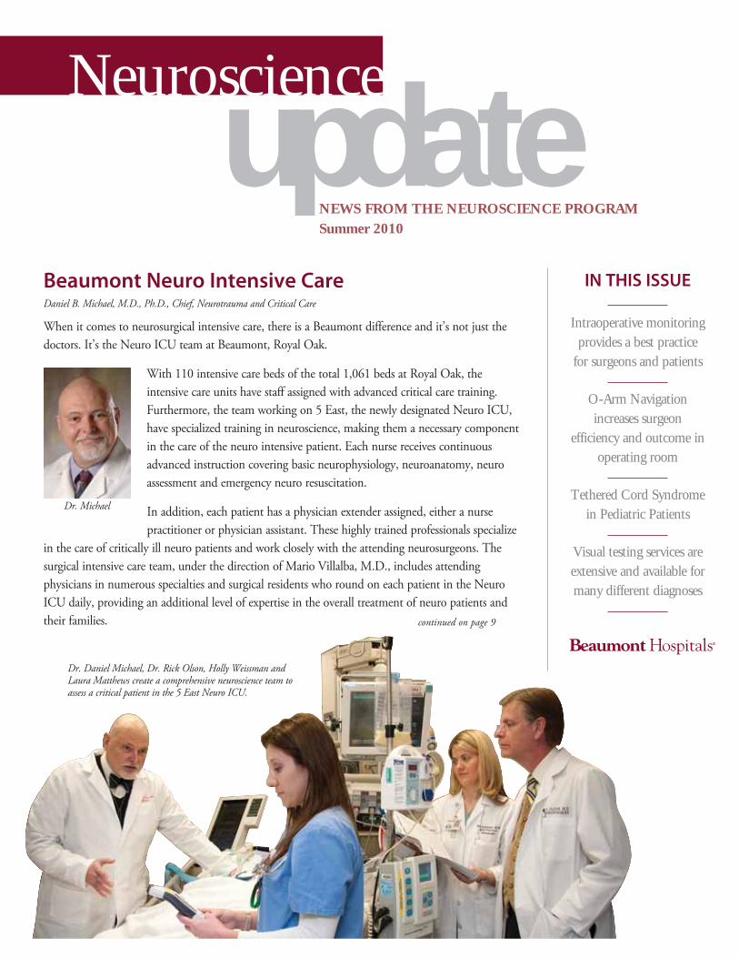
Beaumont Neuro Intensive CareDaniel B. Michael, M.D., Ph.D., Chief, Neurotrauma and Critical Care
When it comes to neurosurgical intensive care, there is a Beaumont difference and it’s not just the doctors. It’s the Neuro ICU team at Beaumont, Royal Oak.
With 110 intensive care beds of the total 1,061 beds at Royal Oak, the intensive care units have staff assigned with advanced critical care training. Furthermore, the team working on 5 East, the newly designated Neuro ICU, have specialized training in neuroscience, making them a necessary component in the care of the neuro intensive patient. Each nurse receives continuous advanced instruction covering basic neurophysiology, neuroanatomy, neuro assessment and emergency neuro resuscitation.
In addition, each patient has a physician extender assigned, either a nurse practitioner or physician assistant. These highly trained professionals specialize
in the care of critically ill neuro patients and work closely with the attending neurosurgeons. The surgical intensive care team, under the direction of Mario Villalba, M.D., includes attending physicians in numerous specialties and surgical residents who round on each patient in the Neuro ICU daily, providing an additional level of expertise in the overall treatment of neuro patients and their families.
updateNEWS FROM THE NEUROSCIENCE PROGRAMSummer 2010
Neuroscience
continued on page 9
Dr. Daniel Michael, Dr. Rick Olson, Holly Weissman and Laura Matthews create a comprehensive neuroscience team to assess a critical patient in the 5 East Neuro ICU.
Dr. Michael

2 • NEUROSCIENCE UPDATE • SUMMER 2010
As the Center for Neuroscience at Royal Oak continues its quest to improve neurological care, I would like to highlight some of the news reported in this issue.
The Center for Neuroscience, in cooperation with hospital administration and the Critical Care division of the department of Surgery, has established the Neurosurgical Intensive Care Unit in 5 East. The 20-bed unit is dedicated to the care of neurosurgical patients admitted from the Emergency Center or from inpatient and surgical suites. The 5 East nursing personnel have worked very effectively in the past with the support of Mario Villalba, M.D., chief of Surgical Intensive Care Units, and all of the critical care physicians to establish an exceptionally solid base from which to build a strong and cohesive neuroscience critical care nursing team. Under the direction of Pat Erwin, the nursing personnel work with the neuroscience mid-level providers to continually improve their skills in the evaluation and management of the critically ill neurosurgical patient. This provides our patients with consistent and predictable nursing care, allows for the establishment of nursing pathways and medical protocols that meet national levels and optimizes the care of our patients.
Technological developments in neurosciences have evolved greatly over the last few years and increased the accuracy and precision of surgical procedures with minimization of surgical access points and improvement in surgical precision. The
O-Arm is one of those tools that are now used regularly in the operating room to improve the care of our patients to help the surgeon plan and complete the procedure with the aid of computer navigation.
Furthermore, electrophysiological monitoring has been increasingly used for the management of patients with aneurysms, brain tumors and spine surgery. The Center for Neuroscience strengthened its very successful electrophysiology monitoring team with the recruitment of Shaila Gowda, M.D., who was recently appointed as the team’s director. Dr. Gowda provides onsite medical support for surgical cases in the South Tower operating room, assisting both neuro and orthopedic surgeons as well as expanding our surgical epilepsy programs at Royal Oak.
Although technological improvements are critical to the continued growth and improvement in patient care, it is the people who work in the Center for Neuroscience who are essential to the work we do. I am extremely confident of the team we have and proud of all the hard work they do in our quest to provide excellent neurological care.
Think Neuro First!
Fernando G. Diaz, M.D., Ph.D.Chief, Neurological Surgery, Beaumont Hospital, Royal Oak
Note from the Chief
Professional interpretation of EEG is now more convenient with the addition of remote viewing software from Persyst Corporation. EEG tracings can be viewed from any computer that has access to the Beaumont Web Portal by accessing the software from the Beaumont server. The Citrix server interface enables the remote computer to use the Persyst software as if it was local. The EEG reader can view any EEG tracing residing in the current data base. In addition to standard EEG tracings, ambulatory EEG data is available.
The Persyst Insight II is full-featured allowing post acquisition manipulation of parameters such as sensitivity, filters,
Technical Enhancements to EEG Reading David Betts, R.EEG/EP T., CNIM, CLTM
continued on page 10

SUMMER 2010 • NEUROSCIENCE UPDATE • 3
Seldom has a field in medicine grown as rapidly as intraoperative neurophysiologic monitoring, or IOM, which provides a real-time assessment of the status of the nervous system during surgery and allows the surgeon to modify the surgical plan in response to changes that indicate impending injury.
As late as the mid-1980s, few people had even heard of IOM. The initial
seminal papers appeared about 20 years ago, documenting the utility of somatosensory- and brainstem auditory-evoked potential monitoring in reducing the morbidity of scoliosis and brainstem surgery; these two modalities monitor the integrity of the body’s sensory and hearing systems, respectively. The last decade has seen remarkable advances in IOM. New modalities, such as motor evoked potentials (a test that monitors the status of the body’s motor function), become available, and the equipment used for monitoring has also improved considerably.
Beaumont’s IOM technologists have worked in the field since its early conception, continue to apply their skill and knowledge daily and provide a level of experience that is nearly unprecedented. These highly trained technologists enjoy a wide referral pattern of diverse cases, allowing each member to exercise and master all manner of IOM testing modalities.
The leading edge IOM lab benefits from the technologists’ diverse scientific and academic background, which contributes to its constant growth and peer-to-peer education. Our tech experience ranges from molecular biology to biomedical engineering, from chemistry to extensive training in evoked potential technology. The group is also involved in newly developed surgical procedures pioneered at Beaumont as well as partnering with industry to help further the use of IOM on an international level.
Intraoperative monitoring provides a best practice for surgeons and patients Josh Mergos, CNIM, BSEE
Welcome Dr. GowdaA recent addition to the group is Shaila Gowda, M.D., the new medical director of Intraoperative Monitoring. Dr. Gowda completed her medical training at Kasturba Medical College in Karnataka, India, and later completed her neurology residency at the University of Mississippi Medical Center, followed by a fellowship in clinical neurophysiolgy/epilepsy at the Cleveland Clinic.
Dr. Gowda served as the director of intraoperative monitoring/epilepsy at Metro Health Medical Center in Cleveland and more recently as the director of EEG and staff neurologist at Providence Hospital. She has been published in the Journal of Neurosurgery: Pediatrics and has special interest in epilepsy surgery, women’s issues in epilepsy and intraoperative neurophysiologic monitoring.
Dr. Gowda joins Beaumont’s IOM group, which consists of six certified neurophysiologic intraoperative monitoring technologists. Surgeries performed at Beaumont using IOM include spinal lumbar and cervical fusions, spinal cord-untetherings, skull-based tumor resections, aneurysm clippings and parotidectomies.
Dr. Gowda
Josh Mergos,CNIM, documents neurologic signals during a cervical spine fusion.
4 • NEUROSCIENCE UPDATE • SUMMER 2010
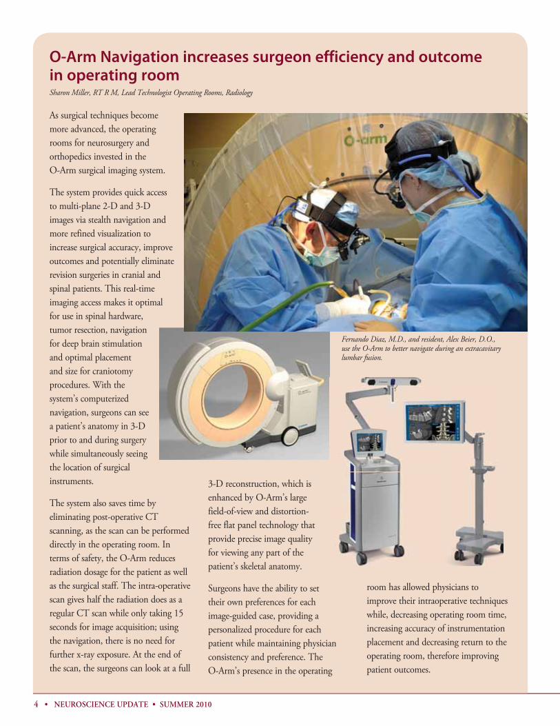
As surgical techniques become more advanced, the operating rooms for neurosurgery and orthopedics invested in the O-Arm surgical imaging system.
The system provides quick access to multi-plane 2-D and 3-D images via stealth navigation and more refined visualization to increase surgical accuracy, improve outcomes and potentially eliminate revision surgeries in cranial and spinal patients. This real-time imaging access makes it optimal for use in spinal hardware, tumor resection, navigation for deep brain stimulation and optimal placement and size for craniotomy procedures. With the system’s computerized navigation, surgeons can see a patient’s anatomy in 3-D prior to and during surgery while simultaneously seeing the location of surgical instruments.
The system also saves time by eliminating post-operative CT scanning, as the scan can be performed directly in the operating room. In terms of safety, the O-Arm reduces radiation dosage for the patient as well as the surgical staff. The intra-operative scan gives half the radiation does as a regular CT scan while only taking 15 seconds for image acquisition; using the navigation, there is no need for further x-ray exposure. At the end of the scan, the surgeons can look at a full
3-D reconstruction, which is enhanced by O-Arm’s large field-of-view and distortion-free flat panel technology that provide precise image quality for viewing any part of the patient’s skeletal anatomy.
Surgeons have the ability to set their own preferences for each image-guided case, providing a personalized procedure for each patient while maintaining physician consistency and preference. The O-Arm’s presence in the operating
room has allowed physicians to improve their intraoperative techniques while, decreasing operating room time, increasing accuracy of instrumentation placement and decreasing return to the operating room, therefore improving patient outcomes.
O-Arm Navigation increases surgeon efficiency and outcome in operating roomSharon Miller, RT R M, Lead Technologist Operating Rooms, Radiology
Fernando Diaz, M.D., and resident, Alex Beier, D.O., use the O-Arm to better navigate during an extracavitary lumbar fusion.

SUMMER 2010 • NEUROSCIENCE UPDATE • 5
Physician spotlight:
Chris Kazmierczak, M.D.Director, Interventional Neuroradiology
Chris Kazmierczak, M.D., is an interventional neuroradiologist, who is board-certified in diagnostic radiology and has a certificate of added qualification in diagnostic neuroradiology.
He graduated from medical school at Wayne State University and completed his residency at Beaumont Hospital. After his residency, he completed his fellowship in diagnostic interventional neuroradiology at the Detroit Medical Center. Dr. Kazmierczak is an integral part of Beaumont’s neuroscience team as well as a leader member of the Joint Commission-accredited Stroke Program.
As a specialist in interventional neuroradiology, Dr. Kazmierczak often consults on a number of diagnoses presenting in the Emergency Center and uses minimally invasive techniques to treat abnormalities involving the brain and spine, such as intracranial aneurysms, stroke or spinal fractures. For the most common EC referral, a ruptured aneurysm, Dr. Kazmierczak performs endovascular aneurysm coiling for treatment, a procedure less invasive than open surgical repair. Emergency treatment services are available around the clock, 365 days a year.
Because patients can have non-emergent vascular abnormalities that require interventional procedures, Dr. Kazmierczak also has a thriving outpatient practice that attracts patients through primary referrals as well secondary referrals from primary care physicians, neurosurgeons and neurologists.
To refer a patient to Dr. Kazmierczak or any member of the Interventional Neuroradiology team, please contact the department at 248-898-2195.
Dr. Kazmierczak in one of the IR cath labs.
Research, new hardware leads to improved ability to examine speech brain responseRichard Merson, Ph.D., coordinator of clinical research, Speech-Language Pathology and Anil Shetty, Ph.D., chief, Imaging Physics; clinical professor, Radiology, Oakland University William Beaumont School of Medicine
Brain imaging of speech and language functions in experimental and clinical research is no longer a rare occurrence. Magnetic resonance imaging, positron emission tomography and magnetoencephalography are being used to identify motor and sensory receptive and expressive speech and language cortical regions of interest.
Clinical research has identified the cortical representations of aphasia, stuttering, dyslexia, cognitive deficits and memory functions through mapping the functional MRI and PET outcomes. These brain imaging studies will allow us to quantify, track and predict speech and language recovery from stroke, traumatic brain injury and perhaps dyslexia patients prior to treatment.
At Beaumont Hospital, Royal Oak, we have used functional MRI with post-stroke patients as well as with patients who have seizure disorders
fMRI speech-imaging of a post-CVA with global aphasia who was in aphasia rehabilitation for several years with a remarkable return of some of his verbal language. The images clearly identify a large left fronto-temporal infarct and compensatory fMRI activation in the non-dominant hemisphere during language activity. continued on page 10

6 • NEUROSCIENCE UPDATE • SUMMER 2010
Tethered Cord Syndrome in Pediatric PatientsHolly S. Gilmer, M,D., Chief, Pediatric Neurosurgery
Tethered cord syndrome can be difficult to diagnose in babies and children since the symptoms may be subtle and insidious over time. The most common symptoms, such as back pain, abnormal gait and urinary accidents are frequently attributed
to other causes during childhood. Despite misdiagnosis of delayed identification, proper treatment can lead to a full recovery.
What is tethered cord syndrome?Tethered cord, or TC, is a disorder in which the spinal cord adheres to a structure within the spine such as dura, scar tissue from a previous operation,
a bony spicule or even a tumor. Although most cases are congenital, the condition may not become symptomatic until later in life as the spine continues to grow and lengthen. TC most often occurs in patients with spina bifida, although it is frequently seen with Chiari malformation. TC can affect people of all ages, but it is most often found from infancy through adolescence. Symptoms become more pronounced during periods of rapid growth due to increased stretching of the spinal cord.
What are the symptoms of tethered cord?Symptoms are age-specific. Older children and adults may experience back pain, leg pain, weakness in the lower extremities and/or fatigue with walking. Recurrent bladder infections, urinary or fecal accidents, as well as urinary frequency, hesitancy and urgency may also be signs of TC, with or without back and lower extremity symptoms.
Babies may experience delayed motor milestones such as late walking. Examination of the back may also show neurocutaneous signs, such as dark or red spots, tufts of hair or soft masses. Those skin lesions associated with TC tend to be midline. Severe cases may show scoliosis or other curvatures of the spine. Leg-length discrepancies, differences in the size of the legs or feet and foot deformities are also seen in severe cases.
How is it diagnosed?A spinal MRI can confirm the diagnosis.
How is tethered cord treated?If the patient has predominantly back pain and mild
weakness, a course of physical therapy may relieve symptoms. This approach requires the patient to be old enough to reliably convey whether the symptoms are worsening or improving. Most TC cases require surgical intervention to prevent neurologic deterioration. A laminectomy is performed, the dura is opened and using the operating microscope, the spinal cord is freed from the tethering structure. If possible, the tethering object is removed to avoid re-tethering, which can happen.
Recovery from the surgery is one to two weeks of very limited activity to ensure proper healing of the surgical site and to prevent leaking of any cerebrospinal fluid. However, most patients are not required to undergo physical therapy post-operatively. Many patients regain normal function and are pain free following surgery.
What happens if TC is left untreated?Patients with untreated tethered cord will continue to experience their current symptoms, and their motor and sensory function may worsen. Particularly in children, lengthening of the spine with growth can lead to paraplegia and loss of bowel and bladder function. Chronic back and leg pain in children is uncommon and should not be taken lightly. Children with symptoms of tethered cord should undergo radiologic imaging and evaluation by a specialist without delay.
Patient, 3mos old, with spinal cord tethered to subcutaneous lipoma at spinal defect, a tumor in this case.
Dr. Gilmer

SUMMER 2010 • NEUROSCIENCE UPDATE • 7
Visual testing services are extensive and available for many different diagnoses Edward Cohn, MD, MBA, MPH
With more than 125 ophthalmologists on staff, Beaumont Hospitals’ Ophthalmology department offers experts in every ophthalmic specialty. The physicians are leaders in their respective specialties, state and national societies as well as internationally recognized teachers and speakers; many are known for their research contributions and regularly participate in National Eye Institute clinical trials. The department is one of the leading international referral centers for pediatric patients with retinopathy of prematurity and related genetic/congenital retinal disorders.
Specialty services offered include:• general ophthalmology• corneal and external disease• anterior segment surgery• refractive surgical services/LASIK • glaucoma• cataract surgery• laser treatment• pediatric ophthalmology• strabismus• ophthalmic plastic and reconstructive
surgery• retina/vitreous disease and surgery• neuro-ophthalmology• low vision evaluation/ vision
rehabilitation• ophthalmic pathology
Ancillary diagnostic testing is available through the Neurology and Ophthalmology departments for:• visual evoked response/potentials, or
VER/VEP – A generalized evaluation of the visual conduction system using either a visual checkerboard pattern or flashing light stimulus
•
• electroretinography, or ERG – A multi-focal ERG, which checks the integrity of the retinal rods and cones
• electrooculography, or EOG – Tests the integrity of the retinal pigment epithelium and retinal pigment epithelial neuro-receptor interfac
• ophthalmic diagnostic imaging includes:
– ultrasounds to measure the eye, scan for intraocular masses and detect ocular structure abnormalities – retinal photography and angiography to document optic nerve appearance and retinal vasculature in CNS disorders, diabetes, macular degeneration, etc. • visual field testing (Humphrey
Automated, Goldmann Kinetic, Tangent Field, Amsler Grid) – Each of these evaluate peripheral vision as well as central vision depending on the specific test utilized.
• color vision testing (Ishihara, Farnsworth Munsel 100, D-15) – can be generalized in determining total loss of color perception, partial color confusion among shades of red/green, or highly specific in determining red (protan), green (deutan), or blue/yellow (tritan) color vision disturbance.
• dark adaptation• optical coherence tomography, or
OCT – An optical signal acquisition and processing method capturing micrometer-resolution, 3-D images of the inner eye.
Visual evoked response or visual evoked potentials test the integrity of the visual conduction system from the optic nerve to the occipital lobe of the brain. Optic Neuritis, MS, Cortical Blindness are some of the visual disturbances where VER is helpful if not diagnostic.
Electroretinogram, or ERG, assists with evaluation of retinal rod and cone function. Retinitis pigmentosa night blindness and other vision loss due to retinal disturbance are aided by ERG, mERG (multifocal ERG for macular function) and electrooculogram, or EOG, studies.

Physician spotlight:
Rick Olson, M.D., Neurosurgery
Rick Olson, M.D., graduated from Wayne State University School of Medicine and completed two years of general surgery residency at Beaumont Hospital, Royal Oak. He completed his neurosurgical residency at the University of Western Ontario under the direction of Charles Drake, M.D., a pioneer in the treatment of basilar artery aneurysms. Dr. Olson is a member of the American College of Surgeons as well as the Royal College of Surgeons of Canada.
In 1983, Dr. Olson returned to Beaumont to begin his career in neurosurgery, joining Fred Latimer, M.D., and Gordon Scratch, M.D. When his partners retired, Dr. Olson formed Oakland Neurosurgical Associates, P.C., with offices in Royal Oak and Troy.
As a neurosurgeon, Dr. Olson treats many areas within his specialty, although he specializes in the treatment of brain tumors. He uses the latest technology in neurosurgery, including neuro-navigational assistance in localizing tumors within the brain. He collaborates with Radiation Oncology staff in treating brain tumors with Gamma Knife therapy. Dr. Olson still enjoys the clinical side of medicine, spending time with his patients while working toward an accurate diagnosis.
For the past seven years, Dr. Olson has been assisted by Christina M. Becker, PA-C. She assists Dr. Olson by seeing patients in the office, rounding on patients in the hospital and assisting in surgery.
From the beginning, Dr. Olson saw Beaumont as a progressive institution, offering the ancillary support including neuroradiology and
neuropathology as well as neurosurgery physician extenders. He is excited about the development of the Oakland University William Beaumont School of Medicine and is looking forward to working with future medical students and residents.
For more information on Dr. Olson’s practice and patient referral, please contact the Physician Referral Line at 800-633-7377.
Dr. Olson and Christina Becker, PA-C evaluate a patient’s films and discuss treatment plans at their Royal Oak clinic.
8 • NEUROSCIENCE UPDATE • SUMMER 2010
Flash CT provides fast, clear images with lower radiation doseRichard Silbergleit, M.D., vice-chief, Diagnostic Radiology, and director, Neuroimaging
Beaumont Hospitals became the first hospital in Michigan to offer patients the newest, safest CT technology when the Royal Oak, Troy and Grosse Pointe hospitals installed Siemens’ Somatom Definition Flash CT scanners.
The Flash CT allows:• unprecedented speed, with studies
performed in less than a second• significant reduction in radiation dose
These scanners use dual-source technology, meaning two X-ray tubes are simultaneously active, which allows the possibility of tissue characterization when the tubes are used at different energies. One important application of this is used in bone subtraction in CT angiography. A major limitation of CT angiography has been difficulty in separating the high attenuation of contrast agent in the blood vessels from the high attenuation of bone at the skull base. Utilizing dual energy, contrast can be more easily distinguished from bone (figure). This takes about minute, which is useful in emergent situations. Post processing by Beaumont’s highly trained 3-D lab technologists yields even higher quality images.
The combination of the speed of the scanner and radiation dose reduction also makes CT perfusion feasible. This application will be used in a variety of clinical conditions but is initially designed for the triage of acute stroke patients at Royal Oak.
The speed of the scanner also allows many scans to be performed in uncooperative
continued on page 10

Neuro Intensive Care continued from page 1
Treatment population
Some patients in the Neuro ICU suffer from stroke, brain tumors or severe traumatic brain injuries, TBI. Others are post-op from a neurosurgical procedure, including insertion of various invasive brain monitoring devices, craniotomy for trauma, cerebral aneurysm repair or tumor resection. Patients having minimally invasive endovascular procedures to repair aneurysms or other cerebral vascular diseases will also be monitored during recovery in the Neuro ICU. In addition to the frequent and expert clinical exams conducted by nurses and other personnel, advanced monitoring techniques including ICP, bedside chemical analysis and continuous electroencephalographic monitoring are routinely employed when appropriate.
Advantages of oneChart EMR
One of the newest features improving Neuro ICU care is oneChart, the electronic health record. This advanced health information technology allows the
team to view and graph patient data for rapid bedside analysis of parameters like intracranial pressure and cerebral perfusion pressure. The patients’ lab information and imaging studies may be viewed at the workstation located in each of the ICU’s 20 private patient rooms. The computer order entry module recently implemented contains neuro-specific order sets based on the latest international guidelines for the treatment of TBI and subarachnoid hemorrhage. oneChart will eventually be used to analyze outcome data to improve future care.
Safety quality measures
The Neuro ICU is a busy place; over the past 12 months, patients on 5 East have averaged 233 ventilator days per month. Continuous improvement programs include monitoring all complications like pneumonia, blood stream, deep venous thrombosis, pressure ulcers and urinary tract and cerebral spinal fluid, or CSF, infections. Although some complications are unavoidable in these very sick, often
comatose patients, constant vigilance and prompt intervention can minimize untoward events. The current ventilator associated pneumonia rate for this unit is 0.07 percent, which is well below the national average for neuro intensive units. The unit’s tracheostomy rate is also below average for comparable ICU patient populations.
Neuro ICU research
Current research in the Neuro ICU at Beaumont includes molecular and cellular analysis of CSF and the utility of advanced imaging techniques such as diffusion tensor imaging in severe TBI and minimally conscious patients. We will also be a participating center for the ProTECT III trial to study the efficacy of progesterone as a treatment for TBI. There is ample opportunity for philanthropic donations to the Neuro ICU at Beaumont as we have plans to add advanced bedside neuro monitoring capabilities including a portable CT scanner.
SUMMER 2010 • NEUROSCIENCE UPDATE • 9
Members of the 5 East Neuro ICU Nursing team.

10 • NEUROSCIENCE UPDATE • SUMMER 2010
Flash CT continued from page 8
patients like pediatric, trauma or confused patients without the use of sedation or general anesthesia, thus allowing for more efficient scanning. Scans that were previously performed on uncooperative patients without sedation and that were limited by motion artifact are now routinely high quality.
Areas of development for using this new technology outside of the neurosciences include cardiac imaging, vascular imaging, and tissue characterization in musculoskeletal imaging. For further information on use of this technology in the neurosciences, please contact a neuroradiologist at 248-898-3184.CTA image of the intracranial circulation performed on the Flash CT. The petrous portions of the internal
carotid arteries have been difficult to visualize on CTA without dual-energy technology and associated software even with dedicated post-processing. This bone subtraction was done in under a minute and the petrous segments of the internal carotid arteries are well seen (arrows).
Speech brain response continued from page 5
before and after neurosurgical procedures. We have examined post-traumatic central nervous system stuttering through MRI and PET. Furthermore, we are attempting to replace the WADA CNS Intercarotid Angiographic procedure to identify cortical hemisphere dominance for speech/language with a standard functional MRI paradigm that will reduce
the invasive nature of cortical angiography and improve the finite description and quantification of speech functioning.
In collaboration with Diagnostic Radiology, Neuroradiology and the Research Institute, we are developing fMRI speech-language models to evaluate post-stroke, traumatic brain injury and seizure
disorder patients. In addition, we are adding new digital hardware to our fMRI unit that will allow improved acquisition of speech stimuli for patient viewing and collection of synchronized speech-language responses. This enhancement dramatically improves our ability to accurately examine speech brain response.
EEG Reading continued from page 2
display time and montages. All of this is done as easily as sitting at any reader station on the hospital’s campus. A full selection of standard montages is available via drop down menu. Custom montages can be created by users on the fly and saved for future use. Laplace and specialize reference montages are available. Comments can be added and stored with the tracing for ease of use.
This new software has advanced features for data analysis and manipulation such as spectral frequency display and interchannel correlation and lag. The EEG tracing and data graphs can be exported to word processing and presentation programs.
Implementation of remote reading has markedly reduced turn around time. No longer is the physician reader required
to come to the Royal Oak campus to view the tracings. EEG can be read throughout the day from any location at the interpreter’s convenience. In the past, physician readers often came just once a day to read the accumulated EEG tracings. Remote reading software provides a major step to a rapid turnaround time resulting in improved service to our patients and referring physicians.

SUMMER 2010 • NEUROSCIENCE UPDATE • 11
Traumatic Brain and Spinal Cord
Injuries – When an accident results in brain or spinal cord injury, treatment often begins at one of Beaumont’s Emergency Centers. Our EC’s offer leading edge imaging and advanced telemetry for fast, accurate diagnosis of injuries.
Our multidisciplinary team of ER staff, trauma surgeons, neurosurgeons and rehab specialists work together to begin immediate treatment, which is essential for best outcomes.
Gamma Knife ® – Beaumont’s Gamma Knife® treats brain tumors and neurological conditions with pinpoint accuracy.
Gamma Knife® delivers a highly therapeutic dose of radiation to the brain accurately and without the risks of open surgery like general anesthesia, infection and bleeding. Only the target tissue receives a significant radiation dose and no incision is required. Recovery is quick and treatment is generally done on an outpatient basis.
In addition to tumors, Gamma Knife® can be used for arteriovenous malformations, trigeminal neuralgia, Parkinson’s disease, essential tremors, epilepsy and obsessive-compulsive disorder.
Neuro Oncology – Our specialists diagnose and treat malignant brain and spine tumors in children and adults using neuroimaging techniques like positron emission tomography, magnetic resonance spectroscopy imaging, Gamma Knife® and spinal radiosurgery. The team includes neuro oncologists, neurosurgeons, radiation therapists and neuroradiologists.
Cranio-Facial and Skull Base – Beaumont’s Cranio-Facial program brings together specialists to provide integrated care for children with a range of craniofacial disorders, such as cleft lip or palate. The multidisciplinary team includes plastic surgeons, otolaryngologists, maxillo facial surgeons and neurosurgeons.
Pediatric Neurosurgery – At Beaumont Children’s Hospital, pediatric neurologists, neurosurgeons and interventional neuroradiologists are available to treat a variety of neurological conditions in infants and children. Our experts are skilled and equipped to handle the most severe head and spine trauma issues in children.
As the only hospital in SE Michigan providing pediatric interventional treatment of brain aneurysms, arteriovenous malformations and vascular tumors, we reduce or eliminate the need for traditional surgery methods.
Epilepsy and Movement Disorders – Our team treats a broad grouping of neurological conditions like epilepsy, Parkinson’s disease, dystonia, spasticity, Huntington’s disease and movement disorders caused by brain injury.
An Epilepsy Monitoring Unit opened at Beaumont Royal Oak as a specialized inpatient unit designed to evaluate, diagnose and treat children and adults with seizures. The EMU offers leading diagnostic capabilities and physician consultation.
Beaumont is also a leader in using deep brain stimulation (DBS) to treat various neurological and movement disorders like Parkinson’s disease, essential tremor, dystonia, tremor due to multiple sclerosis and chronic pain.
Pain Management – Safe and effective pain management is an integral part of treatment. Beaumont’s neuroscience team works in conjunction with anesthesiologists on a variety of treatments and procedures.
Physicians have innovative options for treating pain including dorsal column stimulators that can be adjusted for varying levels of pain as well as an implantable intrathecal drug pump. Beaumont’s advanced radiosurgery equipment is helpful
in treating focal pain conditions because it’s less invasive than surgery and can be repeated if symptoms recur.
Spine – Our team performs minimally invasive surgeries on complex spinal conditions including spinal trauma, spinal deformities, and spinal oncology. This approach significantly improves patient outcomes with less blood loss, less pain, lower infection rates and shorter hospital stays.
Interventional Neuroradiology and
Stroke – Beaumont Royal Oak is designated a Primary Stroke Center by the Joint Commission affirming the success of our comprehensive stroke care. Our team includes specially trained and certified nursing staff, more interventional stroke specialists than any other hospital in Michigan, dedicated ICU beds for stroke patients, interventional neurosurgeons and neuroradiologists who can administer intra-arterial TPA and remove blood clots from the brain, and a dedicated neuroscience unit with 66 beds and 9 monitored critical care beds.
A minimally-invasive approach is provided by our team to treat vascular diseases of the central nervous system and spine including aneurysms, vascular malformations, arterial/venous stenosis, and tumors of the brain, head and neck requiring surgical intervention.
Intraoperative Monitoring – Intraoperative monitoring allows surgeons to check the functionality and integrity of the nervous system during surgery. The use of electrophysiological methods measures brain, nerve and spinal cord activity and has shown to improve patient safety and surgical outcomes.
Beaumont’s IOM team has training and experience exceeding many programs in the nation in the surgical areas of neurosurgery, orthopedics, otolaryngology/head and neck and urology.
Neuroscience program overview

3711 Thirteen Mile RoadRoyal Oak, Michigan 48073
Neuroscience update
is published quarterly in support of the Neuroscience Program at Beaumont Hospitals.
For comments or suggestions, please call Rachael Wade at 248-551-2300
Neurosurgery
Fernando Diaz, M.D., Ph.D.Chief, Neurosurgery
Mick Perez-Cruet, M.D., M.S.Vice-Chief, NeurosurgeryHolly Gilmer, M.D.
Chief, Pediatric NeurosurgeryDaniel Michael, M.D., Ph.D.
Chief, NeurotraumaAttending PhysiciansStephen Boodin, M.D.Phillip Friedman, M.D.
Rick Olson, M.D.Daniel Pieper, M.D.Karol Zakalik, M.D.
Associate PhysiciansRyan Barrett, D.O.Bradley Hall, D.O.
Robert Johnson, M.D.Todd Nida, M.D.
Omar Qahwash, D.O.
Adjunct PhysiciansKonstantin Elisevich, M.D., Ph.D.
Murali Guthikonda, M.D.Steven Ham, D.O.
Fredrick Junn, M.D.Steven Kalkanis, M.D.Ghaus Malik, M.D.
Jack Rock, M.D.Mark Rosenblum, M.D.
Teck Soo, M.D.Sandeep Sood, M.D.
Stroke ProgramSusan Catto, M.D.
Medical Stroke DirectorPhysician Extenders
Holly Weissman, N.P.Chief
Kim Cameron, N.P.Megan Clippard, N.P.Becky Doherty, N.P.
Amanda Griffith, N.P.Lauren Gurski, P.A.Justin Hugelier, P.A.Jennifer Jehle, N.P.Megan Keiser, N.P.
Rosie Mannina, N.P.Kristen McGrath, N.P.Jamie Peysakhov, P.A.
Neuroscience AdministrationLori Sheridan
Administrative ManagerNeuroscience
Rachael WadeAdministrative Assistant
Neuroscience
Hospital AdministrationCharles Shanley, M.D.
Senior Vice President and Chair, Surgery
Victoria Hollingsworth-Schuler, MS, MSA
Administrative DirectorNeuroscience
Dorothy Bernard, BSN, MSADirector, Specialty Nursing
Neurology
Attending PhysiciansMazen Al-Hakim, M.D.Kheir Al-Zouhayli, M.D.
Martin Belkin, D.O.Lawrence Eilender, M.D.Anthony Emmer, D.O.Raina Ernstoff, M.D.
Jonathan Fellows, D.O.Sonia Fernando, M.D.
Michelle Furmaga, M.D.Jodi Ganley, D.O.Neil Gilbert, M.D.
Rashmi Gupta, M.D.Mark Kachadurian, D.O.
Brian Kirschner, M.D.William Leuchter, M.D.Sami Mounayer, M.D.Steven Newman, M.D.Steven Schecter, M.D.
Lalitha Sivaswamy, M.D.Alexander Spitzer, M.D.
Alex Steinbock, D.O.Gary Trock, M.D.
Richard Trosch, M.D.Narayan Verma, M.D.
Associate PhysiciansWilliam Boudouris, D.O.
Norman Burns, M.D.Nancy Jingyang Cao, M.D. Ph.D.
Mitchell Elkiss, D.O.Matthew Holtzman, D.O.
Elizabeth Leleszi, M.D.Zef Lucaj, M.D. Ph.D.
Eileen McCormick, D.O.Andrea Rossi, D.O.
Howard Rossman, D.O.Vijay Samuel, M.D.
Alka Shah, M.D.Mark Silverman, D.O.Bruce Silverman, D.O.Susan Smietana, D.O.Elizabeth Smith, M.D.
Nader Warra, D.O.Danny Watson, M.D.Esther Young, D.O.
Adjunct PhysiciansAaron Ellenbogen, D.O.Shelley Knowles, M.D.Danette Taylor, D.O.
Neuroradiology
Attending PhysiciansChris Kazmierczak, M.D.
Anant Krishnan, M.D.Samir Noujaim, M.D.
Sneha Patel, M.D.Richard Silbergleit, M.D.
Kurt Tech, M.D.Ay-Ming Wang, M.D.Jeffrey Wilseck, D.O.
Neuroscience Faculty and Staff, Royal Oak