Embed Size (px)
Citation preview
*Edited by Erik M. Jorgensen and Joshua M. Kaplan. Last revised February 5, 2008. Published April 7, 2008. This chapter should be cited as: Li,C. and Kim, K. Neuropeptides (April 7, 2008), WormBook, ed. The C. elegans Research Community, WormBook,doi/10.1895/wormbook.1.142.1, http://www.wormbook.org.
Copyright: © 2008 Chris Li and Kyuhyung Kim. This is an open-access article distributed under the terms of the Creative Commons AttributionLicense, which permits unrestricted use, distribution, and reproduction in any medium, provided the original author and source are credited.
§To whom correspondence should be addressed. Tel. (212) 650-8533, Fax: (212) 650-8585. E-mail: [email protected]
Neuropeptides*
Chris Li1§, and Kyuhyung Kim2,
1Department of Biology, City College ofNew York 2Department of Biology, Brandeis University, New York, NY10031 USAWaltham, MA 02454 USA
Table of Contents1. Introduction ............................................................................................................................ 22. Formation of mature neuropeptides ............................................................................................. 2
2.1. Processing of neuropeptide precursor molecules .................................................................. 22.2. Neuropeptides are released from dense core vesicles ............................................................ 4
3. Immunocytochemical localization of neuropeptides ........................................................................ 54. Identification of putative neuropeptide genes ................................................................................. 55. Expression and localization of neuropeptide genes ....................................................................... 236. Biochemical isolation of neuropeptides ...................................................................................... 247. Neuropeptide function ............................................................................................................ 24
7.1. The insulin-like gene family .......................................................................................... 247.2. The flp family ............................................................................................................. 267.3. The nlp family ............................................................................................................ 26
8. Neuropeptide receptors ........................................................................................................... 269. Pharmacology of FLP neuropeptides ......................................................................................... 2810. Summary ............................................................................................................................ 2811. Acknowledgments ................................................................................................................ 2912. References .......................................................................................................................... 29
Abstract
The role of neuropeptides in modulating behavior is slowly being elucidated. With the sequencing of theC. elegans genome, the extent of the neuropeptide genes in C. elegans can be determined. To date, 113neuropeptide genes encoding over 250 distinct neuropeptides have been identified. Of these, 40 genes encodeinsulin-like peptides, 31 genes encode FMRFamide-related peptides, and 42 genes encode non-insulin,non-FMRFamide-related neuropeptides. As in other systems, C. elegans neuropeptides are derived fromprecursor molecules that must be post-translationally processed to yield the active peptides. These precursormolecules contain a single peptide, multiple copies of a single peptide, multiple distinct peptides, or anycombination thereof. The neuropeptide genes are expressed extensively throughout the nervous system,
1
including in sensory, motor, and interneurons. In addition, some of the genes are also expressed in
non-neuronal tissues, such as the somatic gonad, intestine, and vulval hypodermis. To address the effects ofneuropeptides on C. elegans behavior, animals in which the different neuropeptide genes are inactivated oroverexpressed are being isolated. In a complementary approach the receptors to which the neuropeptidesbind are also being identified and examined. Among the knockout animals analyzed thus far, defects inlocomotion, dauer formation, egg laying, ethanol response, and social behavior have been reported. Thesedata suggest that neuropeptides have a modulatory role in many, if not all, behaviors in C. elegans.
1. Introduction
Neuropeptides are short sequences of amino acids that function either directly or indirectly to modulatesynaptic activity. In addition, neuropeptides may also function as primary neurotransmitters. As in mammaliansystems, the number of predicted neuropeptides in C. elegans is well over one hundred (Li et al., 1999; Pierce et al.,2001); however, most of the neuropeptides fall into two large families: the insulin-like peptides (Pierce et al., 2001;Li et al., 2003) and the FMRFamide (Phe-Met-Arg-Phe-NH
2)-related peptides or FaRPs, which are referred to as
FLPs in C. elegans (Li et al., 1998; Li, 2005). The remaining non-insulin, non-FLP peptides are classified as theneuropeptide-like proteins or NLPs. The NLPs are a diverse group of neuropeptides that have little similarity amongeach other (Nathoo et al., 2001). With a few striking exceptions, the functions of the different neuropeptides remainlargely unknown in C. elegans. This overview serves to summarize the current state of the neuropeptide field.
2. Formation of mature neuropeptides
Neuropeptides are typically derived from larger precursor molecules, which undergo posttranslationalprocessing and sometimes modifications to yield mature peptides (see Figure 1). A single neuropeptide precursormolecule can give rise to a single neuropeptide, multiple distinct neuropeptides, multiple copies of a singleneuropeptide, or any combination thereof. As an additional mechanism to increase neuropeptide complexity inmammals, a single precursor molecule can be differentially cleaved to yield different sets of peptides in differentcell types (Strand, 1999; Salio et al., 2006). Furthermore, in the mollusk Aplysia peptides from a single precursormolecule can be sorted into different nerve terminals (Sossin et al., 1990). Whether such differential processing andtrafficking occurs in C. elegans is unclear.
2.1. Processing of neuropeptide precursor molecules
Like mammalian precursor molecules (Steiner, 1998), the initial cleavages in C. elegans occur C-terminal todibasic residues flanking the peptide sequence (Rosoff et al., 1993; Marks et al., 1995, 1997, 1998, 1999a, 2001;Husson et al., 2005, 2006); however, cleavages C-terminal to mono- and tribasic residues have also been reported(Rosoff et al., 1993; Marks et al., 1997, 2001). The enzymes responsible for the initial endoproteolytic cleavage arekex2/subtilisin-like proprotein convertases, which must themselves be cleaved to become active. Cleavage ofproprotein convertase is dependent on a chaperonin protein SBT-1 7B2 (Lindberg et al., 1998; Sieburth et al., 2005).Four C. elegans proprotein convertases, kpc-1, egl-3/kpc-2, aex-5/kpc-3, and bli-4/kpc-4, are present in C. elegans(Thacker and Rose, 2000). egl-3/kpc-2 is expressed in many, but not all neurons in the nervous system (Kass et al.,2001). Loss of egl-3/kpc-2 results in defects in egg-laying, mechanosensation, and locomotion (Kass et al., 2001;Jacob and Kaplan, 2003), indicating that EGL-3/KPC-2 cleaves precursors whose peptides have diverse functions.Moreover, using a polyclonal anti-FMRFamide antibody that recognizes the Arg-Phe-amide but not thenon-amidated Arg-Phe-OH moiety (Marder et al., 1987), Kass and co-workers (2001) found decreasedFMRFamide-like immunoreactivity in egl-3/kpc-2 mutants, suggesting that egl-3/kpc-2 cleaves some, but not allFLP precursors and that other proprotein convertases are active in the same cells. Among the other proproteinconvertases, a kpc-1 deletion mutant shows mild locomotory defects and slow growth, suggesting that KPC-1cleaves precursors of peptides involved in movement and growth (Thacker and Rose, 2000). Mutations inaex-5/kpc-3 cause defecation defects (Thomas, 1990). aex-5/kpc-3 is expressed in muscle (Thacker and Rose, 2000),and has been proposed to cleave a precursor molecule in muscle to produce a peptide that serves as a retrogradesignal to regulate exocytosis (Doi and Iwasaki, 2002). A major function of BLI-4/KPC-4 is to cleave procollageninto collagen so that it can be deposited in the cuticle to give the cuticle structural integrity. Hence, null alleles ofbli-4/kpc-4 cause lethality (Thacker et al., 1995). However, transcripts of bli-4/kpc-4 are also expressed in thenervous system (Thacker et al., 1995; Thacker and Rose, 2000), although phenotypes associated with loss ofbli-4/kpc-4 neural transcripts are unknown. Collectively, these data indicate that multiple proprotein convertases areactive in neurons.
Neuropeptides
2
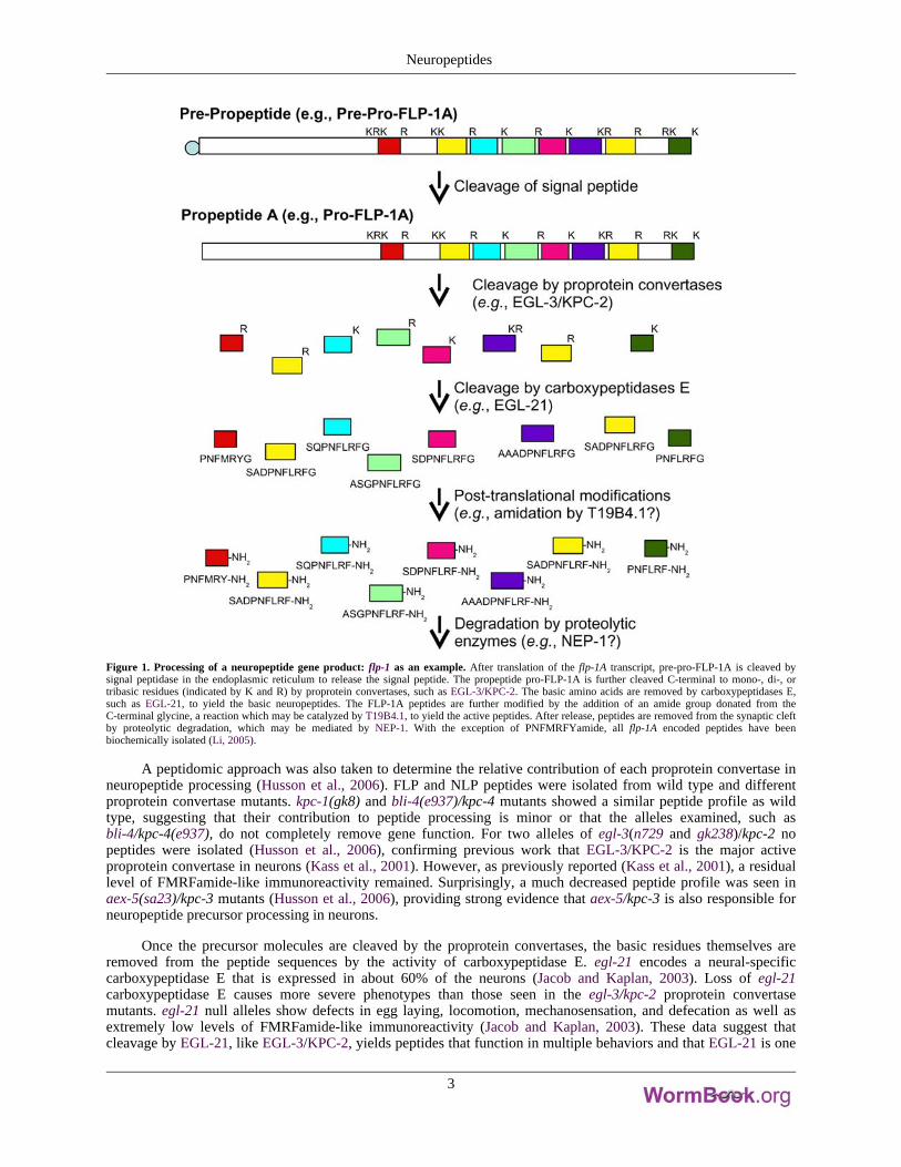
Figure 1. Processing of a neuropeptide gene product: flp-1 as an example. After translation of the flp-1A transcript, pre-pro-FLP-1A is cleaved bysignal peptidase in the endoplasmic reticulum to release the signal peptide. The propeptide pro-FLP-1A is further cleaved C-terminal to mono-, di-, ortribasic residues (indicated by K and R) by proprotein convertases, such as EGL-3/KPC-2. The basic amino acids are removed by carboxypeptidases E,such as EGL-21, to yield the basic neuropeptides. The FLP-1A peptides are further modified by the addition of an amide group donated from theC-terminal glycine, a reaction which may be catalyzed by T19B4.1, to yield the active peptides. After release, peptides are removed from the synaptic cleftby proteolytic degradation, which may be mediated by NEP-1. With the exception of PNFMRFYamide, all flp-1A encoded peptides have beenbiochemically isolated (Li, 2005).
A peptidomic approach was also taken to determine the relative contribution of each proprotein convertase inneuropeptide processing (Husson et al., 2006). FLP and NLP peptides were isolated from wild type and differentproprotein convertase mutants. kpc-1(gk8) and bli-4(e937)/kpc-4 mutants showed a similar peptide profile as wildtype, suggesting that their contribution to peptide processing is minor or that the alleles examined, such asbli-4/kpc-4(e937), do not completely remove gene function. For two alleles of egl-3(n729 and gk238)/kpc-2 nopeptides were isolated (Husson et al., 2006), confirming previous work that EGL-3/KPC-2 is the major activeproprotein convertase in neurons (Kass et al., 2001). However, as previously reported (Kass et al., 2001), a residuallevel of FMRFamide-like immunoreactivity remained. Surprisingly, a much decreased peptide profile was seen inaex-5(sa23)/kpc-3 mutants (Husson et al., 2006), providing strong evidence that aex-5/kpc-3 is also responsible forneuropeptide precursor processing in neurons.
Once the precursor molecules are cleaved by the proprotein convertases, the basic residues themselves areremoved from the peptide sequences by the activity of carboxypeptidase E. egl-21 encodes a neural-specificcarboxypeptidase E that is expressed in about 60% of the neurons (Jacob and Kaplan, 2003). Loss of egl-21carboxypeptidase E causes more severe phenotypes than those seen in the egl-3/kpc-2 proprotein convertasemutants. egl-21 null alleles show defects in egg laying, locomotion, mechanosensation, and defecation as well asextremely low levels of FMRFamide-like immunoreactivity (Jacob and Kaplan, 2003). These data suggest thatcleavage by EGL-21, like EGL-3/KPC-2, yields peptides that function in multiple behaviors and that EGL-21 is one
Neuropeptides
3
of the carboxypeptidases E that cleaves FLP precursor molecules. Two other carboxypeptidases are present in the C.elegans genome, but their roles in neuropeptide processing have not been investigated (Jacob and Kaplan, 2003).
To protect themselves from degradation, neuropeptides are commonly modified at the N- or C-terminus. Inmany circumstances, the modification also confers biological activity to the neuropeptide, including in C. elegans(Schinkmann and Li, 1992). The most common known modification in C. elegans is amidation. Based on thepresence of a C-terminal glycine, which donates an amino group in the amidation process, all of the FLPs and manyof the NLPs are likely to be amidated. In mammals two enzymes, peptidylglycine-alpha-hydroxylatingmonooxygenase (PHM) and peptidyl-alpha-hydroxyglycine alpha-amidating lyase (PAL) act sequentially tocatalyze amidation; the enzymes are synthesized on the same molecule as adjacent domains on a bifunctionalprotein, peptidyl-α-hydroxyglycine α-amidating lyase (PAM; Eipper et al., 1993). C. elegans contains at least onePAM-like and one PHM molecule (Han et al., 2004). Decreased activity of T19B4.1, which encodes amonooxygenase, leads to resistance to the acetylcholinesterase inhibitor aldicarb, suggesting that T19B4.1 may be aPAM-like molecule that processes neuropeptides (Sieburth et al., 2005). Whether T19B4.1 and/or other enzymes areinvolved in neuropeptide amidation has not been determined.
2.2. Neuropeptides are released from dense core vesicles
Bioactive neuropeptides are located in dense core vesicles derived from the trans-Golgi network. By contrast,many of the classical small molecule transmitters are located in small, clear vesicles that are clustered at the synapticzones. The processing of the neuropeptide precursor molecules starts in the endoplasmic reticulum with the removalof the signal peptide and continues in the Golgi complex and the dense core vesicles themselves as the vesicles aretransported to the nerve terminal (Strand, 1999). UNC-104 kinesin is necessary for the transport of small, clearvesicles in C. elegans (Hall and Hedgecock, 1991) and can also function as the motor for dense core vesicles.Mutations in unc-104, for instance, cause an increase of FMRFamide-like immunoreactivity in neuronal cell bodies(Schinkmann, 1994; Jacob and Kaplan, 2003). Furthermore, fast, but not slow, anterograde transport of IDA-1, atransmembrane protein localized to dense core vesicles, is lost in unc-104 mutants, suggesting that at least twodistinct motors can transport dense core vesicles (Zahn et al., 2004). Disruption of unc-116, which encodes a kinesinmolecule distinct from UNC-104, decreases overall FMRFamide-like immunoreactivity (Schinkmann, 1994),suggesting that UNC-116 kinesin also plays a role in dense core vesicle trafficking.
In contrast to small clear vesicles, dense core vesicles are not localized at synaptic zones, but are morediffusely scattered around the nerve terminal (Strand, 1999; Salio et al., 2006). Whereas contents in small clearvesicles can be released by focal increases of calcium at the synaptic zone, release of neuropeptides from dense corevesicles appears to be dependent on a general increase of calcium throughout the nerve terminal, which can occurafter high levels of stimulation (Strand, 1999; Salio et al., 2006). The exact mechanism for dense core vesiclemovement to the cell membrane is unknown. Some mechanisms may be conserved for small clear and dense corevesicles. For instance, in C. elegans UNC-13 is necessary to prime both types of vesicles for release (Richmond etal., 1999; Sieburth et al., 2007). A cytoplasmic protein that promotes vesicle release by bridging between dense corevesicles and the plasma membrane is calcium-dependent activator protein (CAPS; Renden et al., 2001; Grishanin etal., 2002). Similarly, C. elegans UNC-31 CAPS also promotes dense core vesicle release (Sieburth et al., 2007); itsactivity appears to be modulated by IDA-1 (Cai et al., 2004). Mutations in pkc-1 protein kinase I cause increasedpunta fluorescence of neuropeptide precursor molecules, but not of GFP-SNB-1 puncta associated with synapticvesicles, suggesting that PKC-1 is specifically necessary for dense core vesicle release (Sieburth et al., 2007).PKC-1 is expressed in the cholinergic ventral cord motor neurons and not the GABAergic motor neurons (Sieburthet al., 2007), indicating that other protein kinases function to promote dense core vesicle release in the GABAergicmotor neurons. After release of the vesicle's contents, neuropeptides are cleared from the cleft by the action ofproteolytic enzymes, one of which may be NEP-1 neprilysin (Sieburth et al., 2005). Hence, unlike small moleculetransmitters, which can be recycled and re-loaded into synaptic vesicles, neuropeptides must be synthesized de novoin the cell body and transported down to the axon terminal.
In addition to their release at synapses, neuropeptides also act as hormones, i.e., as long range signalingmolecules. Some of the first isolated mammalian neuropeptides, for instance, were hormones released from thepituitary, adrenal glands, and the gut (Strand, 1999). Similarly, neuropeptides in C. elegans released from neurons ornon-neuronal cells (see below) may act as hormones (see below). To monitor expression of neuropeptides into thepseudocoelom, Sieburth et al. (2007) took advantage of the scavenger activity of the coelomocytes, whichcontinuously endocytose fluid from the pseudocoelom (Fares and Grant, 2002), and monitored release ofGFP-tagged neuropeptide precursors into the pseudocoelom by the appearance of GFP in the coelomocytes.
Neuropeptides
4
3. Immunocytochemical localization of neuropeptides
Before the sequencing of the C. elegans genome, one common method to identify neuropeptide candidateswas to use different antibodies from the mammalian field to stain the C. elegans nervous system. Initial workreported immunoreactivity for cholecystokinin, Substance P, melanocyte-stimulating hormone, met-enkephalin,beta-endorphin, and possibly adrenocorticotopic hormone (S. McIntire, pers. comm.). Immunoreactivity was alsodetected for FMRFamide (Schinkmann and Li, 1992), which was initially isolated from invertebrates (Price andGreenberg, 1977) but for which related peptides were later found in mammals (Dockray, 2004). Subsequentsequencing of the C. elegans genome (C. elegans Sequencing Consortium, 1998) indicated that with the exceptionof Substance P- and FMRFamide-related peptides, none of these mammalian peptide families have been identifiedin C. elegans (Nathoo et al., 2001). Because of the similarity to the FLP neuropeptides, anti-cholecystokininantibodies are likely to have cross-reacted with FLPs. The antigens to which the other mammalian antiseracross-reacted are unknown.
4. Identification of putative neuropeptide genes
With the completion of the C. elegans genome, researchers were able to scan the genome for candidate genesencoding neuropeptides. Certain groups focused on specific neuropeptide families. For instance, several groupscollectively identified forty genes that encode insulin-like molecules (see Table 1; Duret et al., 1998; Gregoire et al.,1998; Kawano et al., 2000; Pierce et al., 2001; Li et al., 2003). Our lab group identified twenty-four flp genes (flp-1to flp-23 and flp-28) encoding peptides with a C-terminal RFamide moiety by cDNA isolation and BLAST searches(see Table 2; Li et al., 1998; Kim and Li, 2004; unpubl. obs.), while McVeigh and co-workers (2005; A. Maule,pers. comm.) used EST data mining to identify five additional flp genes, flp-24 to flp-27 and flp-32 (see Table 2).Husson and Schoofs (2007) identified flp-33 by isolating a FLP not encoded by any previously identified flp gene(see Table 2). Some of the flp genes also encode non-FLP peptides (see Table 2). Hart and co-workers usedsimilarity and pattern-based scans in more general BLAST screens to identify other neuropeptide genes (Nathoo etal., 2001). Using the characteristics of neuropeptide precursor processing, the pattern-based scans were designed tosearch for peptide sequences that were flanked by mono- or dibasic sequences. 34 non-insulin-like, non-FLP-likegenes were identified and are referred to collectively as the neuropeptide-like protein or nlp genes (Li et al., 1999;Nathoo et al., 2001; A. Hart, pers. comm.); an additional eight nlp genes were recently identified in a differentscreen and using peptidomic analysis (see Table 3; Couillault et al., 2004; Husson et al., 2005). A total of 113neuropeptide genes encoding over 250 putative neuropeptides have now been identified in C. elegans (see Tables 1,2 and 3).
What is striking about the neuropeptide genes is how many of them are clustered on a chromosome,suggesting that they arise from tandem gene duplications. In some cases, the neuropeptide genes are only a fewhundred or thousand base pairs apart. For instance, within about 25,000 bp on chromosome I there are seven insgenes, ins-24 through ins-30, which do not appear to be part of an operon. Similarly, many of the nlp genes are alsophysically close (Nathoo et al., 2001). While the flp genes are clustered on certain chromosomes, particularly the Xchromosome, only a few (e.g., flp-2, 3, and 28) are as physically close as some of the ins genes.
Because many of the flp neuropeptide genes encode multiple, distinct peptides, a nomenclature was developedto designate the distinct peptides produced by one gene. Each peptide is now designated by the gene name and anumber. For instance, the flp-1 gene encodes eight distinct peptides designated as FLP-1-1, FLP-1-2, etc. (see Table4). As more of the NLP and INS peptides are isolated and their sequences confirmed, these peptides may also begiven specific designations.
Neuropeptides
5
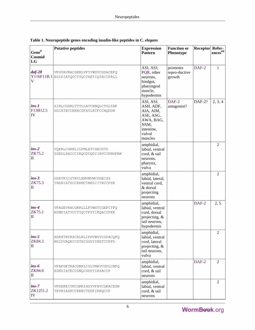
Table 1. Neuropeptide genes encoding insulin-like peptides in C. elegans
Gene#
CosmidLG
Putative peptides ExpressionPattern
Function orPhenotype
Receptor Refer-ences##
daf-28Y116F11B.1V
VPGVAVRACGRRLVPYVWSVCGDACEPQEGIDIATQCCTYQCTAEYIQTACCPRLL
ASI, ASJ,PQR, otherneurons,hindgut,pharyngealmuscle,hypodermis
promotesrepro-ductivegrowth
DAF-2 1
ins-1F13B12.5IV
SIRLCGSRLTTTLLAVCRNQLCTGLTAFGGIATECCEKRCSFAYLKTFCCNQDDN
ASI, ASJ,ASH, ADF,AIA, AIM,ASE, ASG,AWA, BAG,NSM,intestine,vulvalmuscles
DAF-2antagonist?
DAF-2? 2, 3, 4
ins-2ZK75.2II
VQKRLCGRRLILFMLATCGECDTDSSEDLSHICCIKQCDVQDIIRVCCPNSFRK
amphidial,labial, ventralcord, & tailneurons,pharynx,vulva
2
ins-3ZK75.3II
GDKVKICGTKVLKMVMVMCGGECSSTNENIATECCEKMCTMEDITTKCCPSR
amphidial,labial, lateral,ventral cord,& dorsalprojectingneurons
2
ins-4ZK75.1II
VPAGEVRACGRRLLLFVWSTCGEPCTPQEDMDIATVCCTTQCTPSYIKQACCPEK
amphidial,labial, ventralcord, dorsalprojecting, &tail neurons,hypodermis
DAF-2 2, 5
ins-5ZK84.3II
ADRHTNYRSCALRLIPHVWSVCGDACQPQNGIDVAQKCCSTDCSSDYIKETCCPFD
amphidial,labial, ventralcord, lateralprojecting, &tail neurons,vulva
2
ins-6ZK84.6II
VPAPGETRACGRKLISLVMAVCGDLCNPQEGKDIATECCGNQCSDDYIRSACCP
amphidial,labial, ventralcord, & tailneurons
DAF-2 2
ins-7ZK1251.2IV
VPDEKKIYRCGRRIHSYVFAVCGKACESNTEVNIASKCCREECTDDFIRKQCCP
amphidial,labial, ventralcord, & tailneurons
2
Neuropeptides
6
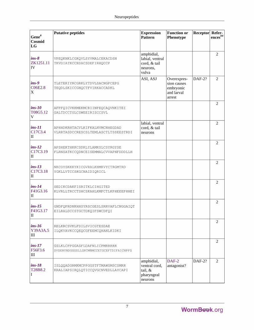
Gene#
CosmidLG
Putative peptides ExpressionPattern
Function orPhenotype
Receptor Refer-ences##
ins-8ZK1251.11IV
VPEQKNKLCGKQVLSYVMALCEKACDSNTKVDIATKCCRDACSDEFIRHQCCP
amphidial,labial, ventralcord, & tailneurons,vulva
2
ins-9C06E2.8X
TLETEKIYRCGRKLYTDVLSACNGPCEPGTEQDLSKICCGNQCTFVIRKACCADKL
ASI, ASJ Overexpres-sion causesembryonicand larvalarrest
DAF-2? 2
ins-10T08G5.12V
AFPFQICVKKMEKMCRIINPEQCAQVNKITEIGALTDCCTGLCSWEEIRISCCSVL
2
ins-11C17C3.4II
APHHDKRHTACVLKIFKALNVMCNHEGDADVLRRTASDCCRESCSLTEMLASCTLTSSEESTRDI
labial, ventralcord, & tailneurons
2
ins-12C17C3.19II
APSHEKTHKKCSDKLYLAMKSLCSYRGYSEFLRNSATKCCQDNCEISEMMALCVVAPNFDDDLLH
2
ins-13C17C3.18II
NKCOYSKKKYKICGVRALKHMKVYCTRGMTRDYGKLLVTCCSKGCNAIDIQRICL
2
ins-14F41G3.16II
SEDIKCDAKFISRITKLCIHGITEDKLVRLLTRCCTSHCSKAHLKMFCTLKPHEEEPHHEI
2
ins-15F41G3.17II
GNDFQPRDNKHHSYRSCGESLSRRVAFLCNGGAIQTEILRALDCCSTGCTDKQIFSWCDFQI
2
ins-16Y39A3A.5III
RELKRCSVKLFDILSVICGTESDAEILQKVAVKCCQEQCGFEEMCQHANLKIDKI
2
ins-17F56F3.6III
GSLKLCPPGGASFLDAFNLICPMRRRRRSVSENYNDGGGSLLGRTMNMCCETGCEFTDIFAICNPFG
2
ins-18T28B8.2I
ISLQQADGRMKMCPPGGSTFTMAWSMSCSMRRKRALIAPSIRQLQTICCQVGCNVEDLLAYCAPI
amphidial,ventral cord,tail, &pharyngealneurons
DAF-2antagonist?
DAF-2? 2
Neuropeptides
7
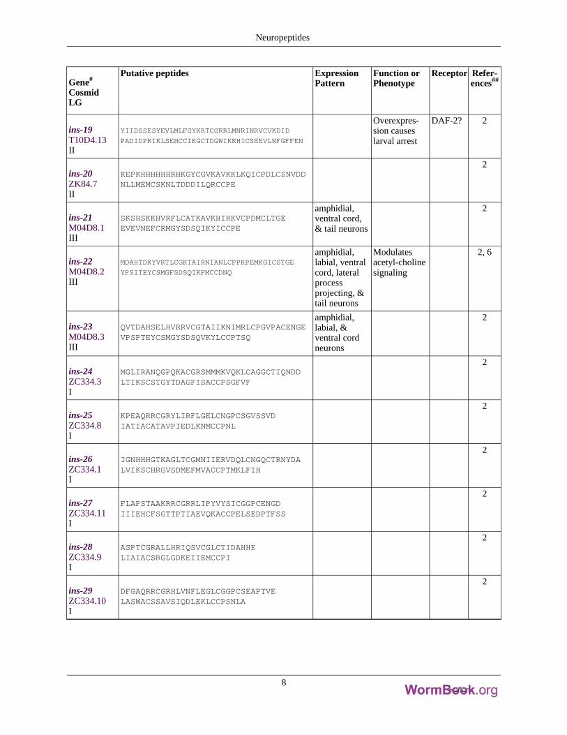
Gene#
CosmidLG
Putative peptides ExpressionPattern
Function orPhenotype
Receptor Refer-ences##
ins-19T10D4.13II
YIIDSSESYEVLMLFGYKRTCGRRLMNRINRVCVKDID
PADIDPKIKLSEHCCIKGCTDGWIKKHICSEEVLNFGFFEN
Overexpres-sion causeslarval arrest
DAF-2? 2
ins-20ZK84.7II
KEPKHHHHHHRHKGYCGVKAVKKLKQICPDLCSNVDDNLLMEMCSKNLTDDDILQRCCPE
2
ins-21M04D8.1III
SKSHSKKHVRFLCATKAVKHIRKVCPDMCLTGEEVEVNEFCRMGYSDSQIKYICCPE
amphidial,ventral cord,& tail neurons
2
ins-22M04D8.2III
MDAHTDKYVRTLCGKTAIRNIANLCPPKPEMKGICSTGE
YPSITEYCSMGFSDSQIKFMCCDNQ
amphidial,labial, ventralcord, lateralprocessprojecting, &tail neurons
Modulatesacetyl-cholinesignaling
2, 6
ins-23M04D8.3III
QVTDAHSELHVRRVCGTAIIKNIMRLCPGVPACENGEVPSPTEYCSMGYSDSQVKYLCCPTSQ
amphidial,labial, &ventral cordneurons
2
ins-24ZC334.3I
MGLIRANQGPQKACGRSMMMKVQKLCAGGCTIQNDDLTIKSCSTGYTDAGFISACCPSGFVF
2
ins-25ZC334.8I
KPEAQRRCGRYLIRFLGELCNGPCSGVSSVDIATIACATAVPIEDLKNMCCPNL
2
ins-26ZC334.1I
IGNHHHGTKAGLTCGMNIIERVDQLCNGQCTRNYDALVIKSCHRGVSDMEFMVACCPTMKLFIH
2
ins-27ZC334.11I
FLAPSTAAKRRCGRRLIPYVYSICGGPCENGDIIIEHCFSGTTPTIAEVQKACCPELSEDPTFSS
2
ins-28ZC334.9I
ASPTCGRALLHRIQSVCGLCTIDAHHELIAIACSRGLGDKEIIEMCCPI
2
ins-29ZC334.10I
DFGAQRRCGRHLVNFLEGLCGGPCSEAPTVELASWACSSAVSIQDLEKLCCPSNLA
2
Neuropeptides
8
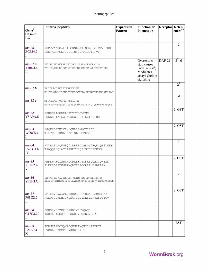
Gene#
CosmidLG
Putative peptides ExpressionPattern
Function orPhenotype
Receptor Refer-ences##
ins-30ZC334.2I
REPVVAAQGAKKTCGRSLLIKIQQLCHGICTVHADDLHETACMKGLTDSQLINSCCPPIPQTPFVF
2
ins-31 aT10D4.4II
FVHHFDHSMFARPEKTCGGLLIRRVDRICPNLNY
TYKIEWELMDNCCEVVCEDQWIKETFCRAPRFNFFGPSF
Overexpres-sion causeslarval arrest$;Modulatesacetyl-cholinesignaling
DAF-2? 2$; 6
ins-31 b KALERSCGPKLFTRVKTVCGE
DINVDNKVKISDHCCTPEGGCTDDWIKENVCKQTRFNFFRQFL
2$
ins-31 c DSPQRSCGPQLFKRVNTLCNE
NINVENNVSVSKSCCESAAGCTDDWIKKNVCTQHKPFVFRPGFY
2$
ins-32Y8A9A.6II
RSRRELICGRRLSKTVTNLCVEMNPQKEEDIATKCCKNKGCSREYIKSIMCPDE
2, OST
ins-33W09C5.4I
HGQKHCGTKIVRKLQMLCPKMCTISDDTLLTEMCSHSLFDDEIQLRCCPKEDE
2, OST
ins-34F52B11.6IV
KTTAAPLAQVNPQCLRRLTLLARGVCRQPCQPSDKPKTSAQQLLQLACSARRPTNEQIISYCCPEKSG
2
ins-35K02E2.4V
KMDENAFGINNRHCQRALKVYSFAICGAICQNYEKILMEGCGSTVMLTMQRTKLICCPEPVDSDELFN
2, OST
ins-36Y53H1A.4I
IRKRHPEGKLVIRDCKRYLIMYSRTICKEKCEKFDERNDITFSINLQFIFTDLLVEGCHSNQTLSNERTRELCCPNAGSN
2
ins-37F08G2.6II
NPIHPVPNAAFLPYRSCGSHLVHRAFEACSGKKDRSSDVDLWKMCCKDECTDLDIKESLCKYASQGYGV
2, OST
ins-38C17C3.20II
SQRSHVFSYKKHCGRRIVSLVQACDIDHDLSIDCCTQNCSSEFVSQRSHVFSY
ins-39F21E9.4X
IFRMTISFCSQVECQNMEAMQKICNTTTPTIHVGELCCPEFFEQVKDDFVTLL
EST
Neuropeptides
9
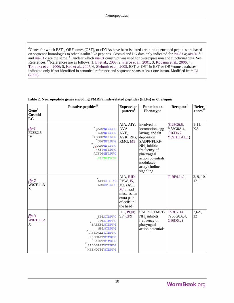
#Genes for which ESTs, ORFeomes (OST), or cDNAs have been isolated are in bold; encoded peptides are basedon sequence homologies to other insulin-like peptides. Cosmid and LG data only indicated for ins-31 a; ins-31 band ins-31 c are the same. $ Unclear which ins-31 construct was used for overexpression and functional data. SeeReferences. ##References are as follows: 1, Li et al., 2003; 2, Pierce et al., 2001; 3, Kodama et al., 2006; 4,Tomioka et al., 2006; 5, Kao et al., 2007; 6, Sieburth et al., 2005. EST or OST in EST or ORFeome databasesindicated only if not identified in canonical reference and sequence spans at least one intron. Modified from Li(2005).
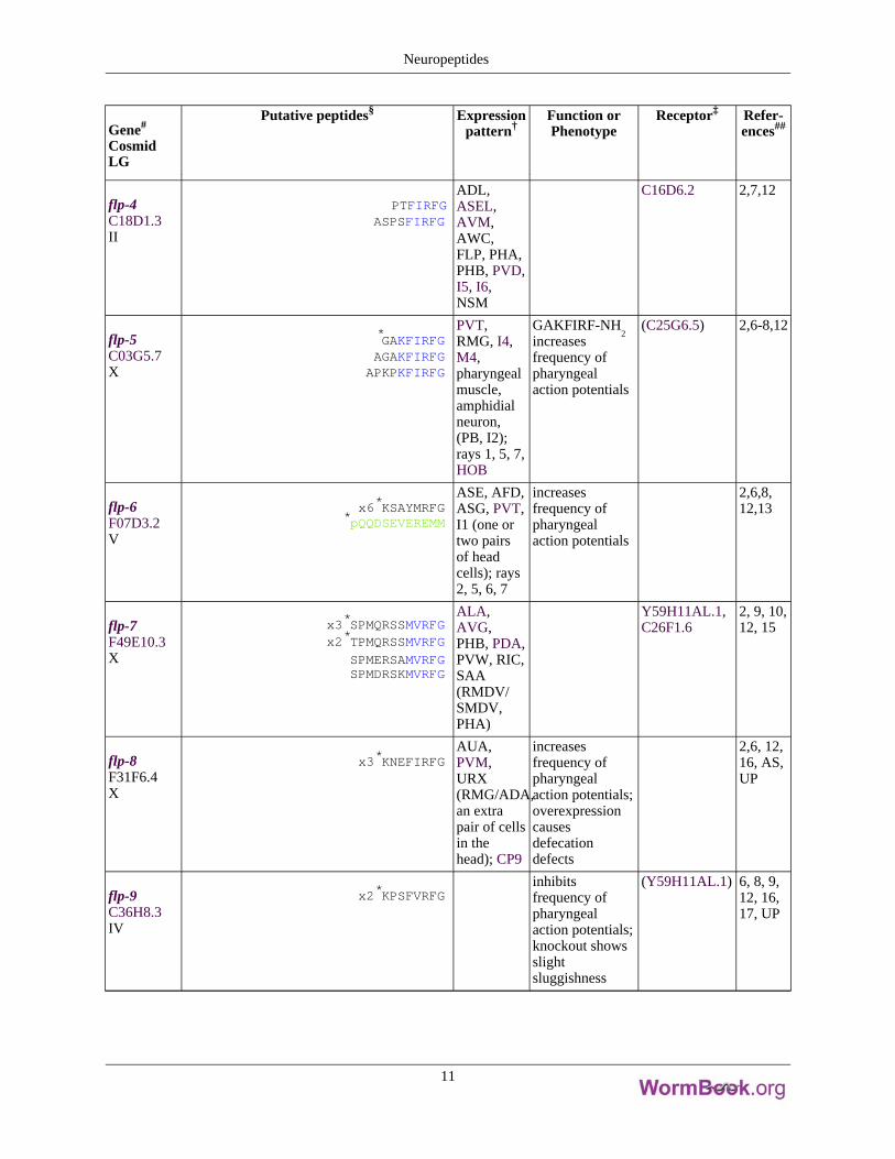
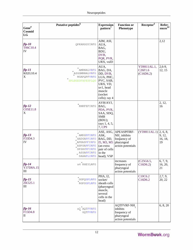
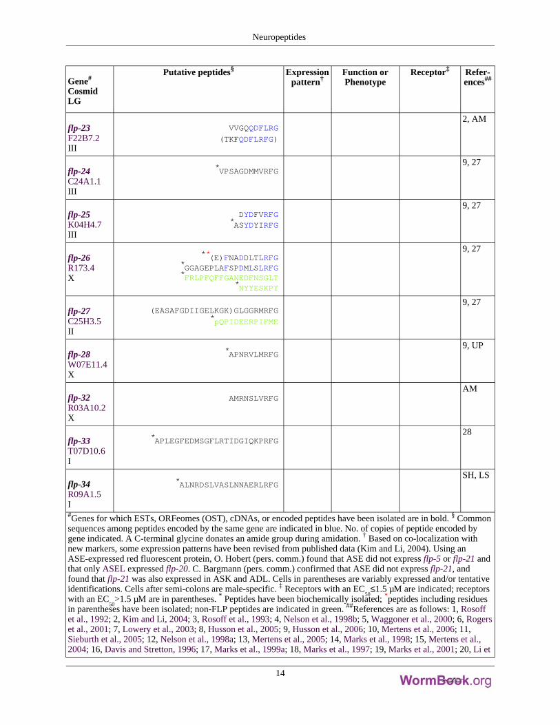
Table 2. Neuropeptide genes encoding FMRFamide-related peptides (FLPs) in C. elegans
Gene#
CosmidLG
Putative peptides§ Expressionpattern†
Function orPhenotype
Receptor‡ Refer-ences##
flp-1F23B2.5IV
*SADPNFLRFG*SQPNFLRFG
*ASGDPNFLRFG*SDPNFLRFG
*AAADPNFLRFG**(K)PNFLRFGAGSDPNFLRFG
(K)PNFMRYG
AIA, AIY,AVA,AVE,AVK, RIG,RMG, M5
involved inlocomotion, egglaying, and fatdeposition;SADPNFLRF-NH
2inhibits
frequency ofpharyngealaction potentials;modulatesacetylcholinesignaling
(C25G6.5,Y58G8A.4,C16D6.2,Y59H11AL.1)
1-11,KA
flp-2W07E11.3X
*SPREPIRFGLRGEPIRFG
AIA, RID,PVW, I5,MC (ASI,M4, headmuscles, anextra pairof cells inthe head)
T19F4.1a/b 2, 9, 10,12
flp-3W07E11.2X
SPLGTMRFG*TPLGTMRFG
*EAEEPLGTMRFGNPLGTMRFG
* ASEDALFGTMRFGEDGNAPFGTMRFG*SAEPFGTMRFG
* SADDSAPFGTMRFG*NPENDTPFGTMRFG
IL1, PQR;SP, CP9
SAEPFGTMRF-NH
2inhibits
frequency ofpharyngealaction potentials
C53C7.1a(Y58G8A.4,C16D6.2)
2,6-9,12
Neuropeptides
10
Gene#
CosmidLG
Putative peptides§ Expressionpattern†
Function orPhenotype
Receptor‡ Refer-ences##
flp-4C18D1.3II
PTFIRFGASPSFIRFG
ADL,ASEL,AVM,AWC,FLP, PHA,PHB, PVD,I5, I6,NSM
C16D6.2 2,7,12
flp-5C03G5.7X
*GAKFIRFGAGAKFIRFGAPKPKFIRFG
PVT,RMG, I4,M4,pharyngealmuscle,amphidialneuron,(PB, I2);rays 1, 5, 7,HOB
GAKFIRF-NH2
increasesfrequency ofpharyngealaction potentials
(C25G6.5) 2,6-8,12
flp-6F07D3.2V
x6*KSAYMRFG*pQQDSEVEREMM
ASE, AFD,ASG, PVT,I1 (one ortwo pairsof headcells); rays2, 5, 6, 7
increasesfrequency ofpharyngealaction potentials
2,6,8,12,13
flp-7F49E10.3X
x3*SPMQRSSMVRFGx2*TPMQRSSMVRFG
SPMERSAMVRFGSPMDRSKMVRFG
ALA,AVG,PHB, PDA,PVW, RIC,SAA(RMDV/SMDV,PHA)
Y59H11AL.1,C26F1.6
2, 9, 10,12, 15
flp-8F31F6.4X
x3*KNEFIRFGAUA,PVM,URX(RMG/ADA,an extrapair of cellsin thehead); CP9
increasesfrequency ofpharyngealaction potentials;overexpressioncausesdefecationdefects
2,6, 12,16, AS,UP
flp-9C36H8.3IV
x2*KPSFVRFGinhibitsfrequency ofpharyngealaction potentials;knockout showsslightsluggishness
(Y59H11AL.1) 6, 8, 9,12, 16,17, UP
Neuropeptides
11
Gene#
CosmidLG
Putative peptides§ Expressionpattern†
Function orPhenotype
Receptor‡ Refer-ences##
flp-10T06C10.4IV
QPKARSGYIRFGAIM, ASI,AUA,BAG,BDU,DVB,PQR, PVR,URX, vulD
2,12
flp-11K02G10.4X
*AMRNALVRFG*ASGGMRNALVRFG* NGAPQPFVRFG
*SPLDEEDFAPESPLQG
AUA,BAG, DA,DD, DVB,LUA, PHC,PVC, SAB,URX, VD,uv1, headmuscle(socketcells); ray 4
Y59H11AL.1,C26F1.6(C16D6.2)
2,6-9,12, 15
flp-12C05E11.8X
*RNKFEFIRFGAVH/AVJ,BAG,PDA, PVR,SAA, SDQ,SMB(BDU);rays 1, 4, 5,7, CP9
2, 12,16
flp-13F33D4.3IV
*AMDSPFIRFG*AADGAPFIRFG*APEASPFIRFG*ASPSAPFIRFG*SPSAVPFIRFG
ASSAPFIRFG* SAAAPLIRFG
ASE, ASG,ASK,BAG, DD,I5, M3, M5(an extrapair of cellsin thehead); VSP
APEASPFIRF-NH
2inhibits
frequency ofpharyngealaction potentials
(Y59H11AL.1) 2, 6, 8,9, 12,16, 18,19
flp-14Y37D8A.15III
x4*KHEYLRFGincreasesfrequency ofpharyngealaction potentials
(C25G6.5,C16D6.2)
6, 7, 9,16, 20,21
flp-15ZK525.1III
*GGPQGPLRFG*RGPSGPLRFG
PHA, I2,socket/sheath cells(pharyngealmuscle,severalcells in thehead)
C10C6.2C16D6.2
2,7, 9,20, 22
flp-16F15D4.8II
x2*AQTFVRFG* GQTFVRFG
AQTFVRF-NH2
inhibitsfrequency ofpharyngealaction potentials
6, 8, 20
Neuropeptides
12

Gene#
CosmidLG
Putative peptides§ Expressionpattern†
Function orPhenotype
Receptor‡ Refer-ences##
flp-17C52D10.11IV
x2 KSAFVRFGKSQYIRFG
BAG, M5(an extrapair of cellsin thehead); rays1, 5, 7
2, 20
flp-18Y48D7A.2X
**(DFD)GAMPGVLRFG* EMPGVLRFG
x3**(SYFDEKK)SVPGVLRFG*EIPGVLRFG*SEVPGVLRFG* DVPGVLRFG
AVA, AIY,RIG, RIM,M2 (M3,two extrapairs ofcells in thehead); rays2, 6
C16D6.2Y58G8A.4C53C7.1aNPR-1(C25G6.5,F41E7.3)
2, 6, 7,9, 15,19, 20,23-26,UP
flp-19M79.4X
*WANQVRFG*ASWASSVRFG
AIN,AWA,BAG,HSN, URX(an extrapair of cellsin the tail);rays 5, 7, 9,CEM
2, 6, 8,9, 20
flp-20E01H11.3X
x2 AMMRFGALM,ASEL,AVM,LUA,PLM, PVC,PVM,PVR,RIB/AIB(PVT)
2, 20,OH
flp-21C26F1.10V
GLGPRPLRFGADL, ASI,ASH, ASJ,ASK, FLP,URA, MC,M4, M2;CP6–9, SP,DVF
mutation causesmild aggregationbehavior
NPR-1C25G6.5Y58G8a.4
2,7,23-25
flp-22F39H2.1I
x3 *SPSAKWMRFGAIM, ASG,AVA,AVG,AVL, CEP,PVD,PVW,RIC/AIZ,RIV, SMD,URA, uv1;6 out of 9CP
(Y59H11AL.1) 2, 6, 8,9, 20
Neuropeptides
13
Gene#
CosmidLG
Putative peptides§ Expressionpattern†
Function orPhenotype
Receptor‡ Refer-ences##
flp-23F22B7.2III
VVGQQDFLRG
(TKFQDFLRFG)
2, AM
flp-24C24A1.1III
*VPSAGDMMVRFG9, 27
flp-25K04H4.7III
DYDFVRFG*ASYDYIRFG
9, 27
flp-26R173.4X
**(E)FNADDLTLRFG*GGAGEPLAFSPDMLSLRFG*FRLPFQFFGANEDFNSGLT
*NYYESKPY
9, 27
flp-27C25H3.5II
(EASAFGDIIGELKGK)GLGGRMRFG*pQPIDEERPIFME
9, 27
flp-28W07E11.4X
*APNRVLMRFG9, UP
flp-32R03A10.2X
AMRNSLVRFGAM
flp-33T07D10.6I
*APLEGFEDMSGFLRTIDGIQKPRFG28
flp-34R09A1.5I
*ALNRDSLVASLNNAERLRFGSH, LS
#Genes for which ESTs, ORFeomes (OST), cDNAs, or encoded peptides have been isolated are in bold. § Commonsequences among peptides encoded by the same gene are indicated in blue. No. of copies of peptide encoded bygene indicated. A C-terminal glycine donates an amide group during amidation. † Based on co-localization withnew markers, some expression patterns have been revised from published data (Kim and Li, 2004). Using anASE-expressed red fluorescent protein, O. Hobert (pers. comm.) found that ASE did not express flp-5 or flp-21 andthat only ASEL expressed flp-20. C. Bargmann (pers. comm.) confirmed that ASE did not express flp-21, andfound that flp-21 was also expressed in ASK and ADL. Cells in parentheses are variably expressed and/or tentativeidentifications. Cells after semi-colons are male-specific. ‡ Receptors with an EC
50≤1.5 µM are indicated; receptors
with an EC50
>1.5 µM are in parentheses.
* Peptides have been biochemically isolated;
*peptides including residuesin parentheses have been isolated; non-FLP peptides are indicated in green. ##References are as follows: 1, Rosoffet al., 1992; 2, Kim and Li, 2004; 3, Rosoff et al., 1993; 4, Nelson et al., 1998b; 5, Waggoner et al., 2000; 6, Rogerset al., 2001; 7, Lowery et al., 2003; 8, Husson et al., 2005; 9, Husson et al., 2006; 10, Mertens et al., 2006; 11,Sieburth et al., 2005; 12, Nelson et al., 1998a; 13, Mertens et al., 2005; 14, Marks et al., 1998; 15, Mertens et al.,2004; 16, Davis and Stretton, 1996; 17, Marks et al., 1999a; 18, Marks et al., 1997; 19, Marks et al., 2001; 20, Li et
Neuropeptides
14
al., 1999; 21, Marks et al., 1995; 22, Kubiak et al., 2003b; 23, Rogers et al., 2003; 24, Kubiak et al., 2003a; 25, deBono and Bargmann, 1998; 26, Kubiak et al., 2008; 27, McVeigh et al., 2005; 28, Husson and Schoofs, 2007; pers.comm.: KA, K. Ashrafi; SH, Steven Husson; AM, A. Maule; LS, Lilianne Schoofs; UP, unpublished results; ESTor OST in EST or ORFeome databases indicated only if not identified in canonical reference and sequence spans atleast one intron. AF, Ascaris suum; PF, Panagrellus redivius. Modified from Li (2005).
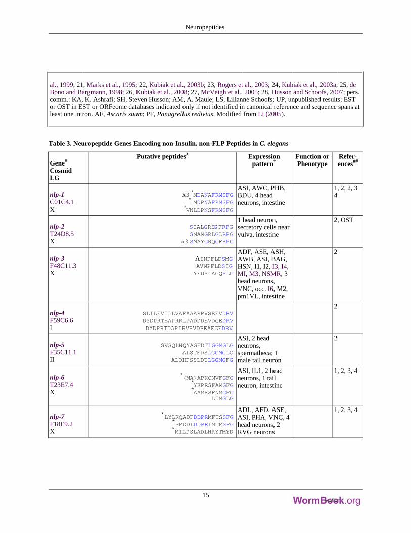
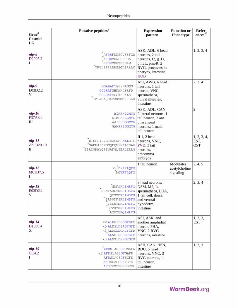
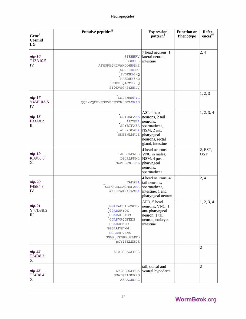
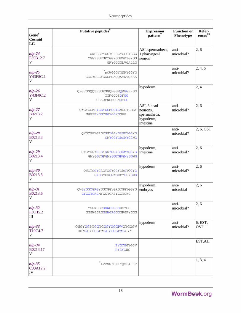
Table 3. Neuropeptide Genes Encoding non-Insulin, non-FLP Peptides in C. elegans
Gene#
CosmidLG
Putative peptides§ Expressionpattern†
Function orPhenotype
Refer-ences##
nlp-1C01C4.1X
x3*MDANAFRMSFG* MDPNAFRMSFG
*VNLDPNSFRMSFG
ASI, AWC, PHB,BDU, 4 headneurons, intestine
1, 2, 2, 34
nlp-2T24D8.5X
SIALGRSGFRPGSMAMGRLGLRPG
x3 SMAYGRQGFRPG
1 head neuron,secretory cells nearvulva, intestine
2, OST
nlp-3F48C11.3X
AINPFLDSMGAVNPFLDSIGYFDSLAGQSLG
ADF, ASE, ASH,AWB, ASJ, BAG,HSN, I1, I2, I3, I4,MI, M3, NSMR, 3head neurons,VNC, occ. I6, M2,pm1VL, intestine
2
nlp-4F59C6.6I
SLILFVILLVAFAAARPVSEEVDRVDYDPRTEAPRRLPADDDEVDGEDRVDYDPRTDAPIRVPVDPEAEGEDRV
2
nlp-5F35C11.1II
SVSQLNQYAGFDTLGGMGLGALSTFDSLGGMGLG
ALQHFSSLDTLGGMGFG
ASI, 2 headneurons,spermatheca; 1male tail neuron
2
nlp-6T23E7.4X
*(MA)APKQMVFGFG*YKPRSFAMGFG*AAMRSFNMGFG
LIMGLG
ASI, IL1, 2 headneurons, 1 tailneuron, intestine
1, 2, 3, 4
nlp-7F18E9.2X
*LYLKQADFDDPRMFTSSFG*SMDDLDDPRLMTMSFG*MILPSLADLHRYTMYD
ADL, AFD, ASE,ASI, PHA, VNC, 4head neurons, 2RVG neurons
1, 2, 3, 4
Neuropeptides
15
Gene#
CosmidLG
Putative peptides§ Expressionpattern†
Function orPhenotype
Refer-ences##
nlp-8D2005.2I
*AFDRFDNSGVFSFGA*AFDRMDNSDFFGA*SFDRMGGTEFGLM
*YPYLIFPASPSSGDSRRLV
ASK, ADL, 6 headneurons, 2 tailneurons, I2, g1D,pm5L, pm5R, 2RVG, processes inpharynx, intestine;HOB
1, 2, 3, 4
nlp-9E03D2.2V
GGARAFYGFYNAGNSGGGRAFNHNANLFRFDGGGRAFAGSWSPYLE
*TPIAEAQGAPEDVDDRRELE
ASI, AWB, 4 headneurons, 1 tailneuron, VNC,spermatheca,vulval muscles,intestine
2, 3, 4
nlp-10F37A8.4III
AIPFNGGMYGSTMPFSGGMYGAAIPFSGGMYGGAMPFSGGMYG
ASK, ADL, CAN,2 lateral neurons, 1tail neuron, 2 ant.pharyngealneurons; 1 maletail neuron
2
nlp-11ZK1320.10II
*HISPSYDVEIDAGNMRNLLDIG*SAPMASDYGNQFQMYNRLIDAG
*SPAISPAYQFENAFGLSEALERAG
IL1, 2 headneurons, VNC,PVD, 3 tailneurons,precommaembryos
1, 2, 3, 4,EST,OST
nlp-12M01D7.5I
x2*DYRPLQFG*DGYRPLQFG
1 tail neuron Modulatesacetylcholinesignaling
2, 4, 5
nlp-13E03D2.1V
*NDFSRDIMSFG*SGNTADLYDRRIMAFG
QPSYDRDIMSFG*SAPSDFSRDIMSFG*SSSMYDRDIMSFG*SPVDYDRPIMAFG*AEDYERQIMAFG
3 head neurons,NSM, M2, I4,spermatheca, LUA,1 tail cell, dorsaland ventralhypoderm,intestine
2, 3, 4
nlp-14D1009.4X
x2 ALDGLDGSGFGFDx5 ALNSLDGAGFGFEx3 ALDGLDGAGFGFD*ALNSLDGQGFGFE
x3 ALNSLDGNGFGFD
ASI, ASK, andanother amphidialneuron, PHA,VNC, 2 RVGneurons, intestine
1, 2, 3,EST
nlp-15CC4.2I
*AFDSLAGSGFDNGFNx2 AFDSLAGSGFGAFN
AFDSLAGSGFSGFDAFDSLAGQGFTGFEAFDTVSTSGFDDFKL
ASH, CAN, HSN,BDU, 5 headneurons, VNC, 3RVG neurons, 1tail neuron,intestine
1, 2, 3
Neuropeptides
16
Gene#
CosmidLG
Putative peptides§ Expressionpattern†
Function orPhenotype
Refer-ences##
nlp-16T13A10.5IV
STEHHRVSEGHPHE
ATHSPEGHIVAKDDHHGHESSDSHHGHQ*SVDEHHGHQ*NAEDHHEHQ
SEHVEHQAEMHEHQSTQEVSGHPEHHLV
7 head neurons, 1lateral neuron,intestine
2, 4
nlp-17Y45F10A.5IV
*GSLSNMMRIGQQEYVQFPNEGVVPCESCNLGTLMRIG
1, 2, 3
nlp-18F33A8.2II
*SPYRAFAFAARYGFA
*SPYRTFAFAASPYGFAFA
*SDEENLDFLE
ASI, 4 headneurons, 2 tailneurons,spermatheca,NSM, 2 ant.pharyngealneurons, rectalgland, intestine
1, 2, 3, 4
nlp-19K09C8.6X
IAGLRLPNFLIGLRLPNML
MGMRLPNIIFL
4 head neurons,VNC in males,NSM, 4 post.pharyngealneurons,spermatheca
2, EST,OST
nlp-20F45E4.8IV
FAFAFA*SGPQAHEGAGMRFAFA
APKEFARFARASFA
4 head neurons, 4tail neurons,spermatheca,intestine, 1 ant.pharyngeal neuron
2, 4
nlp-21Y47D3B.2III
GGARAFSADVGDDY*GGARAFYDE*GGARAFLTEM*GGARVFQGFEDEGGARAFMMD
GGGRAFGDMMGGARAFVENS
GGGRSFPVKPGRLDD)*pQYTSELEEDE
AFD, 5 headneurons, VNC, 1ant. pharyngealneuron, 1 tailneuron, embryo,intestine
1, 2, 3, 4
nlp-22T24D8.3X
SIAIGRAGFRPG2
nlp-23T24D8.4X
LYISRQGFRPASMAIGRAGMRPG
AFAAGWNRG
tail, dorsal andventral hypoderm
2
Neuropeptides
17
Gene#
CosmidLG
Putative peptides§ Expressionpattern†
Function orPhenotype
Refer-ences##
nlp-24F35B12.7V
QWGGGPYGGYGPRGYGGGYGGGYGGYGGRGPYGGYGGRGPYGYGG
GPYGGGGLVGALLG
ASI, spermatheca,1 pharyngealneuron
anti-microbial?
2, 6
nlp-25Y43F8C.1V
*pQWGGGYGNPYGGYGGGGYGGGYGGGFGAQQAYNVQNAA
anti-microbial?
2, 4, 6
nlp-26Y43F8C.2V
QFGFGGQQSFGGRGGQFGGMQRGGFNGN*GGFGQQSQFGG
GGSQFNGRGGNQFGG
hypoderm 2, 4
nlp-27B0213.2V
QWGYGGMPYGGYGGMGGYGMGGYGMGYMWGSPYGGYGGYGGYGGWG
ASI, 3 headneurons,spermatheca,hypoderm,intestine
anti-microbial?
2, 6
nlp-28B0213.3V
QWGYGGYGRGYGGYGGYGRGMYGGYGGMYGGYGRGMYGGWG
anti-microbial?
2, 6, OST
nlp-29B0213.4V
QWGYGGYGRGYGGYGGYGRGMYGGYGGMYGGYGRGMYGGYGRGMYGGWG
hypoderm,intestine
anti-microbial?
2, 6
nlp-30B0213.5V
QWGYGGYGRGYGGYGGYGRGYGGYGGYGGYGRGMWGRPYGGYGWG
hypoderm anti-microbial?
2, 6
nlp-31B0213.6V
QWGYGGYGRGYGGYGGYGRGYGGYGGYGGYGGYGRGMYGGYGRPYGGYGWG
hypoderm,embryos
anti-microbial
2, 6
nlp-32F30H5.2III
YGGWGGRGGWGRGGGRGYGGGGGWGGRGGGWGRGGGGRGFYGGG
anti-microbial?
2, 6
nlp-33T19C4.7V
QWGYGGPYGGYGGGYGGGPWGYGGGWRHWGGYGGGPWGGYGGGPWGGYY
hypoderm anti-microbial?
6, EST,OST
nlp-34B0213.17V
PYGYGGYGGWPYGYGWG
EST,AH
nlp-35C33A12.2IV
*AVVSGYDNIYQVLAPRF1, 3, 4
Neuropeptides
18
Gene#
CosmidLG
Putative peptides§ Expressionpattern†
Function orPhenotype
Refer-ences##
nlp-36B0464.3III
*SMVARQIPQTVVADH1, 3
nlp-37F48B9.4X
*NNAEVVNHILKNFGALDRLGDVG1, 3, 4
nlp-38C01A2.7I
**(ASDDR)VLGWNKAHGLWG*TPQNWNKLNSLWG*SPAQWQRANGLWG
1, 3, 4,AH, EST
nlp-39C54C8.9I
*EVPNFQADNVPEAGGRV1, 3
nlp-40Y74C9A.2I
**APSAPAGLEEKL(R)*pQPAADTFLGFVPQ
1, 3, 4
nlp-41C04H5.8II
*APGLFELPSRSV1, 3, 4
nlp-42Y80D3A.10V
SALLQPENNPEWNQLGWAWGNPDWQDLGFAWG
AH, EST
#Genes for which ESTs, ORFeomes (OST), cDNAs, or encoded peptides have been isolated are in bold. § Commonsequences among peptides encoded by the same gene are indicated in blue. No. of copies of peptide encoded bygene indicated. A C-terminal glycine donates an amide group during amidation. Some nlp peptide predictions havebeen revised. † Based on co-localization with new markers, some expression patterns have been revised frompublished data. Cells in parentheses are variably expressed and/or tentative identifications.
* Peptides have beenbiochemically isolated;
*peptides including residues in parentheses have been isolated; non-FLP peptides areindicated in green.
##References are as follows: 1, Li et al., 1999; 2, Nathoo et al., 2001; 3, Husson et al., 2005; 4,Husson et al., 2006; 5, Sieburth et al., 2005; 6, Couillault et al., 2004; pers. comm.: AH, Anne Hart; EST or OST inEST or ORFeome databases indicated only if not identified in canonical reference and sequence spans at least oneintron. Modified from Li (2005).
Neuropeptides
19
Table 4. Peptides encoded by flp neuropeptide genes
Gene#
CosmidLG
Putative peptides§ Peptide name Name in otherspecies
References##
flp-1F23B2.5IV
*SADPNFLRFG*SQPNFLRFG
*ASGDPNFLRFG*SDPNFLRFG
*AAADPNFLRFG**(K)PNFLRFGAGSDPNFLRFG(K)PNFMRYG
FLP-1-1FLP-1-2FLP-1-3FLP-1-4FLP-1-5FLP-1-6FLP-1-7FLP-1-8
PF2PF1AF26
1-6
flp-2W07E11.3X
*SPREPIRFGLRGEPIRFG
FLP-2-1FLP-2-2
4, 7, 8
flp-3W07E11.2X
SPLGTMRFG*TPLGTMRFG
*EAEEPLGTMRFGNPLGTMRFG
*ASEDALFGTMRFGEDGNAPFGTMRFG*SAEPFGTMRFG
*SADDSAPFGTMRFG*NPENDTPFGTMRFG
FLP-3-1FLP-3-2FLP-3-3FLP-3-4FLP-3-5FLP-3-6FLP-3-7FLP-3-8FLP-3-9
3, 4, 7, 8
flp-4C18D1.3II
PTFIRFGASPSFIRFG
FLP-4-1FLP-4-2
7, 8
flp-5C03G5.7X
*GAKFIRFGAGAKFIRFGAPKPKFIRFG
FLP-5-1FLP-5-2FLP-5-3
2, 7, 8
flp-6F07D3.2V
x6*KSAYMRFG*pQQDSEVEREMM
FLP-6-1FLP-6-2
AF8/PF33, 7-11
flp-7F49E10.3X
x3*SPMQRSSMVRFGx2*TPMQRSSMVRFG
SPMERSAMVRFGSPMDRSKMVRFG
FLP-7-1FLP-7-2FLP-7-3FLP-7-4
4, 7, 8
flp-8F31F6.4X
x3*KNEFIRFG FLP-8 AF17, 8, 12, 13
flp-9C36H8.3IV
x2*KPSFVRFG FLP-93, 4, 8, 12, 14
Neuropeptides
20
Gene#
CosmidLG
Putative peptides§ Peptide name Name in otherspecies
References##
flp-10T06C10.4IV
QPKARSGYIRFG FLP-107, 8
flp-11K02G10.4X
*AMRNALVRFG*ASGGMRNALVRFG*NGAPQPFVRFG
*SPLDEEDFAPESPLQG
FLP-11-1FLP-11-2FLP-11-3FLP-11-4
AF213, 4, 7, 8, 10
flp-12C05E11.8X
*RNKFEFIRFG FLP-12 AF247, 8, 10, 12
flp-13F33D4.3IV
*AMDSPFIRFG*AADGAPFIRFG*APEASPFIRFG*ASPSAPFIRFG*SPSAVPFIRFGASSAPFIRFG*SAAAPLIRFG
FLP-13-1FLP-13-2FLP-13-3FLP-13-4FLP-13-5FLP-13-6FLP-13-7
3, 4, 7, 8, 12, 15,16
flp-14Y37D8A.15III
x4*KHEYLRFG FLP-14 AF2/PF514, 12, 17-20
flp-15ZK525.1III
*GGPQGPLRFG*RGPSGPLRFG
FLP-15-1FLP-15-2
4, 7, 17
flp-16F15D4.8II
x2*AQTFVRFG*GQTFVRFG
FLP-16-1FLP-16-2
AF153, 10, 17
flp-17C52D10.11IV
x2 KSAFVRFGKSQYIRFG
FLP-17-1FLP-17-2
7, 17
flp-18Y48D7A.2X
**(DFD)GAMPGVLRFG*EMPGVLRFG
x3 **(SYFDEKK)SVPGVLRFG*EIPGVLRFG
*SEVPGVLRFG*DVPGVLRFG
FLP-18-1FLP-18-2FLP-18-3FLP-18-4FLP-18-5FLP-18-6
(afp-1)4, 7, 16, 17, 21
flp-19M79.4X
*WANQVRFG*ASWASSVRFG
FLP-19-1GLP-19-2
3, 4, 7, 17
Neuropeptides
21
Gene#
CosmidLG
Putative peptides§ Peptide name Name in otherspecies
References##
flp-20E01H11.3X
x2 AMMRFG FLP-207, 17
flp-21C26F1.10V
GLGPRPLRFG FLP-21 AF97, 9, 17
flp-22F39H2.1I
x3*SPSAKWMRFG FLP-223, 4, 7, 17
flp-23F22B7.2III
VVGQQDFLRG(TKFQDFLRFG)
FLP-237, AM
flp-24C24A1.1III
*VPSAGDMMVRFG FLP-244, 22, UP
flp-25K04H4.7III
DYDFVRFG*ASYDYIRFG
FLP-25-1FLP-25-2
4
flp-26R173.4X
**(E)FNADDLTLRFG*GGAGEPLAFSPDMLSLRFG*FRLPFQFFGANEDFNSGLT
* NYYESKPY
FLP-26-1FLP-26-2FLP-26-3FLP-26-4
4, 22
flp-27C25H3.5II
(EASAFGDIIGELKGK)GLGGRMRFG*pQPIDEERPIFME
FLP-27-1FLP-27-2
4, 22
flp-28W07E11.4X
*APNRVLMRFG FLP-284, UP
flp-32R03A10.2X
AMRNSLVRFG FLP-32AM
flp-33T07D10.6I
*APLEGFEDMSGFLRTIDGIQKPRFG FLP-3323
flp-34R09A1.5I
*ALNRDSLVASLNNAERLRFG FLP-34SH, LS
Neuropeptides
22
#Genes for which ESTs, ORFeomes (OST), cDNAs, or encoded peptides have been isolated are in bold. § Commonsequences among peptides encoded by the same gene are indicated in blue. No. of copies of peptide encoded bygene indicated. A C-terminal glycine donates an amide group during amidation. * Peptides have been biochemicallyisolated; *peptides including residues in parentheses have been isolated; non-FLP peptides are indicated in green.1FLP-14/AF2 has also been found in Haemonchus contortus and Panagrellus redivius. ##References are as follows:1, Rosoff et al., 1992; 2, Rosoff et al., 1993; 3, Husson et al., 2005; 4, Husson et al., 2006; 5, Geary et al., 1992; 6,Yew et al., 2005; 7, Kim and Li, 2004; 8, Nelson et al., 1998a; 9, Cowden and Stretton, 1995; 10, Yew et al., 2005;11, Maule et al., 1994a; 12, Davis and Stretton, 1996; 13, Cowden et al., 1989; 14, Marks et al., 1999a; 15, Markset al., 1997; 16, Marks et al., 2001; 17, Li et al., 1999; 18, Marks et al., 1995; 19, Cowden and Stretton, 1993; 20,Maule et al., 1994b; 21, Edison et al., 1997; 22, McVeigh et al., 2005; 23, Husson et al., 2007; pers. Comm.: SH,Steven Husson; AM, A. Maule; LS, Lilianne Schoofs; UP unpublished results. Modified from Li (2005).
5. Expression and localization of neuropeptide genes
To determine whether a candidate neuropeptide gene is transcribed and to confirm the genomic organizationof the gene, two basic strategies have been used. One is to isolate cDNAs using reverse transcription(RT)-polymerase chain reaction (PCR); the second is to scan the C. elegans EST or ORFeome databases. Throughthese approaches, several groups have determined that 37 of the 40 insulin-encoding genes (Gregoire et al., 1998;Kawano et al., 2000; Pierce et al., 2001; Li et al., 2003), 28 of the 31 flp genes (Rosoff et al., 1992; Nelson et al.,1998a; Kim and Li, 2004; McVeigh et al., 2005; I. Miskelly, N.J. Marks, and A. Maule, pers. comm.; unpublishedresults), and 39 of the 42 nlp genes (Nathoo et al., 2001; Couillault et al., 2004) are expressed. Based on these data,we predict that most, if not all, of the candidate neuropeptide genes are expressed. The neuropeptide genes arerelatively small and often have only a few exons. For instance, the coding and intronic regions of sixteen flp genesare less than 1 kilobase (Kim and Li, 2004; McVeigh et al., 2005). Among the nlp genes, a cluster of six nlp genes(nlp-27, 28, 29, 30, 31, and 34) on chromosome V encode very similar transcripts and have coding and intronicregions that range from 190 to 344 bp (Nathoo et al., 2001). The small size of most neuropeptide genes and thesmall number of exons that comprise the genes contribute to the difficulty of identifying all neuropeptide genes.
Because many of the C. elegans peptides have similar structures (for instance, the insulin-like peptides sharecommon A and B domains and the FLPs all share a common C-terminal Arg-Phe-NH
2), antibodies are difficult to
generate against specific neuropeptides. Most peptide antibodies cross-react with several peptides of similar aminoacid sequences. For example, the anti-FMRFamide antibody (Marder et al., 1987) recognizes FLPs encoded byseveral flp genes (Schinkmann and Li, 1992; Kim and Li, 2004). To determine the expression pattern of differentneuropeptide genes, most researchers have made gene fusions of the promoter region of the neuropeptide gene to thecoding region of a reporter gene, of which green fluorescent protein (GFP) is the most commonly used. Theconstructs are microinjected to generate transgenic animals. This relatively simple method has been used todetermine the expression pattern of 60 neuropeptide genes (see Tables 1, 2 and 3), including 15 insulin-like genes(Pierce et al., 2001; Li et al., 2003), 19 flp genes (Kim and Li, 2004), and 27 nlp genes (Nathoo et al., 2001). Foronly one gene thus far, flp-8, has the GFP expression pattern been confirmed by immunocytochemistry with aFLP-8-specific monoclonal antibody (Sithigorngul et al., 1991; Kim, 2003).
Although there are inherent caveats to these expression patterns, a number of conclusions can be drawn fromthe data. First, the expression of neuropeptides is widespread in C. elegans and includes expression in the nervoussystem as well as in non-neuronal tissue (see Tables 1, 2 and 3). For instance, over 160 neurons, which representover half of the 302 neurons in the C. elegans nervous system, express one or more FLPs (Kim and Li, 2004).Similarly, expression of the ins (Pierce et al., 2001), daf-28 (Li et al., 2003), and nlp (Nathoo et al., 2001) genes iswidespread. Second, there is considerable overlap in the expression patterns. Although each gene appears to beexpressed in a distinct set of neurons, a single neuron can express multiple neuropeptide genes and show aconsiderable diversity of neuropeptide expression. For instance, the chemosensory neuron ASI expresses daf-28,ins-1 and 9, nlp-1, 5, 6, 9, 14, 18, 24, and 27, and flp-2, 10, and 21 (Nathoo et al., 2001; Pierce et al., 2001; Li et al.,2003; Kim and Li, 2004); while some of these expression patterns may be artifactual, nevertheless ASI has thepotential to release a plethora of neuropeptides to modulate neuronal activity. Third, neuropeptide expression is not
Neuropeptides
23
limited to the nervous system. Neuropeptides, including those predicted by the ins, daf-28, flp, and nlp genes, arepredicted to be in non-neuronal tissues, including intestine, somatic gonad, muscle, and hypodermis (Nathoo et al.,2001; Pierce et al., 2001; Li et al., 2003; Kim and Li, 2004). Secretion of neuropeptides from these tissues is likelyto have an endocrine role.
Initially, the expression patterns of different peptides were compared in C. elegans and other nematodes byimmunocytochemistry. The Ascaris nervous system, for instance, showed an extensive distribution ofFMRFamide-like immunoreactivity (Cowden et al., 1993), much more than what was seen in C. elegans(Schinkmann and Li, 1992); however, the extent of flp expression with the GFP reporters in C. elegans now matchesthat in Ascaris. More recently, in situ hybridization has been used to examine the gene-specific expression pattern ofdifferent neuropeptide genes in the parasitic nematode Globodera pallida. For the few Gp-flp genes examined, therewere several differences in the neurons in which the genes were expressed in C. elegans (Kimber et al., 2002). Forinstance, flp-6 is not expressed in C. elegans tail neurons, whereas Gp-flp-6 is expressed in several tail neurons(Kimber et al., 2002). The significance of these differences is unknown, but could suggest that the function of thepeptides diverged in the related nematodes.
6. Biochemical isolation of neuropeptides
To determine whether the predicted peptides are produced, a few groups have begun the biochemical isolationof the different peptides. Thus far, most of the biochemical isolations have focused on the FLPs. To date, 36 FLPsencoded by seventeen flp genes (flp-1, 3, 5, 6, 8, 9, 11, 12, 13, 14, 16, 18, 19, 22, 24, 26, and 33) have been isolated(Rosoff et al., 1993; Marks et al., 1995, 1997, 1998, 1999a, 2001; Davis and Stretton, 1996; Husson et al., 2005; S.Husson and L. Schoofs, pers. comm.; Table 2). Indeed, the identification of flp-33 was based on the biochemicalisolation of the peptide. Two FLPs that are not encoded by any of the identified flp genes have also been isolated(Davis and Stretton, 1996; N. Marks and A. Stretton, pers. comm.), underscoring the difficulty of identifying smallneuropeptide genes with BLAST searches. In the recent peptidomic analysis by Husson and coworkers (2005),twenty nine NLPs encoded by 19 nlp genes were isolated (see Table 3), suggesting that many of the NLPs are alsolikely to be produced in C. elegans. These data also indicate that 113 is likely to be an underestimate of the totalnumber of neuropeptide genes.
Peptides identical to some of the predicted FLPs have been isolated from related nematodes (see Table 4),such as Ascaris suum (Cowden et al., 1989; Cowden and Stretton, 1993, 1995; Yew et all., 2005), Haemonchuscontortus (Keating et al., 1995; Marks et al., 1999b), and Panagrellus redivius (Geary et al., 1992; Maule et al.,1994a,b, 1995). To date, most of the isolated peptides in related nematodes belong to the FLP family (Yew et al.,2005). In addition, the FLP-12 (AF24) and FLP-21 (AF9) peptides have been isolated from Ascaris suum (Cowdenand Stretton, 1995; Yew et al., 2005), suggesting that these peptides are also produced in C. elegans. Similarly,several of the predicted NLPs are similar to peptides isolated from other invertebrates (Nathoo et al., 2001). Thesedata suggest that many of the predicted neuropeptides are indeed produced and highlight the rich diversity ofneuropeptides in C. elegans.
7. Neuropeptide function
Based on the expression pattern of the peptides, the peptides are likely to participate in a multitude ofbehaviors, including dauer formation, locomotion, egg-laying, and mechano- and chemosensation. To determine thefunction of the different neuropeptides, the most common strategy has been to inactivate or overexpress specificneuropeptide genes. This strategy has certain drawbacks with large neuropeptide superfamilies because of thepossible functional overlap among the family members. Several neuropeptides, for instance, may bind and activatethe same receptor. Using RNAi to decrease neuropeptide activity has not been routinely performed because of theinefficiency of RNAi in neurons (Simmer et al., 2002). Nevertheless, recent experiments have shown that several ofthe neuropeptide genes have unique functions.
7.1. The insulin-like gene family
Newly hatched first larval stage (L1) animals sample their environment to assess its qualities for reproductivegrowth. In the absence of food, newly hatched L1 animals will arrest reproductive growth and remain in this arrestedstate until a food source becomes available (Johnson et al., 1984). The decision to enter reproductive growth afterhatching is dependent on the activity of DAF-2, an insulin-like receptor, and ASNA-1, an ATPase that acts non-cellautonomously and regulates the insulin pathway (see below; Gems et al., 1998; Kao et al., 2007).
Neuropeptides
24
Under continued exposure to harsh environmental conditions, such as overcrowding, high temperatures, or ascarce food supply during L1 or L2, C. elegans will undergo an alternative life cycle, referred to as the dauer lifecycle (Cassada and Russell, 1975). After L2, animals enter the dauer state rather than L3 and remain in the dauerstate until conditions improve, whereupon they exit the dauer state and resume the lifecycle as L4 animals (Cassadaand Russell, 1975). The decision to enter reproductive growth or dauer is determined by parallel signaling pathways:the insulin and transforming growth factor β (TGF β) pathways (Riddle and Albert, 1997). Loss of either pathwayresults in constitutive dauer formation, indicating that the pathways function independently. Many of the insulin andTGFβ pathway mutants are temperature sensitive, and show an incompletely penetrant dauer phenotype at thepermissive temperature (Riddle and Albert, 1997). Lowering the activity of an insulin and TGFβ pathway gene has asynergistic effect and causes a stronger dauer phenotype at the permissive temperature than lowering the activity ofeither gene alone (Thomas et al., 1993). Similarly, lowering the activity of asna-1 and a TGFβ pathway gene alsoleads to an enhanced dauer phenotype at the permissive temperature, thereby linking the L1 arrest and dauerdecision to the insulin pathway (Kao et al., 2007). Hence, activation of DAF-2 leads to reproductive growth,whereas inactivation of DAF-2 leads to dauer arrest (Riddle and Albert, 1997). DAF-2 also functions to determinelifespan (Kenyon et al., 1993) and to limit body size (McCulloch and Gems, 2003). DAF-2 is the closest homologueto the mammalian insulin-like receptor superfamily (Kimura et al., 1997), but recently a more divergent family of 56putative insulin-like receptors has been identified in C. elegans (Dlakic, 2002).
Members of the insulin superfamily are encoded by the ins genes and daf-28 in C. elegans; fifteen of the insgenes and daf-28 are expressed in neurons, including some of the amphidial chemosensory neurons (Pierce et al.,2001; Li et al., 2003). ins-1, ins-9, and daf-28 are expressed in ASI and ASJ chemosensory neurons, which arecritical in the decision for dauer formation (Bargmann and Horvitz, 1991), as well as in other neurons; ins-1 anddaf-28 are also expressed in non-neuronal tissue, such as intestinal cells (Pierce et al., 2001; Li et al., 2003). Onlytwo insulin-family genes that have been inactivated by mutation have been examined thus far. INS-1 is most similarto mammalian insulin (Pierce et al., 2001). Loss of ins-1 has no effect on dauer formation or longevity (Pierce et al.,2001). By contrast, the daf-28(sa191) mutation causes transient dauer formation (Malone and Thomas, 1994). Themutation is likely to act as a dominant negative whereby the daf-28(sa191) gene product antagonizes DAF-2 activity(Li et al., 2003). Overexpression of daf-28 in a decreased TGFβ signaling background promotes exit from dauer,indicating that increased activity of the insulin pathway can bypass the TGFβ pathway (Kao et al., 2007). These datasuggest that DAF-28 normally activates the DAF-2 insulin-like receptor to promote reproductive growth (Li et al.,2003). Consistent with this hypothesis, levels of a Pdaf-28::GFP transgene are decreased during starvation orapplication of dauer pheromone (Li et al., 2003), suggesting that daf-28 expression is regulated by environmentalcues, as would be expected for a dauer regulator. Expression of the Pdaf-28::GFP transgene is found in an increasingnumber of cells as the animal ages (Li et al., 2003), suggesting that daf-28 is also involved in other behaviors, suchas aging. ASNA-1 may regulate levels of DAF-28 in the pseudocoelom; asna-1 mutants show decreased levels ofDAF-28::GFP in coelomocytes, indicating that less DAF-28::GFP is being released in the pseudocoelom (Kao et al.,2007).
If other insulin-like peptides also signal through the DAF-2 receptor, then perturbations of other ins genes mayenhance or suppress the phenotypes of daf-2 and daf-28 mutants. The phenotypes caused by overexpression ofseveral ins genes, using their endogenous promoters, were examined in different daf backgrounds. Overexpressionof ins-1 and ins-18 caused a low level of dauer arrest and enhanced the dauer phenotype of daf-2 and/or daf-7 TGFβmutants, whereas no dauer effects were seen with overexpression of ins-9, ins-19, ins-22, and ins-31 (Pierce et al.,2001). However, overexpression of ins-9 or of ins-31 and ins-19 in combination in a wild-type or daf-2 mutantbackground caused embryonic or larval arrest, a phenotype similar to one shown by some daf-2 alleles (Pierce et al.,2001). INS-1 and INS-18 may function to antagonize the activity of DAF-2 or to down-regulate daf-2 to promotedauer formation (Pierce et al., 2001), while INS-9, INS-31, and INS-19 may signal through DAF-2 to affect otheraspects of development. Overexpression of ins-4 or ins-6 can suppress or partially suppress, respectively, thedaf-28(sa191) mutation, suggesting that INS-4 or INS-6 can functionally substitute for DAF-28 and activate theDAF-2 receptor when present at high levels (Li et al., 2003). Furthermore, overexpression of ins-4 can bypass theeffects of a mutation in the TGFβ pathway (Kao et al., 2007). By contrast, overexpression of ins-7, 9, 17, 21, 22, and23 did not suppress the daf-28(sa191) mutation (Li et al., 2003). These data indicate that several insulin-like ligandssignal through or affect DAF-2 activity to affect developmental growth and dauer formation, while other insulin-likeligands signal through other non-DAF-2 insulin receptors.
What are the roles of the other ins genes? Until mutants are isolated, the function of this large class ofneuropeptides is largely unknown. Recently, INS-1 was identified as a key neuropeptide in the integration ofbehavior with the functional state of the animal. When placed on a thermal gradient well-fed animals move towards
Neuropeptides
25
the temperature on which they were cultivated, whereas starved animals avoid the temperature at which they werecultivated (Hedgecock and Russell, 1975). This thermotaxis behavior is mediated by the thermosensory neuronAFD, which signals through the AIY interneuron (Mori and Ohshima, 1995). Well-fed ins-1 mutants exhibit normalthermotaxis, indicating that the basic thermosensory properties of AFD are intact in the mutants. Similar to starvedwild-type animals, starved ins-1 animals slow when encountering food, demonstrating that ins-1 animals canrecognize their starvation state (Kodama et al., 2006). However, starved ins-1 mutants move towards rather thanaway from their cultivation temperature, presumably because they cannot integrate cultivation temperature withstarvation state (Kodama et al., 2006). This integration defect can be rescued by expression of ins-1 in differentneurons and is partially suppressed by mutations in daf-2 and age-1, suggesting that INS-1 acts non-cellautonomously to antagonize signaling through the DAF-2 receptor (Kodama et al., 2006).
INS-1, however, can also activate the DAF-2 receptor. Wild-type animals normally chemotax towards sodiumchloride (NaCl) (Ward, 1973). However, after pre-exposure to NaCl, starved, but not well-fed animals will avoidNaCl; this behavior is referred to as salt chemotaxis learning (Saeki et al., 2001). Several mutants, including daf-2,age-1, pdk-1, akt-1, and ins-1, are defective for salt chemotaxis learning (Tomioka et al., 2006), implicatinginvolvement of the DAF-2 pathway in salt chemotaxis learning. Based on transgenic rescues and laser ablations,Tomioka and co-workers (2006) propose that INS-1 is released from AIA interneurons to activate DAF-2 receptorsin ASER, thereby initiating the DAF-2 signaling cascade. Hence, INS-1 is involved in multiple integration eventsand whether it activates or antagonizes DAF-2 signaling is context dependent.
Acetylcholine is the primary excitatory transmitter at the neuromuscular junction in C. elegans. Aldicarbblocks the effects of acetylcholinesterase, thereby increasing the amount of acetylcholine at the synapse and causingparalysis and lethality (Nguyen et al., 1995). To identify genes that are resistant to aldicarb, a genome-wide RNAiscreen was performed on eri-1;lin-15B or eri-1; dgk-1 lin-15B mutants, which have a sensitized background forRNAi (Sieburth et al., 2005). In addition to the processing enzymes, decreased activity of four neuropeptide genes,two ins genes (ins-22 and ins-31), one flp gene (flp-1), and one nlp gene (nlp-12), conferred aldicarb-resistance,suggesting that the peptides encoded by these genes modulate acetylcholine signaling (Sieburth et al., 2005).
7.2. The flp family
Deletion mutants have been isolated for eleven flp genes (Nelson et al., 1998b; unpubl. obs.). Inactivation offlp-1 causes several defects, including hyperactive movement (Nelson et al., 1998b), defects in the timing of egglaying (Waggoner et al., 2000), thereby causing a decreased number of eggs laid (unpubl. obs.), and decreased fatstores in flp-1(yn2) mutants (K. Ashrafi, pers. comm.). FLP-1 peptides are also necessary for down-regulation of egglaying in the absence of food (Waggoner et al., 2000) and modulation of acetylcholine signaling (see above;Sieburth et al., 2005). The remaining flp mutants are currently being examined. Because so many flp genes haveoverlapping expression patterns, the function of these genes may also overlap and, therefore, be difficult to teaseapart. Hence, deletion mutants are being screened on a large variety of behavioral assays. For instance, theswimming or thrashing assay, which involves placing animals in physiological buffer and counting the number ofthrashes per minute, is more sensitive for detecting locomotion defects than examining the animal's movement on asolid surface. By the thrashing assay, flp-9 was found to thrash significantly less actively than wild-type animals(unpubl. obs.). Hence, to understand how the FLPs function, mutants will be examined for subtle defects, animalscarrying multiple knockouts need to be isolated, and the receptor to which the peptides bind must be identified, aswas done in the case of flp-21. The function of flp-21 will be discussed in conjunction with the function of itsreceptor, NPR-1.
7.3. The nlp family
Although no nlp mutant has been examined thus far, several of the nlp genes may function as anti-microbialpeptides. In microarray analyses to identify genes whose expression levels are changed in response to fungal orbacterial insults, expression of nlp-29, 31, and 33 was induced (Couillault et al., 2004). Furthermore, the peptideencoded by nlp-31 has anti-microbial activity and protects against fungal infection (Couillault et al., 2004). Thepeptides encoded by nlp-24, 25, 27, 28, and 30 are similar to those encoded by nlp-29, 31, and 33, suggesting thatthese peptides also have anti-microbial functions (Couillault et al., 2004). nlp-29 is expressed in the hypoderm andintestine (Nathoo et al., 2001), nlp-31 in the hypoderm and embryos (Nathoo et al., 2001), and nlp-33 exclusively inthe hypoderm (Couillault et al., 2004), suggesting that the peptides encoded by these genes only function asanti-microbial agents. By contrast, nlp-24 and 27 are also expressed in neurons (Nathoo et al., 2001) and mayfunction both as anti-microbial agents and neuropeptides. The expression patterns of nlp-25 and 28 are unknown, so
Neuropeptides
26
nlp-12 is involved in modulating acetylcholine signaling (Sieburth et al., 2005).
8. Neuropeptide receptors
As mentioned above, the function of a specific neuropeptide may be difficult to discern. Not only are multipleneuropeptides expressed in a single cell, but a specific neuropeptide may bind to multiple receptors. As illustratedabove with the insulin-like peptides and their receptors, an alternative strategy to determine the function ofneuropeptides is to inactivate the receptors to which the peptides bind.
Of the 1000 G-protein-coupled receptors in C. elegans, over 50 of them are likely to be neuropeptide receptors(Bargmann, 1998). Sixty G-protein receptors that were predicted to bind either a small molecule transmitter or aneuropeptide were inactivated by RNAi and screened for behavioral deficits (Keating et al., 2003). RNAi of sixreceptors, C16D6.2, C25G6.5, C26F1.6, F35G8.1, F41E7.3, and F59C12.2, resulted in either an increased ordecreased brood size (Keating et al., 2003). Disruption by RNAi of eight receptors, AC7.1 (tachykinin-like),C15B12.5, C10C6.2, C24A8.4, F15A8.5, F59D12.1, T02E9.1, and T05A1.1, causes locomotion defects (Keating etal., 2003). The phenotypes from the RNAi data were confirmed in two cases by the isolation of deletion mutants inT05A1.1 and F35G8.1 (Keating et al., 2003). Several of the ligands for these receptors have now been identified(see below).
For the FLPs, many FaRP receptors have been isolated from other systems. With the exception of a molluscanFMRFamide-gated amiloride-sensitive channel that has homology to the MEC-4 and MEC-6 mechanoreceptors(Lingueglia et al., 1995), other identified FMRFamide receptors are G-protein coupled receptors (Tensen et al.,1998; Bonini et al., 2000; Cazzamali and Grimmelikhuijzen, 2002; Meeusen et al., 2002; Duttlinger et al., 2003). Tomatch FLP ligands to specific G protein-coupled receptor binding partners, several groups have expressed candidatereceptors in either heterologous cells or Xenopus oocytes (Kubiak et al., 2003a, b; Lowery et al., 2003; Rogers et al.,2003; Mertens et al., 2004; Table 2). Different FLP ligands were applied and several assays were used as the readout. Interestingly, all FLP receptors examined thus far can be activated by multiple FLPs. Some of the receptors areactivated by multiple peptides encoded by one gene, while other receptors can be activated by peptides encoded bydifferent genes. A few examples will be described below.
The Upjohn/Pharmacia group (Kubiak et al., 2003a, b; Lowery et al., 2003) transfected candidate receptorsand chimeric G proteins into Chinese hamster ovary (CHO) cells, and used binding to GTPγS, a non-hydrolyzableform of GTP, as the readout. Binding of the cognate ligand to the receptor would presumably activate the receptor,thereby activating G proteins. GTPγS binding to membranes of transfected cells, therefore, indicates ligand binding.The group determined that peptides encoded by flp-15, GGPQGPLRFamide and RGPSGPLRFamide, activateC10C6.2 with an EC
50(concentration which produces 50% maximal activation) of 250 and 160 nM, respectively
(Kubiak et al., 2003b). No other FLP activated the receptor, despite some FLPs, such as FLP-21GLGPRPLRFamide, with high sequence similarity at the C-terminus (Kubiak et al., 2003b).
Mertens and coworkers (2004) expressed candidate receptors and Gα16
in HEK or CHO cells and screened foran increased calcium response, as monitored by an increase in fluorescence. The sensitivity of this system may belower than that when using GTPγS binding as the readout, because the concentration of peptide needed to activatemany receptors is generally much higher (usually in the µM rather than nM range). Nevertheless, Mertens andco-workers found that C26F1.6 binds to peptides encoded by two flp genes (Mertens et al., 2004). Oneflp-7-encoded peptide, FLP-7-2 TPMQRSSMVRFamide, activates C26F1.6 with an EC
50of ~1 µM; by contrast,
FLP-7-1 SPMQRSSMVRFamide, which differs from FLP-7-2 by only one amino acid at the N-terminus, did notactive the receptor at concentrations up to 10 µM. Interestingly, despite less sequence similarity, FLP-11-1AMRNALVRFamide activates C26F1.6 with an EC
50of ~1.33 µM.
A FLP receptor can also bind to a diverse group of peptides with a range of activities. The Y59H11AL.1receptor binds to 15 peptides encoded by 6 flp genes with EC
50values ranging from 25 nM to 5 µM (Mertens et al.,
2006). Only three of the peptides, however, FLP-7-3 SPMERSAMVRFamide (25 nM), FLP-1-8 KPNFMRYamide(100 nM), and FLP-11-1 AMRNALVRFamide (750 nM) have EC
50values less than 1 µM (Mertens et al., 2006).
While FLP-7-3 and FLP-11-1 have C-terminal sequence similarity, the structure of FLP-1-8 appears to be verydifferent. Note that peptides encoded by flp-7 bind to two receptors, C26F1.6 and Y59H11AL.1. Where these tworeceptors are expressed may give us some clues as to how FLP-7 peptides affect behavior. Given that a FLP receptorcan bind to multiple peptides encoded by different genes and a single peptide can bind to multiple receptors, thepotential complexity of peptide actions in C. elegans is astounding.
Neuropeptides
27
whether the peptides encoded by these genes also function as anti-microbial agents is unknown. As described above,
de Bono and coworkers searched specifically for the ligand to the NPR-1 receptor, a homologue to themammalian neuropeptide Y receptor. Mutations in the NPR-1 receptor affect aggregation behavior (de Bono andBargmann, 1998) and tolerance to alcohol (Davies et al., 2004). On an agar plate with a bacterial food source,wild-type animals feed alone (referred to as solitary feeding); a mutation in npr-1 causes the animals to aggregateduring feeding (referred to as social feeding) and accumulate at the edges of the bacteria (referred to as borderingbehavior; de Bono and Bargmann, 1998). The aggregation behavior of npr-1 mutants can be suppressed bymutations in gcy-35 or gcy-36 (Cheung et al., 2004), both of which encode soluble guanylate cyclases (Morton et al.,1999). GCY-35 guanylate cyclase binds oxygen, and the aggregation behavior of npr-1 mutants may be related tooxygen levels in the local environment of the animals (Gray et al., 2004). To identify the NPR-1 ligand, the de Bonogroup injected constructs for NPR-1 as well as inwardly rectifying potassium channels into Xenopus oocytes, andscreened for receptor activation of the potassium channels (Rogers et al., 2003). Because no neuropeptide Y ispresent in C. elegans, the assumption was that the NPR-1 ligand was a FLP. Both Rogers et al. (2003) and Kubiak etal. (2003a) determined that FLP-21 binds to NPR-1; in addition, Rogers et al. (2003) found that peptides encoded byflp-18 also activated NPR-1. Animals carrying a mutation in flp-21 display only mild aggregation compared to npr-1mutants (Rogers et al., 2003). Two explanations may account for the difference in phenotypes between the FLP-21ligand and the NPR-1 receptor mutants. The first is that the flp-21 mutation may be a partial loss of function alleleinstead of a null allele; the second is that FLP-18 ligands, which also bind NPR-1, may functionally substitute forthe loss of FLP-21 ligands (Rogers et al., 2003). As with the other FLP receptors, NPR-1 is promiscuous in itsbinding to multiple FLP ligands produced by different flp genes. As with other FLPs, FLP-18 peptides bind multiplereceptors, including NPR-1 (Kubiak et al., 2003a; Rogers et al., 2003) and Y58G8A.4 receptors (Kubiak et al.,2008).
9. Pharmacology of FLP neuropeptides
Because many of the FLPs have been also found in parasitic nematodes (Cowden et al., 1989; Cowden andStretton, 1993, 1995; Keating et al., 1995; Marks et al., 1999b) and, therefore, present possible targets foranti-helminthetic drugs, there has been an interest in examining the physiological effects of FLPs both in C. elegansas well as in parasitic nematodes. In C. elegans the effects of FLPs have been examined in a pharyngeal preparation(see Table 2; Rogers et al., 2001). Muscles of exposed pharyngeal terminal bulbs were impaled with microelectrodesto record changes of activity and action potential frequency. Surprisingly, many of the tested FLPs modulated theaction potential frequency (Rogers et al., 2001). Peptides encoded by flp-5, 6, 8, and 14 increased the actionpotential frequency, while peptides encoded by flp-1, 3, 9, 13, and 16 decreased the action potential frequency.These data are consistent with the expression of some of these FLPs in the pharynx (Kim and Li, 2004) and suggestthat multiple FLPs are involved in feeding behavior. Whether the different FLPs signal through one or multiple FLPreceptors has yet to be determined.
Similarly, application of peptides encoded by 20 flp genes had a range of effects on body wall, reproductive,and pharyngeal muscle of Ascaris suum (Fellowes et al., 1998; Bowman et al., 2002; Moffett et al., 2003; Trailovicet al., 2005; for reviews see Maule et al., 1996; Brownlee et al., 1996; Brownlee and Walker, 1999; Brownlee et al.,2000), and it is likely that these FLPs have similar effects in C. elegans. On Ascaris muscle, different FLPs canactivate different ion channels. Application of KPNFLRFamide (FLP-1-6) to somatic muscle cells, for instance,opens chloride channels, while application of SDPNFLRFamide (FLP-1-4) opens potassium channels (Bowman etal., 1996, 2002). The effects of KSAYMRFamide (FLP-6) on somatic muscle are context-dependent: application toventral muscles causes contraction while application to dorsal muscles causes relaxation (Maule et al., 1994a). Theresponse to FLPs can also be more complex. For instance, application of KNEFIRFamide (FLP-8) orKHEYLRFamide (FLP-14) to somatic muscle strips elicits biphasic responses consisting of an initialhyperpolarization, followed by an excitatory phase of rhythmic contractions (Cowden and Stretton, 1993; Bowmanet al., 1996). More recently, Stretton and coworkers have begun to characterize the effects of C. elegans and AscarisFLPs on the synaptic activity of Ascaris motor neurons (Davis and Stretton, 2001). Overall, the number of FLPs thatcan elicit physiological effects is striking and highlights the complex and intricate ways that different FLPs canmodulate synaptic and muscle activity.
10. Summary
The diversity of neuropeptides in C. elegans rivals the numbers found in mammals. Although many of thepeptides appear to be invertebrate specific, the insulin-like and FMRFamide-related peptides have counterparts inmammals. The daunting task of determining the regulation and functions of the different peptides remains. This task
Neuropeptides
28
is complicated by the overlapping expression and redundant functions of the different peptides, as well as by theirability to bind multiple receptors, and, likewise, by the ability of the receptors to bind multiple ligands. While most,if not all, FLPs and NLPs are likely to signal through G protein-coupled receptors and the insulin-like ligands tosignal through receptor tyrosine kinases, the identities of these receptors are still largely unknown. Despite thelimited information available thus far, we envision that neuropeptides are involved in all behaviors in C. elegans.
11. Acknowledgments
We thank K. Ashrafi, A. Hart, A. Maule, P. McVeigh, S. McIntire, L. Schoofs, and A. Stretton for sharingunpublished data, O. Hobert with revisions of expression patterns, C. Ferguson for comments on the manuscript, andA. Hart and A. Maule for helpful discussions. This work was supported by grants from the NIH to C.L. and the CityCollege of the City University of New York.
12. References
Bargmann, C.I. (1998). Neurobiology of the Caenorhabditis elegans genome. Science 282, 2028–2033. AbstractArticle
Bargmann, C.I., and Horvitz, H.R. (1991). Control of larval development by chemosensory neurons inCaenorhabditis elegans. Science 251, 1243–1246. Abstract Article
Bonini, J.A., Jones, K.A., Adham, N., Forray, C., Artymyshyn, R., Durkin, M.M., Smith, K.E., Tamm, J.A., Boteju,L.W., Lakhlani, P.P., et al. (2000). Identification and characterization of two G protein-coupled receptors forneuropeptide FF. J. Biol. Chem. 275, 39324–39331. Abstract Article
Bowman, J.W., Friedman, A.R., Thompson, D.P., Maule, A.G., Alexander-Bowman, S.J., and Geary, T.G. (2002).Structure-activity relationships of an inhibitory nematode FMRFamide-related peptide, SDPNFLRFamide (PF1), onAscaris suum muscle. Int. J. Parasitol. 32, 1765–1771. Abstract Article
Bowman, J.W., Friedman, A.R., Thompson, D.P., Ichhpurani, A.K., Kellman, M.F., Marks, N.J., Maule, A.G., andGeary, T.G. (1996). Structure-activity relationships of KNEFIRFamide (AF1), a nematode FMRFamide-relatedpeptide, on Ascaris suum muscle. Peptides 17, 381–387. Abstract Article
Brownlee, D.J., Fairweather, I., Holden-Dye, L., and Walker, R.J. (1996). Nematode neuropeptides: Localization,isolation and functions. Parasitol. Today 12, 343–351. Abstract Article
Brownlee, D., Holden-Dye, L., and Walker, R. (2000). The range and biological activity of FMRFamide-relatedpeptides and classical neurotransmitters in nematodes. Adv. Parasitol. 45, 109–80. Abstract
Brownlee, D.J., and Walker, R.J. (1999). Actions of nematode FMRFamide-related peptides on the pharyngealmuscle of the parasitic nematode, Ascaris suum. Ann. N. Y. Acad. Sci. 897, 228–238. Abstract Article
C. elegans Sequencing Consortium (1998). Genome sequence of the nematode C. elegans: A platform forinvestigating biology. Science 282, 2012–2018. Abstract Article
Cai, T., Fukushige, T., Notkins, A.L., and Krause, M. (2004). Insulinoma-associated protein IA-2, a vesicletransmembrane protein, genetically interacts with UNC-31/CAPS and affects neurosecretion in C. elegans. J.Neurosci. 24, 3115–3124. Abstract Article
Cassada, R.C., and Russell, R.L. (1975). The dauerlarva, a post-embryonic developmental variant of the nematodeCaenorhabditis elegans. Dev. Biol. 46, 326–342. Abstract Article
Cazzamali, G, and Grimmelikhuijzen, C.J. (2002). Molecular cloning and functional expression of the first insectFMRFamide receptor. Proc. Natl. Acad. Sci. U.S.A. 99, 12073–12078. Abstract Article
Cheung, B.H.H., Arellano-Carbajal, F., Rybicki, I., and de Bono, M. (2004). Soluble guanylate cyclases act inneurons exposed to the body fluid to promote C. elegans aggregation behavior. Curr. Biol. 14, 1105–1111. AbstractArticle
Neuropeptides
29
Couillault, C., Pujol, N., Reboul, J., Sabatier, L., Guichou, J.-F., Kohara, Y., and Ewbank, J.J. (2004).TLR-independent control of innate immunity in Caenorhabditis elegans by the TIR domain adaptor protein TIR-1,an ortholog of human SARM. Nat. Immunol. 5, 488–494. Abstract Article
Cowden, C., Sithigorngul, P., Brackley, P., Guastella, J., and Stretton, A.O. (1993). Localization and differentialexpression of FMRFamide-like immunoreactivity in the nematode Ascaris suum. J. Comp. Neurol. 333, 455–468.Abstract Article
Cowden, C., and Stretton, A.O. (1993). AF2, an Ascaris neuropeptide: isolation, sequence, and bioactivity. Peptides14, 423–430. Abstract Article
Cowden, C., and Stretton, A.O. (1995). Eight novel FMRFamide-like neuropeptides isolated from the nematodeAscaris suum. Peptides 16, 491–500. Abstract Article
Cowden, C., Stretton, A.O., and Davis, R.E. (1989). AF1, a sequenced bioactive neuropeptide isolated from thenematode Ascaris suum. Neuron 2, 1465–1473. Abstract Article
Davies, A.G., Bettinger, J.C., Thiele, T.R., Judy, M.E., and McIntire, S.L. (2004). Natural variation in the npr-1gene modifies ethanol responses of wild strains of C. elegans. Neuron 42, 731–743. Abstract Article
Davis, R.E., and Stretton, A.O. (1996). The motornervous system of Ascaris: eletrophysiology and anatomy of theneurons and their control by neuromodulators. Parasitology 113, S97–S117. Abstract
Davis, R.E., and Stretton, A.O. (2001). Structure-activity relationships of 18 endogenous neuropeptides on the motornervous system of the nematode Ascaris suum. Peptides 22, 7–23. Abstract Article
de Bono, M., and Bargmann, C.I. (1998). Natural variation in a neuropeptide Y receptor homolog modifies socialbehavior and food response in C. elegans. Cell 94, 679–689. Abstract Article
Dlakic, M. (2002). A new family of putative insulin receptor-like proteins in C. elegans. Curr. Biol. 12, R155–R157.Abstract Article
Dockray, G.J. (2004). The expanding family of -RFamide peptides and their effects on feeding behaviour. Exp.Physiol. 89, 229–235. Abstract Article
Doi, M. and Iwasaki, K. (2002). Regulation of retrograde signaling at neuromuscular junctions by novel C2 domainprotein AEX-1. Neuron 33, 249–259. Abstract Article
Duret, L., Guex, N., Peitsch, M.C., and Bairoch, A. (1998). New insulin-like proteins with atypical disulfide bondpattern characterized in Caenorhabditis elegans by comparative sequence analysis and homology modeling.Genome Res. 8, 348–353. Abstract
Duttlinger, A., Mispelon, M., and Nichols, R. (2003). The structure of the FMRFamide receptor and activity of thecardioexcitatory neuropeptide are conserved in mosquito. Neuropeptides 37, 120–126. Abstract Article
Edison, A.S., Messinger, L.A., and Stretton, A.O. (1997). afp-1: a gene encoding multiple transcripts of a new classof FMRFamide-like neuropeptides in the nematode Ascaris suum. Peptides 18, 929–935. Abstract Article
Eipper, B.A., Milgram, S.L., Husten, E.J., Yun, H.Y., and Mains, R.E. (1993). Peptidylglycine alpha-amidatingmonooxygenase: a multifunctional protein with catalytic, processing, and routing domains. Protein Sci. 2, 489–497.Abstract
Fares, H. and Grant, B. (2002). Deciphering endocytosis in Caenorhabditis elegans. Traffic 3, 11–19. AbstractArticle
Fellowes, R.A., Maule, A.G., Marks, N.J., Geary, T.G., Thompson, D.P., Shaw, C., Halton, D.W. (1998).Modulation of the motility of the vagina vera of Ascaris suum in vitro by FMRF amide-related peptides.Parasitology 116, 277–287. Abstract Article
Neuropeptides
30
Geary, T.G., Price, D.A., Bowman, J.W., Winterrowd, C.A., Mackenzie, C.D., Garrison, R.D., Williams, J.F., andFriedman, A.R. (1992). Two FMRFamide-like peptides from the free-living nematode Panagrellus redivivus.Peptides 13, 209–214. Abstract Article
Gems, D., Sutton, A.J., Sundermeyer, M.L., Albert, P.S., King, K.V., Edgley, M.L., Larsen, P.L., and Riddle, D.L.(1998). Two pleiotropic classes of daf-2 mutation affect larval arrest, adult behavior, reproduction and longevity inCaenorhabditis elegans. Genetics 150, 129–155. Abstract
Gray, J.M., Karow, D.S., Lu, H., Chang, A.J., Chang, J.S., Ellis, R.E., Marietta, M.A., and Bargmann, C.I. (2004).Oxygen sensation and social feeding mediated by a C. elegans guanylate cyclase homologue. Nature 430, 317–322.Abstract Article
Gregoire, F.M., Chomiki, N., Kachinskas, D., and Warden, C.H. (1998). Cloning and developmental regulation of anovel member of the insulin-like gene family in Caenorhabditis elegans. Biochem. Biophys. Res. Commun. 249,385–390. Abstract Article
Grishanin, R.N., Klenchin, V.A., Loyet, K.M., Kowalchyk, J.A., Ann, K., and Martin, T.F. (2002). Membraneassociation domains in Ca2+-dependent activator protein for secretion mediate plasma membrane and dense-corevesicle binding required for Ca2+-dependent exocytosis. J. Biol. Chem. 277, 22025–22034. Abstract Article
Hall, D.H., and Hedgecock, E.M. (1991). Kinesin-related gene unc-104 is required for axonal transport of synapticvesicles in C. elegans. Cell 65, 837–847. Abstract Article
Han, M., Park, D., Vanderzalm, P.J., Mains, R.E., Eipper, B.A., and Taghert, P.H. (2004). Drosophila uses twodistinct neuropeptide amidating enzymes, dPAL1 and dPAL2. J. Neurochem. 90, 129–141. Abstract Article
Hedgecock, E.M. and Russell, R.L. (1975). Normal and mutant thermotaxis in the nematode Caenorhabditiselegans. Proc. Natl. Acad. Sci. U.S.A. 72, 4061–4065. Abstract Article
Husson, S.J., Clynen, E., Baggerman, G., De Loof, A., and Schoofs, L. (2005). Discovering neuropeptides inCaenorhabditis elegans by two dimensional liquid chromatography and mass spectrometry. Biochm. Biophys. Res.Commun. 335, 76–86. Abstract Article
Husson, S.J., Clynen, E., Baggerman, G., Janssen, T., and Schoofs, L. (2006). Defective processing o fneuropeptideprecursors in Caenorhabditis elegans lacking proprotein converase 2 (KPC-2/ELG-3): mutant analysis by massspectrometry. J. Neurochem. 98, 1999–2012. Abstract Article
Husson, S.J., and Schoofs, L. (2007). Altered neuropeptide profile of Caenorhabditis elegans lacking the chaperoneprotein 7B2 as analyzed by mass spectrometry. FEBS Lett. 581, 4288–4292. Abstract Article
Jacob, T.C., and Kaplan, J.M. (2003). The EGL-21 carboxypeptidase E facilitates acetylcholine release atCaenorhabditis elegans neuromuscular junctions. J. Neurosci. 23, 2122–2130. Abstract
Johnson, T.E., Mitchell, D.H., Kline, S., Kernal, R., and Foy, J. (1984). Arresting development arrests aging in thenematode Caenorhabditis elegans. Mech. Ageing Dev. 28, 23–40. Abstract Article
Kao, G., Nordenson, C., Still, M., Rönnlund, A., Tuck, S., and Naredi, P. (2007). ASNA-1 positively regulatesinsulin secretion in C. elegans and mammalian cells. Cell 128, 577–587. Abstract Article
Kass, J., Jacob, T.C., Kim, P., and Kaplan, J.M. (2001). The EGL-3 proprotein convertase regulates mechanosensoryresponses of Caenorhabditis elegans. J. Neurosci. 21, 9265–9272. Abstract Article
Kawano, T., Ito, Y., Ishiguro, M., Takuwa, K., Nakajima, T., and Kimura, Y. (2000). Molecular cloning andcharacterization of a new insulin/IGF-like peptide of the nematode Caenorhabditis elegans. Biochem. Biophys. Res.Commun. 273, 431–436. Abstract Article
Keating, C.D., Holden-Dye, L., Thorndyke, M.C., Williams, R.G., Mallett, A., and Walker, R.J. (1995). TheFMRFamide-like neuropeptide AF2 is present in the parasitic nematode Haemonchus contortus. Parasitology 111,515–521. Abstract
Neuropeptides
31
Keating, C.D., Kriek, N., Daniels, M., Ashcroft, N.R., Hopper, N.A., Siney, El.J., Holden-Dye, L., and Burke, J.F.(2003). Whole-genome analysis of 60 G protein-coupled receptors in Caenorhabditis elegans by gene knockout withRNAi. Curr. Biol. 13, 1715–1720. Abstract Article
Kenyon, C., Chang, J., Gensch, E., Rudner, A., and Tabtiang, R. (1993). A C. elegans mutant that lives twice aslong as wild type. Nature 366, 461–464. Abstract Article
Kim, K. (2003). Function of a FMRFamide-related neuropeptide gene family in Caenorhabditis elegans. BostonUniversity thesis.
Kim, K., and Li, C. (2004). Expression and regulation of an FMRFamide-related neuropeptide gene family inCaenorhabditis elegans. J. Comp. Neurol. 475, 540–550. Abstract Article
Kimber, M.J., Fleming, C.C., Prior, Al, Jones, J.T., Halton, D.W., and Maule, A. (2002). Localisation of Globoderapallida FMRFamide-related peptide encoding genes using in situ hybridization. Int. J. Parasitol. 32, 1095–1105.Abstract Article
Kimura, K.D., Tissenbaum, H.A., Liu, Y., and Ruvkun, G. (1997). daf-2, an insulin receptor-like gene that regulateslongevity and diapause in Caenorhabditis elegans. Science 277, 942–946. Abstract Article
Kodama, E., Kuhara, A., Mohri-Shiomi, A., Kimura, K.D., Okumura, M., Tomioka, M., Iino, Y., and Mori, I.(2007). Insulin-like signaling and the neural circuit for integrative behavior in C. elegans. Genes Dev. 20,2955–2960. Abstract Article
Kubiak, T.M., Larsen, M.J., Nulf, S.C., Zantello, M.R., Burton, K.J., Bowman, J.W., Modric, T., and Lowery, D.E.(2003a). Differential activation of “social” and “solitary” variants of the Caenorhabditis elegans G protein-coupledreceptor NPR-1 by its cognate ligand AF9. J. Biol. Chem. 278, 33724–33729. Abstract Article
Kubiak, T.M., Larsen, M.J., Zantello, M.R., Bowman, J.W., Nulf, S.C., and Lowery, D.E. (2003b). Functionalannotation of the putative orphan Caenorhabditis elegans G-protein-coupled receptor C10C6.2 as a FLP15 peptidereceptor. J. Biol. Chem. 278, 42115–42120. Abstract Article
Kubiak, T.M., Larsen, M.J., Bowman, J.W., Geary, T.G., and Lowery, D.E. (2008). FMRFamide-like peptides(FLPs) encoded on the flp-18 precursor gene activate two isoforms of the orphan Caenorhabditis elegansG-protein-coupled receptor Y58G8A.4 heterologously expressed in mammalian cells. Biopolymers, in press.Abstract Article
Li, C. (2005). The ever-expanding neuropeptide gene families in the nematode Caenorhabditis elegans. Parasitology131 Suppl. S109–S127. Abstract Article
Li, W., Kennedy, S.G., and Ruvkun, G. (2003). daf-28 encodes a C. elegans insulin superfamily member that isregulated by environmental cues and acts in the DAF-2 signaling pathway. Genes Dev. 17, 844–858. AbstractArticle
Li, C., Kim, K., Nelson, L.S. (1998). FMRFamide-related neuropeptide gene family in Caenorhabditis elegans.Brain Res. 848, 26–34. Abstract Article
Li, C., Nelson, L.S., Kim, K., Nathoo, A., and Hart, A.C. (1999). Neuropeptide gene families in the nematodeCaenorhabditis elegans. Ann. N. Y. Acad. Sci. 897, 239–52. Abstract Article
Lindberg, I, Tu, B., Muller, L., and Dickerson, I.M. (1998). Cloning and functional analysis of C. elegans 7B2.DNA Cell Biol. 17, 727–734. Abstract
Lingueglia, E., Champigny, G., Lazdunski, M., and Barbry, P. (1995). Cloning of the amiloride-sensitiveFMRFamide peptide-gated sodium channel. Nature 378, 730–733. Abstract Article
Lowery, D.E., Geary, T.G., Kubiak, T.M., and Larsen, M.J. (2003). Pharmacia & Upjohn Company, Gprotein-coupled receptor-like receptors and modulators thereof, United States, Patent 6,632,621.
Neuropeptides
32
Malone, E.A., and Thomas, J.H. (1994). A screen for nonconditional dauer-constitutive mutations in Caenorhabditiselegans. Genetics 136, 879–886. Abstract
Marder, E., Calabrese, R.L., Nusbaum, M.P., and Trimmer, B. (1987). Distribution and partial characterization ofFMRFamide-like peptides in the stomatogastric nervous systems of the rock crab, Cancer borealis, and the spinylobster, Panulirus interruptus. J. Comp. Neurol. 259, 150–163. Abstract Article
Marks, N.J., Maule, A.G., Geary, T.G., Thompson, D.P., Li, C., Halton, D.W., and Shaw, C. (1998).KSAYMRFamide (PF3/AF8) is present in the free-living nematode, Caenorhabditis elegans. Biochem. Biophys.Res. Commun. 248, 422–425. Abstract Article
Marks, N.J., Maule, A.G., Geary, T.G., Thompson, D.P., Davis, J.P., Halton, D.W., Verhaert, P., and Shaw, C.(1997). APEASPFIRFamide, a novel FMRFamide-related decapeptide from Caenorhabditis elegans: structure andmyoactivity. Biochem. Biophys. Res. Commun. 231, 591–595. Abstract Article
Marks, N.J., Maule, A.G., Li, C., Nelson, L.S., Thompson, D.P., Alexander-Bowman, S., Geary, T.G., Halton,D.W., Verhaert, P., and Shaw, C. (1999a). Isolation, pharmacology and gene organization of KPSFVRFamide: aneuropeptide from Caenorhabditis elegans. Biochem. Biophys. Res. Commun. 254, 222–230. Abstract Article
Marks, N.J., Sangster, N.C., Maule, A.G., Halton, D.W., Thompson, D.P., Geary, T.G., and Shaw, C. (1999b).Structural characterisation and pharmacology of KHEYLRFamide (AF2) and KSAYMRFamide (PF3/AF8) fromHaemonchus contortus. Mol. Biochem. Parasitol. 100, 185–194. Abstract Article
Marks, N.J., Shaw, C., Halton, D.W., Thompson, D.P., Geary, T.G., Li, C., and Maule, A.G. (2001). Isolation andpreliminary biological assessment of AADGAPLIRFamide and SVPGVLRFamide from Caenorhabditis elegans.Biochem. Biophys. Res. Commun. 286, 1170–1176. Abstract Article
Marks, N.J., Shaw, C., Maule, A.G., Davis, J.P., Halton, D.W., Verhaert, P., Geary, T.G., and Thompson, D.P.(1995). Isolation of AF2 (KHEYLRFamide) from Caenorhabditis elegans: evidence for the presence of more thanone FMRFamide-related peptide-encoding gene. Biochem. Biophys. Res. Commun. 217, 845–851. Abstract Article
Maule, A.G., Bowman, J.W., Thompson, D.P., Marks, N.J., Friedman, A.R., and Geary, T.G. (1996).FMRFamide-related peptides (FaRPs) in nematodes: occurrence and neuromuscular physiology. Parasitology 113,S119–S135. Abstract
Maule, A.G., Shaw, C., Bowman, J.W., Halton, D.W., Thompson, D.P., Geary, T.G., and Thim, L. (1994a).KSAYMRFamide: a novel FMRFamide-related heptapeptide from the free-living nematode, Panagrellus redivivus,which is myoactive in the parasitic nematode, Ascaris suum. Biochem. Biophys. Res. Commun. 200, 973–980.Abstract Article
Maule, A.G., Shaw, C., Bowman, J.W., Halton, D.W., Thompson, D.P., Geary, T.G., and Thim, L. (1994b). TheFMRFamide-like neuropeptide AF2 (Ascaris suum) is present in the free-living nematode, Panagrellus redivivus(Nematoda, Rhabditida). Parasitology 109, 351–356. Abstract
Maule, A.G., Geary, T.G., Bowman, J.W., Marks, N.J., Blair, K.L., Halton, D.W., Shaw, C., and Thompson, D.P.(1995). Inhibitory effects of nematode FMRFamide-related peptides (FaRPs) on muscle strips from Ascaris suum.Invert. Neurosci. 1, 255–265. Abstract Article
McCulloch, D., and Gems, D. (2003). Body size, insulin/IGF signaling and aging in the nematode Caenorhabditiselegans. Exp. Gerontol. 38, 129–136. Abstract Article
McIntire, Sl.L., Jorgensen, E., Kaplan, J., and Horvitz, H.R. (1993). The GABAergic nervous system ofCaenorhabditis elegans. Nature 364, 337–341. Abstract Article
McVeigh, P., Leech, S., Mair, G.R., Marks, N.J.,. Geary, T.G., and Maule, A.G. (2005). Analysis ofFMRFamide-like peptide (FLP) diversity in phylum Nematoda. Int. J. Parasitol. 35, 1043–1060. Abstract Article
Neuropeptides
33
Meeusen, T., Mertens, I., Clynen, E., Baggerman, G., Nichols, R., Nachman, R.J., Huybrechts, R., De Loof, A., andSchoofs, L. (2002). Identification in Drosophila melanogaster of the invertebrate G protein-coupled FMRFamidereceptor. Proc. Natl. Acad. Sci. U.S.A. 99, 15363–15368. Abstract Article
Mertens, I., Clinckspoor, I., Janssen, T., Nachman, R., and Schoofs, L. (2006). FMRFamide-related peptide ligandsactivate the Caenorhabditis elegans orphan GPCR Y59H11AL.1. Peptides 27, 1291–1296. Abstract Article
Mertens, I., Meeusen, T., Janssen, T., Nachman, R., and Schoofs, L. (2005). Molecular characterization of two Gprotein-coupled receptor splice variants as FLP2 receptors in Caenorhabditis elegans. Biochem. Biophys. Res.Commun. 330, 967–974. Abstract Article
Mertens, I., Vandingenen, A., Meeursen, T., Janssen, T., Luyten, W., Nachman, R.J., De Loof, A., and Schoofs, L.(2004). Functional characterization of the putative orphan neuropeptide G-protein coupled receptor C26F1.6 inCaenorhabditis elegans. FEBS Lett. 573, 55–60. Abstract Article
Moffett, C.L., Beckett, A.M., Mousley, A., Geary, T.G., Marks, N.J., Halton, D.W., Thompson, D.P., and Maule,A.G. (2003). The ovijector of Ascaris suum: multiple response types revealed by Caenorhabditis elegansFMRFamide-related peptides. Int. J. Parasitol. 33, 859–876. Abstract Article
Mori, I. and Ohshima, Y. (1995). Neural regulation of thermotaxis in Caenorhabditis elegans. Nature 376, 344–348.Abstract Article
Morton, D.B., Hudson, M.L., Waters, E. and O'Shea, M. (1999). Soluble guanylyl cyclases in Caenorhabditiselegans: NO is not the answer. Curr. Biol. 9, R546–R547. Abstract Article
Nathoo, A.N., Moeller, R.A., Westlund, B.A., and Hart, A.C. (2001). Identification of neuropeptide-like proteingene families in Caenorhabditis elegans and other species. Proc. Natl. Acad. Sci. U.S.A. 98, 14000–14005. AbstractArticle
Nelson, L., Kim, K., Memmott, J., and Li, C. (1998a). FMRFamide-related gene family in the nematodeCaenorhabditis elegans. Mol. Br. Res. 58, 103–111. Abstract Article
Nelson, L.S., Rosoff, M.L., and Li, C. (1998b). Disruption of a neuropeptide gene, flp-1, causes multiple behavioraldefects in Caenorhabditis elegans. Science 281, 1686–1690. Abstract Article
Nguyen, M., Alfonso, A., Johnson, C.D., and Rand, J.B. (1995). Caenorhabditis elegans mutants resistant toinhibitors of acetylcholinesterase. Genetics 140, 527–535. Abstract
Pierce, S.B., Costa, M., Wisotzkey, R., Devadhar, S., Homburger, S.A., Buchman, A.R., Ferguson, K.C., Heller, J.,Platt, D.M., Pasquinelli, A.A. et al. (2001). Regulation of DAF-2 receptor signaling by human insulin and ins-1, amember of the unusually large and diverse C. elegans insulin gene family. Genes Dev. 15, 672–678. AbstractArticle
Price, D.A., and Greenberg, M.J. (1977). Structure of a molluscan cardioexcitatory neuropeptide. Science 197,670–671. Abstract Article
Renden, R., Berwin, B., Davis, W., Kyoungsook, A., Chin, C.-T., Kreber, R., Ganetzky, B, Martin, T.F.J., andBroadie, K. (2001). Drosophila CAPS is an essential gene that regulates dense-cre vesicle release and synapticvesicle fusion. Neuron 31, 421–437. Abstract Article
Richmond, J.E., Davis, W.S, and Jorgensen, E.M. (1999). UNC-13 I srequired for synaptic vesicle fusion in C.elegans. Nat. Neurosci. 2, 959–964. Abstract Article
Riddle, D.L., and Albert, P.S. (1997). Genetic and environmental regulation of dauer larva development. In C.elegans II, D.L. Riddle, T. Blumenthal, B.J. Meyer, and J.R. Priess, eds. (New York, NY: Cold Spring HarborLaboratory Press), pp. 739–768.
Neuropeptides
34
Rogers, C.M., Franks, C.J., Walker, R.J., Burke, J.F., and Holden-Dye, L. (2001). Regulation of the pharynx ofCaenorhabditis elegans by 5-HT, octopamine, and FMRFamide-like neuropeptides. J. Neurobiol. 15, 235–244.Abstract Article
Rogers, C., Reale, V., Kim, K., Chatwin, H., Li, C., Evans, P., and de Bono, M. (2003). Inhibition of Caenorhabditiselegans social feeding by FMRFamide-related peptide activation of NPR-1. Nat. Neurosci. 6, 1178–85. AbstractArticle
Rosoff, M.L., Burglin, T.R., and Li, C. (1992). Alternatively spliced transcripts of the flp-1 gene encode distinctFMRFamide-like peptides in Caenorhabditis elegans. J. Neurosci. 12, 2356–2361. Abstract
Rosoff, M.L., Doble, K.E., Price, D.A., and Li. C. (1993). The flp-1 propeptide is processed into multiple, highlysimilar FMRFamide-like peptides in Caenorhabditis elegans. Peptides 14, 331–338. Abstract Article
Saeki, S., Yamamoto, M., and Iino, Y. (2001). Plasticity of chemotaxis revealed by paired presentation of achemoattractant and starvation in the nematode Caenorhabditis elegans. J. Exp. Biol. 204, 1757–1764. Abstract
Salio, C., Lossi, L., Ferrini, F., and Merighi, A. (2006). Neuropeptides as synaptic transmitters. Cell Tissue Res.326, 583–598. Abstract Article
Schinkmann, K. (1994). FMRFamide-like peptides in the nematodes Caenorhabditis elegans and Caenorhabditisvulgaris. Boston University, Boston, MA, Ph.D. thesis.
Schinkmann, K., and Li, C. (1992). Localization of FMRFamide-like peptides in Caenorhabditis elegans. J. Comp.Neurol. 316, 251–260. Abstract Article
Simmer, F., Tijsterman, M., Parrish, S., Koushika, S.P., Nonet, M.L., Fire, A., Ahringer, J., and Plasterk, R.H.(2002). Loss of the putative RNA-directed RNA polymerase RRF-3 makes C. elegans hypersensitive to RNAi. Curr.Biol. 12, 1317–1319. Abstract Article
Sieburth, D., Ch'ng, Q.L., Dybbs, M., Tavazoie, M., Kennedy, S., Want, D., Dupuy, D., Rual, J.-F., Hill, D.E.,Vidal, M., Ruvkun, G., and Kaplan, J.M. (2005). Systematic analysis of genes required for synapse structure andfunction. Nature 436, 510–517. Abstract Article
Sieburth, D., Madison, J.M., and Kaplan, J.M. (2007). PKC-1 regulates secretion of neuropeptides. Nat. Neurosci.10, 49–57. Abstract Article
Sithigorngul, P., Stretton, A.O., and Cowden, C. (1991). A versatile dot-ELISA method with femtomole sensitivityfor detecting small peptides. J. Immunol. Methods 141, 23–32. Abstract Article
Sossin, W.S., Sweet-Cordero, A., and Scheller, R.H. (1990). Dale's hypothesis revisited: different neuropeptidesderived from a common prohormone are targeted to different processes. Proc. Natl. Acad. Sci. U.S.A. 87,4845–4848. Abstract Article
Steiner, D.F. (1998). The proprotein convertases. Curr. Opin. Chem. Biol. 2, 31–39. Abstract Article
Strand, F.L. (1999). Neuropeptides. MIT Press, Cambridge, MA.
Tensen, C.P., Cox, K.J., Smit, A.B., van der Schors, R.C., Meyerhof, W., Richter, D., Planta, R.J., Hermann, P.M.,van Minnen, J., Geraerts, W.P., et al. (1998). The Lymnaea cardioexcitatory peptide (LyCEP) receptor: aG-protein-coupled receptor for a novel member of the RFamide neuropeptide family. J. Neurosci. 18, 9812–9821.Abstract
Thacker, C., Peters, K., Srayko, M., and Rose, A.M. (1995). The bli-4 locus of Caenorhabditis elegans encodesstructurally distinct Kex2/Subtilisin-like endoproteases essential for early development and adult morphology.Genes Dev. 9, 956–971. Abstract
Thacker, C., and Rose, A.M. (2000). A look at the Caenorhabditis elegans Kex2/Subtilisin-like proproteinconvertase family. BioEssays 22, 545–553. Abstract
Neuropeptides
35
Thomas, J.H. (1990). Genetic analysis of defecation in Caenorhabditis elegans. Genetics 124, 855–872. Abstract
Thomas, J.H., Birnby, D.A., and Vowels, J.J. (1993). Evidence for parallel processing of sensory informationcontrolling dauer formation in Caenorhabditis elegans. Genetics 134, 1105–1117. Abstract
Tomioka, M., Adachi, T., Suzuki, H., Kunitomo, H., Schafer, W.R., and Iino, Y. (2006). The insulin/PI 3-kinasepathway regulates salt chemotaxis learning in Caenorhabditis elegans. Neuron 51, 613–625. Abstract Article
Trailovic, S.M., Clark, C.L., Robertson, A.P., and Martin, R.J. (2005). Brief application of AF2 produces longlasting potentiation of nAChR responses in Ascaris suum. Mol. Biochem. Parasitol. 139, 51–64. Abstract Article
Waggoner, L.E., Hardaker, L.A., Golik, S., and Schafer, W.R. (2000). Effect of a neuropeptide gene on behavioralstates in Caenorhabditis elegans egg-laying. Genetics 154, 1181–1192. Abstract
Ward, S. (1973). Chemotaxis by the nematode Caenorhabditis elegans: Identification of attractants and analysis ofthe response by use of mutants. Proc. Natl. Acad. Sci. U.S.A. 70, 817–821. Abstract Article
Yew, J.Y., Kutz, K.K., Dikler, S., Messinger, L., Li, L., and Stretton, A.O. (2005). Mass spectrometric map ofneuropeptide expression in Ascaris suum. J. Comp. Neurol. 488, 396–413. Abstract Article
Zahn, T.R., Angleson, J.K., MacMorris, M.A., Domke, E., Hutton, J.F., Schwartz, C., and Hutton, J.C. (2004).Dense core vesicle dynamics in Caenorhabditis elegans neurons and the role of kinesin UNC-104. Traffic 5,544–559. Abstract Article
Neuropeptides
36
All WormBook content, except where otherwise noted, is licensed under a Creative Commons Attribution License


![Design, Synthesis and Biological Evaluation of ...elpub.bib.uni-wuppertal.de/servlets/DerivateServlet/...Figure 2: Overview on functions of insect neuropeptides[8] Neuropeptides and](https://img.dokumen.tips/doc/110x75/602c9862fd38af6cb12ca3b8/design-synthesis-and-biological-evaluation-of-elpubbibuni-figure-2-overview.jpg)