Embed Size (px)
Citation preview

Central Journal of Neurological Disorders & Stroke
Cite this article: Yoshida M (2014) Neuropathology of Demyelinating Diseases in Japan. J Neurol Disord Stroke 2(5): 1086.
Review Article
Neuropathology of Demyelinating Diseases in JapanMari Yoshida*Institute for Medical Science of Aging, Aichi Medical University, Japan
Abstract
We reviewed and compared the neuropathology of multiple sclerosis (MS), neuromyelitis optica (NMO), neuromyelitis optica spectrum disorders (NMOSD) and acute disseminated encephalomyelitis (ADEM) in Japan. Demyelinating lesions of MS are well circumscribed as compared with the lesions of NMO and NMOSD, which reveal variable, irregularly shaped and ill-defined borders that extend longitudinally along vessels, causing destructive changes with poor gliosis. Although the optic nerves and chiasm, spinal cord, cerebral white matter, brainstem, and cerebellum are involved in both MS and NMO/NMOSDs, the formation patterns of demyelinating lesions appear to differ between MS and NMO/NMOSD. NMO/NMOSD preferentially exhibit central lesions of the spinal cord with strongly softening features. Furthermore, the expression of myelin basic protein (MBP) is strongly diminished in the demyelinating lesions of MS, without loss of aquaporin-4 (AQP4) or GFAP expression. However, AQP4 and GFAP expression is decreased in the demyelinating lesions of NMO/NMOSD. Therefore, AQP4 and MBP immunoreactivity may distinguish NMO/NMOSD from MS neuropathologically. Serial sections of the spinal cord demonstrate longitudinally extensive lesions in NMO/NMOSD, although some cases with MS also reveal similar longitudinally extensive lesions of the spinal cord. In ADEM, demyelinating lesions form primarily in small perivenous foci that differ from the lesions of MS and NMO/NMOSD. Therefore, the shape and formation patterns of demyelinating lesions appear to be disease specific, and it might be possible to distinguish among MS, NMO and ADEM; the immunoreactivity patterns of MBP, AQP4, and GFAP may also aid diagnosis.
ABBREVIATIONS ADEM: Acute Disseminated Encephalomyelitis; AHL: Acute
Hemorrhagic Leukoencephalopathy; MS: Multiple Sclerosis; NMO: Neuromyelitis Optica; NMOSD: Neuromyelitis Optica Spectrum Disorders; C: Cervical Cord; T: Thoracic Cord; L: Lumbar Cord
INTRODUCTIONThe pathology of demyelination is defined as the destruction
of normal myelin with relative preservation of axons. The term demyelination excludes diseases characterized by a loss of myelin due to an inherited defect of metabolism (leukodystrophies) or a loss of myelin as a result of axonal degeneration.
Multiple sclerosis (MS) is the most common demyelinating disease of the central nervous system. NMO, also known as Dévic’s disease, is characterized by the acute development of optic neuritis and acute transverse myelitis within weeks of each other [1-5]. Until relatively recently, NMO has been classified as a subtype of multiple sclerosis. There has long been controversy concerning whether NMO is a variant of MS, called optic-spinal
multiple sclerosis (OSMS) in Asian countries, or a distinct disease [1-5]. After the identification of AQP4, which is the main water channel protein in the CNS and is densely expressed on astrocyte endfeet at the blood-brain barrier (BBB), the loss of AQP4 in NMO lesions revealed distinct pathogenesis and pathology, distinguishing it from MS [6,7].
The recognition of variable distributions of NMO lesions in regions other than the spinal cord and optic nerves has promoted the concept of NMO spectrum disorders (NMOSD) [8,9]. We use the term NMOSD as a comprehensive concept in this review. We reviewed the neuropathology of 27 autopsied cases with demyelinating diseases in Japan and demonstrated the characteristic features of MS, NMOSD and ADEM. These patients were chosen from the files of the Institute for Medical Science of Aging, Aichi Medical University, between 1980 and 2013. The demographic features of all of the included cases are summarized in the Table 1. The pathological diagnosis revealed 9 cases with MS, 14 cases with NMOSD, 3 cases with ADEM and 1 case with AHL (Table 1). In cases with NMOSD, 2 cases were combined with Sjögren syndrome. It is interesting that NMOSD cases were more frequent than MS cases in Japan, although this may be biased by
*Corresponding authorMari Yoshida, Institute for Medical Science of Aging, Aichi Medical University, 1-1 Yazakokarimata, Nagakute, Aichi,480-1195 Japan, Tel: 81-0561-62-3311; Fax: 81- 0561-63-3531; Email: [email protected]
Submitted: 29 March 2014
Accepted: 17 September 2014
Published: 19 September 2014
Copyright© 2014 Yoshida
OPEN ACCESS
Keywords•Multiple sclerosis•Neuromyelitis optica spectrum disorders•Acute disseminated encephalomyelitis•Myelin basic protein•Aquaporin-4
Special Issue on
Multiple Sclerosis

Central
Yoshida (2014)Email:
J Neurol Disord Stroke 2(5): 1086 (2014) 2/8
Case Age (years)/sex
Disease duration
Pathological diagnosis
Pathological lesionsON Spinal cord (LETM) Distribution of lesions
1 55/F 7 years NMOSD + + (+) ON, spinal cord, cerebral white matter2 23/M 4 years MS + − ON, brainstem, cerebellum, periventricular white matter3 63/F 5 years NMOSD + NE ON, brainstem, thalamus4 43/M 8 years NMOSD + + (+) ON, spinal cord, brainstem, cerebellum, cerebral white matter5 59/M 3 months MS + + (−) ON, spinal cord, cerebral white matter6 54/F 27 years MS + ± (−) ON, spinal cord, brainstem, cerebral white matter & cortices7 82/M 6 years NMOSD + + (+) ON, spinal cord8 64/M 38 days ADEM + + (+) ON, spinal cord, brainstem, cerebral white matter 9 74/F 11 years NMOSD + + (+) ON, spinal cord, brainstem, cerebral white matter
10 46/F 3 years NMOSD + + (+) ON, spinal cord, brainstem, cerebellum, cerebral white matter11 68/F 2 months MS + + (+) ON, spinal cord, brainstem 12 29/M 14 days AHL − + (−) Brainstem, basal ganglia, cerebral white matter 13 74/F 2 months NMOSD − + (+) Spinal cord14 67/F 5 years NMOSD − + (+) Spinal cord15 85/F 11 months NMOSD NE + (+) Spinal cord16 67/M 5 years MS + + (−) ON, spinal cord, brainstem, basal ganglia 17 68/F 22 years NMOSD + + (+) ON, spinal cord18 57/M 8 months NMOSD − + (+) Spinal cord, brainstem19 73/F 17 years NMOSD + + (+) ON, spinal cord, cerebral white matter20 66/M 10 years MS − + (−) Spinal cord, cerebral white matter21 71/F 14 years NMOSD + − ON, brainstem, cerebral white matter22 66/F 4 years MS + + (−) ON, spinal cord, cerebral white matter23 63/M 5 months ADEM − + (+) Spinal cord, brainstem, basal ganglia, thalamus 24 63/M 44 years MS + + (−) ON, spinal cord, cerebral white matter
25 56/M 4 years MS − + (−) Spinal cord, brainstem, basal ganglia, thalamus, cerebral white matter & cortices
26 64/F 23 years NMOSD + + (+) ON, spinal cord, brainstem, cerebral white matter
27 60/F 14 days ADEM − + (+) Spinal cord, brainstem, basal ganglia, thalamus, cerebral white matter & cortices
Table 1: Table Demographics of NMOSD, MS and ADEM patients.
f: Female; m: Male; ADEM: Acute Disseminated Encephalomyelitis; MS: Multiple Sclerosis; NMOSD: Neuromyelitis Optica Spectrum Disorder; NE: Not Examined; LETM: Longitudinally Extensive Transverse Myelitis; ON: Optic Neuritis.
the autopsied series. Some cases that were previously diagnosed as MS were reassessed and classified as NMOSD. We present the macroscopic and microscopic features of MS, NMOSD and ADEM and confirm the discriminations of these demyelinating disorders based on their neuropathologies.
MULTIPLE SCLEROSISThe pathological hallmark of MS is the presence of focal
demyelinated plaques with partial axonal preservation and reactive glial scar formation in the white and gray matter of the CNS [10,11]. In addition, there is considerable axonal loss in small nerve fibers [12,13] as well as diffuse damage throughout the normal-appearing white and gray matter [14-17]. With disease progression, these alterations are associated with increasing global brain atrophy.
Macroscopic findings
In fixed tissues, MS plaques in the acute stage appear as well-circumscribed pale regions. When acute stage plaques are large and extending, the spinal cord may show transverse edema and softening (Figure 1). Chronic lesions in the cerebral white matter appear as round well-defined plaques with discoloration (Figure
5A). Cavitated lesions are observed in longstanding severely damaged plaques (Figure 7A).
Microscopic findings
In the acute stage, well-circumscribed demyelination is observed using myelin staining in the optic nerves and chiasm, the cerebral peduncles of the midbrain, the pons, the medullary tegmentum and the spinal cord (Figure 2). Demyelination with marked edema and softening is observed in cases with a rapidly progressive fatal course, known as acute (Marburg-type) MS. Within the plaque, relative preservation of neurons and numerous foamy macrophages and perivascular lymphocytes are observed (Figure 3A,B). Immunohistochemistry for phosphorylated neurofilament indicates relative preservation of axons and loss of staining for MBP (Figures 3,4). The lesions show different stages in time as well as demyelination and remyelination. In inactive lesions, staining for myelin reveals a well-circumscribed, round demyelinating plaque associated with marked hypocellularity, obvious loss of oligodendroglia and gliosis (Figure 5). In contrast to the marked loss of staining for MBP, GFAP and AQP4 immunoreactivity are preserved (Figure 6). In longstanding MS, extensive demyelinating lesions and axonal loss are observed in the subcortical white matter and at the junction between

Central
Yoshida (2014)Email:
J Neurol Disord Stroke 2(5): 1086 (2014) 3/8
Figure 1 Macroscopic appearance of acute MS of 2 months’ duration (case 11). Scattered pale plaques (arrows) in cerebral peduncles of midbrain (A).Transverse sections in the thoracic cord with remarkable edematous myelitis (B).The numbers indicate the level of thoracic cord and the seventh and lower thoracic cords show severe edema and softening..
Figure 2 Optic nerves and chiasm, brainstem and spinal cord in acute MS (case 11).Staining for myelin reveals well-circumscribed areas of demyelination in the optic nerves and chiasm, midbrain, pons and medulla (A). Well-circumscribed lesions in the cervical cord and extensive longitudinal transverse demyelination with edema in the thoracic cord (B).Klüver-Barrera stain. (Reproduced with permission from Igaku-Shoin [31])
A B
C D
Figure 3 Spinal cord of an acute MS case (case 11).Relative preservation of neurons in the cervical cord (A) and numerous foamy macrophages (B). Immunohistochemistry for phosphorylated neurofilament shows relative preservation of axons (C) and loss of staining for MBP (D).
A B
Figure 4 Immunostaining for myelin basic protein (case 11).Multiple well-defined area of demyelinating plaques (arrows) in the midbrain (A) and medulla (B).
A B
C D Figure 5 Chronic plaque in the cerebral white matter (case 20).Macroscopic appearance of a round, well-defined plaque with discoloration (arrow) in the cerebral white matter (A). Staining for myelin reveals a well-circumscribed, round demyelinating plaque (B). Markedly hypocellular, inactive plaque with obvious loss of oligodendroglia (C). Most of the cells within the plaque are astrocytes (D).(Reproduced with permission from Igaku-Shoin [31])
the cerebral cortex and white matter, resulting in global brain atrophy (Figure 7B) [14-17].
Neuromyelitis optica spectrum disorders (MNOSD)
NMO is characterized by the development of optic neuritis and transverse myelitis within weeks of each other [1,3]. Since the identification of AQP4, a diverse spectrum of AQP4-related demyelination has been recognized [6,7,18-24]. In this review,

Central
Yoshida (2014)Email:
J Neurol Disord Stroke 2(5): 1086 (2014) 4/8
B
C D
A
Figure 6 Immunostaining of a chronic plaque (case 20).Immunohistochemistry for phosphorylated neurofilament shows relative preservation of axons (A) and marked loss of staining for MBP (B). Immunostaining for GFAP (C) and AQP4 (D) is preserved.(Reproduced with permission from Igaku-Shoin [31])
Figure 7 Plaque cavitation in longstanding MS (case 6).Cavitated lesions are present in longstanding, severely damaged plaques (A). Extensive demyelinating lesions in the periventricular and subcortical white matter (B).
Figure 8 Macroscopic appearance of a spinal cord with longstanding NMOSD (case 17).Marked and extensive longitudinal atrophy of the thoracic cord (T) and preservation of the cervical cord (C).(Reproduced with permission from Igaku-Shoin [31])
C3
T1
T6
L1
S1 T10
C8
Figure 9 Transverse sections of the spinal cord with long NMOSD (case 17).Longitudinally extensive and severe atrophy and softening in the thoracic cord.(Reproduced with permission from Igaku-Shoin [31])
Figure 10 Klüver-Barrera staining of transverse sections of a cord with NMOSD (case 17). Irregular demyelination, softening and longitudinally extensive atrophy of the thoracic cord are remarkable, and Wallerian degeneration is present in the posterior column of the cervical cord and in the bilateral pyramidal tract of the lumbar cord.(Reproduced with permission from Igaku-Shoin [31])
of 14 cases with NMOSD, 8 cases had both optic neuritis and longitudinally extensive transverse myelitis pathologically; two cases had localized lesions only within optic neuritis and transverse myelitis (case 7 and 17); there were 3 cases (cases 13,14 and 18) without optic neuritis; and one case (case 21) without transverse myelitis (Table 1).
Macroscopic findings
In the acute stage, the optic nerves and chiasm and affected regions show swollen edema and softening. In chronic stages of long duration, the macroscopic appearance of spinal cords with

Central
Yoshida (2014)Email:
J Neurol Disord Stroke 2(5): 1086 (2014) 5/8
Figure 11 Cerebral white matter lesions in NMOSD (case 10).Multiple irregular cystic plaques in the corpus callosum (A), temporal white matter (B) and periventricular white matter of the occipital lobe (C) on coronal sections and irregular demyelinating lesions on the same slice identified by myelin staining (D-F). (Reproduced with permission from Igaku-Shoin [31])
Figure 12 Demyelinating lesions in the optic chiasm, brainstem and spinal cord in a case of NMOSD of 3 years’ duration (case 10).Multiple irregular lesions in the optic nerves and chiasm, brainstem and spinal cord with loss of staining for myelin. In the spinal cord, longitudinally extensive demyelinating cord lesions are present from the cervical cord to the lower thoracic cord. The cord shows severe atrophy, with irregular cystic destructive lesions extending from the central portion around the central canal to the adjacent gray and white matter.(Reproduced with permission from Igaku-Shoin [31])
Figure 13 A demyelinating lesion of NMOSD that extends along the perivascular area longitudinally (A). Silver impregnation indicates that the axons are relatively preserved (B), although they are decreased in severely damaged cystic areas. Foamy macrophages (C) and hyaline thickening of small vessels (D) in the lesion.
Figure 14 Immunohistochemistry of AQP4 and GFAP in spinal cord lesions (case 17).AQP4 immunoreactivity (A-C) is severely decreased in the cystic lesion (arrow) (C) when compared with the normal-looking lesion (B). AQP4 is highly expressed in the astrocyticendfeet around blood vessels (B). GFAP immunoreactivity (D-F) is decreased in the cystic lesion with severe damage (arrow) (F), although GFAP immunoreactivity remains or has recovered in the area of mild demyelination (arrowhead) (E).
Figure 15 Phagocytizedcorpora amylacea (arrows) in NMO lesions (case 18).
a typical NMOSD presentation shows marked and extensive longitudinal atrophy of the cord (Figures 8-10). Cerebral white matter lesions in NMOSDs show multiple irregular cystic lesions in the corpus callosum, temporal white matter and periventricular white matter (Figure 11).
Microscopic findings
In NMOSDs, demyelinating lesions show multiple irregularly shaped areas of softening, which differs substantially from the sharp margin of MS plaques (Figure 12). The transverse sections of the cord show extensive irregular demyelination and longitudinal softening from the cervical cord to the lower thoracic
Central
Yoshida (2014)Email:
J Neurol Disord Stroke 2(5): 1086 (2014) 6/8
Figure 16 Macroscopic findings of ADEM (case 8). Case of a 64-year-old man who developed transverse myelitis and visual disturbance 2 weeks after the onset of an atypical mycobacterial disease, which resulted in death after 38 days. The brain shows edematous softening in the white matter and multiple large, swollen, confluent demyelinating lesions with small perivascular foci of demyelination (A, B). The resemblance to acute MS is striking.
Figure 17 Basal ganglia in ADEM (case 23).Small disseminated perivenous demyelination (arrows) of the basal ganglia in acute disseminated encephalomyelitis (ADEM) at the chronic stage (A-C). Perivenous infiltration of lymphocytes and macrophages is present (D).
Figure 18 Brainstem in ADEM (case 23).Multiple small softening lesions (arrows) of the medulla (A). Disseminated perivenous demyelination (arrows) is present (B), as shown by Klüver-Barrera staining.
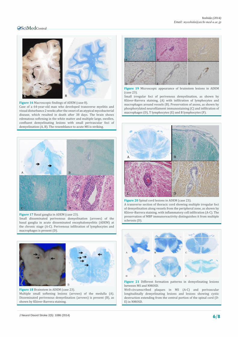
Figure 19 Microscopic appearance of brainstem lesions in ADEM (case 23).Small irregular foci of perivenous demyelination, as shown by Klüver-Barrera staining, (A) with infiltration of lymphocytes and macrophages around vessels (B). Preservation of axons, as shown by phosphorylated neurofilament immunostaining (C) and infiltration of macrophages (D), T lymphocytes (E) and B lymphocytes (F).
Figure 20 Spinal cord lesions in ADEM (case 23).A transverse section of thoracic cord showing multiple irregular foci of demyelination along vessels from the peripheral zone, as shown by Klüver-Barrera staining, with inflammatory cell infiltration (A-C). The preservation of MBP immunoreactivity distinguishes it from multiple sclerosis (D).
Figure 21 Different formation patterns in demyelinating lesions between MS and NMOSD. Well-circumscribed plaques in MS (A-C) and perivascular longitudinally demyelinating lesions and lesions showing cystic destruction extending from the central portion of the spinal cord (D-E) in NMOSD.

Central
Yoshida (2014)Email:
J Neurol Disord Stroke 2(5): 1086 (2014) 7/8
cord, accompanied by Wallerian degeneration in the posterior column and the bilateral pyramidal tract (Figure 12). The irregular lesions on the spinal cord showing cystic destruction extend from the central portion to the adjacent gray and white matter.
The demyelinating lesions of NMOSDs tend to extend along the perivascular region longitudinally (Figure 13A). Axons are more severely decreased in NMOSD lesions than in those of MS (Figure 13B). Foamy macrophages and hyaline thickening of small vessels in the lesion are observed (Figure 13C, D).
AQP4 immunoreactivity is severely decreased in cystic lesions when compared with normal-looking lesions (Figure 14A-C) [18-23]. AQP4 is highly expressed in the astrocytic endfeet around blood vessels. GFAP immunoreactivity is decreased in cystic lesions exhibiting severe damage, whereas GFAP expression remains or may recover in areas showing mild demyelination (Figure 14D-F). Phagocytized corpora amylacea have been reported to be a characteristic feature in acute NMOSD lesions (Figure 15) [25]. The presence of different lesion types is reported to suggest diverse mechanisms of tissue injury in NMO [26].
ACUTE DISSEMINATED ENCEPHALOMYELITIS (ADEM)
ADEM is a multifocal inflammatory disorder of the cen-tral nervous system, presenting perivenous encephalomyelitis, postinfectious encephalomyelitis and post-vaccinal encephalo-myelitis [27-31]. In the acute phase, swelling of the brainstem, spinal cord or cerebral white matter may be observed in severe cases and may resemble acute MS in some cases (Figure 16). In the chronic stage, small areas of disseminated perivenous demy-elination and perivenous infiltration of lymphocytes and macro-phages are visible microscopically (Figures 17-19). In the spinal cord, transverse sections show multiple irregular foci of demyeli-nation along vessels from the peripheral zone and inflammatory cell infiltration (Figure 20). The formation patterns of demyeli-nating lesions in the spinal cord are characteristic of ADEM and different from those of MS and NMOSDs.
CONCLUSIONDifferent formation patterns in demyelinating lesions are
pathologically evident in MS, NMOSD and ADEM. The characteristic pathological features include well-circumscribed plaques in MS and irregular lesions showing perivascular longitudinal demyelination combined with cystic destructive lesions in NMOSDs (Figure 21). Additionally, the immunoreactivities of MBP and AQP4 differ between MS and NMOSDs, which may aid the pathological reevaluation of past cases diagnosed as MS, OSMS or NMO. These pathological images may contribute to understanding the neuroimaging features of demyelinating diseases. Further investigation is needed to determine the pathomechanism of demyelination.
REFERENCES 1. Devic E. Myélitesubaiguëcompliquée de névriteoptique. Bull Med
(Paris). 1894; 8: 1033-1034.
2. Okinaka S, Tsubaki T, Kuroiwa Y, Toyokura Y, Imamura Y. Multiple sclerosis and allied diseases in Japan; clinical characteristics.
Neurology. 1958; 8: 756-763.
3. Wingerchuk DM, Hogancamp WF, O’Brien PC, Weinshenker BG. The clinical course of neuromyelitis optica (Devic’s syndrome). Neurology. 1999; 53: 1107-1114.
4. Misu T, Fujihara K, Nakashima I, Miyazawa I, Okita N, Takase S, et al. Pure optic-spinal form of multiple sclerosis in Japan. Brain. 2002; 125: 2460-2468.
5. Kira J. Multiple sclerosis in the Japanese population. Lancet Neurol. 2003; 2: 117-127.
6. Lennon VA, Wingerchuk DM, Kryzer TJ, Pittock SJ, Lucchinetti CF, Fujihara K, et al. A serum autoantibody marker of neuromyelitis optica: distinction from multiple sclerosis. Lancet. 2004; 364: 2106-2112.
7. Lennon VA, Kryzer TJ, Pittock SJ, Verkman AS, Hinson SR. IgG marker of optic-spinal multiple sclerosis binds to the aquaporin-4 water channel. J Exp Med. 2005; 202: 473-477.
8. Wingerchuk DM, Lennon VA, Pittock SJ, Lucchinetti CF, Weinshenker BG. Revised diagnostic criteria for neuromyelitis optica. Neurology. 2006; 66: 1485-1489.
9. Wingerchuk DM, Lennon VA, Lucchinetti CF, Pittock SJ, Weinshenker BG. The spectrum of neuromyelitis optica. Lancet Neurol. 2007; 6: 805-815.
10. Hu W1, Lucchinetti CF. The pathological spectrum of CNS inflammatory demyelinating diseases. Semin Immunopathol. 2009; 31: 439-453.
11. Kutzelnigg A, Lassmann H. Pathology of multiple sclerosis and related inflammatory demyelinating diseases. In: Handbook of Clinical Neurology, Vol. 122 (3rd series) Multiple sclerosis and related disorders. Goodin DS, editor 2014. Elsevier B.V. 16-58.
12. Ganter P, Prince C, Esiri MM. Spinal cord axonal loss in multiple sclerosis: a post-mortem study. Neuropathol Appl Neurobiol. 1999; 25: 459-467.
13. DeLuca GC, Ebers GC, Esiri MM. Axonal loss in multiple sclerosis: a pathological survey of the corticospinal and sensory tracts. Brain. 2004; 127: 1009-1018.
14. Kidd D, Barkhof F, McConnell R, Algra PR, Allen IV, Revesz T. Cortical lesions in multiple sclerosis. Brain. 1999; 122 : 17-26.
15. Geurts JJ, Bö L, Pouwels PJ, Castelijns JA, Polman CH, Barkhof F. Cortical lesions in multiple sclerosis: combined postmortem MR imaging and histopathology. AJNR Am J Neuroradiol. 2005; 26: 572-577.
16. Geurts JJ, Barkhof F. Grey matter pathology in multiple sclerosis. Lancet Neurol. 2008; 7: 841-851.
17. Calabrese M, Filippi M, Gallo P. Cortical lesions in multiple sclerosis. Nat Rev Neurol. 2010; 6: 438-444.
18. Misu T, Fujihara K, Kakita A, Konno H, Nakamura M, Watanabe S, et al. Loss of aquaporin 4 in lesions of neuromyelitis optica: distinction from multiple sclerosis. Brain. 2007; 130: 1224-1234.
19. Misu T, Takano R, Fujihara K, Takahashi T, Sato S, Itoyama Y. Marked increase in cerebrospinal fluid glial fibrillar acidic protein in neuromyelitis optica: an astrocytic damage marker. J Neurol Neurosurg Psychiatry. 2009; 80: 575-577
20. Yanagawa K, Kawachi I, Toyoshima Y, Yokoseki A, Arakawa M, Hasegawa A, et al. Pathologic and immunologic profiles of a limited form of neuromyelitis optica with myelitis. Neurology. 2009; 73: 1628-1637.
21. Pittock SJ, Lennon VA, Krecke K, Wingerchuk DM, Lucchinetti CF, Weinshenker BG. Brain abnormalities in neuromyelitis optica. Arch Neurol. 2006; 63: 390-396.
Central
Yoshida (2014)Email:
J Neurol Disord Stroke 2(5): 1086 (2014) 8/8
22. Nakamura M, Nakashima I, Sato S, Miyazawa I, Fujihara K, Itoyama Y. Clinical and laboratory features of neuromyelitis optica with oligoclonal IgG bands. Mult Scler. 2007; 13: 332-335.
23. Misu T, Fujihara K, Nakashima I, Sato S, Itoyama Y. Intractable hiccup and nausea with periaqueductal lesions in neuromyelitis optica. Neurology. 2005; 65: 1479-1482.
24. Roemer SF, Parisi JE, Lennon VA, Benarroch EE, Lassmann H, Bruck W, et al. Pattern-specific loss of aquaporin-4 immunoreactivity distinguishes neuromyelitis optica from multiple sclerosis. Brain. 2007; 130: 1194-1205.
25. Suzuki A, Yokoo H, Kakita A, Takahashi H, Harigaya Y, Ikota H, et al. Phagocytized corpora amylacea as a histological hallmark of astrocytic injury in neuromyelitis optica. Neuropathology. 2012; 32: 587-594.
26. Misu T, Höftberger R, Fujihara K, Wimmer I, Takai Y, Nishiyama S, et al. Presence of six different lesion types suggests diverse mechanisms of tissue injury in neuromyelitis optica. Acta Neuropathol. 2013; 125: 815-827.
27. Noorbakhsh F, Johnson RT, Emery D, Power C. Acute disseminated encephalomyelitis: clinical and pathogenesis features. Neurol Clin. 2008; 26: 759-780,
28. Scolding N. Acute disseminated encephalomyelitis and other inflammatory demyelinating variants Handbook of Clinical Neurology, Vol. 122 (3rd series) Multiple Sclerosis and Related Disorders. Goodin DS, editor. 2014 Elsevier BV. 601-611.
29. Sonneville R, Klein I, de Broucker T, Wolff M. Post-infectious encephalitis in adults: diagnosis and management. J Infect. 2009; 58: 321-328.
30. Tenembaum S, Chitnis T, Ness J, Hahn JS; International Pediatric MS Study Group. Acute disseminated encephalomyelitis. Neurology. 2007; 68: S23-36.
31. Yoshida M. [AQP4 immunohistochemistry in neuromyelitis optica and multiple sclerosis: a neuropathological review]. Brain Nerve. 2010; 62: 961-974.
Yoshida M (2014) Neuropathology of Demyelinating Diseases in Japan. J Neurol Disord Stroke 2(5): 1086.
Cite this article