Embed Size (px)
Citation preview
Neuronal and Glial Apoptosis after Traumatic Spinal Cord Injury
Xiao Z. Liu,1 Xiao M. Xu,2 Rong Hu,1 Cheng Du,1 Shu X. Zhang,2 John W. McDonald,1 Hong X. Dong,1Ying J. Wu,1 Guang S. Fan,1 Mark F. Jacquin,1 Chung Y. Hsu,1 and Dennis W. Choi1
1Center for the Study of Nervous System Injury and Department of Neurology, Washington University School of Medicine,Saint Louis, Missouri 63110-1093, and 2Department of Anatomy and Neurobiology, Saint Louis University School ofMedicine, Saint Louis, Missouri 63104-1028
Cell death was examined by studying the spinal cords of ratssubjected to traumatic insults of mild to moderate severity.Within minutes after mild weight drop impact (a 10 gm weightfalling 6.25 mm), neurons in the immediate impact area showeda loss of cytoplasmic Nissl substances. Over the next 7 d, thislesion area expanded and cavitated. Terminal deoxynucleotidyltransferase (TdT)-mediated deoxyuridine triphosphate–biotinnick end labeling (TUNEL)-positive neurons were noted primar-ily restricted to the gross lesion area 4–24 hr after injury, with amaximum presence at 8 hr after injury. TUNEL-positive gliawere present at all stages studied between 4 hr and 14 d, witha maximum presence within the lesion area 24 hr after injury.However 7 d after injury, a second wave of TUNEL-positive glialcells was noted in the white matter peripheral to the lesion andextending at least several millimeters away from the lesion
center. The suggestion of apoptosis was supported by electronmicroscopy, as well as by nuclear staining with Hoechst 33342dye, and by examination of DNA prepared from the lesion site.Furthermore, repeated intraperitoneal injections of cyclohexi-mide, beginning immediately after a 12.5 mm weight dropinsult, produced a substantial reduction in histological evidenceof cord damage and in motor dysfunction assessed 4 weekslater. Present data support the hypothesis that apoptosis de-pendent on active protein synthesis contributes to the neuronaland glial cell death, as well as to the neurological dysfunction,induced by mild-to-moderate severity traumatic insults to therat spinal cord.
Key words: acute SCI; apoptosis; cell death; contusion injury;spinal cord; rat; cycloheximide; motor function
Traumatic insults to the spinal cord induce both immediate me-chanical damage and subsequent tissue degeneration, the latterprogressing in a setting of ischemia, hemorrhage, and edema(Allen, 1914; Ducker et al., 1971; Fairholm and Turnbull, 1971;Green and Wagner, 1973; Osterholm, 1974; Senter and Venes,1978; Young, 1993). There has been a long-standing, primarilyimplicit assumption that such trauma-induced spinal cord celldeath, like the death of brain cells induced by several acuteinsults, represents the form of death called “necrosis” (Kerr et al.,1972; Balentine, 1978a,b; Selina et al., 1989). This assumption isconsistent with the more recent implication of excitotoxicity inthe pathogenesis of traumatic spinal cord damage (Faden andSimon, 1988; Panter et al., 1990; Wrathall et al., 1992), becauseexcitotoxicity is typically marked by early neuronal cell swelling(Coyle et al., 1981) and likely leads preferentially to necrosis(Csernansky et al., 1994; Gwag et al., 1996).
Over the past few years, however, growing evidence has sug-
gested that both global (Goto et al., 1990; Shigeno et al., 1990;Papas et al., 1992; Heron et al., 1993; Okamoto et al., 1993;Roberts-Lewis et al., 1993; Kihara et al., 1994; Nitatori et al.,1995) and focal (Linnik et al., 1993; Tominaga et al., 1993;MacManus et al., 1994; Charriaut-Marlangue et al., 1995; Li etal., 1995; Du et al., 1996a,b) ischemic brain cell loss may in partreflect programmed cell death, resulting in apoptosis (Kerr et al.,1972; Wyllie et al., 1980; Arends and Wyllie, 1991; Johnson et al.,1995). In cortical cell cultures deprived of oxygen and glucose,neurons die predominantly by excitotoxic necrosis (Goldberg andChoi, 1993), but if excitotoxicity is blocked by the combinedapplication of NMDA receptor and AMPA/kainate receptorantagonists, then neurons undergo apoptosis (Gwag et al., 1995)as do cultured sympathetic neurons exposed to hypoxia (Rosen-baum et al., 1994). Apoptosis of brain cells likely also occurs aftertraumatic insults (Rink et al., 1995) and may occur in the settingof neurodegenerative diseases (Portera-Cailliau et al., 1995;Thompson, 1995).
It seemed therefore plausible that apoptosis might also contrib-ute to the spinal cord cell loss occurring after traumatic insults.We initiated the present study to look for apoptosis in the spinalcords of rats subjected to moderate severity traumatic insultsusing a well characterized injury model (Gruner, 1992). In themeantime, independent evidence supporting this idea has beenreported from other laboratories (see Discussion). An abstract onour current research has been published previously (Liu et al.,1996).
MATERIALS AND METHODSSpinal cord injury (SCI). Impact injury was induced using the weight dropdevice developed at New York University (Gruner, 1992). Adult Long–
Received Dec. 23, 1996; revised April 28, 1997; accepted May 2, 1997.This work was supported by National Institute of Neurological Disorders and Stroke
Grant 32636 to D.W.C., as well as by grants from the American Paralysis Associationto C.Y.H. and D.W.C. and from the Daniel Heumann Fund for Spinal Cord Researchto X.M.X. We thank Dr. Q. C. Yu from the University of Chicago for helpfuldiscussion of EM data and Dr. Michael Province from Washington University forassistance with statistical analyses. The rip monoclonal antibody developed by B.Friedman was obtained from the Developmental Studies Hybridoma Bank maintainedby the Department of Pharmacology and Molecular Sciences, Johns Hopkins Univer-sity School of Medicine (Baltimore, MD) and by the Department of BiologicalSciences, University of Iowa (Iowa City, IA) under Contract N01-HD-2-3144 from theNational Institute of Child Health and Human Development.
Correspondence should be addressed to Dr. Dennis W. Choi, Center for theStudy of Nervous System Injury and Department of Neurology, Washington Uni-versity School of Medicine, 660 South Euclid Avenue, Box 8111, Saint Louis, MO63110-1093.Copyright © 1997 Society for Neuroscience 0270-6474/97/175395-12$05.00/0
The Journal of Neuroscience, July 15, 1997, 17(14):5395–5406
Evans female rats (Simonsen Lab, Gilroy, CA) were anesthetized withpentobarbital (50 mg/kg, i.p.). During surgery, rectal temperature wasmaintained at 37.0 6 0.2°C by a thermostatically regulated heating pad(Versa-Therm 2156; Cole-Parmer, Chicago, IL). A laminectomy wasperformed at the T9–T10 level, exposing the cord underneath withoutdisrupting the dura. After the spinous processes of T8 and T11 wereclamped to stabilize the spine, the exposed dorsal surface of the cord wassubjected to weight drop impact using a 10 gm rod (2.5 mm in diameter)dropped at a height of either 6.25 or 12.5 mm and following the proce-dure guidelines established by a multicenter consortium (MulticenterAnimal Spinal Cord Injury Study; Basso et al., 1995, 1996a,b). Animalssubjected to the 6.25 mm insult developed near complete recovery ofhindlimb motor function 4 weeks after injury. Therefore, the 12.5 mminsult was used for testing of therapeutic intervention.
After the injury, the muscles and skin were closed in layers, and ratswere placed in a temperature- and humidity-controlled chamber (Ther-mocare, Incline Village, NV) overnight. Manual bladder expression wasperformed three times per day until reflex bladder emptying was estab-lished. All of the surgical interventions and presurgical and postsurgicalanimal care were provided in accordance with the Laboratory AnimalWelfare Act, the Guide for the Care and Use of Laboratory Animals(National Research Council, 1996), and the Guidelines and Policies forRodent Survival Surgery provided by the Animal Studies Committee ofWashington University School of Medicine.
Cycloheximide administration. Cycloheximide (Sigma, St. Louis, MO)was dissolved in sterile saline at a concentration of 1 mg/ml. Ratsreceiving the 12.5 mm insult were assigned randomly to receive eithercycloheximide (1 mg/kg) or vehicle that was injected intraperitoneallyimmediately after insult and then every third day for 4 weeks.
Behavioral tests. Behavioral tests were performed by investigatorsblinded to the treatment groups and using the Basso–Beattie–Bresnahan(BBB) scales. Testing began 1 d after the 12.5 mm weight drop injury andthen continued twice per week for 4 weeks.
Histopathology. After receiving behavioral testing for 4 weeks, 24animals subjected to the 12.5 mm injury were given a lethal overdose ofpentobarbital and perfused intracardially with normal saline followed by4% paraformaldehyde in PBS, pH 7.4. For histological evaluation, a 15mm cord segment centered at the injury site was removed from thevertebral canal, was placed in the same fixative overnight, and wasembedded in paraffin. Serial 10 mm cross-sections were cut and stainedwith hematoxylin and eosin. The spared areas of the spinal cord at theepicenter (0), at 750 and 1500 mm rostral (2750 and 21500 mm), and at750 and 1500 mm caudal (1750 and 11500 mm) to the epicenter weremeasured using a Metamorph image analysis system (Universal Imagingorporation, West Chester, PA). Measurements were performed andanalyzed by an investigator blind to treatment group assignment.
Another 64 rats were subjected to the 6.25 mm injury. Four animalswere euthanized at 5 min, 4, 8, and 24 hr, and 3, 7, 14, and 30 d afterinsult using the perfusion and embedding procedure described above.Serial 7 mm longitudinal (coronal) sections were cut. These sectionsthrough the central canal were Nissl-stained and examined under anOlympus BX 60 microscope. Longitudinal sections through the anteriorhorn were used for terminal deoxynucleotidyl transferase (TdT)-mediated deoxyuridine triphosphate (dUTP)–biotin nick end labeling(TUNEL), for Hoechst 33342 staining, or for staining for neuronal-specific enolase. Camera lucida drawings of lesion size and TUNEL-positive cells were performed using a drawing tube attached to themicroscope. The rest of the rats were perfused as described above, andthe spinal cords were post-fixed for 4 hr and were then transferred in 30%sucrose overnight. Fourteen micrometer longitudinal sections throughthe anterior horn were cut for rip immunohistochemistry. All the histo-logical analyses were performed blinded to experimental condition.
TUNEL staining. Paraffin-embedded cord sections were deparaffinizedin two changes of xylene for 5 min each. The sections were washed
sequentially in 100, 95, and 75% ethanol before being incubated with 20mg/ml proteinase K (Sigma, St. Louis, MO) for 5 min to strip off nuclearproteins. TUNEL was accomplished using the Apoptag in situ kit ob-tained from Oncor (Gaithersburg, MD). After immersion in equilibra-tion buffer for 10 min, sections were incubated with TdT and dUTP-digoxigenin in a humidified chamber at 37°C for 1 hr and then incubatedin the stop/wash buffer at 37°C for 30 min to stop the reaction. Thesections were washed with PBS once before incubation in antidigoxigen-in–peroxidase solution for 30 min. They were colorized with diamino-benzidine–H2O2 solution (0.2 mg/ml tetrachloride and 0.005% H2O2 in50 mM Tris-HCl buffer) and then counterstained with methyl green.Control sections were treated similarly but incubated in the absence ofTdT enzyme, dUTP-digoxigenin, or anti-digoxigenin antibody.
Double staining. Sections from different time points were double-stained by TUNEL and by immunohistochemical staining with a poly-clonal antibody directed against neuron-specific enolase (Dako, Carpin-
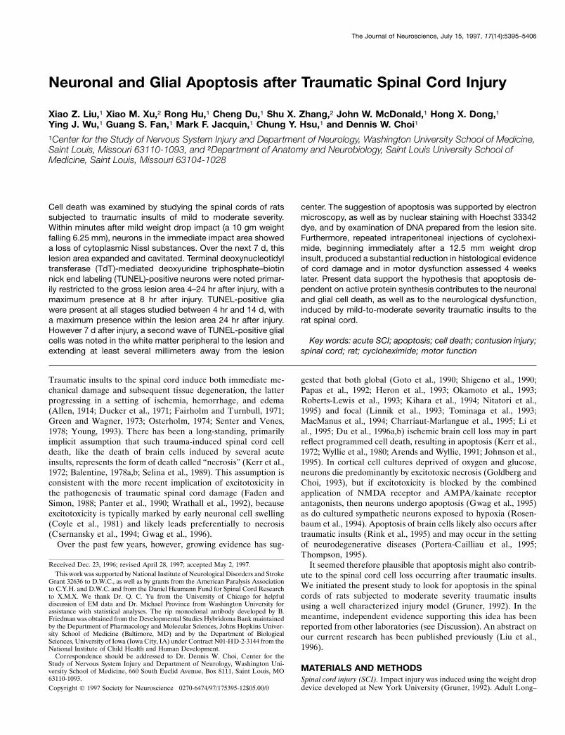
Figure 1. Schematic drawings showing longitudinal (coronal) sectionsthrough the central canal in animals receiving the 6.25 mm impact injuryand being perfused at 5 min, 4, 8, and 24 hr, and 3 d (A); 7 d (B); and 14and 30 d (C). Each contour is the average from four animals. Progressiveexpansion of the lesion was seen, initially defined by the disappearance ofNissl substance from neurons (5 min–4 hr), later defined by the breakingdown of axonal segments and myelin as well as the invasion of blood cellsinto white matter, and still later defined by gross cavitation (7–30 d,cross-hatched areas). By 14 d, the lesion consisted entirely of a cavity andwas somewhat smaller than the lesion defined at 3 d; this cavity wassomewhat increased by 30 d. WM, White matter; GM, gray matter; *Siteof impact. Scale bar, 1 mm.
3
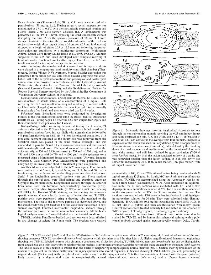
Figure 2. TUNEL-labeled (A–F) and Hoechst 33342-stained (G–I) cells in the spinal cord after a 6.25 mm injury. A, Longitudinal section of the cordshowing numerous TUNEL-positive cells (arrowheads) present within the injury area 8 hr after injury. B, Higher magnification of demarcated region in Ashowing two TUNEL-labeled neurons with chromatin condensation. C, Section showing TUNEL-labeled neuron) (arrowhead) that can be distinguishedfrom labeled glial cells (thin arrows) by its relatively larger nucleus, its prominent cytoplasm, and the pericellular space created by its shrinkage (thick arrows).The labeled nucleus of this neuron is smaller than the nuclei of neighboring morphologically normal neurons (D). E, Section showing breakdown of thenucleus of a glial cell in the white matter near the injury epicenter into several fragments. F, Section showing TUNEL-labeled glial cell, presumably anoligodendrocyte (thick arrow), in the peripheral white matter away from the injury epicenter. Note the close association of the cell with the space (asterisks)likely created by a degenerated axon. A morphologically normal oligodendrocyte nucleus (thin arrow) and a (Figure legend continues)
5396 J. Neurosci., July 15, 1997, 17(14):5395–5406 Liu et al. • Apoptosis after Traumatic SCI

degenerating axonal segment (arrowhead) are also marked for comparison. G–I, Hoechst 33342-stained sections. These show nuclear fragmentation ofa probable neuron (H, arrow) and a glial cell (I, arrow) in the spinal cord 24 hr after injury. The nuclear labeling of a morphologically normal neuron(G, arrow) is presented for comparison. WM, White matter; GM, gray matter. Scale bars: A, 50 mm; B–F, 10 mm; G–I, 10 mm.
Liu et al. • Apoptosis after Traumatic SCI J. Neurosci., July 15, 1997, 17(14):5395–5406 5397
teria, CA) or with the oligodendrocyte-specific monoclonal antibody rip(Developmental Studies Hybridoma Bank, Iowa City, IA) (Friedman etal., 1989). To identify neurons, we first subjected fixed sections toTUNEL and then blocked the sections with 5% horse serum, washedthem in PBS, and incubated them with mouse neuron-specific enolaseantibody. The sections were again washed in PBS and incubated in
biotinylated horse anti-mouse IgG (Vector Laboratories, Burlingame,CA) and in avidin–biotin complex (Vector Laboratories). Peroxidasewas demonstrated with a Vector SG kit (Vector Laboratories). Negativecontrols were stainings performed without primary antibody. To identifyoligodendrocytes, we incubated frozen sections overnight in rip diluted1:200. Sections were washed with PBS for 10 min, incubated with sec-ondary anti-mouse IgG antibody conjugated to fluorescein, and thensubjected to TUNEL using a secondary antibody conjugated to rhoda-mine. Sections were examined under epifluorescence illumination on aOlympus BX 60 microscope.
Hoechst 33342 staining. Paraffin sections were deparaffinized withxylene two times for 5 min each and then rinsed with PBS. The sectionswere first stained with 10 mg/ml Hoechst 33342 from Molecular Probes(Eugene, OR) for 5 min, washed with PBS, and then stained withpropidium iodide (1:1000) from Molecular Probes for 5 min.
Transmission electron microscopy (EM). Cross-sections of the spinalcord were cut serially every 500 mm through the epicenter of the injuryin rats that received the 6.25 mm injury and were perfused at 4, 8, and 24hr after injury. Samples were fixed in 2.5% glutaraldehyde in 0.1 Mcacodylate buffer, pH 7.4, for 60 min at 4°C and then washed in the samebuffer for 80 min. After post-fixation in 1% osmium tetroxide in 0.1 Mcacodylate buffer, pH 7.4, for 60 min at room temperature, the tissueswere dehydrated in graded ethanol and were embedded in Spurr’s epoxyresin (Spurr, 1969). One micrometer semithin plastic sections werestained with 1% toluidine blue for light microscopic observations. Ultra-thin sections of the same specimen were cut and stained with uranylacetate and lead citrate and examined with a Zeiss 108 electronmicroscope.
Quantitation of histone-associated DNA fragmentation. The extent ofhistone-associated DNA fragmentation was assessed using an ELISA kit(Cell Death Detection ELISA) obtained from Boehringer Mannheim(Indianapolis, IN). The assay is based on the quantitative sandwichenzyme immunoassay principle with mouse monoclonal antibodies di-rected against DNA and histones, respectively and detects mononucleo-somes and oligonucleosomes.
Rats subjected to the 6.25 mm injury were euthanized under deepanesthesia at 4 and 24 hr and 3 and 7 d after injury (n 5 3 animals at eachtime point). A 5 mm segment of the injured cord at the epicenter or ofthe normal cord (n 5 3) was dissected, homogenized, and centrifuged(14,890 3 g for 10 min). The supernatant was diluted 1:200 and used asan antigen source in sandwich ELISA with a primary anti-histone anti-body coated to the microliter plate and a secondary anti-DNA antibodycoupled to peroxidase.
DNA gel electrophoresis. Spinal cord DNA was isolated according to themethod of Sambrook et al. (1989) with a few modifications. Briefly, ratsreceiving the 6.25 mm injury were euthanized at 4, 8, and 24 hr, and 50mg of fresh cord tissue was removed and homogenized individually in anEppendorf tube containing 300 ml of cell lysis buffer (10 mM Tris-HCl,100 mM EDTA, and 0.5% SDS). After mixing with an additional 300 mlof cell lysis buffer, the sample was incubated for 1 hr at 65°C and thenincubated with proteinase K (final concentration, 100 mg/ml) overnight at55°C. Extraction was performed with an equal volume of phenol, equil-ibrated with 0.5 M Tris-HCl, and phenol /chloroform/amyl alcohol (25:24:1). Total DNA contained in the aqueous phase was precipitated withethanol. The DNA pellet was washed twice with 70% ethanol anddissolved in 25 ml Tris–EDTA buffer (10 mM Tris-HCl, pH 8.0, and 1 mMEDTA, pH 8.0). The DNA was then treated with DNase-free RNase (10mg/ml) for 1 hr at 37°C and assayed by optical absorption at 260 nm.Equal amounts of DNA samples were subjected to 1.5% agarose gelelectrophoresis.
Statistical analysis. Data are expressed as mean 6 SEM. To analyzedifferences in open field locomotor scores or in volumes of spared tissuebetween groups, we used a repeated measures ANOVA with Tukey’sstudentized range test to correct for multiple comparisons. A two-wayANOVA was also used for comparison of the extent of histone-associated DNA fragmentation between groups. For the difference be-tween groups at each time point, we used a pairwise post hoc Tukey’sstudentized range test. p , 0.05 was considered significant.
RESULTSMorphological featuresFive minutes after a weight drop insult (a 10 gm rod, 2.5 mm indiameter, falling 6.25 mm), a well defined area of gray matterinjury, defined by loss of neuronal Nissl staining and petechiae,
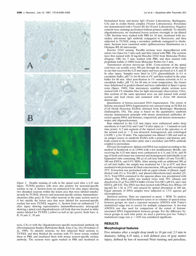
Figure 3. Double staining of cells in the spinal cord after a 6.25 mminjury. TUNEL-positive cells were also positive for neuronal-specificenolase or rip. A, Section from rat euthanized 8 hr after injury showingtwo shrunken neurons within the lesion area that were labeled simulta-neously by TUNEL (brown) and neuronal-specific enolase immunohisto-chemistry (blue gray). B, Two other neurons in the same section used inA but outside the lesion area that were labeled for neuronal-specificenolase but were TUNEL negative. C, Section from rat euthanized 7 dafter injury showing representative immunofluorescence micrographshowing a spinal cord oligodendrocyte cell body that was located in whitematter labeled for TUNEL ( yellow) as well as rip ( green). Scale bars: A,B, 10 mm; C, 10 mm.
5398 J. Neurosci., July 15, 1997, 17(14):5395–5406 Liu et al. • Apoptosis after Traumatic SCI

was apparent (Fig. 1) with a clear boundary between damagedand morphologically normal neurons. The injured region had alongitudinal extent of 2.31 6 0.08 mm (mean 6 SEM, n 5 4animals) and a transverse extent of 1.17 6 0.13 mm. Little grossdamage to white matter was initially apparent.
The gross lesion area expanded over time. By 3 d, the lesionarea extended to 4.62 6 0.08 mm along the cord axis and 2.60 60.05 mm transversely (Fig. 1). White matter injury became grosslyapparent 8 hr after injury, delineated by the breaking down ofaxonal segments and myelin as well as the invasion of blood cellsinto white matter, although changes in white matter could be seen4 hr after injury by EM. EM changes, including axonal swelling,degenerating axonal segments, and thinning or loss of myelinsheaths, were observed, in agreement with earlier studies (Balen-
tine, 1978b; Bresnahan, 1978; Blight, 1983; Kao et al., 1983; Blightand Decrescito, 1986; Beattie et al., 1988).
By 7 d, multiple cavitations were noted within the lesion area.By 14 d, a large central cavity formed that expanded modestlyfurther by 30 d.
TUNELFive min after injury, no TUNEL-positive cells were observed.After 4 hr, many darkly TUNEL-positive cells were present inthe gray matter, confined to the lesion area as defined by loss ofNissl staining and petechiae. Morphology as well as double stain-ing for neuronal-specific enolase indicated that many of theseTUNEL-positive cells were neurons (Figs. 2–4). These TUNEL-positive neurons typically exhibited shrinkage of both cytoplasm
Figure 4. Schematic drawing ofTUNEL-stained longitudinal (coronal)sections of the spinal cord through theventral horns, after a 6.25 mm weightdrop insult and euthanization of the ratsat the indicated times after injury. Fiveminutes after insult, no TUNEL-labeledcells were observed. By 8 hr, manyTUNEL-positive neurons (large dots)and glial cells (small dots) were observedmostly within the lesion area (defined byloss of Nissl substance from neurons). By24 hr, TUNEL-positive neurons were nolonger found, but many TUNEL-positive glial cells were seen within theinjury area. By 7 d, a second wave ofTUNEL-positive glial cells was ob-served mostly outside of the lesion areain the lateral funiculus extending theentire length of the section (1.5 cm). By14 d, fewer TUNEL-labeled glia werefound. WM, White matter; GM, graymatter; *Site of impact. Scale bar, 1 mm.
Liu et al. • Apoptosis after Traumatic SCI J. Neurosci., July 15, 1997, 17(14):5395–5406 5399

and nucleus, creating pericellular space, and nuclear fragmenta-tion. The mean nuclear diameter of TUNEL-labeled neurons was6.74 6 0.34 mm (mean 6 SD, n 5 36), approximately half the sizeof TUNEL-negative neurons in the same sections (13.01 6 0.59mm, mean 6 SD; n 5 42). Neuronal TUNEL positivity wasmaximal at 8 hr after injury and decreased subsequently (Fig. 4).By 24 hr after injury, few TUNEL-positive neurons were seen(Fig. 4).
Apoptotic changes in presumptive glial cells were also observedbeginning 4 hr after injury and with a maximum at 24 hr after
Figure 5. Electron micrograph showing a representative apoptotic cell and a representative necrotic cell in the gray matter of the cord 8 hr after injury.A, Apoptosis. The nucleus has fragmented into several membrane-bounded, highly condensed bodies (thick arrow), and the cell body has shrunk withan intact, infolded cell membrane (arrowheads). B, Necrosis. The nucleus (Nu) is swollen (arrowhead) with scattered granular aggregations of chromatin(thick arrows), and the cell is swollen with dilation of rough endoplasmic reticulum (thin arrows) and mitochondria (asterisks). RBC, Red blood cell. Scalebars, 1 mm.
Figure 6. Ethidium bromide-stained agarose gel showing a DNAladder from a rat spinal cord after a weight drop insult. Data are fromrats euthanized: in Lane 1, after no injury (normal control); in lane 2,4 hr after injury; in lane 3, 8 hr after injury; and in lane 4, 24 hr afterinjury. Molecular weight markers are shown to the lef t of lane 1. DNAladdering is apparent in lane 4 (arrows).
Figure 7. Enrichment of mononucleosomes and oligonucleosomes in thecytoplasmic fraction after a 6.25 mm injury (enrichment factor, absorbanceof the injured tissue/absorbance of the normal control tissue). An ELISAkit (see Materials and Methods) was used to detect mononucleosomes andoligonucleosomes in the cytoplasmic fraction of spinal cord tissue at theindicated times after injury. Evidence of internucleosomal DNA fragmen-tation peaked 24 hr after injury and was reduced by cycloheximidetreatment. Error bars indicate SEM; *p , 0.05; n 5 3 animals per group.
5400 J. Neurosci., July 15, 1997, 17(14):5395–5406 Liu et al. • Apoptosis after Traumatic SCI

injury (Fig. 4, data for 4 hr not shown). Most of these TUNEL-positive glial cells were found within the lesion area, althoughsome were present in the neighboring white matter (Fig. 4). Thecells typically exhibited small, fragmented nuclei with little visiblecytoplasm surrounding them (Fig. 2). The mean nuclear diameterof TUNEL-positive glial cells was 4.53 6 0.25 mm (mean 6 SD,n 5 42), compared with 7.76 6 0.25 mm (mean 6 SD, n 5 36) inTUNEL-negative glia. By 3 (data not shown) and 7 d after injury,the number of TUNEL-positive glial cells within the lesion areahad fallen sharply (Fig. 4; data not shown).
However, by 7 d, a second wave of TUNEL-positive glial cells
was observed in white matter extending the entire 1.5 cm lengthof the coronal section (taken through the ventral horn, Fig. 4).Few TUNEL-positive cells were seen in the white matter at the3 d time point (data not shown). These TUNEL-positive whitematter cells typically showed a close association with degenerat-ing axons (Fig. 2F) and stained for the oligodendrocyte-specificmarker rip (Fig. 3C) (Friedman et al., 1989).
EM
Transmission EM revealed coexistent apoptotic and necroticchanges in cells within the lesion area 4–24 hr after injury.
Figure 8. Hematoxylin- and eosin-stained horizontal cross-sections taken 4 weeks after injury in rats treated with either vehicle (lef t) or cycloheximide(right). B, E, Sections through the lesion epicenter (0). A, D, Sections 750 mm rostral to the epicenter (2750 mm). C, F, Sections 750 mm caudal to theepicenter (1750 mm). Scale bar, 100 mm.
Liu et al. • Apoptosis after Traumatic SCI J. Neurosci., July 15, 1997, 17(14):5395–5406 5401
Apoptotic changes were characterized by cytoplasmic shrinkage,plasma membrane infolding, coarse chromatin condensation, andbreakdown of the nucleus into discrete, membrane-bounded bod-ies (Fig. 5A). Occasionally, well preserved apoptotic bodies con-taining fragmented nuclear chromatin were found within thecytoplasm of macrophages (data not shown). Necrotic changeswere characterized by cell, nuclear, and mitochondrial swellingwith loosely textured chromatin aggregation and dilation of roughendoplasmic reticulum (Fig. 5B).
DNA ladderingDNA prepared from lesion site tissue 4–24 hr after injury and runon an agarose gel revealed a progressive increase in internucleo-somal laddering over this time (Fig. 6).
Quantitation of DNA breakdownTissue DNA breakdown was quantitated using a commercialELISA that measures histone-associated mononucleosomes oroligonucleosomes. DNA fragmentation in lesion site tissue fromrats receiving 6.25 mm insults was detectable as early as 4 hr afterinjury, reached a large peak by 24 hr, and then declined. DNAfragmentation was reduced by cycloheximide treatment (1 mg/kg,i.p.) that began immediately after injury and continued onceevery third day until the animals were euthanized (Fig. 7).
Protective effect of cycloheximide administrationWe turned to the protein synthesis inhibitor cycloheximide to tryto reduce apoptotic cell loss in this injury model (Martin et al.,1988, 1992; Ciutat et al., 1996; Yaginuma et al., 1996). Havingdetected evidence of oligodendrocyte apoptosis 7 d after insult,we decided to use a recurrent injection paradigm. Pavlik andTeisinger (1980) showed that subcutaneous injection of a single0.6 mg/kg dose of cycloheximide resulted in transient (;12 hr)suppression of brain protein synthesis in rats. We administered 1mg/kg intraperitoneally immediately after a 12.5 mm weight dropinjury and followed with 1 mg/kg intraperitoneally every thirdday thereafter, a regimen that was well tolerated by the ratspresumably because protein synthesis was intermittently releasedfrom inhibition. This treatment regimen resulted in the grosssparing of spinal cord tissue compared with vehicle-treated con-trols measured 4 weeks after injury (Figs. 8, 9). The tissue sparingconsisted of a wider rim of tissue appearing to approximatenormal architecture surrounding a hypercellular, vascularizedlesion area with many macrophages (Fig. 10). In addition, openfield motor testing using the BBB Locomotor Rating Scale (Bassoet al., 1995, 1996a,b) showed that this cycloheximide treatmentsubstantially improved hindlimb function compared with vehicle-treated controls (Fig. 11). By 4 weeks after injury, thecycloheximide-treated rats had improved to a BBB score of 19 61.71, compared with 14.75 6 3.17 (mean 6 SEM, n 5 12) invehicle-treated controls (a BBB score of 21 is normal). Thisdifference in BBB score reflects a grossly apparent improvementin gait, although there is coordinated plantar stepping at bothscores of 15 and 19. At a score of 15, there is little toe clearanceduring forward limb advancement, and paws are parallel to thebody only at initial contact. At a score of 19, there is consistenttoe clearance during forward limb advancement, and paws areparallel to the body both at initial contact and at liftoff.
DISCUSSIONData presented here suggest that apoptosis, involving both neu-rons and glia, contributes to spinal cord tissue damage aftertraumatic insults of mild to moderate severity. Our determination
of apoptosis relies on multiple criteria: morphology under bothlight and electron microscopic examination, nuclear chromatinstaining with Hoechst 33342 dye and with TUNEL, DNA ladder-ing on gel electrophoresis, an increase in histone-associatedmononucleosomes and oligonucleosomes by ELISA, and the sen-sitivity of gross tissue damage to inhibition of protein synthesis(Wyllie et al., 1980; Kerr and Harmon, 1991; Johnson et al., 1995).Impact-induced spinal cord cell apoptosis was not confined inspace to the immediate impact site or in time to the immediatepostinjury period. Although there was a burst of neuronal andglial apoptosis in gray and white matter at the lesion site withinapproximately the first 24 hr, a delayed plethora of oligodendro-cyte apoptosis occurred in distant white matter several days later.Because apoptosis is typically a rapid process, over in hours(Bursch et al., 1990), this late apoptosis likely reflects a true waveof delayed oligodendrocyte death.
The idea of delayed oligodendrocyte apoptosis in the spinalcord after traumatic insults was recently proposed by Li et al.(1996), who observed TUNEL-positive, glial fibrillary acidicprotein-negative cells in the white matter of rat spinal cordssubjected to compression injury, and by Bresnahan et al. (1996),who observed apoptotic oligodendrocytes closely associated withdying axons in monkey spinal cords subjected to contusion injury.Perhaps reflecting specific model differences between their injurymodel and the model used here, Li et al. (1996) found littleTUNEL positivity in gray matter, although Katoh et al. (1996)recently reported the occurrence of TUNEL positivity in cellsfrom both gray and white matter after extradural weight compres-sion injury to the rat spinal cord. Furthermore, morphologicalevidence for spinal cord cell apoptosis in rats injured with the
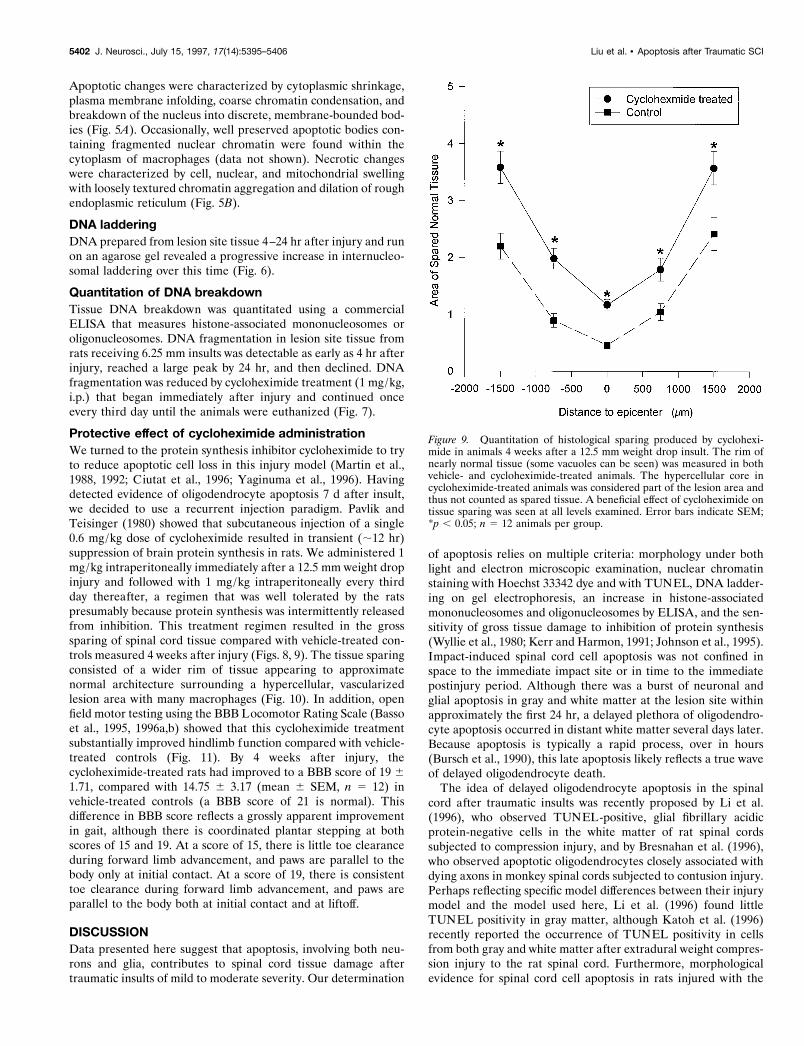
Figure 9. Quantitation of histological sparing produced by cyclohexi-mide in animals 4 weeks after a 12.5 mm weight drop insult. The rim ofnearly normal tissue (some vacuoles can be seen) was measured in bothvehicle- and cycloheximide-treated animals. The hypercellular core incycloheximide-treated animals was considered part of the lesion area andthus not counted as spared tissue. A beneficial effect of cycloheximide ontissue sparing was seen at all levels examined. Error bars indicate SEM;*p , 0.05; n 5 12 animals per group.
5402 J. Neurosci., July 15, 1997, 17(14):5395–5406 Liu et al. • Apoptosis after Traumatic SCI

same New York University impactor used here has been reportedin abstracts by Crowe et al. (1995), Shuman et al. (1996), Swobodaet al. (1996), and Yong et al. (1996).
The evolution of the gross lesion observed here is consistentwith older studies of impact trauma to the spinal cord, which havedescribed the progressive enlargement of an initial lesion incentral gray matter over a period of hours to days to involvecontiguous white matter, eventually leading to central cavitationat higher levels of impact severity (Allen, 1914; Goodkin andCampbell, 1969; Ducker et al., 1971; White, 1975). Balentine(1978a,b) performed a detailed study of lesion evolution usingboth light and electron microscopic methods to examine thespinal cords of Sprague Dawley rats subjected to weight dropinjury. He emphasized the occurrence of immediate (3–5 min)multifocal petechial hemorrhages in central gray matter that werefollowed over the next hours by tissue edema and by the necrosisdeaths of both neurons and glia. These necrosis deaths weremarked by the loss of cytoplasmic detail and by the swelling oforganelles. White matter exhibited progressive “extracellularswelling” thought to represent edema fluid. By 8–72 hr afterinsult, axons were swollen and granular. The only changes de-scribed between 1 and 4 weeks after insult were reactive gliosis,phagocytosis of necrotic debris by macrophages, deposits of cal-cium and hemosiderin, and the formation of multiloculated cysts.
Did apoptosis occur in the study of Balentine (1978a,b)? Pos-sibly not. He did not comment on the possibility, and although hismodel was not identical to ours, he used a more severe insult thatlikely shifted cell death away from apoptosis toward necrosis. Onthe other hand, coarse condensation of nuclear chromatin, rem-iniscent of apoptosis, can be seen in his cell electron micrographs(Balentine, 1978a, his Figs. 12, 15).
The idea that a delayed wave of oligodendrocyte apoptosisoccurs in spinal cord white matter after traumatic insults isespecially intriguing in light of other evidence suggesting thatpoor myelination of axons can persist long after experimental(Blight, 1985) or human (Bunge et al., 1993) spinal cord injury.Further studies will be needed to identify the mechanisms respon-sible for this delayed oligodendrocyte death. Most likely, it wastriggered by evolving axonal degeneration and subsequent loss ofaxonally derived survival signals (Barres et al., 1993). Alterna-tively, delayed oligodendrocyte apoptosis may occur as a result ofslowly evolving adverse changes in the cellular milieu distant tothe impact site, for example, as a result of inflammatory events(Hsu and Dimitrijevic, 1990). In this latter formulation, all cellsmight be exposed to low levels of some injury, but oligodendro-cytes would be especially vulnerable and would succumbselectively.
Apoptosis in the CNS has been classically considered to occur
Figure 10. Hematoxylin- and eosin-stained transverse right hemisection at the lesion epicenter in a rat treated with cycloheximide. A, Section showinga wide rim of spared cord tissue (arrow). In the central region, hypercelluar, vascularized tissue with many macrophage-like cells is seen. B, Highermagnification of demarcated region in A showing a border between the peripheral rim of spared cord tissue and this central hypercellular tissue (dottedline). BV, Blood vessel. Arrowheads indicate probable macrophages. Scale bars, 100 mm.
Liu et al. • Apoptosis after Traumatic SCI J. Neurosci., July 15, 1997, 17(14):5395–5406 5403

only during development, in which it plays a vital role in the sizematching of cell populations and in the formation of propersynaptic connections. Growing evidence that apoptosis contrib-utes importantly to pathological CNS loss raises the excitingpossibility that measures aimed at blocking apoptosis may findtherapeutic use in various disease states. To our knowledge, thedata reported here are the first to provide direct support for theidea that an antiapoptotic treatment can improve outcome afterspinal cord injury. Specifically, intraperitoneal injections of cyclo-heximide at 3 d intervals produced substantial preservation oftissue, reduced central cavitation, and improved recovery of func-tion. However, the possibility cannot be presently excluded thatthe beneficial effects of cycloheximide observed here were medi-ated by mechanisms not related to direct inhibition of pro-grammed cell death. Some alternative mechanisms might beenhancement of cellular glutathione levels because of reducedcysteine use (Ratan et al., 1994) or suppression of neutrophilchemotaxis (Tanabe et al., 1994). Yet another possible mecha-nism is raised by the interesting finding that low concentrations ofcycloheximide that produce only limited inhibition of proteinsynthesis can induce the production of Bcl2 and antioxidantenzymes (Furukawa et al., 1997). However, we think that the 1mg/kg dose of cycloheximide used here should have achievedconsiderable suppression of protein synthesis (Pavlik and Teis-inger, 1980).
Whether indeed the beneficial effect of cycloheximide treat-ment results from reduction of spinal cord cell apoptosis, it will beimportant to determine the extent to which specific populations ofspinal cord cells can be preserved by this treatment, as well as the
contribution of each cycloheximide-preserved population to func-tional benefit. It is possible, for example, that most or even all ofthe functional benefit is unrelated to lesion size reduction andreflects improved axonal myelination caused by preservation ofthe oligodendrocyte population. It will also be important todetermine whether inhibition of apoptosis can be therapeuticallyeffective against insults more severe than those used here (incontrols, sparing ,20% of spinal cord tissue at the lesion core butreducing the BBB score to only 15 after recovery). On theoptimistic side, however, the current regimen of cycloheximideadministration, being intermittent, may not have achieved com-plete inhibition of protein synthesis-dependent apoptosis. Testingof more specific inhibitors of apoptosis, such as the interleukin-converting enzyme family inhibitors (Kondo et al., 1996; Pronk etal., 1996), as well as combined antiapoptotic and antiexcitotoxicmeasures (Du et al., 1996b), offers additional goals for futurestudy that may lead to the development of practical clinicaltherapies.
REFERENCESAllen AR (1914) Remarks on the histopathological changes in the spinal
cord due to impact. An experimental study. J Nerv Ment Dis41:141–147.
Arends MJ, Wyllie AH (1991) Apoptosis: mechanisms and roles in pa-thology. Int Rev Exp Pathol 32:223–254.
Balentine JD (1978a) Pathology of experimental spinal cord trauma. I.The necrotic lesion as a function of vascular injury. Lab Invest39:236–253.
Balentine JD (1978b) Pathology of experimental spinal cord trauma. II.Ultrastructure of axons and myelin. Lab Invest 39:254–266.
Barres BA, Jacobson MD, Schmid R, Sendtner M, Raff MC (1993) Doesoligodendrocyte survival depend on axons? Curr Biol 3:489–497.
Basso DM, Beattie MS, Bresnahan JC (1995) A sensitive and reliablelocomotor rating scale for open field testing in rats. J Neurotrauma12:1–21.
Basso DM, Beattie MS, Bresnahan JC (1996a) Graded histological andlocomotor outcomes after spinal cord contusion using the NYU weightdrop device versus transection. Exp Neurol 139:244–256.
Basso DM, Beattie MS, Bresnahan JC, Anderson DK, Faden AI, GrunerJA, Holford TR, Hsu CY, Noble LJ, Nockels R, Perot PL, SalzmanSK, Young W (1996b) MASCIS evaluation of open field locomotorscores: effects of experience and teamwork on reliability. J Neuro-trauma 13:343–359.
Beattie MS, Stokes BT, Bresnahan JC (1988) Experimental spinal cordinjury: strategies for acute and chronic intervention based on anatomic,physiological, and behavioral studies. In: Pharmacological approachesto the treatment of brain and spinal cord injury (Stein DG, Sabel BA,eds), pp 43–74. New York: Plenum.
Blight AR (1983) Axonal physiology of chronic spinal cord injury in thecat: intracellular recording in vitro. Neuroscience 10:1471–1486.
Blight AR (1985) Delayed demyelination and macrophage invasion: acandidate for secondary cell damage in spinal cord injury. Cent NervSyst Trauma 2:299–315.
Blight AR, Decrescito V (1986) Morphometric analysis of experimentalspinal cord injury in the cat: the relation of injury intensity to survivalof myelinated axons. Neuroscience 19:321–341.
Bresnahan JC (1978) An electron-microscopic analysis of axonal alter-ations following blunt contusion of the spinal cord of the rhesus monkey(Macaca Mulatta). J Neurol Sci 37:59–82.
Bresnahan JC, Shuman SL, Beattie MS (1996) Evidence for apoptosis ofoligodendroglia in long tracts undergoing wallerian degeneration afterspinal cord injury (SCI) in monkeys. Soc Neurosci Abstr 22:1185.
Bunge RP, Puckett WR, Becerra JL, Marcillo A, Quencer RM (1993)Observations on the pathology of human spinal cord injury: a reviewand classification of 22 new cases with details from a case of chroniccord compression with extensive focal demyelination. Adv Neurol59:75–89.
Bursch W, Kleine L, Tenniswood M (1990) The biochemistry of celldeath by apoptosis. Biochem Cell Biol 68:1071–1074.
Charriaut-Marlangue C, Margaill I, Plotkine M, Ben-Ari Y (1995) Early
Figure 11. Long-term beneficial effect of cycloheximide on the hindlimbneurological function of rats after a 12.5 mm weight drop injury, assessedby the BBB Locomotor Rating Scale. Error bars indicate SEM; *p , 0.05;n 5 12 animals per group.
5404 J. Neurosci., July 15, 1997, 17(14):5395–5406 Liu et al. • Apoptosis after Traumatic SCI

endonuclease activation following reversible focal ischemia in the ratbrain. J Cereb Blood Flow Metab 15:385–388.
Ciutat D, Caldero J, Oppenheim RW, Esquerda JE (1996) Schwann cellapoptosis during normal development and after axonal degenerationinduced by neurotoxins in the chick embryo. J Neurosci 16:3979–3990.
Coyle JT, Bird SJ, Evans RH, Gulley RL, Nadler JV, Nicklas WJ, OlneyJW (1981) Excitatory amino acid neurotoxins: selectivity, specificity,and mechanisms of action. Neurosci Res Program Bull 19:1–427.
Crowe MJ, Shuman SL, Masters JN, Bresnahan JC, Beattie MS (1995)Morphological evidence suggesting apoptotic nuclei in spinal cordinjury. Soc Neurosci Abstr 21:232.
Csernansky CA, Canzoniero LMT, Sensi SL, Yu SP, Choi DW (1994)Delayed application of aurintricarboxylic acid reduces glutamate-induced cortical neuronal injury. J Neurosci Res 38:101–108.
Du C, Hu R, Csernansky CA, Hsu CY, Choi DW (1996a) Very delayedinfarction after mild focal cerebral ischemia: a role for apoptosis? JCereb Blood Flow Metab 16:195–201.
Du C, Hu R, Csernansky CA, Liu XZ, Hsu CY, Choi DW (1996b)Additive neuroprotective effects of dextrorphan and cycloheximide inrats subjected to transient focal cerebral ischemia. Brain Res718:233–236.
Ducker TB, Kindt GW, Kempe LG (1971) Pathological findings in acuteexperimental spinal cord trauma. J Neurosurg 35:700–708.
Faden AI, Simon RP (1988) A potential role for excitotoxins in thepathophysiology of spinal cord injury. Ann Neurol 23:623–626.
Fairholm DJ, Turnbull IM (1971) Microangiographic study of experi-mental spinal cord injuries. J Neurosurg 35:277–286.
Friedman B, Hockfield S, Black JA, Woodruff KA, Waxman SG (1989)In situ demonstration of mature oligodendrocytes and their processes:an immunocytochemical study with a new monoclonal antibody, rip.Glia 2:380–390.
Furukawa K, Estus S, Fu WM, Mark RJ, Mattson MP (1997) Neuro-protective action of cycloheximide involves induction of BCL-2 andantioxidant pathways. J Cell Biol 136:1137–1149.
Goldberg MP, Choi DW (1993) Combined oxygen and glucose depriva-tion in cortical cell culture: calcium-dependent and calcium-independent mechanisms of neuronal injury. J Neurosci 13:3510–3524.
Goodkin R, Campbell JB (1969) Sequential pathologic changes in spinalcord injury: a preliminary report. Surg Forum 20:430–432.
Goto K, Ishige A, Sekiguchi K, Iizuka S, Sugimoto A, Yuzurihara M,Aburada M, Hosoya E, Kogure K (1990) Effects of cycloheximide ondelayed neuronal death in rat hippocampus. Brain Res 534:299–302.
Green BA, Wagner Jr FC (1973) Evolution of edema in the acutelyinjured spinal cord: a fluorescence microscopic study. Surg Neurol1:98–101.
Gruner JA (1992) A monitored contusion model of spinal cord injury inthe rat. J Neurotrauma 9:123–128.
Gwag BJ, Lobner D, Koh JY, Wie MB, Choi DW (1995) Blockade ofglutamate receptors unmasks neuronal apoptosis after oxygen-glucosedeprivation in vitro. Neuroscience 68:615–619.
Gwag BJ, Koh JY, DeMaro JA, Ying HS, Jacquin M, Choi DW (1997)Slowly-triggered excitotoxicity occurs by necrosis in cortical cultures.Neuroscience 77:393–401.
Heron A, Pollard H, Dessi F, Moreau J, Lasbennes F, Ben-Ari Y,Charriaut-Marlangue C (1993) Regional variability in DNA fragmen-tation after global ischemia evidenced by combined histological and gelelectrophoresis observations in the rat brain. J Neurochem61:1973–1976.
Hsu CY, Dimitrijevic MR (1990) Methylprednisolone in spinal cordinjury: the possible mechanism of action. J Neurotrauma 7:115–119.
Johnson Jr EM, Greenlund LJ, Akins PT, Hsu CY (1995) Neuronalapoptosis: current understanding of molecular mechanisms and poten-tial role in ischemic brain injury. J Neurotrauma 12:843–852.
Kao CC, Wrathall JR, Kyoshima K (1983) Axonal reaction to transec-tion In: Spinal cord reconstruction (Kao CC, ed), pp 41–57. New York:Raven.
Katoh K, Ikata T, Katoh S, Hamada Y, Nakauchi K, Sano T, Niwa M(1996) Induction and its spread of apoptosis in rat spinal cord aftermechanical trauma. Neurosci Lett 216:9–12.
Kerr JFR, Harmon BV (1991) Definition and incidence of apoptosis: anhistorical perspective. In: Apoptosis: the molecular basis of cell death(Tomei LD, Cope FO, eds), pp 5–29. New York: Cold Spring HarborLaboratory.
Kerr JFR, Wyllie AH, Currie AR (1972) Apoptosis: a basic biological
phenomenon with wide-ranging implications in tissue kinetics. Br JCancer 26:239–257.
Kihara S, Shiraishi T, Nakagawa S, Toda K, Tabuchi K (1994) Visual-ization of DNA double strand breaks in the gerbil hippocampal CA1following transient ischemia. Neurosci Lett 175:133–136.
Kondo S, Kondo Y, Yin D, Barnett GH, Kaakaji R, Peterson JW,Morimura T, Kubo H, Takeuchi J, Barna BP (1996) Involvement ofinterleukin-1 beta-converting enzyme in apoptosis of bFGF-deprivedmurine aortic endothelial cells. FASEB J 10:1192–1197.
Li GL, Brodin G, Farooque M, Funa K, Holtz A, Wang WL, Olsson Y(1996) Apoptosis and expression of BCL-2 after compression traumato rat spinal cord. J Neuropathol Exp Neurol 55:280–289.
Li Y, Sharov VG, Jiang N, Zaloga C, Sabbah HN, Chopp M (1995)Ultrastructural and light microscopic evidence of apoptosis after mid-dle cerebral artery occlusion in the rat. Am J Pathol 146:1045–1051.
Linnik MD, Zobrist RH, Hatfield MD (1993) Evidence supporting arole for programmed cell death in focal cerebral ischemia in rats.Stroke 24:2002–2009.
Liu XZ, Xu XM, Hu R, Du C, Fan GS, Hsu CY, Choi DW (1996) Effectof cycloheximide on DNA breakdown, tissue loss, and behavioraloutcome after spinal cord impact injury. Soc Neurosci Abstr 22:1185.
MacManus JP, Hill IE, Huang ZG, Rasquinha I, Xue D, Buchan AM(1994) DNA damage consistent with apoptosis in transient focal isch-emic neocortex. NeuroReport 5:493–496.
Martin DP, Schmidt RE, DiStefano PS, Lowry OH, Carter JG, JohnsonJr EM (1988) Inhibitors of protein synthesis and RNA synthesis pre-vent neuronal death caused by nerve growth factor deprivation. J CellBiol 106:829–844.
Martin DP, Ito A, Horigome K, Lampe PA, Johnson Jr EM (1992)Biochemical characterization of programmed cell death in NGF-deprived sympathetic neurons. J Neurobiol 23:1205–1220.
Nitatori T, Sato N, Waguri S, Karasawa Y, Araki H, Shibanai K, Komi-nami E, Uchiyama Y (1995) Delayed neuronal death in the CA1pyramidal cell layer of the gerbil hippocampus following transientischemia is apoptosis. J Neurosci 15:1001–1011.
Okamoto M, Matsumoto M, Ohtsuki T, Taguchi A, Mikoshiba K, Yanagi-hara T, Kamada T (1993) Internucleosomal DNA cleavage involved inischemia-induced neuronal death. Biochem Biophys Res Commun196:1356–1362.
Osterholm JL (1974) The pathophysiological response to spinal cordinjury. The current status of related research. J Neurosurg 40:5–33.
Panter SS, Yum SW, Faden AI (1990) Alteration in extracellular aminoacids after traumatic spinal cord injury. Ann Neurol 27:96–99.
Papas S, Crepel V, Hasboun D, Jorquera I, Chinestra P, Ben-Ari Y(1992) Cycloheximide reduces the effects of anoxic insult in vivo and invitro. Eur J Neurosci 4:758–765.
Pavlik A, Teisinger J (1980) Effect of cycloheximide administered to ratsin early postnatal life: prolonged inhibition of DNA synthesis in thedeveloping brain. Brain Res 192:531–541.
Portera-Cailliau C, Hedreen JC, Price DL, Koliatsos VE (1995) Evi-dence for apoptotic cell death in Huntington disease and excitotoxicanimal models. J Neurosci 15:3775–3787.
Pronk GJ, Ramer K, Amiri P, Williams LT (1996) Requirement of anICE-like protease for induction of apoptosis and ceramide generationby REAPER. Science 271:808–810.
Ratan RR, Murphy TH, Baraban JM (1994) Macromolecular synthesisinhibitors prevent oxidative stress-induced apoptosis in embryonic cor-tical neurons by shunting cysteine from protein synthesis to glutathione.J Neurosci 14:4385–4392.
Rink A, Fung KM, Trojanowski JQ, Lee VM, Neugebauer E, McIntoshTK (1995) Evidence of apoptotic cell death after experimental trau-matic brain injury in the rat. Am J Pathol 147:1575–1583.
Roberts-Lewis JM, Marcy VR, Zhao Y, Vaught JL, Siman R, Lewis ME(1993) Aurintricarboxylic acid protects hippocampal neurons fromNMDA- and ischemia-induced toxicity in vivo. J Neurochem61:378–381.
Rosenbaum DM, Michaelson M, Batter DK, Doshi P, Kessler JA (1994)Evidence for hypoxia-induced, programmed cell death of culturedneurons. Ann Neurol 36:864–870.
Sambrook J, Fritsch EF, Maniatis T (1989) Isolation of DNA frommammalian cells: protocol I. In: Molecular cloning: a laboratory man-ual, Ed 2, Chap 9 (Sambrook J, ed), pp 16–19. New York: Cold SpringHarbor Laboratory.
Selina CC, McIntosh TK, Noble LJ (1989) Experimental fluid percus-
Liu et al. • Apoptosis after Traumatic SCI J. Neurosci., July 15, 1997, 17(14):5395–5406 5405
sion brain injury: vascular disruption and neuronal and glial alterations.Brain Res 482:271–282.
Senter HJ, Venes JL (1978) Altered blood flow and secondary injury inexperimental spinal cord trauma. J Neurosurg 49:569–578.
Shigeno T, Yamasaki Y, Kato G, Kusaka K, Mima T, Takakura K,Graham DI, Furukawa S (1990) Reduction of delayed neuronal deathby inhibition of protein synthesis. Neurosci Lett 120:117–119.
Shuman SL, Bresnahan JC, Beattie MS (1996) Morphological evidenceof glial involvement in apoptotic cell death following spinal cord con-tusion. Soc Neurosci Abstr 22:1185.
Spurr AR (1969) A low-viscosity epoxy resin embedding medium forelectron microscopy. J Ultrastruct Res 26:31–43.
Swoboda TK, Li Y, Nockels RP, Chopp M (1996) Evidence of apoptosisin an acute spinal cord injury model. Soc Neurosci Abstr 22:20.
Tanabe J, Watanabe M, Kondoh S, Mue S, Ohuchi K (1994) Possible rolesof protein kinases in neutrophil chemotactic factor production by leuco-cytes in allergic inflammation in rats. Br J Pharmacol 113:1480–1486.
Thompson CB (1995) Apoptosis in the pathogenesis and treatment ofdisease. Science 267:1456–1462.
Tominaga T, Kure S, Narisawa K, Yoshimoto T (1993) Endonucleaseactivation following focal ischemic injury in the rat brain. Brain Res608:21–26.
White RJ (1975) Pathology of spinal cord injury in experimental lesions.Clin Orthop 112:16–26.
Wrathall JR, Teng YD, Choiniere D, Mundt DJ (1992) Evidence thatlocal non-NMDA receptors contribute to functional deficits in contu-sive spinal cord injury. Brain Res 586:140–143.
Wyllie AH, Kerr JFR, Currie AR (1980) Cell death: the significance ofapoptosis. Int Rev Cytol 68:251–306.
Yaginuma H, Tomita M, Takashita N, McKay SE, Cardwell C, YinQW, Oppenheim RW (1996) A novel type of programmed neuronaldeath in the cervical spinal cord of the chick embryo. J Neurosci16:3685–3703.
Yong C, Arnold PM, Zoubine MN, Citron BA, Festoff BW (1996)Apoptosis in injured spinal cord of rat. Soc Neurosci Abstr 22:20.
Young W (1993) Secondary injury mechanisms in acute spinal cordinjury. J Emerg Med 11:13–22.
5406 J. Neurosci., July 15, 1997, 17(14):5395–5406 Liu et al. • Apoptosis after Traumatic SCI


![Computer system for simulation of human perception. Some ...the central nervous system [9,10]. Figure 1 shows a schematic diagram of the glial-neuronal interaction: two astrocytes](https://img.dokumen.tips/doc/110x75/60ecb64657caf315d017ba30/computer-system-for-simulation-of-human-perception-some-the-central-nervous.jpg)