Embed Size (px)
Citation preview

Neuron
Article
Action-Related Properties Shape ObjectRepresentations in the Ventral StreamBradford Z. Mahon,1,2,* Shawn C. Milleville,3 Gioia A.L. Negri,4 Raffaella I. Rumiati,4 Alfonso Caramazza,1,2
and Alex Martin3
1Center for Mind/Brain Sciences, University of Trento, Rovereto (TN) 38068, Italy2Department of Psychology, Harvard University, Cambridge, MA 02138, USA3Laboratory of Brain and Cognition, National Institute of Mental Health, Bethesda, MD 20892-1366, USA4Cognitive Neuroscience Sector, Scuola Internazionale Superiore di Studi Avanzati Trieste, Italy
*Correspondence: [email protected]
DOI 10.1016/j.neuron.2007.07.011
SUMMARY
The principles driving the organization of theventral object-processing stream remain un-known. Here, we show that stimulus-specificrepetition suppression (RS) in one region ofthe ventral stream is biased according to motor-relevant properties of objects. Quantitativeanalysis confirmed that this result was not con-founded with similarity in visual shape. A similarpattern of biases in RS according to motor-relevant properties of objects was observed indorsal stream regions in the left hemisphere.These findings suggest that neural specificityfor ‘‘tools’’ in the ventral stream is driven by sim-ilarity metrics computed over motor-relevantinformation represented in dorsal structures.Support for this view is provided by convergingresults from functional connectivity analyses ofthe fMRI data and a separate neuropsycholog-ical study. More generally, these data suggestthat a basic organizing principle giving rise to‘‘category specificity’’ in the ventral streammay involve similarity metrics computed overinformation represented elsewhere in the brain.
INTRODUCTION
One principle of organization of the primate visual system
is the division of labor between the ventral object-
processing stream, mediating visual object recognition,
and a dorsal object-processing stream, mediating online
object-directed action and spatial analysis (Goodale and
Milner, 1992; Ungerleider and Mishkin, 1982). The ventral
stream projects from primary visual cortex to the lateral
and ventral surfaces of occipital cortex, through to anterior
ventral temporal cortex. The dorsal stream projects from
primary visual cortex to dorsal occipital and lateral tempo-
ral cortex, through to parietal cortex (Ungerleider, 1995).
Within the dorsal object-processing stream, a network of
primarily left-lateralized regions process object-associ-
ated motion (left middle temporal gyrus), online visuo-
motor transformations for grasping objects (posterior pari-
etal cortex), and the motor commands associated with tool
use (inferior parietal lobule) (e.g., Culham et al., 2003;
Beauchamp et al., 2002; Johnson-Frey, 2004). Functional
neuroimaging has shown that these dorsal regions are dif-
ferentially activated when participants view manipulable
objects compared to living things or large nonmanipulable
objects (e.g., Chao and Martin, 2000; Johnson-Frey et al.,
2005; Okada et al., 2000; for review, see Lewis, 2006).
A second characteristic of the organization of the
human visual system concerns the organization of high-
order visual object recognition processes within the
ventral stream, and in particular, within the fusiform gyrus.
The fusiform gyrus processes visual properties of objects
such as color and form (e.g., Martin et al., 1995; Miceli
et al., 2001). A number of functional neuroimaging studies
in humans have found that living things (e.g., faces, ani-
mals), compared to nonliving things, differentially activate
the lateral portion of the fusiform gyrus (in the vicinity of the
Fusiform Face Area—Kanwisher et al., 1999; Chao et al.,
1999a). In contrast, manipulable objects such as tools
and utensils, compared to living things, differentially acti-
vate the medial fusiform gyrus (e.g., Chao et al., 1999b;
Noppeney et al., 2006; although the specificity of this
claim has been challenged: Downing et al., 2006; Mechelli
et al., 2006). Finally, stimuli that may be described as
highly contextualized, such as large nonmanipulable
objects, houses, and scenes, differentially activate the
parahippocampal gyrus (Parahippocampal Place Area—
Epstein et al., 1999; see also Avidan et al., 2002; Barr
and Aminoff, 2003; Downing et al., 2006). These cate-
gory-specific profiles of neural activation have also been
observed at the neuronal level. Single-cell recordings in
humans have documented category specificity in medial
temporal lobe structures that receive input from ventral
temporal-occipital cortex (Kreiman et al., 2000).
Thus, two broad properties of the organization of the hu-
man visual system can be distinguished. On the one hand,
visually presented objects are processed in the ventral
stream for recognition and in the dorsal stream for online
guidance of action. On the other hand, there is articulated
structure within the ventral object-processing stream in
Neuron 55, 507–520, August 2, 2007 ª2007 Elsevier Inc. 507

Neuron
Action Shapes Visual Object Representations
terms of the topography of category-specific neural re-
sponses. These two organizational characteristics of the
visual system are generally viewed as functionally and
physiologically independent. An important and as yet un-
resolved issue is the degree to which there exist functional
interactions between the dorsal and ventral object-
processing streams. This issue is particularly relevant in
addressing the causes of neural specificity in the ventral
stream for manipulable objects. To date, it has been ar-
gued that neural specificity in the ventral stream depends
on similarity metrics that are computed over the informa-
tion that is represented and processed internal to the ven-
tral stream itself. For instance, it has been proposed that
similarity in visual form (Haxby et al., 2001) or in the
distribution of eccentricity preferences (Levy et al., 2001)
explains the causes of category specificity in the ventral
stream.
An alternative conceptual framework that has not to date
been explored is that neural specificity for objects in the
ventral stream is determined, in part, by similarity metrics
computed over information that is stored elsewhere. For
example, neural specificity for manipulable objects in the
ventral stream may depend on information represented
in dorsal stream regions that directly mediate object-
directed action. The left inferior parietal lobule processes
motor commands associated with tool use (e.g., Heilman
et al., 1982; Johnson-Frey et al., 2005; Rumiati et al.,
2004), and the left middle temporal gyrus processes the
rigid and unarticulated motion associated with nonliving
objects (Beauchamp et al., 2002, 2003). In the course of
manipulating and using objects, it is necessary to integrate
the output of object recognition processes (ventral stream)
with information about object motion (left middle temporal
gyrus) and the motor commands necessary to realize the
function of the objects (left inferior parietal lobule). The
efficacy of such an information-processing network would
be increased if the organization of object recognition pro-
cesses already anticipated the processing requirements
of computations implemented ‘‘downstream.’’
The physiology of the primate brain affords the possibil-
ity that inputs from neural structures beyond the ventral
pathway determine, in part, neural specificity within the
medial fusiform gyrus for manipulable objects. There are
anatomical projections between ventral temporal cortex
and the inferior parietal lobule (Rushworth et al., 2006;
Webster et al., 1994; Zhong and Rockland, 2003) as well
as lateral temporal cortex (Saleem et al., 2000). There is
also functional connectivity between ventral temporal
and ventral prefrontal regions (Miller et al., 2003), which
are involved in categorization and the determination of
behavioral goals. Anterior motor areas such as premotor
cortex in turn have substantial functional connectivity
with these ventral prefrontal regions (Rizzolatti and Lup-
pino, 2001). Finally, the basal ganglia, involved in motor
control, and which receive input from frontal, parietal,
and temporal structures, also project to inferior temporal
cortex (Middleton and Strick, 1996) (see also Ungerleider,
1995, and Pisella et al., 2006, for review).
508 Neuron 55, 507–520, August 2, 2007 ª2007 Elsevier Inc.
Clear predictions follow from the view that the organiza-
tion of the ventral stream is driven, in part, by functional
connectivity with dorsal regions directly mediating ob-
ject-directed action. The first prediction is that neural re-
sponses in the medial fusiform gyrus will be sensitive to
the relationship between the physical structure of objects
(in the visual modality, represented in the fusiform gyrus)
and the motor movements associated with the use of
those objects (represented in dorsal stream regions, in-
cluding the left inferior parietal lobule). The second predic-
tion is that perturbation of neural processes in the dorsal
stream may disrupt the equilibrium of processes mediated
by the ventral stream. We tested these predictions with
a combination of functional neuroimaging and neuropsy-
chological methods.
Rapid, event-related fMRI was used to study modula-
tions in stimulus-specific repetition suppression (RS) in
the medial fusiform gyrus as a function of motor-relevant
properties of nonliving things. Stimulus-specific RS can
serve as an index of neural specificity, since it may be ar-
gued that only those sources of signal (i.e., populations of
neurons) that are critically involved in the processing of
a stimulus will show RS to repeated presentations of
that stimulus (e.g., Avidan et al., 2002; Chao et al., 2002;
Dobbins et al., 2004; Grill-Spector et al., 2006; James
et al., 2002). We find that stimulus-specific RS in the left
medial fusiform gyrus is observed only for manipulable
objects with direct relationships between their physical
structure and the motor movements associated with their
use—‘‘tools.’’ A similar pattern of RS biased toward
‘‘tools’’ was observed in the left middle temporal gyrus
and in the left inferior parietal lobule.
The neuropsychological study evaluated regions of the
brain in which lesions predict deficits for using and identi-
fying objects. Converging with the results of the fMRI
study, we find that lesions to the left middle temporal gy-
rus and the left inferior parietal lobule are associated with
impairments for both using and identifying objects. We
further show that when patients are separated on the
anatomical criterion of having lesions involving parietal
cortex, the distribution of performance of the patients for
both identifying and using objects is modulated.
Finally, functional connectivity analyses demonstrated
that the neural responses in the fMRI experiment in the
left medial fusiform gyrus independently predicted neural
responses in the left middle temporal gyrus and the left in-
ferior parietal lobule. These dorsal stream regions identi-
fied by the functional connectivity analyses corresponded
to regions of damaged tissue independently identified in
the lesion overlap analyses.
RESULTS
fMRI Stimulus Characteristics
The experimental stimuli for the fMRI experiment con-
sisted of grayscale photographs of animals, ‘‘tools,’’ arbi-
trarily manipulated objects, and nonmanipulable objects.
‘‘Tools’’ refer to manipulable objects that have systematic

Neuron
Action Shapes Visual Object Representations
Figure 1. fMRI Stimulus Characteristics
The motor-relevant distinctions between
‘‘tools,’’ arbitrarily manipulated objects, and
nonmanipulable objects were confirmed by
ratings (see Supplemental Experimental Pro-
cedures and Figure S4). Graphs (A–C) repre-
sent mean Likert rating (+ SEM), with greater
values indicating greater difficulty (A), more
central (B), or more familiar (C). (A) ‘‘Tools,’’ ar-
bitrarily manipulated objects, and nonmanipu-
lable objects differed monotonically (linear
contrast analysis, p < 0.001; h2 = 0.92) in the
degree to which their identity was predictable
from their associated motor movements. (B)
The motor movements associated with ‘‘tools’’
were more central in determining their function
than were the motor movements associated
with arbitrarily manipulated objects and non-
manipulable objects (ps < 0.001). (C) ‘‘Tools’’
and arbitrarily manipulated objects did not dif-
fer (p = 0.93) with respect to participants’ expe-
rience in physically interacting with the objects.
(D) Previous research (Op de Beeck et al.,
2001) demonstrates that visual shape similarity
can determine the pattern of neuronal re-
sponses in the ventral stream. A quantitative
analysis (see Belongie et al., 2002) of the ex-
perimental stimuli demonstrated that there
was no difference in similarity of visual shape
within stimulus type, among animals, ‘‘tools,’’
and nonmanipulable objects (all ps > 0.05;
see also Figure S5). Within-category similarity
in visual shape was greater for arbitrarily manipulated objects than for the other stimulus types (all ps < 0.05, Bonferroni correction). Larger values
on the y axis of the graph indicate greater dissimilarity in visual shape. Box plot represents medians ± interquartile ranges (IQRs). Outliers (circles)
and extreme values (stars) are defined as values between 1.5 and 3 IQRs and greater than 3 IQRs, respectively, from the tops and bottoms of the
boxes. Not shown in this figure, a separate behavioral experiment measuring naming latencies demonstrated reliable repetition priming (one-way
ANOVAs) for all stimulus types (all ps % 0.05; see Supplemental Experimental Procedures for details).
relationships between their physical form and their man-
ner of manipulation/function (e.g., hammer, scissors,
wrench). Arbitrarily manipulated objects refer to equally
manipulable objects that have variable or nonsystematic
relationships between their physical form and their man-
ner of manipulation/function (e.g., book, wallet, envelope).
The distinction between ‘‘tools’’ and arbitrarily manipu-
lated objects was confirmed by behavioral studies (see
Figure 1 for details). The motor movements associated
with the ‘‘tool’’ stimuli were more central in determining
their function and were more predictive of their identity
than were the motor movements associated with arbi-
trarily manipulated objects. Critically, however, ‘‘tools’’
and arbitrarily manipulated objects did not differ with re-
spect to participants’ experience in physically manipulat-
ing the objects. Stimuli in the set ‘‘nonmanipulable ob-
jects’’ were large objects that may be touched, but
which are not ‘‘taken’’ in the hands (e.g., anchor [of
a ship], fence, desk). The four stimulus types—‘‘tools,’’ ar-
bitrarily manipulated objects, nonmanipulable objects,
and animals—were matched on lexical frequency and
concept familiarity. There was no difference in within-
category similarity in visual shape between animals, tools,
and nonmanipulable objects (see Figure 1D and Supple-
mental Experimental Procedures for details). Furthermore,
there was no difference across the four stimulus types, in
terms of similarity in visual shape between items from
a given stimulus type, and all other items in the experiment
from the other stimulus types (intercategory similarity)
(ANOVA: F < 1).
Stimulus-Specific RS According to Motor-Relevant
Properties of Objects
Ventral Stream
As described in the Introduction, the ventral object-pro-
cessing stream is composed of a set of regions character-
ized by distinct profiles of category specificity. Of interest
in our study is the pattern of neural responses according
to motor-relevant properties of nonliving objects in the
medial fusiform gyrus, on the ventral surface of temporal-
occipital cortex. This region is medial to the well-known
Fusiform Face Area and posterior to the Parahippocampal
Place Area. In this study, we did not use the localizer
approach to identify voxels in the medial fusiform gyrus.
Rather, we used functional contrasts internal to the exper-
imental design that were orthogonal to the effect of interest
(i.e., stimulus-specific RS for each object type).
Neuron 55, 507–520, August 2, 2007 ª2007 Elsevier Inc. 509

Neuron
Action Shapes Visual Object Representations
Figure 2. RS in the Medial Fusiform Gyri Modulated by Motor-Relevant Properties of Objects
(A) The figure shows group-averaged activity superimposed on the brain of an individual subject. Blue indicates regions in the medial fusiform
gyri preferring nonliving things to animals (top row, left to right, y = �54 through y = �42, in steps of 2; bottom row, left to right, z = �16 through
z = �4, in steps of 2). The red cross-sections in the top row indicate the plane of the axial slice directly below (and vice versa). Voxels were defined
at p < 0.001, corrected using family-wise error correction (Monte Carlo simulation), which takes into account cluster size and alpha level.
(B) All histograms and statistical analyses of BOLD responses (here and elsewhere) were computed using mean BOLD responses by experimental
condition, averaged across all voxels in the region. Error bars in all histograms of BOLD responses (here and elsewhere) represent the SEM. The peak
differences for the contrast of nonliving things compared to animals were located (TT coordinates), in the left medial fusiform gyrus (8596 mm3), at
�24, �48, �8, and in the right medial fusiform gyrus (8264 mm3), at 28, �41,�10 (figure shown at z = �12). There were main effects of RS bilaterally
(ps < 0.005), collapsing across the four stimulus types. However, as depicted in the histograms, there was a systematic bias in RS toward ‘‘tools’’ in
the left medial fusiform gyrus and toward ‘‘tools’’ and arbitrarily manipulated objects in the right medial fusiform gyrus.
In line with previously reports (e.g., Avidan et al., 2002;
Chao et al., 1999b; Noppeney et al., 2006), we found in-
creased activity in the medial fusiform gyri, bilaterally,
when subjects named nonliving things (collapsed to-
gether), compared to naming animals (Figure 2A). In addi-
tion, although ‘‘tools’’ failed to elicit more activity in the
medial fusiform gyri than the other nonliving object types
based on responses to the novel trials, there was a sys-
tematic bias in RS effects that followed motor-relevant
properties of the stimuli (see histograms in Figure 2B). In
the left medial fusiform gyrus, RS was observed for only
‘‘tools’’ (p < 0.001; all other Fs < 1), while in the right medial
fusiform gyrus, RS was biased toward ‘‘tools’’ (p < 0.001)
and arbitrarily manipulated objects (p = 0.067; all other
Fs % 1). These modulations in RS effects by stimulus
type within the medial fusiform gyri were confirmed with
510 Neuron 55, 507–520, August 2, 2007 ª2007 Elsevier Inc.
ANOVAs. In the right medial fusiform gyrus, there was a
marginal interaction between RS and stimulus type (p =
0.05); in the left medial fusiform gyrus, the interaction
approached significance (p = 0.10) (for further analysis,
see Figure S1 and Table S1).
As noted in the Introduction, previous research indi-
cates that contextualized visual stimuli (such as the large,
nonmanipulable objects in our study) are differentially pro-
cessed in parahippocampal cortex (Parahippocampal
Place Area—Epstein et al., 1999; see also Barr and Ami-
noff, 2003), while living things such as animals are differ-
entially processed in the lateral fusiform gyrus (e.g.,
Chao et al., 1999a, 1999b), in the vicinity of the Fusiform
Face Area (e.g., Kanwisher et al., 1999). Consistent with
this body of research, we observed stimulus-specific RS
only for nonmanipulable objects in parahippocampal

Neuron
Action Shapes Visual Object Representations
Figure 3. RS by Stimulus Type in the Ventral Stream
(A) Large bilateral regions encompassing medial and lateral regions of the fusiform gyrus were defined by the presence of a main effect of RS (both
ps < 0.002), collapsing across the factor stimulus type (left to right, axial views at z = �17, �12, and �7). Consistent with the broad expanse of
highlighted structures, there were main effects of stimulus type (ps < 0.0001) in both the left and right hemispheres. The interaction between RS
and stimulus type was significant in the right hemisphere (p < 0.041), and there was a trend in the left hemisphere (p = 0.09). The peak TT coordinates
for this contrast were �36, �57, �8 (left hemisphere) and 32, �46, �6 (right hemisphere).
(B) The distinct colors represent RS (p < 0.01) biased toward each of the four stimulus types. We also coded voxels that showed RS biased toward
both ‘‘tools’’ and arbitrarily manipulated objects in order to study potential overlap in RS effects for the two types of manipulable objects. As depicted
in Figure 3B (left to right, axial views at z = �17, �12, and �7), the largest regions, located in the medial fusiform gyri bilaterally, showed RS biased
toward ‘‘tools’’ (blue). In addition, smaller clusters of voxels showing RS biased toward arbitrarily manipulated objects (yellow), and toward both
‘‘tools’’ and arbitrarily manipulated objects (green), were also observed. RS was biased toward animals (red) in the right lateral fusiform gyrus. RS
biased toward nonmanipulable objects (cyan) was restricted to parahippocampal cortex. This effect for nonmanipulable objects was present bilat-
erally (only right hemisphere activation shown).
(C–E) Histograms represent RS effects (difference scores: novel � repeated) for each experimental condition, for the left (red bars) and right (green
bars) medial fusiform gyrus. The corresponding functional group maps are shown (colored blue) next to each histogram (y =�42). As can be seen, RS
was observed for ‘‘tools’’ in the medial fusiform gyrus bilaterally, independently of how this region was defined. For further details of the analyses, see
Table S1; for histograms showing overall BOLD responses for the same contrasts, see Figure S1. (C) Voxels in the medial fusiform gyrus showing
greater activation for ‘‘tools’’ than animals (p < 0.01). (D). Voxels in the medial fusiform gyrus showing greater activation for arbitrarily manipulated
objects than animals (p < 0.01). (E). Voxels in the medial fusiform gyrus showing greater activation for nonmanipulable objects than animals (p < 0.01).
cortex bilaterally and only for animals in the right lateral
fusiform gyrus. These findings are depicted in Figure 3B.
Importantly, stimulus-specific RS was biased toward
‘‘tools’’ in the left medial fusiform gyrus independently of
how voxels in this region were defined. For instance, as
summarized in Figure 3 (panels C–E), RS was observed
for only ‘‘tools’’ in the left medial fusiform, independently
of whether the region was defined by nonmanipulable ob-
jects versus animals, arbitrarily manipulated objects ver-
sus animals, or ‘‘tools’’ versus animals. At the same
time, the region in the medial fusiform gyrus showing an
enhanced response to identifying nonmanipulable objects
included the region defined by arbitrarily manipulated ob-
jects versus animals. Similarly, the region showing en-
hanced activity for arbitrarily manipulated objects com-
pared to animals included the region showing enhanced
activity for ‘‘tools’’ compared to animals (see Table S1
for details). These relative differences in the sizes of ob-
ject-responsive regions remained when bilateral medial
fusiform regions were defined using only novel trials for
each of the nonliving object types compared to animals
(see Figure S1). This means that these relative size differ-
ences were not due to differential RS effects. In contrast,
as depicted in Figure 3B, when voxels were defined in
Neuron 55, 507–520, August 2, 2007 ª2007 Elsevier Inc. 511

Neuron
Action Shapes Visual Object Representations
Figure 4. RS in the Dorsal Stream Modulated by Motor-Relevant Properties of Objects
Histograms represent mean (+SEM) BOLD responses by experimental condition, averaged across all voxels (blue) showing a larger response
(p < 0.01) for ‘‘tools’’ (novel + repeated) than for animals (novel + repeated). The red line in the axial view indicates the plane of the coronal view.
(A) RS was observed for ‘‘tools’’ in the left middle temporal gyrus (1156 mm3; TT: �49, �61, �7; shown at y = �61).
(B) RS was restricted to ‘‘tools’’ in the left caudal IPS (Culham et al., 2003) (3416 mm3; TT: �16, �67, 44) and was observed for both ‘‘tools’’ and
arbitrarily manipulated objects in right caudal IPS (223 mm3; TT: 24, �64, 36) (axial view at z = 34). Caudal IPS is involved in visual analysis of
object affordances for object-directed reaching (e.g., Culham et al., 2003).
terms of the presence of RS effects for a single nonliving
object type, RS for ‘‘tools’’ was observed throughout the
bilateral medial fusiform gyri, while RS for nonmanipulable
objects was restricted to parahippocampal cortex.
Dorsal Stream
In contrast to the pattern observed in the ventral stream,
the contrast of all nonliving things versus animals failed
to elicit activity in any of the dorsal stream regions. More-
over, and as expected, there was no RS for animals (all
Fs < 1) or nonmanipulable objects (the lowest p = 0.22)
in any of the dorsal stream regions identified by the con-
trasts of each of the nonliving object types compared to
animals (see Figure 4, Table S1, and Figure S2 for details).
Here, we focus on the pattern of findings within the regions
showing greater activation for ‘‘tools’’ (novel + repeated)
compared to animals (novel + repeated).
In the left middle temporal gyrus, RS was observed for
only ‘‘tools’’ (p < 0.03) (see Figure 4A and Table S1). As
noted in the Introduction, this region of lateral temporal
cortex is just anterior to motion area MT (Beauchamp
et al., 2002) and is involved in motion analysis of nonliving
things (e.g., Beauchamp et al., 2003; Kable et al., 2002;
Martin, 2007). One possible interpretation of the pattern
of BOLD responses in the left posterior middle temporal
gyrus is that there is a graded degree across the nonliving
object types in the consistency of the movements that are
associated with those objects (see Figure 1A for comple-
mentary behavioral findings).
Of particular interest is the pattern of effects observed
within the left inferior parietal lobule, known to process
512 Neuron 55, 507–520, August 2, 2007 ª2007 Elsevier Inc.
complex object-associated actions (e.g., Heilman et al.,
1982; Johnson-Frey, 2004). Within the left inferior parietal
lobule, there was a reliable BOLD response on novel trials
only for ‘‘tools’’ and RS for only this stimulus type (p < 0.02)
(Figure 5A). The BOLD responses to novel animals and
novel arbitrarily manipulated objects did not differ from
the fixation baseline (ts < 1; one-sample t tests). For novel
nonmanipulable objects, there was a trend toward deacti-
vation (p = 0.09). These data suggest that the left inferior
parietal lobule is a critical structure mediating between
processing of object identity and object use. In particular,
‘‘tools’’ were distinguished (psychophysically) from arbi-
trarily manipulated objects in that, for the former, the mo-
tor movements associated with their use are instrumental
in determining their function (see Figure 1B).
Interestingly, the similarity in neural responses between
the left medial fusiform gyrus and the left inferior parietal
lobule was not limited to analyses of RS. As depicted in
Figure 5B, there was a reliable and positive correlation be-
tween overall BOLD responses (novel + repeated) in the
left medial fusiform gyrus and the left inferior parietal lob-
ule only for ‘‘tool’’ stimuli (for the full correlation matrix, see
Table S2). These data support the view that neural speci-
ficity for ‘‘tools’’ in the left medial fusiform gyrus is related
to processing in the left inferior parietal lobule.
Lesion Overlap Analysis
A large body of evidence (e.g., Goodale and Milner, 1992;
James et al., 2002; Shmuelof and Zohary, 2005) indicates
that the ventral and dorsal object-processing streams can

Neuron
Action Shapes Visual Object Representations
Figure 5. RS Restricted to ‘Tools’ in the
Left Inferior Parietal Lobule
(A) Histograms represent mean (+SEM) BOLD
responses by experimental condition, aver-
aged across all voxels (blue) showing a larger
response (p < 0.01) for ‘‘tools’’ (novel + re-
peated) than for animals (novel + repeated).
The red line in the axial view indicates the plane
of the coronal view, and vice versa. The left in-
ferior parietal lobule (Frey et al., 2005) (1600
mm3; TT: �57, �27, 34; axial view at z = 36)
showed a reliable BOLD response on novel tri-
als only for ‘‘tools,’’ and RS for only this stimu-
lus type.
(B) The b estimates for BOLD signal changes
were extracted for all subjects from the left me-
dial fusiform gyrus region (see also Figure S1)
and the left inferior parietal region described
in (A) above (both regions defined by ‘‘tools’’
[novel + repeated] versus animals [novel + re-
peated]). Intersubject rankings in b estimates
of BOLD responses (novel + repeated) were re-
liably correlated (Spearman) between these
two regions only for ‘‘tools.’’ These data indi-
cate a selective relationship in the distributions
across subjects of BOLD responses in the left
medial fusiform gyrus and the left inferior pari-
etal lobule (see also Figure 8). The 95% confi-
dence intervals around the regression lines
are shown.
operate relatively autonomously with respect to one an-
other and that object processing in the two streams oc-
curs largely in parallel (e.g., Fang and He, 2005). Damage
to the ventral stream can lead to an inability to identify vi-
sually presented objects, despite normal object-directed
action (visual agnosia without optic ataxia). Damage to
dorsal occipital and posterior parietal regions can lead
to an impairment for object-directed grasping despite in-
tact object identification (optic ataxia without visual agno-
sia) (for review, see Goodale and Milner, 1992). It is also
known that damage to the left inferior parietal lobule can
lead to impairments for using objects, despite intact ob-
ject identification (for reviews, see Johnson-Frey, 2004;
Mahon and Caramazza, 2005). These neuropsychological
dissociations mean that the integrity of neural processes
mediating object grasping and use are not necessary in
order for successful object identification to occur. How-
ever, it remains an open possibility whether inputs from
dorsal structures mediating object-directed action modu-
late the efficacy of ventral stream processing.
To address these issues, we studied the lesion corre-
lates in a group of 42 patients associated with impair-
ments for using and identifying objects. All patients
were administered object identification and object use
Neuron 55, 507–520, August 2, 2007 ª2007 Elsevier Inc. 513
Neuron
Action Shapes Visual Object Representations
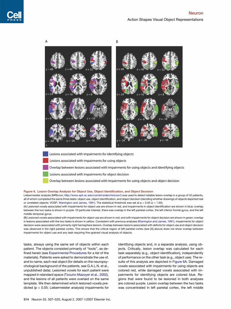
Figure 6. Lesion Overlap Analysis for Object Use, Object Identification, and Object Decision
Liebermeister analysis (MRIcron; http://www.sph.sc.edu/comd/rorden/mricron/) was used to detect reliable lesion overlap in a group of 42 patients,
all of whom completed the same three tasks: object use, object identification, and object decision (deciding whether drawings of objects depicted real
or unrelated objects: VOSP, Warrington and James, 1991). The statistical threshold was set at p < 0.05 (z = 1.65).
(A) Lesioned voxels associated with impairments for object use are shown in red, and impairments in object identification are shown in blue; overlap
between the two tasks is shown in purple. Of particular interest, there was overlap in the left parietal cortex, the left inferior frontal gyrus, and the left
middle temporal gyrus.
(B) Lesioned voxels associated with impairments for object use are shown in red, and with impairments for object decision are shown in green; overlap
in lesions associated with the two tasks is shown in yellow. Consistent with previous analyses (Warrington and James, 1991), impairments for object
decision were associated with primarily right hemisphere lesions. Overlap between lesions associated with deficits for object use and object decision
was observed in the right parietal cortex. This shows that the critical region of left parietal cortex (see [A] above) does not show overlap between
impairments for object use and any task requiring fine-grained visual analysis of objects.
tasks, always using the same set of objects within each
patient. The objects consisted primarily of ‘‘tools’’, as de-
fined herein (see Experimental Procedures for a list of the
materials). Patients were asked to demonstrate the use of,
and to name, each real object (for details on the neuropsy-
chological background of the patients, see G.A.L.N. et al.,
unpublished data). Lesioned voxels for each patient were
mapped in standard space (Tzourio-Mazoyer et al., 2002),
and the lesions of all patients were overlaid on the same
template. We then determined which lesioned voxels pre-
dicted (p < 0.05; Liebermeister analysis) impairments for
514 Neuron 55, 507–520, August 2, 2007 ª2007 Elsevier Inc.
identifying objects and, in a separate analysis, using ob-
jects. Critically, lesion overlap was calculated for each
task separately (e.g., object identification), independently
of performance on the other task (e.g., object use). The re-
sults of this analysis are depicted in Figure 6A. Damaged
voxels associated with impairments for using objects are
colored red, while damaged voxels associated with im-
pairments for identifying objects are colored blue. Re-
gions that were found to be lesioned in both analyses
are colored purple. Lesion overlap between the two tasks
was concentrated in left parietal cortex, the left middle

Neuron
Action Shapes Visual Object Representations
temporal gyrus, and the left inferior frontal gyrus. Notably,
a similar region of the left inferior parietal lobule that
showed RS biased toward ‘‘tools’’ (Figure 5A) in the
fMRI experiment was associated with impaired perfor-
mance in both object use and object identification.
In a second analysis, the 42 patients were separated
into two groups according to whether or not their lesions
involved the parietal cortex (in either the left or the right
hemispheres). We then computed correlations (Pearson),
separately within each group, between performance in us-
ing objects and performance in identifying objects. The re-
sults of this analysis are depicted in Figure 7. There was
a reliable correlation (R2 = 0.595, p < 0.001) only in the
group of patients with lesions involving the parietal cortex
(for the group not having lesions involving the parietal cor-
tex, R2 = 0.013; p = 0.62; see also Figure 7 and Figure S3
for further analyses).
It is important to note that in both groups of patients
(those with and those without parietal cortex lesions) le-
sions to the left middle temporal gyrus were associated
with impairments for object identification and object use.
This is in accord with previous research (e.g., Damasio
et al., 2004; Tranel et al., 1997) that has documented
that lesions to the left middle temporal gyrus are associ-
ated with conceptual impairments for manipulable ob-
jects. The correlational analyses between performance
in object use and object identification indicate that lesions
to the left middle temporal gyrus do not, in and of them-
selves, result in modulation of performance in both tasks.
This is because, while both groups of patients had reliable
lesion overlap in the left middle temporal gyrus, only in the
group of patients with lesions involving the parietal cortex
was there a reliable relation between performance in ob-
ject use and object identification. The implication is that
damage to parietal cortex, in the context of damage to
the left middle temporal gyrus, modulates the relationship
between object use and object identification at the group
level.
Functional Connectivity Analyses of fMRI Data
and Their Relation to Regions of Lesion Overlap
As noted in the Introduction, previous research has de-
scribed anatomical connections between ventral temporal
cortex and both lateral temporal cortex and the inferior pa-
rietal lobule (Rushworth et al., 2006; Saleem et al., 2000;
Webster et al., 1994; Zhong and Rockland, 2003). In order
to provide a more stringent test of the view that neural
specificity in the ventral stream is tied to neural specificity
in the dorsal stream, we carried out functional connectivity
analyses over the BOLD responses in the fMRI experiment
(Gitelman et al., 2003) (see Supplemental Experimental
Procedures for details). The seed voxel for these analyses
was the peak response in the left medial fusiform gyrus for
the contrast of all nonliving object types compared to an-
imals (see Figure 2). In this way, the seed voxel for the
analysis was not defined either in terms of the stimulus
type of interest (‘‘tools’’) or the dependent measure of in-
terest (stimulus-specific RS). We then correlated RS ef-
fects, within stimulus type, between all voxels in the brain
and the seed voxel. This analysis defined regions in the left
middle temporal gyrus and the left inferior parietal lobule
that showed RS for only ‘‘tools’’ (see histograms in Figure 8
for a representation of RS effects by stimulus type in these
regions). This analysis demonstrates functional connectiv-
ity between the left medial fusiform gyrus and the left
middle temporal gyrus, as well as between the left medial
fusiform gyrus and the left inferior parietal lobule.
We then overlaid in standard space the results of this
functional connectivity analysis with the results of the le-
sion analysis described above. As can be seen in Figure 8,
the regions of the left middle temporal gyrus and the left
inferior parietal lobule identified by the functional connec-
tivity analysis corresponded to regions of reliable lesion
overlap independently defined in the neuropsychological
study.
DISCUSSION
Three new findings have been reported:
(1) Using stimulus-specific RS, we found that neural
specificity in the left medial fusiform gyrus tracks
motor-relevant properties of objects. Importantly,
the pattern of RS in the left medial fusiform gyrus
according to motor-relevant properties of objects
cannot be explained by differential similarity in vi-
sual shape between the items within the different
stimulus types (see Figure 1D). A similar pattern of
RS restricted to ‘‘tools’’ was observed in the left
middle temporal gyrus and the left inferior parietal
lobule.
(2) Using the neuropsychological approach, we found
that lesions to the left inferior parietal lobule,
together with lesions to the left middle temporal
gyrus, modulated the relationship between perfor-
mance on object identification and object use at
the group level.
(3) Functional connectivity analyses of the fMRI data
demonstrated that the RS effect for ‘‘tools’’ in the
left medial fusiform gyrus predicted RS effects for
‘‘tools’’ in the left inferior parietal lobule and the
left middle temporal gyrus. The regions of the left
middle temporal gyrus and the left inferior parietal
lobule identified by the functional connectivity anal-
ysis corresponded to the regions independently
identified in the lesion analysis.
On the basis of these data, we suggest that neural spec-
ificity in the left medial fusiform gyrus for ‘‘tools’’ is deter-
mined, in part, by similarity metrics that are computed
over motor-relevant properties of objects. The conver-
gence of the fMRI and neuropsychological data indicate
that the left middle temporal gyrus and the left inferior pa-
rietal lobule represent, at least in part, the similarity met-
rics that determine neural specificity for ‘‘tools’’ in ventral
temporal-occipital cortex. The neural circuit that is
Neuron 55, 507–520, August 2, 2007 ª2007 Elsevier Inc. 515

Neuron
Action Shapes Visual Object Representations
Figure 7. Behavioral Performance of Patients According to Anatomy of Lesions
In a second-level analysis, the 42 patients were separated into two groups according to whether or not their lesions involved parietal cortex (either the
left or right hemispheres). There were 21 patients with lesions involving the parietal cortex (shown in [A]) and 21 whose lesions did not involve the
parietal cortex (shown in [B]). The lesion overlap analyses were recomputed, as described in Figure 6, and we also studied the relationship between
performance in object use and object identification in the two groups.
(A) The behavioral performance in object identification and object use for the group of patients with lesions involving the parietal cortex was correlated
at the group level. As can be seen, there was a strong relationship at the group level between performance profiles in these two tasks (R2 = 0.595,
p < 0.001). For comparison, we carried out the parallel correlational analysis relating performance in object use and object decision; there was no
relationship (R2 = 0.036; p = 0.41). The 95% confidence intervals around the regression line are shown.
(B) There was no relationship between performance in object use and object identification at the group level for patients with lesions that did not
involve parietal cortex (R2 = 0.013; p = 0.62). There was also no relationship between performance in object use and object decision (R2 = 0.016;
p = 0.59). As can be seen in the axial slices, there remained reliable lesion overlap in the left middle temporal gyrus that was associated with joint
impairments for object use and object identification. The 95% confidence intervals around the regression line are shown.
516 Neuron 55, 507–520, August 2, 2007 ª2007 Elsevier Inc.

Neuron
Action Shapes Visual Object Representations
Figure 8. Overlay of Connectivity Analyses and Lesion Overlap Analyses
Functional connectivity analyses were conducted on the event-related fMRI data. The seed voxel for this analysis was the peak response in the left
medial fusiform gyrus showing greater activation for All Nonliving objects compared to animals. Voxels throughout the brain were then identified that
showed a reliable correlation with the seed voxel in terms of stimulus-specific RS, separately for each of the stimulus types. Regions identified by this
analysis included the left middle temporal gyrus (TT:�64,�47, �12) and the left inferior parietal cortex (TT:�51, �34, 36), both of which showed RS
only for ‘‘tools’’ (see Table S3 for all regions identified by this analysis). The histograms represent the strength (t values) of the RS effects in these
regions for each stimulus type. Negative t values indicate that the difference between novel and repeated trials (for example, in the left middle tem-
poral gyrus) was negatively correlated with the corresponding difference in the seed voxel in the left medial fusiform gyrus. The results of the functional
connectivity analysis were then overlaid in Talairach space with the results of the lesion overlap analysis (see text and Figures 6 and 7). The axial slices
(z = 36) in the top row show voxels in parietal cortex associated with impairments for identifying objects (left) and using objects (right). The axial slices
(z = �12) in the bottom row show voxels in temporal cortex associated with impairments for identifying objects (left) and using objects (right). Color
bars indicate z scores for lesion overlap calculated separately for each task. Cross-hairs on all slices show the locations in inferior parietal cortex (top)
and lateral temporal cortex (bottom) that were identified through the functional connectivity analysis on the fMRI data. There is anatomical conver-
gence between the two analyses. Together with the results displayed in Figure 7, these data demonstrate functional interactions between the left
inferior parietal lobule, the left middle temporal gyrus, and the left medial fusiform gyrus.
composed of the left medial fusiform gyrus, the left middle
temporal gyrus, and the left inferior parietal lobule is ‘‘do-
main specific.’’ By ‘‘domain-specific’’ it is meant that the
circuit can be defined with respect to the content of the
object class that is processed, independently of the differ-
ent types of information (i.e., form, motion, action) that are
processed by different components of the circuit (for dis-
cussion, see Caramazza and Mahon, 2006; Martin, 2007).
Relation between Overall BOLD Responses
and Stimulus-Specific RS
An important aspect of the pattern of BOLD responses in
the ventral stream is that they demonstrate a dissociation
between category preferences, as indexed by the ampli-
tude of BOLD responses on novel trials, and stimulus-
specific RS. In the medial fusiform gyri, RS was strongest
for ‘‘tools’’ despite the fact that there was either no differ-
ence on novel trials among the nonliving object types or
that novel nonmanipulable objects elicited greater activa-
tion than ‘‘tools.’’ These findings indicate that the overall
amplitude of BOLD responses cannot be taken, in and
of itself, as an indication of neural specificity.
Previous studies have used overall BOLD responses to
test for dissociations within the medial fusiform gyrus be-
tween different types of nonliving things. In particular,
Downing et al. (2006) tested a wide range of different clas-
ses of nonliving objects and found that the amplitude of
BOLD responses did not distinguish between the different
nonliving stimulus types. On the basis of those data, the
authors concluded that there is neural specificity in the
ventral stream for only faces, places, and body parts.
The pattern of BOLD responses on novel trials that we
Neuron 55, 507–520, August 2, 2007 ª2007 Elsevier Inc. 517
Neuron
Action Shapes Visual Object Representations
have reported (see Figure 2) essentially replicates what
Downing and colleagues reported. However, and as dis-
cussed above, overall BOLD responses cannot be taken,
in and of themselves, as an index of neural specificity.
Thus, contrary to recent arguments (Downing et al.,
2006; Mechelli et al., 2006; Tyler et al., 2003), the findings
reported herein demonstrate neural specificity in the left
medial fusiform gyrus for ‘‘tools.’’
The observation that initial BOLD responses to nonma-
nipulable objects were large in amplitude throughout the
medial fusiform gyrus bilaterally may indicate a high den-
sity of neurons that respond to nonmanipulable objects.
The fact that the same medial fusiform regions show
very little, if any, stimulus-specific RS for nonmanipulable
objects may suggest that there is substantial adaptation of
the BOLD response across different novel presentations
of the nonmanipulable objects. Following this reasoning,
it would follow that neurons in the medial fusiform gyrus
are ‘‘broadly tuned’’ to nonmanipulable objects, whereas
neurons in parahippocampal cortex are ‘‘finely tuned’’ to
nonmanipulable (cf. spatially contextualized) objects.
Similarly, the strong stimulus-specific RS observed for
‘‘tools’’ throughout the left medial fusiform gyrus indicates
a high density of neurons that are finely tuned to this object
type.
Two previous studies used RS to study neural specific-
ity for different types of objects in the ventral stream. Avi-
dan et al. (2002) studied the categories of houses and
faces, while Chao et al. (2002) studied the categories of
‘‘tools’’ and animals. Both studies observed category
preferences, as measured through overall BOLD re-
sponses, with nonliving and living things differentially acti-
vating distinct regions of ventral temporal-occipital cortex.
However, in each study, equivalent RS effects were ob-
served for the preferred as well as the nonpreferred cat-
egory. One possible reason why those studies did not
observe modulation of RS effects by stimulus type is
because both studies used designs in which either all re-
peated or all novel stimuli were presented within a block.
Such a design may lead to stimulus nonspecific modula-
tions in attention (e.g., Wojciulik et al., 1998) that mask
biases in RS by stimulus type.
Conclusion
Clearly, a single dimension is not sufficient to explain all
aspects of the organization of the ventral object-process-
ing stream (e.g., Haxby et al., 2001; Levy et al., 2001).
However, given the pattern of findings reported herein,
one may speculate as to whether neural specificity in the
ventral stream for other types of stimuli, such as faces
and animals, words, and places, may also depend in
part on functional connectivity with neural systems out-
side (or ‘‘downstream’’) from ventral temporal-occipital
cortex (Caramazza and Mahon, 2006). For instance,
neural specificity for faces in the lateral fusiform gyrus (Fu-
siform Face Area) may be driven, in part, by functional
connectivity between this region and regions of the brain
mediating affective reactions, such as the amygdala
518 Neuron 55, 507–520, August 2, 2007 ª2007 Elsevier Inc.
(e.g., Kreiman et al., 2000). Similarly, it may be reasonable
to speculate that neural specificity for written language in
the visual word form area may be driven in part by
functional connectivity with regions of the brain mediating
language processing (Martin, 2006). An important issue for
future research is thus whether functional connectivity be-
tween the ventral stream and structures outside of the
ventral stream is a general organizing principle giving
rise to category specificity in the primate visual system.
EXPERIMENTAL PROCEDURES
fMRI Experimental Stimuli and Design
There were 20 distinct items within each of the four stimulus types, and
four exemplars of each item (total n = 320). The 20 items within each
stimulus type were divided into two sets, A and B. Thus, each set within
a stimulus type had 40 different pictures (four exemplars each of ten
different items). A given participant (both for the fMRI study and the be-
havioral study on naming latencies) either saw set A three times and
Set B once, or the reverse. The factor Set (A and B) was counterbal-
anced across participants so that, across all participants, the same
materials appeared as novel and repeated trials. This design resulted
in 640 trials for each subject (e.g., [Set A: 40 3 3 Presentations 3 4
Stimulus Types] + [Set B: 40 3 1 Presentation 3 4 Stimulus types]). Af-
ter collapsing the first presentations of stimuli from sets A and B into
the ‘‘novel’’ condition, and the second and third presentations into
the ‘‘repeated condition,’’ there were 80 novel trials per stimulus type
and 80 repeated trials per stimulus type. Stimuli from all four stimulus
types, for both novel and repeated events, were intermixed throughout
the entire experiment (see Supplemental Experimental Procedures).
MR Data Collection Parameters
Seventeen subjects (ten female) were recruited from the NIH Healthy
Volunteer pool and paid for their participation in the study. Informed
consent was obtained in writing under an approved National Institute
of Mental Health protocol. fMRI data were collected at the NIH Clinical
Center NMR Research Facility on a GE Signa 3 Tesla scanner using
standard imaging procedures. Before collecting experimental data,
a high-resolution SPGR anatomical sequence (124 axial slices, 1.2 mm
thickness, FOV = 24 cm, Acquisition matrix = 256 3 256) was performed.
Data were collected using an echo-planar series, with a TR of 2000 ms,
and a TE of 30 ms with 3.75 mm in-plane resolution. Volumes were
collected in the axial plane in 24 contiguous, interleaved slices.
fMRI Data Analysis
All MR data were analyzed using the AFNI software package (Cox,
1996). To account for motion artifacts, all scan series were registered
to the volume acquired closest to the high-resolution anatomy. Then
a spatial filter with a 4.5 mm full-width half-maximum Gaussian filter
was applied to each volume. For each individual subject, echo-planar
and anatomical volumes were transformed into the standardized
Talairach and Tournoux (TT) volume (Talairach and Tournoux, 1988).
A random-effects approach within the general linear model in AFNI
was used to analyze the data. The response to each stimulus type
and repetition compared to the fixation baseline was calculated using
multiple regression. There were eight regressors of interest: two for
each of the four stimulus types to represent the novel and repeated
stimuli, and six regressors of no interest (six outputs from volume
registration to account for residual variance from subject motion not
corrected by registration). Within each regressor of interest, delta func-
tions representing the response following stimulus presentation at
each full volume echo-planar acquisition (2 s) in a 12 s window were
fit to the MR signal, resulting in an estimate of the response to a single
stimulus of each stimulus type and repetition with no assumptions

Neuron
Action Shapes Visual Object Representations
about the shape of the hemodynamic response. This resulted in a 12 s
time series for each stimulus type in each voxel.
The amplitude of the response to the stimulus was estimated by
summing the b weights of the regressors representing TRs 2 through
4. The regression model of the estimated amplitude provided a single
estimate of the response to each stimulus type in each voxel for each
subject. A two-way mixed-effects ANOVA was performed on each
voxel in standardized space. Fixed-effects contrasts of the stimulus
and repetition variables were performed, with subjects acting as the
random-effect repeated measure.
Analysis of RS Effects Biased toward Each Stimulus Type
Analyses were performed to isolate voxels showing RS biased toward
each stimulus type (Figure 3B and Figure S2). First, mask files of the RS
effect (novel > repeated) were created for each of the four stimulus
types. Next, the four masks were overlaid to create a color map illus-
trating areas where RS was significant for only a single stimulus
type, as well as regions of overlap for ‘‘tools’’ and arbitrarily manipu-
lated objects.
All TT coordinates reported in the manuscript refer to the location of
the voxel showing the greatest difference for that given contrast.
Lesion Overlap Study
Forty-two consecutive patients with unilateral strokes were recruited
from the rehabilitation ward of the Ospedali Riuniti in Trieste (age:
mean = 64.2; standard deviation [SD] = 11.0 years; education: mean =
9.4; SD = 3.7 years). Only patients with no previous neurological history
and who had CT or MRI scans available were included. The lesions in
each patient were mapped into standard space using MRIcro by a neu-
roradiologist (M.U.). Twenty-five neurologically healthy individuals
matched for age and education (age: mean = 66; SD = 11; education:
mean = 8.96; SD = 4.1) with the patient group were recruited from pa-
tients’ and staff’s relatives, as well as from the rehabilitation ward of the
Ospedali Riuniti in Trieste, where they were treated following orthope-
dic surgery. The Revised Standardized Difference Test (RSDT) was
used to detect impaired performance for single patients against con-
trols (Crawford and Garthwaite, 2006). The scatter plots in Figure 7
plot modified t values as calculated with the procedure described in
Crawford and Garthwaite (2006). Thirty-five of the patients completed
the object identification and object use task using the same set of 29
real objects (bottle, cigarette, coffee mug, comb, dust cloth, eraser,
fork, glass, gun, hammer, iron, jug, key, knife, ladle, lemon squeezer,
light bulb, lipstick, match stick, paintbrush, pen, razor, saw, scissors,
screwdriver, wrench, spoon, tennis racket, tooth brush). Seven pa-
tients completed the same tasks with a subset (n = 20) of those objects
(excluding: bottle, dust cloth, fork, glass, knife, ladle, match stick,
racket, tooth brush). Patients were compared to controls taking into
account only the items in common to patients and controls (control
performance for all 29 items: naming, mean = 99.2; standard deviation
(SD) = 1.5; object use, mean = 96.3; SD = 4.4; control performance for
20 items: naming: mean = 99.6; SD = 1.4; object use, mean = 96.5;
SD = 4.6). No feedback (either positive or negative) was given to either
patients or controls during testing. For further details of the testing
procedures, see Supplemental Experimental Procedures; G.A.L.N.,
R.I.R., A. Zadini, M. Ukmar, B.Z.M., and A.C., unpublished data.
Supplemental Data
The Supplemental Data for this article can be found online at http://
www.neuron.org/cgi/content/full/55/3/507/DC1/.
ACKNOWLEDGMENTS
The fMRI study was supported by the National Institute of Mental
Health, Division of Intramural Research. Preparation of this manuscript
was supported by an NSF Graduate Research Fellowship to B.Z.M.
and NIH grant DC0542 to A.C. We would like to thank Jessica Gilbert
and Kim Vargas for their help in collecting data; Maja Ukmar for assis-
tance in mapping the lesions of the patients; Sabrina Aristei for assis-
tance with the analysis of shape similarity; and Gabriele Miceli, Jens
Schwarzbach, and Massimo Turatto for their comments on earlier
drafts of this manuscript. We are grateful to Antonella Zadini for refer-
ring the patients.
Received: January 31, 2007
Revised: May 30, 2007
Accepted: July 13, 2007
Published: August 1, 2007
REFERENCES
Avidan, G., Hasson, R., Hendler, T., Zohary, E., and Malach, R. (2002).
Analysis of the neuronal selectivity underlying low fMRI signals. Curr.
Biol. 12, 964–972.
Barr, M., and Aminoff, E. (2003). Cortical analysis of visual context.
Neuron 38, 347–358.
Beauchamp, M.S., Lee, K.E., Haxby, J.V., and Martin, A. (2002). Paral-
lel visual motion processing streams for manipulable objects and
human movements. Neuron 34, 149–159.
Beauchamp, M.S., Lee, K.E., Haxby, J.V., and Martin, A. (2003). fMRI
responses to video and point-light displays of moving humans and
manipulable objects. J. Cogn. Neurosci. 15, 991–1001.
Belongie, S., Malik, J., and Puzicha, J. (2002). Shape matching and ob-
ject recognition using shape contexts. IEEE Transactions on Pattern
Analysis and Machine Intelligence 24, 509–522.
Caramazza, A., and Mahon, B.Z. (2006). The organisation of concep-
tual knowledge in the brain: The future’s past and some future direc-
tions. Cogn. Neuropsychol. 23, 13–38.
Chao, L.L., and Martin, A. (2000). Representation of manipulable man-
made objects in the dorsal stream. Neuroimage 12, 478–484.
Chao, L.L., Martin, A., and Haxby, J.V. (1999a). Are face-responsive
regions selective only for faces? Neuroreport 10, 2945–2950.
Chao, L.L., Haxby, J.V., and Martin, A. (1999b). Attribute-based neural
substrates in posterior temporal cortex for perceiving and knowing
about objects. Nat. Neurosci. 2, 913–919.
Chao, L.L., Weisberg, J., and Martin, A. (2002). Experience dependent
modulation of category-related cortical activity. Cereb. Cortex 12,
545–551.
Cox, R.W. (1996). AFNI: Software for analysis and visualization of func-
tional magnetic resonance neuroimages. Comput. Biomed. Res. 29,
162–173.
Crawford, J.R., and Garthwaite, P.H. (2006). Methods of testing for
a deficit in single case studies: Evaluation of statistical power by Monte
Carlo simulation. Cogn. Neuropsych. 23, 877–904.
Culham, J.C., Danckert, S.L., DeSouza, J.F., Gati, J.S., Menon, R.S.,
and Goodale, M.A. (2003). Visually guided grasping produces fMRI ac-
tivation in dorsal but not ventral stream brain areas. Exp. Brain Res.
153, 180–189.
Damasio, H., Tranel, D., Grabowski, T., Adolphs, R., and Damasio, A.
(2004). Neural systems behind word and concept retrieval. Cognition
92, 179–229.
Dobbins, I.G., Schnyer, D.M., Verfaellie, M., and Schacter, D.L. (2004).
Cortical activity reductions during repetition priming can result from
rapid response learning. Nature 428, 316–319.
Downing, P.E., Chan, A.W.-Y., Peelen, M.V., Dodds, C.M., and Kanw-
isher, N. (2006). Domain specificity in visual cortex. Cereb. Cortex 16,
1453–1461.
Epstein, R., Harris, A., Stanley, D., and Kanwisher, N. (1999). The para-
hippocampal place area: recognition, navigation, or encoding? Neuron
23, 115–125.
Neuron 55, 507–520, August 2, 2007 ª2007 Elsevier Inc. 519

Neuron
Action Shapes Visual Object Representations
Fang, F., and He, S. (2005). Cortical responses to invisible objects in
the human dorsal and ventral pathways. Nat. Neurosci. 8, 1380–1385.
Frey, S.H., Vinton, D., Norlund, R., and Grafton, S.T. (2005). Cortical to-
pography of human anterior intraparietal cortex active during visually
guided grasping. Brain Res. Cogn. Brain Res. 23, 397–405.
Gitelman, D.R., Penny, W.D., Ashburner, J., and Friston, K.J. (2003).
Modeling regional and psychophysiologic interactions in fMRI: the im-
portance of hemodynamic deconvolution. Neuroimage 19, 200–207.
Goodale, M.A., and Milner, A.D. (1992). Separate visual pathways for
perception and action. Trends Neurosci. 15, 20–25.
Grill-Spector, K., Henson, R., and Martin, A. (2006). Repetition and the
brain: neural models of stimulus-specific effects. Trends Cogn. Sci. 10,
14–23.
Haxby, J.V., Gobbini, M.I., Furey, M.L., Ishai, A., Schouten, J.L., and
Pietrini, P. (2001). Distributed and overlapping representations of faces
and objects in ventral temporal cortex. Science 293, 2425–2430.
Heilman, K.M., Rothi, L.J., and Valenstein, E. (1982). Two forms of
ideomotor apraxia. Neurology 32, 342–346.
James, T.W., Humphrey, G.K., Gati, J.S., Menon, R.S., and Goodale,
M.A. (2002). Differential effects of viewpoint on object-driven activation
in dorsal and ventral streams. Neuron 35, 793–801.
Johnson-Frey, S.H. (2004). The neural basis of complex tool use in hu-
mans. Trends Cogn. Sci. 8, 71–78.
Johnson-Frey, S.H., Newman-Norlund, R., and Grafton, S.T. (2005). A
distributed left hemisphere network active during planning of everyday
tool use skills. Cereb. Cortex 15, 681–695.
Kable, J.W., Kan, I.P., Wilson, A., Thompson-Schill, S.L., and Chatter-
jee, A. (2002). Conceptual representations of action in the lateral tem-
poral cortex. J. Cogn. Neurosci. 17, 1855–1870.
Kanwisher, N., Stanley, D., and Harris, A. (1999). The fusiform face area
is selective for faces not animals. Neuroreport 10, 183–187.
Kreiman, G., Koch, C., and Fried, I. (2000). Category-specific visual
responses of single neurons in the human medial temporal lobe.
Nat. Neurosci. 3, 946–953.
Levy, I., Hasson, U., Avidan, G., Hendler, T., and Malach, R. (2001).
Center-periphery organization of human object areas. Nat. Neurosci.
4, 533–539.
Lewis, J.W. (2006). Cortical networks related to human use of tools.
Neuroscientist 12, 211–231.
Mahon, B.Z., and Caramazza, A. (2005). The orchestration of the sen-
sory-motor systems: Clues from neuropsychology. Cogn. Neuropsy-
chol. 22, 480–494.
Martin, A. (2006). Shades of Dejerine—Forging a causal link between
the visual word form area and reading. Neuron 50, 173–175.
Martin, A. (2007). The representation of object concepts in the brain.
Annu. Rev. Psychol. 58, 25–45.
Martin, A., Haxby, J.V., Lalonde, F.M., Wiggs, C.L., and Ungerleider,
L.G. (1995). Discrete cortical regions associated with knowledge of
color and knowledge of action. Science 270, 102–105.
Mechelli, A., Sartori, G., Orlandi, P., and Price, C.J. (2006). Semantic
relevance explains category effects in medial fusiform gyri. Neuro-
image 3, 992–1002.
Miceli, G., Fouch, E., Capasso, R., Shelton, J.R., Tomaiuolo, F., and
Caramazza, A. (2001). The dissociation of color from form and function
knowledge. Nat. Neurosci. 4, 662–667.
Middleton, F.A., and Strick, P.L. (1996). The temporal lobe is a target of
output from the basal ganglia. Proc. Natl. Acad. Sci. USA 93, 8683–
8687.
Miller, E.K., Nieder, A., Freedman, D.J., and Wallis, J.D. (2003). Neural
correlates of categories and concepts. Curr. Opin. Neurobiol. 13, 198–
203.
520 Neuron 55, 507–520, August 2, 2007 ª2007 Elsevier Inc.
Noppeney, U., Price, C.J., Penny, W.D., and Friston, K.J. (2006). Two
distinct neural mechanisms for category-selective responses. Cereb.
Cortex 16, 437–445.
Okada, T., Tanakac, S., Nakaid, T., Nishizawa, S., Inuic, T., Sadatob,
N., Yonekurae, Y., and Konishia, J. (2000). Naming of animals and
tools: a functional magnetic resonance imaging study of categorical
differences in the human brain areas commonly used for naming visu-
ally presented objects. Neurosci. Lett. 296, 33–36.
Op de Beeck, H., Wagemans, J., and Vogels, R. (2001). Inferotemporal
neurons represent low-dimensional configurations of parameterized
shapes. Nat. Neurosci. 4, 1244–1252.
Pisella, L., Binkofski, F., Lasek, K., Toni, I., and Rossetti, Y. (2006). No
double-dissociation between optic ataxia and visual agnosia: Multiple
sub-streams for multiple visuo-manual integrations. Neuropsycholo-
gia 44, 2734–2748.
Rizzolatti, G., and Luppino, G. (2001). The cortical motor system. Neu-
ron 31, 889–901.
Rumiati, R.I., Weiss, P.H., Shallice, T., Ottoboni, G., Noth, J., Zilles, K.,
and Fink, G.R. (2004). Neural basis of pantomiming the use of visually
presented objects. Neuroimage 21, 1224–1231.
Rushworth, M.F.S., Behrens, T.E.J., and Johansen-Berg, H. (2006).
Connection patterns distinguish 3 regions of human parietal cortex.
Cereb. Cortex 16, 1418–1430.
Saleem, K.S., Suzuki, W., Tanaka, K., and Hashikawa, T. (2000). Con-
nections between anterior inferotemporal cortex and superior tempo-
ral sulcus regions in the macaque monkey. J. Neurosci. 20, 5083–
5101.
Shmuelof, L., and Zohary, E. (2005). Dissociation between ventral and
dorsal fMRI activation during object action recognition. Neuron 47,
457–470.
Talairach, J., and Tournoux, P. (1988). Co-Planar Stereotaxic Atlas of
the Human Brain (New York: Thieme).
Tranel, D., Damasio, H., and Damasio, A.R. (1997). A neural basis for
the retrieval of conceptual knowledge. Neuropsychologia 35, 1319–
1327.
Tyler, L.K., Bright, P., Dick, E., Tavares, P., Pilgrim, L., Fletcher, P.,
Greer, M., and Moss, H. (2003). Do semantic categories activate dis-
tinct cortical regions? Evidence for a distributed neural semantic sys-
tem. Cogn. Neuropsychol. 20, 541–559.
Tzourio-Mazoyer, N., Landeau, B., Papathanassiou, D., Crivello, F.,
Etard, O., Delcroix, N., Mazoyer, B., and Joliot, M. (2002). Automated
anatomical labeling of activations in SPM using a macroscopic ana-
tomical parcellation of the MNI MRI single-subject brain. Neuroimage
15, 273–289.
Ungerleider, L.G. (1995). Functional brain imaging studies of cortical
mechanisms for memory. Science 270, 769–775.
Ungerleider, L.G., and Mishkin, M. (1982). Two cortical visual systems.
In Analysis of Visual Behavior, D.J. Ingle, M.A. Goodale, and R.J.W.
Mansfield, eds. (Cambridge, MA: The MIT Press), pp. 549–586.
Warrington, E.K., and James, M. (1991). The Visual Object and Space
Perception Battery (Bury St Edmunds, UK: Thames Valley Test Com-
pany).
Webster, M.J., Bachevalier, J., and Ungerleider, L.G. (1994). Connec-
tions of inferior temporal areas TEO and TE with parietal and frontal
cortex in macaque monkeys. Cereb. Cortex 4, 470–483.
Wojciulik, E., Kanwisher, N., and Driver, J. (1998). Covert visual atten-
tion modulates face-specific activity in the human fusiform gyrus: fMRI
study. J. Neurophysiol. 79, 1574–1578.
Zhong, Y.-M., and Rockland, K.S. (2003). Inferior parietal lobule pro-
jections to anterior inferiortemporal cortex (area TE) in Macaque mon-
key. Cereb. Cortex 13, 527–540.