Embed Size (px)
Citation preview

Nervous System

Overview
• 2 ways body communicates to send signals.
• 1st - endocrine system - hormones.
• 2nd - nervous system - no chemical signaling – impulses - travel through network of cells to get to destination.
QuickTime™ and a decompressor
are needed to see this picture.

http://www.drstandley.com/images/nervous5.bmp

• Impulses allow organisms to receive and respond to stimuli in environment.
• Controls all functions of life - ability to move, think, breathe, etc.
QuickTime™ and a decompressor
are needed to see this picture.
http://www.medicalook.com/systems_images/Autonomic_Nervous_System.jpg

Functional unit
• Functional unit of nervous system - neurons.
• Specialized cell designed to transmit electrochemical signals called action potentials (nerve impulses).
• Signals formed by altering of voltage across plasma membrane.
QuickTime™ and a decompressor
are needed to see this picture.


• Basic part of neuron’s structure - cell body, dendrites, axon.
• Cell body contains nucleus, most organelles - site of protein synthesis.
• Dendrites project from body and receive chemical information from other neurons; carry this information to cell body.
QuickTime™ and a decompressor
are needed to see this picture.

http://www.nida.nih.gov/JSP/MOD3/images/NEURON2.gif

• Axon - projection of neuron that transmits information to cell body to target cells.
• Has to be long enough to carry action potential from central nervous system to extremities.
QuickTime™ and a decompressorare needed to see this picture.

Copyright © 2002 Pearson Education, Inc., publishing as Benjamin Cummings
Fig. 48.2
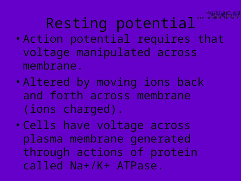
Resting potential• Action potential requires that
voltage manipulated across membrane.
• Altered by moving ions back and forth across membrane (ions charged).
• Cells have voltage across plasma membrane generated through actions of protein called Na+/K+ ATPase.
QuickTime™ and a decompressor
are needed to see this picture.

http://openwetware.org/images/thumb/a/a6/Action-potential.jpg/300px-Action-potential.jpg.png

• Hydrolysis of ATP for energy - protein pumps sodium ions out of cell and potassium into cell.
• Activity essential in maintaining osmotic balance of cells.
• Some potassium leaks back out of cell through potassium channel.
QuickTime™ and a decompressor
are needed to see this picture.

http://scienceblogs.com/clock/upload/2006/08/Potassium-Channel-2-2004.JPG

• Potassium channel is ion channel that selectively allows potassium ions to flow down K+ gradient established by ATPase.
• Resting potential about -70mVolts - most positive ions on outside of cell.
QuickTime™ and a decompressor
are needed to see this picture.


Action potential• Most cells maintain membrane
potential at resting potential.• Membrane excited - potential
changed - allows information to be carried via action potential.
• Neurons, muscle cells have ion channel proteins in plasma membrane that open to allow ions through.
QuickTime™ and a decompressor
are needed to see this picture.

http://www.unm.edu/~lkravitz/MEDIA2/Action%20Potential.jpg

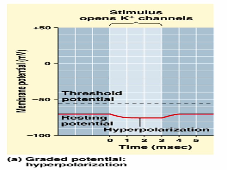
• Happens in response to decrease in membrane potential.
• Protein that does this in neurons - voltage-gated sodium channel.
• When membrane potential more negative than resting potential, (from -70mVolts to -90mVolts), membrane hyperpolarized.
QuickTime™ and a decompressor
are needed to see this picture.

http://courses.washington.edu/conj/membrane/chan.gif

• When membrane potential less negative than usual, (-70mVolts to -50mVolts or 0mVolts) membrane depolarized.
• Voltage-gated sodium channels closed at resting potential - do not let ions through membrane.
• Change in membrane potential causes voltage-gated sodium channels to open to allow sodium through. QuickTime™ and a
decompressorare needed to see this picture.


• If membrane voltage becomes less negative than resting potential (-70mVolts to -50mVolts) voltage-gated sodium channels in membrane will open.
• Voltage at which voltage-gated channels open - threshold potential.
QuickTime™ and a decompressor
are needed to see this picture.

http://openwetware.org/images/thumb/a/a6/Action-potential.jpg/300px-Action-potential.jpg.png

• When channels open, sodium diffuses freely through channel to cross membrane from outside of cell into cytoplasm.
• Opening of channels in one region of membrane, entry of sodium through channels causes membrane depolarization (membrane less polarized, moving toward 0)
QuickTime™ and a decompressor
are needed to see this picture.

QuickTime™ and a decompressor
are needed to see this picture.

• After voltage-gated sodium channels opened and depolarization complete, channels close rapidly again, allowing membrane voltage to normal potential (millisecond).
• Return of voltage to normal negative state - repolarization.
• Section of membrane depolarizes, triggers threshold for voltage-gated channels in next section of membrane to depolarize.
QuickTime™ and a decompressor
are needed to see this picture.
QuickTime™ and a decompressor
are needed to see this picture.
http://www.cidpusa.org/nervous_repolarization.gif

• Action potential moves along length of axon in wavelike manner until it reaches end of neuron at synapse.
• Some vertebrate – neurons have myelin - surrounds axon, allows action potentials to travel more quickly.
QuickTime™ and a decompressor
are needed to see this picture.

QuickTime™ and a decompressor
are needed to see this picture.

• Myelin formed by glial cells - wrap plasma membrane around axons, insulating it.
• Small spaces between myelin - nodes of Ranvier.
• Myelinated neurons - action potential jumps from 1 node to another node bypassing insulated myelin regions where no ions cross membrane.
QuickTime™ and a decompressor
are needed to see this picture.

QuickTime™ and a decompressor
are needed to see this picture.
http://butler.cc.tut.fi/~malmivuo/bem/bembook/02/fi/0201.gif

• Allows action potential to travel much quicker - can jump forward instead of traveling whole length of axon - saltatory conduction.
• Larger neurons carry action potentials more quickly.
QuickTime™ and a decompressorare needed to see this picture.

Copyright © 2002 Pearson Education, Inc., publishing as Benjamin Cummings


Size and frequency of action potentials
• Every action potential in neuron - same size.
• Once membrane reaches threshold for depolarization - fully depolarize - all-or-nothing response.
• Either neuron fires an action potential or it doesn’t. QuickTime™ and a
decompressorare needed to see this picture.

QuickTime™ and a decompressor
are needed to see this picture.
http://education.vetmed.vt.edu/Curriculum/VM8054/Labs/Lab10/IMAGES/MOTOR%20END%20PLATES%20SMALL%201.jpg

• Strength of stimulus does not change size of action potential depolarization or duration of depolarization.
• Strength of stimulus determines intensity of action potential.
• Light touch will trigger less frequent response. QuickTime™ and a
decompressorare needed to see this picture.

QuickTime™ and a decompressor
are needed to see this picture.
http://www.unmc.edu/physiology/Mann/pix_3/f3-17.gif

• Action potential same in strength but frequency of action potentials change with intensity of stimulus.
• When action potential passes through section of membrane - cannot carry action potential again immediately.
• Must first finish depolarizing, then repolarize. QuickTime™ and a
decompressorare needed to see this picture.

QuickTime™ and a decompressor
are needed to see this picture.
http://www.gregalo.com/action_potential.jpg

• Limit to frequency of action potential firing in neuron - refractory period.
• Places upper limit on number of action potentials that can pass through neuron in unit of time.
• Action potentials carried in one direction, from cell body to end of axon.
QuickTime™ and a decompressorare needed to see this picture.

QuickTime™ and a decompressor
are needed to see this picture.

The synapse
• When action potential reaches end of neuron - neurotransmitters released to communicate with next cell across small gap between cells - synapse.
• 2 types of synapses, chemical and electrical.
QuickTime™ and a decompressor
are needed to see this picture.

QuickTime™ and a decompressor
are needed to see this picture.
http://universe-review.ca/I10-40-synapse.jpg

• At chemical synapse - action potential reaches end of axon, comes in contact with rounded terminal filled with vesicles - have neurotransmitters in them.
• Include seratonin, dopamine, acetylcholine, and glutamate.
QuickTime™ and a decompressor
are needed to see this picture.

QuickTime™ and a decompressor
are needed to see this picture.

• When action potential signals it, voltage-gates calcium channels open and allow calcium into cell.
• Calcium causes some vesicles to fuse with plasma membrane and release contents into synapse.
• Synapse and target cell together - synaptic cleft.
QuickTime™ and a decompressor
are needed to see this picture.

QuickTime™ and a decompressor
are needed to see this picture.
http://findlaw.doereport.com/imagescooked/2728W.jpg

• Neurotransmitter diffuses across synaptic cleft and binds to receptors on target cell plasma membrane.
• When bound, receptors will open ion channels - allows specific ions through membrane in response to neurotransmitter.
• Ions cause response in postsynaptic cell (cell after synapse)
QuickTime™ and a decompressor
are needed to see this picture.

Copyright © 2002 Pearson Education, Inc., publishing as Benjamin Cummings
Fig. 48.12

• Each neurotransmitter has specific receptors that it interacts with at synapse; each receptor opens channel that allows specific ion through.
• Excitatory neurotransmitter binds to receptor that depolarizes membrane of postsynaptic cell.
QuickTime™ and a decompressor
are needed to see this picture.

QuickTime™ and a decompressor
are needed to see this picture.
http://www.chemistryexplained.com/images/chfa_03_img0602.jpg

• Acetylcholine used with skeletal muscle, diffuses across synaptic cleft - binds to receptors for it.
• Receptors - ligand-gated ion channels that bind Ach - open to allow sodium ions to diffuse into cell.
• When sodium enters - depolarizes plasma membrane of target muscle cell.
QuickTime™ and a decompressor
are needed to see this picture.

QuickTime™ and a decompressor
are needed to see this picture.
http://thebrain.mcgill.ca/flash/d/d_06/d_06_m/d_06_m_mou/d_06_m_mou_2a.jpg

• If depolarization of target reaches threshold - action potential will be initiated in muscle cell membrane by voltage-gated channels, and will be sent throughout muscle cell membrane, which triggers muscle cell to contract.
QuickTime™ and a decompressor
are needed to see this picture.

QuickTime™ and a decompressor
are needed to see this picture.
http://www.blackwellpublishing.com/korfgenetics/jpg/300_96dpi/Fig16-2.jpg

• More action potentials that reach muscle and more muscle cells involved, stronger muscle contraction.
• Neurotransmitter can bind to receptor -opens to allow chloride to enter postsynaptic membrane causing hyperpolarization.
• Membrane potential moves away from threshold for triggering an action potential.
QuickTime™ and a decompressor
are needed to see this picture.

QuickTime™ and a decompressor
are needed to see this picture.
http://www.gregalo.com/action_potential.jpg

• If neurotransmitter inhibits - more difficult for action potential to start in target cell.
• Most common - GABA.• Neuron can form synapses with
many neurons; release neurotransmitter to alter membrane potential of target cell.
QuickTime™ and a decompressor
are needed to see this picture.

QuickTime™ and a decompressor
are needed to see this picture.
http://huguenard-lab.stanford.edu/beta3/gaba.jpg

• Information from all synapses neuron interacts with combined in cell body of neuron in summation - single neuron processes information from all of its stimulating neurons.
• Then decides whether or not to initiate an action potential itself.QuickTime™ and a decompressorare needed to see this picture.

QuickTime™ and a decompressor
are needed to see this picture.
http://www.biologymad.com/NervousSystem/summation.jpg

• If changes in potential of neuron cause it to reach threshold depolarization to open voltage-gated channels, it will fire an action potential.
• If not, neuron will not fire an action potential.
QuickTime™ and a decompressor
are needed to see this picture.

QuickTime™ and a decompressor
are needed to see this picture.
http://www.unm.edu/~lkravitz/MEDIA2/Action%20Potential.jpg

• To turn off signal - • Once neurotransmitter released
into synaptic cleft, continues to bind to postsynaptic receptors unless removed from synapse.
• One way - neurotransmitter to diffuse into surrounding fluid.
QuickTime™ and a decompressor
are needed to see this picture.

QuickTime™ and a decompressor
are needed to see this picture.
http://www.elmhurst.edu/~chm/vchembook/images2/661synapse.gif

• Another way - enzyme that degrades neurotransmitter.
• Acetylocholinesterase acts on acetylcholine to inactivate it.
• Pesticides, nerve gas inactivate this enzyme.
• 3rd way - take neurotransmitter back up into cells at synapse.
• Happens with norepinephrine,seratonin.
QuickTime™ and a decompressor
are needed to see this picture.

QuickTime™ and a decompressor
are needed to see this picture.

Organization of nervous system
• As complexity of organism increases, complexity of nervous system also increases.
• Simple organisms can respond to simple stimuli, more complex organisms can discern stimuli (i.e.shades of color)
QuickTime™ and a decompressor
are needed to see this picture.

QuickTime™ and a decompressor
are needed to see this picture.

Invertebrate nervous systems• Protozoa - single celled, no
nervous system.• Receptors that respond to stimuli
(heat, light, chemicals).• Sponges - multicellular - have
almost no response to environment, no nerves.
• Cnidarians - network of cells - nerve net, located between inner and outer layers of cells of bodies.
QuickTime™ and a decompressor
are needed to see this picture.

• Annelids - primitive nervous system consisting of ventral nerve cord and anterior brain of fused ganglia.
• Arthropods - better developed nervous system - specialized sense organs, including sight and hearing organs.
QuickTime™ and a decompressor
are needed to see this picture.


Human nervous system• Central nervous system - brain,
spinal cord.• Brain contains all functions
beyond simple reflexes - consists of outer portion containing neuronal cell bodies (gray matter), inner portion containing axons (white matter).
• CNS processes information, sends response out to body through neurons.
QuickTime™ and a decompressor
are needed to see this picture.

QuickTime™ and a decompressor
are needed to see this picture.
http://www.medem.com/MEDEM/images/ama/ama_brain_stroke_lev20_thebraineffectsstroke_01.gif

Human brain• 1Cerebral cortex – all voluntary motor
activity - initiates responses of motor neurons present within spinal cord.
• Controls higher functions (memory, creative thought).
• Cortex divided into hemispheres (left and right), with some specialization of function between them.
QuickTime™ and a decompressor
are needed to see this picture.

QuickTime™ and a decompressor
are needed to see this picture.
QuickTime™ and a decompressor
are needed to see this picture.
http://www.morphonix.com/software/education/science/brain/game/specimens/images/cerebral_cortex.gif

• 2Olfactory lobe – center of reception and integration of olfactory input.
• 3Thalamus – nervous impulses and sensory information relayed and integrated in this section as impulse travels to and from cerebral cortex.
• 4Hypothalamus – hunger, thirst, pain, temperature regulation, water balance controlled here.
QuickTime™ and a decompressorare needed to see this picture.

QuickTime™ and a decompressor
are needed to see this picture.

• 5Cerebellum – muscle activity coordinated, modulated.
• 6Pons –relay center for cortical fibers on their way to cerebellum.
• 7Medulla oblongata –controls vital physiological functions - breathing, heart rate, gastrointestinal activity - has receptors for CO2 levels.
QuickTime™ and a decompressorare needed to see this picture.

Copyright © 2002 Pearson Education, Inc., publishing as Benjamin Cummings
Fig. 48.20

• Spinal cord is part of central nervous system.
• Route axons to travel out of brain.• Serves as center for reflex actions
- do not involve brain.• Dorsal horn of spinal cord is
entrance point for sensory nerve fibers whose cell bodies are contained within dorsal root ganglion.
QuickTime™ and a decompressor
are needed to see this picture.

QuickTime™ and a decompressor
are needed to see this picture.
http://www.futuremedicalsupply.com/scipages/_images/content/spine/spine%252Bnerves.jpg

• Ventral horn contains cell bodies of motor neurons - initiate muscular contractions.
• Lower sections of brain perform more primitive functions (spinal cord, medulla, cerebellum); forebrain and cortex more advanced.
• Cortex and forebrain important in evolution of vertebrates.
QuickTime™ and a decompressorare needed to see this picture.

QuickTime™ and a decompressor
are needed to see this picture.
http://www.cartage.org.lb/en/kids/science/Biology%20Cells/Nervous%20System/Explore%20the%20Nervous%20System/Spinal%20Cord/Segments%20Spinal%20Cord/vert3.gif

Peripheral nervous system
• Carries nerves from CNS to target tissues in body.
• 12 cranial nerves (head and shoulders), 31 spinal nerves (rest of body).
• Cranial nerves exit from brainstem, spinal nerves exit from spinal cord.
• 2 divisions: somatic and autonomic.
QuickTime™ and a decompressor
are needed to see this picture.

QuickTime™ and a decompressor
are needed to see this picture.
http://www.web-books.com/eLibrary/Medicine/Physiology/Nervous/cranial_nerves.jpg

Somatic nervous system• Innervates skeletal muscle,
responsible for voluntary movement.
• Motor neurons release acetylcholine (neurotransmitter - ACh) onto ACh receptors in skeletal muscle.
• Causes depolarization of skeletal muscle leading to muscle contraction. QuickTime™ and a decompressorare needed to see this picture.

QuickTime™ and a decompressor
are needed to see this picture.
http://thebrain.mcgill.ca/flash/d/d_06/d_06_m/d_06_m_mou/d_06_m_mou_2a.jpg

• Somatic nervous system also important in reflexes.
• 2 types of reflexes: monosynaptic (one synapse between sensory neuron and motor neuron) and polysynaptic (sensory neurons synapse with > 1 neuron)
QuickTime™ and a decompressor
are needed to see this picture.

QuickTime™ and a decompressor
are needed to see this picture.
http://nawrot.psych.ndsu.nodak.edu/Courses/Psych465.S.02/Movement/Fig.%208-2a.jpg

• Example of monosynaptic - knee-jerk response.
• When patella hit, stretch receptors sense this - action potentials sent up sensory neuron into spinal cord.
• Sensory neuron synapses with motor neuron in spinal cord - stimulates leg muscles to contract, causing leg to move. QuickTime™ and a
decompressorare needed to see this picture.

QuickTime™ and a decompressor
are needed to see this picture.
http://www.public.iastate.edu/~zool.255/Pics/Q1.jpg

• Example of polysynaptic is withdrawal reflex.
• Person steps on nail - injured leg withdraws in pain, while other leg extends to retain balance.
QuickTime™ and a decompressor
are needed to see this picture.


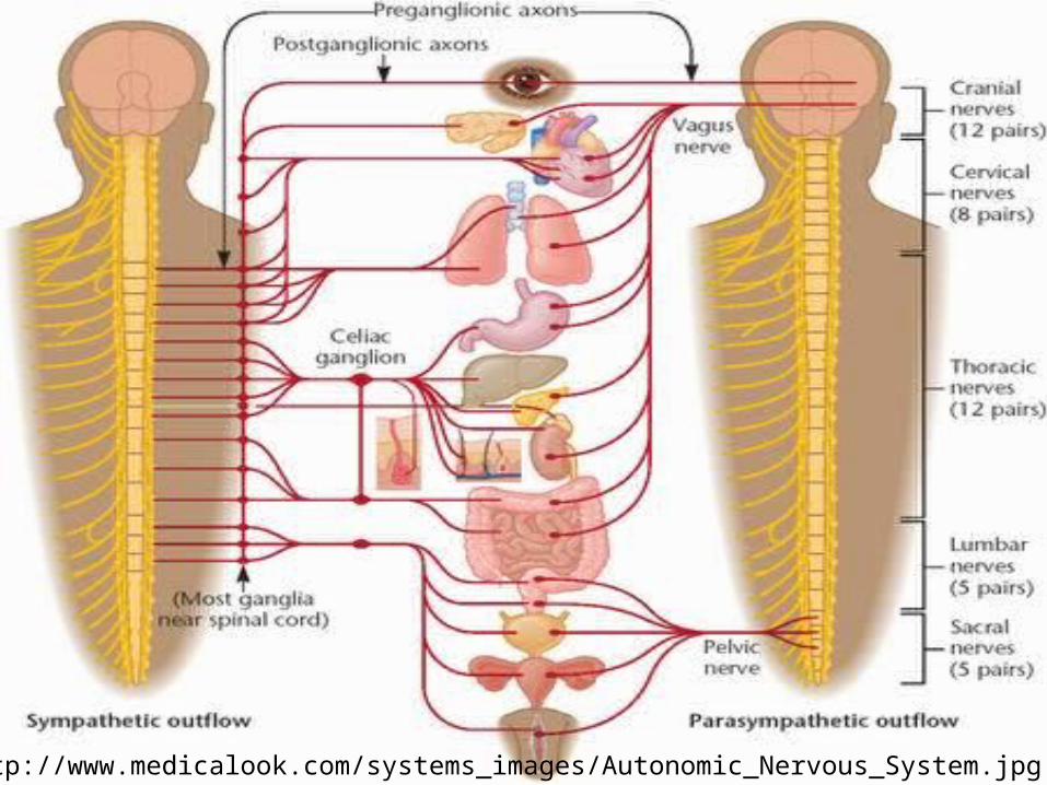
Autonomic nervous system
• Regulates involuntary functions of body.
• Innervates heart and blood vessels, GI tract, urinary system, respiratory system, muscles of eye.
• Innervates glands and smooth muscle (not skeletal muscle).
• Made up of sympathetic and parasympathetic nervous systems.
QuickTime™ and a decompressor
are needed to see this picture.

QuickTime™ and a decompressor
are needed to see this picture.
http://users.rcn.com/jkimball.ma.ultranet/BiologyPages/A/autonomic.gif

Sympathetic nervous system• System uses epinephrine as its
transmitter.• Activates body for emergency
situations and actions (fight or flight response).
• Strengthens heart contractions, increases heart rate, dilates pupils and bronchioles, constricts vessels feeding digestive tract.
QuickTime™ and a decompressor
are needed to see this picture.

QuickTime™ and a decompressor
are needed to see this picture.
http://cache.eb.com/eb/image?id=72120&rendTypeId=35

• Adrenal gland regulated by this system.
• Produces epinephrine in response to stimulation - produces many of same fight or flight responses.
QuickTime™ and a decompressorare needed to see this picture.

QuickTime™ and a decompressor
are needed to see this picture.
http://www.beliefnet.com/healthandhealing/images/exh45027_ma.jpg

Parasympathetic nervous system
• Acetylcholine - primary neurotransmitter for this system.
• System deactivates or slows down activities of muscles and glands.
• Constricts pupils, slows down heart rate, constricts bronchioles, dilates vessels of digestive tract.
• Principle nerve - vagus nerve. QuickTime™ and a decompressor
are needed to see this picture.

QuickTime™ and a decompressor
are needed to see this picture.
http://www.pharmainfo.net/files/images/stories/article_images/Sympathetic%20and%20parasympathetic%20neurons%20with%20ganglion.JPG