Embed Size (px)
Citation preview

Peripheral nerveAnatomy and Physiology

CNS
Sensory division Motor division
Autonomic nervous system
Sympathetic division
Parasympathetic division
PNS
Somatic nervous system

Hystology

Neuron
■ 4 regions

Neuron
■ Cell body Nucleus RNA Proteins

Neuron
■ Dendrites Input form other neurons
Neuron
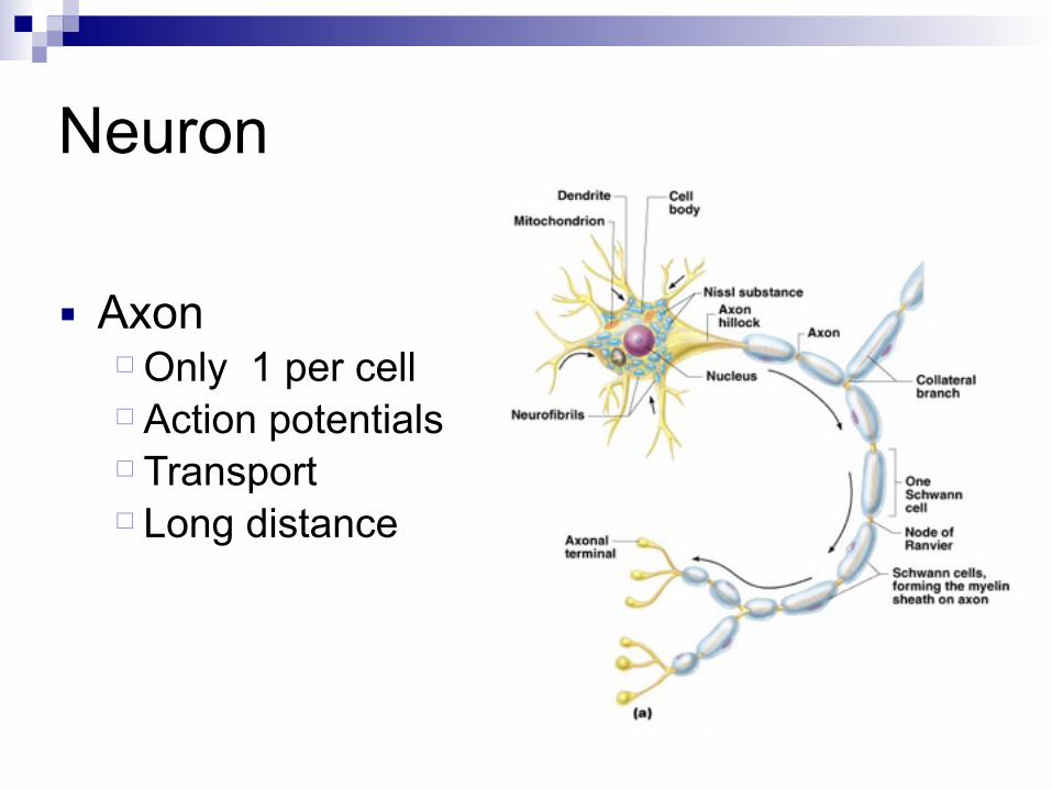
■ Axon Only 1 per cell Action potentials Transport Long distance
Neuron
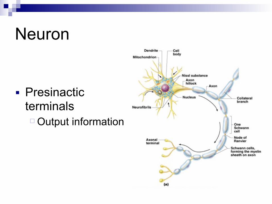
■ Presinactic terminals
Output information

Nervous Tissue: Support Cells (Neuroglia)■ Astrocytes
Abundant, star-shaped cells Brace neurons Form barrier between capillaries and neurons Control the chemical environment of the brain
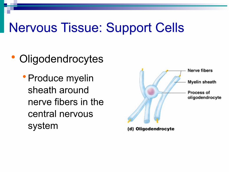
Nervous Tissue: Support Cells
• Oligodendrocytes • Produce myelin
sheath around nerve fibers in the central nervous system

Nervous Tissue: Support Cells
• Satellite cells • Protect neuron cell bodies

• Myelin sheaths in jelly-roll like fashion
• One neuron
Schwann cell

• Up to 500 each neuron
• Nodes of Ranvier
Schwan cell

Myelin
■ 70 % lipid, cholesterol, and phospholipids ■ 30% protein ■ Faster conduction ■ Less energy used

Physiology
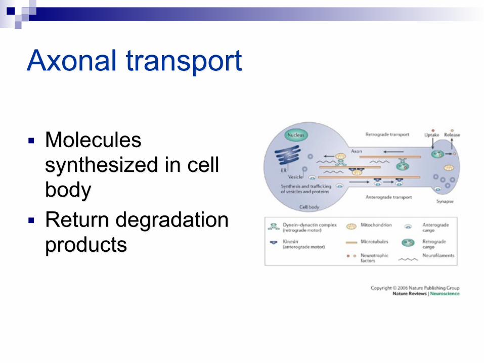
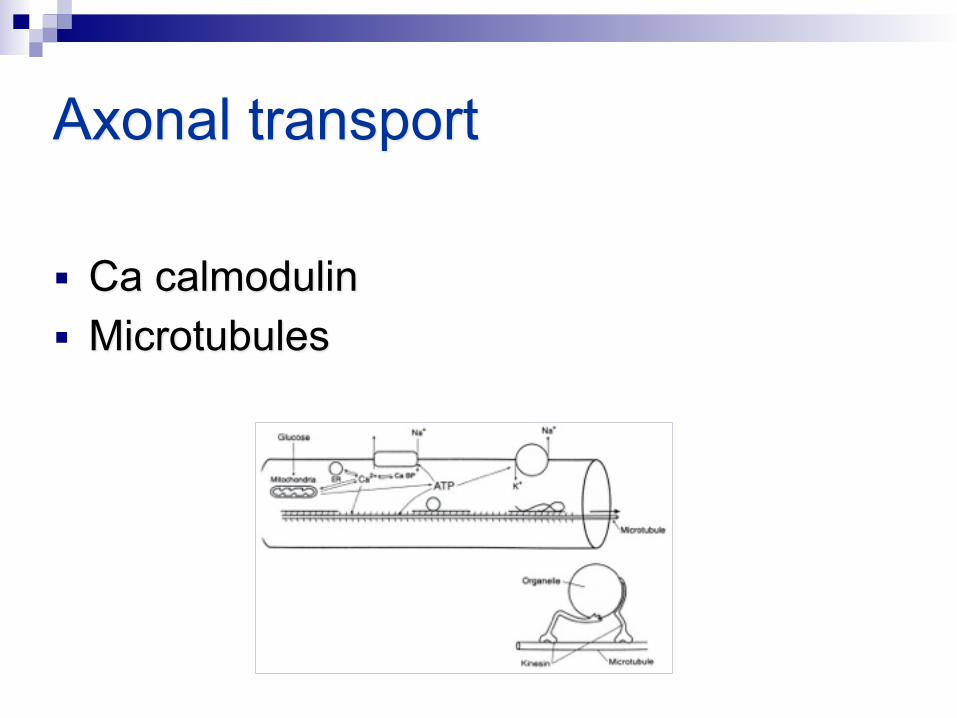
Axonal transport
■ Molecules synthesized in cell body
■ Return degradation products

Axonal transport
■ ATP energy source ■ Oxidative metabolism
Axonal transport
■ Ca calmodulin ■ Microtubules

Axonal transport anterograde
■ Kinesin ■ Fast – strong molecule attach ■ Slow – loose molecule attach ■ Impairment – terminal degeneration

Axonal transport retrograde
■ Fast, ■ 1/3 ½ from anterograde ■ Return molecules, vesicles ■ Nerve grow factor from target organ ■ HSV, Rabies, Polio and Tetanous toxin

Action potential - resting
■ Negative inside ■ Na, K, Cl ■ Passive transport ■ Channel
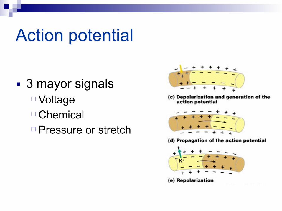
■ 3 mayor signals Voltage Chemical Pressure or stretch
Action potential

■ Increase memebrane resistence ■ Decreace memebrane capacitane ■ Ranvier nodes high channel concentration
Myelin
■ Current thought the membrane ■ Threshold reached ■ Self-propagation
External activation

anatomy
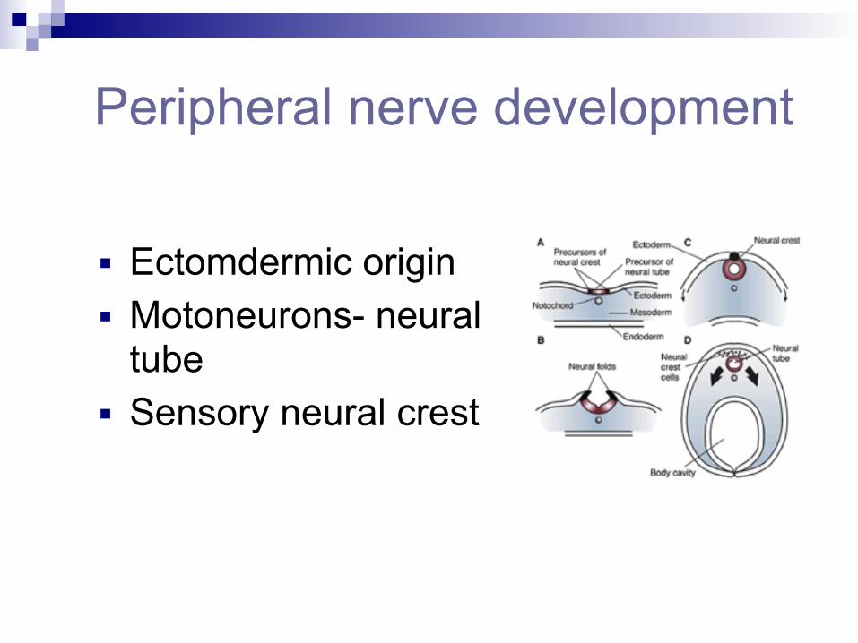
Peripheral nerve development
■ Ectomdermic origin ■ Motoneurons- neural
tube ■ Sensory neural crest
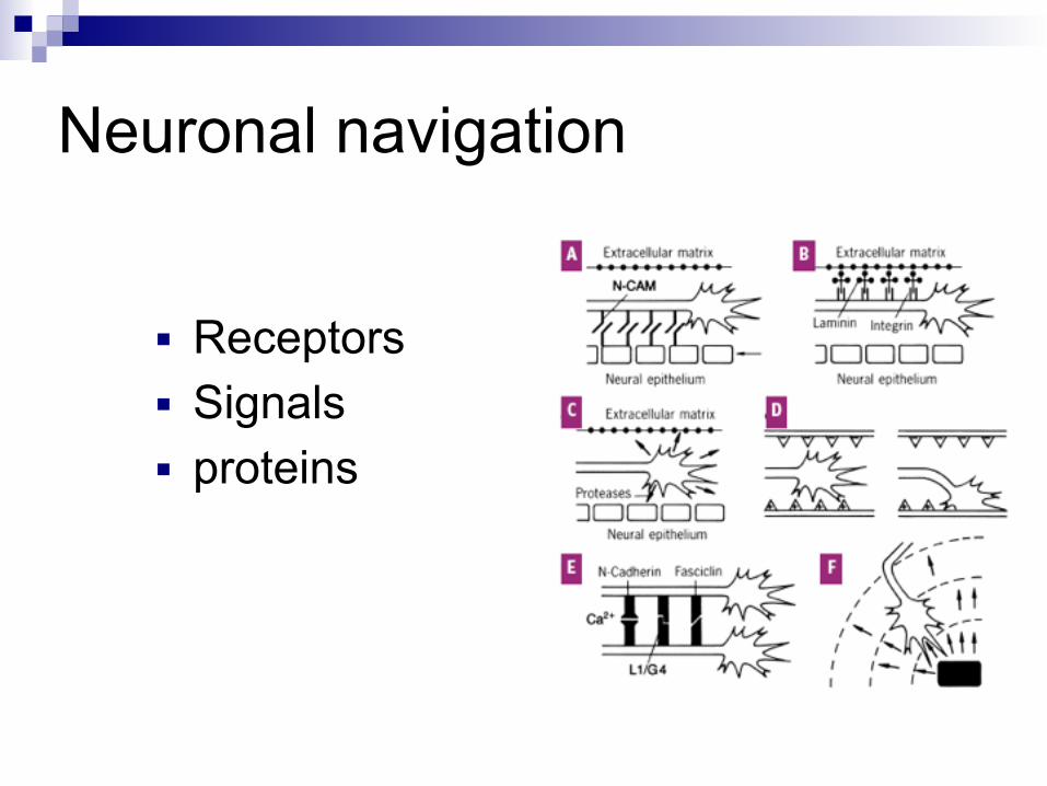
Neuronal navigation
■ Receptors ■ Signals ■ proteins
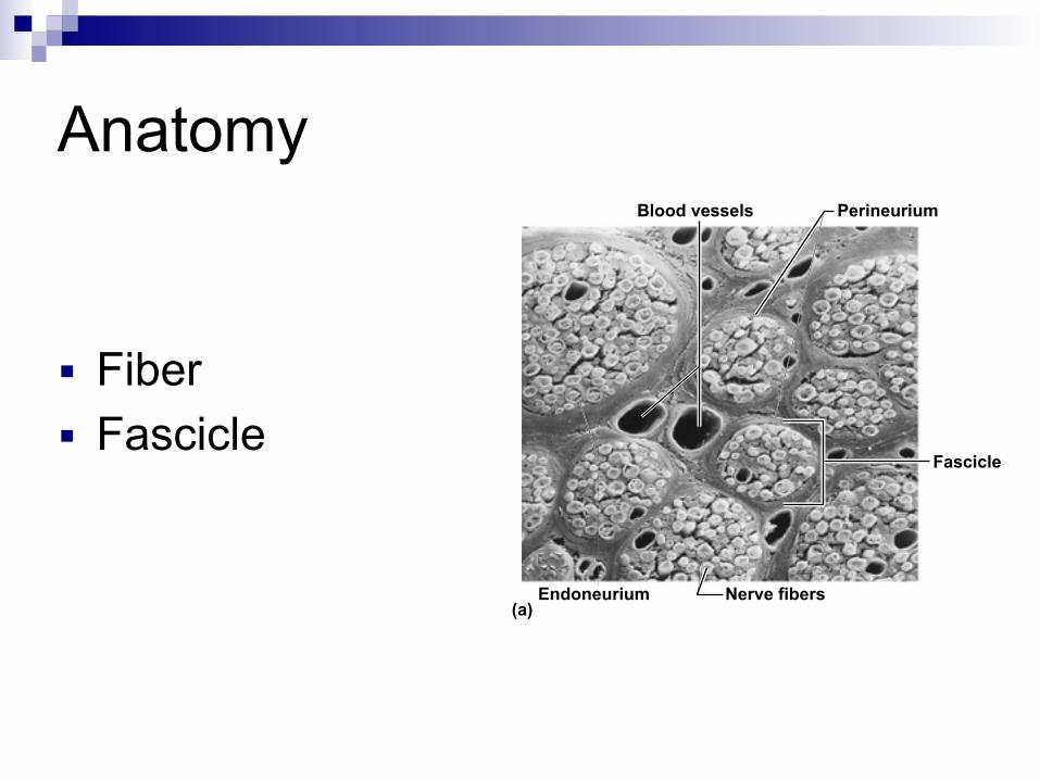
(a)
Fascicle
Perineurium Blood vessels
Endoneurium Nerve fibers
Anatomy
■ Fiber ■ Fascicle
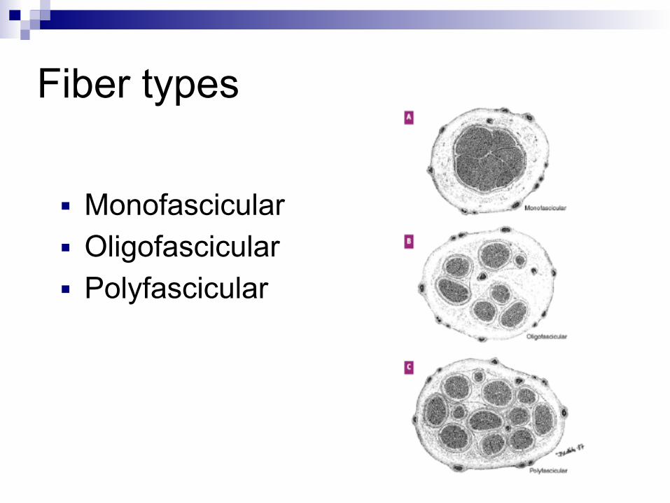
Fiber types
■ Monofascicular ■ Oligofascicular ■ Polyfascicular

(b)
Axon
Endoneurium
Perineurium
Epineurium
Myelin sheath
Blood vessels
Fascicle
Connective tissue
■ Endoneurium Loose collagen Capillary network High resistance Barrier

(b)
Axon
Endoneurium
Perineurium
Epineurium
Myelin sheath
Blood vessels
Fascicle
Connective tissue
■ Perineurium Thin layer

(b)
Axon
Endoneurium
Perineurium
Epineurium
Myelin sheath
Blood vessels
Fascicle
Connective tissue
■ Epinerium Framework Vascular plexus Compression

Vascular supply
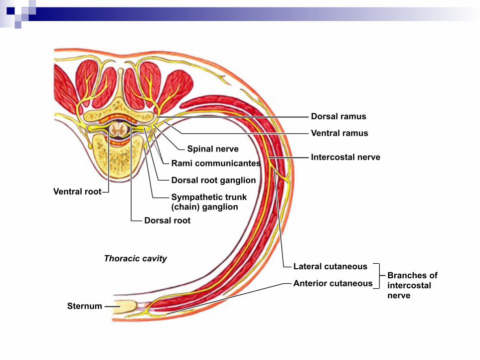
Dorsal ramus
Ventral ramus
Intercostal nerve
Lateral cutaneous
Anterior cutaneous
Spinal nerveRami communicantes
Dorsal root ganglion
Sympathetic trunk(chain) ganglion
Dorsal root
Ventral root
Sternum
Thoracic cavity
Branches of intercostal nerve

Classification of nerve fiber

Sensory receptors
• Free Nerve Endings !
• Encapsulated Nerve Endings !
• Expanded Tip Endings

- No end structure. - Most abundant. - Derived from unmyelinated nerve fibers.
Free nerve endings

-Vertical pattern. - Around the bodies of the sudoriferous glands. - Displacement of the Skin, Painful Stimuli and
Temperature.
Free nerve endings

- Great variety in size and shape. - Consist of a single myelinated nerve axon - Connective tissue sheath.
Encapsulated nerve endings

!Main types: !• Krause Bulbs • Pacinian Corpuscle • Meissner’s Corpuscle • Corpuscles of Ruffini
Encapsulated

•Glabrous skin of the palm and digits, lips, genitals •Rapidly adapting fibers. •Lower Frequency Stimulation less 50 hz
Meissner corpuscles
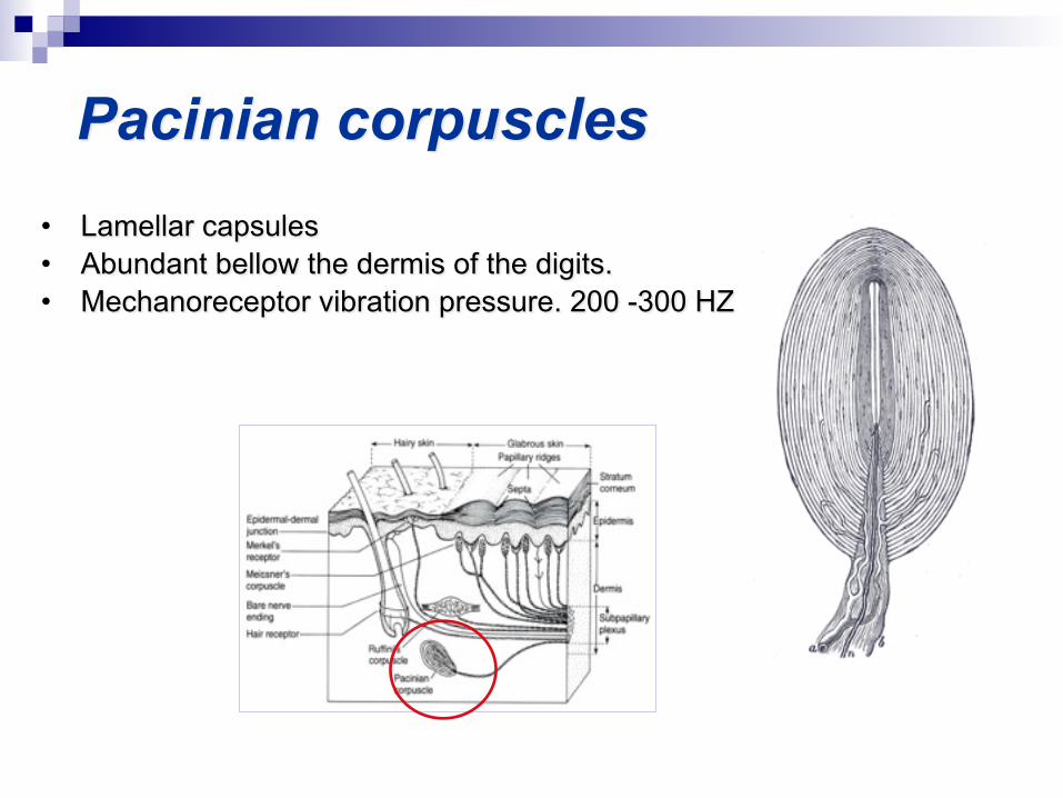
• Lamellar capsules • Abundant bellow the dermis of the digits. • Mechanoreceptor vibration pressure. 200 -300 HZ
Pacinian corpuscles

- Fluid-filled space traversed by collagen fibers. - Respond to Mechanical Stimuli and Tensional Forces on the
Surrounding Tissue. - Subcutaneous tissue finger.
Ruffini corpuscle

• Cylindrical or oval Bodies Semifluid core.
• Synovial membranes of certain joints, digits
• Epineurium of nerve trunks.
Krause bulb

•Epithelial cell specialized epithelial cell •Respond to Static Pressure. •Reading Braille !
Merkel disc
Spinal cord reflexes
■ Definition: Rapid, predictable and involuntary motor response to stimuli through pathways called reflex arcs.

Spinal cord reflexes
■ Two systems Autonomic reflexes (unconscious): digestion, sweating etc. Somatic reflexes: activate skeletal muscles.

Spinal cord reflexes
■ Characteristics: Structurally (number of neurons involved)
Monosynaptic arc: one synapse Polysynaptic arc: one or more association neurons.

The basic components of all human reflex arcs
Stimulus
ReceptorSkin
Sensory neuron
Spinal cord (in cross section)
Integration centerInterneuronMotor neuron
Effector
1
54
2 3

Stretch reflex
■ Monosynaptic ■ Alfa motoneurons ■ Stimulate muscle spindle

The stretch reflex
(a)(b)
Initial stimulus: muscle stretch
Afferent impulses from stretch receptor to spinal cord
Efferent impulses to alpha (α) motor neurons cause contraction of the stretched muscle that resists/reverses the stretch
Efferent impulses to antagonist muscles are damped (reciprocal inhibition)
Spinal cord (L2–L4)
Patella
Cell body of sensory neuron
Key: + Excitatory synapse – Inhibitory synapse
Motor neuron serving quadricepsMotor neuron serving antagonist muscle group (hamstrings)
Muscle spindleQuadriceps (extensors)
Hamstrings (flexors)
Muscle spindle
Patellar ligament
Interneuron
–
1
2
3

Muscle spindle
(a) (b) (d)(c)
Muscle spindleIntrafusal muscle fiber
Primary sensory (la) nerve fiber
Extrafusal muscle fiber
Time Time Time
Unstretched muscle; AP frequency constant
Stretched muscle; AP frequency increased
α − γ Neuron coactivation; AP frequency constant
α Motor neuron stimulation only; no APs, unable to signal length changes
Time
AP: Action Potential

The Golgi tendon reflex
■ Autogenic inhibition
++
+ –
Quadriceps (extensor)
Golgi tendon organ
Hamstrings (flexor)
Afferent fiber from Golgi tendon organEfferent fiber to muscle associated with stretched tendonEfferent fiber to antagonistic muscle
Spinal cord
Interneurons
Key: + Excitatory synapse – Inhibitory synapse

Golgi tendon organ
Tendon
γ Efferent motor fiber to spindle
Golgi tendon organ
CapsuleSensory fiber

Basics nerve injury
Response to trauma
■ Inflammatory response ■ Increase vascular permeability ■ Edema ■ Abnormal axonal transport ■ Vascular deficit

Nerve injury classification
■ Metabolic conduction block ■ Sunderland I – neuropraxia ■ Sunderland II – axonotmesis ■ Sunderland III ■ Sunderland IV ■ Sunderland V – neurotmesis
Metabolic conduction block
■ Local arrest of intraneural microcirculation ■ Immediately reversible when compression
removed

Sunderland I - Neuropraxia
■ Axon continuity preserved ■ Motor paralysis, some sensation spared ■ Spontaneously reversible within 3 months ■ Prolonged latency
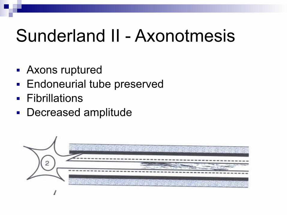
Sunderland II - Axonotmesis
■ Axons ruptured ■ Endoneurial tube preserved ■ Fibrillations ■ Decreased amplitude
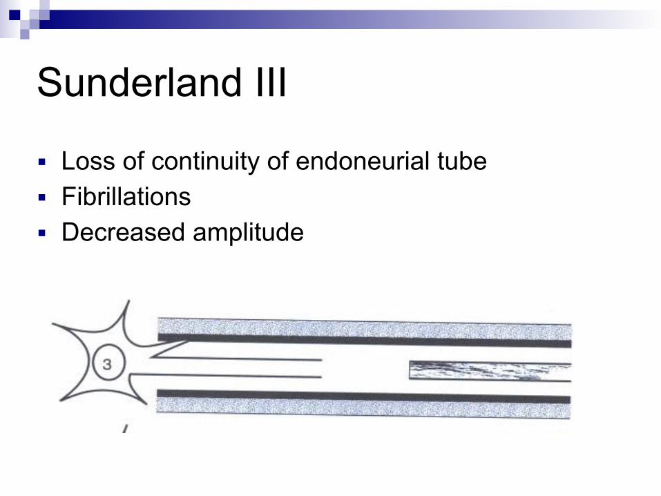
Sunderland III
■ Loss of continuity of endoneurial tube ■ Fibrillations ■ Decreased amplitude

Sunderland IV
■ Loss of continuity of perinerium ■ Completely blocked with scar tissue

Sunderland V - Neurotmesis
■ Complete nerve transection

Wallerian degeneration
■ Axonal degeneration ■ Proteolysis ■ calcium

Schuann cell response
■ Proliferation ■ Band of Bügner ■ Increase NGF receptors

Macrophage response
■ IL 1 stimulate Schuann cell ■ Increase NGF ■ Phagocytic

Cell body respose
■ Phases Reactive Recovery Degenerative