Embed Size (px)
Citation preview
Neonatal Respiratory Distress Syndrome
D. Vidyasagar and S. Adeni
Neonatal respiratory distress syndrome
Respiratory Distress Syndrome (RDS) is one of the common causes for newborn admissions to the Neo- natal Intensive Care Unit (NICU). It is the most frequent cause of death in newborn infants in associ- ation with prematurity.’ The term neonatal respir- atory distress syndrome is often used synonymously with Hyaline Membrane Disease (HMD) and is very much gestational age dependent, having a higher incidence in preterm infants. There is also a predomi- nance of male over female affected infants.
The underlying pathophysiologic mechanism in RDS is a relative deficiency in surface active mater- ials, namely pulmonary surfactant. Surfactant is produced by Type II alveolar cells, and, is a surface tension reducing phospholipid which prevents alveo- lar collapse at end-expiration. Avery and Mead* provided the first clinico-pathological correlation between surfactant deficiency and neonatal respir- atory distress syndrome. It has been demonstrated over the past two decades that the risk for relative surfactant deficiency and subsequent pulmonary disease were greatly increased in infants less than 37 weeks’ gestation. A number of maternal factors delaying pulmonary maturity in the fetus have also been described.3 The most important maternal factors influencing fetal lung maturity are maternal diabetes mellitus, antepartum hemorrhage and peri- natal asphyxia, all of which delay or retard adequate surfactant production in the fetus. Pathology. The post-mortem appearance of the lungs of infants dying from RDS are grossly described as
Dr D. Vidyasagar, Dr S. Adeni, Department of Paediatrics, University of Illinois Hospital, 840 So. Wood Street M/C 8.56, Chicago, Illinois 60611. USA (Correspondence to DV)
airless and having a ruddy appearance. Microscopi- cally, diffuse atelectasis is the hallmark of RDS with the formation of a hyaline membrane being a major characteristic. The hyaline membrane is formed of fibrin and cellular debris and usually is found as an eosinophilic membrane lining the terminal air spaces. Diagnosis. RDS is primarily a disease of premature infants. It is thus important to assess the risk for pulmonary immaturity in any prematurely delivered infant. The most commonly used predictor of lung maturity is the Lecithin/Sphingomyelin ratio (L/S ratio), obtained from amniotic fluid.4 The L/S ratio prior to 32 weeks is usually 5 1.0 and begins to increase thereafter. A L/S ratio of 12.0 is associated with only a minimal risk of pulmonary immaturity.’ Another marker of pulmonary maturity is the presence of phosphatidyl-glycerol (PG). PG is only produced in fetal lungs, and is therefore recommen- ded when conditions which may interfere with the L/S ratio are present, such as maternal diabetes6 A complete list of the tests used to determine fetal maturity is given in Table 1 in order of clinical importance.
Clinical features
Tachypnoea is the most commonly noted sign in the neonate with RDS. The normal range of 40-60 breaths per min is usually exceeded by the neonate with RDS. Respiratory rates 1 60/min must there- fore be viewed as an impending sign of respiratory compromise. However, as the tachypnea continues the infant tends to become exhausted leading to apnea and/or bradycardia. Chest wall retraction, grunting, decreased or poor breath sounds and cya- nosis are other common signs of respiratory failure in the neonate. Grunting is primarily a compensatory

160 CURRENT ANAESTHESIA AND CRITICAL CARE
Table 1
Tests for Fetal Lung maturity in order of importance
1. US Ratio Amniotic fluid 2. Presence of PG (Phosphatidyl-Glycerol) 3. Gestational age of fetus as judged by Obstetric Ultra
Sonography 4. Foam stability/shake test 5. Fluorescent polarimetry for viscosity of amniotic fluid 6. Optical density measurement of amniotic fluid at 650nm 7. Amniotic fluid creatinine concentration 8. Osmolality of amniotic fluid 9. Nile blue staining of cells from amniotic fluid
10. Optical density at 450nm of amniotic fluid
mechanism in which air is exhaled through narrow vocal cords. This creates a physiologic end-expiratory pressure, preventing alveloar collapse at end-expir- ation. Auscultation of the chest may provide impor- tant information. In moderately severe RDS, the breath sounds are usually decreased secondary to widespread atelectasis. A common pitfall to be mentioned is that in the very small neonate, due to the small thoracic size, auscultation may provide only a rough estimate of pulmonary function. Also, differ- ences in breath sounds between right and left lung may not be readily recognised. This makes it difficult to diagnose by auscultation alone even some catastro- phic lesions such as a pneumothorax. A very careful evaluation is therefore required in all premature neonates with respiratory distress syndrome. To aid in objective and methodical evaluation of the severity of respiratory distress, a number of scoring systems have been developed. Table 2 shows such a scoring system found to be helpful by the authors. The score allows one to quantitate the severity of the respira- tory distress and also provides some guidelines for therapeutic intervention.’ A score of 2-4 would be consistent with mild respiratory distress and a score of 5-7 or 8- 10 would be consistent with moderate and severe respiratory distress respectively.
The roentgenographic appearance of the neonatal chest in RDS is confirmatory. The classic picture of RDS described by Donald et al in 1953 has been known to vary.8 In moderately severe cases of RDS the lung fields are opaque with the presence of ‘air bronchograms’ which delineate the bronchial tree in a poorly aerated background of the lung fields. In severe cases, the lung fields are totally opaque and a ‘white-out’ appearance of the chest X-ray is char- acteristic. In this state, the cardiac and pulmonary borders become indistinguishable from each other. The most important differential diagnosis to be considered in the presence of respiratory distress and a ‘white out’ chest X-ray in neonate is the presence of neonatal pneumonia caused by group B streptococci which produces a X-ray picture essentially indistinguishable from RDS.
Other clinical features of RDS include cyanosis, peripheral edema, pallor and persistent metabolic acidosis as well as hypoglycemia and hypocalcemia all
of which result directly or indirectly from the stressed state of the neonate with RDS. Table 3 summarises the clinical features of an infant with RDS.
Biochemical abnormalities
Hypoxaemia and acidosis are the most consistent features of RDS. The initial P,O, is usually 5 60- mmHg with a significant alveolar/arterial gradient. In very sick infants, the Pao:! even in 100% oxygen and on mechanical ventilation may not exceed 60-80 mmHg. The pH is usually between 7.25-7.30 in the first few hours of life in an infant with RDS. A predominantly respiratory acidosis is an indicator of poor ventilation and may require the use of mechani- cal ventilation. Marked metabolic acidosis is commonly seen in neonates with perinatal asphyxia and a stormy delivery room course. Continuing metabolic acidosis in the face of adequate respiratory management may indicate sepsis, hypovolemia or shock. The occurence of a severe intracranial hemor- rhage in severely depressed infants with RDS is common and may be another cause of continuing metabolic acidosis.
Alveolar hypoventilation is responsible for both hypoxemia and hypercarbia. Widespread atelectasis causes V/Q mismatching which in turn aggravates the hypercarbia caused by alveolar hypoventilation.
Monitoring the infant with RDS
All infants with suspected or proven RDS should be monitored in the NICU. Since hypoxemia is the most critical biochemical abnormality, all infants with RDS should have their arterial blood gases moni- tored. An indwelling umbilical arterial line facilitates frequent blood sampling and continuous arterial blood pressure monitoring. In recent years the prac- tice of pulse oximetry has gained widespread accept- ance as a non-invasive mode of monitoring arterial oxygen saturation. The oxygen saturation monitor is a welcome addition to the non-invasive monitoring apparatus available in the NICU. However, there are some limitations to its use which have recently been reviewed by us. 9 The usual recommendation is to keep the O2 saturation between 87-95% and if
Table 2
Clinical Respiratory Distress Scoring System Score
Clinical Sign 0 1 2
Respiratory rate 60 60-80 >80 or apneic episode (per minute) Cyanosis None In air In 40% OX Retractions None Mild Moderate to severe Grunting None Audible with Audible without
stethoscope stethoscope Air entry* Clear Delayed or Barely audible (crying) decreased
* Air entry represents the quality of inspiratory breath sounds heard in the mid-axillary line.
NEONATAL RESPIRATORY DISTRESS SYNDROME 161
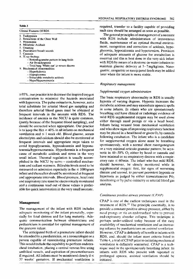
Table 3
Clinical Features Of RDS
1. Tachypnoea 2. Retractions of the Chest Wall 3. Hypoxemia 4. Metabolic Acidosis 5. Grunting 6. Poor/absent breath sounds 7. Cyanosis 8. X-ray findings
- Reticulogranular pattern in lung fields - Air Bronchograms - Total lung ‘White out’ in severe disease
9. Biochemical Abnormalities - Hyperkalemia - Hypoglycemia - Intractable metabolic acidosis - Hypo/Hypernatremia
295%. our practice is to decrease the inspired oxygen concentration to minimize the hazards associated with hyperoxia. The pulse oximeter is, however, not a total substitute for arterial blood gas sampling and therefore arterial blood gases must be obtained at frequent intervals in the neonate with RDS. The incidence of anemia in the NICU is quite common, (partly because of the frequent blood sampling), and should be corrected when appropriate. Our practice is to keep the Hct 2 40% in all infants on mechanical ventilation and I 1 week old. Blood glucose, serum electrolytes and calcium should also be monitored at frequent intervals and appropriate steps taken to avoid hypoglycaemia, hypocalcaemia and hypona- traemia/hypernatraemia. Hypothermia is a frequent cause of metabolic acidosis and stress in the very small infant. Thermal regulation is usually accom- plished in the NICU by servo - controlled mechan- isms and radiant warmers. Bilirubin levels need to be measured on admission especially in the I 1OOOgram infant and thereafter should be monitored at frequent and appropriate intervals. Blood pressure, heart rate and respiratory rate must be electronically monitored and a continuous read out of these values is prefer- able for quick intervention in the very small neonate.
Management
The management of the infant with RDS includes adequate monitoring of the infant prenatally, espe- cially for fetal distress and for lung maturity. Ade- quate communication between obstetricians and pediatricians is essential for optimal management of the preterm infant.
The anticipated birth of a premature infant should be attended by a paediatrician, or a neonatologist or a person capable of resuscitating premature infants. This would include the capability to perform endotra- cheal intubation, placing a central venous line using umbilical vessels, and aspiration of pneumothoraces if required. All infants must be monitored closely if 5 37 weeks’ gestation. If mechanical ventilation is
required, transfer to a facility capable of providing such care should be arranged as soon as possible.
The general principles of management of a neonate with RDS include administration of intravenous fluids, maintenance of an optimal thermal environ- ment, recognition and correction of acidosis, hypo- glycemia, hypocalcemia and hypotension. Provision of adequate amounts of glucose for metabolism is essential and this is best done in the very sick infant with RDS by means of a dextrose-in-water infusion to maintain glucose delivery at 5-7mg/kg/min. Naso- gastric, orogastric or nasojejunal feeds may be added later when the infant is more stable.
Respiratory management
Supplemental oxygen administration
The basic respiratory abnormality in RDS is usually hypoxia of varying degrees. Hypoxia increases the metabolic acidosis and may exacerbate apnoeic spells in some infants. In others who are spontaneously breathing and have clinical or radiologic evidence of mild RDS supplemental oxygen may be used alone either through nasal prongs or via a head hood. Infants being weaned from mechanical ventilation and who show signs of improving respiratory function may be placed in a head hood or given 02 by cannula following extubation. The infant who is a candidate for supplemental oxygen alone must be breathing spontaneously, with a normal chest roentgenogram or a very minimal reticula-granular pattern; be acya- notic in 40-50% 02 either by hood or cannula and have minimal or no respiratory distress with a respir- atory rate r 60/min. The infant who has mild RDS, should however, be closely monitored while in supplemental oxygen: first, for signs of worsening disease and second, to prevent persistent hypoxia or hyperoxia as judged by either transcutaneous PO, monitoring or by pulse oximetry or arterial blood gas analysis.
Continuous positive airway pressure (CPAP)
CPAP is one of the earliest techniques used in the treatment of RDS. lo The principle essentially, is to provide a constant positive airway pressure, either by nasal prongs or via an endotracheal tube to prevent end-expiratory alveolar collapse. This technique is perhaps under-utilised today because of the easy availability of mechanical ventilation and an increas- ing reliance by paediatricians on assisted ventilation. However, CPAP is definitely of benefit in infants with RDS, and, should the infant meet criteria listed in Table 4, a trial of CPAP prior to initiating mechanical ventilation is definitely warranted. CPAP is a tech- nique used in the spontaneously breathing infant. In infants with RDS who present with repeated or prolonged apnoea, assisted ventilation should be initiated.
162 CURRENT ANAESTHESIA AND CRITICAL CARE
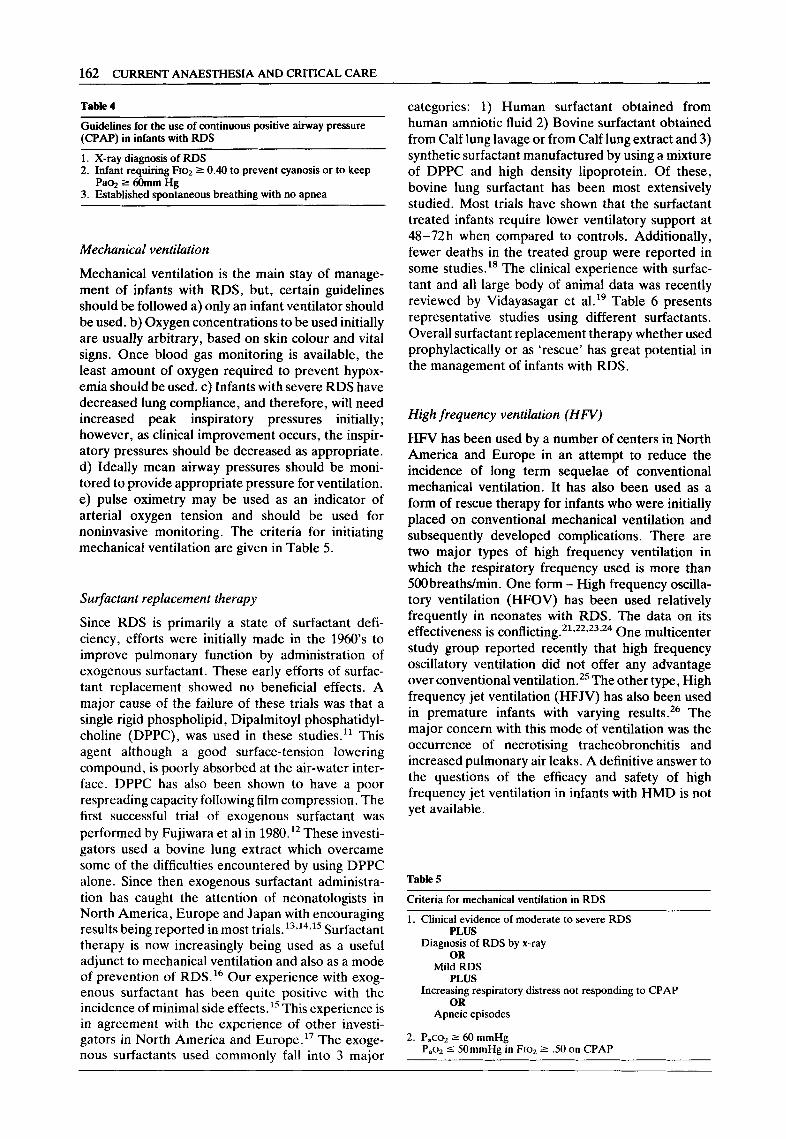
Table 4
Guidelines for the use of continuous positive airway pressure (CPAP) in infants with RDS
1. X-ray diagnosis of RDS 2. Infant requiring FIO~ 2 0.40 to prevent cyanosis or to keep
Paoz 2 60mm Hg 3. Established spontaneous breathing with no apnea
Mechanical ventilation
Mechanical ventilation is the main stay of manage- ment of infants with RDS, but, certain guidelines should be followed a) only an infant ventilator should be used. b) Oxygen concentrations to be used initially are usually arbitrary, based on skin colour and vital signs. Once blood gas monitoring is available, the least amount of oxygen required to prevent hypox- emia should be used. c) Infants with severe RDS have decreased lung compliance, and therefore, will need increased peak inspiratory pressures initially; however, as clinical improvement occurs, the inspir- atory pressures should be decreased as appropriate. d) Ideally mean airway pressures should be moni- tored to provide appropriate pressure for ventilation. e) pulse oximetry may be used as an indicator of arterial oxygen tension and should be used for noninvasive monitoring. The criteria for initiating mechanical ventilation are given in Table 5.
Su$actant replacement therapy
Since RDS is primarily a state of surfactant defi- ciency, efforts were initially made in the 1960’s to improve pulmonary function by administration of exogenous surfactant. These early efforts of surfac- tant replacement showed no beneficial effects. A major cause of the failure of these trials was that a single rigid phospholipid, Dipalmitoyl phosphatidyl- choline (DPPC), was used in these studies.” This agent although a good surface-tension lowering compound, is poorly absorbed at the air-water inter- face. DPPC has also been shown to have a poor respreading capacity following film compression. The first successful trial of exogenous surfactant was performed by Fujiwara et al in 198O.l* These investi- gators used a bovine lung extract which overcame some of the difficulties encountered by using DPPC alone. Since then exogenous surfactant administra- tion has caught the attention of neonatologists in North America, Europe and Japan with encouraging results being reported in most trials.13*14*15 Surfactant therapy is now increasingly being used as a useful adjunct to mechanical ventilation and also as a mode of prevention of RDS.16 Our experience with exog- enous surfactant has been quite positive with the incidence of minimal side effects.15 This experience is in agreement with the experience of other investi- gators in North America and Europe.” The exoge- nous surfactants used commonly fall into 3 major
categories: 1) Human surfactant obtained from human amniotic fluid 2) Bovine surfactant obtained from Calf lung lavage or from Calf lung extract and 3) synthetic surfactant manufactured by using a mixture of DPPC and high density lipoprotein. Of these, bovine lung surfactant has been most extensively studied. Most trials have shown that the surfactant treated infants require lower ventilatory support at 4%72h when compared to controls. Additionally, fewer deaths in the treated group were reported in some studies.18 The clinical experience with surfac- tant and all large body of animal data was recently reviewed by Vidayasagar et al.19 Table 6 presents representative studies using different surfactants. Overall surfactant replacement therapy whether used prophylactically or as ‘rescue’ has great potential in the management of infants with RDS.
High frequency ventilation (HFV)
HFV has been used by a number of centers in North America and Europe in an attempt to reduce the incidence of long term sequelae of conventional mechanical ventilation. It has also been used as a form of rescue therapy for infants who were initially placed on conventional mechanical ventilation and subsequently developed complications. There are two major types of high frequency ventilation in which the respiratory frequency used is more than %Obreaths/min. One form - High frequency oscilla- tory ventilation (HFOV) has been used relatively frequently in neonates with RDS. The data on its effectiveness is conflicting.21*22*23,24 One multicenter study group reported recently that high frequency oscillatory ventilation did not offer any advantage over conventional ventilation.25 The other type, High frequency jet ventilation (HFJV) has also been used in premature infants with varying results.*‘j The major concern with this mode of ventilation was the occurrence of necrotising tracheobronchitis and increased pulmonary air leaks. A definitive answer to the questions of the efficacy and safety of high frequency jet ventilation in infants with HMD is not yet available.
Table 5
Criteria for mechanical ventilation in RDS
1. Clinical evidence of moderate to severe RDS PLUS
Diagnosis of RDS by x-ray OR
Mild RDS PLUS
Increasing respiratory distress not responding to CPAP OR
Apneic episodes
2. P,co* 2 60 mmHg PaoZ 5 SOmmHg in FIO? 2 .50 on CPAP

NEONATAL RESPIRATORY DISTRESS SYNDROME 163
Table 6
Author (Reference) Type of
surfactant Number of patients Gestational age (+ S.D.)
Birthweight (grams) (+ SD.) Outcome
1. Fujiwara (12)
2. Gitlin (14)
3. Raju (15)
4. Halliday (16)
5. Merritt (18)
6. Morley et al (20)
7. Shapiro et al (28)
Bovine lung extract
Bovine lung extract
Bovine lung extract
DPPC + HDL
Human surfactant
DPPC + PG (7:3)
Calf lung lavage surfac- tant extract
10
18 treated (tx) 23 control (c)
17 treated (tx) 13 control (c)
49 treated (tx) 51 control (c)
31 treated (tx) 29 control (c)
159 treated (tx) 149 control (c)
16 treated (tx) 16 control (c)
30.2 + 1.9 1489 + 334
29 + 1.6 (tx) 29 + 1.5 (c)
28.4 & 3 (tx) 27.6 f 2 (c)
31.0 f 2.1 (tx) 30.3 z!z 7.4 (c)
24 - 29 wks
27.6 + 1.3 (tx) 27.6 + 1.3 (c)
27.4 + (tx) 27.3 f 0.7 (c)
1238 + 153 (tx) 1214 + 151 (c)
1119 z!z 204 (tx) 1096 + 220 (c)
1591 +- 425 (tx) 1486 + 421 (c)
938 + 286 (tx) 964 + 174 (c)
1093 + 310 (tx) 1070 + 251 (c)
1004 + 160 (tx) 932 + 112 (c)
Prompt increase in oxygenation. Radiologic evidence of lung improvement within 3 h
Decrease in O2 require- ment and ventilator support; no significant differences in mortality or morbidity
Decrease in ventilatory requirements; lower in incidence of Broncho- pulmonary dysplasia in treated infants
No difference in mortality
Treated group had less mortality, BPD and pneumothoraces
23 deaths in tx group vs. 40 in control
No difference in mortality, morbidity
or PDA. TX group had less severe distress at 12 and 24h
Complications
Most of the complications of RDS are related to the therapies employed in RDS. Mechanical ventilation is life saving in the infant with RDS, yet, it is not entirely without adverse effects. The complications of RDS may be divided into short and long term effects. The most common long term side effect of mechani- cal ventilation in the neonate is the development of bronchopulmonary dysplasia (BPD). An infant is defined as having BPD when he or she remains oxygen dependent at 28 days of life following mech- anical ventilation in the first week of life. The incidence of BPD is between lo-20% in infants with RDS who receive mechanical ventilation.”
Other complications of RDS are listed in Table 7. The most devastating long term complications found in survivors of RDS are intraventricular hemorrhage, which causes a significant amount of morbidity, and, retinopathy of prematurity which although less common today, is still a major concern of all neonato- logists because of the danger of blindness.
Summary
RDS today continues to be a major reason for neonatal admissions to the NICU world wide. Over the past decades the management of these infants has changed from empirical supportive treatment to that of prenatal detection and prevention. In recent years
the possibility of correction of the underlying bio- chemical deficiency has paved the way for new hopes. In spite of improved treatment regimens and the introduction of surfactant replacement therapy, it is possible that RDS will remain the most important cause for neonatal morbidity well into the next decade. Only prevention of prematurity may impact on the incidence of RDS and its associated compli- cations.
Table 7
Complications of RDS
I. Short term (early): - Severe metabolic acidosis - Hypoglycaemia - Intractable hypoxia - Pneumothorax - Pneumomediastinum - Pneumoperitonem - Pulmonary hemorrhage
II. Late: - Patent ductus arteriosus - Increased risk of necrotizing enterocolitis (NEC) - Intracranial hemorrhage - Pulmonary interstitial emphysema (PIE) - Formation of lung cysts - Subglottic stenosis - Palatal groove or cleft formation from the endotracheal
tube - Bronchopulmonary dysplasia (BPD) - Retinopathy of prematurity - Increased risk of nosocomial infection

164 CURRENT ANAESTHESIA AND CRITICAL CARE
References
1. Perelman RH, Farrell PM: Analysis of causes of neonatal death in the U.S. with specific emphasis on fetal Hyaline Membrane Disease. Paediatrics 1982; 70: 570-575
2. Avery ME, Mead J: Surface properties in relation to atelectasis and Hyaline Membrane Disease. Am J Dis Child 1959; 97: 517-523
3. Avery ME et al: The lung and its disorders in the newborn infant, Ed 4, Philadelphia, WB Saunders 1981,1969
4. Gluck L, Kulovitch M: US ratio in amniotic fluid in normal and abnormal pregnancy. Am J Obstet Gynecol1975; 115, 53%546,1973,
5. Harvey D: Risk of RDS. Lancet 1975; 1: 42 6. Creasv G. Simon N: Am J Perinatal 1984: 1: 302 7. Downks JJ, Vidyasagar D, Morrow GM, et al. Respiratory
distress syndrome of newborn infants. I. New clinical scoring system with acid-base and blood gas correlations. Clin Pediatr 1970; 9 (6): 325-326
8. Donald I, Steiner RE: Radiography in the diagnosis of HMD. Lancet 1953; 2: 846-849
9. Dziedzic K, Vidyasagar D: Pulse oximetry in Neonatal Intensive Care. Clin oerinatol. 16. 1. March 1989
10. Gregory GA: Respiratory Care of Newborn Infants. Ped Chn N Am 1972; 19: 311-324
11. Notter RH, Shapiro DL: Lung surfactant for replacement therapy. Clin Perinatol 1987; 14,3,433-470
12. Fujiwara T, Chida S, et al: Artificial surfactant therapy in HMD. Lancet 1980; I: 55-58
13. Enhorning G et al: Prevention of Neonatal Respiratory Distress Syndrome by tracheal instillation of surfactant. A randomized clinica trial. Pediatrics 1985; 76: 145-153
14. Gitlin JD, Sol1 RF et al: Randomised controlled trial of exogenous surfactant for the treatment of HMD. Pediatrics 1987; 79: 31-37
15. Raju TNK, Vidyasagar D, Bhat R et al: Double-blind controlled trial of single dose treatment with bovine surfactant in severe HMD. Lancet 1987; I: 651-656
16. Halliday HL et al: Controlled trial of artificial surfactant to prevent respiratory distress syndrome. Lancet 1984; I: 476- 478,
17. Collaborative European Multicenter Study Group: Surfactant replacement therapy for severe neonatal RDS. An International randomised clinical trial. Pediatrics, 1988; 82: 683-691
18. Merritt TA, Hallman M et al: Prophylactic treatment of very premature infants with Human Surfactant. N Engl. J Med 1986; 315: 785-790
19. Vidyasagar D, Raju TNK et al: Surfactant replacement therapy: Clinical and experimental studies. Clin Perinatol, September 1987; 14: 3
20. Ten Center Study Group: Ten Center trial of artificial surfactant in very premature babies. Br Med J 1987; 294: 991996
21. Boros SJ, Mammel MC et al: Neonatal HFJV: 4 years experience. Pediatrics 1985; 75: 651-53
22. Frantz ID, Westhammer J. et al: HFV in premature infants with lung disease: Adequate gas exchange at low tracheal pressures. Pediatrics 1983: 71: 483-488
23. High frequency ventilation for immature infants: Report of a conference, March 2-4,1982. Pediatrics 1983; 71: 280-287
24. Marchak BE, Thompson WK et al: Treatment of RDS by HFOV: A preliminary report. J. Pediatr, 99,287-92, 1981: Pediatrics 1983; 71: 280-287
25. The HiFI study group: HFOV compared with conventional mechanical ventilation in the treatment of respiratory failure in preterm infants. N Engl J Med 1989; 320: 88-93
26. Carlo WA, Chatbura RL et al: Decrease in airway pressure during HFJV in infants with RDS. J Pediatr 1987; 110: 275-282
27. Bancalari E and Gerhardt T: Bronchopulmonary Dysplasia: Ped Clin N. Am 33, 1, l-23, February 1986
28. Shapiro DL, Notter RA, Morin FC, et al: Double blind, randomised trail of calf lung surfactant extract administered at birth to very premature infants for prevention of respiratory distress syndrome. Pediatrics 1985; 76: 593-599