Embed Size (px)
Citation preview

Neonatal respiratory distress including CPAP: Clinical Learning Resource
To be used in conjunction with: Queensland Clinical
Guideline - Neonatal respiratory distress including CPAP
http://www.health.qld.gov.au/qcg/documents/g-cpap.pdf
Refer to local policies and procedures: • Hand hygiene • Non-pharmacological reduction of pain and discomfort • Patient identification

Neonatal respiratory distress including CPAP CLR V2 Page 2 of 58
Neonatal respiratory distress including CPAP Clinical Learning Resource
Published by the State of Queensland (Queensland Health), January 2015
This document is licensed under a Creative Commons Attribution 3.0 Australia licence. To view a copy of this licence, visit creativecommons.org/licenses/by/3.0/au
© State of Queensland (Queensland Health) 2015
You are free to copy, communicate and adapt the work, as long as you attribute the State of Queensland (Queensland Health).
For permissions beyond the scope of this licence contact: Intellectual Property Officer, Department of Health, GPO Box 48, Brisbane QLD 4001, email [email protected], phone (07) 3234 1479.
For further information contact : Workforce Development & Education Unit, Centre for Clinical Nursing, Royal Brisbane & Women’s Hospital, Herston, 4029 Queensland, at [email protected] , phone (07) 3646 0382.
An electronic version of this document is available at www.health.qld.gov.au/qcg
Disclaimer: The content presented in this publication is distributed by the Queensland Government as an information source only. The State of Queensland makes no statements, representations or warranties about the accuracy, completeness or reliability of any information contained in this publication. The State of Queensland disclaims all responsibility and all liability (including without limitation for liability in negligence) for all expenses, losses, damages and costs you might incur as a result of the information being inaccurate or incomplete in any way, and for any reason reliance was placed on such information.

Neonatal respiratory distress including CPAP CLR V2 Page 3 of 58
Contents Contents ............................................................................................................. 3 Figures ............................................................................................................... 5 Tables ................................................................................................................ 5 Summary ............................................................................................................ 6 How to use the CLR ........................................................................................... 6 Assessment ........................................................................................................ 6 Resources required to complete the package .................................................... 7 Objectives........................................................................................................... 7 Authors ............................................................................................................... 8 Acknowledgments .............................................................................................. 8 Continuing Professional Development ............................................................... 8 Version Control ................................................................................................... 8 Unit 1: Physiology of Respiratory Distress of the Newborn ........................... 9
Learning Objectives ............................................................................................. 9 Reading 1 ................................................................................................... 9
1.1 Compliance ................................................................................................ 9 1.2 Resistance ................................................................................................ 11
Reading 2 ................................................................................................. 12 Reading 3 ................................................................................................. 12 Activity 1 ................................................................................................... 12
Unit 2: Physiology of CPAP ........................................................................ 13 Learning Objectives ........................................................................................... 13 2.1 Continuous Positive Airway Pressure ....................................................... 13
Reading 4 ................................................................................................. 13 Activity 2 ................................................................................................... 14
2.2 CPAP Delivery Systems ........................................................................... 15 2.3 Patient Interfaces ...................................................................................... 16
2.3.2 Nasopharyngeal Tube (NPT) use .................................................... 17 2.3.3 Non-Invasive Positive Pressure Ventilation (NIPPV) ........................ 18 2.3.4 Heated High Flow Nasal Cannula (HHFNC) .................................... 18 Activity 3 ................................................................................................... 18 Activity 4 ................................................................................................... 18 Clinical Tips – Sizing Prongs .................................................................... 19
Unit 3: Humidification of CPAP ................................................................... 20 Learning Objectives ........................................................................................... 20
Reading 5 ................................................................................................. 20 Reading 6 ................................................................................................. 20 Activity 5 ................................................................................................... 21
3.1 Two limb circuit ......................................................................................... 21 3.2 Co-axial circuits ........................................................................................ 21 3.3 What temperature? ................................................................................... 22
Activity 6 ................................................................................................... 22 Clinical Tips – Humidification .................................................................... 22
Unit 4: Complications of CPAP ................................................................... 23 Learning Objectives ........................................................................................... 23 4.1 Contraindications ...................................................................................... 23 4.2 Pulmonary Air Leaks ................................................................................. 23

Neonatal respiratory distress including CPAP CLR V2 Page 4 of 58
Reading 7 ................................................................................................. 24 Activity 7 ................................................................................................... 24
4.3 Abdominal Distension ............................................................................... 24 Activity 8 ................................................................................................... 25
4.4 Failure of CPAP ........................................................................................ 25 Activity 9 ................................................................................................... 25
4.5 Nasal Trauma ........................................................................................... 26 Unit 5: Nursing Care of the Baby on CPAP ................................................. 27
Learning Objectives ........................................................................................... 27 Reading 8 ................................................................................................. 27 Activity 10 ................................................................................................. 28 Clinical Tips – Check Equipment Fit ......................................................... 28 Reading 9 ................................................................................................. 28
Clinical Scenario ................................................................................................ 28 Clinical Tips – CPAP Trauma ................................................................... 29 Activity 10.1 .............................................................................................. 29
5.1 Developmental Care ................................................................................. 30 Reading 10 ............................................................................................... 30 5.1.1 Positioning the Baby on CPAP ........................................................ 30 Clinical Tips – Preventing Pressure Injuries .............................................. 33 Activity 10.2 .............................................................................................. 33 Activity 10.3 .............................................................................................. 33 Activity 10.4 .............................................................................................. 34 Activity 10.5 .............................................................................................. 34
5.2 Supportive Care ........................................................................................ 34 5.2.1 Analgesia ........................................................................................ 34 5.2.2 Feeding ........................................................................................... 35 Activity 11 ................................................................................................. 35
Conclusion........................................................................................................ 35 Appendix 1 Glossary of terms ...................................................................... 36 Appendix 2 Nasal Prong Tube CPAP ........................................................... 37
Indications ................................................................................................ 37 Principles .................................................................................................. 37 Equipment ................................................................................................ 38 Insertion .................................................................................................... 39 Ongoing Care: .......................................................................................... 43 Complications: .......................................................................................... 43 NPT Suctioning ......................................................................................... 43 Indications ................................................................................................ 44 Equipment ................................................................................................ 44 Procedure ................................................................................................. 45 Complications ........................................................................................... 47
Appendix 3 Bubble CPAP: Minimising Rain Out .......................................... 48 Further tips ............................................................................................... 49
Appendix 4 Clinical Skills Assessment Tool ................................................. 51 References ....................................................................................................... 54

Neonatal respiratory distress including CPAP CLR V2 Page 5 of 58
Figures Figure 1 Nasal Prong Tube ..................................................................................... 17
Figure 2 NPT Tapes ............................................................................................... 39
Figure 3 Measuring for NPT.................................................................................... 40
Figure 4 Cut the NPT .............................................................................................. 40
Figure 5 Lubricate the NPT ..................................................................................... 40
Figure 6 Hydrocolloid strips for NPT ....................................................................... 41
Figure 7 Base tapes for NPT .................................................................................. 41
Figure 8 Secure the NPT ........................................................................................ 42
Figure 9 Second trouser leg ................................................................................... 42
Figure 10 Secure orogastric tube .............................................................................. 42
Figure 11 Length of NPT for suction ......................................................................... 46
Figure 12 Correct humidifier set up ........................................................................... 48
Figure 13 Correct probe set up ................................................................................. 48
Figure 14 Correct probe placement .......................................................................... 49
Figure 15 Removal of extension tubing ..................................................................... 49
Tables Table 1 CPAP Delivery Systems ........................................................................... 15
Table 2 CPAP Interfaces ....................................................................................... 17
Table 3 Guide to weight appropriate NPT sizes ..................................................... 39
Table 4 Appropriate suction catheter sizes ............................................................ 44
Neonatal respiratory distress including CPAP CLR V2 Page 6 of 58
Summary This Clinical Learning Resource (CLR) will assist nursing staff (Registered Nurses and Midwives with an interest in neonatal care) to perform their role in the management of the neonate in respiratory distress requiring the administration of Continuous Positive Airway Pressure (CPAP).
At the completion of this CLR, the participant will be able to:
Manage the care of the neonate in respiratory distress within their scope of practice Identify infants at risk of developing respiratory distress of the newborn Initiate therapy and provide clinical management of the neonate requiring CPAP Demonstrate knowledge of associated policies and procedures
How to use the CLR To complete this CLR:
Read through the reading material, including recommended readings and related policies and guidelines
Complete written activities and discuss your answers with a resource person Complete the clinical skills assessment tool (Appendix 4) on completion of this
package
Assessment Assessment of this CLR will be demonstrated through successful completion of specific activities utilising the resources provided or identified throughout the CLR. Resource staff, Clinical Facilitators or Nurse Educators should review and discuss the responses of all activities listed in the CLR to determine knowledge and awareness of the specific issues addressed. To gain competency for administering CPAP to neonates, the following must be completed:
Completion of this CLR and response booklet Successful completion of the clinical skills assessment tool will be upon direct
supervision by a Resource Person, Clinical Facilitator or Nurse Educator competent in the care of the baby on CPAP in accordance with the assessment guidelines within the package
Completion of this resource package and associated assessment is optional for nurses/midwives with previous experience in the care of the baby requiring CPAP, i.e. previous completion of an accredited education program in neonatal nursing, including recent experience in the care of the baby requiring CPAP.
Neonatal respiratory distress including CPAP CLR V2 Page 7 of 58
Resources required to complete the package The following resources will assist with completion of this CLR:
Recommended readings or textbooks Access to QHEPS Access to Health Service Policy website Resource person or Clinical Facilitator or Nurse Educator
Objectives The purpose of this CLR is to assist the participant to develop skills and knowledge in accordance with their scope of practice to be able to competently care for the neonate with respiratory distress requiring treatment with CPAP.
Upon completion of this CLR, the participant will be able to:
Demonstrate an understanding of respiratory distress of the newborn Identify the signs and symptoms of respiratory distress Review and define the physiology of CPAP Demonstrate knowledge of the indications for initiating CPAP to neonates in
accordance with the Queensland Clinical Guidelines - Neonatal respiratory distress including CPAP
Discuss the complications associated with CPAP delivery Develop sound knowledge of the complexities of nursing management of neonates
receiving CPAP
Please note: The mention of specific companies or of certain manufacturers’ products
does not imply that they are endorsed or recommended by Queensland Clinical
Guidelines or this CLR’s stakeholders in preference to others of a similar nature that
are not mentioned. Errors and omissions excepted, the names of proprietary products
are distinguished by initial capital letters.

Neonatal respiratory distress including CPAP CLR V2 Page 8 of 58
Authors Initiated by: Queensland Clinical Guidelines
Developed by: Version 1: Eliza Hughes, Project Officer
Version 2: Colette McIntyre, Nurse Educator
Project Funding: Queensland Clinical Guidelines
Additional support: Jacinta Lee, Christine Burridge, Allison Bowen, Tami
Photinos, Trish Smith, Karen Hose, Meshell Curtis,
Lynette Chapple, Liz Chappell, Anne Dawbney
Acknowledgments The authors wish to acknowledge the additional support, contributions and direction provided by:
Queensland Clinical Guidelines (QCG) Nursing & Midwifery Board of Australia Intensive Care Nursery Nurse Unit Manager, Clinical Nurse Consultant & Nurse
Educators, Royal Brisbane &Women’s Hospital (RBWH) Workforce Development & Education Unit/Centre for Clinical Nursing for education
standards RBWH Nursing & Midwifery Executive Council (NMEC) RBWH NMEC Subgroup - Workforce Development Education Group for
endorsement
Continuing Professional Development Completion of this package, if relevant to the context of practice, attracts 28 Continuing Professional Development (CPD) hours of learning. CPD hours can contribute to the nurse/midwife CPD requirements as per the Nursing and Midwifery Board of Australia Continuing Professional Development.
Version Control This is Version 2.0 of the Neonatal respiratory distress including CPAP CLR and will remain current until 2018. The current version will be available for access on the internet on the Queensland Clinical Guidelines website http://www.health.qld.gov.au/qcg
Neonatal respiratory distress including CPAP CLR V2 Page 9 of 58
Unit 1: Physiology of Respiratory Distress of the Newborn
Learning Objectives On completion of this unit the participant will be able to:
Explain the physiology of respiratory distress of the newborn Identify physical signs of respiratory distress Identify the predisposing factors to developing respiratory distress Demonstrate knowledge of lung compliance and the role of surfactant
In understanding physiology relevant to neonatal ventilation, it is important that one has a sound knowledge of the terminology of lung volumes and various elements of both internal and external respiration. In Appendix A, you will find a Glossary of Terms which may help you to revise your knowledge of foundational aspects of ventilation. You will find it helpful to understand what is meant by the terms: tidal volume (TV), functional residual capacity (FRC), total lung capacity (TLC), physiologic dead space and mechanical dead space.
The following reading will provide you with an overview of fetal lung development, influences on lung development and transition to extra uterine life, as well as a review of neonatal lung mechanics. Gaining an understanding of lung compliance and airway resistance will be of benefit later when the physiology of CPAP is discussed.
Reading 1
Diehl-Jones, B. (2012). Physiological Principles of the Respiratory System.
In D. Fraser (Ed.), Acute Respiratory Care of the Neonate (3rd ed.)(pp.1-
28). Petaluma, California: NICU Ink.1
1.1 Compliance Reading 1 provided an overview of some of the concepts relevant to lung compliance. This is such a critical concept in neonatal care, as much of the work is directed at overcoming the consequences of poor compliance or ‘stiff lungs’. Respiratory distress syndrome is a common example of a disease process in which lung poor compliance is a significant problem. A simple explanation of the physiology of breathing explains this concept. Like blowing up a balloon, the initial inflation of the newborn lung requires a reasonable degree of pressure for a small change in volume. This pressure increase is required to overcome surface tension and recruit (inflate) alveoli. Once inflated it is important that the alveoli are able to be stabilised, and easily reinflated with the next breaths. This phase is followed by one where, for a small increase in pressure, a large volume of gas is able to enter the lungs more easily.
It is useful to note that as the lungs near the peak of inspiration, there is only a small change in lung volume in response to a moderate delivery of pressure. Following
Neonatal respiratory distress including CPAP CLR V2 Page 10 of 58
expiration in the healthy newborn lung, the lung volume does not return to zero but retains a small volume of gas. This is known as the establishment of a functional residual capacity (FRC).
In contrast, the stiff or non-compliant lung in Respiratory Distress Syndrome (RDS) requires very high pressure to achieve only small changes in lung volume, and at the end of expiration lung volume returns to near zero. The resulting failure to establish a FRC has the effect of increasing the work of breathing for these babies, as each breath requires a large effort to recruit alveoli for gas exchange. Low FRC indicates reduced lung compliance and requires the generation of higher pressure to move the same amount of air as compared with a normal lung. An additional and significant problem in newborns with RDS is surfactant deficiency. Surfactant is a soapy substance which reduces surface tension, or in other words, prevents the natural tendency of the alveolar walls to close in on themselves (collapse) on expiration. Surfactant coats the inner lining of the alveolar wall and stabilises it, preventing collapse on expiration. It is important to understand that babies with RDS have surfactant deficiency, which contributes to the loss of FRC and an increase in respiratory effort. In treating RDS, CPAP and positive end expiratory pressure (PEEP) are instrumental in preventing atelectasis and reducing the work of breathing. These strategies both work to maintain a small volume of air in the lungs. Refer to Figure 1-12, page 18 from Reading 1, to gain an understanding of the pressure-volume curves of newborn lungs which will help in providing insight into the significance of CPAP and positive end expiratory pressure (PEEP).
The consequences of a low FRC have been discussed. In using CPAP and PEEP it is important to recognise that excessively high FRC, can also lead to a decrease in volume for a given pressure change. This occurs either from gas trapping within the alveoli (so much pressure applied that gas cannot be exhaled sufficiently), or excessive ventilation pressure as the limits of lung and chest wall expansion are approached.
Preterm babies differ from term babies both structurally and functionally. Issues of lung compliance are particularly relevant to neonatal care; however, there is another aspect of compliance that is also relevant. This relates to chest wall compliance. In contrast to lung compliance, which is often decreased in the preterm infant, chest wall compliance is increased in this population. In adults, chest wall compliance is low because of the rigidity provided by the rib cage. However, the poorly ossified bony structures of the preterm infant thorax allow for greater compliance. Babies with sternal recession are a graphic representation of the combination of non-compliant lungs and very compliant chest walls. As a point of interest, the presence of chest wall oedema may decrease chest wall compliance.1

Neonatal respiratory distress including CPAP CLR V2 Page 11 of 58
1.2 Resistance Reading 1 outlined two sources of friction (and resultant loss of energy) that cause resistance to air flow in the neonatal lung and airways. Airway frictional resistance accounts for approximately 80% of total pulmonary resistance and can be caused by anatomical structures or ventilatory appliances.1 Airway resistance caused by ventilatory appliances is directly proportional to the length and size of the breathing apparatus, that is the endotracheal tube (ETT), nasopharyngeal tube (NPT), and bi-nasal CPAP prongs. Decreasing the radius of a tube increases its resistance, therefore as the length of the tube increases and the radius of the tube decreases, it may be necessary to increase pressure to overcome the increased airway resistance.1
The second cause of resistance to airflow is ‘viscous resistance,’ which accounts for nearly 20% of total airway resistance. Viscous resistance can be related to any neonatal lung pathology that causes an increase in pulmonary fluid (e.g. delayed reabsorption, basement membrane leaking, left-to-right shunting and patent ductus arteriosis), and is also created by tissue moving against tissue within the lungs themselves as seen with surfactant deficiency.2
Considering that babies have increased airway resistance due to their narrower airway lumen, lung pathology and prematurity, ill-fitting ventilatory appliances can contribute to a proportionally greater increase in airway resistance.1 The interest for neonatal clinicians in understanding airway resistance lies in determining ways to reduce it. For neonatal nurses/midwives this often relates to the artificial airway and ventilation circuit.
Some ways to reduce resistance include:
Decreasing turbulence by ensuring that ventilator circuits are free from unnecessary curves and twists, and reducing the presence of condensation that has accumulated in the circuitry
Minimising the length of circuit tubing Improving flow by shortening the length of ETT or NPT (Shortening the tube also
reduces the work of breathing for spontaneous breaths) Ensuring humidification is adequate is critical for the reduction of thick and viscous
airway secretions and avoidance of mucus plugging narrowing or blocking airways Reduce dead space in the circuit by filling the humidifier to the specified fill level Ensuring that bi-nasal prongs are measured to fit the nares snugly
Reflect on your current practice and think of instances where you might have addressed the issue of airway resistance. You may be able to add to the above list.
Ensuring that all steps are being taken to minimise resistance to flow in a baby on assisted ventilation is vitally important when providing nursing care.

Neonatal respiratory distress including CPAP CLR V2 Page 12 of 58
Reading 2
Soltau, D.T. & Waldemar, A.C. (2014). Respiratory System. In C. Kenner
& J. Wright Lott (Eds.), Comprehensive Neonatal Nursing Care (5th ed.)
(pp. 140-145). New York: Springer Publishing Company.3
This reading provides a simple guide to understanding the causes and the clinical presentation of respiratory distress. In neonatal nursing, it is anticipated that a preterm baby will encounter some degree of respiratory distress. Respiratory distress can have several aetiologies and whilst they may incur differing treatment regimens, CPAP may ultimately be the only treatment option for preterm and term babies in outlying hospitals. Whilst we have covered the theory behind neonatal lung development and respiratory distress, it is also important to understand the skills required for assessment of a baby’s oxygenation and respiratory effort. The following reading will provide an overview of those nursing assessment skills.
Reading 3
Fraser, D. (2015).Chest and lungs. In E.P. Tapperro & M. E. Honeyfield
(Eds.). Physical Assessment of the Newborn (5th Ed.) (pp.79-91).
Petaluma, California: NICU Ink.4
Activity 1
Utilising the information in the above readings and the Queensland Clinical Guideline - Neonatal respiratory distress including CPAP5 answer the following in your response booklet:
a) Identify the clinical signs of respiratory distress of the newborn.
b) List the major causes of respiratory distress of the newborn and identify for each cause, rationale for why it happens.

Neonatal respiratory distress including CPAP CLR V2 Page 13 of 58
Unit 2: Physiology of CPAP
Learning Objectives On completion of this unit the participant will be able to:
Define CPAP. Demonstrate knowledge of the effect of CPAP on respiratory function. Explain and apply the various modes of CPAP delivery.
2.1 Continuous Positive Airway Pressure Before CPAP and its different delivery methods and interfaces are discussed, it is important to develop an understanding of the physiology of CPAP from the following reading.
Reading 4
Wiswell, T.E. & Courtney, S.E. (2011). Non-Invasive Respiratory Support.
In J.P Goldsmith & E.H. Karotkin (Eds.), Assisted Ventilation of the
Neonate (5th ed.)(pp 140 -162). St Louis: Elsevier.6
CPAP is defined as providing air and/or oxygen, into the lungs under pressure.7 By maintaining a positive pressure throughout the whole respiratory cycle, collapse of the alveoli at the end of expiration is minimised. As a result less energy is needed to reopen stiff alveoli and initiate a breath and the total work of breathing is decreased.8 By reopening alveoli, CPAP increases the functional residual capacity of the lungs and improves pulmonary artery oxygen (PaO2), thus improving gas exchange. CPAP as a means of supporting respiratory function in newborn babies with respiratory distress was first published in 1971 and continues to be an important strategy for treatment of babies with respiratory problems.9 CPAP has been used with a number of delivery devices and pressure generating systems, each seeking to provide safe and consistent pressure delivery, while minimising adverse equipment related effects.
CPAP has long been documented as contributing to reduced intubation rates, reduced incidence of chronic lung disease and improved survival.10,11 Early trials reported improved survival rates and lower rates of chronic lung disease. The following years saw an upsurge in the use of CPAP in very low birth weight (VLBW) infants and an increased use of CPAP in non-tertiary centres.11 Whilst CPAP has been used for over 4 decades12, there is still a great deal of ongoing research into CPAP’s remaining unanswered questions. For example, recent studies are now focussing on the INSURE method to identify if there are benefits to intubating babies for the purpose of giving surfactant, and then extubating to CPAP13 or alternatively, only giving Surfactant to infants requiring ventilation.14 Additional studies are also reviewing the benefits of Heated High Flow Nasal Cannula (HHFNC) as an alternative to CPAP.15 Research is ongoing in these areas and currently there is no change to practice in Queensland relating to these therapies in neonates. Please refer to the Queensland Clinical

Neonatal respiratory distress including CPAP CLR V2 Page 14 of 58
Guideline – Neonatal respiratory distress including CPAP for further information about managing respiratory distress and CPAP in your centre.5
Focussing on the timing of CPAP initiation, the COIN trial studied mortality, respiratory morbidity and early childhood health and development outcomes comparing early CPAP versus early intubation, and concluded some positive results primarily in the reduction of chronic lung disease though observed that the incidence of pneumothoraces were slightly higher in the CPAP group.16 It could be questioned that perhaps the higher incidence of pneumothoraces in the CPAP group is attributed to the fact that this group did not receive surfactant. Interestingly, research is currently being undertaken with the goal to potentially administer surfactant to babies on CPAP without the need for intubation.17,18 It remains to be seen if changing the way surfactants are administered may prove to reduce or eliminate the most severe complications related to intubation and the treatment of very low birth weight (VLBW) and extremely low birth weight (ELBW) babies with CPAP; hence research in this area is ongoing.17
It is absolutely critical for all who care for sick babies and their families to be continuously looking for the evidence to support practice. Research is changing and improving practices all the time, so as you move further through
this module, it is suggested that the Cochrane Library is accessed for further information.
Registered nurses/midwives have a vested interest in being skilled and knowledgeable in this area. Firstly, nurses/midwives must have an understanding of both the known and unknown risks and benefits of a therapy they apply. Next, nurses/midwives must be versed in the advantages and disadvantages of the various delivery and pressure generating devices, and very significantly nurses /midwives are required to develop considerable skill in managing CPAP systems. Despite CPAP being perceived as less invasive than intubation and mechanical ventilation, nursing work is considerable, often requiring repeated handling of the baby to prevent the development of complications and to maintain airway pressure.
Nurses and midwives with experience in the nursery setting may have been able to identify particular groups of babies most likely to receive CPAP. Your readings so far have explored how CPAP works, and how forcing a set continuous pressure into babies’ lungs, lessens the symptoms of respiratory distress. Reflect on your current knowledge about what you already know about CPAP and complete the activity below.
Activity 2
Cathy has just started work in the nursery and you are assigned to preceptor her on her first day caring for a baby on CPAP. In your response booklet, please describe how you would teach Cathy the following:
a) Identify the main issues that CPAP is used for.
b) Identify how CPAP lessens babies’ respiratory symptoms.

Neonatal respiratory distress including CPAP CLR V2 Page 15 of 58
2.2 CPAP Delivery Systems CPAP can be generated by a variety of devices including an infant ventilator, Bubble CPAP apparatus and Infant flow drivers.19 Each system may differ from institution to institution so one system may be more familiar than another. There is limited evidence to suggest one method of generating CPAP pressure is beneficial over another.20 The following section explores some popular devices further and does not endorse the use of one product or equipment type over another.
Table 1 CPAP Delivery Systems
Bubble CPAP
Bubble CPAP generates CPAP by submerging the expiratory component of the CPAP tubing under water to gain a desired level of PEEP. The level of CPAP is determined by the number of centimetres below the water level the limb is submerged (e.g. 6 cm below the surface = 6 cm H20).19 Gas flow is attached to an air/02 blender system and is humidified prior to delivery into the inspiratory tubing.21 Confirmation of delivery of the prescribed CPAP is gained via visualisation of active bubbling in the water chamber and an adequate seal at the delivery point/nares.
Absent bubbling may ultimately mean air leak in the system and by occluding the prongs, you will be able to determine if the problem with a loss of CPAP is related to the circuit, or the baby. When occluding the prongs, if a bubble is achieved, the problem is likely to be at the baby end of the circuit (e.g. prong size or position, open mouth). If occlusion of the prongs does not generate bubble, troubleshooting the circuit will be necessary (e.g. condensation blocking the tubing, leakage of gas from the circuit connections).22 Occasionally an increase in flow is required to gain the desired level of CPAP; this should be the last approach to troubleshooting loss of bubble/pressure.
The availability, ease of use and inexpensive nature of Bubble CPAP means that it is often the preferred method of delivering CPAP to infants with respiratory distress.22

Neonatal respiratory distress including CPAP CLR V2 Page 16 of 58
Infant Ventilator
CPAP via an infant ventilator is generated via the exhalation valve and there are no pressure oscillations. If the set pressure falls too low, the ventilator alarms.22 CPAP is manually adjusted independent of flow, and flow rates can be changed manually depending on the type of ventilator. Gas flow is through the attached humidification system.
Infant Flow Driver
The infant flow driver (IFD) provides a variable flow. It has an integrated nasal interface/pressure generator and the pressure is affected by flow. Gas is delivered in response to the infant’s respiratory efforts. There is limited data to support its superiority over other pressure generating devices.23
Images courtesy of QCG & Herston Multimedia Unit
2.3 Patient Interfaces Nasal CPAP is designed to deliver a consistent pressure at the nasal opening, which promotes continuous distending pressure within the airway and results in an increased functional residual capacity.19 CPAP can be delivered utilising a variety of patient interfaces, such as nasal prongs, nasal cannula, face mask, NPT or ETT.23 Most centres now use bi-nasal prongs since research demonstrated that the use of short bi-nasal prongs reduces respiratory failure rates and re-intubation in neonates24,25, in comparison to the single long nasopharyngeal prong. There is however, no evidence recommending the use of midline devices over other available bi-nasal prongs.22
Use of mask CPAP varies between centres with some units alternating between mask and bi-nasal prong, others using mask alone and some centres utilising mask CPAP once pressure areas become a problem using bi-nasal prong CPAP. A recent study concluded that mask CPAP was more effective at reducing intubation in babies <31 weeks if used in the first 72 hours of life.25 There remain unanswered questions in relation to the use of masks however, and research in this area continues.

Neonatal respiratory distress including CPAP CLR V2 Page 17 of 58
Table 2 CPAP Interfaces
Regardless of the interface used, the following principles apply to bi-nasal CPAP:
It is important to ensure the prongs are correctly fitting; too small and they will not create the seal necessary to generate the prescribed CPAP. In addition, they will move within the nare potentially causing skin/mucosal trauma, and also may increase airway resistance.22 If the prongs are too large, damage to the surrounding tissue can lead to blanching, erosion and necrosis.22,23
Regardless of the type of hat being used, it is important to have a well-fitting hat; a loose hat will impact upon achieving an effective seal and thus the delivery of CPAP, as well as causing movement of the CPAP device with subsequent injury to the surrounding skin and nasal structures.
Additionally, too tight a hat will lead to pressure area development and potential head moulding.22
Images courtesy of QCG & Herston Multimedia Unit
2.3.2 Nasopharyngeal Tube (NPT) use Using NPT’s increases work of breathing due to the increased resistance produced by the length of the tube23 and has been shown to be a less effective method of CPAP delivery when compared with short bi-nasal prongs.25
Figure 1 Nasal Prong Tube
NPT’s are flexible, ivory PortexTM tubes. It is essential that supplies of these are kept very separate to ETT stock as intubating and ventilating a baby using the ivory tube is difficult and creates problems for ventilation.
There remains a place for NPT CPAP, when other methods are not appropriate or effective, these include:
Conditions such as Pierre-Robin Sequence and Treacher Collins Syndrome. The upper airway obstruction that is a feature of these conditions can achieve better airway patency with the longer NPT
Management of significant septal columella pressure areas when other interfaces are not available or appropriate
Post operatively following ventriculo-peritineal shunt insertion

Neonatal respiratory distress including CPAP CLR V2 Page 18 of 58
Congenital nasal conditions in which bi-nasal devices cannot create a seal (e.g. certain types of cleft lip/palate), or the nasal passages are not patent (choanal atresia).26
Appendix 2 has a guide to equipment required and insertion principles for NPT use in the event it is needed to stabilise a baby with any of the above conditions whilst awaiting retrieval.
2.3.3 Non-Invasive Positive Pressure Ventilation (NIPPV) NIPPV is used in tertiary centres to provide a spontaneously breathing neonate with both PEEP and intermittent positive pressure breaths. These breaths are either synchronised and triggered with the baby’s breathing or non-synchronised. NIPPV is delivered via a ventilator and bi-nasal or mask CPAP interface.27 Due to patient acuity, the specific conditions requiring NIPPV support, and the associated risks for this patient cohort, ongoing NIPPV therapy should be delivered in a tertiary hospital setting.
2.3.4 Heated High Flow Nasal Cannula (HHFNC) HHFNC has been prominent within research in recent years as a potential alternative to CPAP, and as a method of weaning off CPAP and/or ventilation. Research in this area is ongoing. Due to its unpredictable and inconsistent pressure delivery, it has not yet been demonstrated that HHFNC is a safe alternative to CPAP for treating acute lung disease.28
The first step in troubleshooting issues with CPAP and avoiding complications is to develop an understanding of the advantages and disadvantages of the systems available. Being well aware of the risks associated with a particular device can lead to providing appropriate treatment and prevention strategies.
Reading 4 outlined the types of CPAP used and some of the associated complications. Much additional evidence is available and one of the best sites to access this is online at the Cochrane Library. In this next activity, consider the benefits and limitations of the most frequently used CPAP devices.
Activity 3 A colleague who has just started in the nursery comments that she is unfamiliar with using short bi-nasal prongs and Bubble CPAP. After a demonstration of applying these devices and caring for a baby on these types of CPAP, she asks you about the advantages and disadvantages of each system. Using an evidence based approach, document your answer in your response booklet.
Activity 4
In your response booklet, read through the clinical scenario provided and answer the subsequent questions.
Neonatal respiratory distress including CPAP CLR V2 Page 19 of 58
Clinical Tips – Sizing Prongs
Having answered the previous activity, consider the nursing management of nasal trauma resulting from CPAP. For example if the baby’s septal columella is looking excoriated, what nursing strategies may be implemented to prevent this? In assessing the baby, determine whether the prongs are being pushed in too far and are creating pressure on the nasal columella; perhaps the prongs are too small and are therefore allowing too much free movement in and out of the nostril. Observing the nare regularly and systematically re-measuring the baby’s nares and septal diameter will provide prompts as to whether the baby requires prong upsizing.
With the development of experience in caring for babies on bi-nasal CPAP, nurses/ midwives are aware that whilst it is essential to size correctly in the first instance, it can prove difficult to achieve a perfect fit and may require experimentation. Be aware that a prong upsize should not cause the nose to appear blanched for a period longer than the initial insertion process.

Neonatal respiratory distress including CPAP CLR V2 Page 20 of 58
Unit 3: Humidification of CPAP
Learning Objectives On completion of this unit the participant will be able to:
Explain the fundamentals of humidity. Identify and demonstrate the management of humidification of non-invasive
ventilation.
Humidification of inspired gases for ventilated babies and babies on CPAP has become a fundamental tool in preventing complications. Inadequate humidification has been associated with airway obstruction, pneumothorax and trauma to the respiratory epithelium.16 Normally the nasal passages and upper respiratory tract play an important role in the humidification of inspired gases. Air is inhaled as a cool, dry gas and is subsequently exhaled warm and moist. The vasculature of the nose and nasal sinuses are equipped to compensate for this problem. Thus the nose, pharynx and trachea recover heat and moisture through normal physiologic processes. Bypassing this normal physiologic process through the use of endotracheal tubes, nasopharyngeal tubes, or bi-nasal prongs necessitates active management of gas humidification.16
The implications of humidification for nursing practice go beyond simply turning on the humidifier. The following readings outline the physiologic processes of the mucociliary support system and its role in prevention and reduction of the risk of pneumothorax, respiratory epithelial damage and tube obstruction. It will also provide you with the basic fundamentals to help you understand the concepts of humidity. The next two readings will help you answer the activities that follow.
Reading 5
Reading 6
De Klerk, A. (2012). Humidification during noninvasive respiratory support
of the newborn: Continuous positive pressure ventilation, and humidified
high-flow nasal cannula. In A.M. Esquinas (Ed.), Humidification in the
Intensive Care Unit (pp. 271-284). Berlin: Springer-Verlag.29
De Klerk, A. (2012). Physiology of humidification in critically ill neonates. In
A.M. Esquinas (Ed.), Humidification in the Intensive Care Unit (pp. 253-265).
Berlin: Springer-Verlag.16

Neonatal respiratory distress including CPAP CLR V2 Page 21 of 58
Activity 5
Inadequately humidified ventilatory gases delivered to intubated babies can cause significant respiratory morbidity. Utilising your response booklet, explain seven (7) respiratory changes that occur as a result of poor humidification.
Take a minute to examine the relationship between temperature and humidity of inspired gas temperatures in Reading 5, Figure 30.1.
The graph and reading discuss the concept of ‘saturation’ of gas and shows that to increase the total capacity of gas to hold water, gases must first be heated. If the inspired gases are not already saturated with water vapour (100% relative humidity) the gases will take up water from the lung mucosa, thus drying it (and the baby) out.29 So, what is known about heating gases?
Humidification for neonatal ventilation is mostly managed by heating a chamber of water through which the inspiratory gases must pass on the way to the baby. Innovations in circuit design have led to the availability configurations other than the traditional two limb circuit that incorporates a water trap.
3.1 Two limb circuit A heating wire is threaded through the inspiratory line to prevent ‘rain out’ or condensation. Rain-out or condensation occurs when warm gas that is holding water cools after leaving the humidifier, thus lowering its ability to hold water. This highlights another important point you need to remember about humidification – that the amount of humidity delivered to the baby is affected by environmental temperature (and this includes incubator temperature), unless steps are taken to counter it.
The internal heating wire minimises the potential for rain-out that occurs when humidified gases come in contact with cooler room or cot temperatures. The temperature, to which the heating wire will warm inspiratory gases, is thermostatically controlled by the set patient temperature and sensed by a circuit (patient) temperature probe.16 When the circuit probe senses that the set temperature has been reached, the heating wire is automatically switched off. Though ‘rain-out’ can still occur, manufacturers have configured the circuit to largely reduce the impact of environmental temperatures.
3.2 Co-axial circuits The introduction of co-axial neonatal ventilator circuits appears to have eliminated many of the rain-out problems experienced with the traditional circuit configuration. The co-axial circuits have the inspiratory limb inside the expiratory limb. The expiratory limb insulates and heats the inspiratory limb, and also has a short heater wire leading to the expiratory port. The dual heater wires eliminate the need for a water trap, as all the gases are heated, preventing cooling and rainout.
Neonatal respiratory distress including CPAP CLR V2 Page 22 of 58
3.3 What temperature? So far, it has been established that heating gases is required to maximise humidity. But how do we know what temperature is the right temperature to heat it to? Research has indicated that just as too little humidity can be problematic so too is too much humidity. So what temperature range is required? Aiming for at least basic physiologic conditions is thought to be adequate.
There is a demonstrated, significant fall in inspired gas temperature between the points of the heater wire ending and the airway opening.30 Though it has been observed that the fall is less in co-axial circuits, it has led to the belief that higher set temperatures may be necessary in an attempt to achieve the physiologically plausible temperature of 37°C at the airway opening.16
Many humidifiers currently used in neonatal nurseries employ an airway temperature of 40oC with a humidifier temperature of 37oC, neither of which is able to be altered by the user.
Appendix 3 contains a resource developed by Fisher & PaykelTM regarding management of rain out with a CPAP circuit. Whilst the information and diagrams are specific to Bubble CPAP, many of the hints and tips can be applied to ventilator driven CPAP and other humidification/tubing devices also.
Activity 6
Consider the management of humidification for ventilation gases in your nursery. Review your nursery’s policy and practices and examine the configuration of the circuit used in the nursery. Using your readings and Appendix 3, answer the questions in your response booklet.
Clinical Tips – Humidification
Humidification is central to preventing respiratory problems in newborns. So what’s the one thing that should always be remembered? NEVER let the humidifier run dry!
Many neonatal nurseries have a practice of checking the humidifier water level every hour with observations and replacing the bag of water or filling up the chamber as necessary.

Neonatal respiratory distress including CPAP CLR V2 Page 23 of 58
Unit 4: Complications of CPAP
Learning Objectives On completion of this unit the participant will be able to:
Identify the complications relating to CPAP treatment: pneumothorax, nasal trauma, abdominal distension and failure of CPAP
Identify the signs and symptoms of these complications Access and utilise the Queensland Clinical Guideline - Neonatal respiratory distress
including CPAP5 Identify strategies to prevent and manage these issues
Nursing experience highlights the fact that many treatments provided to patients ultimately have adverse effects. Some adverse effects are worse than others, and some can be completely avoided or at least reduced with a simple change in medical or nursing management. This unit will cover some of the nursing and medical management of the complications related to CPAP; however, discussion on the nursing care of the baby on CPAP will take place later in the package.
4.1 Contraindications Prior to a discussion of the complications of CPAP, the reasons why initiating CPAP would be avoided will be identified. In babies’ that are critically unwell, it would compromise their health status further if intubation and ventilation were delayed. Other factors for consideration prior to initiating CPAP and when it would be advisable to avoid CPAP, are when infants are diagnosed with upper airway abnormalities like cleft palate, choanal atresia, tracheo-oesophageal fistula, unrepaired gastroschisis or diaphragmatic hernia and recurrent apnoeic episodes.19 In some of these identified conditions, it would be technically difficult to pass NPT’s or due to obstructions difficult to achieve FRC. In other conditions, the potentially harmful nature of abdominal distension secondary to CPAP is another contraindication.
4.2 Pulmonary Air Leaks Pulmonary interstitial emphysema, pneumomediastinum and pneumothoraces are the most common air leaks experienced amongst neonates.31 Pneumothoraces occur in 1-2% of all newborns with as little as 0.07% being symptomatic.32 A number of factors can cause a baby to develop a pneumothorax, mostly these are related to the progression of their respiratory distress but often they can be attributed to the treatment modality or resuscitation they have been given.
As the aetiologies of respiratory distress were previously discussed, it would be expected that pneumothorax is a potential clinical dilemma for all of the patient groups that may endure a degree of respiratory distress whether they are supported with ventilation or not. Recent studies show an overall decrease in the incidence of pneumothorax when treated with CPAP in comparison to control groups.33 However,

Neonatal respiratory distress including CPAP CLR V2 Page 24 of 58
the variety of timing and methods of managing intubation and surfactant delivery along with the different CPAP devices and interfaces utilised in trials, makes it difficult to compare populations and outcomes.
Pneumothoraces can have a significant impact on the health outcomes of a baby. The following reading has been included as it provides a succinct summary of pneumothorax and the associated pathophysiology, clinical indications and management. Refer to the Queensland Clinical Guideline - Neonatal respiratory distress including CPAP5 for the procedure of draining an air leak and further management instructions. Following the reading and clinical scenario, complete the short activity below.
Reading 7
Papoff, P. & Moretti, C. (2012). Pulmonary air leakage. In G.
Buonocore, R. Bracci & M. Weindling (Eds.) Neonatology (pp. 460-
468). Milano, Italy: Springer.31
Activity 7
Read through the clinical scenario and answer the subsequent questions in your response booklet.
4.3 Abdominal Distension Abdominal distension has been documented in the neonatal population for some years, particularly in reference to babies being treated with CPAP. As CPAP is forced into the airways, air subsequently enters the oesophagus and then the stomach. For this reason, it is important to ensure that the flow is set as low as possible, whilst still maintaining mean airway pressure or bubble, in an effort to reduce the amount of gas entering the stomach. Insertion of a size 8Fg or gastric tube is imperative whilst a baby is being treated with CPAP to allow for gastric venting.
Additionally, observation of abdominal girth27 is an important aspect of nursing assessment and it is important to differentiate whether the abdominal distension is actually “CPAP Belly” where the resultant gastric aspirate would largely be made up of air, or if it is a sign of a more sinister problem like necrotising enterocolitis (NEC). The nursing management of a baby with abdominal distension caused by CPAP administration is crucial in preventing failure of CPAP and a worsening of the baby’s health status. As some babies may experience a range of health issues related to abdominal distension, for example apnoea and feed intolerance, it is therefore an important component of the nursing care of these babies to implement continuous gastric venting and regular aspirates.

Neonatal respiratory distress including CPAP CLR V2 Page 25 of 58
Activity 8
Consider the current nursing management of abdominal distension and the prevention strategies used in the nursery. Answer the following questions within your response booklet.
• How often do babies in your nursery have gastric aspirates? • Is gastric venting a routine procedure? • How is CPAP belly and signs of NEC differentiated?
4.4 Failure of CPAP Indicating when a baby has failed CPAP is an important aspect of medical and nursing management. A problem lies in the fact that there is no universally accepted definition of what CPAP failure really is and as health professionals, nurses/midwives are well aware of the possible disadvantages of extubating a baby or removing CPAP too early. Some researchers believe that by considering variables such as gestational age, maternal history, antenatal steroid administration along with CPAP pressure and FiO2 the likelihood of CPAP failure can be predicted.34 Some suggested indicators of CPAP failure include: increasing apnoea, increased oxygen requirement, increased work of breathing, intubation and mechanical ventilation.35 Regardless of the failure criteria, it is important to eliminate or remedy causes as to why the baby’s condition is worsening prior to re-intubation or seeking retrieval to a tertiary centre. Consider the following:
If there are oral or nasal secretions If the orogastric tube has been aspirated recently Is the baby achieving the set mean airway pressure? Is it necessary to resize the nasal prongs?
Activity 9
Consult the Queensland Clinical Guideline - Neonatal respiratory distress including CPAP5 and complete the activity below in your response booklet. Identify the signs of failure of CPAP as described in the guideline and the nursing actions that would be initiated.
Neonatal respiratory distress including CPAP CLR V2 Page 26 of 58
4.5 Nasal Trauma Many studies have been undertaken to see if there is any one CPAP apparatus that causes significantly more adverse effects than another.36 Unfortunately there is no one CPAP interface that has been proven to be less detrimental to skin integrity than any other. It is therefore absolutely critical for all who are involved in the care of sick babies and their families to be continuously looking for the evidence to support practice. Astoundingly nasal trauma accounts for between 20% and 60% of the total complications of CPAP administration and is in the most part preventable.37 Current evidence suggests that nasal trauma has an impact on sepsis, reintubation rates, patient discomfort and developmental outcomes, clear reasons that nursing staff should be aware of their practice and be motivated to achieve excellence in managing a baby on CPAP.36

Neonatal respiratory distress including CPAP CLR V2 Page 27 of 58
Unit 5: Nursing Care of the Baby on CPAP
Learning Objectives On completion of this unit the participant will be able to:
Demonstrate techniques to avoid complications of CPAP Troubleshoot issues related to CPAP Measure CPAP appliances and their components relative to each baby Describe the importance of appropriately positioning of the baby on CPAP Describe strategies for supportive care for the baby on CPAP Demonstrate knowledge of assessment of the baby on CPAP Formulate a plan of care to nurse a baby on CPAP
Nurses/midwives have such an important role to play in caring for a baby on CPAP. Providing nursing care for these babies however, is a skilful and time-consuming exercise. Along with good airway management such as keeping the airway clear, monitoring vital signs, monitoring activity pattern, perfusion, fluid balance and providing family centred care, the nurse/midwife also needs to closely monitor the integrity of the CPAP device. This is done with the dual goals of providing a constant source of distending pressure and minimising the risk of complications. Perfecting the art of CPAP delivery is a time-consuming exercise. The information contained in this reading and the one to follow will be of benefit to nurses/midwives in caring for babies on CPAP.
Reading 8
Newnam et al (2013). An integrative review of skin breakdown in the
preterm infant associated with nasal continuous positive airway pressure.
JOGNN, 42(5), 508-516.36
In caring for a baby on CPAP, it is fundamental to ensure you have the appropriate equipment to begin with. Trauma to the nasal septum, columella and philtrum is a significant complication of nasal CPAP occurring in 20-60% of babies and potentially resulting in short and long term consequences38; making an informed equipment choice can go a long way to minimising this complication.
Refer to the manufacturer’s information for the prongs used in the nursery or the tertiary referral centre to determine how to get the right fit of equipment. If there is no access to product information in the nursery, check the product website for advice.
Evidence suggests that the incidence of nasal trauma is equal between both mask and bi-nasal CPAP25 and that prevention of nasal trauma is largely affected by nursing experience and expertise with CPAP and positioning.19,39 Additional implications of nasal trauma include pain and discomfort for the baby, parental stress and potential increased length of hospitalisation.40

Neonatal respiratory distress including CPAP CLR V2 Page 28 of 58
Nasal skin trauma is thought to occur early in treatment with prevention being key.38 Research is ongoing into interface design and potential skin barriers to reduce pressure and prevent the skin breakdown associated with CPAP.37
Activity 10
Using your response booklet, list the measurements needed to determine the appropriate equipment size to minimise trauma and maximise efficacy of CPAP prior to application
Clinical Tips – Check Equipment Fit
It is vitally important that regular assessments are made for the duration of the CPAP therapy as to the fit of the prongs, bonnet, and CPAP interface. Not only do babies grow, but their nares dilate and the bonnets stretch and lose elasticity. The baby’s mean airway pressure should be monitored continually throughout the shift (either through the ventilator or via bubbling in the chamber depending on the device used) and close observation of the baby’s septum and any other pressure areas should be maintained. Adjustments may need to be made to either the sizing, or the baby’s positioning on a regular basis. However, aim for weekly measurements with circuit changes to ensure appropriately sized consumables are still being used.
The following reading presents some useful information regarding prevention strategies for nasal injuries. The reading will assist in answering the following clinical scenario and activity.
Reading 9
Squires, A.J. & Hyndman, M. (2009). Prevention of nasal injuries
secondary to NCPAP application in the ELBW infant. Neonatal Network,
28,13-27.37
Clinical Scenario Jack was born at 32 weeks gestation by spontaneous vaginal delivery (SVD), after the onset of preterm labour. Due to the precipitous nature of his birth, his mother was not fully steroid loaded. Soon after birth he developed respiratory distress. His saturation monitoring revealed the need for supplemental oxygen. Over the following hours, his oxygen requirement had increased to 30% FiO2. He was showing signs of increased work of breathing with sub-sternal and sub-costal recession and was occasionally grunting. Based on his worsening clinical presentation and chest x-ray, a diagnosis of respiratory distress syndrome was made. He was commenced on CPAP via short bi-nasal prongs with the pressure generated via Bubble CPAP. Now that Jack has

Neonatal respiratory distress including CPAP CLR V2 Page 29 of 58
commenced CPAP, you need to spend some time considering a plan of care to ensure that the therapy continues to be provided safely. You will document your plan of care in an activity later in the resource package.
Clinical Tips – CPAP Trauma
1. Running the ventilator or ‘bubble’ circuit tubing through the silastic ports on the sides of the isolette rather than the top porthole will provide greater stability and less drag on the circuit and nasal prongs. Subsequently this will decrease the pressure against the baby’s philtrum and septum.
2. It is vitally important in the attempt to reduce or eliminate nasal trauma to always maintain a gap between the nasal prongs and septum. Aiming for at least a 2mm gap will be the number one trauma prevention strategy. The Nurse/Midwife should be checking the presence of a gap on a regular basis (and documenting hourly with observations). With experience, the Nurse/Midwife will become highly skilled at troubleshooting this aspect of caring for a baby on CPAP. Achieving a gap is not always as simple as pulling back the nasal prongs. Often, to achieve a gap, Nurse/Midwife may need to consider changing the position of the infant and circuit, and re-sizing prongs. Avoidance of bed propping will minimise the risk of the baby ‘hanging off’ the prongs and will help achieve a gap between prongs and septum.
3. In the unfortunate incidence of septal or nasal trauma, it is important to identify what the nursing strategy will be. It is likely that continuing on the current method of delivering CPAP will cause further trauma. Some nurseries decide at this point to change to another brand of bi-nasal prongs. For example, if Fisher & Paykel™ midline interface is causing pressure to the septum, changing over to Hudson™ prongs may be an option. Alternatively, alternating mask and bi-nasal prongs may also help.
Activity 10.1
In an effort to minimise nasal trauma, in your response booklet identify how the presented strategies will be addressed.
Neonatal respiratory distress including CPAP CLR V2 Page 30 of 58
5.1 Developmental Care Developmental Care may well be one of the biggest challenges nurses and midwives face in caring for a baby on CPAP. Developmental care incorporates minimal handling, minimising noxious and painful stimuli, good positioning and family-centred care. Good positioning is vital to preventing nasal trauma and keeping the baby comfortable.37 This will be discussed further in your next reading.
Reading 10
Reid, T. & Freer, Y. (2010). Developmentally focused nursing care. In
Boxwell, G. (Ed.). Neonatal Intensive Care Nursing (2nd ed.) (pp. 17-37).
New York: Routledge.41
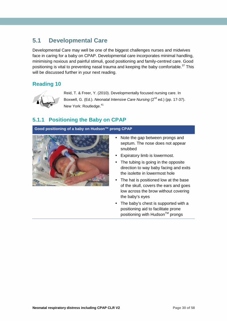
5.1.1 Positioning the Baby on CPAP Good positioning of a baby on Hudson™ prong CPAP
Note the gap between prongs and septum. The nose does not appear snubbed
Expiratory limb is lowermost. The tubing is going in the opposite
direction to way baby facing and exits the isolette in lowermost hole
The hat is positioned low at the base of the skull, covers the ears and goes low across the brow without covering the baby’s eyes
The baby’s chest is supported with a positioning aid to facilitate prone positioning with HudsonTM prongs

Neonatal respiratory distress including CPAP CLR V2 Page 31 of 58
Poor positioning of a baby on Hudson™ prong CPAP
This baby is clearly not contained and is unsettled. This goes against developmental care principles and will present difficulties keeping equipment in place and maintaining a seal
Providing a nest/supportive bedding may assist with comfort and containment of this baby
Also, the expiratory limb is uppermost in this picture (problematic for rain out)
The bonnet is positioned very high on this baby’s brow which is impacting the placement of the prongs. The nose then appears snubbed and a pressure area is starting to form
Good positioning of a baby on a midline interface e.g. Fisher & PaykelTM
Note the gap between prongs and septum. The baby’s nose does not appear snubbed
The hat is low on the brow and rear of skull
The baby has been positioned in accordance to developmental care principles (e.g. baby well contained and facilitating hand to mouth movement)
Poor positioning of a baby on a midline interface e.g. Fisher & PaykelTM
Though the gap between prongs and septum cannot be observed in this photo, the prongs are being forced upwards and therefore ‘snubbing’ the nose
The bonnet has been positioned poorly (too low and too tight), as seen by the skin fold above the eyes
Developmental care positioning is inadequate which will result in dislodgement of the prongs or movement upwards against the septum leading to pressure damage
Images courtesy of the Grantley Stable Neonatal Nursery Image Library, with permission.

Neonatal respiratory distress including CPAP CLR V2 Page 32 of 58
So what positioning is best? Adhering to the basic principles of positioning a baby is necessary; it will be a little more challenging and time consuming to get the baby in a good position whilst he/she is being nursed on CPAP. Positions that enhance containment, reduce disorganised behaviours and promote hand to mouth movements will ultimately settle the infant. There is controversy surrounding the thought that prone and supine positioning may be more beneficial to oxygenation than side lying22,42; ultimately optimal positioning will result in a contained and comfortable baby, clinically stable, with no pressure generating on the nares or septum.
Variation of positioning is important to ensure adequate development of head shape, flexed support of joints, to facilitate hand to mouth gestures and later, walking and crawling.41 The literature supports position changes on a 3-6 hourly basis.22 Due to issues with oxygen and energy consumption, thermoregulation and neurodevelopment, it is necessary to ensure that babies in the acute stages of respiratory distress are handled minimally and monitored carefully3; unit policy and clinical signs such a tolerance to handling, cardiorespiratory stability and skin integrity need to be considered when making clinical judgements regarding frequency of cares. During the acute phase of respiratory distress, atelectasis will occur when the infant spending time spent off CPAP and may take hours for the baby to re-recruit collapsed alveoli21; this increases energy expenditure unnecessarily and can often lead to an increased oxygen requirement. For this reason it is ideal to have two person cares at this time. One nurse/midwife applying CPAP whilst the other performs cares.
The nurse/midwife caring for a baby on CPAP should advocate for Kangaroo care as a measure to promote comfort, reduce pain and encourage parent-infant bonding.37 Each baby should be individually assessed as to their stability to manage kangaroo care. At times this may not be advisable to allow for kangaroo care especially over the first couple of days when the baby’s respiratory distress may be at its most acute. At this time it would be wise to disturb the infant only when absolutely necessary.
Though somewhat challenging, it is important that nurses/midwives implement strategies in relation to positioning and care giving techniques to achieve good developmental outcomes for all newborns.
Nurses/midwives should be aware that the nursery environment is not conducive to developmental care. This is particularly significant for the sick or preterm baby who undergoes numerous medical and nursing interventions and may endure a long period of time in an isolette.
Neonatal respiratory distress including CPAP CLR V2 Page 33 of 58
Clinical Tips – Preventing Pressure Injuries
Some suggestions to help when caring for the baby on CPAP and trying to maintain airway patency and patient comfort:
Use a chin strap if mean airway pressure decreases or bubble has stopped but resumes when you gently close the baby’s mouth
Position the baby prone with their hand tucked under their chin to facilitate closure of the mouth and good neurodevelopmental positioning
Use comfort measures such as swaddling, decreased light and noise Use a roll under the baby’s neck or under their chest to help keep the airway
patent
If a product is used on the baby’s nose to assist with maintaining a seal e.g.
ComfeelTM, it is essential the dressing is checked regularly for trapped moisture,
changed at regular intervals to allow for assessment of underlying skin integrity21 and
removed for trials off CPAP.
Reflect on your current practice and think of ways that you currently incorporate developmental care in your nursery?
Activity 10.2
In order to address airway patency, how would the following strategies be addressed?
a) Removal of nasal secretions (frequency/technique)
b) Prevention of drying of secretions
Activity 10.3
In order to maintain mean airway pressure, how will the following strategies will be addressed?
a) Maintain prongs in nares
b) Prong size prevents air leak
c) Minimise oral air leaks

Neonatal respiratory distress including CPAP CLR V2 Page 34 of 58
Activity 10.4
In an effort to reduce gastric distention, how would you address the following?
a) Assessment of gastric distention
b) Gastric decompression
Let’s have another look at how Jack is progressing…
Over the last few hours, Jack has shown signs of worsening respiratory distress. His work of breathing and oxygen requirement are increasing despite being settled and handled minimally. You have suctioned his mouth and nares which yielded only minimal secretions and have determined that the prescribed level of CPAP is being delivered and there is no major leak. You aspirated the stomach contents which showed only a scant amount of air. You discuss the infant’s clinical presentation and deteriorating blood gases with the medical officer, who decides to increase the CPAP from 7 cm H2O first to 8 and then to 9 cm H2O.
Activity 10.5
Despite the increased pressure Jack shows no signs of improvement. In your response booklet, outline why higher levels of CPAP may not improve Jack’s condition.
5.2 Supportive Care Supportive care is equally important to the care of a baby on CPAP in ensuring that trauma, respiratory distress and hospital stay is reduced, this includes use of minimal handling and comfort measures such as analgesia and feeding. Consider what measures can be taken to reduce handling for the baby on CPAP. Ensuring that cares are clustered is vital. Any sick or preterm baby should be cared for in this method by ensuring that nappy changes, positioning, suctions, temperature monitoring and feeds all occur together at specific times unless otherwise necessary. Sometimes too many interventions clustered together can be too overwhelming; monitor the baby’s response and individualise your caregiving activities accordingly.
5.2.1 Analgesia It’s important to note that babies may experience some discomfort and in some cases pain whilst on CPAP. Review local nursery policies on pain relief in neonates especially if your nursery uses more invasive types of CPAP devices, such as the single long prong nasopharyngeal tube. Not all babies will require analgesics; ongoing skilled assessment of infant cues will determine appropriate and timely intervention. Also consider the use of non-pharmacological methods of pain relief as appropriate.

Neonatal respiratory distress including CPAP CLR V2 Page 35 of 58
5.2.2 Feeding Refer to the Queensland Clinical Guideline - Neonatal respiratory distress including CPAP5 for guidelines on supportive care. The guidelines recommend small trophic feeds as research suggest that it may reduce the duration of both respiratory distress and hospital admission. Remembering to vent the orogastric tube between feeds will minimise the risk of gastric distension and vomiting and increase comfort for the baby.22
Activity 11
In your response booklet, formulate a nursing care plan based on Jack’s scenario. Utilise the Queensland Clinical Guideline5, local policy, readings provided within this package and nursing experience to formulate a plan of care for baby Jack.
Jack eventually stabilises, and after two days begins to show signs of improvement. The CPAP is ceased after three days, without the development of any complications. Great work!!
Conclusion Caring for the neonate on CPAP requires nursing staff to be meticulous in their nursing assessment, observations and attention to detail. In addition to knowledge of the physiology behind respiratory distress and CPAP, an awareness of the available equipment and interfaces being used is essential, along with the potential advantages and disadvantages of those methods. CPAP is increasingly being used for more preterm neonates and in an increasing number of non-tertiary centres. It is often more time consuming than caring for a ventilated baby and requires skill in positioning the baby, troubleshooting the CPAP device and caring for the family unit as a whole. Maintaining current clinical and theoretical knowledge is key to maintaining a high standard of nursing care of the baby on CPAP.
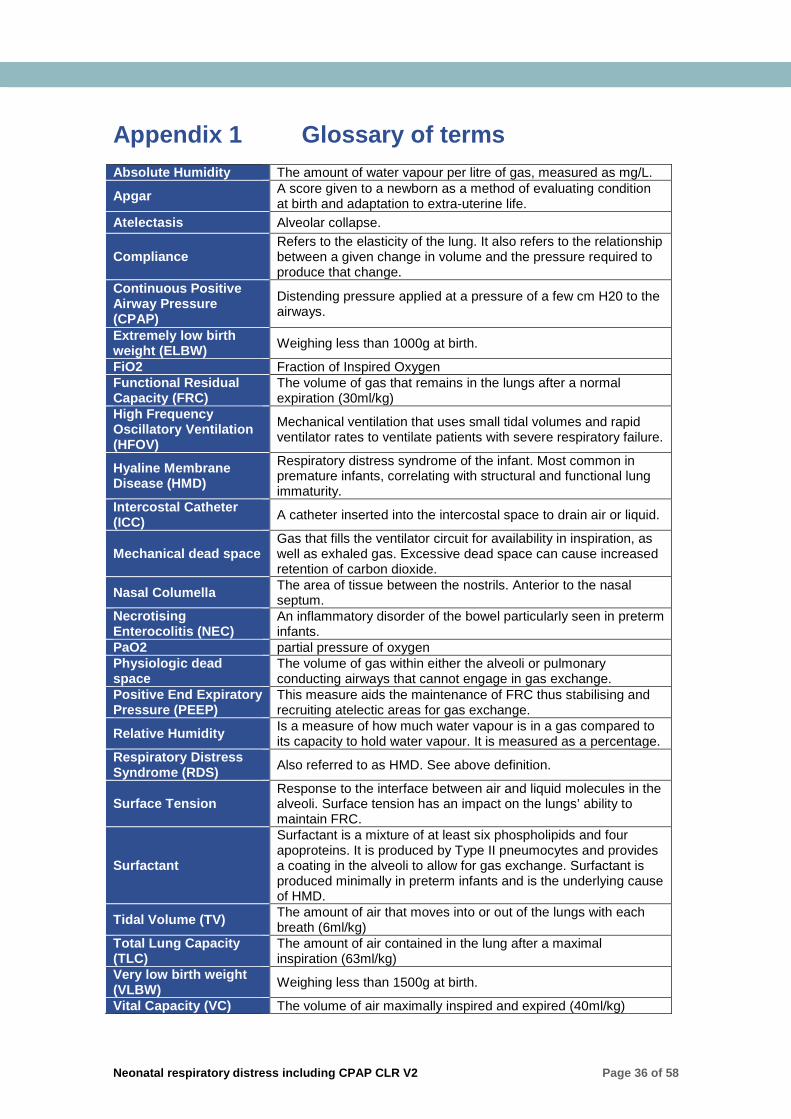
Neonatal respiratory distress including CPAP CLR V2 Page 36 of 58
Appendix 1 Glossary of terms Absolute Humidity The amount of water vapour per litre of gas, measured as mg/L.
Apgar A score given to a newborn as a method of evaluating condition at birth and adaptation to extra-uterine life.
Atelectasis Alveolar collapse.
Compliance Refers to the elasticity of the lung. It also refers to the relationship between a given change in volume and the pressure required to produce that change.
Continuous Positive Airway Pressure (CPAP)
Distending pressure applied at a pressure of a few cm H20 to the airways.
Extremely low birth weight (ELBW) Weighing less than 1000g at birth.
FiO2 Fraction of Inspired Oxygen Functional Residual Capacity (FRC)
The volume of gas that remains in the lungs after a normal expiration (30ml/kg)
High Frequency Oscillatory Ventilation (HFOV)
Mechanical ventilation that uses small tidal volumes and rapid ventilator rates to ventilate patients with severe respiratory failure.
Hyaline Membrane Disease (HMD)
Respiratory distress syndrome of the infant. Most common in premature infants, correlating with structural and functional lung immaturity.
Intercostal Catheter (ICC) A catheter inserted into the intercostal space to drain air or liquid.
Mechanical dead space Gas that fills the ventilator circuit for availability in inspiration, as well as exhaled gas. Excessive dead space can cause increased retention of carbon dioxide.
Nasal Columella The area of tissue between the nostrils. Anterior to the nasal septum.
Necrotising Enterocolitis (NEC)
An inflammatory disorder of the bowel particularly seen in preterm infants.
PaO2 partial pressure of oxygen Physiologic dead space
The volume of gas within either the alveoli or pulmonary conducting airways that cannot engage in gas exchange.
Positive End Expiratory Pressure (PEEP)
This measure aids the maintenance of FRC thus stabilising and recruiting atelectic areas for gas exchange.
Relative Humidity Is a measure of how much water vapour is in a gas compared to its capacity to hold water vapour. It is measured as a percentage.
Respiratory Distress Syndrome (RDS) Also referred to as HMD. See above definition.
Surface Tension Response to the interface between air and liquid molecules in the alveoli. Surface tension has an impact on the lungs’ ability to maintain FRC.
Surfactant
Surfactant is a mixture of at least six phospholipids and four apoproteins. It is produced by Type II pneumocytes and provides a coating in the alveoli to allow for gas exchange. Surfactant is produced minimally in preterm infants and is the underlying cause of HMD.
Tidal Volume (TV) The amount of air that moves into or out of the lungs with each breath (6ml/kg)
Total Lung Capacity (TLC)
The amount of air contained in the lung after a maximal inspiration (63ml/kg)
Very low birth weight (VLBW) Weighing less than 1500g at birth.
Vital Capacity (VC) The volume of air maximally inspired and expired (40ml/kg)

Neonatal respiratory distress including CPAP CLR V2 Page 37 of 58
Appendix 2 Nasal Prong Tube CPAP The following information has been taken from the Royal Brisbane & Women’s Hospital CPAP Procedure26 with permission.
Indications The nasopharyngeal tube (NPT) CPAP may be utilised in the following circumstances:
To overcome upper airway obstruction due to syndromes, i.e. Pierre-Robin Sequence and Treacher Collins Syndrome
Post-operatively following insertion of a ventriculo-peritoneal shunt or reservoir If significant loss of skin/septal integrity has occurred following use of a bi-nasal
prong Congenital nasal conditions that are unable to be managed with a bi-nasal device
(e.g. certain types of cleft lip/palate; unilateral choanal atresia)
Principles Single prong NPT insertion is always a two person procedure, with an assistant
(Registered Nurse/Midwife (RN/RM) or Medical Officer) to physically contain the neonate and observe cardiorespiratory monitoring
Suctioning at least 6th hourly is a requirement for all neonates on single prong NPT CPAP due to the increased amount and tenacity of secretions. Frequency should be individualised according to the neonate’s needs, with some neonates requiring 3 or 4 hourly suction to maintain patency of the NPT. Refer to steps below on nasopharyngeal tube suctioning
Single prong NPT should be changed on a daily basis, with the exception of neonates in the first 96hrs of acute lung disease where second daily changes would be preferable (period of inability to tolerate handling coupled with minimal secretions). Each new NPT must be placed in the alternate nostril. This will minimise the opportunity for development of complications such as septal deviation and allow for complete removal of secretions behind and surrounding the NPT

Neonatal respiratory distress including CPAP CLR V2 Page 38 of 58
Equipment Initial insertion only (or when resizing) Disposable tape measure Laryngoscope (size 1 MacIntosh Blade)– All Insertions One (1) IVORY PORTEX tube – size dependant on the neonate’s size and gestational age (Table 3 below)
Sterile dressing towel (or use the inside of the sterile packaging of the portex tube)
Sterile scissors to cut tube to the appropriate measured length
Suction equipment (tested for full function) with size 6 and 8 Fg suction catheters
Lubricant (e.g. KY Jelly TM) Barrier wipes (e.g. Cavilon TM or Comfeel TM)
Two (2) hydrocolloid skin protection strips (e.g. Comfeel TM) – cut and shaped as per Figure 2 below to a size appropriate for the neonate
½ inch Leukoplast TM pieces (cut and shaped as per Figure 2 below to a size appropriate for the neonate) to secure the tube
Adhesive removal wipe for removal of tapes if prong already insitu Wet and dry wash cloth
0.9 % sodium chloride drawn up in a 1 mL syringe for facilitating secretion removal (dictated by neonate’s individual requirements)
Peanut shaped pillow or other device to maintain midline positioning during NPT insertion
10 mL syringe (to aspirate orogastric tube (OGT) if applicable)
Consider non-pharmacological pain relief as appropriate
NPT to maintain open airway e.g. Pierre-Robin Sequence or Treacher Collins Syndrome Humidification device (e.g. swedish nose) will be necessary
NPT for delivery of CPAP: Ventilator/Bubble CPAP device with parameters set to the individual neonate’s requirements including: CPAP mode Appropriate flow (recommended
6 L/min) PEEP/FiO2
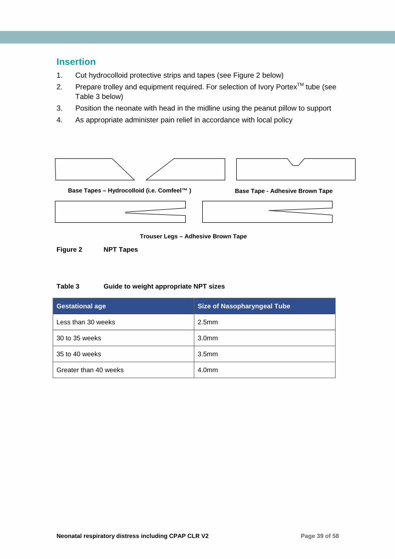
Neonatal respiratory distress including CPAP CLR V2 Page 39 of 58
Insertion 1. Cut hydrocolloid protective strips and tapes (see Figure 2 below) 2. Prepare trolley and equipment required. For selection of Ivory PortexTM tube (see
Table 3 below) 3. Position the neonate with head in the midline using the peanut pillow to support 4. As appropriate administer pain relief in accordance with local policy
Figure 2 NPT Tapes Table 3 Guide to weight appropriate NPT sizes
Gestational age Size of Nasopharyngeal Tube
Less than 30 weeks 2.5mm
30 to 35 weeks 3.0mm
35 to 40 weeks 3.5mm
Greater than 40 weeks 4.0mm
Base Tapes – Hydrocolloid (i.e. Comfeel™ ) Base Tape - Adhesive Brown Tape
Trouser Legs – Adhesive Brown Tape

Neonatal respiratory distress including CPAP CLR V2 Page 40 of 58
5. For the first insertion of the NPT for the neonate:
Operator Measures the distance from the corner of the nostril, to the tragus of the ear; to
calculate the TAPING distance for the NPT Calculates the CUTTING distance of the NPT by adding 1.5 cm to this
measurement; Aspirates the OGT/NGT insitu (OGTs are preferred for the duration of CPAP
therapy)
Figure 3 Measuring for NPT Figure 4 Cut the NPT Figure 5 Lubricate the NPT
Assistant: 6. Cuts the tube at the calculated length, reconnects the blue connector and places
onto the sterile field (green drape or inside NPT packaging) with the lubricant 7. Positions, gently restrain the neonate to promote ease of insertion If there is already an NPT insitu:
Operator: 8. Unwinds the brown tapes securing the NPT and removes the tube and tapes 9. Suctions the neonate: Oropharynx first, then nostrils (to prevent oral contents
being aspirated during nasopharyngeal suction) Note: neonates who have had a NPT insitu for a period of time usually have large amounts of thick nasopharyngeal secretions, therefore a clear airway must always be ensured
10. Check the condition of the hydrocolloid film. If the film looks moist, white in appearance, or is lifting at the edges, it should be replaced. If it is in good condition you do not have to replace it
11. If replacing the hydrocolloid protective film, use an adhesive removal wipe when removing tapes; this is critical in order to maintain the neonate’s skin integrity. Ensure excess fluid from the adhesive removal wipe does not spill into the neonate’s eye area; use a cotton applicator soaked in the adhesive remover fluid instead if necessary
12. Wash neonate’s face with a warm damp cloth and then pat dry 13. Apply the barrier wipe from cheek to cheek and allow to dry 14. Apply hydrocolloid strips to each cheek (Refer to Figure 6 below)
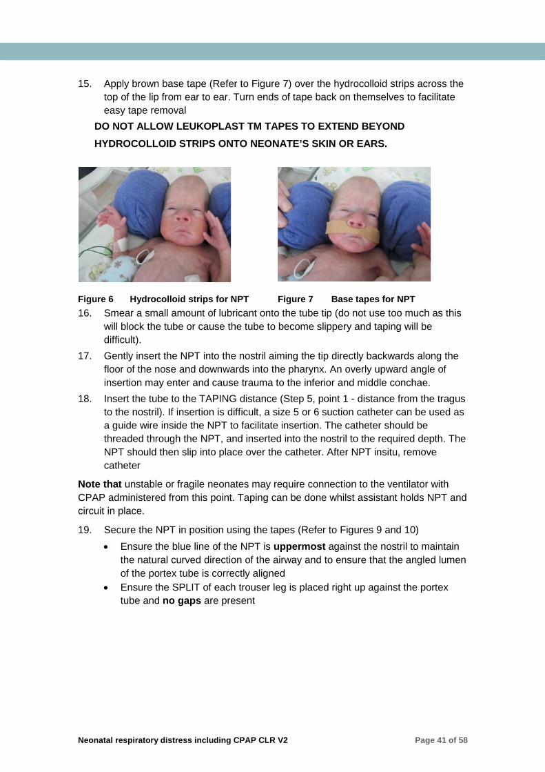
Neonatal respiratory distress including CPAP CLR V2 Page 41 of 58
15. Apply brown base tape (Refer to Figure 7) over the hydrocolloid strips across the top of the lip from ear to ear. Turn ends of tape back on themselves to facilitate easy tape removal
DO NOT ALLOW LEUKOPLAST TM TAPES TO EXTEND BEYOND HYDROCOLLOID STRIPS ONTO NEONATE’S SKIN OR EARS.
Figure 6 Hydrocolloid strips for NPT Figure 7 Base tapes for NPT 16. Smear a small amount of lubricant onto the tube tip (do not use too much as this
will block the tube or cause the tube to become slippery and taping will be difficult).
17. Gently insert the NPT into the nostril aiming the tip directly backwards along the floor of the nose and downwards into the pharynx. An overly upward angle of insertion may enter and cause trauma to the inferior and middle conchae.
18. Insert the tube to the TAPING distance (Step 5, point 1 - distance from the tragus to the nostril). If insertion is difficult, a size 5 or 6 suction catheter can be used as a guide wire inside the NPT to facilitate insertion. The catheter should be threaded through the NPT, and inserted into the nostril to the required depth. The NPT should then slip into place over the catheter. After NPT insitu, remove catheter
Note that unstable or fragile neonates may require connection to the ventilator with CPAP administered from this point. Taping can be done whilst assistant holds NPT and circuit in place.
19. Secure the NPT in position using the tapes (Refer to Figures 9 and 10) • Ensure the blue line of the NPT is uppermost against the nostril to maintain
the natural curved direction of the airway and to ensure that the angled lumen of the portex tube is correctly aligned
• Ensure the SPLIT of each trouser leg is placed right up against the portex tube and no gaps are present

Neonatal respiratory distress including CPAP CLR V2 Page 42 of 58
If this is the first NPT insertion, the NPT depth must be checked with laryngoscope by a Medical Officer or Senior Nurse/Midwife. NPT tip should only just appear behind the uvula and is positioned in the upper pharynx. The position of the tube may need to be re-adjusted following this check.
20. Connect/reconnect the neonate to the ventilator. Check CPAP and FiO2 settings ordered
21. If not already in situ insert OGT and tape to the top lip. Refer to Figure 11 and local OGT insertion policy
An OGT will be required for the duration of CPAP to help deflate the stomach.
Figure 10 Secure orogastric tube 22. Position neonate comfortably and secure ventilation tubing to ensure there is no
traction 23. Dispose of used equipment according to local policy 24. Document details of the procedure on observation sheet, nursing care plan and in
the patient record, including: • Size, cut length and taped length of NPT • Ventilation (CPAP) settings • Suction results and cares performed
Application 1st trouser leg: Lower leg across lip 2nd leg over, then around tube in a
spiral manner
Application 2nd trouser leg Top leg goes over bridge of nose 2nd leg goes under, then around tube in
a spiral manner
Figure 8 Secure the NPT Figure 9 Second trouser leg

Neonatal respiratory distress including CPAP CLR V2 Page 43 of 58
25. Observe the neonate for signs of respiratory distress during the procedure. Liaise with RMO if was intolerant of this procedure or has subsequently developed increased work of breathing/oxygen requirement. Document any evidence of nasal trauma such as bloody secretions or blood stained plugs obtained during suction
26. Ensure parents are kept informed of neonate’s progress and condition
Ongoing Care: 27. Gastric venting (aspiration) should be attended to at least 6th hourly. This should
be individualised based on the neonate’s needs as some neonates may require venting to commence 1 hour after feeds (to allow for digestion) and continuing on until next feed time
28. Neonate should be positioned according to developmental care principles 29. NPT suctioning should be undertaken as per procedure outlined below
Complications: Nasal trauma/ turbinate damage/nasal bridge cellulitis/inflammation or pressure
erosion of the septum Increased nasal secretions leading to airway obstruction Apnoea, bradycardia and oxygen desaturation Gastric distension Pain and discomfort Pneumothorax Trauma to skin of cheeks, upper lip and nasal bridge from frequent changing of
tapes
NPT Suctioning NPT suctioning is performed to remove secretions via the nasopharyngeal tube. Frequency should be based on the individual neonate’s respiratory status and tolerance. However, as a minimum, suctioning via the NPT should be performed at least 6th hourly to ensure patency is maintained. An appropriately sized suction catheter should be used to ensure that the NPT is not totally occluded and decrease the potential for inadvertent generation of excessive positive pressure.
Instillation of saline to assist secretion removal is not routine but may also be required to loosen secretions
Pre-oxygenating is not a standard practice within the GSNU; however it may be required in some neonates who demonstrate poor tolerance to and recovery from suction
The suction catheter should only be passed to the measured length of the NPT plus 3 cm for the connector. Deep suction (beyond the measured length of the NPT + connector) is not routine and may cause adverse outcomes related to mucosal trauma. Mucosal trauma can occur anywhere in a neonatal airway when the suction catheter comes in contact with mucosal tissue
NPT suction is always a two person procedure.
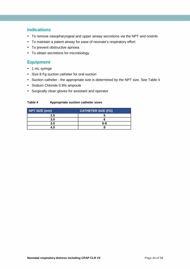
Neonatal respiratory distress including CPAP CLR V2 Page 44 of 58
Indications To remove nasopharyngeal and upper airway secretions via the NPT and nostrils To maintain a patent airway for ease of neonate’s respiratory effort To prevent obstructive apnoea To obtain secretions for microbiology
Equipment 1 mL syringe Size 8 Fg suction catheter for oral suction Suction catheter - the appropriate size is determined by the NPT size. See Table 4 Sodium Chloride 0.9% ampoule Surgically clean gloves for assistant and operator Table 4 Appropriate suction catheter sizes
NPT SIZE (mm) CATHETER SIZE (FG) 2.5 5 3.0 6 3.5 6-8 4.0 8
Neonatal respiratory distress including CPAP CLR V2 Page 45 of 58
Procedure 1. Gather equipment and place on neonate’s incubator 2. If indicated draw up 0.3–0.5 mL of Sodium Chloride 0.9% (more if necessary for
each individual baby). Check saline with assistant 3. Check oxygen and suction equipment ensuring a safe vacuum pressure of less
than 100mmHg is obtained 4. Assistant:
• Silences alarms • Removes the Fg 8 suction catheter already attached to the suction tubing and
makes adjustments to suction apparatus as necessary • Opens one end of the suction catheter packet outside the cot portholes (to
minimize noise inside cot for neonate) • Washes hands or applies alcohol gel after touching suction catheter packet
5. Operator: • Gathers the suction catheter in the designated clean suctioning hand without
contaminating the catheter tip (leaving the first 5-10 cm untouched) and • Connects sterile suction catheter to the suction tubing
6. Assistant: • Supports the NPT and gently restrains the neonate throughout the procedure • Disconnects the NPT from the ventilator circuit manifold; and instils the 0.1
mL of Sodium Chloride 0.9% to loosen secretions (if required)
If this results in a bradycardic episode or desaturation, wait if possible until these have been stabilised before suctioning.
7. Operator: • Waits for 2-3 breaths of the neonate (if possible) before inserting the suction
catheter into the NPT. This allows the Sodium Chloride 0.9% to loosen secretions (the NPT can be reconnected to the ventilator while waiting; which will maintain functional residual capacity of the lung)
• Advances the suction catheter (without applying suction) to the pre-measured distance (cut length of NPT + connector. See Fig.12 below) using the designated gloved hand
• Applies suction and waits 2-3 seconds, maintaining suction during slow withdrawal of suction catheter Too rapid withdrawal may not secure secretions within the suction catheter, leaving them in the airway
8. Assistant reconnects manifold to NPT between suctions and allow the neonate to recover before continuing
9. Assess vital signs, oxygen saturation, chest wall movement and colour throughout the procedure. If neonate requires increased support, the assistant may manipulate oxygen settings to meet need
10. The operator will advise the assistant when to continue with the procedure according to neonate’s tolerance

Neonatal respiratory distress including CPAP CLR V2 Page 46 of 58
11. Depending on each individual neonate, repeat this procedure in relation to the amount and tenacity of secretions present.
Figure 11 Length of NPT for suction
Distance = cut length of NPT + length of NPT connector (3 cm)
12. Further saline flushes may be necessary to loosen secretions 13. Ensure NPT is securely attached to ventilator circuit manifold for remainder of
procedure 14. When NPT suctioning is complete, remove small NPT suction catheter and
replace with the new Fg 8 suction catheter 15. Perform oral suction; THEN suction the nasopharynx. This ensures that oral
contents are not aspirated • For oro/nasopharyngeal suction; (oro) measure from the tragus (ear) to
corner of mouth to determine the distance to the back of the pharynx; (naso) measure from the tragus (ear) to corner of nostril to determine the distance to the back of the pharynx
16. Use the rest of the Sodium Chloride 0.9% to clear the suction catheter 17. Assistant
• Removes the used suction equipment via the “dirty” porthole, NOT the side portholes
• Turns suction unit off to minimise noise in the neonate’s surroundings. • Re-activates alarms
18. Operator
• Checks position and traction on NPT
• Changes position of the neonate as necessary
• Ensure CPAP resumes original settings in place prior to the procedure according to neonate’s tolerance
• Documents the procedure on observation sheet and in the patient record including:
o Neonate’s response/tolerance to procedure
o Consistency, colour and amount of secretions
19. Refer any changes in secretions or concerns to the Medical Officer
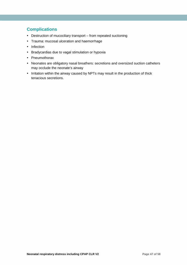
Neonatal respiratory distress including CPAP CLR V2 Page 47 of 58
Complications Destruction of mucociliary transport – from repeated suctioning Trauma: mucosal ulceration and haemorrhage Infection Bradycardias due to vagal stimulation or hypoxia Pneumothorax Neonates are obligatory nasal breathers: secretions and oversized suction catheters
may occlude the neonate’s airway Irritation within the airway caused by NPTs may result in the production of thick
tenacious secretions.

Neonatal respiratory distress including CPAP CLR V2 Page 48 of 58
Appendix 3 Bubble CPAP: Minimising Rain Out The following information and images are courtesy of Fisher & PaykelTM
Set-Up for all Fisher & Paykel HealthcareTM humidified Circuits (stated from position viewing front of humidifier)
Figure 12 Correct humidifier set up
Figure 13 Correct probe set up
Gas Source i.e. Inlet Manifold LHS Blue Inspiratory Tube
RHS
Probes on the right – You’ve got it right!
NB: Views from front of Humidifier Inset Probes into circuit fully – i.e.: no ‘blue’ showing Turn on MR850 Humidifier & use default temperature setting for
Bubble CPAP – i.e. ‘Invasive Mode’
Position Water Chamber with Auto Feed Tubing at back – chamber groove directed at front
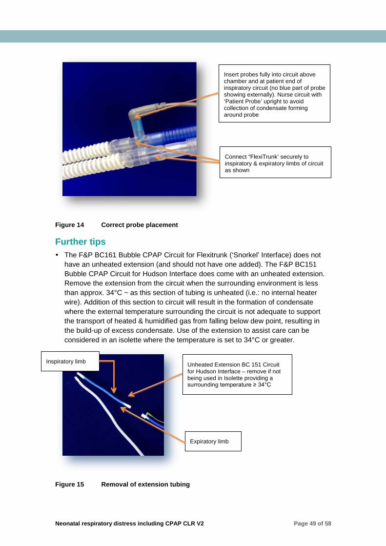
Neonatal respiratory distress including CPAP CLR V2 Page 49 of 58
Figure 14 Correct probe placement
Further tips The F&P BC161 Bubble CPAP Circuit for Flexitrunk (‘Snorkel’ Interface) does not
have an unheated extension (and should not have one added). The F&P BC151 Bubble CPAP Circuit for Hudson Interface does come with an unheated extension. Remove the extension from the circuit when the surrounding environment is less than approx. 34°C ~ as this section of tubing is unheated (i.e.: no internal heater wire). Addition of this section to circuit will result in the formation of condensate where the external temperature surrounding the circuit is not adequate to support the transport of heated & humidified gas from falling below dew point, resulting in the build-up of excess condensate. Use of the extension to assist care can be considered in an isolette where the temperature is set to 34°C or greater.
Figure 15 Removal of extension tubing
Insert probes fully into circuit above chamber and at patient end of inspiratory circuit (no blue part of probe showing externally). Nurse circuit with ‘Patient Probe’ upright to avoid collection of condensate forming around probe
Connect “FlexiTrunk’ securely to inspiratory & expiratory limbs of circuit as shown
Unheated Extension BC 151 Circuit for Hudson Interface – remove if not being used in Isolette providing a surrounding temperature ≥ 34°C
Inspiratory limb
Expiratory limb

Neonatal respiratory distress including CPAP CLR V2 Page 50 of 58
If the temperature reading of the gases measured by the proximal airway probe closest to the patient decreases, check for condensation forming around it. When this ‘patient probe’ has been in contact with cooling condensate it may register temperatures lower than that set. To avoid this, position the ‘patient probe’ tip down. If you have to remove excess water from the probe, remove from the circuit and wipe with an alcohol swab
To minimise the influence of external environment & ambient air +/- drafts (e.g. air-conditioning, fans, etc.) on circuit performance, ensure all circuit and probe connections are secure and tight, with probes pushed ‘all the way home’ exposing no blue part of probe
Where the circuit is exposed to radiant heat (e.g. open resuscitaire or mobile infant warmers for instance), the use of adhesive protective probe covers can reduce the impact of heat sources affecting probe readings & possible resultant condensate formation
Circuit should be positioned to drain condensate away from the patient – avoid using circuit stands and support devices that elevate tubing such that condensate is drained inappropriately towards patient
Humidifier chamber should be positioned below the patient to encourage any formation of condensate to drain away from the patient & return to the chamber
Corrugations in inspiratory tubing allow flexibility for tube positioning and trap droplets of condensate to control migration and pooling – prior to repositioning or moving patient, manipulate tubing including interface to drain any formed condensate back to the humidifier chamber before moving the patient
Condensate in the white/clear expiratory limb is normal & utilised to promote auto-filling of the CPAP generator (‘Bubble Box’). As with the inspiratory limb, position this so expected condensate drains away from patient & towards the CPAP generator. Excess fluid in the CPAP generator will spill over into the collection chamber for easy removal and emptying without disruption to therapy or a break in the circuit
Additionally, ensure the inspiratory limb is placed uppermost to assist with the drainage of ‘rain out’ through the expiratory limb

Neonatal respiratory distress including CPAP CLR V2 Page 51 of 58
Appendix 4 Clinical Skills Assessment Tool Nursing care of the baby with respiratory distress requiring CPAP
Assessor to ensure that the following are complete prior to clinical assessment:
� Neonatal respiratory distress including CPAP CLR
The participant will demonstrate: The ability to correctly assess the neonate’s needs and perform the clinical skills required to manage the neonate with respiratory distress requiring CPAP in a safe manner.
PERFORMANCE CRITERIA: Achieved Not
Achieved
Demonstrated awareness of and performs in accordance with current research, local policy and the Queensland Clinical Guideline – Neonatal respiratory distress including CPAP by identifying the following:
• Signs and symptoms of respiratory distress.
• Management of oxygenation. • Blood glucose management.
• Thermoregulation. • Contraindications to CPAP use.
• Demonstrated familiarity with available CPAP generator/s, manipulation of settings and relevant safety aspects.
• Management of CPAP complications including air leaks, pressure injury, abdominal distention and over inflation.
• Signs of CPAP failure. • The rationale for humidification of the CPAP circuit.
• Guidelines for weaning and ceasing CPAP.
• Emergency equipment required for pneumothorax management.
• The correct candidates and the process for consultation and referral to a tertiary centre.
• Frequency of cares required and rationale.
• Frequency of observations.
• The differences, benefits and risks associated with the varying types of CPAP interfaces.
• The rationale for 1 or 2 person cares.
Demonstrated ability to correctly set up CPAP generator with appropriate circuit and humidification.
Demonstrated ability to correctly measure and fit CPAP appliance.
Demonstrated ability to describe the physiology of respiratory distress in the newborn and how CPAP supports the anatomical and physiological difficulties experienced by these babies.
Performed a safety check of the cotside at the commencement of the shift: • Checks safety and resuscitation equipment available and
functional, alarm parameters, CPAP settings against written orders, floors clear of spills/cords and fluid orders against fluids infusing.
• Maintains an awareness of the evacuation procedure for the unit.

Neonatal respiratory distress including CPAP CLR V2 Page 52 of 58
PERFORMANCE CRITERIA: Achieved Not Achieved
Performed a comprehensive physical assessment of the baby using a systematic approach • Utilises other related information to inform assessment e.g. ABG’s,
biochemistry, haematology, microbiology, chest x-ray, CT/MRI scans and antenatal and perinatal history.
Formulated an individualised plan of nursing care incorporating: • Included the family in the formulation of the plan where
appropriate and individualises the plan according to the family’s needs as able e.g. religious or cultural mores.
• Used assessment data as a basis for the plan. • Formulated a predicted outcome of the nursing care plan. • Identified potential problems that may adversely affect the patient. • Identified nursing interventions to address potential problems and
provides rationale. • Contributed to and participates in decision making on the ward
round. • Includes other members of the heath care team e.g.
physiotherapist, social worker, stomal therapist or pharmacist. • Structured nursing interventions/activities to support
neurodevelopment. • Formulated criteria for evaluation of predicted outcomes. • Recognised own abilities and incorporates other nursing staff to
assist or provide guidance if necessary
Managed therapeutic interventions as evidenced by: • Demonstrated accountability for own actions at all times. • Communicated with the baby before commencing any nursing
interventions. • Monitored and responded effectively to any changes in the
patient’s condition and is able to give rationale for response. • Altered equipment settings in collaboration with the
multidisciplinary health care team as required e.g. infusion rates, ventilator and monitor alarms.
• Demonstrated ability to describe the pharmacological effect of medications administered.
• Involved family members in the baby’s care when possible. • Communicated with, and supports the baby’s parents and other
family members. • Reported any deterioration or improvement in the patient’s
condition to the team leader and/or medical officer. • Supported continuity of care by providing a comprehensive
handover to the team leader of the current shift and the nursing staff on the next shift.
Documented assessment findings, baby’s response to handling, nursing care provided and any relevant changes to baby’s status or care: • Documentation is correct and precise and incorporates all aspects
of care.
Demonstrated evidence of therapeutic interaction by:
• Used the correct patient identification process. • Provided privacy as able. • Explained any procedures to the family. • Obtained informed consent from the parents as appropriate.
Positioned the baby in accordance with developmental care principles and takes the disease process into consideration.
Aligned practice to local policy/procedures and the Queensland Clinical Guideline – Neonatal respiratory distress including CPAP.
Applied principles of hand hygiene and aseptic non-touch technique (ANTT) throughout the procedure.
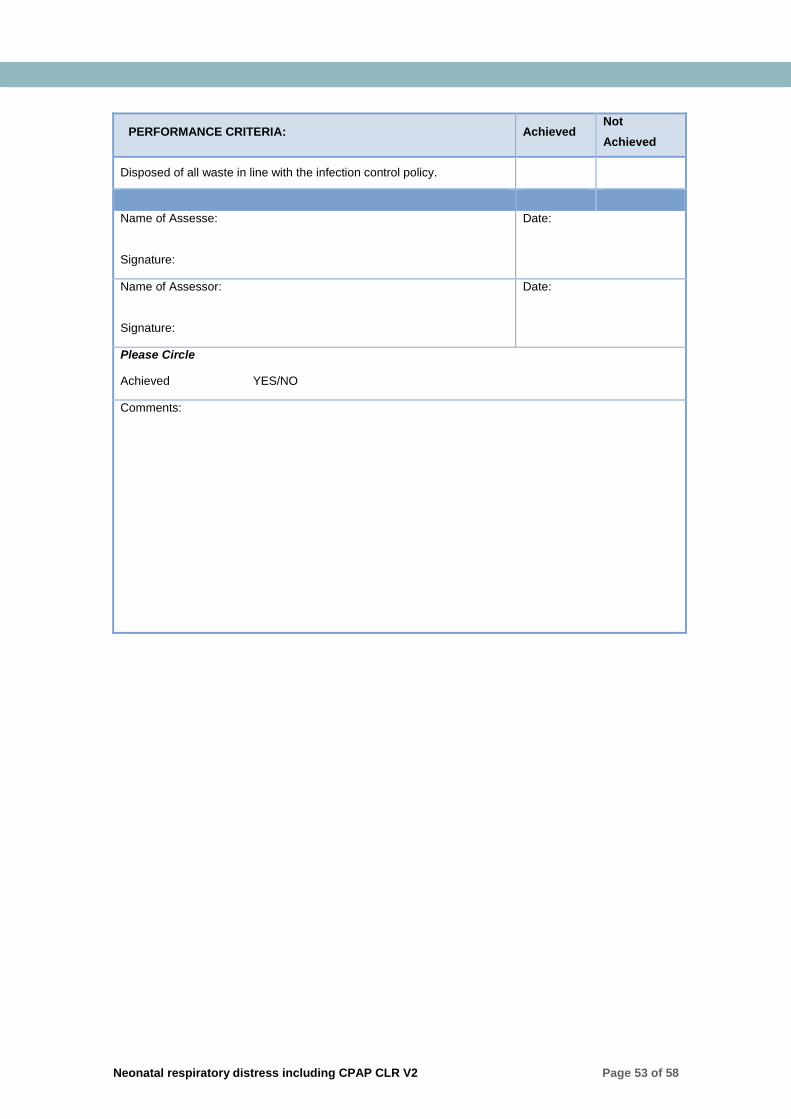
Neonatal respiratory distress including CPAP CLR V2 Page 53 of 58
PERFORMANCE CRITERIA: Achieved Not Achieved
Disposed of all waste in line with the infection control policy.
Name of Assesse:
Signature:
Date:
Name of Assessor:
Signature:
Date:
Please Circle
Achieved YES/NO
Comments:

Neonatal respiratory distress including CPAP CLR V2 Page 54 of 58
References 1. Diehl-Jones B. Physiological Principles of the Respiratory System. In: Fraser D, editor. Acute Respiratory Care of the Neonate. 3rd ed. Petaluma, California: NICUInk; 2012.
2. Peterson SW. Understanding the sequence of pulmonary injury in the extremely low birth weight, surfactant-deficient infant. Neonatal Netw. 2009; 28(4):221-9; quiz 255-8.
3. Soltau DTW, A.C. Respiratory System. In: Lott CKJW, editor. Comprehensive Neonatal Nursing Care. 5th ed. New York: Springer Publishing Company; 2014.
4. Fraser D. Chest and Lungs. In: Tapperro EPH, M.E., editor. Physical Assessment of the Newborn. 5th Ed. ed. Petaluma, California: NICU Ink; 2015.
5. Queensland Clinical Guidelines. Neonatal respiratory distress including CPAP. 2014:1-21.
6. Wiswell TEC, S.E. Non-Invasive Respiratory Support. In: Karotkin JPGEH, editor. Assisted Ventilation of the Neonate. 5th ed. St Louis: Elsevier; 2011.
7. Buckmaster A. Nasal continuous positive airway pressure for respiratory distress in non-tertiary care centres: what is needed and where to from here? J Paediatr Child Health. 2012; 48(9):747-52.
8. Koti J, Murki S, Gaddam P, Reddy A, Reddy MD. Bubble CPAP for respiratory distress syndrome in preterm infants. Indian Pediatr. 2010; 47(2):139-43.
9. Mai CL, Yaster M, Firth P. The development of continuous positive airway pressure: an interview with Dr. George Gregory. Paediatr Anaesth. 2013; 23(1):3-8.
10. Sai Sunil Kishore M, Dutta S, Kumar P. Early nasal intermittent positive pressure ventilation versus continuous positive airway pressure for respiratory distress syndrome. Acta Paediatr. 2009; 98(9):1412-5.
11. Roberts CL, Badgery-Parker T, Algert CS, Bowen JR, Nassar N. Trends in use of neonatal CPAP: a population-based study. BMC Pediatrics. 2011; 11(89).
12. Todd DA, Wright A, Broom M, Chauhan M, Meskell S, Cameron C, et al. Methods of weaning preterm babies <30 weeks gestation off CPAP: a multicentre randomised controlled trial. Arch Dis Child Fetal Neonatal Ed. 2012; 97(4):F236-40.
13. Committee on fetus and newborn. Respiratory support in preterm infants at birth. Pediatrics. 2014; 133(1):171-4.
14. Saxena A, Thapar RK, Sondhi V, Chandra P. Continuous positive airway pressure for spontaneously breathing premature infants with respiratory distress syndrome. Indian J Pediatr. 2012; 79(9):1185-91.
15. Yoder BA, Stoddard RA, Li M, King J, Dirnberger DR. Heated, Humidified High-Flow Nasal Cannula Versus Nasal CPAP for Respiratory Support in Neonates. Pediatrics. 2013; 131(5):e1482-e1490.
16. de Klerk A. In: Physiology of Humidification in Critically Ill Neonates. 2012 Springer Berlin Heidelberg.
17. Lopez E, Gascoin G, Flamant C, Merhi M, Tourneux P, Baud O. Exogenous surfactant therapy in 2013: what is next? Who, when and how should we treat newborn infants in the future? BMC Pediatr. 2013; 13:165.
18. Gopel W, Kribs A, Ziegler A, Laux R, Hoehn T, Wieg C, et al. Avoidance of mechanical ventilation by surfactant treatment of spontaneously breathing preterm infants (AMV): an open-label, randomised, controlled trial. Lancet. 2011; 378(9803):1627-34.
19. Diblasi RM. Nasal continuous positive airway pressure (CPAP) for the respiratory care of the newborn infant. Respir Care. 2009; 54(9):1209-35.

Neonatal respiratory distress including CPAP CLR V2 Page 55 of 58
20. Tagare A, Kadam S, Vaidya U, Pandit A, Patole S. Bubble CPAP versus ventilator CPAP in preterm neonates with early onset respiratory distress--a randomized controlled trial. J Trop Pediatr. 2013; 59(2):113-9.
21. McCoskey L. Nursing Care Guidelines for prevention of nasal breakdown in neonates receiving nasal CPAP. Adv Neonatal Care. 2008; 8(2):116-24.
22. Bonner KM, Mainous RO. The nursing care of the infant receiving bubble CPAP therapy. Adv Neonatal Care. 2008; 8(2):78-95; quiz 96-7.
23. Chowdhury O, Wedderburn CJ, Duffy D, Greenough A. CPAP review. European Journal of Pediatrics. 2012; 171(10):1441-1448. Available from: ccm.
24. De Paoli A, Davis P, Faber B, Morley C. Devices and pressure sources for administration of nasal continuous positive airway pressure (NCPAP) in preterm neonates. Cochrane Database of Systematic Reviews 2008, Issue 1. Art. No.: CD002977. DOI:10.1002/14651858.CD002977.pub2. 2008.
25. Kieran EA, Twomey AR, Molloy EJ, Murphy JF, O'Donnell CP. Randomized trial of prongs or mask for nasal continuous positive airway pressure in preterm infants. Pediatrics. 2012; 130(5):e1170-6.
26. Queensland Health. In: 21032/Proc Continuous Positivie Airway Pressure (CPAP) Management, Neonate. 2013. Brisbane, Australia: Queensland Government.
27. DiBlasi RM. Neonatal noninvasive ventilation techniques: do we really need to intubate? Respir Care. 2011; 56(9):1273-94; discussion 1295-7.
28. Ramanathan R. Nasal respiratory support through the nares: its time has come. J Perinatol. 2010; 30 Suppl:S67-72.
29. De Klerk A. Humidification during noninvasive respiratory support of the newborn: Continuous positive pressure ventilation, and humidified high-flow nasal cannula. In: Esquinas AM, editor. Humidification in the Intensive Care Unit. Berlin: Springer-Verlag; 2012.
30. Jardine LA, Dunster KR, Davies MW. An experimental model for the measurement of inspired gas temperatures in ventilated neonates. Pediatr Pulmonol. 2008; 43(1):29-33.
31. Papoff PM, C. . Pulmonary air leakage. In: G. Buonocore RB, M. Weindling, editor. Neonatology. Milano, Italy: Springer-Verlag Italia; 2012.
32. Fraser D. Complications of positive pressure ventilation. In: Fraser D, editor. Acute Respiratory Care of the Neonate. 3rd ed. Petaluma, California: NICU Ink; 2012.
33. Carlo WA. Gentle ventilation: the new evidence from the SUPPORT, COIN, VON, CURPAP, Colombian Network, and Neocosur Network trials. Early Human Development. 2012; 88:S81-3. Available from: ccm.
34. Pillai MS, Sankar MJ, Mani K, Agarwal R, Paul VK, Deorari AK. Clinical prediction score for nasal CPAP failure in pre-term VLBW neonates with early onset respiratory distress. J Trop Pediatr. 2011; 57(4):274-9.
35. Jardine L, Davies M. Withdrawal of neonatal continuous positive airway pressure: current practice in Australia. Pediatrics International. 2008; 50(4):572–5.
36. Newnam KM, McGrath JM, Estes T, Jallo N, Salyer J, Bass WT. An integrative review of skin breakdown in the preterm infant associated with nasal continuous positive airway pressure. J Obstet Gynecol Neonatal Nurs. 2013; 42(5):508-16.
37. Squires AJ, Hyndman M. Prevention of nasal injuries secondary to NCPAP application in the ELBW infant. Neonatal Netw. 2009; 28(1):13-27.
38. Fischer C, Bertelle V, Hohlfeld J, Forcada-Guex M, Stadelmann-Diaw C, Tolsa J-F. Nasal trauma due to continuous positive airway pressure in neonates. Arch Dis Child Fetal Neonatal Ed. 2010; 95:447-451.

Neonatal respiratory distress including CPAP CLR V2 Page 56 of 58
39. de Winter JP, Machteld, A. G. de Vries, Zimmermann, L. J. Clinical Practice Noninvase respiratory support in newborns. European Journal of Pediatrics. 2010; 169:777-782.
40. Lamburne S. An assessment tool for infants requiring nasal continuous positive airway pressure. Infant. 2014; 10(4):123-126. Available from: ccm.
41. Reid TRF, Y. . Developmentally focused nursing care. In: Boxwell G, editor. Neonatal Intensive Care Nursing. 2nd ed. New York: Routledge; 2010.
42. Brunherotti MA, Martinez EZ, Martinez FE. Effect of body position on preterm newborns receiving continuous positive airway pressure. Acta Paediatr. 2014; 103(3):e101-5.

Neonatal respiratory distress including CPAP CLR V2 Page 57 of 58
Resource Package Final Assessment Sheet
Name of Participant: Position:
Work Unit: Service Line:
Participant Signature: Date:
This assessment sheet when signed is documentation of completion of this package which attracts 28 Continuing Professional Development (CPD) hours of learning.
Components of
Competency
Date/s of
completion
Name of
Assessor
Position of
Assessor
Assessor Signature
All learning
activities
Clinical skill
assessment tool
completed:
Has the participant’s responses met expected standards for all components? Yes No
If no, please detail what further evidence is required:
To be completed when further evidence is provided to meet expected standards.
Participant's Signature: _______________________Date: _________________________
Assessor's Signature: _______________________Date: __________________________
On completion of the above, please present this assessment sheet to the Nurse/Midwifery Educator for recording on Nursing & Midwifery Initiatives Spreadsheet (NMIS) as evidence of training and to your line manager for insertion to your professional development plan and file.

Neonatal respiratory distress including CPAP CLR V2 Page 58 of 58
Department of Health
Neonatal Respiratory Distress including CPAP: Clinical Learning Resource
www.health.qld.gov.au