Embed Size (px)
Citation preview

CHHS17/244
Canberra Hospital and Health ServicesClinical ProcedureNeonatal NeurologyContents
Contents...................................................................................................................................1
Purpose.................................................................................................................................... 2
Scope........................................................................................................................................2
Section 1: Bedside aEEG monitoring........................................................................................2
Section 2: Cooling for HIE.........................................................................................................6
Section 4: Neurovascular Assessment & Observation..............................................................9
Section 5: Seizure Management...............................................................................................9
Section 6: Lumbar Puncture...................................................................................................13
Related Policies, Procedures, Guidelines and Legislation.......................................................16
References..............................................................................................................................16
Search Terms..........................................................................................................................17
Doc Number Version Issued Review Date Area Responsible PageCHHS17/244 1 25/10/2017 01/10/2021 WY&C -
Neonatology1 of 17
Do not refer to a paper based copy of this policy document. The most current version can be found on the ACT Health Policy Register

CHHS17/244
Purpose
This procedure outlines the management of infants with neurological problems in the Department of Neonatology.
Scope
This document pertains to infants nursed in the Department of Neonatology at the Centenary Hospital for Women & Children.
This document applies to the following Canberra Hospital Health Services (CHHS) staff working within their scope of practice: Medical Officers Registered Nurses and Midwives Student Nurses and Midwives working under supervision
Section 1: Bedside aEEG monitoring
BackgroundBedside aEEG monitoring provides a continuous record of electrocortical acitivity of the brain.
Bedside aEEG monitoring allows for early detection and treatment of pathological events, surveillance of interventions, and their effects and a more selective use of formal EEG in the Department of Neonatology.
The criteria for bedside aEEG monitoring includes: Definite or questionable seizures Perinatal asphyxia (pH <7 and encephalopathy 2-3) Apnoea in term or near- term infants Muscle relaxed infants (If muscle relaxation for days and hypoxia/ ischaemia likely)
All abnormal aEEG recordings should be confirmed by multi-channel EEG from the Neurology Department. However, treatment of seizures should not be delayed waiting for a multi-channel EEG.
aEEG findings are to be recorded every 8 hours in the clinical notes, detailing: background activity presence and duration of seizures
How to record aEEGDoc Number Version Issued Review Date Area Responsible PageCHHS17/244 1 25/10/2017 01/10/2021 WY&C -
Neonatology2 of 17
Do not refer to a paper based copy of this policy document. The most current version can be found on the ACT Health Policy Register

CHHS17/244
Equipment Bedside EEG monitoring Neonatal sensor set (electrodes) Positioning aid (measuring tape provided) Skin marker Skin preparation treatment Sterile water Cotton applicator Gauze pads
Procedure1. Position the infant supine2. Use supplied measuring tape and measure from the sagittal suture to the ear tragus and
note the letter (A, B etc) mark the site for electrode placement making sure the picture of the infant’s face on the tape measure is facing the same way as the infant
3. Reverse the tape measure and mark the site on the other side of the head4. Clean the marked areas with sterile water and gauze, parting the hair in a line towards
the top of the head5. Gently pat dry with gauze maintaining the part in the hair6. Clean the area with skin prep using a small swab stick7. Remove the skin prep with gauze swab and water continuing to maintain the part in the
hair8. Pat the area dry with the gauze swab maintaining the part in the hair9. Site the electrodes as follows:
9.1 black electrode at the back and direct leads toward the top of the head9.2 white electrode in front of the black lead ensuring only a 5 mm distance between
the 2 electrodes, direct the leads toward the top of the head9.3 gently turn the infant’s head over and repeat the process
Doc Number Version Issued Review Date Area Responsible PageCHHS17/244 1 25/10/2017 01/10/2021 WY&C -
Neonatology3 of 17
Do not refer to a paper based copy of this policy document. The most current version can be found on the ACT Health Policy Register

CHHS17/244
9.4 reference electrode may be placed on either side of the infant, behind the ear or on the shoulder away from the hair after preparing the skin as above
For Insertion of Needle Electrodes1. Cleanse area that needle electrodes are inserted 2. Hold skin taut and insert needle electrodes at a <30o angle(sub-dermal) just under the
skin parallel to each other
3. When using needle electrodes the reference electrode should be hydrogel4. Place reference electrode as for standard hydrogel electrodes
5. Secure the sub-dermal needle electrode in place with steri-strip as below
Doc Number Version Issued Review Date Area Responsible PageCHHS17/244 1 25/10/2017 01/10/2021 WY&C -
Neonatology4 of 17
Do not refer to a paper based copy of this policy document. The most current version can be found on the ACT Health Policy Register

CHHS17/244
6. On the CFM open the screen using the following:6.1. user name = NICU6.2. password = NICU
7. Push assess patient box on the screen to record the data for a new patient8. If electrodes lift, reapply using a drop of sterile water and apply warmth with warm
cloth. Do not stop the machine if electrodes lift9. Mark events on the recording, i.e. suction, cares, xray etc: to ensure that any changes
are interpreted correctly10. If readout demonstrates:
10.1. impulses outside normal limits contact Medical Officer10.2. contact impedance as displayed by red or yellow alert reapply electrode as above10.3. green light indicates effective electrode function
To change date/time1. Go to ‘Home’2. Touch ‘Tools’ 3. Touch ‘System’ and then ‘Exit to maintenance’4. Touch ‘Date/time’ button then choose either ‘Date’ or ‘Time’ and adjust using arrows5. Touch ‘Accept’ button and return to monitor
Any abnormalities should be discussed with the Neonatal Registrar. The neonatal Registrars are to be informed of seizures immediately.
The type of trace must be recorded on the Neonatal Encephalopathy Chart by the medical staff the trace must be discussed at each ward round
Any significant change must be reported to the Neonatologist by the registrar or nursing staff.
Back to Table of Contents
Doc Number Version Issued Review Date Area Responsible PageCHHS17/244 1 25/10/2017 01/10/2021 WY&C -
Neonatology5 of 17
Do not refer to a paper based copy of this policy document. The most current version can be found on the ACT Health Policy Register

CHHS17/244
Section 2: Cooling for HIE
BackgroundHypoxic ischemic encephalopathy (HIE) is the brain injury caused by hypoxia and ischaemia during the perinatal period. Whole body cooling for infants with moderate-severe encephalopathy has been shown to reduce long-term brain injury. Infants diagnosed with HIE who fulfil the criteria as below, will be whole body cooled within 6 hours of birth to a rectal temperature of 33.0-34.0oC for 72 hours.
The infants that will be cooled must meet the following four criteria:1. More than or equal to 35 weeks gestational age2. Less than 6 hours of age3. Evidence of asphyxia as defined by the presence of at least two of the following-
a) Apgar less than 6 at 10 minutes or continued need for resuscitation with positive pressure ventilation +/- chest compressions at 10 minutes
b) Any acute perinatal event that may result in HIE (ie. Abruption placentae, cord prolapsed, severe FHR abnormality etc.)
c) Cord pH less than 7.0 or base excess of -12mmol/L or lessd) If cord pH is not available, arterial pH less than 7.0 or BE less than -12mmol/L
within 60 minutes of birth.4. The presence of moderate/severe HIE: defined as seizures OR presence of signs in at
least three of the six categories given below:
Category Moderate Encephalopathy Severe EncephalopathyLevel of consciousness Lethargy Stupor/coma
Spontaneous activity Decreased activity No activity
Posture Arms flexed, legs extended(decorticate)
Arms and legs extended
Tone Hypotonia Flaccid
Primitive reflexes Weak suck, incomplete Moro Absent suck, absent MoroAutonomic system (any one of)PupilsHeart rateRespirations
ConstrictedBradycardiaPeriodic breathing
Dilated/non-reactiveVariable heart rateApnoea
Doc Number Version Issued Review Date Area Responsible PageCHHS17/244 1 25/10/2017 01/10/2021 WY&C -
Neonatology6 of 17
Do not refer to a paper based copy of this policy document. The most current version can be found on the ACT Health Policy Register

CHHS17/244
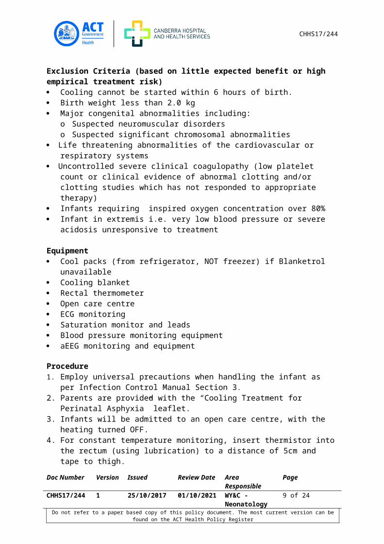
Exclusion Criteria (based on little expected benefit or high empirical treatment risk) Cooling cannot be started within 6 hours of birth. Birth weight less than 2.0 kg Major congenital abnormalities including:
o Suspected neuromuscular disorderso Suspected significant chromosomal abnormalities
Life threatening abnormalities of the cardiovascular or respiratory systems Uncontrolled severe clinical coagulopathy (low platelet count or clinical evidence of
abnormal clotting and/or clotting studies which has not responded to appropriate therapy)
Infants requiring inspired oxygen concentration over 80% Infant in extremis i.e. very low blood pressure or severe acidosis unresponsive to
treatment
Equipment Cool packs (from refrigerator, NOT freezer) if Blanketrol unavailable Cooling blanket Rectal thermometer Open care centre ECG monitoring Saturation monitor and leads Blood pressure monitoring equipment aEEG monitoring and equipment
Procedure1. Employ universal precautions when handling the infant as per Infection Control Manual
Section 3.2. Parents are provided with the “Cooling Treatment for Perinatal Asphyxia” leaflet. 3. Infants will be admitted to an open care centre, with the heating turned OFF.4. For constant temperature monitoring, insert thermistor into the rectum (using
lubrication) to a distance of 5cm and tape to thigh.5. Commence continuous monitoring of ECG, respirations, saturations and blood pressure.6. Observations must be recorded hourly throughout the entire cooling and rewarming
process.7. Attach infant to the aEEG machine as per Neonatal Practice Guidelines and monitor for
at least 72 hours, ideally for 24 hours after normothermia has been achieved and aEEG is normal.
8. Strict fluid balance must be monitored, particular attention must be payed to urine output.
9. Document encephalopathy score, therapy and aEEG findings and investigations on the Neonatal Encephalopathy chart
10. Use the Blanketrol cooling blanket to cool the infant-see operating guide for use11. If cooling blanket is not available, cool the infant by turning the radiant warmer to the
OFF position and by exposing the infant to ambient temperature. If this does not result
Doc Number Version Issued Review Date Area Responsible PageCHHS17/244 1 25/10/2017 01/10/2021 WY&C -
Neonatology7 of 17
Do not refer to a paper based copy of this policy document. The most current version can be found on the ACT Health Policy Register

CHHS17/244
in a decrease in temperature by 1oC within 2 hours, cool packs (from fridge, NOT freezer) may be applied to the back of the neck, head and across the torso.
12. Active cooling will be reduced when the rectal temperature falls below 34.5OC.13. Active cooling is stopped when the temperature falls below 34OC.14. If the temperature falls below 33.5OC the heater output on the radiant warmer will be
manually adjusted to maintain a rectal temperature of approximately 33.5OC.
The following blood tests should be done at least daily:14.1. Electrolytes14.2. Urea, creatinine 14.3. Liver function test 14.4. Full blood count including platelets14.5. Coagulation profile
15. Monitor Arterial Blood Gases, glucose levels and lactate daily or more frequently if clinically indicated.
AlertIf the inspired oxygen increases by more than 20% then active cooling should be reduced (i.e. remove the cool packs or turn the cooling blanket off) Infants treated with anticonvulsants may become hypothermic as a result of the anticonvulsant therapy; therefore ACTIVE cooling may not be necessary.
Cooling may be stopped if there is: Persistent hypoxaemia in 100% oxygen Life threatening coagulopathy Arrythmia requiring medical treatment (not sinus bradycardia)
16. After 72 hours rewarming will occur at a rate not exceeding 0.5OC every two hours using the rewarming facility on the blanketrol
17. Infants will take up to 12 hours to be rewarmed to a target rectal temperature of 37oC.18. Rewarming is a risk for seizures to occur/recur – aEEG monitoring must be continued
duing this time19. To prevent rebound hypothermia the rectal probe measurements should be continued
for six hours after the infant has been rewarmed to a normal body temperature temperature.
20. Accurate documentation of the cooling and rewarming process must be kept.21. MRI of the brain should be done ideally 7-10 days after birth
Back to Table of Contents
Doc Number Version Issued Review Date Area Responsible PageCHHS17/244 1 25/10/2017 01/10/2021 WY&C -
Neonatology8 of 17
Do not refer to a paper based copy of this policy document. The most current version can be found on the ACT Health Policy Register
CHHS17/244
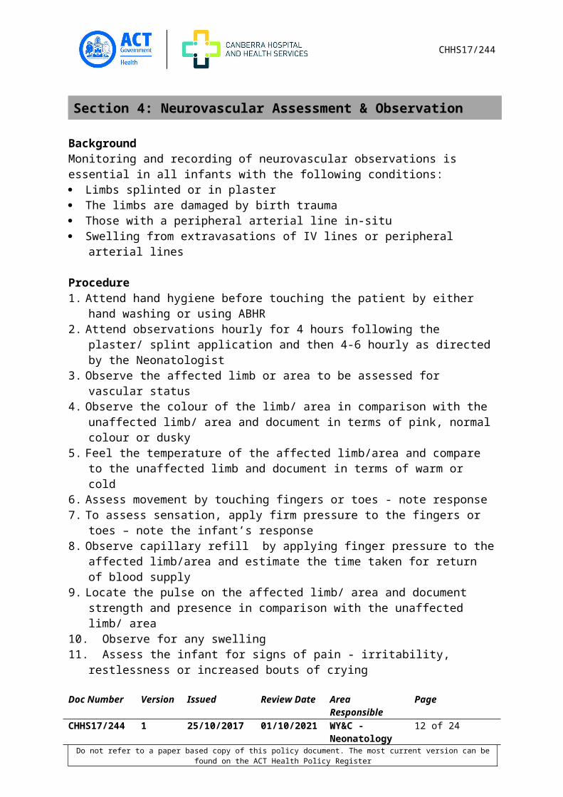
Section 4: Neurovascular Assessment & Observation
BackgroundMonitoring and recording of neurovascular observations is essential in all infants with the following conditions: Limbs splinted or in plaster The limbs are damaged by birth trauma Those with a peripheral arterial line in-situ Swelling from extravasations of IV lines or peripheral arterial lines
Procedure1. Attend hand hygiene before touching the patient by either hand washing or using ABHR2. Attend observations hourly for 4 hours following the plaster/ splint application and then
4-6 hourly as directed by the Neonatologist3. Observe the affected limb or area to be assessed for vascular status4. Observe the colour of the limb/ area in comparison with the unaffected limb/ area and
document in terms of pink, normal colour or dusky5. Feel the temperature of the affected limb/area and compare to the unaffected limb and
document in terms of warm or cold6. Assess movement by touching fingers or toes - note response7. To assess sensation, apply firm pressure to the fingers or toes – note the infant’s
response8. Observe capillary refill by applying finger pressure to the affected limb/area and
estimate the time taken for return of blood supply9. Locate the pulse on the affected limb/ area and document strength and presence in
comparison with the unaffected limb/ area10. Observe for any swelling11. Assess the infant for signs of pain - irritability, restlessness or increased bouts of crying12. Compare observations with previous assessment and report any changes to the team
leader13. Document observations on flow chart and any abnormal findings in the infant’s progress
notes14. Settle the infant in a position of comfort with limbs in natural alignment
Back to Table of Contents
Section 5: Seizure Management
Neonatal seizures are the most common neurological adverse event during the neonatal period and are a sign of an underlying disease process. Treatment is aimed at the underlying cause first, and then at controlling seizures.
Investigations:
Doc Number Version Issued Review Date Area Responsible PageCHHS17/244 1 25/10/2017 01/10/2021 WY&C -
Neonatology9 of 17
Do not refer to a paper based copy of this policy document. The most current version can be found on the ACT Health Policy Register

CHHS17/244
Detailed neurologic examination should be performed, paying attention to level of consciousness, tone, reflexes and head circumference. Please record on the encephalopathy measurement /monitoring chart
Seizures are frequently under-diagnosed, can be brief or mimic normal behaviour.If an infant is suspected of having seizures, an aEEG or EEG should be recorded as soon as possible to confirm seizure activity (see Section 1)
Differential diagnosis Accurate seizure diagnosis remains a challenge. Any unusual or stereotypical movement
may represent a seizure. (refer to Clinical seizure activity table) Some normal behaviour of preterm and term infants may increase suspicion of seizures.
Normal behaviours include: Stretching, non-specific random movements that can be sudden (particularly in preterm
infants), random sucking, coughing or gagging Physiological myoclonus known as benign neonatal myoclonus which occurs during
active sleep (rapid eye movement (REM)) and quiet or non-REM sleep Jitteriness occurs primarily in response to minor stimulation. It is important to
distinguish seizures from jitteriness
Differentiation between jitteriness and seizuresSign Jitteriness SeizureStimulus provoked Yes NoPredominant movement Rapid, oscillatory, tremor Clonic, tonicMovements cease when limb is held Yes NoConscious state Awake or asleep AlteredEye deviation No Yes
Clinical seizure activity Neonatal seizures can present in several ways and several types may be seen in the same infant over several hours. Seizure activity is classified according to clinical presentation. (refer to table below)
Seizure type Proportion of neonatal seizures Clinical signsSubtle 10-35%
Depending on maturityMore common in term infantsOccur in infants with severe global insult (e.g. HIE and intraventricular haemorrhage)
Eye – staring, blinking, horizontal deviation,Oral – mouthing, chewing, sucking, tongue thrusting, lip smackingLimb – boxing, swimming movements of the arms, pedallingAutonomic – apnoea, tachycardia, unstable blood pressure
Clonic 50% Consciousness usually preserved
Doc Number Version Issued Review Date Area Responsible PageCHHS17/244 1 25/10/2017 01/10/2021 WY&C -
Neonatology10 of 17
Do not refer to a paper based copy of this policy document. The most current version can be found on the ACT Health Policy Register

CHHS17/244
Seizure type Proportion of neonatal seizures Clinical signsMore common in term infants Rhythmic jerking (1-3/second)
Focal – limbs or one side of face or body.May suggest underlying focal neuropathy (e.g. cerebral artery infarction) but can occur in metabolic disturbanceMultifocal – irregular, fragmentary, non-ordered migratory pattern
Tonic 20%More common in preterm infants
May involve one extremity or the whole bodyGeneralised extension of upper and lower limbs with opisthotonic posturingFocal – sustained posturing of limb
Myoclonic 5% Rapid isolated jerks (distinguish from benign neonatal myoclonus)Focal (one extremity) or multifocal (several body extremities)
Immediate management Equipment Resuscitation equipment at the cot side Monitoring equipment Cannulation equipment Pathology tubes for investigations as requested by the medical staff aEEG monitoring Emergency trolley Seizure observation chart.
1. Observe seizure activity, noting time of commencement and completion, description of motor movements, eye deviations and respiratory status including vital signs and level of consciousness.
2. Notify medical staff 3. Assess ventilation: If compromised initiate immediate respiratory support noting
airway, breathing, circulation and temperature 4. Obtain assistance from other staff members if necessary. Do not leave infant
unattended to do this.
Ongoing management
Doc Number Version Issued Review Date Area Responsible PageCHHS17/244 1 25/10/2017 01/10/2021 WY&C -
Neonatology11 of 17
Do not refer to a paper based copy of this policy document. The most current version can be found on the ACT Health Policy Register
CHHS17/244
1. If necessary assist with cannulation and intubation as required
Alert :The administration of certain anticonvulsants may impair ventilation.
2. Assist with fluid and electrolyte administration 3. Apply aEEG 4. Inform parents of event and explain planned managementAccurately document the event in patient’s notes and seizure observation chart.
Investigations:If seizures are confirmed clinically, by aEEG or EEG, investigations include:1. Blood test
a. glucose, electrolytes (include magnesium, ionised calcium), b. full blood countc. CRPd. blood culturee. blood gas (lactate, pH)
2. Urinea. cultureb. urine analysis (pH, ketones, leucocytes)
3. Neuro-imaging:a. Head US (bleed, anatomical abnormality, stroke)b. Consider MRI (diffusion weighted images)
4. Othera. Consider metabolic screen
Maternal history:1. GBS status, other risk factors for sepsis2. Overseas travel, viral history including herpes simplex3. Antenatal US results4. Medications5. Family history of neonatal seizures6. Details of delivery (forceps, vacuum, Apgar scores, cord pH)
Treatment:All clinical seizures lasting greater than 3 minutes, brief but serial seizures (>3 per hour), and all electrical (subclinical) seizures (even in the absence of clinically apparent seizures) should be treated with antiepileptic medicationAll suspected clinical neonatal seizures need EEG or aEEG confirmation.
MedicationFor detailed information regarding medications refer to the medication manual.
Doc Number Version Issued Review Date Area Responsible PageCHHS17/244 1 25/10/2017 01/10/2021 WY&C -
Neonatology12 of 17
Do not refer to a paper based copy of this policy document. The most current version can be found on the ACT Health Policy Register

CHHS17/244
The consensus order for escalating medications is listed below. Each medication should be given to a recommended maximum dose before adding another anti-epileptic drug as the apoptotic effects are increased with increasing number of medications. 1. First line: Intravenous (IV) Phenobarbitone, 2. Second line: IV Lignocaine 3. Third line: Levetiracetam 4. Consider a trial of Pyridoxine5. Fourth line: Midazolam ivThere may be some individual variation in treatment prescribed by the neonatologist dependant on the nature of the seizures, response to treatment and aetiology. If the seizures are resistant to treatment multichannel EEG and second opinion is recommended.
Back to Table of Contents
Section 6: Lumbar Puncture
BackgroundA lumbar puncture is performed to collect a sample of cerebro-spinal fluid (CSF).The indications for a Lumbar puncture include: Suspected/exclusion of meningitis Unexplained apnoea Unexplained seizures Analysis in metabolic conditions Post-haemorrhagic hydrocephalus – therapeutic Other
Contra-indications Coagulopathy Thrombocytopenia (<50) Superficial infection around lumbar puncture site Musculo-skeletal anomalies
Equipment Sucrose Sterile basic dressing pack 3 x swab sticks soaked in antiseptic solution Lumbar Puncture needle or 23g needle
Note: 25g needle may be required for a very low birth weight infant (LP needles with a stylet are used in order to avoid later formation of a dermoid cyst. 23 or 25g needles are occasionally used by experienced practitioners when a lumbar puncture cannot be satisfactorily achieved with a standard LP needle)
CSF specimen tubes (3) x3
Doc Number Version Issued Review Date Area Responsible PageCHHS17/244 1 25/10/2017 01/10/2021 WY&C -
Neonatology13 of 17
Do not refer to a paper based copy of this policy document. The most current version can be found on the ACT Health Policy Register

CHHS17/244
Bandaid Mask and sterile gloves and gown Sterile drapes (1 normal and 1 fenestrated) 1ml syringe Pacifier
Procedure1. Ensure parents have been informed of the procedure by the medical officer and have
given verbal consent2. Collect equipment3. Medical Officer to don mask, scrub hands, gown and glove4. Maintain the infant in a thermo-neutral environment5. Aspirate gastric contents if the infant has been fed less than 3 hours prior to the
procedure6. Set up equipment using a sterile technique7. Administer orally 0.25mls sucrose/EBM +/- pacifier 2 minutes prior to procedure for pain
relief document on medication chart8. Take comparative capillary/venous blood glucose level prior to commencement of the
procedure9. Use ECG/O2 saturation electrodes to monitor unwell or very low birth weight babies10. Position the neonate in the left or right lateral position with the head, hips and knees
well flexed to allow the MO to identify the inter-vertebral spaces between vertebrae L4 & L5
11. The spinal cord in infants extends further down the spinal canal than in older children. Lumbar Punctures should be performed at or below the L4 level
Note:Diagram demonstrating area of puncture and position of infant during the procedure
12. Ensure that the patient’s respiratory status is not compromised during the procedure by preventing complete flexion of the head
13. Continually observe colour and respiratory effort and assess level of discomfort
The Medical Officer will:Doc Number Version Issued Review Date Area Responsible PageCHHS17/244 1 25/10/2017 01/10/2021 WY&C -
Neonatology14 of 17
Do not refer to a paper based copy of this policy document. The most current version can be found on the ACT Health Policy Register

CHHS17/244
1. Clean the skin with the antiseptic solution ensuring a sterile environment2. Identify the L4 landmark - the line of the top of the iliac crests3. Insert the needle in the midline between vertebrae L4 and 5 with a steady force aiming
towards the umbilicus4. A “pop” may be felt when the needle tip penetrates the dura mater5. Remove the stylette (lumbar puncture needle), check for appearance of fluid6. Collect 5-10 drops of CSF in each of the (3) CSF specimen tubes for:
o Gram stain, culture and sensitivity o Glucose and protein concentrationo Cell count and differentialo A 4th pot may be required when metabolic disorders are suspected
7. Number the bottles 1, 2 & 3 respectively 8. Withdraw needle and immediately extend the legs to try to close the hole in the dura
Alert: If viral infection is being considered, collect 2-3 extra drops in viral transport medium obtained from pathology department
9. Maintain pressure on the puncture site until leakage has ceased10. Apply bandaid to puncture site11. Observe for leakage at the puncture site following the procedure
Alert: Lumbar puncture adverse effects include trauma and infection; however, these complications at rare
12. Label specimens send to pathology ASAP13. Dispose of used equipment as per OH&S guidelines14. Document the procedure on the observation chart, problem sheet and in the progress
notes
Care of Infant Following Procedure1. Continue routine monitoring of infant2. Check temperature after procedure3. Discontinue cardio respiratory and oxygen saturation monitoring (if not otherwise
indicated) 1 hour following procedure4. If sedation with narcotics was administered prior to procedure, continue oxygen
saturation monitoring for four hours post procedure5. Sedated infants should remain nil orally for two hours post procedure6. Lie infants in the horizontal position for 60 minutes following the procedure, to prevent
distress due to headache, however if the infant is distressed offer dummy or consider the use of paracetamol
7. Position infant using the developmental guidelines8. In the absence of compelling evidence it is advised that the infant remain horizontal for
60 minutes after the procedure.
Doc Number Version Issued Review Date Area Responsible PageCHHS17/244 1 25/10/2017 01/10/2021 WY&C -
Neonatology15 of 17
Do not refer to a paper based copy of this policy document. The most current version can be found on the ACT Health Policy Register

CHHS17/244
Back to Table of Contents
Related Policies, Procedures, Guidelines and Legislation
Policies ACT Health Nursing and Midwifery Continuing Competence Policy CHHS Consent and Treatment Policy CHHS Patient Identification and Procedure Matching Policy Medication Handling Policy
Procedures CHHS Healthcare Associated Infections Clinical Procedure CHHS Patient Identification and Procedure Matching Procedure
Guidelines Neonatal Intensive Care Unit Drug manual
Legislation Health Records (Privacy and Access) Act 1997 Human Rights Act 2004 Work Health and Safety Act 2011
Back to Table of Contents
References
1. Altizer, L. (2002) Neurovascular assessment. Orthopaedic Nursing 21(4) 48-502. Klebermass, K., Kuhle, S., Koulhauser, C., Pollak, A. & Weninger, M. (2001) Evaluation of
the cerebral function monitor as a tool for neutophysiologic surveillance in neonatal intensive care patients. Childs Nervous System. 17 554-560
3. Vries, L. & Hellstrom-Westas., L (2002) Role of the cerebral functioning monitoring in the newborn. Achieves of Diseases in Childhood fetal neonatal Ed 90 f201-f207
4. Rennie, J., Chorley, G., Boylan, G., Pressler, R., Ngyyen, Y. & Hooper, R (2004) Non expert use of cerebral function monitor for neonatal seizure detection. Achieves of Diseases in Childhood fetal neonatal Ed 90 f37-f40
5. McDermott, S & Nolan, L (2010) Clinical Skills in Children’s Nursing. Chapter 13 351-367 Oxford Press, New York
6. Glass HC, Wirrell E. Controversies in Neonatal Seizure Management. Journal of Child Neurology. 2009; 24(5):591-599.
7. Levene M. Recognition and management of neonatal seizures. Paediatrics and Child Health. 2008; 18(4):178-182.
8. Murray DM, Boylan GB, Ali I, Ryan CA, Murphy BP, Connolly S. Defining the gap between electrographic seizure burden, clinical expression, and staff recognition of neonatal seizures. Arch Dis Child Fetal Neonatal Ed 2007.
9. Boxwell G (2010). Neonatal Intensive Care. London Routledge.
Doc Number Version Issued Review Date Area Responsible PageCHHS17/244 1 25/10/2017 01/10/2021 WY&C -
Neonatology16 of 17
Do not refer to a paper based copy of this policy document. The most current version can be found on the ACT Health Policy Register

CHHS17/244
10. Halliday, H., McClure, B. & Reid, M (2003) “Handbook of Neonatal Intensive Care” 5th Ed. London, W.B. Saunders
11. Phillips B (2006) Towards evidence based medicine for paediatricians Achieves of Diseases in Childhood 91 74-83
12. WHO. WHO Guidelines Approved by the Guidelines Review Committee. Guidelines on Neonatal Seizures. World Health Organization. Copyright (c) World Health Organization 2011., Geneva, 2011.
Back to Table of Contents
Search Terms
Neonatal Intensive Care, Maternity, Infant, newborn, Neonate, Seizure, Lumbar puncture, aEEG, Brainz, Cooling, HIE, Hypoxic Ishcaemic Encephalopathy, MRI, Neurovascular
Back to Table of Contents
Disclaimer: This document has been developed by the Canberra Hospital and Health Service specifically for its own use. Use of this document and any reliance on the information contained therein by any third party is at his or her own risk and Health Directorate assumes no responsibility whatsoever.
Date Amended Section Amended Approved ByEg: 17 August 2017 Section 1 ED/CHHSPC Chair
Doc Number Version Issued Review Date Area Responsible PageCHHS17/244 1 25/10/2017 01/10/2021 WY&C -
Neonatology17 of 17
Do not refer to a paper based copy of this policy document. The most current version can be found on the ACT Health Policy Register