Embed Size (px)
Citation preview

Pediatr Radiol (1989) 20:80-84 Pediatric Radiology �9 Springer-Verlag 1989
Neonatal mucolipidosis 2. The spontaneous evolution of early bone lesions and the effect of vitamin D treatment
Report of two cases
U. E. Pazzaglia 1, G. Beluffi 2, C. Danesino 3, P.V. Frediani 4, G. Pagani 5 and G. Zatti 1
I Clinica Ortopedica dell'Universit~ di Pavia and 2 Servizio di Radiodiagnostica, IRCCS Policlinico S. Matteo, Pavia, 3 Cattedra di Genetica Umana, Universit/t di Sassari, Sassari, 40spedale dei Bambini Umberto I, Brescia, s Divisione di Patologia Neonatale, Ospedale S. Anna, Como, Italy
Abstract. Evolution of the early bone lesions in two children with mucolipidosis 2 was followed from birth. The progression of the bone changes did not differ from healing of rickets. Low levels of 1,25- (OH)2-D3 were found in one child and he was treated with vitamin D; resolution of the rachitic changes was more rapid than in the untreated child. It is suggested that in mucolipidosis 2 bone reacts to two independent factors, one controlling calcium metabolism, the other depending on the primary ly- sosomal enzyme defect. Since ricket-like features are not present in the other mucolipidoses or mucopoly- saccharidoses, the defect of calcium metabolism seems to be related to the specific enzyme defect of mucolipidosis 2.
respectively. The child was noted to have wizened facial features, a long philtrum and gingival hypertrophy. The lower limbs were short with bowing of the tibiae.
Radiographic features are reported in the next section. Piastfinopenia was present at birth, but by 12 days of age pla-
telets count was normal. Karyogram was 46 XY; urinary MPS ex-
When mucolipidosis 2 is detected at birth peculiar radiographic changes of the bones have been de- scribed [1-5], which strongly resemble rickets or os- teomalacia [6, 7]. Histologic examination confirmed the presence of rachitic lesions and signs of hyper- parathyroidism [8]. At later ages the disease presents with the typical, Hurler-like signs of dysostosis multiplex. As neonatal mucolipidosis 2 has an un- eventful course, evolution of the radiographic changes has been documented in only a few cases [9, 101.
In the present study bone changes were followed from birth in two cases; on the basis of the previous histological findings [8] one child was treated with vitamin D and the results are described.
Case reports
Patient I - G.L., male, was the 2300 g term product of a 36-year- old gravida-1 para-0 abortus-I mother and a 38-year-old non con- sanguineous father. At birth Apgar scores were 8 and 9 at 1' and 5'
Fig. 1. Case 1. Evolution of radiographic changes in femura from 6 month to 1 year of age. No vitamin D or other treatment was given to the child
brought to you by COREView metadata, citation and similar papers at core.ac.uk
provided by Archivio istituzionale della ricerca - Università di Brescia

U. E. Pazzaglia et al.: Neonatal mucolipidosis 2.
Table 1. Phospho-calcium metabolism parameters available in the two I-cell disease cases
81
Case 1 Case 2
Age 3 days
Calcium Mg/dl 8.7
Phosphorus Mg/dl 5.1
Alcaline phosphatase mU/ml 503
Calcium-urine Mg/dl
Phosphorus-urine Mg/dl
Creatinine-urine Mg/dl
PTH ng/ml (0.2-0.8) a
25-OH-D3 ng% (20-60) a
1,25-(OH)2-D3 pg% (32.8 ----_ 7) a
Idrossiproline-urine Mg/24 h (10-40) ~
6 days 1 year 3 days 5 days 8 days 20 days 30 days
9.2 10.3 9.1 9.5 9.8 10.8 9.9
3.4 4.85 - 2.8 2.3 5.9 5
725 523 - 2008 6450 3796
- 5.07 - 0.80 -
88.5 - 7.0 -
0.39 1.6
- - > 80
- - - 26.1
38.1
2 months 4months 6months 8months
10.2 9.7 10.3 9.7
5.8 5.6 5.3 4.2
2720 2055 1522 1243
9.26 13.4
49.4 102
38
1.16 - -
a 0 Normal values
Table 2, Acid hidrolases determinations in plasma and cultured fibrobtasts
Case 1 Case 2
1 month 1 year birth 6 month
Plasma
Fibroblasts
Beta-exosaminidase Alpha-mannosidase Alpha-fucosidase Arylsulphatase A Alpha-n-acetit glucosaminidase
Beta-exosaminidase Alpha-fucosidase Arylsulphatase A Beta-galattosidase
457 (6.8 -12.8) a,c 58 (0.25- 0.59) a,c 45 (1.8 - 6.7) a,c 32 (0.4 - 1) a,c
12254.4 (500-3000) a
67.55 (0.5-2.5) a 259.59 (70-25) a
784.93 (2500-10000) 3.16 (30-130) a
12.81 (150-500) a 12,73 (300-800) a
13104.48 b
39.77 b 93.81 b
948.37 b 2.52 b
20.61 b 18.73 b
9102.90 b
7 3 . 0 5 b
216.4 b
Activity expressed as n m / m g / h o u r in fibroblasts, as n m / m l / h o u r in serum a 0 Normal values b Normal values as in the second column c Normal values are different in case 1 at 1 month because determinations were carried out in another lab
cretion was normal. Mucolipidosis 2 was diagnosed at two months of age on the basis of the increased activity of beta-hexos- aminidase, alfa-mannosidase, arylsulfatase A and alfa-L-fucosi- dase in serum (Table 1).
Growth was slow and motor development delayed. There was progressive coarsening of the facial features and gingival hyper- trophy, wi~ fully expressed Hurler-like features by six months of age.
Patient 2 - C.D., male, was the third son of healthy non-consan- guineous parents. The older sister was normal but a brother with mucolipidosis 2 died at 1 year of age. Genetic counselling was given to the parents after the second pregnancy, but they refused prenatal diagnosis. Fetal bone dysplasia was documented by ultrasound performed in the 6th month of pregnacy. Tlae child was delivered at 38 week by Caesarean section.
Weight was 2270 g (below the 3rd percentile); with an Apgar score of 8 at birth. The patient had a wizened facies, high fore- head, flat nose with anteverted nostrils, high-arched palate, long philtrum and gingival hypertrophy. Length was 42.5 cm (below the 3rd percentile) and head circumference 32 cm (15th percen- tile).
A deformed thorax, splenomegaly, short upper limbs and arachnodactyly, short lower limbs, club feet and striking radio- graphic skeletal abnormalities were present.
The diagnosis of mucolipidosis 2 was established soon after birth on the basis of increased serum hydrolase activity, inclusions in cultured fibroblasts and decreased hydrolase activity in leuko- cytes and cultured fibroblasts (Table 1). Serum calcium, phospho- rus, alkaline phosphatase, parathormone and 1,25-(OH)2-D3 as well as urine calcium and phosphorus, were determined and are reported in Table 2.

82
Treatment with 25-OH-D3 (5~tg/Kg) was begun at 7 days of age; the dose was increased on the 15th (10jxg/ Kg) and 23rd day (15 ~tg/Kg) and maintained up to 6 months of age.
Fig.2. Case 1. Age 6 month. Stippling of tarsal bones. Cupping of the distal metaphyses of tibia and fibula is still evident as well as mesh-like trabecular structure of the bone
U. E. Pazzaglia et al.: Neonatal mucolipidosis 2.
Radiographic findings Severe osteopenia was present in case i at birth. A thoraco-lumbar kyphosis of the spine was present. The lower thoracic and lumbar vertebrae had an oval shape. Thorax was narrow and the ribs were wide. The long bones had a mesh-like trabecular structure with periosteal cloaking, metaphyseal cupping and bowing of the tibiae and femura (with a sharp angle of the left femur). Stippling of the tarsal bones was present.
Although calcification of the long bones had improved at six months of age they were still osteoporotic. A transverse, radio- paque band was present in the proximal humeral and both femu- ral metaphyses. Periosteal calcifications appeared more dense; in the humeri they extended for the entire length of the diaphysis and doubled the thickness and size of the bone; in the femora it was re- stricted to the proximal part, where it filled the outer space be- tween the transverse, radiopaque band and the diaphysis (Fig. 1). Cupping of the distal tibial and fibular metaphyses was still evi- dent (Fig. 2). At 12 months bone density was normal and morpho- logy also approached a normal pattern with well structured corti- cal bone in the diaphyses and well calcified trabeculae in the metaphyses. Bowing was still evident. The humeri had undergone remodelling and were shorter than normal but they were not as thick as before and the major metaphyseal abnormalities had dis- appeared. A similar evolution was observed in the femora (Fig. 1).
On the contrary, the classic Hurler-like signs of expanded and proximally pointed metacarpals and bullet-shaped phalanges had become apparent in the hands.
Fig.3. Case 2. Evolution of radiographic changes in the left humerus from birth to 6 months of age. 25-OH-D3 was given to the child from the 7th day

U. E. Pazzaglia et al.: Neonatal mucolipidosis 2. 83
Fig.4. Case 2. Right lower limb changes from birth to 6 month of age
radiopacity of bone and subperiosteal apposition, remodeling to the funnelled shape of the metaphyses. Progression was the same in the upper and lower limb, but at each control the femur was in a more advanced phase than the humerus. At six months initial pointing of metacarpals and coning of phalanges was observed (Fig.5).
Fig.5. Case 2. X-rays of the hand at 6 month of age. Initial point- ing of metacarpals and bullet shape of phalanges is observed
Case 2 showed the similar deformities of the spine and thorax as well as marked osteopenia and structural abnormalities of the long bones. The changes in the radiographic features (humerus and femur) from birth to six months of age were qualitatively simi- lar to those observed in case 1, but with a much more rapid evol- ution illustrated in Figs. 3 and 4; this process proceeded through appearance of the metaphyseal, radiopaque band, increased
Discussion
No satisfactory explanation of the difference in the skeletal abnormalities observed in mucolipidosis 2 at birth and dysostosis multiplex few months later has been presented to now. Autopsy findings in two cases showed ticket-like lesions of the bones and hyperparathyroidism [8], giving a pathological basis to the radiographic findings in neonatal cases.
This study further supports such a hypothesis since low levels of 1,25-(OH)2-D3 were demon- strated in one child. The spontaneous evolution of early mucolipidosis 2 lesions in the other did not dif- fer from healing of rickets. On this assumption case 2 was treated from birth with vitamin D with faster resolution of the rachitic signs.
The tendency to spontaneous resolution of ra- chitic signs and at the same time the progression to lesions characteristic of a storage disease could sug- gest a response of bone to two independent factors, one controlling calcium metabolism and the other related to the primary lysosomal enzyme defect. This hypothesis raises two questions:
1) The transient nature of the defective calcium con- trolling mechanism.
2) Relationship to the primary enzyme defect.
84 U. E. Pazzaglia et al. : Neonatal mucolipidosis 2.
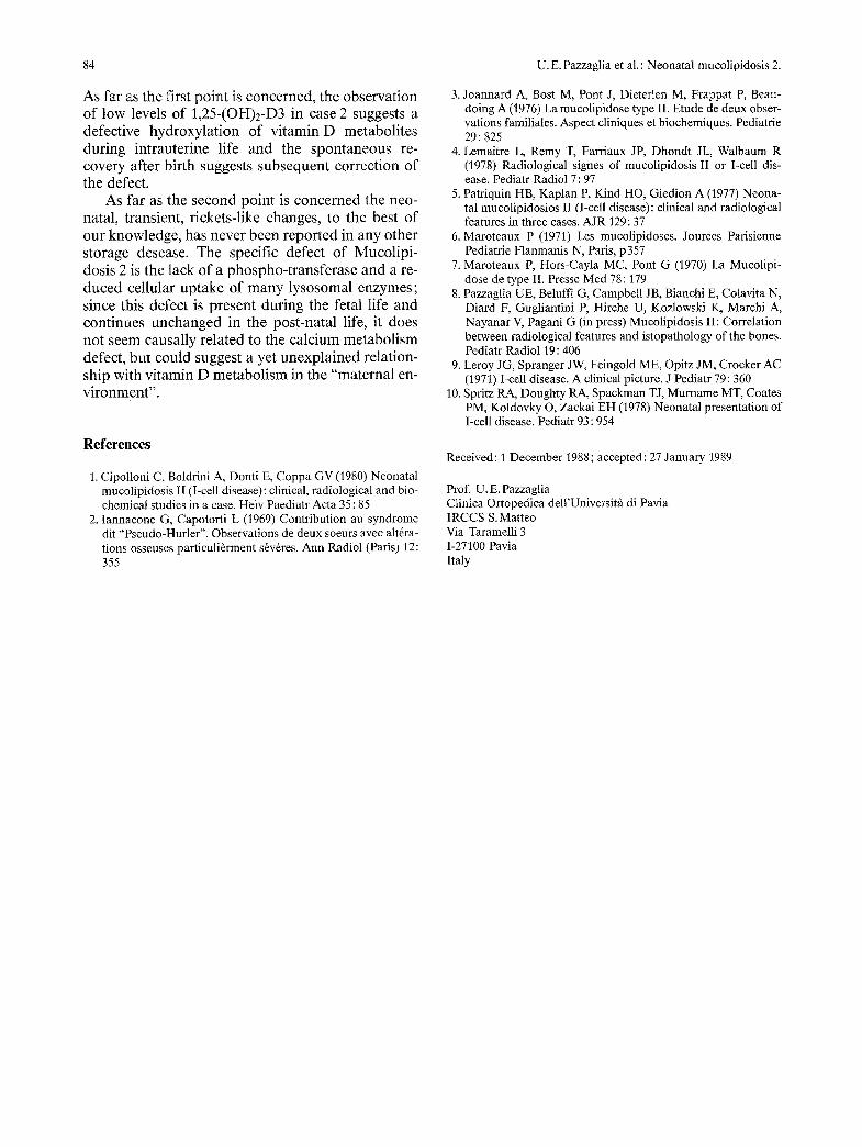
As far as the first point is concerned, the observation of low levels of 1,25-(OH)2-D3 in case 2 suggests a defective hydroxylation of vitamin D metabolites during intrauterine life and the spontaneous re- covery after birth suggests subsequent correction of the defect.
As far as the second point is concerned the neo- natal, transient, rickets-like changes, to the best of our knowledge, has never been reported in any other storage desease. The specific defect of Mucolipi- dosis 2 is the lack of a phospho-transferase and a re- duced cellular uptake of many lysosomal enzymes; since this defect is present during the fetal life and continues unchanged in the post-natal life, it does not seem causally related to the calcium metabolism defect, but could suggest a yet unexplained relation- ship with vitamin D metabolism in the "maternal en- vironment".
References
1. Cipolloni C, Boldrini A, Donti E, Coppa GV (1980) Neonatal mucolipidosis II (I-cell disease): clinical, radiological and bio- chemical studies in a case. Helv Paediatr Acta 35: 85
2. Iannacone G, Capotorti L (1969) Contribution au syndrome dit "Pseudo-Hurler". Observations de deux soeurs avec altrra- tions osseuses particulirrment srvrres. Ann Radiol (Paris) 12: 355
3. Joannard A, Bost M, Pont J, Dieterlen M, Frappat P, Beau- doing A (1976) La mucolipidose type II. Etude de deux obser- vations familiales. Aspect cliniques et biochemiques. Pediatrie 29: 825
4. Lemaitre L, Remy T, Farriaux JP, Dhondt JL, Walbaum R (1978) Radiological signes of mucolipidosis II or I-cell dis- ease. Pediatr Radiol 7:97
5. Patfiquin HB, Kaplan P, Kind HO, Giedion A (1977) Neona- tal mucolipidosios II (I-cell disease): clinical and radiological features in three cases. AJR 129:37
6. Maroteaux P (1971) Les mucolipidoses. Jourees Pafisienne Pediatrie Flanmanis N, Paris, p357
7. Maroteaux P, Hors-Cayla MC, Pont G (1970) La Mucolipi- dose de type II. Presse Med 78:179
8. Pazzaglia UE, Beluffi G, Campbell JB, Bianchi E, Colavita N, Diard F, Gugliantini P, Hirche U, Kozlowski K, Marchi A, Nayanar V, Pagani G (in press) Mucolipidosis II: Correlation between radiological features and istopathology of the bones. Pediatr Radiol 19:406
9. Leroy JG, Spranger JW, Feingold ME, Opitz JM, Crocker AC (1971) I-cell disease. A clinical picture. J Pediatr 79:360
10. Spritz RA, Doughty RA, Spackman TJ, Murname MT, Coates PM, Koldovky O, Zackai EH (1978) Neonatal presentation of I-cell disease. Pediatr 93 : 954
Received: 1 December 1988; accepted: 27 January 1989
Prof. U.E. Pazzaglia Clinica Ortopedica dell'Universitfi di Pavia IRCCS S. Matteo Via Taramelli 3 1-27100 Pavia Italy