Embed Size (px)
Citation preview

Continue
NCCN.org
Version 2.2019, 12/17/18 © 2018 National Comprehensive Cancer Network® (NCCN®), All rights reserved. NCCN Guidelines® and this illustration may not be reproduced in any form without the express written permission of NCCN.
NCCN Clinical Practice Guidelines in Oncology (NCCN Guidelines®)
Version 2.2019 — December 17, 2018
Continue
Primary Cutaneous Lymphomas

NCCNMary Dwyer, MSHema Sundar, PhD
ContinueNCCN Guidelines Panel Disclosures
† Medical oncology‡ Hematology/
Hematology oncology§ Radiotherapy/
Radiation oncologyξ Bone marrow
transplantation
≠ PathologyÞInternal medicine ϖ Dermatologyʘ Plastic surgery¥ Patient advocacy* Discussion Writing
Committee Member
Bradley M. Haverkos, MD, MPH, MS † University of Colorado Cancer Center
Richard T. Hoppe, MD § Stanford Cancer Institute
Eric Jacobsen, MD † Dana-Farber/Brigham and Women's Cancer Center
Deepa Jagadeesh, MD, MPH † ‡ Case Comprehensive Cancer Center/University Hospitals Seidman Cancer Center and Cleveland Clinic Taussig Cancer Institute
Youn H. Kim, MD ϖ † Stanford Cancer Institute
Matthew A. Lunning, DO † Þ ξ Fred & Pamela Buffett Cancer Center
Amitkumar Mehta, MD † ‡ Þ University of Alabama at Birmingham Comprehensive Cancer Center
Neha Mehta-Shah, MD † ‡ Siteman Cancer Center at Barnes- Jewish Hospital and Washington University School of Medicine
Elise A. Olsen, MD ϖ † Duke Cancer Institute
Barbara Pro, MD † Robert H. Lurie Comprehensive Cancer Center of Northwestern University
Saurabh A. Rajguru, MD † ‡ University of Wisconsin Carbone Cancer Center
Satish Shanbhag, MBBS, MPH † ‡ ξ The Sidney Kimmel Comprehensive Cancer Center at Johns Hopkins
Andrei Shustov, MD † Fred Hutchinson Cancer Research Center/Seattle Cancer Care Alliance
Lubomir Sokol, MD, PhD † ‡ Þ Moffitt Cancer Center
Pallawi Torka, MD † ‡ Roswell Park Cancer Institute
Carlos Torres-Cabala, MD ≠ The University of Texas MD Anderson Cancer Center
Ryan Wilcox, MD, PhD † University of Michigan Rogel Cancer Center
Basem M. William, MD ‡ The Ohio State University Comprehensive Cancer Center - James Cancer Hospital and Solove Research Institute
Jasmine Zain, MD † City of Hope National Medical Center
*Steven M. Horwitz, MD/Chair † Þ Memorial Sloan Kettering Cancer Center
*Stephen Ansell, MD, PhD/Vice-Chair ‡ Mayo Clinic Cancer Center
Weiyun Z. Ai, MD, PhD † ‡ UCSF Helen Diller Family Comprehensive Cancer Center
Jeffrey Barnes, MD, PhD † Massachusetts General Hospital Cancer Center
Stefan K. Barta, MD, MRCP, MS † ‡ Þ Fox Chase Cancer Center
Mark W. Clemens, MD ʘ The University of Texas MD Anderson Cancer Center
Ahmet Dogan, MD, PhD ≠ Memorial Sloan Kettering Cancer Center
Francine M. Foss, MD † ‡ ξ Yale Cancer Center/Smilow Cancer Hospital
Aaron M. Goodman, MD ‡ ξ UC San Diego Moores Cancer Center
Joan Guitart, MD ≠ ϖ Robert H. Lurie Comprehensive Cancer Center of Northwestern University Ahmad Halwani, MD ‡ Huntsman Cancer Institute at the University of Utah
NCCN Guidelines Version 2.2019Primary Cutaneous Lymphomas
Version 2.2019, 12/17/18 © 2018 National Comprehensive Cancer Network® (NCCN®), All rights reserved. NCCN Guidelines® and this illustration may not be reproduced in any form without the express written permission of NCCN.
NCCN Guidelines IndexTable of Contents
Discussion

Clinical Trials: NCCN believes that the best management for any patient with cancer is in a clinical trial. Participation in clinical trials is especially encouraged. To find clinical trials online at NCCN Member Institutions, click here:nccn.org/clinical_trials/physician.html.NCCN Categories of Evidence and Consensus: All recommendations are category 2A unless otherwise specified. See NCCN Categories of Evidence and Consensus.
NCCN Categories of Preference:All recommendations are consideredappropriate.See NCCN Categories of Preference
NCCN Primary Cutaneous Lymphomas Panel MembersSummary of the Guidelines Updates
Primary Cutaneous B-Cell Lymphomas• Diagnosis and Workup (CUTB-1)• Primary Cutaneous Marginal Zone Lymphoma (CUTB-2)• Primary Cutaneous Follicle Center Lymphoma (CUTB-2)• TNM Classification of Cutaneous Lymphoma other than
MF/SS (CUTB-A)• Treatment References (CUTB-B)
The NCCN Guidelines® are a statement of evidence and consensus of the authors regarding their views of currently accepted approaches to treatment. Any clinician seeking to apply or consult the NCCN Guidelines is expected to use independent medical judgment in the context of individual clinical circumstances to determine any patient’s care or treatment. The National Comprehensive Cancer Network® (NCCN®) makes no representations or warranties of any kind regarding their content, use or application and disclaims any responsibility for their application or use in any way. The NCCN Guidelines are copyrighted by National Comprehensive Cancer Network®. All rights reserved. The NCCN Guidelines and the illustrations herein may not be reproduced in any form without the express written permission of NCCN. ©2018.
Use of Immunophenotyping/Genetic Testing in Differential Diagnosis of Mature B-Cell and NK/T-Cell Neoplasms (See NCCN Guidelines for B-Cell Lymphomas - NHODG-A)
For Primary Cutaneous Diffuse Large B-cell Lymphoma, Leg Type (See NCCN Guidelines for B-Cell Lymphomas - DLBCL)
Classification and Staging (ST-1)
Primary Cutaneous CD30+ T-Cell Lymphoproliferative Disorders • Overview and Definition (PCTLD/INTRO-1)• Diagnosis (PCTLD-1)• Workup (PCTLD-2)• Primary Cutaneous ALCL (PCTLD-3))• Lymphomatoid Papulosis (PCTLD-4)• Therapy References (PCTLD-A)
Mycosis Fungoides/Sezary Syndrome (MF/SS)• Overview of Definition and Diagnosis (MFSS/INTRO-1)• General Principles (MFSS/INTRO-2)• Diagnosis (MFSS-1)• Workup (MFSS-2)• TNMB Classification and Staging (MFSS-3)• Clinical Staging (MFSS-4)• Stage IA (Limited Skin Involvement Alone, <10% BSA) (MFSS-6)• Stage IB (Skin Only Disease with ≥10% BSA) - Stage IIA (MFSS-7)• Stage IIB (Tumor Stage Disease) (MFSS-8) • Stage III (Erythrodermic Disease) (MFSS-10)• Stage IV (MFSS-11)• Large Cell Transformation (LCT) (MFSS-12)• Suggested Treatment Regimens (MFSS-A)• Supportive Care (MFSS-B)
• Principles of Radiation Therapy (LYMP-A)
• Principles of Molecular Analysis in T-Cell Lymphomas (LYMP-B)
• Supportive Care (LYMP-C)
NCCN Guidelines Version 2.2019Primary Cutaneous Lymphomas
Version 2.2019, 12/17/18 © 2018 National Comprehensive Cancer Network® (NCCN®), All rights reserved. NCCN Guidelines® and this illustration may not be reproduced in any form without the express written permission of NCCN.
NCCN Guidelines IndexTable of Contents
Discussion

UPDATES
Global changes• The following algorithms were combined and published with the Guidelines
name, "Primary Cutaneous Lymphomas"�Primary Cutaneous B-Cell Lymphomas�Mycosis Fungoides/Sezary Syndrome (moved from T-cell Lymphomas)�Primary Cutaneous CD30+ T-Cell Lymphoproliferative Disorders (moved
from T-cell Lymphomas)• Suggested treatment regimen references were updated throughout the
guidelines.• A footnote to the new "Principles of Molecular Analysis in T-Cell Lymphomas
(LYMP-B)" was added to the Diagnosis heading for all subtypes.• A footnote was added to PET/CT scan as appropriate throughout the
guidelines, "Patients with T-cell lymphomas often have extranodal disease, which may be inadequately imaged by CT. PET scan may be preferred in these instances."
• Workup, Useful�Bullet was added to appropriate pages, "Discussion of fertility and sperm
banking, if fertility-impacting therapy is planned."
Updates in Version 1.2019 of the NCCN Guidelines for Primary Cutaneous Lymphomas from Version 2.2018 include:Primary Cutaneous B-Cell LymphomasCUTB-1• Diagnosis, Useful�1st bullet, 1st sub-bullet was revised, "IHC panel may include: Ki-67,
CD5, CD43, CD21, CD23, Cyclin D1, kappa/lambda, EBER."�2nd bullet was revised, "Cytogenetics or (FISH and karyotype):..." �3rd bullet was revised, "If adequate biopsy material available, flow
cytometry or PCR IgH gene rearrangement studies..."• Workup, Essential�5th bullet was revised, "Hepatitis C testing" was added.�6th bullet was revised, "Chest/abdominal/pelvic CT with contrast
and/or PET/CT scan (may be omitted if clinically indicated)."�Bullet was deleted, "Bone marrow biopsy, if PC-DLBCL, leg type."
• Footnote e was revised, "Often reserved for patient with unexplained cytopenias or if there is clinical suspicion of other subtypes (eg, PC-DLBCL, leg type)."
CUTB-2• Initial therapy�"Topicals" was clarified as, "Skin-directed therapies." Also for
CUTB-3.• Footnote k was revised, "There are case reports showing efficacy of
topicals, which include steroids, imiquimod, nitrogen mustard, and bexarotene (useful in pediatric patients)."
CUTB-3• Initial therapy�Local RT was revised by removing "for symptoms."
Updates in Version 2.2019 of the NCCN Guidelines for Primary Cutaneous Lymphomas from Version 1.2019 include:
Primary Cutaneous CD30+ T-Cell Lymphoproliferative DisordersPCTLD-3• Cutaneous ALCL with regional nodes, primary treatment�Brentuximab vedotin + CHP (cyclophosphamide, doxorubicin, and prednisone) for CD30+ cases" was added as an other recommended option
with a category 2A designation.
Continued
NCCN Guidelines Version 2.2019Primary Cutaneous Lymphomas
Version 2.2019, 12/17/18 © 2018 National Comprehensive Cancer Network® (NCCN®), All rights reserved. NCCN Guidelines® and this illustration may not be reproduced in any form without the express written permission of NCCN.
NCCN Guidelines IndexTable of Contents
Discussion

UPDATES
Updates in Version 1.2019 of the NCCN Guidelines for Primary Cutaneous Lymphomas from Version 2.2018 include:Mycosis Fungoides/Sezary SyndromeMFSS/INTRO-1• A new overview page related to the definition and diagnosis of MF and SS
was added.
MFSS/INTRO-2• A new page with the "General Principles of MFSS" was added.
MFSS-1• Diagnosis, Essential�IHC panel was revised by removing, "CD25, CD56, TIA1, granzyme B,
ßF1, TCR-CγM1" and adding "CD25, CD56, TIA1, granzyme B, ßF1, TCRß, TCRẟ" to Useful Under Certain Circumstances."
• Diagnosis, Useful�The following bullet and corresponding footnote were made consistent
"Molecular analysis to detect clonal T-cell antigen receptor (TCR) gene rearrangements or other assessment of clonality (karyotype, array-CGH or FISH analysis to detect somatic mutations or genetic alterations)." Footnote f, "Clonal TCR gene rearrangements can be assessed by PCR or by high throughput sequencing techniques. Results should be interpreted with caution since clonal TCR gene rearrangements can also be seen in patients with non-malignant conditions. A negative result in the setting of high clinical suspicion does not exclude the diagnosis of MF/SS. Demonstration of identical clones in skin, blood, and/or lymph nodes may be helpful in selected cases. See Principles of Molecular Analysis in T-Cell Lymphomas (LYMP-A)." Also for PCTLD-1.
• Diagnosis, Useful�3rd bullet was revised from, "Core needle biopsy (FNA is often
inadequate) of suspicious lymph nodes (if biopsy of skin is not diagnostic)" to "Biopsy of enlarged lymph nodes or suspected extracutaneous sites (if biopsy of skin is not diagnostic.)...Rebiopsy if consult material is nondiagnostic."
MFSS-2• Workup, Useful�2nd bullet was revised from, "Core needle biopsy (FNA is often
inadequate) of suspicious lymph nodes or suspected extracutaneous sites" to "Biopsy of enlarged lymph nodes or suspected extracutaneous sites (if biopsy of skin is not diagnostic)...Rebiopsy if consult material is nondiagnostic."
MFSS-4• The Clinical Staging of MF and SS table was revised to include information
about stage; T, N, M; and the appropriate guidelines page for each stage.• Two new footnotes were added, �Footnote q, "Folliculotropism is a histologic feature that can occur
irrespective of stage. Histologic evidence of folliculotropic MF is associated with higher risk of disease progression. In selected cases or inadequate response, consider primary treatment for stage IIB (tumor stage disease)."
�Footnote r, "Large-cell transformation (LCT) is a histologic feature that can occur irrespective of clinical stage. LCT often but not always corresponds to a more aggressive growth rate requiring systemic therapies."
MFSS-5• "Dutch Criteria for Lymph Nodes" was added.
MFSS-6 through MFSS-12• The algorithm pages were all extensively revised.• Large cell transformed (LCT) treatment was added.
MFSS-A 1 of 6• Skin-directed therapies�Topical carmustine was added as a category 2B.�Phototherapy was revised, "(...PUVA/UVA-1 for thicker plaques)."
MFSS-A 2 of 6• Systemic therapies�For SYST-CAT A and SYST-CAT B, the Categories of Preference was
applied. �SYST-CAT A, Methotrexate dose was changed from "≤100 mg weekly" to
"≤50 mg weekly."�SYST-CAT B, other therapies were moved to Useful under certain
circumstances, Relapsed/refractory disease requiring systemic therapy." The new list also applies to LCT.
�The previous Category C (SYST-CAT C) were moved to "Preferred regimens" for LCT.
�Footnotes g, i, j, l, m, n, and p were added.MFSS-A 3 of 6• Combination therapies were put in alphabetical order.• "Erythrodermic disease/Sezary syndrome" treatment options were added.
Continued
NCCN Guidelines Version 2.2019Primary Cutaneous Lymphomas
Version 2.2019, 12/17/18 © 2018 National Comprehensive Cancer Network® (NCCN®), All rights reserved. NCCN Guidelines® and this illustration may not be reproduced in any form without the express written permission of NCCN.
NCCN Guidelines IndexTable of Contents
Discussion
UPDATES
Updates in Version 1.2019 of the NCCN Guidelines for Primary Cutaneous Lymphomas from Version 2.2018 include:
Principles of Radiation TherapyLYMP-A• The principles for all three algorithms were combined into one page of
Principles and updated as appropriate.
Supportive CareLYMP-C• A new section for anti-infective prophylaxis was added.
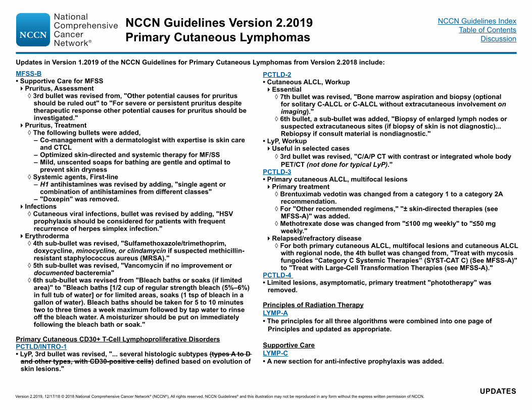
PCTLD-2• Cutaneous ALCL, Workup�Essential
◊ 7th bullet was revised, "Bone marrow aspiration and biopsy (optional for solitary C-ALCL or C-ALCL without extracutaneous involvement on imaging)."
◊ 6th bullet, a sub-bullet was added, "Biopsy of enlarged lymph nodes or suspected extracutaneous sites (if biopsy of skin is not diagnostic)...Rebiopsy if consult material is nondiagnostic."
• LyP, Workup�Useful in selected cases
◊ 3rd bullet was revised, "C/A/P CT with contrast or integrated whole body PET/CT (not done for typical LyP)."
PCTLD-3• Primary cutaneous ALCL, multifocal lesions �Primary treatment
◊ Brentuximab vedotin was changed from a category 1 to a category 2A recommendation.
◊ For "Other recommended regimens," "± skin-directed therapies (see MFSS-A)" was added.
◊ Methotrexate dose was changed from "≤100 mg weekly" to "≤50 mg weekly."
�Relapsed/refractory disease ◊ For both primary cutaneous ALCL, multifocal lesions and cutaneous ALCL with regional node, the 4th bullet was changed from, "Treat with mycosis fungoides “Category C Systemic Therapies” (SYST-CAT C) (See MFSS-A)" to "Treat with Large-Cell Transformation Therapies (see MFSS-A)."
PCTLD-4 • Limited lesions, asymptomatic, primary treatment "phototherapy" was
removed.
MFSS-B• Supportive Care for MFSS�Pruritus, Assessment
◊ 3rd bullet was revised from, "Other potential causes for pruritus should be ruled out" to "For severe or persistent pruritus despite therapeutic response other potential causes for pruritus should be investigated."
�Pruritus, Treatment ◊ The following bullets were added,
– Co-management with a dermatologist with expertise is skin care and CTCL
– Optimized skin-directed and systemic therapy for MF/SS – Mild, unscented soaps for bathing are gentle and optimal to prevent skin dryness
◊ Systemic agents, First-line – H1 antihistamines was revised by adding, "single agent or combination of antihistamines from different classes"
– "Doxepin" was removed. �Infections
◊ Cutaneous viral infections, bullet was revised by adding, "HSV prophylaxis should be considered for patients with frequent recurrence of herpes simplex infection."
�Erythroderma ◊ 4th sub-bullet was revised, "Sulfamethoxazole/trimethoprim, doxycycline, minocycline, or clindamycin if suspected methicillin-resistant staphylococcus aureus (MRSA)."
◊ 5th sub-bullet was revised, "Vancomycin if no improvement or documented bacteremia"
◊ 6th sub-bullet was revised from "Bleach baths or soaks (if limited area)" to "Bleach baths [1/2 cup of regular strength bleach (5%–6%) in full tub of water] or for limited areas, soaks (1 tsp of bleach in a gallon of water). Bleach baths should be taken for 5 to 10 minutes two to three times a week maximum followed by tap water to rinse off the bleach water. A moisturizer should be put on immediately following the bleach bath or soak."
Primary Cutaneous CD30+ T-Cell Lymphoproliferative DisordersPCTLD/INTRO-1• LyP, 3rd bullet was revised, "... several histologic subtypes (types A to D
and other types, with CD30-positive cells) defined based on evolution of skin lesions."
NCCN Guidelines Version 2.2019Primary Cutaneous Lymphomas
Version 2.2019, 12/17/18 © 2018 National Comprehensive Cancer Network® (NCCN®), All rights reserved. NCCN Guidelines® and this illustration may not be reproduced in any form without the express written permission of NCCN.
NCCN Guidelines IndexTable of Contents
Discussion
CUTB-1
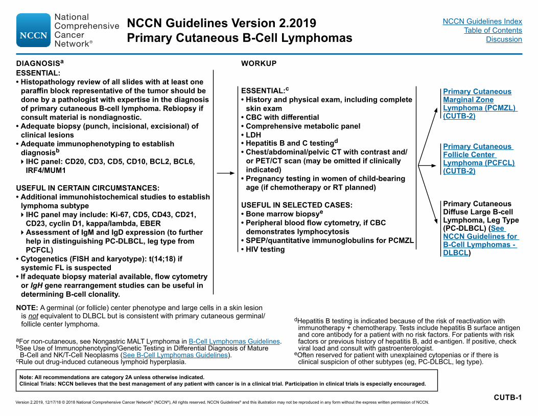
DIAGNOSISa WORKUP
aFor non-cutaneous, see Nongastric MALT Lymphoma in B-Cell Lymphomas Guidelines.bSee Use of Immunophenotyping/Genetic Testing in Differential Diagnosis of Mature
B-Cell and NK/T-Cell Neoplasms (See B-Cell Lymphomas Guidelines).cRule out drug-induced cutaneous lymphoid hyperplasia.
dHepatitis B testing is indicated because of the risk of reactivation with immunotherapy + chemotherapy. Tests include hepatitis B surface antigen and core antibody for a patient with no risk factors. For patients with risk factors or previous history of hepatitis B, add e-antigen. If positive, check viral load and consult with gastroenterologist.
eOften reserved for patient with unexplained cytopenias or if there is clinical suspicion of other subtypes (eg, PC-DLBCL, leg type).
N OTE: A germinal (or follicle) center phenotype and large cells in a skin lesion is not equivalent to DLBCL but is consistent with primary cutaneous germinal/follicle center lymphoma.
ESSENTIAL:• Histopathology review of all slides with at least one
paraffin block representative of the tumor should be done by a pathologist with expertise in the diagnosis of primary cutaneous B-cell lymphoma. Rebiopsy if consult material is nondiagnostic.
• Adequate biopsy (punch, incisional, excisional) of clinical lesions
• Adequate immunophenotyping to establish diagnosisb
�IHC panel: CD20, CD3, CD5, CD10, BCL2, BCL6, IRF4/MUM1
USEFUL IN CERTAIN CIRCUMSTANCES: • Additional immunohistochemical studies to establish
lymphoma subtype�IHC panel may include: Ki-67, CD5, CD43, CD21,
CD23, cyclin D1, kappa/lambda, EBER�Assessment of IgM and IgD expression (to further
help in distinguishing PC-DLBCL, leg type from PCFCL)
• Cytogenetics (FISH and karyotype): t(14;18) if systemic FL is suspected
• If adequate biopsy material available, flow cytometry or IgH gene rearrangement studies can be useful in determining B-cell clonality.
ESSENTIAL:c• History and physical exam, including complete
skin exam• CBC with differential• Comprehensive metabolic panel • LDH• Hepatitis B and C testingd
• Chest/abdominal/pelvic CT with contrast and/or PET/CT scan (may be omitted if clinically indicated)
• Pregnancy testing in women of child-bearing age (if chemotherapy or RT planned)
USEFUL IN SELECTED CASES:• Bone marrow biopsye
• Peripheral blood flow cytometry, if CBC demonstrates lymphocytosis
• SPEP/quantitative immunoglobulins for PCMZL• HIV testing
Primary Cutaneous Marginal Zone Lymphoma (PCMZL) (CUTB-2)
Primary Cutaneous Follicle Center Lymphoma (PCFCL) (CUTB-2)
Primary Cutaneous Diffuse Large B-cell Lymphoma, Leg Type (PC-DLBCL) (See NCCN Guidelines for B-Cell Lymphomas - DLBCL)
Version 2.2019, 12/17/18 © 2018 National Comprehensive Cancer Network® (NCCN®), All rights reserved. NCCN Guidelines® and this illustration may not be reproduced in any form without the express written permission of NCCN.
Note: All recommendations are category 2A unless otherwise indicated.Clinical Trials: NCCN believes that the best management of any patient with cancer is in a clinical trial. Participation in clinical trials is especially encouraged.
NCCN Guidelines Version 2.2019Primary Cutaneous B-Cell Lymphomas
NCCN Guidelines IndexTable of Contents
Discussion

CUTB-2
PRIMARY CUTANEOUS MARGINAL ZONE LYMPHOMA OR FOLLICLE CENTER LYMPHOMAf
STAGEg INITIAL THERAPYh
fAdditional imaging studies during the course of treatment are not needed. PET/CT (strongly preferred) or C/A/P CT with contrast at the end of treatment are needed to assess response. This can be repeated if there is clinical suspicion of progressive disease.
gSee TNM Classification of Cutaneous Lymphoma other than MF/SS (CUTB-A).hSee Treatment References (CUTB-B).
iLocal RT is the preferred initial treatment, but not necessarily the preferred treatment for relapse. See Principles of Radiation Therapy (LYMP-A).
jWhen RT or surgical treatment is neither feasible nor desired.kThere are case reports showing efficacy of topicals, which include steroids,
imiquimod, nitrogen mustard, and bexarotene (useful in pediatric patients).
Extracutaneous disease
Solitary/regional, T1-2
Local RT (preferred)iand/orExcision
In selected cases:Observationj orSkin-directed therapieskor Intralesional steroids
Responsef
Refractory diseasef
Relapsed or progressive diseasef
Regional
Generalized disease (extracutaneous disease)
Generalized disease (skin only)
For PCFCL, manage as Follicular Lymphoma in the NCCN Guidelines for B-Cell Lymphomas (see FOLL-4)or For PCMZL, manage as Nodal Marginal Zone Lymphoma in the NCCN Guidelines for B-Cell Lymphomas (see NODE-2)
Observe
For PCFCL, manage as Follicular Lymphoma in the NCCN Guidelines for B-Cell Lymphomas (see FOLL-4)or For PCMZL, manage as Nodal Marginal Zone Lymphoma in the NCCN Guidelines for B-Cell Lymphomas (see NODE-2)
Generalized disease (skin only), T3
See CUTB-3
See Generalized disease (skin only), T3 (CUTB-3)
See Generalized disease (skin only), T3 (CUTB-3)
Version 2.2019, 12/17/18 © 2018 National Comprehensive Cancer Network® (NCCN®), All rights reserved. NCCN Guidelines® and this illustration may not be reproduced in any form without the express written permission of NCCN.
Note: All recommendations are category 2A unless otherwise indicated.Clinical Trials: NCCN believes that the best management of any patient with cancer is in a clinical trial. Participation in clinical trials is especially encouraged.
NCCN Guidelines Version 2.2019Primary Cutaneous B-Cell Lymphomas
NCCN Guidelines IndexTable of Contents
Discussion

CUTB-3
fAdditional imaging studies during the course of treatment are not needed. PET/CT (strongly preferred) or C/A/P CT with contrast at the end of treatment are needed to assess response. This can be repeated if there is clinical suspicion of progressive disease.
gSee TNM Classification of Cutaneous Lymphoma other than MF/SS (CUTB-A).hSee Treatment References (CUTB-B).iLocal RT is the preferred initial treatment, but not necessarily the preferred
treatment for relapse. See Principles of Radiation Therapy (LYMP-A).kThere are case reports showing efficacy of topicals, which include steroids,
imiquimod, nitrogen mustard, and bexarotene (useful in pediatric patients).
lSee monoclonal antibody and viral reactivation (See NCCN Guidelines B-Cell Lymphoma).
mConsidered appropriate in asymptomatic patients.nRituximab and hyaluronidase human injection for subcutaneous use may
be substituted for rituximab after patients have received the first full dose of rituximab by intravenous infusion. This substitution cannot be made for rituximab used in combination with ibritumomab tiuxetan.
oIn rare circumstances for very extensive or refractory disease, other combination chemotherapy regimens listed in NCCN Guidelines for B-Cell Lymphomas, FOLL-B are used.
Generalized disease (skin only), T3
ObservationmorSkin-directed therapieskorLocal RTiorIntralesional steroidsorRituximabnorOther systemic therapyo
Responsef
Refractory diseasef
Relapsed or progressive diseasef
Treat with alternate initial therapy
Generalized disease (skin only)
Generalized disease (extracutaneous disease)
Observe
For PCFCL, manage as Follicular Lymphoma in the NCCN Guidelines for B-Cell Lymphomas (see FOLL-4)or For PCMZL, manage as Nodal Marginal Zone Lymphoma in the NCCN Guidelines for B-Cell Lymphomas (see NODE-2)
PRIMARY CUTANEOUS MARGINAL ZONE LYMPHOMA OR FOLLICLE CENTER LYMPHOMAf
STAGEg INITIAL THERAPYh,l
Version 2.2019, 12/17/18 © 2018 National Comprehensive Cancer Network® (NCCN®), All rights reserved. NCCN Guidelines® and this illustration may not be reproduced in any form without the express written permission of NCCN.
Note: All recommendations are category 2A unless otherwise indicated.Clinical Trials: NCCN believes that the best management of any patient with cancer is in a clinical trial. Participation in clinical trials is especially encouraged.
NCCN Guidelines Version 2.2019Primary Cutaneous B-Cell Lymphomas
NCCN Guidelines IndexTable of Contents
Discussion
CUTB-A 1 OF 2
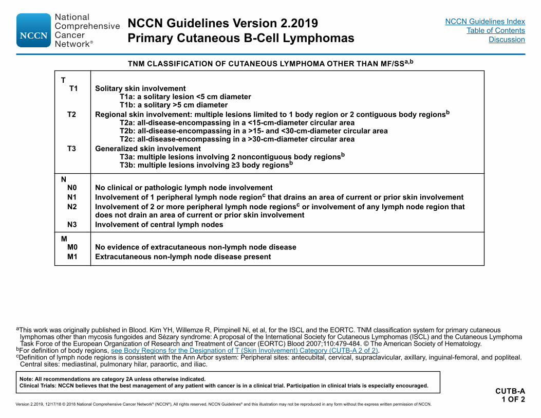
aThis work was originally published in Blood. Kim YH, Willemze R, Pimpinell Ni, et al, for the ISCL and the EORTC. TNM classification system for primary cutaneous lymphomas other than mycosis fungoides and Sézary syndrome: A proposal of the International Society for Cutaneous Lymphomas (ISCL) and the Cutaneous Lymphoma Task Force of the European Organization of Research and Treatment of Cancer (EORTC) Blood 2007;110:479-484. © The American Society of Hematology.
bFor definition of body regions, see Body Regions for the Designation of T (Skin Involvement) Category (CUTB-A 2 of 2).cDefinition of lymph node regions is consistent with the Ann Arbor system: Peripheral sites: antecubital, cervical, supraclavicular, axillary, inguinal-femoral, and popliteal.
Central sites: mediastinal, pulmonary hilar, paraortic, and iliac.
TNM CLASSIFICATION OF CUTANEOUS LYMPHOMA OTHER THAN MF/SSa,b
T T1 Solitary skin involvement T1a: a solitary lesion <5 cm diameter T1b: a solitary >5 cm diameter T2 Regional skin involvement: multiple lesions limited to 1 body region or 2 contiguous body regionsb T2a: all-disease-encompassing in a <15-cm-diameter circular area T2b: all-disease-encompassing in a >15- and <30-cm-diameter circular area T2c: all-disease-encompassing in a >30-cm-diameter circular area T3 Generalized skin involvement T3a: multiple lesions involving 2 noncontiguous body regionsb T3b: multiple lesions involving ≥3 body regionsb
N N0 No clinical or pathologic lymph node involvement N1 Involvement of 1 peripheral lymph node regionc that drains an area of current or prior skin involvement N2 Involvement of 2 or more peripheral lymph node regionsc or involvement of any lymph node region that
does not drain an area of current or prior skin involvement N3 Involvement of central lymph nodes
M M0 No evidence of extracutaneous non-lymph node disease M1 Extracutaneous non-lymph node disease present
Version 2.2019, 12/17/18 © 2018 National Comprehensive Cancer Network® (NCCN®), All rights reserved. NCCN Guidelines® and this illustration may not be reproduced in any form without the express written permission of NCCN.
Note: All recommendations are category 2A unless otherwise indicated.Clinical Trials: NCCN believes that the best management of any patient with cancer is in a clinical trial. Participation in clinical trials is especially encouraged.
NCCN Guidelines Version 2.2019Primary Cutaneous B-Cell Lymphomas
NCCN Guidelines IndexTable of Contents
Discussion

CUTB-A 2 OF 2
aThis work was originally published in Blood. Kim YH, Willemze R, Pimpinell Ni, et al, for the ISCL and the EORTC. TNM classification system for primary cutaneous lymphomas other than mycosis fungoides and Sézary syndrome: A proposal of the International Society for Cutaneous Lymphomas (ISCL) and the Cutaneous Lymphoma Task Force of the European Organization of Research and Treatment of Cancer (EORTC) Blood 2007;110:479-484. © The American Society of Hematology.
dLeft and right extremities are assessed as separate body regions. The designation of these body regions are based on regional lymph node drainage patterns.eDefinition of body regions: Head and neck: inferior border—superior border of clavicles, T1 spinous process. Chest: superior border—superior border of clavicles; inferior
border—inferior margin of rib cage; lateral borders—midaxillary lines, glenohumeral joints (inclusive of axillae). Abdomen/genital: superior border—inferior margin of rib cage; inferior border—inguinal folds, anterior perineum; lateral borders—mid-axillary lines. Upper back: superior border—T1 spinous process; inferior border—inferior margin of rib cage; lateral borders—mid-axillary lines. Lower back/buttocks: superior border—inferior margin of rib cage; inferior border—inferior gluteal fold, anterior perineum (inclusive of perineum); lateral borders—midaxillary lines. Each upper arm: superior borders—glenohumeral joints (exclusive of axillae); inferior borders— ulnar/radial-humeral (elbow) joint. Each lower arm/hand: superior borders—ulnar/radial-humeral (elbow) joint. Each upper leg (thigh): superior borders—inguinal folds, inferior gluteal folds; inferior borders—mid-patellae, midpopliteal fossae. Each lower leg/foot: superior borders—mid-patellae, mid-popliteal fossae.
BODY REGIONS FOR THE DESIGNATION OF T (SKIN INVOLVEMENT) CATEGORYa,d,e
Version 2.2019, 12/17/18 © 2018 National Comprehensive Cancer Network® (NCCN®), All rights reserved. NCCN Guidelines® and this illustration may not be reproduced in any form without the express written permission of NCCN.
Note: All recommendations are category 2A unless otherwise indicated.Clinical Trials: NCCN believes that the best management of any patient with cancer is in a clinical trial. Participation in clinical trials is especially encouraged.
NCCN Guidelines Version 2.2019Primary Cutaneous B-Cell Lymphomas
NCCN Guidelines IndexTable of Contents
Discussion

CUTB-B
TREATMENT REFERENCESRituximabMorales AV, Advani R, Horwitz SM, et al. Indolent primary cutaneous B-cell lymphoma: experience using systemic rituximab. J Am Acad Dermatol 2008;59:953-957.Heinzerling LM, Urbanek M, Funk JO, et al. Reduction of tumor burden and stabilization of disease by systemic therapy with anti-CD20 antibody (rituximab) in patients with primary cutaneous B-cell lymphoma. Cancer 2000;89:1835-1844.Valencak J, Weihsengruber F, Rappersberger K, et al. Rituximab monotherapy for primary cutaneous B-cell lymphoma: Response and follow-up in 16 patients. Ann Oncol 2009;20:326-330.Senff NJ, Noordijk EM, Kim YH, et al. European Organization for Research and Treatment of Cancer and International Society for Cutaneous Lymphoma consensus recommendations for the management of cutaneous B-cell lymphomas. Blood 2008;112:1600-1609.Heinzerling L, Dummer R, Kempf W, Schmid MH, Burg G. Intralesional therapy with anti-CD20 monoclonal antibody rituximab in primary cutaneous B-cell lymphoma. Arch Dermatol 2000;136:374-378.TopicalsTopical/intralesional corticosteroidsBekkenk MW, Vermeer MH, Geerts ML, et al. Treatment of multifocal primary cutaneous B-cell lymphoma: a clinical follow-up study of 29 patients. J Clin Oncol 1999;17:2471-2478.Perry A, Vincent BJ, Parker SR. Intralesional corticosteroid therapy for primary cutaneous B-cell lymphoma. Br J Dermatol 2010;163:223-225. Topical nitrogen mustardBachmeyer C, Orlandini V, Aractingi S. Topical mechlorethamine and clobetasol in multifocal primary cutaneous marginal zone-B cell lymphoma. B J Dermatol 2006;154:1207-1209. Topical bexaroteneTrent JT, Romanelli P, Kerdel FA. Topical Targretin and Intralesional Interferon Alfa for Cutaneous Lymphoma of the Scalp. Arch Dermatol 2002;138:1421-1423. Topical imiquimodCoors EA, Schuler G, Von Den Driesch P. Topical imiquimod as treatment for different kinds of cutaneous lymphoma. Eur J Dermatol 2006;16:391-393.Stavrakoglou A, Brown VL, Coutts I. Successful treatment of primary cutaneous follicle centre lymphoma with topical 5% imiquimod. Br J Dermatol 2007;157:620-622.
ChemotherapyHoefnagel JJ, Vermeer MH, Jansen PM, et al. Primary cutaneous marginal zone B-cell lymphoma: Clinical and therapeutic features in 50 cases. Arch Dermatol 2005;141:1139-1145.Bekkenk MW, Vermeer MH, Geerts ML, et al. Treatment of multifocal primary cutaneous B-cell lymphoma: a clinical follow-up study of 29 patients. J Clin Oncol 1999;17:2471-2478.Senff NJ, Noordijk EM, Kim YH, et al. European Organization for Research and Treatment of Cancer and International Society for Cutaneous Lymphoma consensus recommendations for the management of cutaneous B-cell lymphomas. Blood 2008;112:1600-1609.Grange F, Beylot-Barry M, Courville P, et al. Primary cutaneous diffuse large B-cell lymphoma, leg type: clinicopathologic features and prognostic analysis in 60 cases. Arch Dermatol 2007;143:1144-1150.Brice P, Cazals D, Mounier N, et al. Primary cutaneous large-cell lymphoma: analysis of 49 patients included in the LNH87 prospective trial of polychemotherapy for high-grade lymphomas. Groupe d'Etude des Lymphomes de l'Adulte. Leukemia 1998;12:213-219.Rijlaarsdam JU, Toonstra J, Meijer OW, Noordijk EM, Willemze R. Treatment of primary cutaneous B-cell lymphomas of follicle center cell origin: A clinical follow-up study of 55 patients treated with radiotherapy or polychemotherapy. J Clin Oncol 1996;14:549-555.Vermeer MH, Geelen FA, van Haselen CW, et al. Primary cutaneous large B-cell lymphomas of the legs. A distinct type of cutaneous B-cell lymphoma with an intermediate prognosis. Dutch Cutaneous Lymphoma Working Group. Arch Dermatol 1996;132:1304-1308.Palliative low-dose RT Neelis KJ, Schimmel EC, Vermeer MH, et al. Low-dose palliative radiotherapy for cutaneous B- and T-cell lymphomas. Int J Radiat Oncol Biol Phys 2009;74:154-158.ChemoimmunotherapyGrange F, Joly P, Barbe C, et al. Improvement of survival in patients with primary cutaneous diffuse large B-cell lymphoma, leg type, in France. JAMA Dermatol 2014;150:535-541.
Version 2.2019, 12/17/18 © 2018 National Comprehensive Cancer Network® (NCCN®), All rights reserved. NCCN Guidelines® and this illustration may not be reproduced in any form without the express written permission of NCCN.
Note: All recommendations are category 2A unless otherwise indicated.Clinical Trials: NCCN believes that the best management of any patient with cancer is in a clinical trial. Participation in clinical trials is especially encouraged.
NCCN Guidelines Version 2.2019Primary Cutaneous B-Cell Lymphomas
NCCN Guidelines IndexTable of Contents
Discussion

MFSS/INTRO-1
OVERVIEW
Definition• Mycosis fungoides (MF)�MF is the most common cutaneous T-cell lymphoma (CTCL) and many clinicopathologic variants of MF have been described.a�Most patients with MF exhibit an indolent clinical course with intermittent, stable, or slow progression of the lesions. �Extracutaneous involvement may be seen in advanced stages, with involvement of lymph nodes, blood, or less commonly other organs.a
• Sézary syndrome (SS)�SS is closely related to MF but has unique characteristics. SS is rare, accounting for less than 5% of cutaneous lymphomas and
predominantly affects older individuals.�SS is characterized by the presence of atypical T cells (Sézary cells) in skin (erythroderma), lymph nodes (generalized lymphadenopathy),
and peripheral blood (count of Sézary cells ≥1000 cells/µL; CD4:CD8 ratio ≥ 10; loss of one or more panT-cell antigens).c�SS is thought to arise from thymic memory T cells, while skin resident effector memory T-cells are the cells of origin of MF. This supports the
contention that SS is a process distinct from MF.d Cases presenting clinically as an overlap of these two conditions exist.
Diagnosis• The histopathologic findings of MF, even in cases showing classic features, need to be correlated with clinical presentation in order to reach a
definitive diagnosis.b• Patch lesions are often difficult for conclusive diagnosis; thus, in some instances multiple skin biopsies may be necessary for diagnosis.
Stopping skin-directed therapy for 2–3 weeks or longer to individual lesions before obtaining a skin biopsy is advisable and may aid in diagnosis.a
• Awareness of specific clinicopathologic variants may aid in accurate diagnosis:�Folliculotropic MF presents as folliculocentric lesions on sun-exposed areas such as the head and neck, often associated with alopecia, and
may be more resistant to local therapy. �Unilesional, pagetoid reticulosis and CD8+ MF variants tend to be associated with an indolent course. �Granulomatous slack skin is rare and presents with redundant skin resembling cutis laxa on flexural areas.
• The tumor cells are usually CD3+, CD4+, and CD8-, although CD8+ variants are not uncommon. • Large-cell transformation (LCT) of MF is defined histologically as greater than 25% of the tumor cells displaying large size. CD30 expression
may be seen but is not included in the definition of LCT.• The histopathologic findings of SS in skin are in generally similar to, but may be more subtle than those seen in MF. Correlation with clinical
and laboratory findings in blood is essential for a definitive diagnosis.aSwerdlow SH, Campo E, Pileri SA, et al. The 2016 revision of the World Health Organization classification of lymphoid neoplasms. Blood 2016;127:2375-2390. bPimpinelli N, Olsen EA, Santucci M, et al. Defining early mycosis fungoides. J Am Acad Dermatol 2005;53:1053-1063.cOlsen E, Vonderheid E, Pimpinelli N, et al. Revisions to the staging and classification of mycosis fungoides and Sezary syndrome: a proposal of the International
Society for Cutaneous Lymphomas (ISCL) and the cutaneous lymphomas task force of the European Organization of Research and Treatment of Cancer (EORTC). Blood 2007;110:1713-1722.
dCampbell JJ, Clark RA, Watanabe R, Kupper TS. Sezary syndrome and mycosis fungoides arise from distinct T-cell subsets: a biologic rationale for their distinct clinical behaviors. Blood 2010;116:767-771.
See General Principles of MF/SS (MFSS/INTRO-2)
NCCN Guidelines Version 2.2019
Version 2.2019, 12/17/18 © 2018 National Comprehensive Cancer Network® (NCCN®), All rights reserved. NCCN Guidelines® and this illustration may not be reproduced in any form without the express written permission of NCCN.
Note: All recommendations are category 2A unless otherwise indicated.Clinical Trials: NCCN believes that the best management of any patient with cancer is in a clinical trial. Participation in clinical trials is especially encouraged.
Mycosis Fungoides/Sezary SyndromeNCCN Guidelines Index
Table of ContentsDiscussion
MFSS/INTRO-2
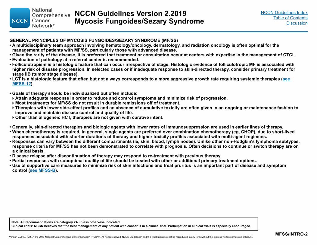
GENERAL PRINCIPLES OF MYCOSIS FUNGOIDES/SEZARY SYNDROME (MF/SS)• A multidisciplinary team approach involving hematology/oncology, dermatology, and radiation oncology is often optimal for the
management of patients with MF/SS, particularly those with advanced disease.• Given the rarity of the disease, it is preferred that treatment or consultation occur at centers with expertise in the management of CTCL. • Evaluation of pathology at a referral center is recommended. • Folliculotropism is a histologic feature that can occur irrespective of stage. Histologic evidence of folliculotropic MF is associated with
higher risk of disease progression. In selected cases or if inadequate response to skin-directed therapy, consider primary treatment for stage IIB (tumor stage disease).
• LCT is a histologic feature that often but not always corresponds to a more aggressive growth rate requiring systemic therapies (see MFSS-12).
• Goals of therapy should be individualized but often include:�Attain adequate response in order to reduce and control symptoms and minimize risk of progression.�Most treatments for MF/SS do not result in durable remissions off of treatment.�Therapies with lower side-effect profiles and an absence of cumulative toxicity are often given in an ongoing or maintenance fashion to
improve and maintain disease control and quality of life. �Other than allogeneic HCT, therapies are not given with curative intent.
• Generally, skin-directed therapies and biologic agents with lower rates of immunosuppression are used in earlier lines of therapy. • When chemotherapy is required, in general, single agents are preferred over combination chemotherapy (eg, CHOP), due to short-lived
responses associated with shorter durations of therapy and higher toxicity profiles associated with multi-agent regimens.• Responses can vary between the different compartments (ie, skin, blood, lymph nodes). Unlike other non-Hodgkin's lymphoma subtypes,
response criteria for MF/SS has not been demonstrated to correlate with prognosis. Often decisions to continue or switch therapy are on a clinical basis.
• Disease relapse after discontinuation of therapy may respond to re-treatment with previous therapy.• Partial responses with suboptimal quality of life should be treated with other or additional primary treatment options.• Use of supportive care measures to minimize risk of skin infections and treat pruritus is an important part of disease and symptom
control (see MFSS-B).
NCCN Guidelines Version 2.2019
Version 2.2019, 12/17/18 © 2018 National Comprehensive Cancer Network® (NCCN®), All rights reserved. NCCN Guidelines® and this illustration may not be reproduced in any form without the express written permission of NCCN.
Note: All recommendations are category 2A unless otherwise indicated.Clinical Trials: NCCN believes that the best management of any patient with cancer is in a clinical trial. Participation in clinical trials is especially encouraged.
Mycosis Fungoides/Sezary SyndromeNCCN Guidelines Index
Table of ContentsDiscussion

MFSS-1
aSee Principles of Molecular Analysis in T-Cell Lymphomas (LYMP-B).bPresence of transformation or areas of folliculotropism may have important
implications for selection of therapy and outcome and should be included in pathology reports.
cClinically suspicious and histologically non-diagnostic cases. Pimpinelli N, Olsen EA, Santucci M, et al, for the International Society for Cutaneous Lymphoma. Defining early mycosis fungoides. J Am Acad Dermatol 2005;53:1053-1063.
dSee Use of Immunophenotyping/Genetic Testing in Differential Diagnosis of Mature B-Cell and NK/T-Cell Neoplasms (See B-Cell Lymphomas Guidelines).
eTypical immunophenotype: CD2+ CD3+ CD5+ CD7- CD4+ CD8- (rarely CD8+) CD30-/+ cytotoxic granule proteins negative.
fClonal TCR gene rearrangement can be assessed by PCR or by high throughput sequencing techniques. Results should be interpreted with caution since clonal TCR gene rearrangements can also be seen in patients with non-malignant conditions. A negative result in the setting of high clinical suspicion does not exclude the diagnosis of MF/SS. Demonstration of identical clones in skin, blood, and/or lymph nodes may be helpful in selected cases. See Principles of Molecular Analysis in T-Cell Lymphomas (LYMP-B).
gSee map for prevalence of HTLV-1 by geographic region.
DIAGNOSISa
ESSENTIAL:• Biopsy of suspicious skin sites�Multiple biopsies may be necessary to capture the pathologic variability of disease at diagnosis
• Dermatopathology review of slidesb
• IHC panel of skin biopsyc,d,e �CD2, CD3, CD4, CD5, CD7, CD8, CD20, CD30
• Molecular analysis to detect clonal T-cell antigen receptor (TCR) gene rearrangements or other assessment of clonality (karyotype, array-CGH, or FISH analysis to detect somatic mutations or genetic alterations)a,f
USEFUL UNDER CERTAIN CIRCUMSTANCES:• Assessment of peripheral blood for Sézary cells (in extensive skin disease where skin biopsy is not
diagnostic and/or strongly of advanced-stage disease) including:�Sezary cell prep�Flow cytometry (CD3, CD4, CD7, CD8, CD26 to assess for expanded CD4+ cells with increased CD4/CD8
ratio or with abnormal immunophenotype, including loss of CD7 or CD26) • IHC panel of skin biopsyb,c �CD25, CD56, TIA1, granzyme B, ßF1, TCRß, TCRẟ
• Biopsy of enlarged lymph nodes or suspected extracutaneous sites (if biopsy of skin is not diagnostic). Excisional or incisional biopsy is preferred over core needle biopsy. An FNA alone is not sufficient for the initial diagnosis of lymphoma. A core needle biopsy is not optimal but can be used under certain circumstances. In certain circumstances, when a lymph node is not easily accessible for excisional or incisional biopsy, a combination of core needle biopsy and FNA in conjunction with appropriate ancillary techniques may be sufficient for diagnosis. Rebiopsy if consult material is nondiagnostic.
• Assessment of HTLV-1g by serology or other methods in at-risk populations.
See Workup (MFSS-2)
NCCN Guidelines Version 2.2019
Version 2.2019, 12/17/18 © 2018 National Comprehensive Cancer Network® (NCCN®), All rights reserved. NCCN Guidelines® and this illustration may not be reproduced in any form without the express written permission of NCCN.
Note: All recommendations are category 2A unless otherwise indicated.Clinical Trials: NCCN believes that the best management of any patient with cancer is in a clinical trial. Participation in clinical trials is especially encouraged.
Mycosis Fungoides/Sezary SyndromeNCCN Guidelines Index
Table of ContentsDiscussion

MFSS-2
aSee Principles of Molecular Analysis in T-Cell Lymphomas (LYMP-B).hSezary syndrome (B2) is as defined on MFSS-3. iSee Discussion for when Sezary flow cytometric study is appropriate in T1 disease. jPatients with T-cell lymphomas often have extranodal disease, which may be inadequately imaged by CT. PET scan may be preferred in these instances.kMany skin-directed and systemic therapies are contraindicated or of unknown safety in pregnancy. Refer to individual drug information.
WORKUPESSENTIAL:• History and complete physical examination:�Complete skin examination: assessment of % body surface area (BSA) (palm plus digits ≈1%
BSA) and type of skin lesion (ie, patch/plaque, tumor, erythroderma)�Palpation of peripheral lymph node regions�Palpation for organomegaly/masses
• Laboratory studies:h�CBC with Sezary screen (manual slide review, "Sezary cell prep")�Sezary flow cytometric study (optional for T1i)�TCR gene rearrangement in peripheral blood lymphocytes if blood involvement suspecteda�Comprehensive metabolic panel�LDH
• Imaging studies:�C/A/P CT with contrast or integrated whole body PET/CTj (arms/legs included when disease
assessment of entire body is needed); for ≥T2b or large-cell transformed or folliculotropic MF, or with palpable adenopathy or abnormal laboratory studies; consider for T2a (patch disease with >10% BSA)
USEFUL IN SELECTED CASES:• Bone marrow biopsy in patients with unexplained hematologic abnormality• Biopsy of enlarged lymph nodes or suspected extracutaneous sites (if biopsy of skin is not
diagnostic). Excisional or incisional biopsy is preferred over core needle biopsy. An FNA alone is not sufficient for the initial diagnosis of lymphoma. A core needle biopsy is not optimal but can be used under certain circumstances. In certain circumstances, when a lymph node is not easily accessible for excisional or incisional biopsy, a combination of core needle biopsy and FNA in conjunction with appropriate ancillary techniques may be sufficient for diagnosis. Rebiopsy if consult material is nondiagnostic.
• Rebiopsy skin if suspicious of LCT• Neck CT with contrast• Pregnancy testing in women of child-bearing age if contemplating treatments that are
contraindicated in pregnancyk
• Discussion of fertility and sperm banking, if fertility impacting therapy is planned
For TNMB Classification, see MFSS-3and For Clinical Staging of MF and SS, see MFSS-4
NCCN Guidelines Version 2.2019
Version 2.2019, 12/17/18 © 2018 National Comprehensive Cancer Network® (NCCN®), All rights reserved. NCCN Guidelines® and this illustration may not be reproduced in any form without the express written permission of NCCN.
Note: All recommendations are category 2A unless otherwise indicated.Clinical Trials: NCCN believes that the best management of any patient with cancer is in a clinical trial. Participation in clinical trials is especially encouraged.
Mycosis Fungoides/Sezary SyndromeNCCN Guidelines Index
Table of ContentsDiscussion

MFSS-3
lAdapted from Olsen E, Vonderheid E, Pimpinelli N, et al. Blood 2007;110:1713-1722 and Olsen E, Whittaker S, Kim Y, et al. J Clin Oncol 2011;29:2598-2607.mSezary syndrome is defined by B2 blood involvement and a clonal rearrangement of TCR in the blood (clones should be relevant to clone in the skin). nPatch = Any size skin lesion without significant elevation or induration.
Presence/absence of hypo- or hyperpigmentation, scale, crusting, and/or poikiloderma should be noted.oPlaque = Any size skin lesion that is elevated or indurated. Presence or absence of scale, crusting, and/or poikiloderma should be noted. Histologic features such as
folliculotropism or LCT (≥25% large cells), CD30+ or CD30-, and clinical features such as ulceration are important to document.pTumor = at least one >1 cm diameter solid or nodular lesion with evidence of depth and/or vertical growth. Note total number of lesions, total volume of lesions, largest
size lesion, and region of body involved. Also note if histologic evidence of LCT has occurred. Phenotyping for CD30 is encouraged.
TNMB TNMB Classification and Staging of Mycosis Fungoides and Sezary Syndromel,m
Skin T1 Limited patches,n papules, and/or plaqueso covering <10% of the skin surface T2 Patches,n papules, and/or plaquesn covering ≥10% of the skin surface T2a Patch only T2b Plaque ± patch T3 One or more tumorsp (≥1 cm in diameter) T4 Confluence of erythema ≥80% body surface area
Node N0 No abnormal lymph nodes; biopsy not required N1 Abnormal lymph nodes; histopathology Dutch Gr 1 or NCI LN 0-2 N2 Abnormal lymph nodes; histopathology Dutch Gr 2 or NCI LN 3 N3 Abnormal lymph nodes; histopathology Dutch Gr 3-4 or NCI LN 4 NX Abnormal lymph nodes; no histologic confirmation
Visceral M0 No visceral organ involvement M1 Visceral involvement (must have pathology confirmation and organ involved should be specified) MX Abnormal visceral site; no histologic confirmation
Blood B0 Absence of significant blood involvement: ≤5% of peripheral blood lymphocytes or <250/mcL are atypical (Sezary) cells or <15% CD4+/CD26- or CD4+/CD7- cells of total lymphocytes B1 Low blood tumor burden: >5% of peripheral blood lymphocytes are atypical (Sezary) cells or >15% CD4+CD26- or
CD4+CD7- of total lymphocytes but do not meet the criteria of B0 or B2 B2 High blood tumor burden: ≥1000/mcL Sezary cellsm (CD4+/CD26- or CD4+/CD7- cells by flow cytometry) or CD4/ CD8 ≥10 or ≥40% CD4+/CD7- or ≥30% CD4+/CD26- cells of total lymphocytes
See NCI Lymph Node Classification on MFSS-5
See Dutch Criteria for lymphnodes on MFSS-5
See Clinical Staging of MF and SS on MFSS-4
NCCN Guidelines Version 2.2019
Version 2.2019, 12/17/18 © 2018 National Comprehensive Cancer Network® (NCCN®), All rights reserved. NCCN Guidelines® and this illustration may not be reproduced in any form without the express written permission of NCCN.
Note: All recommendations are category 2A unless otherwise indicated.Clinical Trials: NCCN believes that the best management of any patient with cancer is in a clinical trial. Participation in clinical trials is especially encouraged.
Mycosis Fungoides/Sezary SyndromeNCCN Guidelines Index
Table of ContentsDiscussion
MFSS-4
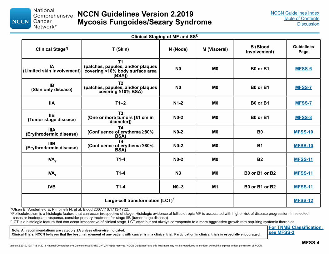
kOlsen E, Vonderheid E, Pimpinelli N, et al. Blood 2007;110:1713-1722.qFolliculotropism is a histologic feature that can occur irrespective of stage. Histologic evidence of folliculotropic MF is associated with higher risk of disease progression. In selected
cases or inadequate response, consider primary treatment for stage IIB (tumor stage disease)rLCT is a histologic feature that can occur irrespective of clinical stage. LCT often but not always corresponds to a more aggressive growth rate requiring systemic therapies.
Clinical Staging of MF and SSk
Clinical Stageq T (Skin) N (Node) M (Visceral) B (Blood Involvement)
Guidelines Page
IA (Limited skin involvement)
T1 (patches, papules, and/or plaques covering <10% body surface area
[BSA])N0 M0 B0 or B1 MFSS-6
IB (Skin only disease)
T2 (patches, papules, and/or plaques
covering ≥10% BSA)N0 M0 B0 or B1 MFSS-7
IIA T1–2 N1-2 M0 B0 or B1 MFSS-7
IIB (Tumor stage disease)
T3 (One or more tumors [≥1 cm in
diameter])N0-2 M0 B0 or B1 MFSS-8
IIIA (Erythrodermic disease)
T4 (Confluence of erythema ≥80%
BSA)N0-2 M0 B0 MFSS-10
IIIB (Erythrodermic disease)
T4 (Confluence of erythema ≥80%
BSA)N0-2 M0 B1 MFSS-10
IVA1 T1-4 N0-2 M0 B2 MFSS-11
IVA2 T1-4 N3 M0 B0 or B1 or B2 MFSS-11
IVB T1-4 N0–3 M1 B0 or B1 or B2 MFSS-11
Large-cell transformation (LCT)r MFSS-12
For TNMB Classification, see MFSS-3
NCCN Guidelines Version 2.2019
Version 2.2019, 12/17/18 © 2018 National Comprehensive Cancer Network® (NCCN®), All rights reserved. NCCN Guidelines® and this illustration may not be reproduced in any form without the express written permission of NCCN.
Note: All recommendations are category 2A unless otherwise indicated.Clinical Trials: NCCN believes that the best management of any patient with cancer is in a clinical trial. Participation in clinical trials is especially encouraged.
Mycosis Fungoides/Sezary SyndromeNCCN Guidelines Index
Table of ContentsDiscussion

MFSS-5
NCI-VA Lymph Node ClassificationLN0: no atypical lymphocytesLN1: occasional and isolated atypical lymphocytes (not arranged in clusters)LN2: many atypical lymphocytes or in 3–6 cell clustersLN3: aggregates of atypical lymphocytes; nodal architecture preservedLN4: partial/complete effacement of nodal architecture by atypical lymphocytes or frankly neoplastic cells
Clendenning WE, Rappaport HW. Report of the Committee on Pathology of Cutaneous T Cell Lymphomas. Cancer Treat Rep 1979;63:719-724.
Dutch Criteria for Lymph NodesGrade 1: Dermatopathic lymphadenopathyGrade 2: Early involvement by mycosis fungoides (presence of cerebriform nuclei >7.5 micrometers)Grade 3: Partial effacement of lymph node architecture; many atypical cerebriform mononuclear cellsGrade 4: Complete effacement of lymph node architecture
Scheffer E, Meijer CJLM, van Vloten WA. Dermatopathic lymphadenopathy and lymph node involvement in mycosis fungoides. Cancer 1980;45:137-148.
NCCN Guidelines Version 2.2019
Version 2.2019, 12/17/18 © 2018 National Comprehensive Cancer Network® (NCCN®), All rights reserved. NCCN Guidelines® and this illustration may not be reproduced in any form without the express written permission of NCCN.
Note: All recommendations are category 2A unless otherwise indicated.Clinical Trials: NCCN believes that the best management of any patient with cancer is in a clinical trial. Participation in clinical trials is especially encouraged.
Mycosis Fungoides/Sezary SyndromeNCCN Guidelines Index
Table of ContentsDiscussion

MFSS-6
sSee Principles for Mycosis Fungoides/Sezary Syndrome (MFSS/INTRO-1).tIn rare cases of confirmed unilesional MF, RT has been shown to provide long-term remission.uRebiopsy if suspect LCT; if histologic evidence of LCT, see MFSS-12.vIn patients with histologic evidence of folliculotropic MF, skin disease may be less responsive to topical therapies.wSee Principles of Radiation Therapy (LYMP-A).
STAGEs PRIMARY TREATMENT RESPONSE TO THERAPY See Supportive Care for MF/SS (MFSS-B)
Stage IA (limited skin involvement alone, <10% BSA)t,u
Skin-directed therapiesv (skin-limited/local) (MFSS-A) (may be alone or in combination with other skin-directed therapies) or If B1 blood involvement, consider primary treatment for stage III, B1 MFSS-10 (category 2B)
Inadequate response
Progression to >stage IA on skin-directed therapies or Refractory disease to multiple previous therapies or Persistent T1 skin disease
Relapse with T1 skin disease
Systemic therapy (SYST-CAT A, MFSS-A) ± skin-directed therapy (MFSS-A) or Consider RT if not previously usedw or Clinical trial
(MFSS-3 and MFSS-4)
CR/PR Relapse with>stage IA disease
MFSS-4 for appropriate clinical stage
NCCN Guidelines Version 2.2019
Version 2.2019, 12/17/18 © 2018 National Comprehensive Cancer Network® (NCCN®), All rights reserved. NCCN Guidelines® and this illustration may not be reproduced in any form without the express written permission of NCCN.
Note: All recommendations are category 2A unless otherwise indicated.Clinical Trials: NCCN believes that the best management of any patient with cancer is in a clinical trial. Participation in clinical trials is especially encouraged.
Mycosis Fungoides/Sezary SyndromeNCCN Guidelines Index
Table of ContentsDiscussion

MFSS-7
PRIMARY TREATMENT RESPONSE TO THERAPYx
See Supportive Care for MF/SS (MFSS-B)
Stage IB (skin only disease with ≥10% BSA) - Stage IIAu Skin-directed therapiesv
(generalized) (MFSS-A)orSystemic therapies (SYST-CAT A, MFSS-A) ± skin-directed therapiesv (MFSS-A)orCombination therapies (MFSS-A) ± skin-directed therapiesv (MFSS-A)
Progression to >stage IB-IIA or
Refractory disease to multiple previous therapies or Persistent T1-T2 skin disease
Relapse with T1-T2 disease
Clinical trialorTSEBTw (if not previously administered)orCombination therapies (MFSS-A) ± skin-directed therapyv (MFSS-A)
orIf blood B1 involvement, consider primary treatment for stage III B1 MFSS-10 (category 2B)
Inadequate response
CR/PR
Relapse with >stage IB-IIA disease See MFSS-4 for appropriate clinical stage
sSee Principles for Mycosis Fungoides/Sezary Syndrome (MFSS/INTRO-1).uRebiopsy if suspect LCT; if histologic evidence of LCT, see MFSS-12.vIn patients with histologic evidence of folliculotropic MF, skin disease may be less responsive to topical therapies.wSee Principles of Radiation Therapy (LYMP-A).xImaging indicated when suspicious of clinical extracutaneous disease with modalities used in workup.
Lower skin disease burden (eg, predominantly patch disease)
Higher skin disease burden (eg, predominantly plaque disease)
Skin-directed therapiesv (skin-limited/local) (MFSS-A) (may be alone or in combination with other skin-directed therapies) Inadequate
response
CR/PR
Relapse with low skin disease burdenRelapse with high skin disease burden (see below)
Progression to >stage IB-IIASee MFSS-4 for appropriate clinical stage
High skin disease burden (see below)
STAGEs
(MFSS-3 and MFSS-4)
NCCN Guidelines Version 2.2019
Version 2.2019, 12/17/18 © 2018 National Comprehensive Cancer Network® (NCCN®), All rights reserved. NCCN Guidelines® and this illustration may not be reproduced in any form without the express written permission of NCCN.
Note: All recommendations are category 2A unless otherwise indicated.Clinical Trials: NCCN believes that the best management of any patient with cancer is in a clinical trial. Participation in clinical trials is especially encouraged.
Mycosis Fungoides/Sezary SyndromeNCCN Guidelines Index
Table of ContentsDiscussion
MFSS-8
PRIMARY TREATMENT RESPONSE TO THERAPYx
See Supportive Care for MF/SS (MFSS-B)
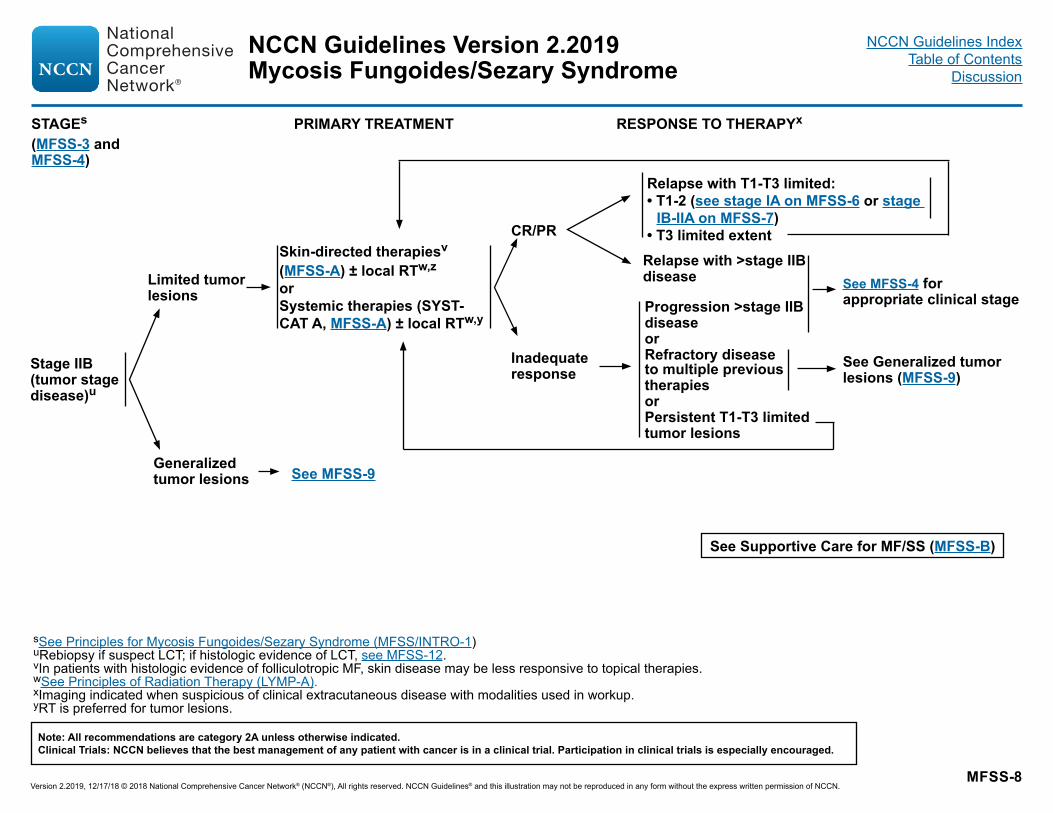
Stage IIB (tumor stage disease)u
sSee Principles for Mycosis Fungoides/Sezary Syndrome (MFSS/INTRO-1)uRebiopsy if suspect LCT; if histologic evidence of LCT, see MFSS-12.vIn patients with histologic evidence of folliculotropic MF, skin disease may be less responsive to topical therapies.wSee Principles of Radiation Therapy (LYMP-A).xImaging indicated when suspicious of clinical extracutaneous disease with modalities used in workup.yRT is preferred for tumor lesions.
Limited tumor lesions
Generalized tumor lesions
Skin-directed therapiesv
(MFSS-A) ± local RTw,z
orSystemic therapies (SYST-CAT A, MFSS-A) ± local RTw,y
Progression >stage IIB disease or Refractory disease to multiple previous therapies or Persistent T1-T3 limited tumor lesions
Relapse with T1-T3 limited:• T1-2 (see stage IA on MFSS-6 or stage
IB-IIA on MFSS-7)• T3 limited extent
See MFSS-9
See Generalized tumor lesions (MFSS-9)
Inadequate response
CR/PR
Relapse with >stage IIB disease See MFSS-4 for
appropriate clinical stage
STAGEs
(MFSS-3 and MFSS-4)
NCCN Guidelines Version 2.2019
Version 2.2019, 12/17/18 © 2018 National Comprehensive Cancer Network® (NCCN®), All rights reserved. NCCN Guidelines® and this illustration may not be reproduced in any form without the express written permission of NCCN.
Note: All recommendations are category 2A unless otherwise indicated.Clinical Trials: NCCN believes that the best management of any patient with cancer is in a clinical trial. Participation in clinical trials is especially encouraged.
Mycosis Fungoides/Sezary SyndromeNCCN Guidelines Index
Table of ContentsDiscussion
MFSS-9
PRIMARY TREATMENT RESPONSE TO THERAPYx
See Supportive Care for MF/SS (MFSS-B)
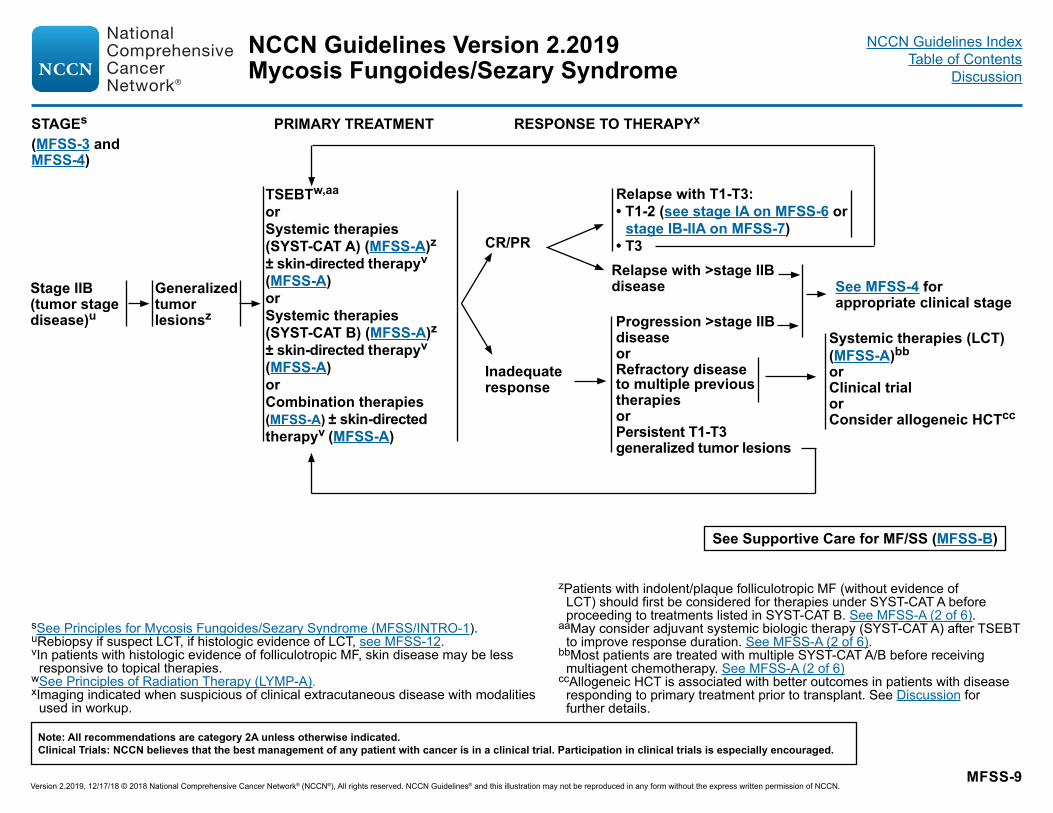
Stage IIB (tumor stage disease)u
sSee Principles for Mycosis Fungoides/Sezary Syndrome (MFSS/INTRO-1).uRebiopsy if suspect LCT, if histologic evidence of LCT, see MFSS-12.vIn patients with histologic evidence of folliculotropic MF, skin disease may be less
responsive to topical therapies.wSee Principles of Radiation Therapy (LYMP-A).xImaging indicated when suspicious of clinical extracutaneous disease with modalities
used in workup.
zPatients with indolent/plaque folliculotropic MF (without evidence of LCT) should first be considered for therapies under SYST-CAT A before proceeding to treatments listed in SYST-CAT B. See MFSS-A (2 of 6).
aaMay consider adjuvant systemic biologic therapy (SYST-CAT A) after TSEBT to improve response duration. See MFSS-A (2 of 6).
bbMost patients are treated with multiple SYST-CAT A/B before receiving multiagent chemotherapy. See MFSS-A (2 of 6)
ccAllogeneic HCT is associated with better outcomes in patients with disease responding to primary treatment prior to transplant. See Discussion for further details.
Generalized tumor lesionsz
TSEBTw,aa
orSystemic therapies (SYST-CAT A) (MFSS-A)z ± skin-directed therapyv
(MFSS-A)or Systemic therapies (SYST-CAT B) (MFSS-A)z ± skin-directed therapyv
(MFSS-A)orCombination therapies (MFSS-A) ± skin-directed therapyv (MFSS-A)
Relapse with T1-T3:• T1-2 (see stage IA on MFSS-6 or
stage IB-IIA on MFSS-7)• T3
Systemic therapies (LCT)(MFSS-A)bborClinical trialorConsider allogeneic HCTcc
Progression >stage IIB disease or Refractory disease to multiple previous therapies or Persistent T1-T3 generalized tumor lesions
Inadequate response
Relapse with >stage IIB disease
CR/PR
See MFSS-4 for appropriate clinical stage
STAGEs
(MFSS-3 and MFSS-4)
NCCN Guidelines Version 2.2019
Version 2.2019, 12/17/18 © 2018 National Comprehensive Cancer Network® (NCCN®), All rights reserved. NCCN Guidelines® and this illustration may not be reproduced in any form without the express written permission of NCCN.
Note: All recommendations are category 2A unless otherwise indicated.Clinical Trials: NCCN believes that the best management of any patient with cancer is in a clinical trial. Participation in clinical trials is especially encouraged.
Mycosis Fungoides/Sezary SyndromeNCCN Guidelines Index
Table of ContentsDiscussion
MFSS-10
See Supportive Care for MF/SS (MFSS-B)
PRIMARY TREATMENT RESPONSE TO THERAPYx
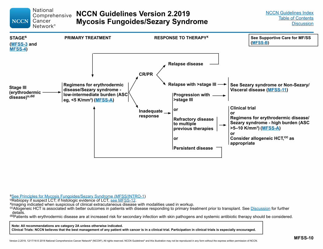
Stage III (erythrodermic disease)u,dd
Regimens for erythrodermic disease/Sezary syndrome - low-intermediate burden (ASC eg, <5 K/mm3) (MFSS-A)
Relapse disease
Clinical trialorRegimens for erythrodermic disease/Sezary syndrome - high burden (ASC >5–10 K/mm3) (MFSS-A)orConsider allogeneic HCT,cc as appropriate
sSee Principles for Mycosis Fungoides/Sezary Syndrome (MFSS/INTRO-1)uRebiopsy if suspect LCT; if histologic evidence of LCT, see MFSS-12.xImaging indicated when suspicious of clinical extracutaneous disease with modalities used in workup.ccAllogeneic HCT is associated with better outcomes in patients with disease responding to primary treatment prior to transplant. See Discussion for further
details. ddPatients with erythrodermic disease are at increased risk for secondary infection with skin pathogens and systemic antibiotic therapy should be considered.
Inadequate response
CR/PR
Progression with >stage III or Refractory disease to multiple previous therapies or Persistent disease
Relapse with >stage III See Sezary syndrome or Non-Sezary/Visceral disease (MFSS-11)
STAGEs
(MFSS-3 and MFSS-4)
NCCN Guidelines Version 2.2019
Version 2.2019, 12/17/18 © 2018 National Comprehensive Cancer Network® (NCCN®), All rights reserved. NCCN Guidelines® and this illustration may not be reproduced in any form without the express written permission of NCCN.
Note: All recommendations are category 2A unless otherwise indicated.Clinical Trials: NCCN believes that the best management of any patient with cancer is in a clinical trial. Participation in clinical trials is especially encouraged.
Mycosis Fungoides/Sezary SyndromeNCCN Guidelines Index
Table of ContentsDiscussion

MFSS-11
PRIMARY TREATMENT RESPONSE TO THERAPYff
sSee Principles for Mycosis Fungoides/Sezary Syndrome (MFSS/INTRO-1).uRebiopsy if suspect LCT; if histologic evidence of LCT, see MFSS-12.wSee Principles of Radiation Therapy (LYMP-A).bbMost patients are treated with multiple SYST-CAT A/B before receiving multiagent chemotherapy. See MFSS-A (2 of 6).ccAllogeneic HCT is associated with better outcomes in patients with disease responding to primary treatment prior to transplant. See Discussion for further details. eePatients with stage IV non-Sezary/visceral disease may present with more aggressive growth characteristics. If there is no evidence of more aggressive growth,
systemic therapies from SYST-CAT B are appropriate. If aggressive growth is seen, then systemic therapies listed for LCT are preferred. See MFSS-A (2 of 6).ffIf disease in lymph nodes and/or viscera or suspicious of disease progression, imaging indicated with modalities used in workup as clinically indicated based on
distribution of disease.
See Supportive Care for MF/SS (MFSS-B)
Stage IVu
Non-Sezaryor Visceral disease (solid organ)
• Regimens for erythrodermic disease/Sezary syndrome
(MFSS-A)�Low-intermediate burden
(eg, ASC <5 K/mm3) or �High burden
(eg, ASC >5 K/mm3)
Systemic therapiesee (SYST-CAT B, MFSS-A)orSystemic therapies (LCT)u,bb (MFSS-A) ± RT for local controlw
Relapse
• Consider allogeneic HCT,cc as appropriate
• Clinical trial
RelapseorClinical trialorConsider allogeneic HCT,cc as appropriate
• Clinical trial• Consider allogeneic HCT,cc
as appropriate
Repeat imaging with modalities used in workup (frequency as clinically indicated)ff
Inadequate response
CR/PR
Persistent disease or Refractory disease to multiple previous therapies
Inadequate response
CR/PR
Refractory disease to multiple previous therapies or Persistent disease
Sezary syndrome
STAGEs
(MFSS-3 and MFSS-4)
NCCN Guidelines Version 2.2019
Version 2.2019, 12/17/18 © 2018 National Comprehensive Cancer Network® (NCCN®), All rights reserved. NCCN Guidelines® and this illustration may not be reproduced in any form without the express written permission of NCCN.
Note: All recommendations are category 2A unless otherwise indicated.Clinical Trials: NCCN believes that the best management of any patient with cancer is in a clinical trial. Participation in clinical trials is especially encouraged.
Mycosis Fungoides/Sezary SyndromeNCCN Guidelines Index
Table of ContentsDiscussion

MFSS-12
PRIMARY TREATMENT RESPONSE TO THERAPYx See Supportive Care for MF/SS (MFSS-B)
LCT
Limited cutaneous lesions with LCT
Generalized cutaneous or extracutaneous lesions with LCT
Systemic therapies (LCT) (MFSS-A) ± skin-directed therapyv (MFSS-A)
• Concurrent management of co-existing disease based on clinical stage and systemic therapies (LCT) (MFSS-A)
• Consider allogeneic HCT,cc as appropriate
• Clinical trial
RelapseorClinical trialorConsider allogeneic HCT,dd as appropriate
• Clinical trial• Consider allogeneic
HCT,ccas appropriate
Repeat imaging with modalities used in workup (frequency as clinically indicated)
Inadequate response
CR/PR
Refractory disease to multiple previous therapies or Persistent disease
Inadequate response
CR/PR
Refractory disease to multiple previous therapies or Persistent disease
RT (preferred) to lesions with LCTw and Concurrent management of co-existing disease based on clinical stage
Relapse
sSee Principles for Mycosis Fungoides/Sezary Syndrome (MFSS/INTRO-1)vIn patients with histologic evidence of folliculotropic MF, skin disease may be less responsive to topical therapies.wSee Principles of Radiation Therapy (LYMP-A).xImaging indicated when suspicious of clinical extracutaneous disease with modalities used in workup.ccAllogeneic HCT is associated with better outcomes in patients with disease responding to primary treatment prior to transplant. See Discussion for further details.
STAGEs
(MFSS-3 and MFSS-4)
NCCN Guidelines Version 2.2019
Version 2.2019, 12/17/18 © 2018 National Comprehensive Cancer Network® (NCCN®), All rights reserved. NCCN Guidelines® and this illustration may not be reproduced in any form without the express written permission of NCCN.
Note: All recommendations are category 2A unless otherwise indicated.Clinical Trials: NCCN believes that the best management of any patient with cancer is in a clinical trial. Participation in clinical trials is especially encouraged.
Mycosis Fungoides/Sezary SyndromeNCCN Guidelines Index
Table of ContentsDiscussion
MFSS-A 1 OF 6
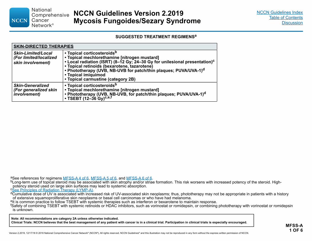
SUGGESTED TREATMENT REGIMENSa
SKIN-DIRECTED THERAPIESSkin-Limited/Local (For limited/localized skin involvement)
• Topical corticosteroidsb• Topical mechlorethamine [nitrogen mustard]• Local radiation (ISRT) (8–12 Gy; 24–30 Gy for unilesional presentation)c• Topical retinoids (bexarotene, tazarotene)• Phototherapy (UVB, NB-UVB for patch/thin plaques; PUVA/UVA-1)d• Topical imiquimod• Topical carmustine (category 2B)
Skin-Generalized(For generalized skin involvement)
• Topical corticosteroidsb• Topical mechlorethamine [nitrogen mustard]• Phototherapy (UVB, NB-UVB, for patch/thin plaques; PUVA/UVA-1)d• TSEBT (12–36 Gy)c,e,f
aSee references for regimens MFSS-A 4 of 6, MFSS-A 5 of 6, and MFSS-A 6 of 6.bLong-term use of topical steroid may be associated with skin atrophy and/or striae formation. This risk worsens with increased potency of the steroid. High-
potency steroid used on large skin surfaces may lead to systemic absorption.cSee Principles of Radiation Therapy (LYMP-A).dCumulative dose of UV is associated with increased risk of UV-associated skin neoplasms; thus, phototherapy may not be appropriate in patients with a history
of extensive squamoproliferative skin neoplasms or basal cell carcinomas or who have had melanoma.eIt is common practice to follow TSEBT with systemic therapies such as interferon or bexarotene to maintain response. fSafety of combining TSEBT with systemic retinoids or HDAC inhibitors, such as vorinostat or romidepsin, or combining phototherapy with vorinostat or romidepsin
is unknown.
NCCN Guidelines Version 2.2019
Version 2.2019, 12/17/18 © 2018 National Comprehensive Cancer Network® (NCCN®), All rights reserved. NCCN Guidelines® and this illustration may not be reproduced in any form without the express written permission of NCCN.
Note: All recommendations are category 2A unless otherwise indicated.Clinical Trials: NCCN believes that the best management of any patient with cancer is in a clinical trial. Participation in clinical trials is especially encouraged.
Mycosis Fungoides/Sezary SyndromeNCCN Guidelines Index
Table of ContentsDiscussion

MFSS-A 2 OF 6
SUGGESTED TREATMENT REGIMENSa
SYSTEMIC THERAPIESPreferred regimensg (alphabetical order) Other recommended regimens Useful under certain circumstances
SYST-CAT A• Brentuximab vedotinh,i,j• Bexarotenef • Extracorporeal photopheresis (ECP)k• Interferons (IFN-alpha, IFN-gamma)• Methotrexate (≤50 mg q week)• Mogamulizumabl• Romidepsinf• Vorinostatf
• Acitretinf • All-trans retinoic acidf• Isotretinoin [13-cis-retinoic
acid]f
SYST-CAT B• Brentuximab vedotinh,i,j• Gemcitabine• Liposomal doxorubicin• Pralatrexate (low-dose or standard dose)
• Relapsed/refractory disease requiring systemic therapy; alphabetical order by category)�Alemtuzumabj,n�Chlorambucil�Cyclophosphamide�Etoposide�Pentostatin�Temozolomide for CNS involvement�Bortezomib (category 2B)�Pembrolizumab (category 2B)o,p�See TCEL-B 2 of 5 for regimens listed
for PTCL-NOSm
Large-Cell Transformation (LCT)
• Brentuximab vedotinh,i,j• Gemcitabine• Liposomal doxorubicin• Pralatrexate (low-dose or standard dose)• Romidepsin• See TCEL-B 2 of 5 for regimens listed for
PTCL-NOSm
aSee references for regimens MFSS-A 4 of 6, MFSS-A 5 of 6, and MFSS-A 6 of 6. fSafety of combining TSEBT with systemic retinoids or HDAC inhibitors, such as vorinostat or romidepsin, or
combining phototherapy with vorinostat or romidepsin is unknown.gRegimens are listed in alphabetical order. The optimal treatment for any patient at any given time is often
individualized based on symptoms of disease, route of administration, toxicities, and overall goals of therapy.hA randomized phase 3 trial comparing brentuximab vedotin (BV) with physician’s choice of oral bexarotene or
methotrexate, showed superior clinical outcome of BV in patients with CD30+ MF and pcALCL. CD30 positivity was defined as CD30 expression ≥10% of total lymphoid cells in at least 1 of minimal 2 skin biopsies required to evaluate for eligibility. Fourty-four percent of eligible patients with MF had at least 1 screening skin biopsy with CD30 <10%. In the two previously reported investigator-initiated studies, clinical responses with BV were observed across all CD30 expression levels including in those with negligible CD30 expression.
iPatients with Sezary syndrome were excluded from the ALCANZA trial.jSee Supportive Care for Brentuximab Vedotin and Alemtuzumab (LYMP-C).kPhotopheresis may be more appropriate as systemic therapy in patients with some blood involvement (B1 or B2).
lPatients with LCT were excluded from the MAVORIC trial.mMultiagent chemotherapy regimens are generally reserved for patients with
relapsed/refractory or extracutaneous disease. Most patients are treated with multiple SYST-CAT A/B before receiving multiagent chemotherapy.
nLower doses of alemtuzumab administered subcutaneously have shown lower incidence of infectious complications.
oPreliminary phase II data in patients with MF and SS. Disease flare is seen in some patients (especially in erythrodermic skin/Sezary patients) and should be distinguished from disease progression. Khodadoust M, Rook A, Porcu P, et al. Pembrolizumab for treatment of relapsed/refractory mycosis fungoides and Sezary syndrome: Clinical efficacy in a CITN multicenter phase 2 study [abstract]. Blood 2018;125:Abstract 181.
pRapid progression has been reported in HTLV positive patients receiving pembrolizumab.
NCCN Guidelines Version 2.2019
Version 2.2019, 12/17/18 © 2018 National Comprehensive Cancer Network® (NCCN®), All rights reserved. NCCN Guidelines® and this illustration may not be reproduced in any form without the express written permission of NCCN.
Note: All recommendations are category 2A unless otherwise indicated.Clinical Trials: NCCN believes that the best management of any patient with cancer is in a clinical trial. Participation in clinical trials is especially encouraged.
Mycosis Fungoides/Sezary SyndromeNCCN Guidelines Index
Table of ContentsDiscussion

MFSS-A 3 OF 6
SUGGESTED TREATMENT REGIMENSa
COMBINATION THERAPIES (alphabetical order)
Skin-directed + Systemic• Phototherapy + ECPk• Phototherapy + IFN• Phototherapy + retinoid• TSEBT + ECPh
Systemic + Systemic • ECPk+ IFN• ECPk + retinoid• ECPk + retinoid + IFN• Retinoid + IFN
ERYTHRODERMIC DISEASE/SEZARY SYNDROMEPreferred regimens Other recommended regimens
Low-intermediate burden (eg, ASC <5 K/mm3)
• Combination therapies (see above)• SYST-CAT A ± skin-directed therapies (skin-
generalized) (See MFSS-A 2 of 6)
• SYST-CAT B ± skin-directed therapies (skin-generalized) (See MFSS-A 2 of 6)
• Alemtuzumabj,n• Pembrolizumabo,p
High burden (eg, ASC >5 K/mm3)
• Combination therapies (see above)• Mogamulizumab ± skin-directed therapies
(skin-generalized)• Romidepsin ± skin-directed therapies (skin-
generalized)
• SYST-CAT A (options not listed under preferred regimens) (See MFSS-A 2 of 6)
• SYST-CAT B (See MFSS-A 2 of 6)• Alemtuzumabj,n• Pembrolizumabo,p
aSee references for regimens MFSS-A 4 of 6, MFSS-A 5 of 6, and MFSS-A 6 of 6. jSee Supportive Care for Brentuximab Vedotin and Alemtuzumab (LYMP-C).kPhotopheresis may be more appropriate as systemic therapy in patients with some blood involvement (B1 or B2).nLower doses of alemtuzumab administered subcutaneously have shown lower incidence of infectious complications.oPreliminary phase II data in patients with MF and SS. Disease flare is seen in some patients (especially in erythrodermic skin/Sezary patients) and should be
distinguished from disease progression. Khodadoust M, Rook A, Porcu P, et al. Pembrolizumab for treatment of relapsed/refractory mycosis fungoides and Sezary syndrome: Clinical efficacy in a CITN multicenter phase 2 study [abstract]. Blood 2018;125:Abstract 181.
pRapid progression has been reported in HTLV positive patients receiving pembrolizumab.
NCCN Guidelines Version 2.2019
Version 2.2019, 12/17/18 © 2018 National Comprehensive Cancer Network® (NCCN®), All rights reserved. NCCN Guidelines® and this illustration may not be reproduced in any form without the express written permission of NCCN.
Note: All recommendations are category 2A unless otherwise indicated.Clinical Trials: NCCN believes that the best management of any patient with cancer is in a clinical trial. Participation in clinical trials is especially encouraged.
Mycosis Fungoides/Sezary SyndromeNCCN Guidelines Index
Table of ContentsDiscussion
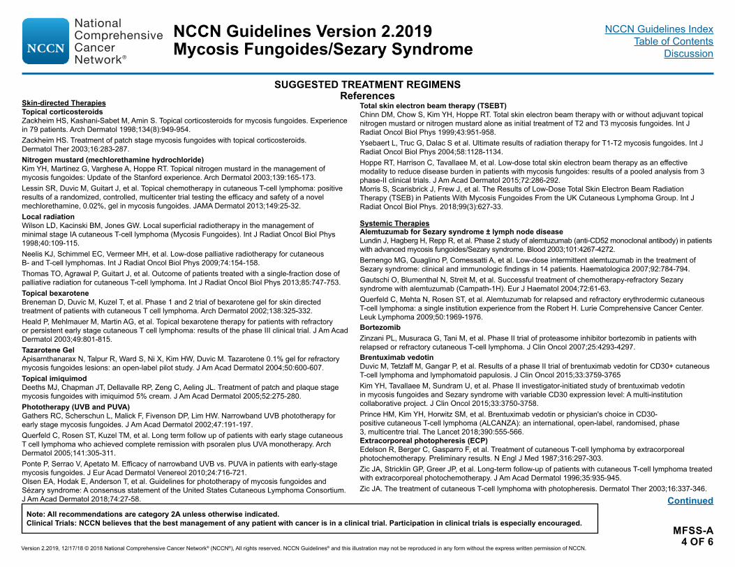
MFSS-A 4 OF 6
SUGGESTED TREATMENT REGIMENSReferences
Continued
Skin-directed TherapiesTopical corticosteroidsZackheim HS, Kashani-Sabet M, Amin S. Topical corticosteroids for mycosis fungoides. Experience in 79 patients. Arch Dermatol 1998;134(8):949-954.Zackheim HS. Treatment of patch stage mycosis fungoides with topical corticosteroids. Dermatol Ther 2003;16:283-287.Nitrogen mustard (mechlorethamine hydrochloride)Kim YH, Martinez G, Varghese A, Hoppe RT. Topical nitrogen mustard in the management of mycosis fungoides: Update of the Stanford experience. Arch Dermatol 2003;139:165-173.Lessin SR, Duvic M, Guitart J, et al. Topical chemotherapy in cutaneous T-cell lymphoma: positive results of a randomized, controlled, multicenter trial testing the efficacy and safety of a novel mechlorethamine, 0.02%, gel in mycosis fungoides. JAMA Dermatol 2013;149:25-32. Local radiation Wilson LD, Kacinski BM, Jones GW. Local superficial radiotherapy in the management of minimal stage IA cutaneous T-cell lymphoma (Mycosis Fungoides). Int J Radiat Oncol Biol Phys 1998;40:109-115.Neelis KJ, Schimmel EC, Vermeer MH, et al. Low-dose palliative radiotherapy for cutaneous B- and T-cell lymphomas. Int J Radiat Oncol Biol Phys 2009;74:154-158. Thomas TO, Agrawal P, Guitart J, et al. Outcome of patients treated with a single-fraction dose of palliative radiation for cutaneous T-cell lymphoma. Int J Radiat Oncol Biol Phys 2013;85:747-753.Topical bexarotene Breneman D, Duvic M, Kuzel T, et al. Phase 1 and 2 trial of bexarotene gel for skin directed treatment of patients with cutaneous T cell lymphoma. Arch Dermatol 2002;138:325-332.Heald P, Mehlmauer M, Martin AG, et al. Topical bexarotene therapy for patients with refractory or persistent early stage cutaneous T cell lymphoma: results of the phase III clinical trial. J Am Acad Dermatol 2003;49:801-815.Tazarotene GelApisarnthanarax N, Talpur R, Ward S, Ni X, Kim HW, Duvic M. Tazarotene 0.1% gel for refractory mycosis fungoides lesions: an open-label pilot study. J Am Acad Dermatol 2004;50:600-607.Topical imiquimodDeeths MJ, Chapman JT, Dellavalle RP, Zeng C, Aeling JL. Treatment of patch and plaque stage mycosis fungoides with imiquimod 5% cream. J Am Acad Dermatol 2005;52:275-280.Phototherapy (UVB and PUVA)Gathers RC, Scherschun L, Malick F, Fivenson DP, Lim HW. Narrowband UVB phototherapy for early stage mycosis fungoides. J Am Acad Dermatol 2002;47:191-197.Querfeld C, Rosen ST, Kuzel TM, et al. Long term follow up of patients with early stage cutaneous T cell lymphoma who achieved complete remission with psoralen plus UVA monotherapy. Arch Dermatol 2005;141:305-311.Ponte P, Serrao V, Apetato M. Efficacy of narrowband UVB vs. PUVA in patients with early-stage mycosis fungoides. J Eur Acad Dermatol Venereol 2010;24:716-721. Olsen EA, Hodak E, Anderson T, et al. Guidelines for phototherapy of mycosis fungoides and Sézary syndrome: A consensus statement of the United States Cutaneous Lymphoma Consortium. J Am Acad Dermatol 2018;74:27-58.
Total skin electron beam therapy (TSEBT) Chinn DM, Chow S, Kim YH, Hoppe RT. Total skin electron beam therapy with or without adjuvant topical nitrogen mustard or nitrogen mustard alone as initial treatment of T2 and T3 mycosis fungoides. Int J Radiat Oncol Biol Phys 1999;43:951-958.Ysebaert L, Truc G, Dalac S et al. Ultimate results of radiation therapy for T1-T2 mycosis fungoides. Int J Radiat Oncol Biol Phys 2004;58:1128-1134.Hoppe RT, Harrison C, Tavallaee M, et al. Low-dose total skin electron beam therapy as an effective modality to reduce disease burden in patients with mycosis fungoides: results of a pooled analysis from 3 phase-II clinical trials. J Am Acad Dermatol 2015;72:286-292.Morris S, Scarisbrick J, Frew J, et al. The Results of Low-Dose Total Skin Electron Beam Radiation Therapy (TSEB) in Patients With Mycosis Fungoides From the UK Cutaneous Lymphoma Group. Int J Radiat Oncol Biol Phys. 2018;99(3):627-33.
Systemic TherapiesAlemtuzumab for Sezary syndrome ± lymph node diseaseLundin J, Hagberg H, Repp R, et al. Phase 2 study of alemtuzumab (anti-CD52 monoclonal antibody) in patients with advanced mycosis fungoides/Sezary syndrome. Blood 2003;101:4267-4272.Bernengo MG, Quaglino P, Comessatti A, et al. Low-dose intermittent alemtuzumab in the treatment of Sezary syndrome: clinical and immunologic findings in 14 patients. Haematologica 2007;92:784-794.Gautschi O, Blumenthal N, Streit M, et al. Successful treatment of chemotherapy-refractory Sezary syndrome with alemtuzumab (Campath-1H). Eur J Haematol 2004;72:61-63.Querfeld C, Mehta N, Rosen ST, et al. Alemtuzumab for relapsed and refractory erythrodermic cutaneous T-cell lymphoma: a single institution experience from the Robert H. Lurie Comprehensive Cancer Center. Leuk Lymphoma 2009;50:1969-1976.BortezomibZinzani PL, Musuraca G, Tani M, et al. Phase II trial of proteasome inhibitor bortezomib in patients with relapsed or refractory cutaneous T-cell lymphoma. J Clin Oncol 2007;25:4293-4297.Brentuximab vedotinDuvic M, Tetzlaff M, Gangar P, et al. Results of a phase II trial of brentuximab vedotin for CD30+ cutaneous T-cell lymphoma and lymphomatoid papulosis. J Clin Oncol 2015;33:3759-3765 Kim YH, Tavallaee M, Sundram U, et al. Phase II investigator-initiated study of brentuximab vedotin in mycosis fungoides and Sezary syndrome with variable CD30 expression level: A multi-institution collaborative project. J Clin Oncol 2015;33:3750-3758.Prince HM, Kim YH, Horwitz SM, et al. Brentuximab vedotin or physician's choice in CD30-positive cutaneous T-cell lymphoma (ALCANZA): an international, open-label, randomised, phase3, multicentre trial. The Lancet 2018;390:555-566.Extracorporeal photopheresis (ECP)Edelson R, Berger C, Gasparro F, et al. Treatment of cutaneous T-cell lymphoma by extracorporeal photochemotherapy. Preliminary results. N Engl J Med 1987;316:297-303.Zic JA, Stricklin GP, Greer JP, et al. Long-term follow-up of patients with cutaneous T-cell lymphoma treated with extracorporeal photochemotherapy. J Am Acad Dermatol 1996;35:935-945.Zic JA. The treatment of cutaneous T-cell lymphoma with photopheresis. Dermatol Ther 2003;16:337-346.
NCCN Guidelines Version 2.2019
Version 2.2019, 12/17/18 © 2018 National Comprehensive Cancer Network® (NCCN®), All rights reserved. NCCN Guidelines® and this illustration may not be reproduced in any form without the express written permission of NCCN.
Note: All recommendations are category 2A unless otherwise indicated.Clinical Trials: NCCN believes that the best management of any patient with cancer is in a clinical trial. Participation in clinical trials is especially encouraged.
Mycosis Fungoides/Sezary SyndromeNCCN Guidelines Index
Table of ContentsDiscussion
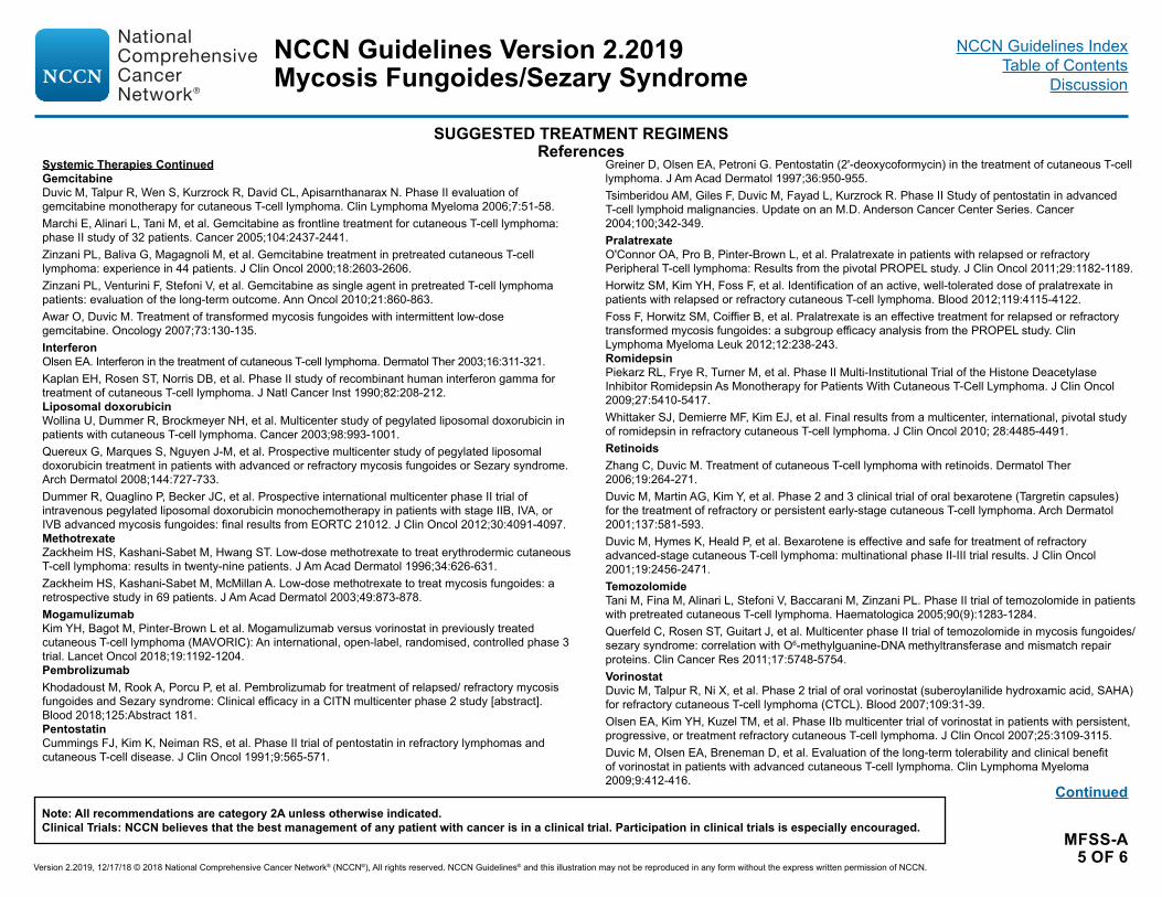
MFSS-A 5 OF 6
SUGGESTED TREATMENT REGIMENSReferences
Continued
Systemic Therapies ContinuedGemcitabineDuvic M, Talpur R, Wen S, Kurzrock R, David CL, Apisarnthanarax N. Phase II evaluation of gemcitabine monotherapy for cutaneous T-cell lymphoma. Clin Lymphoma Myeloma 2006;7:51-58.Marchi E, Alinari L, Tani M, et al. Gemcitabine as frontline treatment for cutaneous T-cell lymphoma: phase II study of 32 patients. Cancer 2005;104:2437-2441.Zinzani PL, Baliva G, Magagnoli M, et al. Gemcitabine treatment in pretreated cutaneous T-cell lymphoma: experience in 44 patients. J Clin Oncol 2000;18:2603-2606.Zinzani PL, Venturini F, Stefoni V, et al. Gemcitabine as single agent in pretreated T-cell lymphoma patients: evaluation of the long-term outcome. Ann Oncol 2010;21:860-863. Awar O, Duvic M. Treatment of transformed mycosis fungoides with intermittent low-dose gemcitabine. Oncology 2007;73:130-135.InterferonOlsen EA. Interferon in the treatment of cutaneous T-cell lymphoma. Dermatol Ther 2003;16:311-321.Kaplan EH, Rosen ST, Norris DB, et al. Phase II study of recombinant human interferon gamma for treatment of cutaneous T-cell lymphoma. J Natl Cancer Inst 1990;82:208-212.Liposomal doxorubicinWollina U, Dummer R, Brockmeyer NH, et al. Multicenter study of pegylated liposomal doxorubicin in patients with cutaneous T-cell lymphoma. Cancer 2003;98:993-1001.Quereux G, Marques S, Nguyen J-M, et al. Prospective multicenter study of pegylated liposomal doxorubicin treatment in patients with advanced or refractory mycosis fungoides or Sezary syndrome. Arch Dermatol 2008;144:727-733.Dummer R, Quaglino P, Becker JC, et al. Prospective international multicenter phase II trial of intravenous pegylated liposomal doxorubicin monochemotherapy in patients with stage IIB, IVA, or IVB advanced mycosis fungoides: final results from EORTC 21012. J Clin Oncol 2012;30:4091-4097.MethotrexateZackheim HS, Kashani-Sabet M, Hwang ST. Low-dose methotrexate to treat erythrodermic cutaneous T-cell lymphoma: results in twenty-nine patients. J Am Acad Dermatol 1996;34:626-631.Zackheim HS, Kashani-Sabet M, McMillan A. Low-dose methotrexate to treat mycosis fungoides: a retrospective study in 69 patients. J Am Acad Dermatol 2003;49:873-878.MogamulizumabKim YH, Bagot M, Pinter-Brown L et al. Mogamulizumab versus vorinostat in previously treated cutaneous T-cell lymphoma (MAVORIC): An international, open-label, randomised, controlled phase 3 trial. Lancet Oncol 2018;19:1192-1204.PembrolizumabKhodadoust M, Rook A, Porcu P, et al. Pembrolizumab for treatment of relapsed/ refractory mycosis fungoides and Sezary syndrome: Clinical efficacy in a CITN multicenter phase 2 study [abstract]. Blood 2018;125:Abstract 181.PentostatinCummings FJ, Kim K, Neiman RS, et al. Phase II trial of pentostatin in refractory lymphomas and cutaneous T-cell disease. J Clin Oncol 1991;9:565-571.
Greiner D, Olsen EA, Petroni G. Pentostatin (2'-deoxycoformycin) in the treatment of cutaneous T-cell lymphoma. J Am Acad Dermatol 1997;36:950-955.Tsimberidou AM, Giles F, Duvic M, Fayad L, Kurzrock R. Phase II Study of pentostatin in advanced T-cell lymphoid malignancies. Update on an M.D. Anderson Cancer Center Series. Cancer 2004;100;342-349.PralatrexateO'Connor OA, Pro B, Pinter-Brown L, et al. Pralatrexate in patients with relapsed or refractory Peripheral T-cell lymphoma: Results from the pivotal PROPEL study. J Clin Oncol 2011;29:1182-1189.Horwitz SM, Kim YH, Foss F, et al. Identification of an active, well-tolerated dose of pralatrexate in patients with relapsed or refractory cutaneous T-cell lymphoma. Blood 2012;119:4115-4122.Foss F, Horwitz SM, Coiffier B, et al. Pralatrexate is an effective treatment for relapsed or refractory transformed mycosis fungoides: a subgroup efficacy analysis from the PROPEL study. Clin Lymphoma Myeloma Leuk 2012;12:238-243.RomidepsinPiekarz RL, Frye R, Turner M, et al. Phase II Multi-Institutional Trial of the Histone Deacetylase Inhibitor Romidepsin As Monotherapy for Patients With Cutaneous T-Cell Lymphoma. J Clin Oncol 2009;27:5410-5417.Whittaker SJ, Demierre MF, Kim EJ, et al. Final results from a multicenter, international, pivotal study of romidepsin in refractory cutaneous T-cell lymphoma. J Clin Oncol 2010; 28:4485-4491.RetinoidsZhang C, Duvic M. Treatment of cutaneous T-cell lymphoma with retinoids. Dermatol Ther 2006;19:264-271.Duvic M, Martin AG, Kim Y, et al. Phase 2 and 3 clinical trial of oral bexarotene (Targretin capsules) for the treatment of refractory or persistent early-stage cutaneous T-cell lymphoma. Arch Dermatol 2001;137:581-593.Duvic M, Hymes K, Heald P, et al. Bexarotene is effective and safe for treatment of refractory advanced-stage cutaneous T-cell lymphoma: multinational phase II-III trial results. J Clin Oncol 2001;19:2456-2471.TemozolomideTani M, Fina M, Alinari L, Stefoni V, Baccarani M, Zinzani PL. Phase II trial of temozolomide in patients with pretreated cutaneous T-cell lymphoma. Haematologica 2005;90(9):1283-1284.Querfeld C, Rosen ST, Guitart J, et al. Multicenter phase II trial of temozolomide in mycosis fungoides/sezary syndrome: correlation with O6-methylguanine-DNA methyltransferase and mismatch repair proteins. Clin Cancer Res 2011;17:5748-5754.VorinostatDuvic M, Talpur R, Ni X, et al. Phase 2 trial of oral vorinostat (suberoylanilide hydroxamic acid, SAHA) for refractory cutaneous T-cell lymphoma (CTCL). Blood 2007;109:31-39.Olsen EA, Kim YH, Kuzel TM, et al. Phase IIb multicenter trial of vorinostat in patients with persistent, progressive, or treatment refractory cutaneous T-cell lymphoma. J Clin Oncol 2007;25:3109-3115.Duvic M, Olsen EA, Breneman D, et al. Evaluation of the long-term tolerability and clinical benefit of vorinostat in patients with advanced cutaneous T-cell lymphoma. Clin Lymphoma Myeloma 2009;9:412-416.
NCCN Guidelines Version 2.2019
Version 2.2019, 12/17/18 © 2018 National Comprehensive Cancer Network® (NCCN®), All rights reserved. NCCN Guidelines® and this illustration may not be reproduced in any form without the express written permission of NCCN.
Note: All recommendations are category 2A unless otherwise indicated.Clinical Trials: NCCN believes that the best management of any patient with cancer is in a clinical trial. Participation in clinical trials is especially encouraged.
Mycosis Fungoides/Sezary SyndromeNCCN Guidelines Index
Table of ContentsDiscussion

MFSS-A 6 OF 6
SUGGESTED TREATMENT REGIMENSReferences
Combination TherapiesSkin-directed + SystemicRupoli S, Goteri G, Pulini S, et al. Long term experience with low dose interferon alpha and PUVA in the management of early mycosis fungoides. Eur J Haematol 2005;75:136-145.Kuzel T, Roenigk H Jr, Samuelson E, et al. Effectiveness of interferon alfa-2a combined with phototherapy for mycosis fungoides and the Sézary syndrome. J Clin Oncol 1995;13:257-263.McGinnis K, Shapiro M, Vittorio C, et al. Psoralen plus long wave UV A (PUVA) and bexarotene therapy: An effective and synergistic combined adjunct to therapy for patients with advanced cutaneous T cell lymphoma. Arch Dermatol 2003;139:771-775.Wilson LD, Jones GW, Kim D, et al. Experience with total skin electron beam therapy in combination with extracorporeal photopheresis in the management of patients with erythrodermic (T4) mycosis fungoides. J Am Acad Dermatol 2000;43:54-60.Stadler R, Otte H-G, Luger T, et al. Prospective randomized multicenter clinical trial on the use of interferon alpha -2a plus acitretin versus interferon alpha -2a plus PUVA in patients with cutaneous T-cell lymphoma stages I and II. Blood 1998;92:3578-3581.Systemic + SystemicStraus DJ, Duvic M, Kuzel T, et al. Results of a phase II trial of oral bexarotene (Targretin) combined with interferon alfa 2b (Intron A) for patients with cutaneous T cell lymphoma. Cancer 2007;109:1799-1803.Talpur R, Ward S, Apisarnthanarax N, Breuer Mcham J, Duvic M. Optimizing bexarotene therapy for cutaneous T cell lymphoma. J Am Acad Dermatol 2002;47:672-684.Suchin KR, Cucchiara AJ, Gottleib SL, et al. Treatment of cutaneous T-cell lymphoma with combined immunomodulatory therapy: a 14-year experience at a single institution. Arch Dermatol. 2002;138:1054-1060.Raphael BA, Shin DB, Suchin KR, et al. High clinical response rate of Sezary syndrome to immunomodulatory therapies: prognostic markers of response. Arch Dermatol 2011;147:1410-1415.
Allogeneic hematopoietic cell transplantde Masson A, Beylot-Barry M, Bouaziz J, et al. Allogeneic stem cell transplantation for advanced cutaneous T-cell lymphomas: a study from the French Society of Bone Marrow Transplantation and French Study Group on Cutaneous Lymphomas. Haematologica 2014;99:527-534.Duarte R, Boumendil A, Onida F, et al. Long-term outcome of allogeneic hematopoietic cell transplantation for patients with mycosis fungoides and Sézary syndrome: a European society for blood and marrow transplantation lymphoma working party extended analysis. J Clin Oncol 2014;32:3347-3348. Duarte RF, Schmitz N, Servitje O, Sureda A. Haematopoietic stem cell transplantation for patients with primary cutaneous T-cell lymphoma. Bone Marrow Transplant 2008;41:597-604.Hosing C, Bassett R, Dabaja B, et al. Allogeneic stem-cell transplantation in patients with cutaneous lymphoma: updated results from a single institution. Ann Oncol 2015;26:2490-2495. Lechowicz M, Lazarus H, Carreras J, et al. Allogeneic hematopoietic cell transplantation for mycosis fungoides and Sezary syndrome. Bone Marrow Transplant 2014;49:1360-1365.Wu PA, Kim YH, Lavori PW, Hoppe RT, Stockerl-Goldstein KE. A meta-analysis of patients receiving allogeneic or autologous hematopoietic stem cell transplant in mycosis fungoides and Sezary syndrome. Biol Blood Marrow Transplant 2009;15:982-990.
NCCN Guidelines Version 2.2019
Version 2.2019, 12/17/18 © 2018 National Comprehensive Cancer Network® (NCCN®), All rights reserved. NCCN Guidelines® and this illustration may not be reproduced in any form without the express written permission of NCCN.
Note: All recommendations are category 2A unless otherwise indicated.Clinical Trials: NCCN believes that the best management of any patient with cancer is in a clinical trial. Participation in clinical trials is especially encouraged.
Mycosis Fungoides/Sezary SyndromeNCCN Guidelines Index
Table of ContentsDiscussion

MFSS-B 1 OF 2
SUPPORTIVE CARE FOR MF/SS
Pruritus• Assessment�Pruritus should be assessed �Correlation between sites of disease and localization of pruritus may
be useful in tailoring therapy�For severe or persistent pruritus despite therapeutic response other
potential causes for pruritus should be investigated
• Treatment�Co-management with a dermatologist with expertise in skin care and
CTCL�Optimized skin-directed and systemic therapy for MF/SS �Mild, unscented soaps for bathing are gentle and optimal to prevent
skin dryness�Moisturizers/ emollients �Topical steroid application (appropriate strength for body region) ±
occlusion1
�Topical over-the-counter preparations �Systemic agents
◊ First-line – H1 antihistamines; single agent or combination of antihistamines from different classes2
– Gabapentin3,4
◊ Second-line – Aprepitant5-8
– Mirtazapine4
– Selective serotonin reuptake inhibitors9
◊ Third-line – Naltrexone10
Collaboration with dermatologist for supportive care is essential.Infections• Active or Suspected Infections�Cutaneous viral infections
◊ High risk for skin dissemination of localized viral infections (HSV/VZV). HSV prophylaxis should be considered for patients with frequent recurrence of herpes simplex infection.
�Erythroderma: ◊ Swab of skin, nares, or other areas for cultures of Staphylococcus aureus (S. aureus) infection or colonization
◊ Intranasal mupirocin for S. aureus carriers ◊ Oral dicloxacillin or cephalexin ◊ Sulfamethoxazole/trimethoprim, doxycycline, minocycline, or clindamycin if suspected methicillin-resistant staphylococcus aureus (MRSA)
◊ Vancomycin if no improvement or documented bacteremia ◊ Bleach baths [1/2 cup of regular strength bleach (5%–6%) in full tub of water] or for limited areas, soaks (1 tsp of bleach in a gallon of water). Bleach baths should be taken for 5 to 10 minutes two to three times a week maximum followed by tap water to rinse off the bleach water. moisturizer should be put on immediately following the bleach bath or soak.
�Ulcerated and necrotic tumors: ◊ Infection or colonization with Gram-negative rods should be considered in addition to the more common gram-positive organisms.
• Prophylaxis�Optimize skin barrier protection with moisturizing of skin�Consider Mupirocin in nares for S. aureus carriage �Diluted bleach baths or soaks (if limited area) as noted above �Minimize use of central lines when possible�For patients receiving alemtuzumab, see LYMP-C.
Continued
NCCN Guidelines Version 2.2019
Version 2.2019, 12/17/18 © 2018 National Comprehensive Cancer Network® (NCCN®), All rights reserved. NCCN Guidelines® and this illustration may not be reproduced in any form without the express written permission of NCCN.
Note: All recommendations are category 2A unless otherwise indicated.Clinical Trials: NCCN believes that the best management of any patient with cancer is in a clinical trial. Participation in clinical trials is especially encouraged.
Mycosis Fungoides/Sezary SyndromeNCCN Guidelines Index
Table of ContentsDiscussion

MFSS-B 2 OF 2
SUPPORTIVE CARE FOR MF/SS
1Yosipovitch G, Szolar C, Hui XY, Maibach H. High-potency topical corticosteroid rapidly decrease histamine-induced itch but not thermal sensation and pain in human beings. J Am Acad Dermatol 1996;35:118-120.
2Eschler D, Klein PA. An evidence-based review of the efficacy of topical antihistamines in the relief of pruritus. J Drugs Dermatol 2010;9:992-997.3Matsuda KM, Sharma D, Schonfeld AR, Kwatra SG. Gabapentin and pregabalin for the treatment of chronic pruritus. J Am Acad Dermatol 2018;75:619-625.4Demierre MF, Taverna J. Mirtazapine and gabapentin for reducing pruritus in cutaneous T-cell lymphoma. Am Acad Dermatol 2006;55:543-544.5Jiménez Gallo D, Albarrán Planelles C, Linares Barrios M, et al. Treatment of pruritus in early-stage hypopigmented mycosis fungoides with aprepitant. Dermatol
Ther 2014;27:178-182. 6Duval A, Dubertret L. Aprepitant as an antipruritic agent? N Engl J Med 2009;361:1415-1416. 7Booken N, Heck M, Nicolay JP, Klemke CD, Goerdt S, Utikal J. Oral apepritant in the therapy of refractory pruritus in erythrodermic cutaneous T-cell lymphomas. Br
J Dermatol 2011;164:665–667.8Ladizinski B, Bazakas A, Olsen EA. Aprepitant: A novel neurokinin-1 receptor/substance P antagonist as antipruritic therapy in cutaneous T-cell lymphoma. Am
Acad Dermatol 2012;67:E198-E199.9Ständer S, Böckenholt B, Schürmeyer-Horst F, et al. Treatment of chronic pruritus with the selective serotonin re-uptake inhibitors paroxetine and fluvoxamine:
results of an open-labelled, two-arm proof-of-concept study. Acta Derm Venereol 2009;89:45-51. 10Brune A, Metze D, Luger T , Ständer S. Antipruritic therapy with the oral opioid receptor antagonist naltrexone. Open, non-placebo controlled administration in 133
patients. Hautarzt 2004;55:1130-1136.
REFERENCES
NCCN Guidelines Version 2.2019
Version 2.2019, 12/17/18 © 2018 National Comprehensive Cancer Network® (NCCN®), All rights reserved. NCCN Guidelines® and this illustration may not be reproduced in any form without the express written permission of NCCN.
Note: All recommendations are category 2A unless otherwise indicated.Clinical Trials: NCCN believes that the best management of any patient with cancer is in a clinical trial. Participation in clinical trials is especially encouraged.
Mycosis Fungoides/Sezary SyndromeNCCN Guidelines Index
Table of ContentsDiscussion
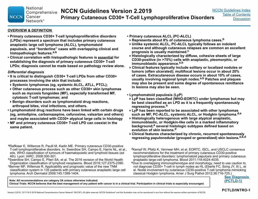
PCTLD/INTRO-1
See Diagnosis (PCTLD-1)
aRalfkiaer E, Willemze R, Paulli M, Kadin ME. Primary cutaneous CD30-positive T-cell lymphoproliferative disorders. In: Swerdlow SH, Campo E, Harris NL, et al., eds. WHO classification of tumours of haematopoietic and lymphoid tissues (ed 4th). Lyon: IARC; 2008:300-301.
bSwerdlow SH, Campo E, Pileri SA, et al. The 2016 revision of the World Health Organization classification of lymphoid neoplasms. Blood 2016;127:2375-2390.
cBenner MF, Willemze R. Applicability and prognostic value of the new TNM classification system in 135 patients with primary cutaneous anaplastic large cell lymphoma. Arch Dermatol 2009;145:1399-1404.
dKempf W, Pfaltz K, Vermeer MH, et al. EORTC, ISCL, and USCLC consensus recommendations for the treatment of primary cutaneous CD30-positive lymphoproliferative disorders: lymphomatoid papulosis and primary cutaneous anaplastic large-cell lymphoma. Blood 2011;118:4024-4035.
eDue to overlapping immunophenotype and morphology, need to use caution to not diagnose CD30+ T-cell in lymph nodes as HL (Eberle FC, Song JY, Xi L, et al. Nodal involvement by cutaneous CD30-positive T-cell lymphoma mimicking classical Hodgkin lymphoma. Amer J Surg Pathol 2012;36:716-725.)
• Primary cutaneous CD30+ T-cell lymphoproliferative disorders (LPDs) represent a spectrum that includes primary cutaneous anaplastic large cell lymphoma (ALCL), lymphomatoid papulosis, and “borderline” cases with overlapping clinical and histopathologic features.a,b
• Clinical correlation with histopathologic features is essential for establishing the diagnosis of primary cutaneous CD30+ T-cell LPDs; diagnosis cannot be made based on pathology review alone.
Differential diagnosis• It is critical to distinguish CD30+ T-cell LPDs from other CD30+
processes involving the skin that include:�Systemic lymphomas (eg, systemic ALCL, ATLL, PTCL);�Other cutaneous process such as other CD30+ skin lymphomas
such as mycosis fungoides (MF), especially transformed MF, cytotoxic T-cell lymphomas; and �Benign disorders such as lymphomatoid drug reactions,
arthropod bites, viral infections, and others. • Lymphomatoid drug reactions have been linked with certain drugs
(eg, amlodipine, carbamazepine, cefuroxime, valsartan and others) and maybe associated with CD30+ atypical large cells in histology
• MF and primary cutaneous CD30+ T-cell LPD can coexist in the same patient.
OVERVIEW & DEFINITION • Primary cutaneous ALCL (PC-ALCL)�Represents about 8% of cutaneous lymphoma cases.b�Unlike systemic ALCL, PC-ALCL typically follows an indolent
course and although cutaneous relapses are common an excellent prognosis is usually maintained.c�Histologically characterized by diffuse, cohesive sheets of large
CD30-positive (in >75%) cells with anaplastic, pleomorphic, or immunoblastic appearance.a,b�Clinical features typically include solitary or localized nodules or
tumors (often ulcerated); multifocal lesions occur in about 20% of cases. Extracutaneous disease occurs in about 10% of cases, usually involving regional lymph nodes.a,b Patches and plaques may also be present and some degree of spontaneous remittance in lesions may also be seen.
• Lymphomatoid papulosis (LyP) �LyP has been classified (WHO-EORTC) under lymphomas but may
be best classified as an LPD as it is a frequently spontaneously regressing process.b�LyP has been reported to be associated with other lymphomas
such as MF, PC-ALCL, systemic ALCL, or Hodgkin lymphoma.d,e�Histologically heterogenous with large atypical anaplastic,
immunoblastic, or Hodgkin-like cells in a marked inflammatory background;a several histologic subtypes defined based on evolution of skin lesions.d�Clinical features characterized by chronic, recurrent spontaneously
regressing papulonodular (grouped or generalized) skin lesions.a,b,d
NCCN Guidelines Version 2.2019
Version 2.2019, 12/17/18 © 2018 National Comprehensive Cancer Network® (NCCN®), All rights reserved. NCCN Guidelines® and this illustration may not be reproduced in any form without the express written permission of NCCN.
Note: All recommendations are category 2A unless otherwise indicated.Clinical Trials: NCCN believes that the best management of any patient with cancer is in a clinical trial. Participation in clinical trials is especially encouraged.
Primary Cutaneous CD30+ T-Cell Lymphoproliferative DisordersNCCN Guidelines Index
Table of ContentsDiscussion
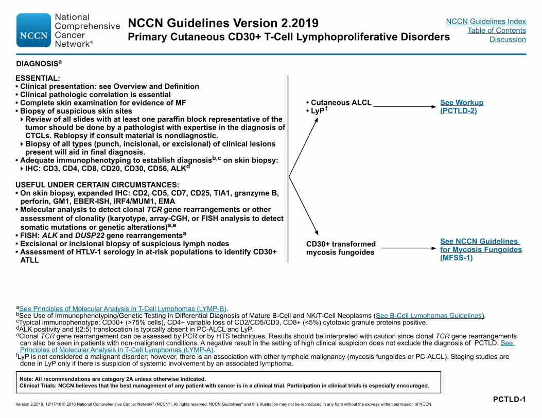
PCTLD-1
aSee Principles of Molecular Analysis in T-Cell Lymphomas (LYMP-B).bSee Use of Immunophenotyping/Genetic Testing in Differential Diagnosis of Mature B-Cell and NK/T-Cell Neoplasms (See B-Cell Lymphomas Guidelines).cTypical immunophenotype: CD30+ (>75% cells), CD4+ variable loss of CD2/CD5/CD3, CD8+ (<5%) cytotoxic granule proteins positive.dALK positivity and t(2;5) translocation is typically absent in PC-ALCL and LyP.eClonal TCR gene rearrangement can be assessed by PCR or by HTS techniques. Results should be interpreted with caution since clonal TCR gene rearrangements
can also be seen in patients with non-malignant conditions. A negative result in the setting of high clinical suspicion does not exclude the diagnosis of PCTLD. See Principles of Molecular Analysis in T-Cell Lymphomas (LYMP-A).
fLyP is not considered a malignant disorder; however, there is an association with other lymphoid malignancy (mycosis fungoides or PC-ALCL). Staging studies are done in LyP only if there is suspicion of systemic involvement by an associated lymphoma.
DIAGNOSISa
ESSENTIAL:• Clinical presentation: see Overview and Definition• Clinical pathologic correlation is essential• Complete skin examination for evidence of MF• Biopsy of suspicious skin sites�Review of all slides with at least one paraffin block representative of the
tumor should be done by a pathologist with expertise in the diagnosis of CTCLs. Rebiopsy if consult material is nondiagnostic. �Biopsy of all types (punch, incisional, or excisional) of clinical lesions
present will aid in final diagnosis.• Adequate immunophenotyping to establish diagnosisb,c on skin biopsy:�IHC: CD3, CD4, CD8, CD20, CD30, CD56, ALKd
USEFUL UNDER CERTAIN CIRCUMSTANCES:• On skin biopsy, expanded IHC: CD2, CD5, CD7, CD25, TIA1, granzyme B,
perforin, GM1, EBER-ISH, IRF4/MUM1, EMA• Molecular analysis to detect clonal TCR gene rearrangements or other
assessment of clonality (karyotype, array-CGH, or FISH analysis to detect somatic mutations or genetic alterations)a,e
• FISH: ALK and DUSP22 gene rearrangementsa• Excisional or incisional biopsy of suspicious lymph nodes• Assessment of HTLV-1 serology in at-risk populations to identify CD30+
ATLL
• Cutaneous ALCL• LyPf
CD30+ transformed mycosis fungoides
See Workup (PCTLD-2)
See NCCN Guidelines for Mycosis Fungoides (MFSS-1)
NCCN Guidelines Version 2.2019
Version 2.2019, 12/17/18 © 2018 National Comprehensive Cancer Network® (NCCN®), All rights reserved. NCCN Guidelines® and this illustration may not be reproduced in any form without the express written permission of NCCN.
Note: All recommendations are category 2A unless otherwise indicated.Clinical Trials: NCCN believes that the best management of any patient with cancer is in a clinical trial. Participation in clinical trials is especially encouraged.
Primary Cutaneous CD30+ T-Cell Lymphoproliferative DisordersNCCN Guidelines Index
Table of ContentsDiscussion

PCTLD-2
fLyP is not considered a malignant disorder; however, there is an association with other lymphoid malignancy (mycosis fungoides or PC-ALCL). Staging studies are done in LyP only if there is suspicion of systemic involvement by an associated lymphoma.
gMonitoring the size and number of lesions will assist with response assessment.hPatients with T-cell lymphomas often have extranodal disease, which may be inadequately imaged by CT. PET scan may be preferred in these instances.iDue to overlapping immunophenotype and morphology, need to use caution to not diagnose CD30+ T-cell in lymph nodes as HL (Eberle FC, Song JY, Xi L, et al. Nodal
involvement by cutaneous CD30-positive T-cell lymphoma mimicking classical Hodgkin lymphoma. Amer J Surg Pathol 2012;36:716-725.)jConsider systemic ALCL, regional lymph node involvement with PC-ALCL, or lymph node involvement with transformed MF.kConsider PC-ALCL if in draining lymph nodes only.lMany skin-directed and systemic therapies are contraindicated or of unknown safety in pregnancy. Refer to individual drug information.mOnly done to exclude an associated lymphoma.
WORKUP
Cutaneous ALCL
ESSENTIAL:• History and complete physical examination including complete skin examination;g
palpation of peripheral lymph node regions; liver or spleen enlargement
• CBC with differential• Comprehensive metabolic panel• LDH• C/A/P CT with contrast or integrated whole body PET/CTh • Biopsy suspicious nodesi,j,k�Biopsy of enlarged lymph nodes or suspected extracutaneous sites (if biopsy of skin is not
diagnostic). Excisional or incisional biopsy is preferred over core needle biopsy. An FNA alone is not sufficient for the initial diagnosis of lymphoma. A core needle biopsy is not optimal but can be used under certain circumstances. In certain circumstances, when a lymph node is not easily accessible for excisional or incisional biopsy, a combination of core needle biopsy and FNA in conjunction with appropriate ancillary techniques may be sufficient for diagnosis. Rebiopsy if consult material is nondiagnostic.
• Bone marrow aspiration and biopsy (optional for solitary C-ALCL or C-ALCL without extracutaneous involvement on imaging)
LyPf
ESSENTIAL:• History and complete physical examination
including complete skin examination;l palpation of peripheral lymph node regions; liver or spleen enlargement
• CBC with differential• Comprehensive metabolic panel• LDH
Primary cutaneous ALCL
Cutaneous ALCL with regional node
Systemic ALCL
See Primary Treatment (PCTLD-3)
See NCCN Guidelines for T-Cell Lymphomas - Peripheral T-Cell (TCEL-1)
See Primary Treatment for LyP (PCTLD-4)
USEFUL IN SELECTED CASES:• Pregnancy testing in women of child-bearing agel• Discussion of fertility and sperm banking, if fertility-impacting therapy is planned
USEFUL IN SELECTED CASES:• Pregnancy testing in women of child-bearing agel• Discussion of fertility and sperm banking, if fertility-
impacting therapy is planned• C/A/P CT with contrast or integrated whole body
PET/CTf,h,m (not done for typical LyP)• Bone marrow aspiration and biopsy (not done for
typical LyP)f,m
NCCN Guidelines Version 2.2019
Version 2.2019, 12/17/18 © 2018 National Comprehensive Cancer Network® (NCCN®), All rights reserved. NCCN Guidelines® and this illustration may not be reproduced in any form without the express written permission of NCCN.
Note: All recommendations are category 2A unless otherwise indicated.Clinical Trials: NCCN believes that the best management of any patient with cancer is in a clinical trial. Participation in clinical trials is especially encouraged.
Primary Cutaneous CD30+ T-Cell Lymphoproliferative DisordersNCCN Guidelines Index
Table of ContentsDiscussion
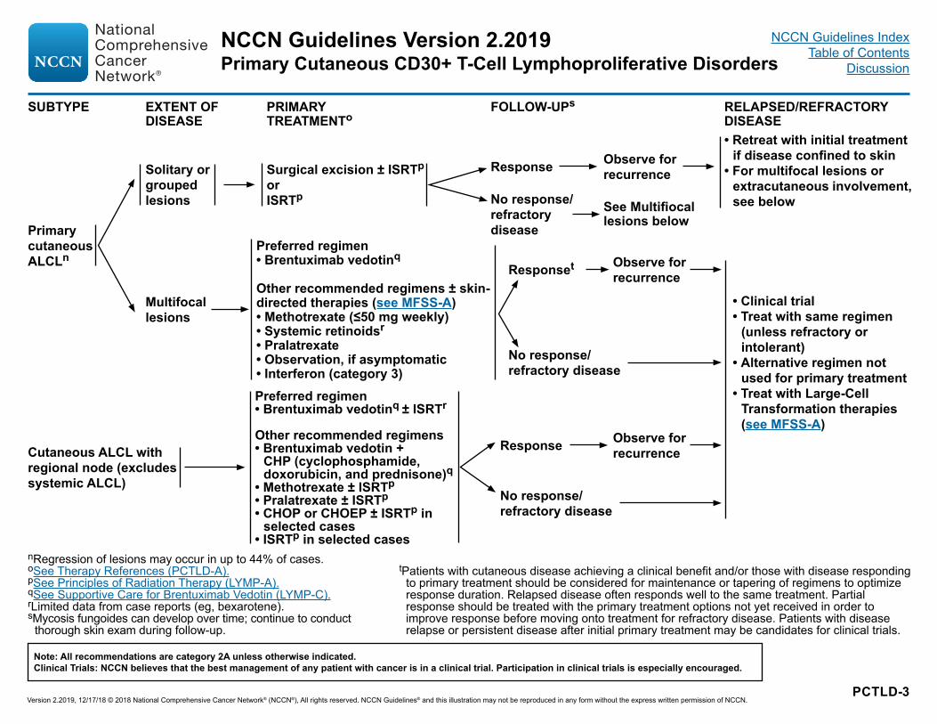
PCTLD-3
nRegression of lesions may occur in up to 44% of cases.oSee Therapy References (PCTLD-A).pSee Principles of Radiation Therapy (LYMP-A).qSee Supportive Care for Brentuximab Vedotin (LYMP-C).rLimited data from case reports (eg, bexarotene).sMycosis fungoides can develop over time; continue to conduct
thorough skin exam during follow-up.
SUBTYPE EXTENT OF DISEASE
PRIMARY TREATMENTo
FOLLOW-UPs RELAPSED/REFRACTORY DISEASE
Primary cutaneous ALCLn
Cutaneous ALCL with regional node (excludes systemic ALCL)
Solitary or grouped lesions
Multifocal lesions
Surgical excision ± ISRTp
orISRTp
Preferred regimen • Brentuximab vedotinq
Other recommended regimens ± skin-directed therapies (see MFSS-A) • Methotrexate (≤50 mg weekly)• Systemic retinoidsr • Pralatrexate• Observation, if asymptomatic• Interferon (category 3)Preferred regimen• Brentuximab vedotinq ± ISRTr
Other recommended regimens• Brentuximab vedotin +
CHP (cyclophosphamide, doxorubicin, and prednisone)q
• Methotrexate ± ISRTp• Pralatrexate ± ISRTp• CHOP or CHOEP ± ISRTp in
selected cases • ISRTp in selected cases
Response
No response/ refractory disease
Observe for recurrence
• Retreat with initial treatment if disease confined to skin
• For multifocal lesions or extracutaneous involvement, see below
Responset
No response/ refractory disease
Observe for recurrence
• Clinical trial• Treat with same regimen
(unless refractory or intolerant)
• Alternative regimen not used for primary treatment
• Treat with Large-Cell Transformation therapies (see MFSS-A)
Response
No response/ refractory disease
Observe for recurrence
tPatients with cutaneous disease achieving a clinical benefit and/or those with disease responding to primary treatment should be considered for maintenance or tapering of regimens to optimize response duration. Relapsed disease often responds well to the same treatment. Partial response should be treated with the primary treatment options not yet received in order to improve response before moving onto treatment for refractory disease. Patients with disease relapse or persistent disease after initial primary treatment may be candidates for clinical trials.
See Multifiocal lesions below
NCCN Guidelines Version 2.2019
Version 2.2019, 12/17/18 © 2018 National Comprehensive Cancer Network® (NCCN®), All rights reserved. NCCN Guidelines® and this illustration may not be reproduced in any form without the express written permission of NCCN.
Note: All recommendations are category 2A unless otherwise indicated.Clinical Trials: NCCN believes that the best management of any patient with cancer is in a clinical trial. Participation in clinical trials is especially encouraged.
Primary Cutaneous CD30+ T-Cell Lymphoproliferative DisordersNCCN Guidelines Index
Table of ContentsDiscussion

PCTLD-4
oSee Therapy References (PCTLD-A).qSee Supportive Care for Brentuximab Vedotin (LYMP-C).rLimited data from case reports (eg, bexarotene).uKempf W, Pfaltz K, Vermeer MH, et al. EORTC, ISCL, and USCLC consensus recommendations for the treatment of primary cutaneous CD30-positive
lymphoproliferative disorders: lymphomatoid papulosis and primary cutaneous anaplastic large-cell lymphoma. Blood 2011;118:4024-4035.vLife-long follow-up is warranted due to high risks for second lymphoid malignancies; continue to conduct thorough skin exam during follow-up.wPatients with a clinical benefit and/or those with disease responding to primary treatment should be considered for maintenance or tapering of regimens to optimize
response duration. Disease relapse often responds well to the same treatment. Partial response should be treated with the other options in the primary treatment options not received before to improve response before moving onto treatment for refractory disease. Patients with disease relapse or persistent disease after initial primary treatment may be candidates for clinical trials.
SUBTYPE EXTENT OF DISEASE
PRIMARY TREATMENTo FOLLOW-UPv RELAPSED/REFRACTORY DISEASE
LyP
Limited lesions, asymptomatic
Extensive lesions
Observation (preferred)orTopical steroids
Observation (preferred for asymptomatic)orMethotrexate (10–35 mg weeklyu)orPhototherapy orSystemic retinoidsrorTopical steroids orTopical mechlorethamine (nitrogen mustard)
Response
No response/refractory disease
Responsew
No response/ refractory disease
Observe for recurrence
Continue current management
Treat with alternative regimen not used for primary treatmentorOther regimens or Clinical trial
Clinical trialorObservationorRetreat with primary treatment or treat with alternative regimen not used for primary treatment
Refractory disease
Clinical trial orBrentuximab vedotinq
Clinical trial or Treat with alternative regimen not used for primary treatment
Limited lesions, symptomatic
Topical steroidsor PhototherapyorObservation
NCCN Guidelines Version 2.2019
Version 2.2019, 12/17/18 © 2018 National Comprehensive Cancer Network® (NCCN®), All rights reserved. NCCN Guidelines® and this illustration may not be reproduced in any form without the express written permission of NCCN.
Note: All recommendations are category 2A unless otherwise indicated.Clinical Trials: NCCN believes that the best management of any patient with cancer is in a clinical trial. Participation in clinical trials is especially encouraged.
Primary Cutaneous CD30+ T-Cell Lymphoproliferative DisordersNCCN Guidelines Index
Table of ContentsDiscussion
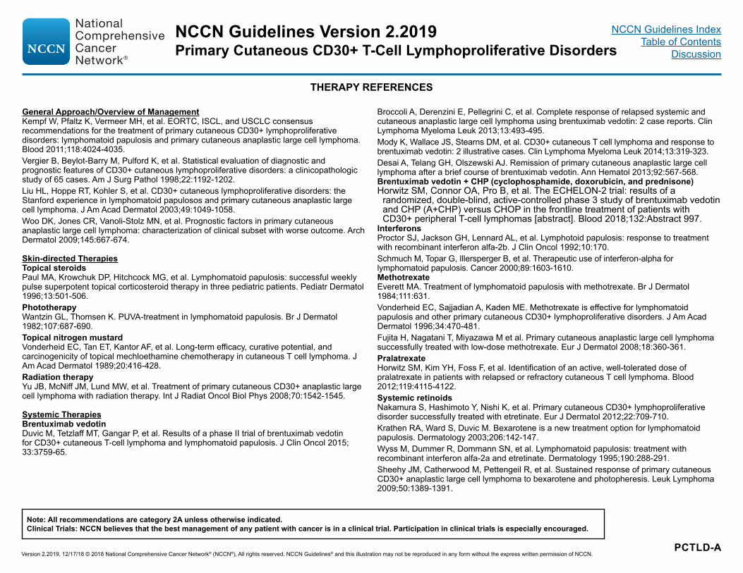
PCTLD-A
THERAPY REFERENCES
General Approach/Overview of ManagementKempf W, Pfaltz K, Vermeer MH, et al. EORTC, ISCL, and USCLC consensus recommendations for the treatment of primary cutaneous CD30+ lymphoproliferative disorders: lymphomatoid papulosis and primary cutaneous anaplastic large cell lymphoma. Blood 2011;118:4024-4035.Vergier B, Beylot-Barry M, Pulford K, et al. Statistical evaluation of diagnostic and prognostic features of CD30+ cutaneous lymphoproliferative disorders: a clinicopathologic study of 65 cases. Am J Surg Pathol 1998;22:1192-1202.Liu HL, Hoppe RT, Kohler S, et al. CD30+ cutaneous lymphoproliferative disorders: the Stanford experience in lymphomatoid papulosos and primary cutaneous anaplastic large cell lymphoma. J Am Acad Dermatol 2003;49:1049-1058.Woo DK, Jones CR, Vanoli-Stolz MN, et al. Prognostic factors in primary cutaneous anaplastic large cell lymphoma: characterization of clinical subset with worse outcome. Arch Dermatol 2009;145:667-674.
Skin-directed TherapiesTopical steroidsPaul MA, Krowchuk DP, Hitchcock MG, et al. Lymphomatoid papulosis: successful weekly pulse superpotent topical corticosteroid therapy in three pediatric patients. Pediatr Dermatol 1996;13:501-506.PhototherapyWantzin GL, Thomsen K. PUVA-treatment in lymphomatoid papulosis. Br J Dermatol 1982;107:687-690.Topical nitrogen mustardVonderheid EC, Tan ET, Kantor AF, et al. Long-term efficacy, curative potential, and carcinogenicity of topical mechloethamine chemotherapy in cutaneous T cell lymphoma. J Am Acad Dermatol 1989;20:416-428.Radiation therapyYu JB, McNiff JM, Lund MW, et al. Treatment of primary cutaneous CD30+ anaplastic large cell lymphoma with radiation therapy. Int J Radiat Oncol Biol Phys 2008;70:1542-1545.
Systemic TherapiesBrentuximab vedotinDuvic M, Tetzlaff MT, Gangar P, et al. Results of a phase II trial of brentuximab vedotin for CD30+ cutaneous T-cell lymphoma and lymphomatoid papulosis. J Clin Oncol 2015; 33:3759-65.
Broccoli A, Derenzini E, Pellegrini C, et al. Complete response of relapsed systemic and cutaneous anaplastic large cell lymphoma using brentuximab vedotin: 2 case reports. Clin Lymphoma Myeloma Leuk 2013;13:493-495.Mody K, Wallace JS, Stearns DM, et al. CD30+ cutaneous T cell lymphoma and response to brentuximab vedotin: 2 illustrative cases. Clin Lymphoma Myeloma Leuk 2014;13:319-323.Desai A, Telang GH, Olszewski AJ. Remission of primary cutaneous anaplastic large cell lymphoma after a brief course of brentuximab vedotin. Ann Hematol 2013;92:567-568.Brentuximab vedotin + CHP (cyclophosphamide, doxorubicin, and prednisone)Horwitz SM, Connor OA, Pro B, et al. The ECHELON-2 trial: results of a
randomized, double-blind, active-controlled phase 3 study of brentuximab vedotin and CHP (A+CHP) versus CHOP in the frontline treatment of patients with CD30+ peripheral T-cell lymphomas [abstract]. Blood 2018;132:Abstract 997.
InterferonsProctor SJ, Jackson GH, Lennard AL, et al. Lymphotoid papulosis: response to treatment with recombinant interferon alfa-2b. J Clin Oncol 1992;10:170.Schmuch M, Topar G, Illersperger B, et al. Therapeutic use of interferon-alpha for lymphomatoid papulosis. Cancer 2000;89:1603-1610. MethotrexateEverett MA. Treatment of lymphomatoid papulosis with methotrexate. Br J Dermatol 1984;111:631.Vonderheid EC, Sajjadian A, Kaden ME. Methotrexate is effective for lymphomatoid papulosis and other primary cutaneous CD30+ lymphoproliferative disorders. J Am Acad Dermatol 1996;34:470-481.Fujita H, Nagatani T, Miyazawa M et al. Primary cutaneous anaplastic large cell lymphoma successfully treated with low-dose methotrexate. Eur J Dermatol 2008;18:360-361.PralatrexateHorwitz SM, Kim YH, Foss F, et al. Identification of an active, well-tolerated dose of pralatrexate in patients with relapsed or refractory cutaneous T cell lymphoma. Blood 2012;119:4115-4122.Systemic retinoidsNakamura S, Hashimoto Y, Nishi K, et al. Primary cutaneous CD30+ lymphoproliferative disorder successfully treated with etretinate. Eur J Dermatol 2012;22:709-710.Krathen RA, Ward S, Duvic M. Bexarotene is a new treatment option for lymphomatoid papulosis. Dermatology 2003;206:142-147.Wyss M, Dummer R, Dommann SN, et al. Lymphomatoid papulosis: treatment with recombinant interferon alfa-2a and etretinate. Dermatology 1995;190:288-291.Sheehy JM, Catherwood M, Pettengeil R, et al. Sustained response of primary cutaneous CD30+ anaplastic large cell lymphoma to bexarotene and photopheresis. Leuk Lymphoma 2009;50:1389-1391.
NCCN Guidelines Version 2.2019
Version 2.2019, 12/17/18 © 2018 National Comprehensive Cancer Network® (NCCN®), All rights reserved. NCCN Guidelines® and this illustration may not be reproduced in any form without the express written permission of NCCN.
Note: All recommendations are category 2A unless otherwise indicated.Clinical Trials: NCCN believes that the best management of any patient with cancer is in a clinical trial. Participation in clinical trials is especially encouraged.
Primary Cutaneous CD30+ T-Cell Lymphoproliferative DisordersNCCN Guidelines Index
Table of ContentsDiscussion

LYMP-A 1 OF 3
PRINCIPLES OF RADIATION THERAPYa
General Principles: • The general intent of RT is to treat the evident skin disease with adequate margin both circumferentially and in depth.
Target Volumes:• Involved-site radiation therapy (ISRT) for cutaneous lesions:�ISRT is recommended as the appropriate field for treating primary cutaneous lymphomas. �Planning to define the clinical target volume (CTV) may often only require a careful physical exam. However, when the depth of disease is
not evident or when disease extends around curved surfaces, treatment planning may be facilitated by ultrasound imaging or CT-based simulation and planning. Incorporating other modern imaging like PET and MRI may enhance treatment volume determination in some cases.�ISRT targets the site of skin involvement. The volume encompasses the clinically evident disease with adequate margins. �The visible or palpable disease defines the gross tumor volume (GTV) and provides the basis for determining the CTV. Concerns for
questionable subclinical disease and uncertainties in original imaging accuracy or localization will lead to expansion of the CTV and are determined individually using clinical judgment but generally includes a margin of 1-2 cm both circumferentially and in depth. The CTV need not be expanded into intact bone.�The planning target volume (PTV) is an additional expansion of the CTV that accounts only for setup variations (see ICRU definitions).�The treatment plan is designed using conventional or 3-D conformal techniques using clinical treatment planning considerations of
coverage and dose reductions for organs at risk (OAR). • Involved-site radiation therapy (ISRT) for nodal disease:�See Principles of Radiation Therapy for T-cell Lymphomas (Target Volumes: ISRT for nodal disease).�See Principles of Radiation Therapy for B-cell Lymphomas (Target Volumes: ISRT for nodal disease).
Continued
aSee references on LYMP-A 3 of 3.
NCCN Guidelines Version 2.2019Primary Cutaneous Lymphomas
Version 2.2019, 12/17/18 © 2018 National Comprehensive Cancer Network® (NCCN®), All rights reserved. NCCN Guidelines® and this illustration may not be reproduced in any form without the express written permission of NCCN.
Note: All recommendations are category 2A unless otherwise indicated.Clinical Trials: NCCN believes that the best management of any patient with cancer is in a clinical trial. Participation in clinical trials is especially encouraged.
NCCN Guidelines IndexTable of Contents
Discussion

LYMP-A 2 OF 3
PRINCIPLES OF RADIATION THERAPYa
General Dose Guidelines: (RT in conventional fraction sizes)• PCMZL and PCFCL:�Optimal initial management for solitary/regional disease is with 24–30 Gy external beam radiation therapy (EBRT).
◊ Surface margins beyond area of clinically evident disease will vary depending on lesion size and body site and must take into account dosimetry of the beam being used. Surface margins of 1.0–1.5 cm are generally adequate.
◊ Margins in depth should include the volume at risk for involvement. ◊ Generally, treatment with 6–9 MeV electrons (with surface bolus) provides an adequate depth of treatment. Alternatively, low-energy x-rays (~100 Kv) may be used.
�RT for relapsed disease: 4 Gy EBRT may be adequate.
• MF/SS�Treatment of Individual Plaques or Tumors
◊ Optimal management for individual plaque and tumor lesions is with EBRT, 8–12 Gy, 8 Gy may be given in a single fraction. For unilesional MF, 24–30 Gy presentation.
◊ Surface margins beyond area of clinically evident disease will vary depending on lesion size and body site and must take into account dosimetry of the beam being used. Surface margins of 1.0–1.5 cm are generally adequate.
◊ Margins in depth should include the volume at risk for involvement. ◊ Generally, treatment with 6–9 MeV electrons (with surface bolus) provides an adequate depth of treatment. Alternatively, low energy x-rays (~100 Kv) may be used.
◊ For certain body surfaces, higher energy photon fields and opposed-field treatment (with bolus) may be required.�Total Skin Electron Beam Therapy (TSEBT)
◊ A variety of techniques may be utilized to cover the entire cutaneous surface. Patients are generally treated in the standing position on a rotating platform or with multiple body positions to ensure total skin coverage.
◊ The dose range is 12–36 Gy, generally 4–6 Gy per week. The advantage of lower total dose includes fewer short-term complications and better ability to re-treat for relapsed disease.
◊ “Shadowed” areas may need to be supplemented with individual electron fields. ◊ Individual tumors may be boosted with doses of 4–12 Gy. ◊ For patients with recalcitrant sites after generalized skin treatment, additional local treatment may be needed.
• Primary cutaneous ALCL: �RT for curative treatment: 24–36 Gy �Palliative RT: 2 Gy x 2
Treatment Modalities:• Treatment with photons or electrons may all be appropriate, depending on clinical circumstances.
aSee references on LYMP-A 3 of 3.
NCCN Guidelines Version 2.2019Primary Cutaneous Lymphomas
Version 2.2019, 12/17/18 © 2018 National Comprehensive Cancer Network® (NCCN®), All rights reserved. NCCN Guidelines® and this illustration may not be reproduced in any form without the express written permission of NCCN.
Note: All recommendations are category 2A unless otherwise indicated.Clinical Trials: NCCN believes that the best management of any patient with cancer is in a clinical trial. Participation in clinical trials is especially encouraged.
NCCN Guidelines IndexTable of Contents
Discussion
LYMP-A 3 OF 3
PRINCIPLES OF RADIATION THERAPY
Hoppe RT, Harrison C, Tavallaee M, et al. Low-dose total skin electron beam therapy as an effective modality to reduce disease burden in patients with mycosis fungoides: results of a pooled analysis from 3 phase-II clinical trials. J Am Acad Dermatol 2015;72:286-292.
Million L, Yi EJ, Wu F, et al. Radiation therapy for primary cutaneous anaplastic large cell lymphoma: An International Lymphoma Radiation Oncology Group Multiinstitutional Experience. Int J Radiat Oncol Biol Phys 2018;95:1454-1459.
Neelis KJ, Schimmel EC, Vermeer MH, et al. Low-dose palliative radiotherapy for cutaneous B- and T-cell lymphomas. Int J Radiat Oncol Biol Phys 2009;74:154-158.
Specht L, Dabaja B, Illidge T, et al. Modern radiation therapy for primary cutaneous lymphomas: field and dose guidelines from the International Lymphoma Radiation Oncology Group. Int J Radiat Oncol Biol Phys 2015;92:32-39.
Thomas TO, Agrawal P, Guitart J, et al. Outcome of patients treated with a single-fraction dose of palliative radiation for cutaneous T-cell lymphoma. Int J Radiat Oncol Biol Phys 2013;85:747-753.
REFERENCES
NCCN Guidelines Version 2.2019Primary Cutaneous Lymphomas
Version 2.2019, 12/17/18 © 2018 National Comprehensive Cancer Network® (NCCN®), All rights reserved. NCCN Guidelines® and this illustration may not be reproduced in any form without the express written permission of NCCN.
Note: All recommendations are category 2A unless otherwise indicated.Clinical Trials: NCCN believes that the best management of any patient with cancer is in a clinical trial. Participation in clinical trials is especially encouraged.
NCCN Guidelines IndexTable of Contents
Discussion

LYMP-B 1 OF 3
PRINCIPLES OF MOLECULAR ANALYSIS IN T-CELL LYMPHOMASa
Continued
• Molecular technologies, including HTS technologies, that detect gene signatures are often tremendously informative and in some cases essential for an accurate and precise diagnostic and prognostic assessment of T-cell lymphomas.
T-cell Antigen Receptor (TCR) Gene Rearrangements• TCR gene rearrangement testing is recommended to confirm a diagnosis of T-cell lymphoma. • Diseases: �PTCLs; mycosis fungoides/Sezary syndrome; primary cutaneous CD30+ T-cell lymphoproliferative disorders (CD30+ T-cell LPD); T-cell
LGLL; T-cell prolymphocytic leukemia; extranodal NK/T-cell lymphoma, nasal type; and hepatosplenic gamma-delta T-cell lymphoma • Description: �TCR gene rearrangement is indicative of T-cell clonal expansion. The test targets the gamma and/or beta TCR genes using PCR methods
with capillaroscopy or gel electrophoresis detection methods. Alternatively, HTS methods are increasingly utilized. HTS methods are more sensitive, precise, and capable of providing a unique sequence of the T-cell clone, which allows for comparison and confirmation of disease evolution and monitoring during remission. Clonal T-cell expansions can also be detected using V beta families in blood or tissue with flow cytometry methods.
• Diagnostic value: �Clonal TCR gene rearrangements without cytologic and immunophenotypic evidence of abnormal T-cell population does not constitute
a diagnosis of T-cell lymphoma since it can be identified in patients with non-malignant conditions. Conversely, a negative result does not exclude the diagnosis of T-cell lymphoma, which occasionally may fail TCR amplification. Nonetheless, it often provides essential information and increased precision for many of these complex diagnoses.
• Prognostic value: �Determination of clonal TCR gene rearrangement is an ancillary confirmatory test without prognostic value, except when used to assess
relapse or residual disease.
ALK Gene Rearrangement• A subset of CD30-positive ALCLs expresses anaplastic lymphoma kinase (ALK) by immunohistochemistry. ALK expression is often
associated with t(2;5)(p23;q35), leading to the fusion of nucleophosmin (NPM1) to ALK and resulting in a chimeric protein. • Detection: �FISH using probes to ALK (2p23)
• Diagnostic value: �The present WHO classification of ALCLs includes two entities distinguishing ALK-positive and ALK-negative variants.
• Prognostic value: �Systemic ALK-positive ALCL with t(2,5) and ALK-negative ALCL with DUSP22 rearrangement (to a lesser extent) have been associated with
a favorable prognosis. ALK inhibition can be an effective therapeutic strategy.
aSee References on LYMP-B 3 of 3.
NCCN Guidelines Version 2.2019Primary Cutaneous Lymphomas
Version 2.2019, 12/17/18 © 2018 National Comprehensive Cancer Network® (NCCN®), All rights reserved. NCCN Guidelines® and this illustration may not be reproduced in any form without the express written permission of NCCN.
Note: All recommendations are category 2A unless otherwise indicated.Clinical Trials: NCCN believes that the best management of any patient with cancer is in a clinical trial. Participation in clinical trials is especially encouraged.
NCCN Guidelines IndexTable of Contents
Discussion
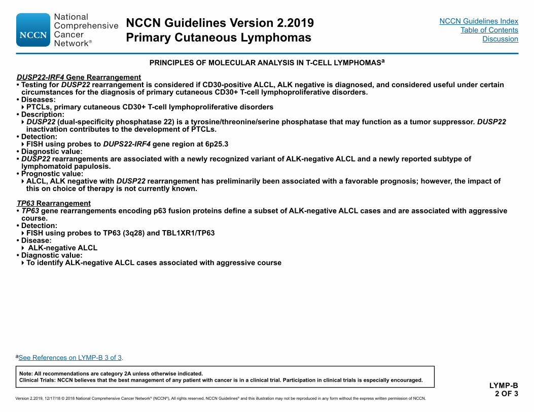
LYMP-B 2 OF 3
PRINCIPLES OF MOLECULAR ANALYSIS IN T-CELL LYMPHOMASa
DUSP22-IRF4 Gene Rearrangement• Testing for DUSP22 rearrangement is considered if CD30-positive ALCL, ALK negative is diagnosed, and considered useful under certain
circumstances for the diagnosis of primary cutaneous CD30+ T-cell lymphoproliferative disorders.• Diseases: �PTCLs, primary cutaneous CD30+ T-cell lymphoproliferative disorders
• Description: �DUSP22 (dual-specificity phosphatase 22) is a tyrosine/threonine/serine phosphatase that may function as a tumor suppressor. DUSP22
inactivation contributes to the development of PTCLs. • Detection: �FISH using probes to DUPS22-IRF4 gene region at 6p25.3
• Diagnostic value: • DUSP22 rearrangements are associated with a newly recognized variant of ALK-negative ALCL and a newly reported subtype of
lymphomatoid papulosis. • Prognostic value: �ALCL, ALK negative with DUSP22 rearrangement has preliminarily been associated with a favorable prognosis; however, the impact of
this on choice of therapy is not currently known.
TP63 Rearrangement• TP63 gene rearrangements encoding p63 fusion proteins define a subset of ALK-negative ALCL cases and are associated with aggressive
course.• Detection: �FISH using probes to TP63 (3q28) and TBL1XR1/TP63
• Disease:� ALK-negative ALCL
• Diagnostic value: �To identify ALK-negative ALCL cases associated with aggressive course
aSee References on LYMP-B 3 of 3.
NCCN Guidelines Version 2.2019Primary Cutaneous Lymphomas
Version 2.2019, 12/17/18 © 2018 National Comprehensive Cancer Network® (NCCN®), All rights reserved. NCCN Guidelines® and this illustration may not be reproduced in any form without the express written permission of NCCN.
Note: All recommendations are category 2A unless otherwise indicated.Clinical Trials: NCCN believes that the best management of any patient with cancer is in a clinical trial. Participation in clinical trials is especially encouraged.
NCCN Guidelines IndexTable of Contents
Discussion
LYMP-B 3 OF 3
PRINCIPLES OF MOLECULAR ANALYSIS IN T-CELL LYMPHOMAS REFERENCES
Chiarle R, Voena C, Ambrogio C, Piva R, Inghirami G. The anaplastic lymphoma kinase in the pathogenesis of cancer. Nat Rev Cancer 2008;8:11-23.
De Schouwer P, Dyer M, Brito-Babapulle V, et al. T-cell prolymphocytic leukemia: antigen receptor gene rearrangement and a novel mode of MTCP1-B1 activation. Br J Haematol 2000;110:831-838.
Hu Z, Medeiros L, Fang L, et al. Prognostic significance of cytogenetic abnormalities in T-cell prolymphocytic leukemia. Am J Hematol 2017;92:441-447.
Morris SW, Kirstein M, Valentine M, et al. Fusion of a kinase gene, ALK, to a nucleolar protein gene, NPM, in non-Hodgkin’s Lymphoma. Science 1994;263:1281-1284.
Odejide O, Weigert O, Lane A, et al. A targeted mutational landscape of angioimmunoblastic T-cell lymphoma. Blood 2014;123:1293-1296.
Pedersen M, Hamilton-Dutoit S, Bendix K, et al. DUSP22 and TP63 rearrangements predict outcome of ALK-negative anaplastic large cell lymphoma: a Danish cohort study. Blood 2017;130:554-557.
Wada D, Law M, Hsi E, et al. Specificity of IRF4 translocations for primary cutaneous anaplastic large cell lymphoma: a multicenter study of 204 skin biopsies. Mod Pathol 2011;24:596-605.
NCCN Guidelines Version 2.2019Primary Cutaneous Lymphomas
Version 2.2019, 12/17/18 © 2018 National Comprehensive Cancer Network® (NCCN®), All rights reserved. NCCN Guidelines® and this illustration may not be reproduced in any form without the express written permission of NCCN.
Note: All recommendations are category 2A unless otherwise indicated.Clinical Trials: NCCN believes that the best management of any patient with cancer is in a clinical trial. Participation in clinical trials is especially encouraged.
NCCN Guidelines IndexTable of Contents
Discussion
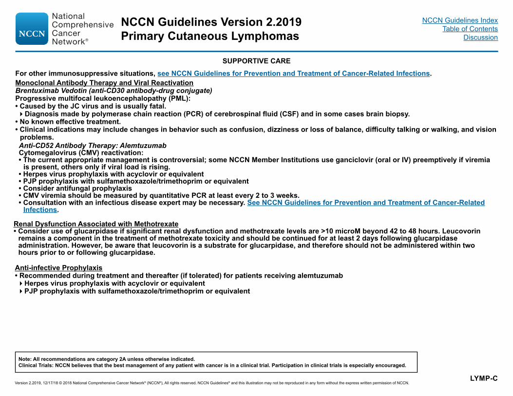
LYMP-C
SUPPORTIVE CAREFor other immunosuppressive situations, see NCCN Guidelines for Prevention and Treatment of Cancer-Related Infections.Monoclonal Antibody Therapy and Viral Reactivation Brentuximab Vedotin (anti-CD30 antibody-drug conjugate)Progressive multifocal leukoencephalopathy (PML):• Caused by the JC virus and is usually fatal.�Diagnosis made by polymerase chain reaction (PCR) of cerebrospinal fluid (CSF) and in some cases brain biopsy.
• No known effective treatment. • Clinical indications may include changes in behavior such as confusion, dizziness or loss of balance, difficulty talking or walking, and vision
problems.Anti-CD52 Antibody Therapy: AlemtuzumabCytomegalovirus (CMV) reactivation:• The current appropriate management is controversial; some NCCN Member Institutions use ganciclovir (oral or IV) preemptively if viremia
is present, others only if viral load is rising. • Herpes virus prophylaxis with acyclovir or equivalent• PJP prophylaxis with sulfamethoxazole/trimethoprim or equivalent• Consider antifungal prophylaxis• CMV viremia should be measured by quantitative PCR at least every 2 to 3 weeks.• Consultation with an infectious disease expert may be necessary. See NCCN Guidelines for Prevention and Treatment of Cancer-Related
Infections.
Renal Dysfunction Associated with Methotrexate• Consider use of glucarpidase if significant renal dysfunction and methotrexate levels are >10 microM beyond 42 to 48 hours. Leucovorin
remains a component in the treatment of methotrexate toxicity and should be continued for at least 2 days following glucarpidase administration. However, be aware that leucovorin is a substrate for glucarpidase, and therefore should not be administered within two hours prior to or following glucarpidase.
Anti-infective Prophylaxis• Recommended during treatment and thereafter (if tolerated) for patients receiving alemtuzumab�Herpes virus prophylaxis with acyclovir or equivalent�PJP prophylaxis with sulfamethoxazole/trimethoprim or equivalent
NCCN Guidelines Version 2.2019Primary Cutaneous Lymphomas
Version 2.2019, 12/17/18 © 2018 National Comprehensive Cancer Network® (NCCN®), All rights reserved. NCCN Guidelines® and this illustration may not be reproduced in any form without the express written permission of NCCN.
Note: All recommendations are category 2A unless otherwise indicated.Clinical Trials: NCCN believes that the best management of any patient with cancer is in a clinical trial. Participation in clinical trials is especially encouraged.
NCCN Guidelines IndexTable of Contents
Discussion
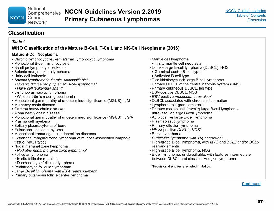
*Provisional entities are listed in italics.
Continued
Classification Table 1WHO Classification of the Mature B-Cell, T-Cell, and NK-Cell Neoplasms (2016)Mature B-Cell Neoplasms• Chronic lymphocytic leukemia/small lymphocytic lymphoma• Monoclonal B-cell lymphocytosis• B-cell prolymphocytic leukemia• Splenic marginal zone lymphoma • Hairy cell leukemia• Splenic lymphoma/leukemia, unclassifiable*�Splenic diffuse red pulp small B-cell lymphoma*�Hairy cell leukemia-variant*
• Lymphoplasmacytic lymphoma�Waldenström’s macroglobulinemia
• Monoclonal gammopathy of undetermined significance (MGUS), IgM• Mu heavy chain disease • Gamma heavy chain disease• Alpha heavy chain disease• Monoclonal gammopathy of undetermined significance (MGUS), IgG/A• Plasma cell myeloma• Solitary plasmacytoma of bone• Extraosseous plasmacytoma• Monoclonal immunoglobulin deposition diseases• Extranodal marginal zone lymphoma of mucosa-associated lymphoid
tissue (MALT type)• Nodal marginal zone lymphoma �Pediatric nodal marginal zone lymphoma*
• Follicular lymphoma�In situ follicular neoplasia�Duodenal-type follicular lymphoma
• Pediatric-type follicular lymphoma• Large B-cell lymphoma with IRF4 rearrangement• Primary cutaneous follicle center lymphoma
• Mantle cell lymphoma�In situ mantle cell neoplasia
• Diffuse large B-cell lymphoma (DLBCL), NOS�Germinal center B-cell type�Activated B-cell type
• T-cell/histiocyte-rich large B-cell lymphoma• Primary DLBCL of the central nervous system (CNS)• Primary cutaneous DLBCL, leg type• EBV-positive DLBCL, NOS• EBV-positive mucocutaneous ulcer*• DLBCL associated with chronic inflammation• Lymphomatoid granulomatosis• Primary mediastinal (thymic) large B-cell lymphoma• Intravascular large B-cell lymphoma• ALK-positive large B-cell lymphoma• Plasmablastic lymphoma• Primary effusion lymphoma• HHV8-positive DLBCL, NOS*• Burkitt lymphoma• Burkitt-like lymphoma with 11q aberration*• High-grade B-cell lymphoma, with MYC and BCL2 and/or BCL6
rearrangements• High-grade B-cell lymphoma, NOS• B-cell lymphoma, unclassifiable, with features intermediate
between DLBCL and classical Hodgkin lymphoma
ST-1
NCCN Guidelines Version 2.2019Primary Cutaneous Lymphomas
Version 2.2019, 12/17/18 © 2018 National Comprehensive Cancer Network® (NCCN®), All rights reserved. NCCN Guidelines® and this illustration may not be reproduced in any form without the express written permission of NCCN.
NCCN Guidelines IndexTable of Contents
Discussion
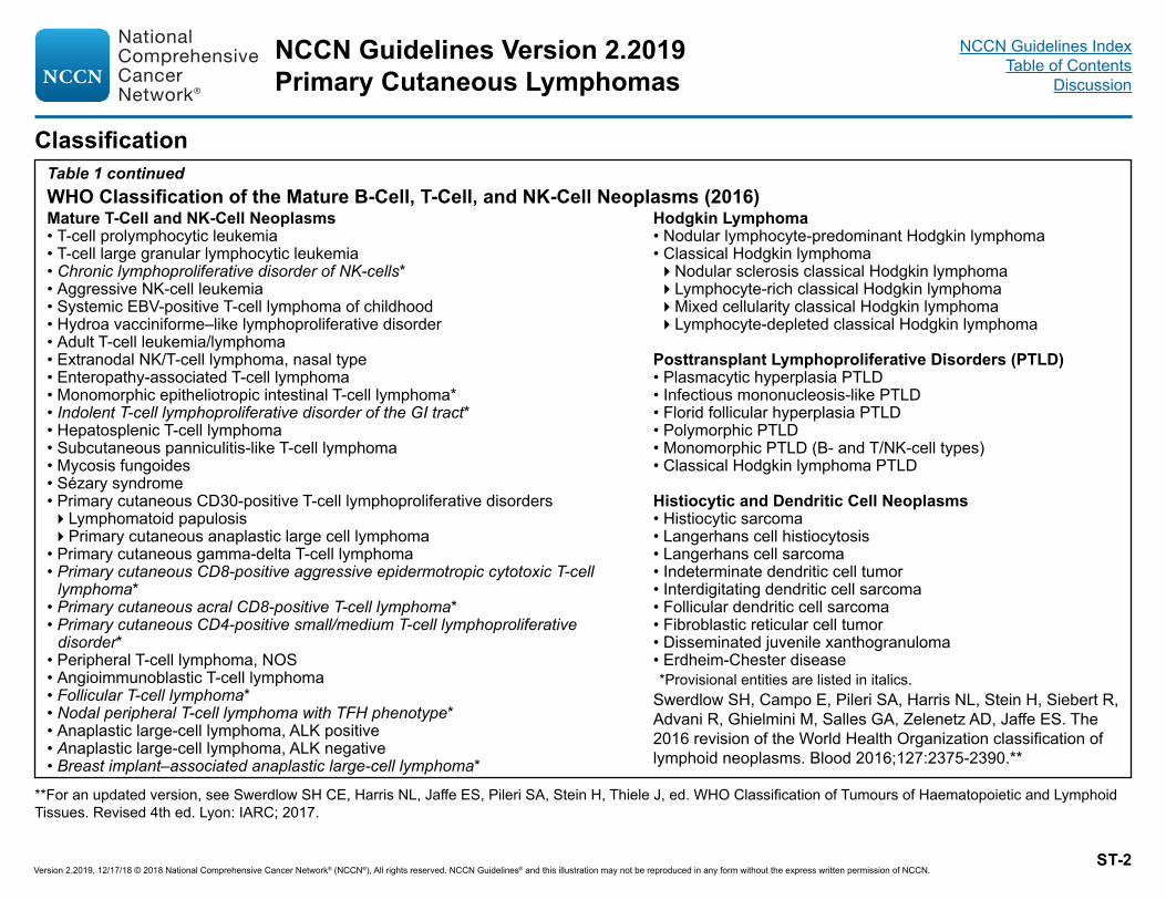
Classification Table 1 continued
Mature T-Cell and NK-Cell Neoplasms• T-cell prolymphocytic leukemia• T-cell large granular lymphocytic leukemia• Chronic lymphoproliferative disorder of NK-cells*• Aggressive NK-cell leukemia• Systemic EBV-positive T-cell lymphoma of childhood• Hydroa vacciniforme–like lymphoproliferative disorder • Adult T-cell leukemia/lymphoma• Extranodal NK/T-cell lymphoma, nasal type• Enteropathy-associated T-cell lymphoma• Monomorphic epitheliotropic intestinal T-cell lymphoma*• Indolent T-cell lymphoproliferative disorder of the GI tract*• Hepatosplenic T-cell lymphoma• Subcutaneous panniculitis-like T-cell lymphoma• Mycosis fungoides• Sézary syndrome• Primary cutaneous CD30-positive T-cell lymphoproliferative disorders�Lymphomatoid papulosis�Primary cutaneous anaplastic large cell lymphoma
• Primary cutaneous gamma-delta T-cell lymphoma• Primary cutaneous CD8-positive aggressive epidermotropic cytotoxic T-cell
lymphoma*• Primary cutaneous acral CD8-positive T-cell lymphoma*• Primary cutaneous CD4-positive small/medium T-cell lymphoproliferative
disorder*• Peripheral T-cell lymphoma, NOS• Angioimmunoblastic T-cell lymphoma• Follicular T-cell lymphoma*• Nodal peripheral T-cell lymphoma with TFH phenotype*• Anaplastic large-cell lymphoma, ALK positive• Anaplastic large-cell lymphoma, ALK negative• Breast implant–associated anaplastic large-cell lymphoma*
*Provisional entities are listed in italics.
Hodgkin Lymphoma• Nodular lymphocyte-predominant Hodgkin lymphoma• Classical Hodgkin lymphoma�Nodular sclerosis classical Hodgkin lymphoma�Lymphocyte-rich classical Hodgkin lymphoma�Mixed cellularity classical Hodgkin lymphoma�Lymphocyte-depleted classical Hodgkin lymphoma
Posttransplant Lymphoproliferative Disorders (PTLD)• Plasmacytic hyperplasia PTLD• Infectious mononucleosis-like PTLD• Florid follicular hyperplasia PTLD• Polymorphic PTLD• Monomorphic PTLD (B- and T/NK-cell types)• Classical Hodgkin lymphoma PTLD
Histiocytic and Dendritic Cell Neoplasms• Histiocytic sarcoma• Langerhans cell histiocytosis• Langerhans cell sarcoma• Indeterminate dendritic cell tumor• Interdigitating dendritic cell sarcoma• Follicular dendritic cell sarcoma• Fibroblastic reticular cell tumor• Disseminated juvenile xanthogranuloma• Erdheim-Chester disease
WHO Classification of the Mature B-Cell, T-Cell, and NK-Cell Neoplasms (2016)
ST-2
Swerdlow SH, Campo E, Pileri SA, Harris NL, Stein H, Siebert R, Advani R, Ghielmini M, Salles GA, Zelenetz AD, Jaffe ES. The 2016 revision of the World Health Organization classification of lymphoid neoplasms. Blood 2016;127:2375-2390.**
**For an updated version, see Swerdlow SH CE, Harris NL, Jaffe ES, Pileri SA, Stein H, Thiele J, ed. WHO Classification of Tumours of Haematopoietic and Lymphoid Tissues. Revised 4th ed. Lyon: IARC; 2017.
NCCN Guidelines Version 2.2019Primary Cutaneous Lymphomas
Version 2.2019, 12/17/18 © 2018 National Comprehensive Cancer Network® (NCCN®), All rights reserved. NCCN Guidelines® and this illustration may not be reproduced in any form without the express written permission of NCCN.
NCCN Guidelines IndexTable of Contents
Discussion

Staging
*Extent of disease is determined by PET/CT for avid lymphomas, and CT for non-avid histologiesNote: Tonsils, Waldeyer’s ring, and spleen are considered nodal tissue**Whether II bulky is treated as limited or advanced disease may be determined by histology and a number of prognostic factors.Categorization of A versus B has been removed from the Lugano Modification of Ann Arbor Staging.
Reprinted with permission. © 2014 American Society of Clinical Oncology. All rights reserved. Cheson B, Fisher R, Barrington S, et al. Recommendations for initial evaluation, staging and response assessment of Hodgkin and non-Hodgkin lymphoma – the Lugano classification. J Clin Oncol 2014;32:3059-3068.
ST-3
Lugano Modification of Ann Arbor Staging System* (for primary nodal lymphomas)
Stage Involvement Extranodal (E) StatusLimited
Stage I One node or a group of adjacent nodes Single extranodal lesions without nodal involvement
Stage II Two or more nodal groups on the same side of the diaphragm
Stage I or II by nodal extent with limited contiguous extranodal involvement
Stage II bulky** II as above with “bulky” disease Not applicable
AdvancedStage III Nodes on both sides of the diaphragm
Nodes above the diaphragm with spleen involvement
Not applicable
Stage IV Additional non-contiguous extralymphatic involvement
Not applicable
NCCN Guidelines Version 2.2019Primary Cutaneous Lymphomas
Version 2.2019, 12/17/18 © 2018 National Comprehensive Cancer Network® (NCCN®), All rights reserved. NCCN Guidelines® and this illustration may not be reproduced in any form without the express written permission of NCCN.
NCCN Guidelines IndexTable of Contents
Discussion
Version 2.2019, 12/17/18 © 2018 National Comprehensive Cancer Network© (NCCN©), All rights reserved. NCCN Guidelines® and this illustration may not be reproduced in any form without the express written permission of NCCN.
NCCN Guidelines Index Table of Contents
Discussion NCCN Guidelines Version 2.2019 Primary Cutaneous Lymphomas
MS-1
Discussion
NCCN Categories of Evidence and Consensus
Category 1: Based upon high-level evidence, there is uniform NCCN consensus that the intervention is appropriate.
Category 2A: Based upon lower-level evidence, there is uniform NCCN consensus that the intervention is appropriate.
Category 2B: Based upon lower-level evidence, there is NCCN consensus that the intervention is appropriate.
Category 3: Based upon any level of evidence, there is major NCCN disagreement that the intervention is appropriate.
All recommendations are category 2A unless otherwise noted.
Table of Contents
Primary Cutaneous B-Cell Lymphomas………………………………………………………………………………………………………………………MS-2
Mycosis Fungoides and Sézary Syndrome………………………………………………………………………………………………………. MS-13
Primary Cutaneous CD30+ T-Cell Lymphoproliferative Disorders………………………………………………………………………………………MS-39
Version 2.2019, 12/17/18 © 2018 National Comprehensive Cancer Network© (NCCN©), All rights reserved. NCCN Guidelines® and this illustration may not be reproduced in any form without the express written permission of NCCN.
NCCN Guidelines Index Table of Contents
Discussion NCCN Guidelines Version 2.2019 Primary Cutaneous Lymphomas
MS-2
Primary Cutaneous B-Cell Lymphomas Primary cutaneous B-cell lymphomas (PCBCLs) are a group of B-cell lymphomas originating in and usually confined to the skin. PCBCLs represent approximately 20% of all extranodal non-Hodgkin’s lymphomas (NHLs). In the United States, the SEER data from the NCI indicated that the incidence of cutaneous T-cell lymphomas accounted for 71%, whereas PCBCLs accounted for 29% from 2001 to 2005.1 The WHO-EORTC classification for cutaneous lymphomas distinguishes 3 main types of PCBCLs:2
• Primary cutaneous marginal zone lymphoma (PCMZL); • Primary cutaneous follicle-center lymphoma (PCFCL); and • Primary cutaneous diffuse large B-cell lymphoma, leg type (PCDLBCL,
leg type)
In addition to the aforementioned subtypes, PCDLBCL, not otherwise specified (PCDLBCL-NOS) with clinicopathologic features intermediate between PCFCL and PCDLBCL, leg type has also been described.3
PCFCL and PCMZL are generally indolent or slow growing. PCFCL is more prevalent in the scalp and the forehead, whereas the trunk and extremities are the most common sites for PCMZL. PCDLBCL, leg type is usually aggressive, associated with a generally poorer prognosis (mainly due to the higher frequency of extracutaneous relapses), and most commonly arises on the leg although it can arise at other sites.4,5 In an Italian series of 467 patients with PCBCL, PCFCL, PCMZL, and PCDLBCL, leg type were reported in 57%, 31%, and 11% of patients, respectively.4 Extracutaneous involvement eventually developed in 6% of patients with PCMZL, 11% with PCFCL, and 17% with PCDLBCL, leg type. The 5-year overall survival (OS) rate was significantly higher for
patients with PCMZL and PCFCL than for patients with PCDLBCL, leg type (97%, 96%, and 73%, respectively; P < .0001).4 In patients with PCMZL and PCFCL, the disease-free survival (DFS) and OS rates were significantly higher for patients with single lesions compared with those with regional or disseminated lesions (5-year DFS, 62% vs. 44%; 5-year OS, 97% vs. 85%), whereas the difference in outcomes between single and regional or disseminated lesions was not significant in patients with PCDLBCL, leg type (5-year DFS rate 55% vs. 44%; 5-year OS rate 79% vs. 67% for single and regional or disseminated lesions, respectively).4 In an analysis of 300 patients with PCBCL from the Dutch Cutaneous Lymphoma Registry, PCFCL, PCMZL, and PCDLBCL comprised 57%, 24%, and 19% of cases, respectively.5 The incidence of extracutaneous relapse was 47% among patients with PCDLBCL, leg type compared to 11% and 9%, respectively, for patients with PCFCL and PCMZL. The 5-year disease-specific survival rates in this series were 95%, 98%, and 50%, respectively.
Literature Search Criteria and Guidelines Update Methodology Prior to the update of this version of the NCCN Guidelines® for Primary Cutaneous B-Cell Lymphomas, a literature search was performed to obtain key literature published between May 2016 and October 2017, using the following search terms: cutaneous diffuse large B-cell lymphoma, cutaneous follicle center lymphoma, and cutaneous marginal zone lymphoma. The PubMed database was chosen as it remains the most widely used resource for medical literature and indexes only peer-reviewed biomedical literature.6
The search results were narrowed by selecting studies in humans published in English. Results were confined to the following article types: Clinical Trial, Phase II; Clinical Trial, Phase III; Clinical Trial, Phase IV; Guideline; Randomized Controlled Trial; Meta-Analysis; Systematic Reviews; and Validation Studies.
This discussion is being updated to correspond with the newly updated algorithm. Last updated on 01/10/18

Version 2.2019, 12/17/18 © 2018 National Comprehensive Cancer Network© (NCCN©), All rights reserved. NCCN Guidelines® and this illustration may not be reproduced in any form without the express written permission of NCCN.
NCCN Guidelines Index Table of Contents
Discussion NCCN Guidelines Version 2.2019 Primary Cutaneous Lymphomas
MS-3
The PubMed search resulted in 28 citations and their potential relevance was examined. The data from key PubMed articles deemed as relevant to these Guidelines and discussed by the panel have been included in this version of the Discussion section. Recommendations for which high-level evidence is lacking are based on the panel’s review of lower-level evidence and expert opinion.
The complete details of the Development and Update of the NCCN Guidelines are available on the NCCN webpage.
Diagnosis The diagnosis of PCBCLs is established by adequate biopsy of skin lesions. Incisional, excisional, or punch biopsy is preferred to shave biopsy, as PCBCLs have primarily dermal infiltrates, often deep, which are less well-sampled and can be missed by a shave biopsy. Review of the slides by a pathologist with expertise in the diagnosis of PCBCL is recommended. Adequate immunophenotyping of the biopsy sample is essential for the diagnosis of the exact subtype of PCBCL. In addition, immunophenotyping is also useful to rule out cutaneous lymphoid hyperplasia (also known as pseudolymphoma or lymphocytoma cutis)7-9 and in the differential diagnosis of intravascular large B-cell lymphoma, which often manifests in skin and is associated with a poor prognosis.10
Gene expression profiling studies have shown that PCFCL has a germinal center B-cell (GCB) phenotype and PCDLBCL, leg type has an activated B-cell (ABC) phenotype.11,12 In nodal DLBCL, the GCB phenotype is associated with a better prognosis than the ABC phenotype. Thus, a germinal (or follicle) center phenotype and large cells in a skin lesion is not equivalent to DLBCL but is consistent with PCFCL with a GCB phenotype.
PCFCL is consistently BCL6-positive, whereas CD10 and BCL2 are expressed in only a few cases with a follicular growth pattern and the detection of BCL2 rearrangement is associated with extracutaneous
spread.13,14 PCMZLs are always negative for BCL6 and CD10, but are often BCL2-positive.15 PCDLBCL, leg type tumors are of ABC origin with expression of CD20, IRF4/MUM1, FOXP1, and BCL2; many cases express BCL6 and lack expression of CD10.5,16,17
While the diagnosis of PCMZL is generally straightforward and reproducible among pathologists, it is more difficult to distinguish between PCFCL and PCDLBCL, leg type, partly because the cell size (large vs. small) is not a defining feature as it is in nodal B-cell lymphomas. PCFCL and PCDLBCL are CD20- and BCL6-positive. BCL2 is usually negative in PCFCL but highly expressed in PCDLBCL, leg type.14 In addition, PCFCL is usually IRF4/MUM1-negative while PCDLBCL, leg type is usually IRF4/MUM1-positive and shows strong expression of FOXP1.17
The initial IHC panel should include CD20, CD3, CD5, CD10, BCL2, BCL6, and IRF4/MUM1. Under certain circumstances, evaluation of additional immunohistochemical markers such as Ki-67, CD43, CD21, CD23, cyclin D1, and kappa/lambda may be useful to further establish the lymphoma subtype. Additionally, assessment of surface IgM and IgD expression may also be helpful in distinguishing PCDLBCL, leg type from PCFCL.18
A high prevalence of MYD88 L265P mutation (occurring in about 60% of patients) has been reported in patients with PCDLBCL, leg type and is associated with inferior clinical outcomes.19 In a retrospective analysis of 61 patients diagnosed with PCDLBCL, leg type, MYD88 L265P mutation was associated with shorter disease-specific survival and was also an independent adverse prognostic factor for OS. The 3- and 5-year disease-specific survival rates for those with MYD88 L265P mutation were 65.7% and 60.2%, respectively, compared to 85% and 72%, respectively, for patients with the wild-type allele.19 In a more recent report that evaluated the prevalence of MYD88 L265P mutation in patients with PCFCL (21 patients) and PCDLBCL (25 patients), leg type identified in the
Version 2.2019, 12/17/18 © 2018 National Comprehensive Cancer Network© (NCCN©), All rights reserved. NCCN Guidelines® and this illustration may not be reproduced in any form without the express written permission of NCCN.
NCCN Guidelines Index Table of Contents
Discussion NCCN Guidelines Version 2.2019 Primary Cutaneous Lymphomas
MS-4
French Cutaneous Lymphoma Study Group Database, MYD88 L265P mutation was detected in 76% of the patients with PCDLBCL, leg type and was absent in all of the patients with PCFCL.20 These findings suggest that determination of MYD88 L265P mutation status could be helpful to further distinguish PCDLBCL, leg type from PCFCL.
Mantle cell lymphoma (MCL) is not a primary cutaneous lymphoma and finding it in the skin requires a careful search for extracutaneous disease. Clinical presentation on the leg and blastoid cytology along with high proliferative index and expression of BCL2, IRF4/MUM1, and IgM would often represent MCL with skin involvement.21 The use of cyclin D1 may be useful to differentiate PCMZL (negative for CD5 and cyclin D1) from MCL (positive for CD5 and cyclin D1).
The t(14;18) translocation only rarely occurs in CBCLs. Therefore, the detection of a t(14;18) translocation in CBCL suggests the presence of systemic follicular lymphoma (FL).22 Cytogenetics or FISH to detect t(14;18) may be useful if systemic FL is suspected. The feasibility of flow cytometric immunophenotyping of skin biopsies for the assessment of B-cell clonality has been reported, although it has not been widely used.9 If adequate biopsy material is available, molecular analysis or flow cytometry could be useful in determining B-cell clonality.
Workup The initial workup is geared toward evaluating extent of disease on the skin and seeking extracutaneous disease. The absence of extracutaneous disease at diagnosis is part of the definition of primary CBCL. The workup includes a complete physical examination, a comprehensive skin examination, and CT and/or PET/CT of the chest, abdomen, and pelvis.23 PET/CT may have higher sensitivity in the detection of both local and distant metastases than CT.24 However, this is not validated and the higher rates of false-positive findings can create confusion.
Bone marrow biopsy is essential for PCDLBCL, leg type, since this is an aggressive lymphoma that will probably require systemic treatment; its role is unclear for PCFCL and PCMZL. Recent studies have indicated that bone marrow biopsy is an essential or more often a valuable component of staging in PCFCL first presenting in the skin, whereas it appears to have a more limited value in PCMZL presenting in the skin, and may be considered only in selected patients.23,25 The International Society for Cutaneous Lymphomas (ISCL) and the EORTC Task Force recommend that bone marrow biopsy be obtained for cutaneous lymphomas with intermediate to aggressive behaviors and should be considered for cutaneous lymphomas with indolent behavior and when there is any evidence of extracutaneous disease, as indicated by other staging assessments (eg, radiographic evidence or serologic clues such as elevated monoclonal or polyclonal immunoglobulins).23 Senff et al evaluated 275 patients with histologic features consistent with marginal zone lymphoma (MZL; n = 82) or follicle center lymphoma (FCL; n = 193) first presenting in the skin.25 Bone marrow involvement was seen in about 11% of patients in the FCL group compared with 2% in the MZL group. FCL patients with skin lesions and a positive bone marrow had a significantly worse prognosis compared with those with PCFCL; the 5-year OS rate was 44% and 84%, respectively.25 The guidelines recommend considering bone marrow biopsy for patients with unexplained cytopenias or if there is a clinical suspicion of other subtypes. Peripheral blood flow cytometry will be useful in selected cases, if complete blood cell (CBC) count demonstrates lymphocytosis.
Treatment Options RT is very effective when used as initial local therapy as well as for cutaneous relapses in most patients with indolent PCBCL.26-29 In a retrospective study of 34 patients with PCBCL treated with RT, 5-year relapse-free survival (RFS) rates ranged from 62% to 73% for PCFCL and PCMZL but were only 33% for patients with PCDLBCL, leg type.27 The
Version 2.2019, 12/17/18 © 2018 National Comprehensive Cancer Network© (NCCN©), All rights reserved. NCCN Guidelines® and this illustration may not be reproduced in any form without the express written permission of NCCN.
NCCN Guidelines Index Table of Contents
Discussion NCCN Guidelines Version 2.2019 Primary Cutaneous Lymphomas
MS-5
5-year OS rate was 100% for PCFCL and PCMZL but was 67% for PCDLBCL, leg type. Senff et al evaluated the outcome of 153 patients with PCBCL (25 with PCMZL; 101 with PCFCL; and 27 with PCDLBCL) who were initially treated with RT with a curative intent.28 Overall, 45% of patients had single lesions while localized or disseminated lesions were seen in 43% and 12% of patients, respectively. Complete response (CR) was obtained in 151 of 153 patients (99%). Relapse rates for PCMZL, PCFCL, and PCDLBCL, leg type were 60%, 29%, and 64%, and the 5-year disease-specific survival rates were 95%, 97%, and 59%, respectively. The PCFCLs presenting on the legs also had a higher relapse rate (63%) and a lower 5-year disease-specific survival (44%) compared with PCFCLs occurring at other sites (25% and 99%, respectively).28
Low-dose involved-field RT (4 Gy in two fractions) is an effective treatment option for palliation of symptoms in patients with persistent (initial) lesions or recurrent symptomatic disease.30 The results of a more recent retrospective study also showed that RT ≤12 Gy (4 Gy for relapsed disease) was equally effective as RT >12 Gy in patients with indolent PCBCL (42 patients; 16 patients had PCFCL).31
RT and excision were also associated with higher response rates compared to chemotherapy in patients with indolent histologies, but were generally used for those with more limited disease; therefore, a direct comparison cannot be made.4,32-34 In a large retrospective analysis by the Italian Study Group for Cutaneous Lymphomas involving 467 patients with PCBCL, the CR rate and the 5- and 10-year OS rates for all patients with PCFCL and PCMZL who received first-line treatment (RT in 53%, with total dose of 35–45 Gy; chemotherapy in 25%, mainly with CHOP; surgery in 23%) were 92% to 95%, 96% to 97%, and 89% to 91%, respectively.4 The relapse rate was 44% to 46.5% and extracutaneous spread was observed in 6% to 11% of patients. Relapse rate did not vary by the type
of initial therapy. In patients with PCDLBCL, leg type, the CR rate and 5- and 10-year OS rates were 82%, 73%, and 47%, respectively. PCDLBCL, leg type was associated with higher relapse rates (55%) and higher incidences of extracutaneous spread (17%) — a higher relapse rate was confirmed both for patients with single or regional lesions treated with RT and for patients with disseminated cutaneous involvement treated with chemotherapy.4 In a retrospective analysis of 137 patients with PC-MZL, initial treatment with surgical excision, RT, or a combination of both resulted in a CR rate of 88% (93% for patients with solitary or localized disease and 71% for those with multifocal lesions).34 Although there were no significant differences in the rate of recurrences between the treatment modalities, surgery alone was associated with more recurrences at the initial site.
Chemotherapy is effective for multifocal skin lesions in patients with PCFCL or PCMZL.35-37 Rituximab has been shown to be effective for indolent PCBCL with multiple lesions that cannot be managed effectively with local therapy.38-42 In a retrospective analysis of 15 patients with indolent PCBCL, rituximab resulted in an overall response rate (ORR) of 87% (60% CR). The ORR was 100% for patients with PCFCL and 60% for PCMZL. With a median follow-up of 36 months, the median duration of response was 24 months.41 In another series of 16 patients with PCBCL, 14 patients (87.5%) achieved a CR with rituximab monotherapy; 35% of these patients with CR eventually relapsed between 6 and 37 months.42
The feasibility and efficacy of intralesional rituximab has also been demonstrated in a small series of patients with PCMZL and PCFCL.43-46 In an observational multicenter study conducted by the Spanish Working Group on Cutaneous Lymphoma (17 patients with PCMZL and 18 patients with PCFCL), intralesional rituximab induced CR and partial response (PR) in 71% and 23% of patients, respectively, with a median DFS of 114 weeks.44 The response rates were similar among patients with PCMZL
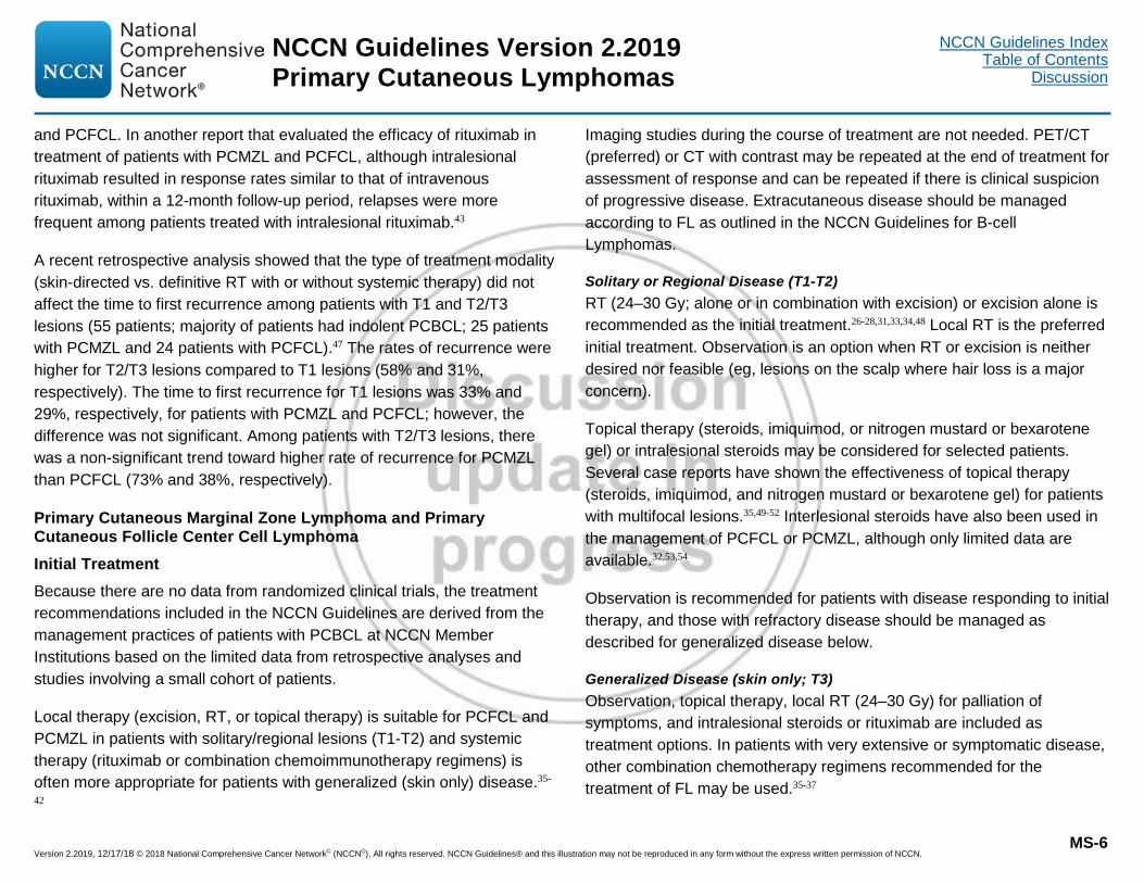
Version 2.2019, 12/17/18 © 2018 National Comprehensive Cancer Network© (NCCN©), All rights reserved. NCCN Guidelines® and this illustration may not be reproduced in any form without the express written permission of NCCN.
NCCN Guidelines Index Table of Contents
Discussion NCCN Guidelines Version 2.2019 Primary Cutaneous Lymphomas
MS-6
and PCFCL. In another report that evaluated the efficacy of rituximab in treatment of patients with PCMZL and PCFCL, although intralesional rituximab resulted in response rates similar to that of intravenous rituximab, within a 12-month follow-up period, relapses were more frequent among patients treated with intralesional rituximab.43
A recent retrospective analysis showed that the type of treatment modality (skin-directed vs. definitive RT with or without systemic therapy) did not affect the time to first recurrence among patients with T1 and T2/T3 lesions (55 patients; majority of patients had indolent PCBCL; 25 patients with PCMZL and 24 patients with PCFCL).47 The rates of recurrence were higher for T2/T3 lesions compared to T1 lesions (58% and 31%, respectively). The time to first recurrence for T1 lesions was 33% and 29%, respectively, for patients with PCMZL and PCFCL; however, the difference was not significant. Among patients with T2/T3 lesions, there was a non-significant trend toward higher rate of recurrence for PCMZL than PCFCL (73% and 38%, respectively).
Primary Cutaneous Marginal Zone Lymphoma and Primary Cutaneous Follicle Center Cell Lymphoma Initial Treatment Because there are no data from randomized clinical trials, the treatment recommendations included in the NCCN Guidelines are derived from the management practices of patients with PCBCL at NCCN Member Institutions based on the limited data from retrospective analyses and studies involving a small cohort of patients.
Local therapy (excision, RT, or topical therapy) is suitable for PCFCL and PCMZL in patients with solitary/regional lesions (T1-T2) and systemic therapy (rituximab or combination chemoimmunotherapy regimens) is often more appropriate for patients with generalized (skin only) disease.35-
42
Imaging studies during the course of treatment are not needed. PET/CT (preferred) or CT with contrast may be repeated at the end of treatment for assessment of response and can be repeated if there is clinical suspicion of progressive disease. Extracutaneous disease should be managed according to FL as outlined in the NCCN Guidelines for B-cell Lymphomas.
Solitary or Regional Disease (T1-T2) RT (24–30 Gy; alone or in combination with excision) or excision alone is recommended as the initial treatment.26-28,31,33,34,48 Local RT is the preferred initial treatment. Observation is an option when RT or excision is neither desired nor feasible (eg, lesions on the scalp where hair loss is a major concern).
Topical therapy (steroids, imiquimod, or nitrogen mustard or bexarotene gel) or intralesional steroids may be considered for selected patients. Several case reports have shown the effectiveness of topical therapy (steroids, imiquimod, and nitrogen mustard or bexarotene gel) for patients with multifocal lesions.35,49-52 Interlesional steroids have also been used in the management of PCFCL or PCMZL, although only limited data are available.32,53,54
Observation is recommended for patients with disease responding to initial therapy, and those with refractory disease should be managed as described for generalized disease below.
Generalized Disease (skin only; T3) Observation, topical therapy, local RT (24–30 Gy) for palliation of symptoms, and intralesional steroids or rituximab are included as treatment options. In patients with very extensive or symptomatic disease, other combination chemotherapy regimens recommended for the treatment of FL may be used.35-37
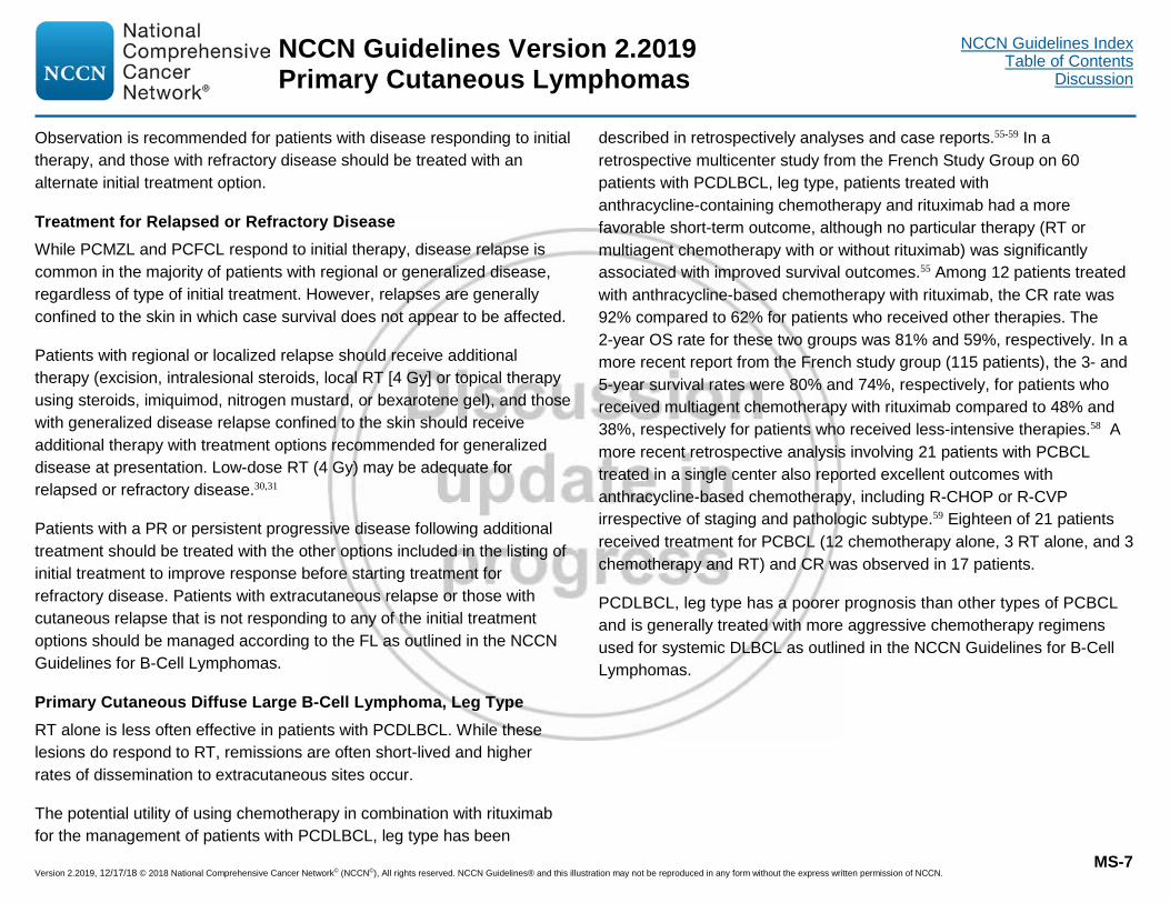
Version 2.2019, 12/17/18 © 2018 National Comprehensive Cancer Network© (NCCN©), All rights reserved. NCCN Guidelines® and this illustration may not be reproduced in any form without the express written permission of NCCN.
NCCN Guidelines Index Table of Contents
Discussion NCCN Guidelines Version 2.2019 Primary Cutaneous Lymphomas
MS-7
Observation is recommended for patients with disease responding to initial therapy, and those with refractory disease should be treated with an alternate initial treatment option.
Treatment for Relapsed or Refractory Disease While PCMZL and PCFCL respond to initial therapy, disease relapse is common in the majority of patients with regional or generalized disease, regardless of type of initial treatment. However, relapses are generally confined to the skin in which case survival does not appear to be affected.
Patients with regional or localized relapse should receive additional therapy (excision, intralesional steroids, local RT [4 Gy] or topical therapy using steroids, imiquimod, nitrogen mustard, or bexarotene gel), and those with generalized disease relapse confined to the skin should receive additional therapy with treatment options recommended for generalized disease at presentation. Low-dose RT (4 Gy) may be adequate for relapsed or refractory disease.30,31
Patients with a PR or persistent progressive disease following additional treatment should be treated with the other options included in the listing of initial treatment to improve response before starting treatment for refractory disease. Patients with extracutaneous relapse or those with cutaneous relapse that is not responding to any of the initial treatment options should be managed according to the FL as outlined in the NCCN Guidelines for B-Cell Lymphomas.
Primary Cutaneous Diffuse Large B-Cell Lymphoma, Leg Type RT alone is less often effective in patients with PCDLBCL. While these lesions do respond to RT, remissions are often short-lived and higher rates of dissemination to extracutaneous sites occur.
The potential utility of using chemotherapy in combination with rituximab for the management of patients with PCDLBCL, leg type has been
described in retrospectively analyses and case reports.55-59 In a retrospective multicenter study from the French Study Group on 60 patients with PCDLBCL, leg type, patients treated with anthracycline-containing chemotherapy and rituximab had a more favorable short-term outcome, although no particular therapy (RT or multiagent chemotherapy with or without rituximab) was significantly associated with improved survival outcomes.55 Among 12 patients treated with anthracycline-based chemotherapy with rituximab, the CR rate was 92% compared to 62% for patients who received other therapies. The 2-year OS rate for these two groups was 81% and 59%, respectively. In a more recent report from the French study group (115 patients), the 3- and 5-year survival rates were 80% and 74%, respectively, for patients who received multiagent chemotherapy with rituximab compared to 48% and 38%, respectively for patients who received less-intensive therapies.58 A more recent retrospective analysis involving 21 patients with PCBCL treated in a single center also reported excellent outcomes with anthracycline-based chemotherapy, including R-CHOP or R-CVP irrespective of staging and pathologic subtype.59 Eighteen of 21 patients received treatment for PCBCL (12 chemotherapy alone, 3 RT alone, and 3 chemotherapy and RT) and CR was observed in 17 patients.
PCDLBCL, leg type has a poorer prognosis than other types of PCBCL and is generally treated with more aggressive chemotherapy regimens used for systemic DLBCL as outlined in the NCCN Guidelines for B-Cell Lymphomas.

Version 2.2019, 12/17/18 © 2018 National Comprehensive Cancer Network© (NCCN©), All rights reserved. NCCN Guidelines® and this illustration may not be reproduced in any form without the express written permission of NCCN.
NCCN Guidelines Index Table of Contents
Discussion NCCN Guidelines Version 2.2019 Primary Cutaneous Lymphomas
MS-8
References
1. Bradford PT, Devesa SS, Anderson WF, Toro JR. Cutaneous lymphoma incidence patterns in the United States: a population-based study of 3884 cases. Blood 2009;113:5064-5073. Available at: http://www.ncbi.nlm.nih.gov/pubmed/19279331.
2. Willemze R, Jaffe ES, Burg G, et al. WHO-EORTC classification for cutaneous lymphomas. Blood 2005;105:3768-3785. Available at: http://www.ncbi.nlm.nih.gov/pubmed/15692063.
3. Lucioni M, Berti E, Arcaini L, et al. Primary cutaneous B-cell lymphoma other than marginal zone: clinicopathologic analysis of 161 cases: Comparison with current classification and definition of prognostic markers. Cancer Med 2016;5:2740-2755. Available at: https://www.ncbi.nlm.nih.gov/pubmed/27665744.
4. Zinzani PL, Quaglino P, Pimpinelli N, et al. Prognostic factors in primary cutaneous B-cell lymphoma: the Italian Study Group for Cutaneous Lymphomas. J Clin Oncol 2006;24:1376-1382. Available at: http://www.ncbi.nlm.nih.gov/pubmed/16492713.
5. Senff NJ, Hoefnagel JJ, Jansen PM, et al. Reclassification of 300 primary cutaneous B-Cell lymphomas according to the new WHO-EORTC classification for cutaneous lymphomas: comparison with previous classifications and identification of prognostic markers. J Clin Oncol 2007;25:1581-1587. Available at: http://www.ncbi.nlm.nih.gov/pubmed/17353548.
6. U.S. National Library of Medicine Key MEDLINE® Indicators Available at: http://www.nlm.nih.gov/bsd/bsd_key.html.
7. Baldassano MF, Bailey EM, Ferry JA, et al. Cutaneous lymphoid hyperplasia and cutaneous marginal zone lymphoma: comparison of morphologic and immunophenotypic features. Am J Surg Pathol 1999;23:88-96. Available at: https://www.ncbi.nlm.nih.gov/pubmed/9888708.
8. Leinweber B, Colli C, Chott A, et al. Differential diagnosis of cutaneous infiltrates of B lymphocytes with follicular growth pattern. Am J Dermatopathol 2004;26:4-13. Available at: https://www.ncbi.nlm.nih.gov/pubmed/14726817.
9. Schafernak KT, Variakojis D, Goolsby CL, et al. Clonality assessment of cutaneous B-cell lymphoid proliferations: a comparison of flow cytometry immunophenotyping, molecular studies, and immunohistochemistry/in situ hybridization and review of the literature. Am J Dermatopathol 2014;36:781-795. Available at: https://www.ncbi.nlm.nih.gov/pubmed/24335516.
10. Murase T, Yamaguchi M, Suzuki R, et al. Intravascular large B-cell lymphoma (IVLBCL): a clinicopathologic study of 96 cases with special reference to the immunophenotypic heterogeneity of CD5. Blood 2007;109:478-485. Available at: https://www.ncbi.nlm.nih.gov/pubmed/16985183.
11. Hoefnagel JJ, Dijkman R, Basso K, et al. Distinct types of primary cutaneous large B-cell lymphoma identified by gene expression profiling. Blood 2005;105:3671-3678. Available at: http://www.ncbi.nlm.nih.gov/pubmed/15308563.
12. Pham-Ledard A, Prochazkova-Carlotti M, Andrique L, et al. Multiple genetic alterations in primary cutaneous large B-cell lymphoma, leg type support a common lymphomagenesis with activated B-cell-like diffuse large B-cell lymphoma. Mod Pathol 2014;27:402-411. Available at: https://www.ncbi.nlm.nih.gov/pubmed/24030746.
13. de Leval L, Harris NL, Longtine J, et al. Cutaneous b-cell lymphomas of follicular and marginal zone types: use of Bcl-6, CD10, Bcl-2, and CD21 in differential diagnosis and classification. Am J Surg Pathol 2001;25:732-741. Available at: https://www.ncbi.nlm.nih.gov/pubmed/11395550.
14. Pham-Ledard A, Cowppli-Bony A, Doussau A, et al. Diagnostic and prognostic value of BCL2 rearrangement in 53 patients with follicular lymphoma presenting as primary skin lesions. Am J Clin Pathol 2015;143:362-373. Available at: https://www.ncbi.nlm.nih.gov/pubmed/25696794.
Version 2.2019, 12/17/18 © 2018 National Comprehensive Cancer Network© (NCCN©), All rights reserved. NCCN Guidelines® and this illustration may not be reproduced in any form without the express written permission of NCCN.
NCCN Guidelines Index Table of Contents
Discussion NCCN Guidelines Version 2.2019 Primary Cutaneous Lymphomas
MS-9
15. Hoefnagel JJ, Vermeer MH, Jansen PM, et al. Bcl-2, Bcl-6 and CD10 expression in cutaneous B-cell lymphoma: further support for a follicle centre cell origin and differential diagnostic significance. Br J Dermatol 2003;149:1183-1191. Available at: http://www.ncbi.nlm.nih.gov/pubmed/14674895.
16. Kodama K, Massone C, Chott A, et al. Primary cutaneous large B-cell lymphomas: clinicopathologic features, classification, and prognostic factors in a large series of patients. Blood 2005;106:2491-2497. Available at: http://www.ncbi.nlm.nih.gov/pubmed/15947086.
17. Hoefnagel JJ, Mulder MMS, Dreef E, et al. Expression of B-cell transcription factors in primary cutaneous B-cell lymphoma. Mod Pathol 2006;19:1270-1276. Available at: http://www.ncbi.nlm.nih.gov/pubmed/16778825.
18. Koens L, Vermeer MH, Willemze R, Jansen PM. IgM expression on paraffin sections distinguishes primary cutaneous large B-cell lymphoma, leg type from primary cutaneous follicle center lymphoma. Am J Surg Pathol 2010;34:1043-1048. Available at: http://www.ncbi.nlm.nih.gov/pubmed/20551823.
19. Pham-Ledard A, Beylot-Barry M, Barbe C, et al. High frequency and clinical prognostic value of MYD88 L265P mutation in primary cutaneous diffuse large B-cell lymphoma, leg-type. JAMA Dermatol 2014;150:1173-1179. Available at: https://www.ncbi.nlm.nih.gov/pubmed/25055137.
20. Menguy S, Gros A, Pham-Ledard A, et al. MYD88 somatic mutation is a diagnostic criterion in primary cutaneous large B-cell lymphoma. J Invest Dermatol 2016;136:1741-1744. Available at: https://www.ncbi.nlm.nih.gov/pubmed/27189828.
21. Wehkamp U, Pott C, Unterhalt M, et al. Skin involvement of mantle cell lymphoma may mimic primary cutaneous diffuse large B-cell lymphoma, leg type. Am J Surg Pathol 2015;39:1093-1101. Available at: https://www.ncbi.nlm.nih.gov/pubmed/26034867.
22. Child F, Russell-Jones R, Woolford A, et al. Absence of the t(14;18) chromosomal translocation in primary cutaneous B-cell lymphoma. British
Journal of Dermatology 2001;144:735-744. Available at: http://www.ncbi.nlm.nih.gov/pubmed/11298531.
23. Kim YH, Willemze R, Pimpinelli N, et al. TNM classification system for primary cutaneous lymphomas other than mycosis fungoides and Sezary syndrome: a proposal of the International Society for Cutaneous Lymphomas (ISCL) and the Cutaneous Lymphoma Task Force of the European Organization of Research and Treatment of Cancer (EORTC). Blood 2007;110:479-484. Available at: http://www.ncbi.nlm.nih.gov/pubmed/17339420.
24. Kumar R, Xiu Y, Zhuang HM, Alavi A. 18F-fluorodeoxyglucose-positron emission tomography in evaluation of primary cutaneous lymphoma. Br J Dermatol 2006;155:357-363. Available at: http://www.ncbi.nlm.nih.gov/pubmed/16882175.
25. Senff N, Kluin-Nelemans H, Willemze R. Results of bone marrow examination in 275 patients with histological features that suggest an indolent type of cutaneous B-cell lymphoma. Br J Haematol 2008;142:52-56. Available at: http://www.ncbi.nlm.nih.gov/pubmed/18422781.
26. Eich HT, Eich D, Micke O, et al. Long-term efficacy, curative potential, and prognostic factors of radiotherapy in primary cutaneous B-cell lymphoma. Int J Radiat Oncol Biol Phys 2003;55:899-906. Available at: http://www.ncbi.nlm.nih.gov/pubmed/12605967.
27. Smith BD, Glusac EJ, McNiff JM, et al. Primary cutaneous B-cell lymphoma treated with radiotherapy: a comparison of the European Organization for Research and Treatment of Cancer and the WHO classification systems. J Clin Oncol 2004;22:634-639. Available at: http://www.ncbi.nlm.nih.gov/pubmed/14966086.
28. Senff NJ, Hoefnagel JJ, Neelis KJ, et al. Results of radiotherapy in 153 primary cutaneous B-Cell lymphomas classified according to the WHO-EORTC classification. Arch Dermatol 2007;143:1520-1526. Available at: http://www.ncbi.nlm.nih.gov/pubmed/18087001.
29. Pedretti S, Urpis M, Leali C, et al. Primary cutaneous non-Hodgkin lymphoma: results of a retrospective analysis in the light of the recent

Version 2.2019, 12/17/18 © 2018 National Comprehensive Cancer Network© (NCCN©), All rights reserved. NCCN Guidelines® and this illustration may not be reproduced in any form without the express written permission of NCCN.
NCCN Guidelines Index Table of Contents
Discussion NCCN Guidelines Version 2.2019 Primary Cutaneous Lymphomas
MS-10
ILROG guidelines. Tumori 2017:[Epub ahead of print]. Available at: https://www.ncbi.nlm.nih.gov/pubmed/28218382.
30. Neelis KJ, Schimmel EC, Vermeer MH, et al. Low-dose palliative radiotherapy for cutaneous B- and T-cell lymphomas. Int J Radiat Oncol Biol Phys 2009;74:154-158. Available at: https://www.ncbi.nlm.nih.gov/pubmed/18834672.
31. Akhtari M, Reddy JP, Pinnix CC, et al. Primary cutaneous B-cell lymphoma (non-leg type) has excellent outcomes even after very low dose radiation as single-modality therapy. Leuk Lymphoma 2016;57:34-38. Available at: https://www.ncbi.nlm.nih.gov/pubmed/25860237.
32. Senff NJ, Noordijk EM, Kim YH, et al. European Organization for Research and Treatment of Cancer and International Society for Cutaneous Lymphoma Consensus recommendations for the management of cutaneous B-cell lymphomas. Blood 2008;112:1600-1609. Available at: http://www.ncbi.nlm.nih.gov/pubmed/18567836.
33. Pashtan I, Mauch PM, Chen YH, et al. Radiotherapy in the management of localized primary cutaneous B-cell lymphoma. Leuk Lymphoma 2013;54:726-730. Available at: https://www.ncbi.nlm.nih.gov/pubmed/22916994.
34. Servitje O, Muniesa C, Benavente Y, et al. Primary cutaneous marginal zone B-cell lymphoma: response to treatment and disease-free survival in a series of 137 patients. J Am Acad Dermatol 2013;69:357-365. Available at: https://www.ncbi.nlm.nih.gov/pubmed/23796549.
35. Bekkenk MW, Vermeer MH, Geerts ML, et al. Treatment of multifocal primary cutaneous B-cell lymphoma: a clinical follow-up study of 29 patients. J Clin Oncol 1999;17:2471-2478. Available at: http://www.ncbi.nlm.nih.gov/pubmed/10561311.
36. Rijlaarsdam JU, Toonstra J, Meijer OW, et al. Treatment of primary cutaneous B-cell lymphomas of follicle center cell origin: a clinical follow-up study of 55 patients treated with radiotherapy or polychemotherapy. J Clin Oncol 1996;14:549-555. Available at: http://www.ncbi.nlm.nih.gov/pubmed/8636770.
37. Brice P, Cazals D, Mounier N, et al. Primary cutaneous large-cell lymphoma: analysis of 49 patients included in the LNH87 prospective trial of polychemotherapy for high-grade lymphomas. Groupe d'Etude des Lymphomes de l'Adulte. Leukemia 1998;12:213-219. Available at: http://www.ncbi.nlm.nih.gov/pubmed/9519784
38. Heinzerling LM, Urbanek M, Funk JO, et al. Reduction of tumor burden and stabilization of disease by systemic therapy with anti-CD20 antibody (rituximab) in patients with primary cutaneous B-cell lymphoma. Cancer 2000;89:1835-1844. Available at: http://www.ncbi.nlm.nih.gov/pubmed/11042581.
39. Heinzerling L, Dummer R, Kempf W, et al. Intralesional therapy with anti-CD20 monoclonal antibody rituximab in primary cutaneous B-cell lymphoma. Arch Dermatol 2000;136:374-378. Available at: http://www.ncbi.nlm.nih.gov/pubmed/10724200.
40. Gellrich S, Muche JM, Wilks A, et al. Systemic eight-cycle anti-CD20 monoclonal antibody (rituximab) therapy in primary cutaneous B-cell lymphomas--an applicational observation. Br J Dermatol 2005;153:167-173. Available at: http://www.ncbi.nlm.nih.gov/pubmed/16029344.
41. Morales AV, Advani R, Horwitz SM, et al. Indolent primary cutaneous B-cell lymphoma: experience using systemic rituximab. J Am Acad Dermatol 2008;59:953-957. Available at: http://www.ncbi.nlm.nih.gov/pubmed/18817999.
42. Valencak J, Weihsengruber F, Rappersberger K, et al. Rituximab monotherapy for primary cutaneous B-cell lymphoma: response and follow-up in 16 patients. Ann Oncol 2009;20:326-330. Available at: http://www.ncbi.nlm.nih.gov/pubmed/18836086.
43. Kerl K, Prins C, Saurat JH, French LE. Intralesional and intravenous treatment of cutaneous B-cell lymphomas with the monoclonal anti-CD20 antibody rituximab: report and follow-up of eight cases. Br J Dermatol 2006;155:1197-1200. Available at: https://www.ncbi.nlm.nih.gov/pubmed/17107389.

Version 2.2019, 12/17/18 © 2018 National Comprehensive Cancer Network© (NCCN©), All rights reserved. NCCN Guidelines® and this illustration may not be reproduced in any form without the express written permission of NCCN.
NCCN Guidelines Index Table of Contents
Discussion NCCN Guidelines Version 2.2019 Primary Cutaneous Lymphomas
MS-11
44. Penate Y, Hernandez-Machin B, Perez-Mendez LI, et al. Intralesional rituximab in the treatment of indolent primary cutaneous B-cell lymphomas: an epidemiological observational multicentre study. The Spanish Working Group on Cutaneous Lymphoma. Br J Dermatol 2012;167:174-179. Available at: https://www.ncbi.nlm.nih.gov/pubmed/22356294.
45. Vakeva L, Ranki A, Malkonen T. Intralesional rituximab treatment for primary cutaneous B-cell lymphoma: nine Finnish cases. . Acta Derm Venereol 2016;96:396-397. Available at: http://www.ncbi.nlm.nih.gov/pubmed/26525093.
46. Eberle FC, Holstein J, Scheu A, et al. Intralesional anti-CD20 antibody for low-grade primary cutaneous B-cell lymphoma: Adverse reactions correlate with favorable clinical outcome. J Dtsch Dermatol Ges 2017;15:319-323. Available at: https://www.ncbi.nlm.nih.gov/pubmed/28177583.
47. Haverkos B, Tyler K, Gru AA, et al. Primary Cutaneous B-Cell Lymphoma: management and patterns of recurrence at the multimodality cutaneous lymphoma clinic of the Ohio State University. Oncologist 2015;20:1161-1166. Available at: https://www.ncbi.nlm.nih.gov/pubmed/26306900.
48. Specht L, Dabaja B, Illidge T, et al. Modern radiation therapy for primary cutaneous lymphomas: field and dose guidelines from the International Lymphoma Radiation Oncology Group. Int J Radiat Oncol Biol Phys 2015;92:32-39. Available at: https://www.ncbi.nlm.nih.gov/pubmed/25863751.
49. Trent JT, Romanelli P, Kerdel FA. Topical targretin and intralesional interferon alfa for cutaneous lymphoma of the scalp. Arch Dermatol 2002;138:1421-1423. Available at: http://www.ncbi.nlm.nih.gov/pubmed/12437444.
50. Bachmeyer C, Orlandini V, Aractingi S. Topical mechlorethamine and clobetasol in multifocal primary cutaneous marginal zone-B cell lymphoma. British Journal of Dermatology 2006;154:1207-1209. Available at: http://www.ncbi.nlm.nih.gov/pubmed/16704661.
51. Coors EA, Schuler G, Von Den Driesch P. Topical imiquimod as treatment for different kinds of cutaneous lymphoma. Eur J Dermatol 2006;16:391-393. Available at: http://www.ncbi.nlm.nih.gov/pubmed/16935796.
52. Stavrakoglou A, Brown VL, Coutts I. Successful treatment of primary cutaneous follicle centre lymphoma with topical 5% imiquimod. Br J Dermatol 2007;157:620-622. Available at: http://www.ncbi.nlm.nih.gov/pubmed/17553050.
53. Perry A, Vincent BJ, Parker SR. Intralesional corticosteroid therapy for primary cutaneous B-cell lymphoma. Br J Dermatol 2010;163:223-225. Available at: http://www.ncbi.nlm.nih.gov/pubmed/20394622.
54. Kollipara R, Hans A, Hall J, Lisle A. A case report of primary cutaneous marginal zone lymphoma treated with intralesional steroids. Dermatol Online J 2015;21. Available at: https://www.ncbi.nlm.nih.gov/pubmed/26437162.
55. Grange F, Beylot-Barry M, Courville P, et al. Primary cutaneous diffuse large B-cell lymphoma, leg type: clinicopathologic features and prognostic analysis in 60 cases. Arch Dermatol 2007;143:1144-1150. Available at: http://www.ncbi.nlm.nih.gov/pubmed/17875875.
56. Posada Garcia C, Florez A, Pardavila R, et al. Primary cutaneous large B-cell lymphoma, leg type, successfully treated with rituximab plus chemotherapy. Eur J Dermatol 2009;19:394-395. Available at: http://www.ncbi.nlm.nih.gov/pubmed/19467966.
57. Grange F, Maubec E, Bagot M, et al. Treatment of cutaneous B-cell lymphoma, leg type, with age-adapted combinations of chemotherapies and rituximab. Arch Dermatol 2009;145:329-330. Available at: http://www.ncbi.nlm.nih.gov/pubmed/19289772.
58. Grange F, Joly P, Barbe C, et al. Improvement of survival in patients with primary cutaneous diffuse large B-cell lymphoma, leg type, in France. JAMA Dermatol 2014;150:535-541. Available at: https://www.ncbi.nlm.nih.gov/pubmed/24647650.
Version 2.2019, 12/17/18 © 2018 National Comprehensive Cancer Network© (NCCN©), All rights reserved. NCCN Guidelines® and this illustration may not be reproduced in any form without the express written permission of NCCN.
NCCN Guidelines Index Table of Contents
Discussion NCCN Guidelines Version 2.2019 Primary Cutaneous Lymphomas
MS-12
59. Kim MJ, Hong ME, Maeng CH, et al. Clinical features and treatment outcomes of primary cutaneous B-cell lymphoma: a single-center analysis in South Korea. Int J Hematol 2015;101:273-278. Available at: https://www.ncbi.nlm.nih.gov/pubmed/25552248.

Version 2.2019, 12/17/18 © 2018 National Comprehensive Cancer Network© (NCCN©), All rights reserved. NCCN Guidelines® and this illustration may not be reproduced in any form without the express written permission of NCCN.
NCCN Guidelines Index Table of Contents
Discussion NCCN Guidelines Version 2.2019 Primary Cutaneous Lymphomas
MS-13
Mycosis Fungoides and Sézary Syndrome Overview Cutaneous T-cell lymphomas (CTCLs) are a group of NHLs of mature T- cells that primarily present in the skin, and at times progress to involve lymph nodes, blood, and visceral organs. MF is the most common subtype with primary cutaneous involvement and SS is an erythrodermic, leukemic variant of CTCL that is characterized by significant blood involvement and lymphadenopathy.1 MF accounts for about 50% to 70% of CTCLs while SS accounts for only 1% to 3% of CTCLs.2,3 In a population-based study of 3884 patients with cutaneous lymphomas diagnosed during 2001 to 2005, MF and SS were diagnosed in 1487 patients (38%) and 33 patients (less than 1%; 0.8%), respectively.3 In 2016, an estimated 1620 people were diagnosed with MF and 70 people were diagnosed with SS in the United States.4
Literature Search Criteria and Guidelines Update Methodology Prior to the update of this version of the NCCN Guidelines® for T-Cell Lymphomas, an electronic search of the PubMed database was performed to obtain key literature in MF and SS published between May 2016 and November 2017 using the following search terms: cutaneous T-cell lymphomas, mycosis fungoides, and Sezary syndrome. The PubMed database was chosen as it remains the most widely used resource for medical literature and indexes only peer-reviewed biomedical literature.5
The search results were narrowed by selecting studies in humans published in English. Results were confined to the following article types: Clinical Trial, Phase II; Clinical Trial, Phase III; Clinical Trial, Phase IV; Guideline; Randomized Controlled Trial; Meta-Analysis; Systematic Reviews; and Validation Studies.
The PubMed search resulted in 193 citations and their potential relevance was examined. The data from key PubMed articles selected by the panel for review during the Guidelines update meeting as well as articles from additional sources deemed as relevant to these Guidelines and discussed by the panel have been included in this version of the Discussion section (eg, e-publications ahead of print, meeting abstracts). Recommendations for which high-level evidence is lacking are based on the panel’s review of lower-level evidence and expert opinion.
The complete details of the Development and Update of the NCCN Guidelines are available on the NCCN website.
Staging The TNM staging system was first developed by the Mycosis Fungoides Cooperative Group (MFCG) and has since been revised by the EORTC and the International Society for Cutaneous Lymphomas (ISCL) based on new data that emerged in the area of immunohistochemistry, biology, and prognosis of MF and SS.6,7
In the revised staging system, T1 disease is defined as less than 10% of the skin surface involvement with patches, papules, and/or plaques and T4 disease is defined as erythroderma with at least 80% of the skin surface diffusely involved. However, this criterion of 80% is subjective and the surface area can fluctuate in patients with erythrodermic CTCL. Thus, other features including keratoderma, ectropion, or leg edema should also be evaluated in patients with erythrodermic CTCL. The extent of skin involvement is based on the percentage of body surface area (BSA) where the patient’s palm (without digits) is equivalent to 0.5% BSA and the palm with all 5 digits is equivalent to 1% BSA.6,7
Lymph node biopsy for staging is recommended only for clinically abnormal nodes (>1.5 cm in diameter). Patients can have lymphadenopathy that is clinically reactive or dermatopathic; thus, not all
This discussion is being updated to correspond with the newly updated algorithm. Last updated on 08/13/18

Version 2.2019, 12/17/18 © 2018 National Comprehensive Cancer Network© (NCCN©), All rights reserved. NCCN Guidelines® and this illustration may not be reproduced in any form without the express written permission of NCCN.
NCCN Guidelines Index Table of Contents
Discussion NCCN Guidelines Version 2.2019 Primary Cutaneous Lymphomas
MS-14
enlarged lymph nodes are sampled. The designation “Nx” may be used for abnormal lymph nodes without histologic evaluation. Visceral disease with the involvement of an organ (eg, spleen, liver) other than the skin, nodes, or blood should be documented using imaging studies. The designation “Mx” can be used for presence of abnormal visceral sites without histologic evaluation.
Blood involvement is classified into three groups: B0 is associated with the absence of significant blood involvement (≤5% of peripheral blood lymphocytes are atypical [Sézary] cells or <15% of total lymphocytes are CD4+/CD26- or CD4+/CD7- or otherwise aberrant in phenotype); B1 is defined as having a low tumor burden (>5% of peripheral blood lymphocytes are atypical [Sézary] cells or >15% of total lymphocytes are CD4+/CD26- or CD4+/CD7- or otherwise aberrant but do not meet the criteria for B2); and B2 is associated with high tumor burden with more than 1000 Sézary cells/mcL or increase in CD4+ cells with an abnormal phenotype (≥40% of total lymphocytes are CD4+/CD7- or ≥30% of total lymphocytes are CD+/CD26-).6,7 According to the revised criteria, stage III disease is further divided into two subgroups, stages IIIA and IIIB, based on the extent of blood involvement (B0 and B1, respectively). SS is defined by B2 blood involvement and the presence of clonal T-cell antigen receptor (TCR) gene rearrangements in the blood (clonally related to neoplastic T cells in the skin).6,7
Prognosis Age at presentation, overall stage, extent and type of skin involvement (T classification), presence of extracutaneous disease, the extent of peripheral blood involvement (as defined by flow cytometric measurements of Sézary cell counts), elevated LDH, and the presence of large cell transformation (LCT) and folliculotropism have been identified as the most significant prognostic factors for survival in patients with MF.8-16 In a retrospective cohort study of 525 patients with MF and SS, patient age,
T classification, and presence of extracutaneous disease retained independent prognostic value in a multivariate analysis.11 The risk of disease progression, development of extracutaneous disease, or death due to MF correlated with initial T classification. Limited patch or plaque disease has an excellent prognosis compared to tumor stage disease or erythrodermic skin involvement and extracutaneous disease is associated with a poor prognosis.13,14
LCT has been documented in a subgroup of patients with MF and the incidence of LCT is strongly dependent on the stage of the disease at diagnosis (1.4% in early-stage disease, compared with 27% for stage IIB disease and 56%–67% for stage IV disease).17 LCT is often, but not always, aggressive. Age >60 years, advanced stage, high levels of LDH, and CD30 expression <10% were identified as risk factors for disease progression.18,19 LCT is diagnosed when large cells are present in more than 25% of lymphoid/tumor cell infiltrates in a skin lesion biopsy.20,21 CD30 expression is associated with LCT in MF or SS in 30% to 50% of cases and this finding may have potential implications for CD30-directed therapies.17,21,22 Expert hematopathology review is needed to confirm the diagnosis, as LCT may not be easily distinguishable from other lymphoproliferative disorders.
Folliculotropic MF (FMF) may be an adverse prognostic variant of MF characterized by the infiltration of hair follicles by atypical T lymphocytes.23-26 FMF typically presents as plaques and tumors mainly on the head/neck that are less responsive to skin-directed therapies and are also associated with higher risk of disease progression. Recent studies have reported that FMF presents with two distinct patterns of clinicopathologic features with different prognostic implications (early stage and advanced stage).27,28 The 5-year and 10-year OS rates were 92% and 72%, respectively, for early skin-limited FMF and the corresponding survival rates were 55% and 28%, respectively, for
Version 2.2019, 12/17/18 © 2018 National Comprehensive Cancer Network© (NCCN©), All rights reserved. NCCN Guidelines® and this illustration may not be reproduced in any form without the express written permission of NCCN.
NCCN Guidelines Index Table of Contents
Discussion NCCN Guidelines Version 2.2019 Primary Cutaneous Lymphomas
MS-15
advanced skin-limited FMF.28 Also, the risk profile for folliculotropism varies with stage of the disease. In early-stage MF (IA-IIA), folliculotropism is associated with either risk of disease progression or worse survival outcome, but in advanced-stage MF (IIB-IV) or SS, this feature is not an independent prognostic factor.16
In the Cutaneous Lymphoma International Consortium (CLIC) study that evaluated the relevance of prognostic markers on overall survival (OS) in 1275 patients with advanced-stage MF and SS, stage IV disease, age 60 years, LCT, and LDH levels were identified as independent prognostic markers that could be used together in a prognostic model to identify 3 risk groups with significantly different survival outcomes.16 The 5-year survival rates were 68%, 44%, and 28%, respectively, for low-risk, intermediate-risk, and high-risk groups. A prospective international study by CLIC is underway to identify any new prognostic markers and validate the refined prognostic index model to optimize risk-stratified management in patients with MF and SS.
Diagnosis
Biopsy of suspicious skin sites and immunohistochemical studies of skin biopsy are essential to confirm the diagnosis. Bone marrow biopsy is not required for disease staging, but may be helpful in those with an unexplained hematologic abnormality.6,7 Fine-needle aspiration (FNA) sampling is often inadequate. Excisional (preferred) or core needle biopsy of suspicious lymph nodes (ie, palpable nodes >1.5 cm in diameter and/or firm, irregular, clustered, or fixed nodes) and/or assessment of peripheral blood for Sézary cells are recommended in the absence of a definitive skin diagnosis.
MF and SS cells are typically characterized by the following immunophenotype: CD2+, CD3+, CD5+, CD4+, CD8-, CCR4+, TCR-beta+, and CD45RO+ and they lack certain T-cell markers, CD7 and
CD26.29 However, there are subtypes of MF that are CD8+ (especially the hypopigmented variant) or CD4/CD8 dual negative (in those with LCT), although rare. The T cells also express cutaneous lymphocyte antigen (CLA) and TH2 cytokines. They are also associated with a loss of TH1 and IL-12 cytokines. The immunohistochemical panel may include CD2, CD3, CD4, CD5, CD7, CD8, CD20, CD30, CD25, CD56, TIA1, granzyme B, and ßF1 (TCR-beta).
Molecular analysis to detect clonal TCR gene rearrangements is a useful technique to support the diagnosis of MF/SS and to distinguish MF from inflammatory dermatoses, especially if identical clones are demonstrated in more than one skin site.30 A recent study evaluated the sensitivity and specificity of PCR-based TCR gamma and TCR beta clonality tests in distinguishing MF from inflammatory dermatoses, and reported that the combined use of these tests (in sequence) was more useful than a TCR gamma test alone. The researchers proposed an algorithm for the sequential use of these tests in patients with intermediate pretest probabilities of having MF.31 However, results of the clonal TCR gene rearrangement analysis should be interpreted with caution since TCR clonal rearrangements can also be seen in non-malignant conditions or may not be demonstrated in all cases of MF/SS. Demonstration of identical clones in skin, blood, and/or lymph nodes may be helpful in selected cases.
Assessment of peripheral blood for Sézary cells, including Sézary cell prep and flow cytometry to assess for expanded CD4+ cells with increased CD4/CD8 ratio or with abnormal immunophenotype (including loss of CD7 or CD26), would be useful in cases where skin biopsy is not diagnostic and/or strongly suspicious of advanced-stage disease. Assessment of HTLV-1 status, either by HTLV-1 serology or other methods, may be useful in at-risk populations.

Version 2.2019, 12/17/18 © 2018 National Comprehensive Cancer Network© (NCCN©), All rights reserved. NCCN Guidelines® and this illustration may not be reproduced in any form without the express written permission of NCCN.
NCCN Guidelines Index Table of Contents
Discussion NCCN Guidelines Version 2.2019 Primary Cutaneous Lymphomas
MS-16
Workup
The initial workup of patients diagnosed with MF or SS involves a complete skin examination to assess the extent of the disease (ie, percent of BSA), type of skin lesion (eg, patch/plaque, tumor, erythroderma), and lymph nodes or other masses for the evaluation of lymphadenopathy or organomegaly.6 Laboratory studies should include a complete blood count (CBC) with Sézary screen (manual slide review to identify Sézary cells) and Sézary flow cytometric study (optional for T1 disease). A comprehensive metabolic panel and assessment of LDH levels should also be part of the initial laboratory studies. Analysis of clonal TCR gene arrangement of peripheral blood lymphocytes is recommended if blood involvement is suspected. CT with contrast of the chest, abdomen, and pelvis or integrated whole body PET/CT scan is recommended for patients with unfavorable features (T2b or higher, FMF or LCT, palpable adenopathy, or abnormal laboratory studies) and should be considered for patients with T2a (patch disease with 10% or more BSA). A CT scan of the neck may be useful in some circumstances. Integrated PET/CT was found to be more sensitive for the detection of lymph node involvement than CT alone and can help direct biopsies.32 Pregnancy testing should be done in women of child-bearing age if contemplating treatments that are contraindicated during pregnancy.
Treatment Options
Skin-Directed Therapies
Topical therapy with corticosteroids, mechlorethamine hydrochloride (nitrogen mustard), topical retinoids (eg, bexarotene) or topical imiquimod, or RT are indicated for patients with localized disease. Phototherapy [UVB or PUVA (psoralen and UVA)] and total skin electron beam therapy (TSEBT) are indicated for patients with widespread skin involvement (see Skin-Directed Therapies in the algorithm on MFSS-A).
Topical corticosteroids are effective, especially for the treatment of patch-stage MF, producing response rates of over 90%.33 However, long-term use of a topical steroid may lead to skin atrophy or striae formation and the risk becomes greater with increased potency of the steroid. Moreover, high-potency steroids used on large skin surfaces may lead to systemic absorption.
Topical nitrogen mustard has been used for the management of MF for many decades. Long-term follow-up results from a retrospective cohort study in 203 patients with stage I-III MF have confirmed the activity and safety of topical nitrogen mustard.34 The overall response rate (ORR) was 83% (CR in 50%). The 5-year relapse-free survival (RFS) rate for patients with a CR was 42%. The median OS for the entire cohort was 16 years and the actuarial 10-year OS rate was 71%.34 Patients with T1 disease had a higher ORR (93% vs. 72%), CR rate (65% vs. 34%), longer median OS (21 months vs. 15 months), and higher 5-year OS rate (97% vs. 72%) than those with T2 disease.34 The efficacy with topical nitrogen mustard was similar for aqueous and ointment preparations, although the ointment was associated with reduced hypersensitivity reactions. A multicenter randomized phase II trial evaluated the efficacy of a topical gel formulation of the nitrogen mustard and the compounded ointment formulation in 260 patients with stage IA or IIA MF who had not been treated with topical nitrogen mustard within 2 years of study enrollment and had not received prior therapy with topical nitrogen mustard.35 Response rate based on Composite Assessment of Index Lesion Severity was 59% with the gel formulation compared with 48% for the ointment; these outcomes met non-inferiority criteria for the gel formulation arm. No study treatment-related serious adverse events were reported, and no systemic absorption was detected.35 These positive results led to the FDA approval of the topical gel formulation in 2013.
Version 2.2019, 12/17/18 © 2018 National Comprehensive Cancer Network© (NCCN©), All rights reserved. NCCN Guidelines® and this illustration may not be reproduced in any form without the express written permission of NCCN.
NCCN Guidelines Index Table of Contents
Discussion NCCN Guidelines Version 2.2019 Primary Cutaneous Lymphomas
MS-17
Bexarotene gel, the only FDA-approved synthetic retinoid for topical therapy in patients with MF and SS, was evaluated in two open-label, historically controlled clinical studies involving 117 patients with CTCL.36,37 In the phase I-II trial involving 67 patients with early-stage MF, the ORR was 63% (CR in 21%) and the estimated median response duration was 99 weeks.36 Response rates were higher among the patients who had no prior therapy compared with those who had received prior topical therapies (75% vs. 67%). In the phase III multicenter study of 50 patients with early-stage refractory MF, the ORR was 44% (CR in 8%).37 In a small open-label pilot study in patients (n = 20) with early patch or plaque MF lesions (stable or refractory to therapy), tazarotene 0.1% topical gel was reported to be a well-tolerated and active adjuvant therapy by clinical and histologic assessments.38 Imiquimod has also demonstrated activity in a small number of patients with early-stage MF refractory to other therapies.39-42 Given the common skin irritation toxicity observed with topical retinoids and imiquimod, these agents are best for treatment of localized, limited areas.
Radiation Therapy MF is extremely radiosensitive and patients with stage IA MF may be managed effectively with local RT without adjuvant therapy.43-45 In patients with unilesional MF (n = 18), treatment with local RT (most patients received an RT dose of 30.6 Gy) resulted in an ORR of 100%, with a 10-year RFS and OS rates of 86% and 100%, respectively.43 Local superficial RT (median surface dose was 20 Gy) was associated with high disease-free survival (DFS) rates (75% at 5 years; 64% at 10 years) in patients with stage IA MF.44 The 10-year DFS rate was 85% for patients with unilesional disease and the DFS rate was 91% for patients treated with ≥20 Gy. Low-dose IFRT has also been reported to result in high response rates without any toxicity in patients with MF.46-48 In a study that included 31 patients with MF, low-dose RT (4 Gy in 2 fractions) resulted in a CR rate of only 30% whereas increasing the dose to 8 Gy in two
fractions yielded a CR rate of 92%.46 Patients in whom low-dose RT failed were retreated with 20 Gy in 8 fractions. In a large series of 58 patients treated with 8 Gy in a single fraction, the CR rate was 94% for individual lesions, after a median follow-up of 41 months.47
TSEBT has been shown to be effective in patients with early-stage MF, either alone or in combination with adjuvant therapy.49,50 In a retrospective analysis involving 148 patients with T2 and T3 disease, TSEBT alone or in combination with adjuvant topical nitrogen mustard yielded significantly higher CR rates compared with nitrogen mustard alone (76% vs. 39% for T2; 44% vs. 8% for T3).49 In another study involving patients with T1 or T2 disease (n = 57), TSEBT alone (mean total RT dose of 30 Gy) resulted in an ORR of 95% (CR 88% for patients with T1 disease and 85% for patients with T2 disease).50 After a median follow-up of 114 months, the 5-year DFS and OS rates were 50% and 90%, respectively. The 10-year OS rate was 65%.
Recent studies suggest that lower-dose TSEBT may be sufficiently active.51,52 In a retrospective study of patients with T2 to T4 disease (n = 102, excluding those with extracutaneous disease), TSEBT doses of 5 Gy to <30 Gy resulted in an ORR (>50% improvement) of 96% and CR rate of 31%.51 The ORR among the subgroup that received 5 Gy to <10 Gy (n = 19), 10 Gy to <20 Gy (n = 52), and 20 Gy to <30 Gy (n = 32) were 90%, 98%, and 97%, respectively. In patients with T2 or T3 disease, the CR rate with TSEBT 5 Gy to <30 Gy was higher among patients with T2 compared with T3 disease (41% vs. 17%). However, the OS and PFS outcomes were not significantly different by dose groups and were comparable to that of standard-dose TSEBT (≥30 Gy).51 The efficacy of low-dose TSEBT (10–12 Gy over a period of 2–3 weeks) for stage IB-IV MF has also been confirmed in recent studies.52-55 A pooled analysis of 3 phase II clinical trials that evaluated low-dose TSEBT (12 Gy; 1 Gy per fraction over 3 weeks) in 33 patients with MF reported an ORR of 88% (including 9

Version 2.2019, 12/17/18 © 2018 National Comprehensive Cancer Network© (NCCN©), All rights reserved. NCCN Guidelines® and this illustration may not be reproduced in any form without the express written permission of NCCN.
NCCN Guidelines Index Table of Contents
Discussion NCCN Guidelines Version 2.2019 Primary Cutaneous Lymphomas
MS-18
patients with a CR).53 The median time to response and median duration of clinical benefit were 8 weeks and 71 weeks. The advantage of lower total dose includes fewer short-term complications and better ability to re-treat for PD. Further studies are warranted to confirm the use of low-dose TSEBT in combined modality regimens.
Phototherapy Phototherapy with UVB (including narrowband) and photochemotherapy with PUVA are effective alternative treatment options for patients with early-stage MF.56-60 In a retrospective analysis of patients with stage IA or IB, phototherapy with narrowband UVB (n = 21) and PUVA (n = 35) produced similar CR rates (81% vs. 71%) and mean relapse-free interval (24.5 months vs. 23 months).57 In another retrospective analysis of patients with early-stage MF (stages IA–IIA) who achieved a CR with PUVA (n = 66), 10-year DFS rates were 30% for patients with stage IA disease and 50% for those with stage IB/IIA disease.56 The median follow-up time was 94 months. The 10-year OS rates were 82% and 69%, respectively. Interestingly, OS outcomes were not different by relapse status. A third of patients developed signs of chronic photodamage and secondary cutaneous malignancies.56 In another retrospective study in a larger group of patients with early-stage MF (stages IA–IIA; n = 114), treatment with narrowband UVB (n = 19) and PUVA (n = 95) also resulted in similar CR rates (68% vs. 62%) and median time to relapse (11.5 months vs.14 months).59 It should be noted that cumulative doses of UV are associated with increased risk of UV-associated skin malignancies. Thus, phototherapy may not be appropriate for patients with a history of squamous or basal cell carcinoma or melanoma. Since narrowband UVB has less skin toxicity than broadband and PUVA, it is preferred to start with narrowband UVB than PUVA in patients with early patch-stage or thin-plaque disease.
Systemic Therapies
There are extensive data on many systemic therapeutic options for MF/SS, primarily from small clinical studies. Historically, the response criteria for MF/SS were poorly defined and validated response assessments were lacking. More recent studies have incorporated consensus response assessments and newer FDA-approved agents have undergone central review for efficacy outcomes.
Conventional systemic chemotherapy has only modest activity in MF/SS and is used as a primary treatment only for patients with stages IIB-IV or LCT and as second-line therapy for stages IA-IIA refractory to skin-directed therapies and systemic biologic therapies.61 Extracorporeal photopheresis (ECP), interferons (IFNs), systemic retinoids (bexarotene, all-trans retinoic acid [ATRA], isotretinoin [13-cis retinoic acid], and acitretin), histone deacetylase (HDAC) inhibitors (vorinostat or romidepsin), low-dose methotrexate (≤ 100 mg once a week), or brentuximab vedotin are preferred over conventional chemotherapy regimens for patients who do not respond to initial skin-directed therapies (see SYST-CAT A in the algorithm on MFSS-A). Multiagent chemotherapy is generally reserved only for patients who do not respond to multiple prior therapies (including single-agent chemotherapy and combination regimens) or those with bulky lymph node or solid organ disease.
ECP is an immunomodulatory therapy in which patient’s leukocytes are removed by leukapheresis, treated extracorporeally with 8-methoxypsoralen and UVA, and then returned to the patient. ECP is generally given for at least 6 months and is particularly indicated in patients with or at risk of blood involvement (erythrodermic stage III disease or IVA with SS). In small retrospective studies, ECP has resulted in ORR ranging from about 50% to 70% (15%–30% CR). The median OS was 6 to 8 years, and the 5-year OS rate was reported to be 80% in one study.62-66 Long-term follow-up data also confirmed the durability of
Version 2.2019, 12/17/18 © 2018 National Comprehensive Cancer Network© (NCCN©), All rights reserved. NCCN Guidelines® and this illustration may not be reproduced in any form without the express written permission of NCCN.
NCCN Guidelines Index Table of Contents
Discussion NCCN Guidelines Version 2.2019 Primary Cutaneous Lymphomas
MS-19
responses in patients with MF/SS treated with ECP (31 patients with T4 disease and 8 patients with T2 disease).66 After a median follow-up of 7 months, ECP resulted in a skin ORR of 74% (33% of patients achieved ≥50% partial skin response) and 41% of patients achieved ≥90% improvement after a median of 19.6 months. In a meta-analysis involving more than 400 patients with MF/SS, ECP as monotherapy resulted in 55.5% ORR with 15% CR.67 The corresponding response rates were 58% (15% CR) for erythrodermic disease (T4) and 43% (9.5% CR) for SS.
IFN alpha as a single agent has produced ORR greater than 70% with CR rates greater than 20%.68 IFN gamma has been shown to be effective in the treatment of patients with various stages of MF/SS that is refractory to IFN alpha and other topical or systemic therapies.69
Oral bexarotene has been evaluated for the treatment of refractory or persistent early- and advanced-stage MF/SS in two multicenter clinical trials.70,71 In patients with stages IA-IIA MF/SS refractory to prior treatment, bexarotene (300 mg/m2/day) was well tolerated and induced an ORR of 54%.71 The rate of disease progression was 21%, and the median duration of response had not been reached at the time of the report. In patients with stages IIB–IVB MF/SS refractory to prior treatments, bexarotene (300 mg/m2/day) induced clinical CR and PR in 45% of patients. At doses greater than 300 mg/m2/day, the ORR was 55%, including a 13% clinical CR.70 Side effects were reversible and manageable with appropriate medications prior to initiation of treatment. In a retrospective comparison study, ATRA and bexarotene were reported to induce similar outcomes with modest single-agent activity in the treatment of patients with relapsed MF and SS.72 Bexarotene (oral capsules) is approved by the FDA for the treatment of refractory MF/SS.
Vorinostat was the first HDAC inhibitor to receive FDA approval for the treatment of patients with progressive, persistent, or recurrent MF/SS, on or following two systemic therapies. In a phase IIB study involving 74
patients (median 3 prior therapies) with persistent, progressive, or refractory stage IB to IVA MF/SS, vorinostat resulted in an ORR of 30% and median time to progression (TTP) of 5 months.73 Median TTP was greater than 9.8 months in responders with advanced disease (stage IIB or higher).73 The response rates and median response durations appeared to be comparable to those obtained with bexarotene capsules. A post-hoc subset analysis of patients who experienced clinical benefit with vorinostat in the previous phase IIB study and received 2 or more years of vorinostat therapy (n = 6) provided some evidence for the long-term safety and clinical benefit of vorinostat in heavily pretreated patients, regardless of previous treatment failures.74
Romidepsin, another HDAC inhibitor, also has demonstrated significant activity in MF/SS and is approved by the FDA for the treatment of patients with MF/SS who have received at least one prior systemic therapy.75-77 In the pivotal phase IIB study (GPI-04-0001; 96 patients with stage IB to IVA MF/SS; 71% had advanced-stage disease ≥ stage IIB; median 2 prior systemic therapies), romidepsin resulted in an ORR of 34% (CR in 6%). Among patients with advanced stages of disease, 38% achieved an objective response (CR in 7%).76 The median time to response was 2 months and the median duration of response was 15 months. Improvement in pruritus was observed in 28 of 65 patients (43%) with moderate to severe symptoms at baseline, including in 11 patients who did not achieve an objective response.76 An updated subanalysis from this pivotal trial confirmed that romidepsin has clinical activity across all disease compartments (skin, lymph nodes, and blood; no patient with visceral involvement was enrolled in the trial).77 The compartment-specific ORRs were 40%, 35%, 32%, and 27%, respectively, for skin involvement, erythroderma, blood involvement, and lymphadenopathy.
Alemtuzumab, a humanized anti-CD52 monoclonal antibody, has shown promising activity in patients with advanced MF and SS.78-83 In studies

Version 2.2019, 12/17/18 © 2018 National Comprehensive Cancer Network© (NCCN©), All rights reserved. NCCN Guidelines® and this illustration may not be reproduced in any form without the express written permission of NCCN.
NCCN Guidelines Index Table of Contents
Discussion NCCN Guidelines Version 2.2019 Primary Cutaneous Lymphomas
MS-20
using standard-dose alemtuzumab (IV or subcutaneous [SC]; 30 mg 3 times a week for up to 12 weeks) in heavily pretreated patients with advanced MF or SS, the ORR was 38% to 84% (CR in 0%–47%); most patients progressed within 4 to 6 months.78,79,82 The ORR was higher in patients with SS than those with advanced MF. In one multicenter retrospective analysis of 39 patients with SS (n = 23) or advanced MF (n =16), alemtuzumab resulted in an ORR of 51% for the whole study group (70% in patients with SS and 25% in patients with MF [P = .009]) and the median TTP was 3 months.83 Major toxicities with alemtuzumab included myelotoxicities and infectious complications (including those attributed to CMV reactivation), thus prompting the investigation of lower doses of alemtuzumab.80 In a study of patients with SS (n = 14; relapsed/refractory SS, n = 11), SC alemtuzumab at low doses (3–15 mg per administration) given for a short time period based on Sézary cell count was associated with an ORR of 86% (CR in 21%) with an acceptable toxicity profile.80 The median time to treatment failure was 12 months. None of the patients who received the 10-mg dose developed hematologic toxicities or infections, which suggested that low-dose alemtuzumab (up to 10 mg per dose) may be a reasonable regimen for patients with pretreated SS.
Low-dose methotrexate has been used to treat early-stage MF and SS for many years, although only limited data are available.84,85 Gemcitabine as a single agent has been evaluated in patients with advanced, heavily pretreated MF/SS and as front-line therapy in untreated patients.86-88 Nucleoside analog pentostatin has shown activity either as a single agent or in combination with IFN alpha in patients with advanced MF or SS.89,90 Limited data also suggest some activity for the oral alkylating agent temozolomide and the proteasome inhibitor bortezomib in patients with previously treated MF.91,92
Pralatrexate is a folate analog with demonstrated activity in patients with MF/SS.93-95 In a multicenter dose-finding study, pralatrexate 10 mg/m2 to
30 mg/m2 (given weekly for 2 of 3 weeks or 3 of 4 weeks) was evaluated in patients with relapsed or refractory MF/SS (n = 54; MF, n = 38 [70%]; SS, n = 15 [28%]).93 Patients had received a median of 4 prior systemic therapies (range, 1–11). The recommended dose was identified as 15 mg/m2 weekly for 3 weeks of a 4-week cycle. The ORR for all evaluable patients in this study was 41% (CR in 5.5%). Among the patients (in the dose-finding cohort and expansion cohort) who received the recommended dose (as above; n = 29), the ORR was 45% (CR in 3%).93 Thus, low-dose pralatrexate was shown to have high activity in patients with heavily pretreated MF/SS. In the subgroup of patients with relapsed/refractory transformed MF (n = 12) treated on the PROPEL trial that evaluated pralatrexate (30 mg/m2 weekly for 6 weeks of a 7-week cycle) in patients with PTCL, the ORR based on investigator assessment and by independent review was 58% and 25%, respectively.94 Based on investigator assessment, the median duration of response, median PFS, and OS were 4 months, 5 months, and 13 months, respectively.
Pegylated liposomal doxorubicin has shown substantial single-agent activity in patients with pretreated, advanced, or refractory MF/SS.96-99 In a small prospective phase II trial in patients with previously treated MF/SS (n = 19; MF, n = 13 [including transformed MF in n = 3]; SS, n = 3), pegylated liposomal doxorubicin induced an ORR of 84% (CR in 42%) with no significant differences between patients with stage I-IIA and IIB-IV disease.96 After a median follow-up of 23 months, the median EFS and OS were 18 months and 34 months, respectively. In another prospective study in patients with advanced or refractory MF/SS (n = 25), the ORR was 56% (CR in 20%) with pegylated liposomal doxorubicin.97 The median OS was 44 months. A phase II multicenter trial from the EORTC evaluated pegylated liposomal doxorubicin in patients with advanced MF (stage IIB, IVA, IVB) that was refractory or relapsed after at least 2 prior systemic therapies (n = 49).98 The ORR was 41% (CR in 6%). The median TTP was 7 months, and the median duration of response was 6 months.

Version 2.2019, 12/17/18 © 2018 National Comprehensive Cancer Network© (NCCN©), All rights reserved. NCCN Guidelines® and this illustration may not be reproduced in any form without the express written permission of NCCN.
NCCN Guidelines Index Table of Contents
Discussion NCCN Guidelines Version 2.2019 Primary Cutaneous Lymphomas
MS-21
Single-agent therapy with pegylated liposomal doxorubicin was well tolerated with no grade 3 or 4 hematologic toxicities; the most common grade 3 or 4 toxicities included dermatologic toxicity other than hand and foot reaction (6%), constitutional symptoms (4%), gastrointestinal toxicities (4%), and infection (4%).98 A recent phase II study evaluated pegylated liposomal doxorubicin followed sequentially by oral bexarotene in patients with advanced-stage or refractory MF/SS (n = 37; stage IV, n = 22 [including SS, n = 7]; stage IIB, n = 10; refractory, n = 6).99 Treatment with 8 doses (16 weeks) of liposomal doxorubicin resulted in an ORR of 41% including clinical CR in 2 patients (n = 34 evaluable) with a median PFS of 5 months. The maximum response was observed after 16 weeks of treatment with liposomal doxorubicin; sequential bexarotene did not improve the response rate or duration.
Brentuximab vedotin, a CD30-targeting antibody-drug conjugate has been evaluated in patients with refractory or advanced MF and SS.100,101 In a phase II study of 32 patients with refractory or advanced MF and SS (negligible to 100% CD30 expression levels), brentuximab vedotin resulted in an ORR of 70% (21 of 30 evaluable patients achieved an objective global response).101 Although clinical responses with brentuximab vedotin were observed across all CD30 expression levels (including negligible CD30 expression), those with <5% CD30 expression had a lower likelihood of global response than those with ≥5% CD30 expression (P < .005). The safety and efficacy of brentuximab vedotin were further confirmed in a phase III randomized study.102 In this study, 131 patients with previously treated CD30-expressing MF/SS (≥10% CD30-positive malignant cells or lymphoid infiltrate; 97 patients with MF/SS) were randomized to receive either brentuximab vedotin or physician’s choice (methotrexate or bexarotene). At a median follow-up of 23 months, the primary endpoint, ORR lasting for ≥4 months was significantly higher for brentuximab vedotin compared to the physician’s choice of treatment (56% vs. 13%; P < .0001) in the intent-to treat population. The proportion
of patients achieving CR was also higher with brentuximab vedotin than with physician’s choice (16% vs. 2%). Peripheral neuropathy was the most common adverse event reported in 67% of patients treated with brentuximab vedotin compared to 6% of patients in the physician’s choice group.
Pembrolizumab, an immune checkpoint inhibitor, also has significant clinical activity in patients with previously treated MF/SS.103 In a phase II study of 24 patients with MF/SS (stage IIB-IV) treated with at least one prior systemic therapy, at a median follow-up of 40 weeks, pembrolizumab resulted in an ORR of 38%. The median PFS has not yet been reached and the one-year PFS rate was 69%. Skin flare reaction occurred exclusively in patients with SS and it should be distinguished from disease progression.
Mogamulizumab, a humanized anti-CCR4 monoclonal antibody, was recently approved by the FDA for the treatment of relapsed or refractory MFSS after at least one prior systemic therapy. The approval was based on the results of a phase III randomized, open-label, multicenter trial (MAVORIC).104 In this trial, 372 eligible patients with relapsed or refractory MFSS were randomized to either mogamulizumab (n = 186) or vorinostat (n = 186). Crossover to mogamulizumab was allowed for patients with disease progression or intolerance despite dose reduction and appropriate management of side-effects after at least 2 cycles of treatment with vorinostat. Patients could continue treatment with mogamulizumab until disease progression, drug intolerance, unacceptable toxicity, or any other criteria for treatment discontinuation were met. Mogamulizumab resulted in significantly higher investigator-assessed ORR (28% vs. 5%; P < .0001) and superior investigator-assessed median PFS (8 months vs 3 months; P < .0001) compared with vorinostat, after a median follow-up of 17 months. The ORR was higher in patients with SS than those with MF (37% vs. 21%). Among the 186 patients randomly assigned to vorinostat, 136

Version 2.2019, 12/17/18 © 2018 National Comprehensive Cancer Network© (NCCN©), All rights reserved. NCCN Guidelines® and this illustration may not be reproduced in any form without the express written permission of NCCN.
NCCN Guidelines Index Table of Contents
Discussion NCCN Guidelines Version 2.2019 Primary Cutaneous Lymphomas
MS-22
patients (109 patients with disease progression and 27 patients after intolerable toxicity) crossed over to the mogamulizumab. The ORR was 31% for the 133 patients who crossed over from vorinostat to mogamulizumab and subsequently received mogamulizumab. In the post-hoc subgroup analysis by clinical stage, the ORR for mogamulizumab were higher for patients with stage III (23%) or stage IV disease (36%) than those with stage IIB (16%) or stage IB/IIA disease (19%). Mogamulizumab also resulted higher ORR than vorinostat across all disease compartments. The compartment-specific ORRs for mogamulizumab were 42%, 68% and 17%, respectively, for skin, blood involvement, and lymph nodes. The corresponding ORRs for vorinostat were 16%, 19% and 4%, respectively. This trial, however, was not powered to detect OS differences between the two groups within the defined follow-up period. The most common adverse events associated with mogamulizumab were mostly grade 1-2 and manageable (infusion-related reactions [37%], skin eruptions [25%] and diarrhea [14%]). Pyrexia (4%) and cellulitis (3%) were the most common grade 3 adverse events in the mogamulizumab group. Mogamulizumab is included as a systemic therapy option (SYST-CAT A) for MF and SS.
Combination Therapies
Combinations of biologic or non-cytotoxic therapies are used when single-agent therapies fail or for advanced, progressive, or refractory disease (see Combination Therapies in the algorithm on MFSS-A). The rationale for such systemic combination strategies is to provide synergistic efficacy without additive toxicities. Combinations of systemic and skin-directed therapies are often used to maximize clinical responses in the skin compartment. Most commonly used combination regimens include phototherapy plus either IFN or systemic retinoid,105-112 and ECP plus either IFN or systemic retinoid or both.63,113,114
PUVA, when used in combination with IFN alfa, produced an ORR of 93% (CR in 80%) in patients with stage IB to stage IVB disease evaluated in a phase I trial (n = 15); the median duration of response exceeded 23 months.105 In a prospective randomized study that evaluated IFN combined with PUVA versus IFN combined with retinoids in patients with stage I or II CTCL (n = 82 evaluable), the combination of IFN with PUVA resulted in significantly higher CR rates in this patient population (70% vs. 38%).107 In a phase II trial in patients with symptomatic MF/SS (n = 63; stages IA-IIA, n = 43; stages IIA-IIB, n = 6; and stages III-IVA, n = 14), IFN combined with PUVA (followed by PUVA maintenance in patients with a CR) resulted in a CR in 75% of patients, with a median duration of response of 32 months.108 The 5-year DFS and OS rates were 75% and 91%, respectively. In another prospective phase II trial in patients with early-stage MF (stages IA-IIA; n = 89), the combination of low-dose IFN alfa with PUVA resulted in an ORR of 98% (CR in 84%).110 However, a phase III randomized study from the EORTC reported no significant differences in outcomes using the combination of bexarotene with PUVA compared with PUVA alone in patients with early-stage MF (stage IB and IIA; n = 93).111 The ORR with the combination was 77% (CR in 31%) compared with 71% (CR in 22%) with PUVA alone; the median duration of response was 5.8 months and 9.7 months, respectively. A trend towards fewer PUVA sessions and lower UVA doses to achieve CR was observed with the combination arm, although the differences were not significant.111 This trial was closed prematurely due to low patient accrual. A small prospective study evaluated the combination of low-dose bexarotene in combination with PUVA maintenance in 21 patients with MF/SS (stages IB-IV) resistant or intolerant to previous therapies.112 The ORR was 85.6% after induction therapy with bexarotene (93.4% for early-stage disease and 66.6% for advanced disease). At the end of maintenance, the ORR was 76.2% (33.3% CR) and the median EFS for the whole group was 31 months.
Version 2.2019, 12/17/18 © 2018 National Comprehensive Cancer Network© (NCCN©), All rights reserved. NCCN Guidelines® and this illustration may not be reproduced in any form without the express written permission of NCCN.
NCCN Guidelines Index Table of Contents
Discussion NCCN Guidelines Version 2.2019 Primary Cutaneous Lymphomas
MS-23
The combination of IFN or systemic retinoids with ECP has been shown to improve response rates in patients with advanced-stage CTCL.63,113,114 In a retrospective study involving patients with advanced CTCL (n = 47), ECP with or without IFN or systemic retinoids resulted in an ORR of 79% (CR in 26%) with a median OS of 74 months.113 The median OS in the subgroup of patients with stage III or IV disease with blood involvement was 55 months. The combined modality therapy (ECP with IFN and/or systemic retinoids) resulted in improved response rates (84% vs. 75%) and median OS (74 months vs. 66 months) compared with ECP alone despite poor prognostic features among patients treated with combined modality therapy; however, these differences in outcomes were not statistically significant.113 In a retrospective cohort study of patients with SS (n = 98) who received at least 3 months of ECP combined with 1 or more biologic agents (ie, IFN alfa, systemic retinoid, IFN gamma, GM-CSF), the ORR was 75% with CR in 30% of patients.114 Most patients in this study received ECP in combination with IFN alfa (89%) and/or systemic retinoids (86%); 30% of the patients were treated with ECP combined with both IFN alfa and systemic retinoids. The 5-year OS rate from time of diagnosis was 55% and the median OS was 65%.114 The 5-year OS rates for the subgroups of patients with stage IIIB, IVA1, IVA2, and IVB were 80%, 80%, 76%, and 0%, respectively. A higher monocyte percentage at baseline was significantly associated with CR rates.114
Systemic retinoids have also been studied in combination with IFN in patients with advanced disease. The combination of low-dose bexarotene and low-dose IFN alfa was reported to have synergistic activity in a small case series of patients with erythrodermic CTCL and follicular MF.115 In a phase II study in patients with CTCL (n = 22; all stages), oral bexarotene (at standard doses; 300 mg/m2/day for at least 8 weeks) was evaluated in combination with IFN alfa (added in cases of <CR after 8 weeks of bexarotene alone).116 Among evaluable patients (n = 18), the ORR for the combined regimen was 39% (CR in 6%). Although the regimen was well
tolerated, response rates were not improved relative to the ORR expected with bexarotene alone.70,71 Combined modality therapy with oral isotretinoin and IFN alfa (followed by TSEBT and maintenance therapy with topical nitrogen mustard and IFN alfa) was evaluated in patients with MF (n = 95; stages IA-IIA, n = 50; stages IIB-IVB, n = 45) in a long-term follow-up study.117 The ORR was 85% with CR in 60% of patients; the CR rate was 76% among patients with early-stage MF (remission >5 years in 24% of responders) and 40% among those with advanced-stage disease (remission duration >5 years in 17%). The median DFS and OS rates for patients with early-stage disease was 62 months and 145 months, respectively. The corresponding endpoints for patients with advanced-stage disease were 7 months and 36 months, respectively. The 5-year estimated OS rate was 94% for patients with early-stage and 35% for advanced-stage MF. Disease stage was the only independent prognostic factor for survival based on multivariate analysis.117
Allogeneic Hematopoietic Cell Transplantation Autologous hematopoietic stem cell transplantation (HCT) has been used infrequently for patients with CTCL. In general, the duration of response have been short, thus limiting its utility and uptake.118 Allogeneic HCT for patients with advanced MF and SS has been reported in small prospective series or in retrospective studies.119-123 In a multicenter retrospective analysis of 37 patients with advanced-stage primary CTCL treated with allogeneic HCT (24 patients [65%] had stage IV MFSS or disseminated nodal or visceral involvement), after a median follow-up of 29 months, the incidence of relapse was 56% and the estimated 2-year OS and PFS rates were 57% and 31%, respectively.119 In a retrospective analysis of patients with advanced-stage MF/SS in the European Group for Blood and Marrow Transplantation (EBMT) database (n = 60) treated with allogeneic HCT, the 5-year PFS and OS rates were 32% and 46%, respectively. The corresponding 7-years survival rates were 44% and 30%, respectively.120 The non-relapse mortality (NRM) rate at 7 years was 22%. Outcomes

Version 2.2019, 12/17/18 © 2018 National Comprehensive Cancer Network© (NCCN©), All rights reserved. NCCN Guidelines® and this illustration may not be reproduced in any form without the express written permission of NCCN.
NCCN Guidelines Index Table of Contents
Discussion NCCN Guidelines Version 2.2019 Primary Cutaneous Lymphomas
MS-24
were not significantly different between histology types. However, patients with advanced-stage disease had an increased risk of relapse or progression as well as lower PFS and myeloablative conditioning was associated with poorer NRM and OS. In addition, transplants from unrelated donors had a statistically borderline impact on NRM and a significantly lower PFS as well as OS. In a prospective case series of 47 patients with advanced-stage MF/SS who underwent allogeneic HCT after failure of standard therapy, the estimated 4-year OS and PFS rates were 51% and 26%, respectively.122 While there was no statistical difference in the OS in patients who had MF alone, SS, MF with LCT, or SS with LCT, the 4-year PFS rate was superior in patients who had SS versus those who did not (52% vs. 10%; P = .02).
A meta-analysis compared the outcome of allogeneic versus autologous HCT in patients with MF and SS based on patient cases derived from the literature (n = 35).124 The analysis suggested that OS outcomes and response durations were more favorable among the patients who received allogeneic HCT.124 In the allogeneic HCT group, the majority (70%) of patients experienced persistent graft-versus-host disease (GVHD), which was primarily mild to moderate in severity. Whereas the majority of the deaths among patients undergoing autologous HCT may be attributable to PD,124 deaths associated with allogeneic HCT may be more due to NRM. The incidence of NRM in published reports with allogeneic HCT is about 21% to 25%. In a study that evaluated TSEBT with allogeneic HSCT in patients with advanced CTCL (n = 19), the ORR was 68% (CR in 58%) with median OS not reached at the time of the report; the treatment-related mortality (TRM) rate was 21%.
Allogeneic HCT appears to be a promising therapeutic strategy in patients with advanced CTCL. Further data from prospective studies are needed to establish the role of allogeneic HCT in these patients.
Treatment Recommendations Based on Clinical Stage The NCCN Guidelines panel recommends that patients diagnosed with MF/SS be treated at specialized centers with expertise in the management of this disease. Due to the rarity of the condition and the need for an individualized approach, referral to a multidisciplinary academic specialty center is preferred.
Primary Treatment
Stage IA Disease Stage IA disease is managed primarily with skin-directed therapies, alone or in combination with other skin-directed therapies including local RT (8–12 Gy. 8 Gy may be given in a single-fraction Gy).47,48 Local RT (24–30 Gy) is recommended particularly for unilesional presentation. Treatment options include topical corticosteroids, topical chemotherapy (mechlorethamine), topical retinoids (bexarotene or tazarotene), topical imiquimod, and/or phototherapy (UVB for patch or thin plaques; PUVA for thicker plaques) (see Skin-Directed Therapies in the algorithm on MFSS-A).
Stage IB-IIA Disease Patients with stage IB-IIA disease require generalized skin treatment. In addition to the other skin-directed therapies used for stage IA disease (as mentioned above), TSEBT (12–36 Gy; 4 Gy per week) is another treatment option for those with severe skin symptoms or generalized thick plaque or tumor disease.53,55 It is common practice to follow TSEBT with systemic therapies such as IFN or bexarotene to maintain response. Topical retinoids are not recommended for generalized skin involvement because these treatments can cause substantial irritation.
Stage IIB Disease Patients with limited tumor disease can be managed with skin-directed therapies or systemic therapies (SYST-CAT A: retinoids, IFNs, HDAC
Version 2.2019, 12/17/18 © 2018 National Comprehensive Cancer Network© (NCCN©), All rights reserved. NCCN Guidelines® and this illustration may not be reproduced in any form without the express written permission of NCCN.
NCCN Guidelines Index Table of Contents
Discussion NCCN Guidelines Version 2.2019 Primary Cutaneous Lymphomas
MS-25
inhibitors, ECP, methotrexate [≤100 mg per week], or brentuximab vedotin, mogamulizumab) with or without local RT for tumor lesions. Patients with generalized tumor disease are treated with TSEBT or systemic therapy, with or without skin-directed therapy. For patients treated with TSEBT, adjuvant systemic biologic therapy (such as IFN or bexarotene) can be considered to improve response duration. For systemic therapy, recommended options include treatments listed under SYST-CAT A (as listed above), SYST-CAT B (brentuximab vedotin, gemcitabine, liposomal doxorubicin, or low-dose pralatrexate are included as preferred regimens; chlorambucil, pentostatin, etoposide, cyclophosphamide, temozolomide, methotrexate [>100 mg per week], pembrolizumab, or bortezomib are included as other options), SYST-CAT C (bortezomib, brentuximab vedotin, gemcitabine, liposomal doxorubicin, low-dose or standard-dose pralatrexate, or romidepsin regimens are recommended for PTCL in the NCCN Guidelines for T-Cell Lymphomas), or combination therapies.
Stage III Disease Management of patients with stage III disease depends on the extent of blood involvement. Stage III disease with no significant blood involvement (B0) should be managed with generalized skin-directed therapies similar to those recommended for stage IB-IIA disease. Mid-potency steroids should be used in combination with systemic therapy to reduce skin symptoms. Antibiotic therapy should be considered for this group of patients since they are at increased risk of developing secondary infections. TSEBT may not be well tolerated in patients with stage III disease and should be used with caution. In these patients, TSBET may be used with lower doses and slower fractionation.
Stage III disease with blood involvement (B1) should be managed with systemic therapy options listed under SYST-CAT A, with or without skin-directed therapy.
Stage IV Disease Stage IV disease includes SS and non-Sézary or visceral (solid organ) disease. SS is treated with single-agent systemic therapy (agents listed in SYST-CAT A) or combination therapies. Safety data on the use of TSEBT in combination with systemic retinoids or HDAC inhibitors (vorinostat or romidepsin) are currently lacking. Non-Sézary or solid organ disease is frequently managed with systemic therapy (SYST-CAT B, SYST-CAT C, or multiagent chemotherapy) with or without RT for local control. Stage IV disease may present with more aggressive growth characteristics. If there is no evidence of aggressive growth, systemic therapies from SYST-CAT B would be more appropriate. In cases where aggressive growth is observed, the regimens listed under SYST-CAT C would be preferred. Adjuvant biologic therapy may be considered following chemotherapy to improve response duration.
Additional Therapy Based on Response to Primary Treatment Response criteria for MF/SS have not been demonstrated to correlate with prognosis. The decisions to continue with or switch treatment regimens are often made based on clinical parameters. Imaging with the same modalities used in workup is indicated when there is suspicion of disease progression or extracutaneous disease. A proposal for the standardization of definition of response in skin, nodes, blood, and viscera has been published.7
All patients (stage IA through stage IV) with a clinical benefit and/or those with disease responding to primary treatment should be considered for maintenance or tapering of regimens to optimize response duration. Disease relapse often responds well to the same treatment. Following completion of primary therapy, patients with persistent T1 or T2 disease should be treated with skin-directed therapies for limited (T1) or generalized (T2) skin involvement. Patients with persistent T3 limited tumor disease should continue to receive local RT with adjuvant systemic

Version 2.2019, 12/17/18 © 2018 National Comprehensive Cancer Network© (NCCN©), All rights reserved. NCCN Guidelines® and this illustration may not be reproduced in any form without the express written permission of NCCN.
NCCN Guidelines Index Table of Contents
Discussion NCCN Guidelines Version 2.2019 Primary Cutaneous Lymphomas
MS-26
therapy (SYST-CAT A), or systemic therapy (with or without skin-directed therapies and with or without RT). Patients with persistent T3 generalized disease should continue to receive TSEBT, systemic therapies, or combination therapies, with or without skin-directed therapies.
PR or inadequate response should be treated with the other primary treatment options not received before to improve response before moving onto treatment for refractory disease.
Large-cell Transformed or Folliculotropic Mycosis Fungoides Histologic evidence of FMF or LCT may be associated with higher risk of disease progression and skin disease may be less responsive to topical therapies. Among patients with LCT, advanced age, LCT at the time of initial diagnosis of MF, high levels of LDH, and CD30 expression <10% are associated with disease progression.19 Recent studies have reported that in a subgroup of patients with early skin-limited disease, FMF has an indolent disease course and a favorable prognosis.27,28 Patients with early-stage FMF may benefit from standard skin-directed therapies used for the treatment of early-stage MF.125 In a report from the Dutch Cutaneous Lymphoma Group that evaluated the treatment outcomes in patients with FMF (203 patients; 84 patients with early-stage FMF, 102 patients with advanced-stage FMF, and 17 patients with extracutaneous FMF), treatment with topical steroids and phototherapy with UVB or PUVA were more effective in patients with early-stage FMF resulting in an ORR of 83% (28% CR), 83%, and 88%, respectively. Local RT, TSEBT, and PUVA combined with RT were more effective in patients with advanced-stage FMF resulting in an ORR of 100% (63% CR), 100% (59% CR), and 75% (5% CR), respectively.
Primary treatment as described for stage IIB disease could be considered in selected patients with histologic evidence of FMF (indolent/plaque FMF without evidence of LCT). Patients with refractory disease with multiple therapies or disease progression should initially be considered for options
under SYST-CAT A before resorting to treatment options listed under SYST-CAT B or SYST-CAT C. Systemic therapy is the initial treatment for patients with LCT (see MFSS-6 and MFSS-A in the algorithm). If there is no evidence of aggressive growth, systemic therapies from SYST-CAT A or SYST-CAT B are appropriate. For LCT with aggressive growth, the guidelines recommend systemic therapy with options listed under SYST-CAT C. Combination regimens are generally reserved for patients with relapsed or refractory or extracutaneous disease.
Refractory or Progressive Disease Participation in a clinical trial is recommended for all patients with relapsed disease or PD.
Stage IA-IIA Disease Clinical trial or systemic therapy (single agent or combination therapy with regimens listed under SYST-CAT A) is recommended for patients with stage IA, IB-IIA disease that is progressive or refractory to multiple skin-directed therapies. Skin-directed therapy can be used as adjuvant treatment to reduce skin symptoms. Patients who do not respond to treatment with agents under SYST-CAT A should be considered for clinical trial, TSEBT (if not previously administered), and single-agent systemic chemotherapy regimens listed under SYST-CAT B.
Stage IIB Stage IIB limited tumor disease that is progressive or refractory to multiple previous therapies should be treated with TSEBT, systemic chemotherapy, or combination therapies—with or without skin-directed therapies. Adjuvant systemic therapy (SYST-CAT A) after TSEBT may be considered to improve response duration.
Stage IIB generalized tumor disease that is progressive or refractory to multiple previous therapies should be managed with multiagent chemotherapy or clinical trial. Most patients are generally treated with
Version 2.2019, 12/17/18 © 2018 National Comprehensive Cancer Network© (NCCN©), All rights reserved. NCCN Guidelines® and this illustration may not be reproduced in any form without the express written permission of NCCN.
NCCN Guidelines Index Table of Contents
Discussion NCCN Guidelines Version 2.2019 Primary Cutaneous Lymphomas
MS-27
multiple agents from SYST-CAT A or SYST-CAT B or with combination therapies before receiving multiagent chemotherapy.
Stage III Combination therapy or clinical trial should be considered for stage III disease that is progressive or refractory to multiple previous therapies. If the disease remains refractory or progresses during second-line therapy, then clinical trial, systemic therapy with agents listed under SYST-CAT B, or alemtuzumab may be considered. Lower doses of SC alemtuzumab is associated with lower incidence of infectious complications.
Stage IV SS that is progressive or refractory to multiple previous therapies should be managed with systemic therapy with agents listed under SYST-CAT B, alemtuzumab, or clinical trial. Clinical trial should be considered for patients with non-Sézary or visceral disease that is progressive or refractory to multiple previous therapies.
Indications for Allogeneic HCT Currently there is no definitive treatment for advanced disease that can produce reliable durable remissions or curative results, other than possibly allogeneic HCT. Patients with relapsed disease or PD only in the skin should not be referred for transplant.
Allogeneic HCT may be considered for patients with stage IIB-IV disease that is progressive or refractory to primary treatment options. Appropriate patients (with stage IIB or stage III MF who have failed multiple systemic therapies/combination therapies and adequate trial of skin-directed therapy; high-risk stage IV patients with relapse or inadequate response following primary treatment with systemic therapies; combination therapies and/or multiagent chemotherapy) may be referred for a transplant consultation. In general, patients should have failed biologic options and
single-agent chemotherapy prior to allogeneic HCT. When appropriate, TSEBT may be considered as cytoreductive therapy before transplant.
The ideal timing for allogeneic HCT is when the disease is well controlled with induction therapy and before the disease has progressed to a state where the chance of response or survival with allogeneic HCT is low. This is particularly true for patients with high-risk stage IV disease that has relapsed (or has persistent disease) after primary treatment. For these patients, consideration of allogeneic HCT should be made earlier in the treatment phase to optimize response to induction therapy prior to transplant. Thus, for high-risk stage IV disease, allogeneic HCT should not be a “last resort” option.
Supportive Care for Patients with MF/SS Management of Pruritus Symptoms of pruritus can be present in a large majority (nearly 90%) of patients with CTCL, and may be associated with decreased quality of life for patients.126-128 Patients with MF/SS should be evaluated for pruritus at each visit. Other potential causes of pruritus (eg, contact dermatitis, atopic dermatitis, psoriasis, other inflammatory skin conditions) should be ruled out. The extent of pruritus should be determined (localized vs. generalized), and potential correlation between disease site and localization of pruritus should be noted.
The treatment of pruritus requires optimizing skin-directed and systemic treatments. Daily use of moisturizers and emollients are helpful in maintaining and protecting the skin barrier. Topical steroids (with or without occlusion) can be effective in managing the disease and accompanying pruritus in early-stage disease.128,129 First-line options with systemic therapies include antihistamines, the tricyclic antidepressant doxepin, or the anticonvulsant gabapentin.130-132 In the second-line setting, systemic therapy with the neurokinin-1 receptor antagonist aprepitant,133-135

Version 2.2019, 12/17/18 © 2018 National Comprehensive Cancer Network© (NCCN©), All rights reserved. NCCN Guidelines® and this illustration may not be reproduced in any form without the express written permission of NCCN.
NCCN Guidelines Index Table of Contents
Discussion NCCN Guidelines Version 2.2019 Primary Cutaneous Lymphomas
MS-28
the tetracyclic antidepressant mirtazapine, or selective serotonin reuptake inhibitors may be considered.130,136 Treatment with the oral opioid receptor antagonist naltrexone may be considered if symptoms of pruritus do not resolve with the above agents.137,138
Prevention and Treatment of Infections Infectious complications are frequent among patients with MF/SS, particularly cutaneous bacterial infections and cutaneous herpes viral infections (eg, HSV or HZV infections).139 Bacteremia/sepsis and bacterial pneumonia were reported as the major cause of death due to infections in a retrospective cohort study of patients with MF/SS.139 Several preventive measures can be incorporated to minimize infectious complications in patients with MF/SS. These measures include maintaining/protecting the skin barrier (routine use of skin moisturizers and/or emollients), bleach bath or soaks (for limited areas only), avoidance of central lines (particularly for erythrodermic patients), and prophylactic use of mupirocin in cases of Staphylococcus aureus (S. aureus) colonization. Patients with MF/SS undergoing treatment with alemtuzumab-containing regimens should be closely monitored for CMV reactivation and preemptively treated with antivirals to avoid overt CMV disease (see Monoclonal Antibody Therapy and Viral Reactivation in algorithm).
For active or suspected infection in patients with erythroderma, cultures from skin swab and nares (nostrils) should be taken to evaluate for S. aureus colonization/infection. Bleach baths or soaks may be helpful if the affected area is limited. Antimicrobial treatments may include intranasal mupirocin and/or oral dicloxacillin or cephalexin. For cases of suspected methicillin-resistant S. aureus (MRSA) infection, trimethoprim/sulfamethoxazole (TMP/SMX) or doxycycline should be considered. If no improvements in infection status are observed with the above agents, or if bacteremia is suspected, vancomycin should be initiated. Further information on the appropriate use of vancomycin is
included in the NCCN Guidelines for the Prevention and Treatment of Cancer-Related Infections.
Infection with Gram-negative rods is common in necrotic tumors, and may lead to serious complications such as bacteremia/sepsis. For active or suspected infections in patients with ulcerated and necrotic tumors, blood cultures should be obtained and empiric therapy with antibacterials should be considered even in the absence of a fever. An antimicrobial agent with broad-spectrum coverage (including coverage for both Gram-negative rods and Gram-positive cocci) should be chosen initially. The role of skin/wound culture is not clear in this setting. Further information on empiric therapy in cancer patients at risk for infections is included in the NCCN Guidelines for the Prevention and Treatment of Cancer-Related Infections.

Version 2.2019, 12/17/18 © 2018 National Comprehensive Cancer Network© (NCCN©), All rights reserved. NCCN Guidelines® and this illustration may not be reproduced in any form without the express written permission of NCCN.
NCCN Guidelines Index Table of Contents
Discussion NCCN Guidelines Version 2.2019 Primary Cutaneous Lymphomas
MS-29
References
1. Willemze R, Jaffe ES, Burg G, et al. WHO-EORTC classification for cutaneous lymphomas. Blood 2005;105:3768-3785. Available at: http://www.ncbi.nlm.nih.gov/pubmed/15692063.
2. Criscione VD, Weinstock MA. Incidence of cutaneous T-cell lymphoma in the United States, 1973-2002. Arch Dermatol 2007;143:854-859. Available at: http://www.ncbi.nlm.nih.gov/pubmed/17638728.
3. Bradford PT, Devesa SS, Anderson WF, Toro JR. Cutaneous lymphoma incidence patterns in the United States: a population-based study of 3884 cases. Blood 2009;113:5064-5073. Available at: http://www.ncbi.nlm.nih.gov/pubmed/19279331.
4. Teras LR, DeSantis CE, Cerhan JR, et al. 2016 US lymphoid malignancy statistics by World Health Organization subtypes. CA Cancer J Clin 2016;66:443-459. Available at: https://www.ncbi.nlm.nih.gov/pubmed/27618563.
5. U.S. National Library of Medicine Key MEDLINE® Indicators Available at: http://www.nlm.nih.gov/bsd/bsd_key.html.
6. Olsen E, Vonderheid E, Pimpinelli N, et al. Revisions to the staging and classification of mycosis fungoides and Sezary syndrome: a proposal of the International Society for Cutaneous Lymphomas (ISCL) and the cutaneous lymphoma task force of the European Organization of Research and Treatment of Cancer (EORTC). Blood 2007;110:1713-1722. Available at: http://www.ncbi.nlm.nih.gov/pubmed/17540844.
7. Olsen EA, Whittaker S, Kim YH, et al. Clinical end points and response criteria in mycosis fungoides and Sezary syndrome: a consensus statement of the International Society for Cutaneous Lymphomas, the United States Cutaneous Lymphoma Consortium, and the Cutaneous Lymphoma Task Force of the European Organisation for Research and Treatment of Cancer. J Clin Oncol 2011;29:2598-2607. Available at: https://www.ncbi.nlm.nih.gov/pubmed/21576639.
8. Kim YH, Bishop K, Varghese A, Hoppe RT. Prognostic factors in erythrodermic mycosis fungoides and the Sezary syndrome. Arch Dermatol 1995;131:1003-1008. Available at: http://www.ncbi.nlm.nih.gov/pubmed/7661601.
9. Kim YH, Chow S, Varghese A, Hoppe RT. Clinical characteristics and long-term outcome of patients with generalized patch and/or plaque (T2) mycosis fungoides. Arch Dermatol 1999;135:26-32. Available at: http://www.ncbi.nlm.nih.gov/pubmed/9923777.
10. de Coninck EC, Kim YH, Varghese A, Hoppe RT. Clinical characteristics and outcome of patients with extracutaneous mycosis fungoides. J Clin Oncol 2001;19:779-784. Available at: http://www.ncbi.nlm.nih.gov/pubmed/11157031.
11. Kim YH, Liu HL, Mraz-Gernhard S, et al. Long-term outcome of 525 patients with mycosis fungoides and Sezary syndrome: clinical prognostic factors and risk for disease progression. Arch Dermatol 2003;139:857-866. Available at: http://www.ncbi.nlm.nih.gov/pubmed/12873880.
12. Vidulich KA, Talpur R, Bassett RL, Duvic M. Overall survival in erythrodermic cutaneous T-cell lymphoma: an analysis of prognostic factors in a cohort of patients with erythrodermic cutaneous T-cell lymphoma. Int J Dermatol 2009;48:243-252. Available at: http://www.ncbi.nlm.nih.gov/pubmed/19261011.
13. Agar NS, Wedgeworth E, Crichton S, et al. Survival outcomes and prognostic factors in mycosis fungoides/Sezary syndrome: validation of the revised International Society for Cutaneous Lymphomas/European Organisation for Research and Treatment of Cancer staging proposal. J Clin Oncol 2010;28:4730-4739. Available at: http://www.ncbi.nlm.nih.gov/pubmed/20855822.
14. Talpur R, Singh L, Daulat S, et al. Long-term outcomes of 1,263 patients with mycosis fungoides and Sezary syndrome from 1982 to 2009. Clin Cancer Res 2012;18:5051-5060. Available at: http://www.ncbi.nlm.nih.gov/pubmed/22850569.

Version 2.2019, 12/17/18 © 2018 National Comprehensive Cancer Network© (NCCN©), All rights reserved. NCCN Guidelines® and this illustration may not be reproduced in any form without the express written permission of NCCN.
NCCN Guidelines Index Table of Contents
Discussion NCCN Guidelines Version 2.2019 Primary Cutaneous Lymphomas
MS-30
15. Alberti-Violetti S, Talpur R, Schlichte M, et al. Advanced-stage mycosis fungoides and Sezary syndrome: survival and response to treatment. Clin Lymphoma Myeloma Leuk 2015;15:e105-112. Available at: https://www.ncbi.nlm.nih.gov/pubmed/25817937.
16. Scarisbrick JJ, Prince HM, Vermeer MH, et al. Cutaneous lymphoma international consortium study of outcome in advanced stages of mycosis fungoides and Sezary syndrome: Effect of specific prognostic markers on survival and development of a prognostic model. J Clin Oncol 2015;33:3766-3773. Available at: https://www.ncbi.nlm.nih.gov/pubmed/26438120.
17. Arulogun SO, Prince HM, Ng J, et al. Long-term outcomes of patients with advanced-stage cutaneous T-cell lymphoma and large cell transformation. Blood 2008;112:3082-3087. Available at: http://www.ncbi.nlm.nih.gov/pubmed/18647960.
18. Pulitzer M, Myskowski PL, Horwitz SM, et al. Mycosis fungoides with large cell transformation: clinicopathological features and prognostic factors. Pathology 2014;46:610-616. Available at: https://www.ncbi.nlm.nih.gov/pubmed/25393251.
19. Talpur R, Sui D, Gangar P, et al. Retrospective analysis of prognostic factors in 187 cases of transformed mycosis fungoides. Clin Lymphoma Myeloma Leuk 2016;16:49-56. Available at: https://www.ncbi.nlm.nih.gov/pubmed/26702474.
20. Diamandidou E, Colome-Grimmer M, Fayad L, et al. Transformation of mycosis fungoides/Sezary syndrome: clinical characteristics and prognosis. Blood 1998;92:1150-1159. Available at: http://www.ncbi.nlm.nih.gov/pubmed/9694702.
21. Vergier B, de Muret A, Beylot-Barry M, et al. Transformation of mycosis fungoides: clinicopathological and prognostic features of 45 cases. French Study Group of Cutaneious Lymphomas. Blood 2000;95:2212-2218. Available at: http://www.ncbi.nlm.nih.gov/pubmed/10733487.
22. Benner MF, Jansen PM, Vermeer MH, Willemze R. Prognostic factors in transformed mycosis fungoides: a retrospective analysis of 100 cases. Blood 2012;119:1643-1649. Available at: http://www.ncbi.nlm.nih.gov/pubmed/22160616.
23. van Doorn R, Scheffer E, Willemze R. Follicular mycosis fungoides, a distinct disease entity with or without associated follicular mucinosis: a clinicopathologic and follow-up study of 51 patients. Arch Dermatol 2002;138:191-198. Available at: https://www.ncbi.nlm.nih.gov/pubmed/11843638.
24. Gerami P, Rosen S, Kuzel T, et al. Folliculotropic mycosis fungoides: an aggressive variant of cutaneous T-cell lymphoma. Arch Dermatol 2008;144:738-746. Available at: https://www.ncbi.nlm.nih.gov/pubmed/18559762.
25. Lehman JS, Cook-Norris RH, Weed BR, et al. Folliculotropic mycosis fungoides: single-center study and systematic review. Arch Dermatol 2010;146:607-613. Available at: https://www.ncbi.nlm.nih.gov/pubmed/20566923.
26. Wieser I, Wang C, Alberti-Violetti S, et al. Clinical characteristics, risk factors and long-term outcome of 114 patients with folliculotropic mycosis fungoides. Arch Dermatol Res 2017;309:453-459. Available at: https://www.ncbi.nlm.nih.gov/pubmed/28516243.
27. Hodak E, Amitay-Laish I, Atzmony L, et al. New insights into folliculotropic mycosis fungoides (FMF): A single-center experience. J Am Acad Dermatol 2016;75:347-355. Available at: https://www.ncbi.nlm.nih.gov/pubmed/27245278.
28. van Santen S, Roach RE, van Doorn R, et al. Clinical staging and prognostic factors in folliculotropic mycosis fungoides. JAMA Dermatol 2016;152:992-1000. Available at: https://www.ncbi.nlm.nih.gov/pubmed/27276223.
29. Kim EJ, Hess S, Richardson SK, et al. Immunopathogenesis and therapy of cutaneous T cell lymphoma. J Clin Invest 2005;115:798-812. Available at: http://www.ncbi.nlm.nih.gov/pubmed/15841167.
Version 2.2019, 12/17/18 © 2018 National Comprehensive Cancer Network© (NCCN©), All rights reserved. NCCN Guidelines® and this illustration may not be reproduced in any form without the express written permission of NCCN.
NCCN Guidelines Index Table of Contents
Discussion NCCN Guidelines Version 2.2019 Primary Cutaneous Lymphomas
MS-31
30. Thurber SE, Zhang B, Kim YH, et al. T-cell clonality analysis in biopsy specimens from two different skin sites shows high specificity in the diagnosis of patients with suggested mycosis fungoides. J Am Acad Dermatol 2007;57:782-790. Available at: http://www.ncbi.nlm.nih.gov/pubmed/17646032.
31. Zhang B, Beck AH, Taube JM, et al. Combined use of PCR-based TCRG and TCRB clonality tests on paraffin-embedded skin tissue in the differential diagnosis of mycosis fungoides and inflammatory dermatoses. J Mol Diagn 2010;12:320-327. Available at: http://www.ncbi.nlm.nih.gov/pubmed/20203005.
32. Tsai EY, Taur A, Espinosa L, et al. Staging accuracy in mycosis fungoides and sezary syndrome using integrated positron emission tomography and computed tomography. Arch Dermatol 2006;142:577-584. Available at: http://www.ncbi.nlm.nih.gov/pubmed/16702495.
33. Zackheim HS. Treatment of patch-stage mycosis fungoides with topical corticosteroids. Dermatol Ther 2003;16:283-287. Available at: http://www.ncbi.nlm.nih.gov/pubmed/14686970.
34. Kim YH, Martinez G, Varghese A, Hoppe RT. Topical nitrogen mustard in the management of mycosis fungoides: update of the Stanford experience. Arch Dermatol 2003;139:165-173. Available at: http://www.ncbi.nlm.nih.gov/pubmed/12588222.
35. Lessin SR, Duvic M, Guitart J, et al. Topical chemotherapy in cutaneous T-cell lymphoma: positive results of a randomized, controlled, multicenter trial testing the efficacy and safety of a novel mechlorethamine, 0.02%, gel in mycosis fungoides. JAMA Dermatol 2013;149:25-32. Available at: http://www.ncbi.nlm.nih.gov/pubmed/23069814.
36. Breneman D, Duvic M, Kuzel T, et al. Phase 1 and 2 trial of bexarotene gel for skin-directed treatment of patients with cutaneous T-cell lymphoma. Arch Dermatol 2002;138:325-332. Available at: http://www.ncbi.nlm.nih.gov/pubmed/11902983.
37. Heald P, Mehlmauer M, Martin AG, et al. Topical bexarotene therapy for patients with refractory or persistent early-stage cutaneous T-cell lymphoma: results of the phase III clinical trial. J Am Acad Dermatol 2003;49:801-815. Available at: http://www.ncbi.nlm.nih.gov/pubmed/14576658.
38. Apisarnthanarax N, Talpur R, Ward S, et al. Tazarotene 0.1% gel for refractory mycosis fungoides lesions: an open-label pilot study. J Am Acad Dermatol 2004;50:600-607. Available at: http://www.ncbi.nlm.nih.gov/pubmed/15034511.
39. Deeths MJ, Chapman JT, Dellavalle RP, et al. Treatment of patch and plaque stage mycosis fungoides with imiquimod 5% cream. J Am Acad Dermatol 2005;52:275-280. Available at: http://www.ncbi.nlm.nih.gov/pubmed/15692473.
40. Coors EA, Schuler G, Von Den Driesch P. Topical imiquimod as treatment for different kinds of cutaneous lymphoma. Eur J Dermatol 2006;16:391-393. Available at: http://www.ncbi.nlm.nih.gov/pubmed/16935796.
41. Martinez-Gonzalez MC, Verea-Hernando MM, Yebra-Pimentel MT, et al. Imiquimod in mycosis fungoides. Eur J Dermatol 2008;18:148-152. Available at: http://www.ncbi.nlm.nih.gov/pubmed/18424373.
42. Lewis DJ, Byekova YA, Emge DA, Duvic M. Complete resolution of mycosis fungoides tumors with imiquimod 5% cream: a case series. J Dermatolog Treat 2017;28:567-569. Available at: https://www.ncbi.nlm.nih.gov/pubmed/28635518.
43. Micaily B, Miyamoto C, Kantor G, et al. Radiotherapy for unilesional mycosis fungoides. Int J Radiat Oncol Biol Phys 1998;42:361-364. Available at: http://www.ncbi.nlm.nih.gov/pubmed/9788416.
44. Wilson LD, Kacinski BM, Jones GW. Local superficial radiotherapy in the management of minimal stage IA cutaneous T-cell lymphoma (Mycosis Fungoides). Int J Radiat Oncol Biol Phys 1998;40:109-115. Available at: http://www.ncbi.nlm.nih.gov/pubmed/9422565.
Version 2.2019, 12/17/18 © 2018 National Comprehensive Cancer Network© (NCCN©), All rights reserved. NCCN Guidelines® and this illustration may not be reproduced in any form without the express written permission of NCCN.
NCCN Guidelines Index Table of Contents
Discussion NCCN Guidelines Version 2.2019 Primary Cutaneous Lymphomas
MS-32
45. Piccinno R, Caccialanza M, Cuka E, Recalcati S. Localized conventional radiotherapy in the treatment of Mycosis Fungoides: our experience in 100 patients. J Eur Acad Dermatol Venereol 2014;28:1040-1044. Available at: https://www.ncbi.nlm.nih.gov/pubmed/23998331.
46. Neelis KJ, Schimmel EC, Vermeer MH, et al. Low-dose palliative radiotherapy for cutaneous B- and T-cell lymphomas. Int J Radiat Oncol Biol Phys 2009;74:154-158. Available at: http://www.ncbi.nlm.nih.gov/pubmed/18834672.
47. Thomas TO, Agrawal P, Guitart J, et al. Outcome of patients treated with a single-fraction dose of palliative radiation for cutaneous T-cell lymphoma. Int J Radiat Oncol Biol Phys 2013;85:747-753. Available at: https://www.ncbi.nlm.nih.gov/pubmed/22818412.
48. Specht L, Dabaja B, Illidge T, et al. Modern radiation therapy for primary cutaneous lymphomas: field and dose guidelines from the International Lymphoma Radiation Oncology Group. Int J Radiat Oncol Biol Phys 2015;92:32-39. Available at: https://www.ncbi.nlm.nih.gov/pubmed/25863751.
49. Chinn DM, Chow S, Kim YH, Hoppe RT. Total skin electron beam therapy with or without adjuvant topical nitrogen mustard or nitrogen mustard alone as initial treatment of T2 and T3 mycosis fungoides. Int J Radiat Oncol Biol Phys 1999;43:951-958. Available at: http://www.ncbi.nlm.nih.gov/pubmed/10192339.
50. Ysebaert L, Truc G, Dalac S, et al. Ultimate results of radiation therapy for T1-T2 mycosis fungoides (including reirradiation). Int J Radiat Oncol Biol Phys 2004;58:1128-1134. Available at: http://www.ncbi.nlm.nih.gov/pubmed/15001254.
51. Harrison C, Young J, Navi D, et al. Revisiting low-dose total skin electron beam therapy in mycosis fungoides. Int J Radiat Oncol Biol Phys 2011;81:e651-657. Available at: http://www.ncbi.nlm.nih.gov/pubmed/21489711.
52. Kamstrup MR, Gniadecki R, Iversen L, et al. Low-dose (10-Gy) total skin electron beam therapy for cutaneous T-cell lymphoma: an open clinical study and pooled data analysis. Int J Radiat Oncol Biol Phys 2015;92:138-143. Available at: https://www.ncbi.nlm.nih.gov/pubmed/25863761.
53. Hoppe RT, Harrison C, Tavallaee M, et al. Low-dose total skin electron beam therapy as an effective modality to reduce disease burden in patients with mycosis fungoides: results of a pooled analysis from 3 phase-II clinical trials. J Am Acad Dermatol 2015;72:286-292. Available at: https://www.ncbi.nlm.nih.gov/pubmed/25476993.
54. Kroeger K, Elsayad K, Moustakis C, et al. Low-dose total skin electron beam therapy for cutaneous lymphoma : Minimal risk of acute toxicities. Strahlenther Onkol 2017;193:1024-1030. Available at: https://www.ncbi.nlm.nih.gov/pubmed/28785772.
55. Morris S, Scarisbrick J, Frew J, et al. The Results of Low-Dose Total Skin Electron Beam Radiation Therapy (TSEB) in Patients With Mycosis Fungoides From the UK Cutaneous Lymphoma Group. Int J Radiat Oncol Biol Phys 2017;99:627-633. Available at: https://www.ncbi.nlm.nih.gov/pubmed/28843374.
56. Gathers RC, Scherschun L, Malick F, et al. Narrowband UVB phototherapy for early-stage mycosis fungoides. J Am Acad Dermatol 2002;47:191-197. Available at: http://www.ncbi.nlm.nih.gov/pubmed/12140464.
57. Diederen PV, van Weelden H, Sanders CJ, et al. Narrowband UVB and psoralen-UVA in the treatment of early-stage mycosis fungoides: a retrospective study. J Am Acad Dermatol 2003;48:215-219. Available at: http://www.ncbi.nlm.nih.gov/pubmed/12582391.
58. Querfeld C, Rosen ST, Kuzel TM, et al. Long-term follow-up of patients with early-stage cutaneous T-cell lymphoma who achieved complete remission with psoralen plus UV-A monotherapy. Arch Dermatol 2005;141:305-311. Available at: http://www.ncbi.nlm.nih.gov/pubmed/15781671.

Version 2.2019, 12/17/18 © 2018 National Comprehensive Cancer Network© (NCCN©), All rights reserved. NCCN Guidelines® and this illustration may not be reproduced in any form without the express written permission of NCCN.
NCCN Guidelines Index Table of Contents
Discussion NCCN Guidelines Version 2.2019 Primary Cutaneous Lymphomas
MS-33
59. Ponte P, Serrao V, Apetato M. Efficacy of narrowband UVB vs. PUVA in patients with early-stage mycosis fungoides. J Eur Acad Dermatol Venereol 2010;24:716-721. Available at: http://www.ncbi.nlm.nih.gov/pubmed/19929938.
60. Almohideb M, Walsh S, Walsh S, et al. Bath psoralen-ultraviolet A and narrowband ultraviolet B phototherapy as initial therapy for early-stage mycosis fungoides: A retrospective cohort of 267 cases at the University of Toronto. Clin Lymphoma Myeloma Leuk 2017;17:604-612. Available at: https://www.ncbi.nlm.nih.gov/pubmed/28711574.
61. Hughes CF, Khot A, McCormack C, et al. Lack of durable disease control with chemotherapy for mycosis fungoides and Sezary syndrome: a comparative study of systemic therapy. Blood 2015;125:71-81. Available at: https://www.ncbi.nlm.nih.gov/pubmed/25336628.
62. Zic JA, Stricklin GP, Greer JP, et al. Long-term follow-up of patients with cutaneous T-cell lymphoma treated with extracorporeal photochemotherapy. J Am Acad Dermatol 1996;35:935-945. Available at: http://www.ncbi.nlm.nih.gov/pubmed/8959953.
63. Gottlieb SL, Wolfe JT, Fox FE, et al. Treatment of cutaneous T-cell lymphoma with extracorporeal photopheresis monotherapy and in combination with recombinant interferon alfa: a 10-year experience at a single institution. J Am Acad Dermatol 1996;35:946-957. Available at: http://www.ncbi.nlm.nih.gov/pubmed/8959954.
64. Bisaccia E, Gonzalez J, Palangio M, et al. Extracorporeal photochemotherapy alone or with adjuvant therapy in the treatment of cutaneous T-cell lymphoma: a 9-year retrospective study at a single institution. J Am Acad Dermatol 2000;43:263-271. Available at: http://www.ncbi.nlm.nih.gov/pubmed/10906649.
65. Talpur R, Demierre MF, Geskin L, et al. Multicenter photopheresis intervention trial in early-stage mycosis fungoides. Clin Lymphoma Myeloma Leuk 2011;11:219-227. Available at: https://www.ncbi.nlm.nih.gov/pubmed/21575927.
66. Knobler R, Duvic M, Querfeld C, et al. Long-term follow-up and survival of cutaneous T-cell lymphoma patients treated with extracorporeal photopheresis. Photodermatol Photoimmunol Photomed 2012;28:250-257. Available at: https://www.ncbi.nlm.nih.gov/pubmed/22971190.
67. Zic JA. The treatment of cutaneous T-cell lymphoma with photopheresis. Dermatol Ther 2003;16:337-346. Available at: http://www.ncbi.nlm.nih.gov/pubmed/14686977.
68. Olsen EA. Interferon in the treatment of cutaneous T-cell lymphoma. Dermatol Ther 2003;16:311-321. Available at: http://www.ncbi.nlm.nih.gov/pubmed/14686974.
69. Kaplan EH, Rosen ST, Norris DB, et al. Phase II study of recombinant human interferon gamma for treatment of cutaneous T-cell lymphoma. J Natl Cancer Inst 1990;82:208-212. Available at: http://www.ncbi.nlm.nih.gov/pubmed/2104937.
70. Duvic M, Hymes K, Heald P, et al. Bexarotene is effective and safe for treatment of refractory advanced-stage cutaneous T-cell lymphoma: multinational phase II-III trial results. J Clin Oncol 2001;19:2456-2471. Available at: http://www.ncbi.nlm.nih.gov/pubmed/11331325.
71. Duvic M, Martin AG, Kim Y, et al. Phase 2 and 3 clinical trial of oral bexarotene (Targretin capsules) for the treatment of refractory or persistent early-stage cutaneous T-cell lymphoma. Arch Dermatol 2001;137:581-593. Available at: http://www.ncbi.nlm.nih.gov/pubmed/11346336.
72. Querfeld C, Rosen ST, Guitart J, et al. Comparison of selective retinoic acid receptor- and retinoic X receptor-mediated efficacy, tolerance, and survival in cutaneous t-cell lymphoma. J Am Acad Dermatol 2004;51:25-32. Available at: http://www.ncbi.nlm.nih.gov/pubmed/15243520.
73. Olsen EA, Kim YH, Kuzel TM, et al. Phase IIB multicenter trial of vorinostat in patients with persistent, progressive, or treatment refractory cutaneous T-cell lymphoma. J Clin Oncol 2007;25:3109-3115. Available at: http://www.ncbi.nlm.nih.gov/pubmed/17577020.
Version 2.2019, 12/17/18 © 2018 National Comprehensive Cancer Network© (NCCN©), All rights reserved. NCCN Guidelines® and this illustration may not be reproduced in any form without the express written permission of NCCN.
NCCN Guidelines Index Table of Contents
Discussion NCCN Guidelines Version 2.2019 Primary Cutaneous Lymphomas
MS-34
74. Duvic M, Olsen EA, Breneman D, et al. Evaluation of the long-term tolerability and clinical benefit of vorinostat in patients with advanced cutaneous T-cell lymphoma. Clin Lymphoma Myeloma 2009;9:412-416. Available at: http://www.ncbi.nlm.nih.gov/pubmed/19951879.
75. Piekarz RL, Frye R, Turner M, et al. Phase II multi-institutional trial of the histone deacetylase inhibitor romidepsin as monotherapy for patients with cutaneous T-cell lymphoma. J Clin Oncol 2009;27:5410-5417. Available at: http://www.ncbi.nlm.nih.gov/pubmed/19826128.
76. Whittaker SJ, Demierre MF, Kim EJ, et al. Final results from a multicenter, international, pivotal study of romidepsin in refractory cutaneous T-cell lymphoma. J Clin Oncol 2010;28:4485-4491. Available at: http://www.ncbi.nlm.nih.gov/pubmed/20697094.
77. Kim EJ, Kim YH, Rook AH, et al. Clinically significant responses achieved with romidepsin across disease compartments in patients with cutaneous T-cell lymphoma. Leuk Lymphoma 2015:1-8. Available at: http://www.ncbi.nlm.nih.gov/pubmed/25791237.
78. Kennedy GA, Seymour JF, Wolf M, et al. Treatment of patients with advanced mycosis fungoides and Sezary syndrome with alemtuzumab. Eur J Haematol 2003;71:250-256. Available at: http://www.ncbi.nlm.nih.gov/pubmed/12950233.
79. Lundin J, Hagberg H, Repp R, et al. Phase 2 study of alemtuzumab (anti-CD52 monoclonal antibody) in patients with advanced mycosis fungoides/Sezary syndrome. Blood 2003;101:4267-4272. Available at: http://www.ncbi.nlm.nih.gov/pubmed/12543862.
80. Bernengo MG, Quaglino P, Comessatti A, et al. Low-dose intermittent alemtuzumab in the treatment of Sezary syndrome: clinical and immunologic findings in 14 patients. Haematologica 2007;92:784-794. Available at: http://www.ncbi.nlm.nih.gov/pubmed/17550851.
81. Alinari L, Geskin L, Grady T, et al. Subcutaneous alemtuzumab for Sezary Syndrome in the very elderly. Leuk Res 2008;32:1299-1303. Available at: http://www.ncbi.nlm.nih.gov/pubmed/18096224.
82. Querfeld C, Mehta N, Rosen ST, et al. Alemtuzumab for relapsed and refractory erythrodermic cutaneous T-cell lymphoma: a single institution experience from the Robert H. Lurie Comprehensive Cancer Center. Leuk Lymphoma 2009;50:1969-1976. Available at: http://www.ncbi.nlm.nih.gov/pubmed/19860617.
83. de Masson A, Guitera P, Brice P, et al. Long-term efficacy and safety of alemtuzumab in advanced primary cutaneous T-cell lymphomas. Br J Dermatol 2014;170:720-724. Available at: https://www.ncbi.nlm.nih.gov/pubmed/24438061.
84. Zackheim HS, Kashani-Sabet M, Hwang ST. Low-dose methotrexate to treat erythrodermic cutaneous T-cell lymphoma: results in twenty-nine patients. J Am Acad Dermatol 1996;34:626-631. Available at: http://www.ncbi.nlm.nih.gov/pubmed/8601652.
85. Zackheim HS, Kashani-Sabet M, McMillan A. Low-dose methotrexate to treat mycosis fungoides: a retrospective study in 69 patients. J Am Acad Dermatol 2003;49:873-878. Available at: http://www.ncbi.nlm.nih.gov/pubmed/14576667.
86. Duvic M, Talpur R, Wen S, et al. Phase II evaluation of gemcitabine monotherapy for cutaneous T-cell lymphoma. Clin Lymphoma Myeloma 2006;7:51-58. Available at: http://www.ncbi.nlm.nih.gov/pubmed/16879770.
87. Marchi E, Alinari L, Tani M, et al. Gemcitabine as frontline treatment for cutaneous T-cell lymphoma: phase II study of 32 patients. Cancer 2005;104:2437-2441. Available at: http://www.ncbi.nlm.nih.gov/pubmed/16216001.
88. Pellegrini C, Stefoni V, Casadei B, et al. Long-term outcome of patients with advanced-stage cutaneous T cell lymphoma treated with gemcitabine. Ann Hematol 2014;93:1853-1857. Available at: https://www.ncbi.nlm.nih.gov/pubmed/24908331.
89. Foss FM, Ihde DC, Breneman DL, et al. Phase II study of pentostatin and intermittent high-dose recombinant interferon alfa-2a in advanced
Version 2.2019, 12/17/18 © 2018 National Comprehensive Cancer Network© (NCCN©), All rights reserved. NCCN Guidelines® and this illustration may not be reproduced in any form without the express written permission of NCCN.
NCCN Guidelines Index Table of Contents
Discussion NCCN Guidelines Version 2.2019 Primary Cutaneous Lymphomas
MS-35
mycosis fungoides/Sezary syndrome. J Clin Oncol 1992;10:1907-1913. Available at: http://www.ncbi.nlm.nih.gov/pubmed/1453206.
90. Tsimberidou AM, Giles F, Duvic M, et al. Phase II study of pentostatin in advanced T-cell lymphoid malignancies: update of an M.D. Anderson Cancer Center series. Cancer 2004;100:342-349. Available at: http://www.ncbi.nlm.nih.gov/pubmed/14716770.
91. Tani M, Fina M, Alinari L, et al. Phase II trial of temozolomide in patients with pretreated cutaneous T-cell lymphoma. Haematologica 2005;90:1283-1284. Available at: http://www.ncbi.nlm.nih.gov/pubmed/16154858.
92. Zinzani PL, Musuraca G, Tani M, et al. Phase II trial of proteasome inhibitor bortezomib in patients with relapsed or refractory cutaneous T-cell lymphoma. J Clin Oncol 2007;25:4293-4297. Available at: http://www.ncbi.nlm.nih.gov/pubmed/17709797.
93. Horwitz SM, Kim YH, Foss F, et al. Identification of an active, well-tolerated dose of pralatrexate in patients with relapsed or refractory cutaneous T-cell lymphoma (CTCL). Blood 2012;119:4115-4122. Available at: http://www.ncbi.nlm.nih.gov/pubmed/22394596.
94. Foss F, Horwitz SM, Coiffier B, et al. Pralatrexate is an effective treatment for relapsed or refractory transformed mycosis fungoides: a subgroup efficacy analysis from the PROPEL study. Clin Lymphoma Myeloma Leuk 2012;12:238-243. Available at: http://www.ncbi.nlm.nih.gov/pubmed/22542448.
95. Talpur R, Thompson A, Gangar P, Duvic M. Pralatrexate alone or in combination with bexarotene: long-term tolerability in relapsed/refractory mycosis fungoides. Clin Lymphoma Myeloma Leuk 2014;14:297-304. Available at: https://www.ncbi.nlm.nih.gov/pubmed/24589156.
96. Pulini S, Rupoli S, Goteri G, et al. Pegylated liposomal doxorubicin in the treatment of primary cutaneous T-cell lymphomas. Haematologica 2007;92:686-689. Available at: http://www.ncbi.nlm.nih.gov/pubmed/17488695.
97. Quereux G, Marques S, Nguyen JM, et al. Prospective multicenter study of pegylated liposomal doxorubicin treatment in patients with advanced or refractory mycosis fungoides or Sezary syndrome. Arch Dermatol 2008;144:727-733. Available at: http://www.ncbi.nlm.nih.gov/pubmed/18559761.
98. Dummer R, Quaglino P, Becker JC, et al. Prospective international multicenter phase II trial of intravenous pegylated liposomal doxorubicin monochemotherapy in patients with stage IIB, IVA, or IVB advanced mycosis fungoides: final results from EORTC 21012. J Clin Oncol 2012;30:4091-4097. Available at: http://www.ncbi.nlm.nih.gov/pubmed/23045580.
99. Straus DJ, Duvic M, Horwitz SM, et al. Final results of phase II trial of doxorubicin HCl liposome injection followed by bexarotene in advanced cutaneous T-cell lymphoma. Annals of Oncology 2014;25:206-210. Available at: http://www.ncbi.nlm.nih.gov/pubmed/24285015.
100. Duvic M, Tetzlaff MT, Gangar P, et al. Results of a phase II trial of brentuximab vedotin for CD30+ cutaneous T-cell lymphoma and lymphomatoid papulosis. J Clin Oncol 2015;33:3759-3765. Available at: https://www.ncbi.nlm.nih.gov/pubmed/26261247.
101. Kim YH, Tavallaee M, Sundram U, et al. Phase II Investigator-Initiated Study of Brentuximab Vedotin in Mycosis Fungoides and Sezary Syndrome With Variable CD30 Expression Level: A Multi-Institution Collaborative Project. J Clin Oncol 2015;33:3750-3758. Available at: https://www.ncbi.nlm.nih.gov/pubmed/26195720.
102. Prince HM, Kim YH, Horwitz SM, et al. Brentuximab vedotin or physician's choice in CD30-positive cutaneous T-cell lymphoma (ALCANZA): an international, open-label, randomised, phase 3, multicentre trial. Lancet 2017;390:555-566. Available at: https://www.ncbi.nlm.nih.gov/pubmed/28600132.
103. Khodadoust M, Rook AH, Porcu P, et al. Pembrolizumab for treatment of relapsed/refractory mycosis fungoides and Sezary syndrome: Clinical efficacy in a CITN multicenter phase 2 study [abstract]. Blood

Version 2.2019, 12/17/18 © 2018 National Comprehensive Cancer Network© (NCCN©), All rights reserved. NCCN Guidelines® and this illustration may not be reproduced in any form without the express written permission of NCCN.
NCCN Guidelines Index Table of Contents
Discussion NCCN Guidelines Version 2.2019 Primary Cutaneous Lymphomas
MS-36
2016;128:Abstract 181. Available at: http://www.bloodjournal.org/content/128/22/181.abstract.
104. Kim YH, Bagot M, Pinter-Brown L, et al. Mogamulizumab versus vorinostat in previously treated cutaneous T-cell lymphoma (MAVORIC): an international, open-label, randomised, controlled phase 3 trial. Lancet Oncol 2018;19:1192-1204. Available at: https://www.ncbi.nlm.nih.gov/pubmed/30100375.
105. Roenigk HH, Jr., Kuzel TM, Skoutelis AP, et al. Photochemotherapy alone or combined with interferon alpha-2a in the treatment of cutaneous T-cell lymphoma. J Invest Dermatol 1990;95:198S-205S. Available at: http://www.ncbi.nlm.nih.gov/pubmed/2258636.
106. Kuzel TM, Roenigk HH, Jr., Samuelson E, et al. Effectiveness of interferon alfa-2a combined with phototherapy for mycosis fungoides and the Sezary syndrome. J Clin Oncol 1995;13:257-263. Available at: http://www.ncbi.nlm.nih.gov/pubmed/7799028.
107. Stadler R, Otte HG, Luger T, et al. Prospective randomized multicenter clinical trial on the use of interferon -2a plus acitretin versus interferon -2a plus PUVA in patients with cutaneous T-cell lymphoma stages I and II. Blood 1998;92:3578-3581. Available at: http://www.ncbi.nlm.nih.gov/pubmed/9808550.
108. Chiarion-Sileni V, Bononi A, Fornasa CV, et al. Phase II trial of interferon-alpha-2a plus psolaren with ultraviolet light A in patients with cutaneous T-cell lymphoma. Cancer 2002;95:569-575. Available at: http://www.ncbi.nlm.nih.gov/pubmed/12209749.
109. McGinnis KS, Shapiro M, Vittorio CC, et al. Psoralen plus long-wave UV-A (PUVA) and bexarotene therapy: An effective and synergistic combined adjunct to therapy for patients with advanced cutaneous T-cell lymphoma. Arch Dermatol 2003;139:771-775. Available at: http://www.ncbi.nlm.nih.gov/pubmed/12810509.
110. Rupoli S, Goteri G, Pulini S, et al. Long-term experience with low-dose interferon-alpha and PUVA in the management of early mycosis
fungoides. Eur J Haematol 2005;75:136-145. Available at: http://www.ncbi.nlm.nih.gov/pubmed/16000130.
111. Whittaker S, Ortiz P, Dummer R, et al. Efficacy and safety of bexarotene combined with psoralen-ultraviolet A (PUVA) compared with PUVA treatment alone in stage IB-IIA mycosis fungoides: final results from the EORTC Cutaneous Lymphoma Task Force phase III randomized clinical trial (NCT00056056). Br J Dermatol 2012;167:678-687. Available at: http://www.ncbi.nlm.nih.gov/pubmed/22924950.
112. Rupoli S, Canafoglia L, Goteri G, et al. Results of a prospective phase II trial with oral low-dose bexarotene plus photochemotherapy (PUVA) in refractory and/or relapsed patients with mycosis fungoides. Eur J Dermatol 2016;26:13-20. Available at: https://www.ncbi.nlm.nih.gov/pubmed/26678311.
113. Suchin KR, Cucchiara AJ, Gottleib SL, et al. Treatment of cutaneous T-cell lymphoma with combined immunomodulatory therapy: a 14-year experience at a single institution. Arch Dermatol 2002;138:1054-1060. Available at: http://www.ncbi.nlm.nih.gov/pubmed/12164743.
114. Raphael BA, Shin DB, Suchin KR, et al. High clinical response rate of Sezary syndrome to immunomodulatory therapies: prognostic markers of response. Arch Dermatol 2011;147:1410-1415. Available at: http://www.ncbi.nlm.nih.gov/pubmed/21844430.
115. McGinnis KS, Junkins-Hopkins JM, Crawford G, et al. Low-dose oral bexarotene in combination with low-dose interferon alfa in the treatment of cutaneous T-cell lymphoma: clinical synergism and possible immunologic mechanisms. J Am Acad Dermatol 2004;50:375-379. Available at: http://www.ncbi.nlm.nih.gov/pubmed/14988678.
116. Straus DJ, Duvic M, Kuzel T, et al. Results of a phase II trial of oral bexarotene (Targretin) combined with interferon alfa-2b (Intron-A) for patients with cutaneous T-cell lymphoma. Cancer 2007;109:1799-1803. Available at: http://www.ncbi.nlm.nih.gov/pubmed/17366595.
117. Duvic M, Apisarnthanarax N, Cohen DS, et al. Analysis of long-term outcomes of combined modality therapy for cutaneous T-cell lymphoma. J

Version 2.2019, 12/17/18 © 2018 National Comprehensive Cancer Network© (NCCN©), All rights reserved. NCCN Guidelines® and this illustration may not be reproduced in any form without the express written permission of NCCN.
NCCN Guidelines Index Table of Contents
Discussion NCCN Guidelines Version 2.2019 Primary Cutaneous Lymphomas
MS-37
Am Acad Dermatol 2003;49:35-49. Available at: http://www.ncbi.nlm.nih.gov/pubmed/12833006.
118. Duarte RF, Schmitz N, Servitje O, Sureda A. Haematopoietic stem cell transplantation for patients with primary cutaneous T-cell lymphoma. Bone Marrow Transplant 2008;41:597-604. Available at: http://www.ncbi.nlm.nih.gov/pubmed/18176611.
119. de Masson A, Beylot-Barry M, Bouaziz JD, et al. Allogeneic stem cell transplantation for advanced cutaneous T-cell lymphomas: a study from the French Society of Bone Marrow Transplantation and French Study Group on Cutaneous Lymphomas. Haematologica 2014;99:527-534. Available at: https://www.ncbi.nlm.nih.gov/pubmed/24213148.
120. Duarte RF, Boumendil A, Onida F, et al. Long-term outcome of allogeneic hematopoietic cell transplantation for patients with mycosis fungoides and Sezary syndrome: a European society for blood and marrow transplantation lymphoma working party extended analysis. J Clin Oncol 2014;32:3347-3348. Available at: https://www.ncbi.nlm.nih.gov/pubmed/25154828.
121. Lechowicz MJ, Lazarus HM, Carreras J, et al. Allogeneic hematopoietic cell transplantation for mycosis fungoides and Sezary syndrome. Bone Marrow Transplant 2014;49:1360-1365. Available at: https://www.ncbi.nlm.nih.gov/pubmed/25068422.
122. Hosing C, Bassett R, Dabaja B, et al. Allogeneic stem-cell transplantation in patients with cutaneous lymphoma: updated results from a single institution. Ann Oncol 2015;26:2490-2495. Available at: https://www.ncbi.nlm.nih.gov/pubmed/26416896.
123. Shiratori S, Fujimoto K, Nishimura M, et al. Allogeneic hematopoietic stem cell transplantation following reduced-intensity conditioning for mycosis fungoides and Sezary syndrome. Hematol Oncol 2016;34:9-16. Available at: https://www.ncbi.nlm.nih.gov/pubmed/25312300.
124. Wu PA, Kim YH, Lavori PW, et al. A meta-analysis of patients receiving allogeneic or autologous hematopoietic stem cell transplant in mycosis fungoides and Sezary syndrome. Biol Blood Marrow Transplant
2009;15:982-990. Available at: http://www.ncbi.nlm.nih.gov/pubmed/19589488.
125. van Santen S, van Doorn R, Neelis KJ, et al. Recommendations for treatment in folliculotropic mycosis fungoides: report of the Dutch Cutaneous Lymphoma Group. Br J Dermatol 2017;177:223-228. Available at: https://www.ncbi.nlm.nih.gov/pubmed/28132406.
126. Demierre MF, Gan S, Jones J, Miller DR. Significant impact of cutaneous T-cell lymphoma on patients' quality of life: results of a 2005 National Cutaneous Lymphoma Foundation Survey. Cancer 2006;107:2504-2511. Available at: http://www.ncbi.nlm.nih.gov/pubmed/17048251.
127. Sampogna F, Frontani M, Baliva G, et al. Quality of life and psychological distress in patients with cutaneous lymphoma. Br J Dermatol 2009;160:815-822. Available at: http://www.ncbi.nlm.nih.gov/pubmed/19120325.
128. Meyer N, Paul C, Misery L. Pruritus in cutaneous T-cell lymphomas: frequent, often severe and difficult to treat. Acta Derm Venereol 2010;90:12-17. Available at: http://www.ncbi.nlm.nih.gov/pubmed/20107719.
129. Trautinger F, Knobler R, Willemze R, et al. EORTC consensus recommendations for the treatment of mycosis fungoides/Sezary syndrome. Eur J Cancer 2006;42:1014-1030. Available at: http://www.ncbi.nlm.nih.gov/pubmed/16574401.
130. Demierre MF, Taverna J. Mirtazapine and gabapentin for reducing pruritus in cutaneous T-cell lymphoma. J Am Acad Dermatol 2006;55:543-544. Available at: http://www.ncbi.nlm.nih.gov/pubmed/16908377.
131. Eschler DC, Klein PA. An evidence-based review of the efficacy of topical antihistamines in the relief of pruritus. J Drugs Dermatol 2010;9:992-997. Available at: https://www.ncbi.nlm.nih.gov/pubmed/20684150.

Version 2.2019, 12/17/18 © 2018 National Comprehensive Cancer Network© (NCCN©), All rights reserved. NCCN Guidelines® and this illustration may not be reproduced in any form without the express written permission of NCCN.
NCCN Guidelines Index Table of Contents
Discussion NCCN Guidelines Version 2.2019 Primary Cutaneous Lymphomas
MS-38
132. Matsuda KM, Sharma D, Schonfeld AR, Kwatra SG. Gabapentin and pregabalin for the treatment of chronic pruritus. J Am Acad Dermatol 2016;75:619-625 e616. Available at: https://www.ncbi.nlm.nih.gov/pubmed/27206757.
133. Duval A, Dubertret L. Aprepitant as an antipruritic agent? N Engl J Med 2009;361:1415-1416. Available at: http://www.ncbi.nlm.nih.gov/pubmed/19797294.
134. Booken N, Heck M, Nicolay JP, et al. Oral aprepitant in the therapy of refractory pruritus in erythrodermic cutaneous T-cell lymphoma. Br J Dermatol 2011;164:665-667. Available at: http://www.ncbi.nlm.nih.gov/pubmed/21039410.
135. Jimenez Gallo D, Albarran Planelles C, Linares Barrios M, et al. Treatment of pruritus in early-stage hypopigmented mycosis fungoides with aprepitant. Dermatol Ther 2014;27:178-182. Available at: https://www.ncbi.nlm.nih.gov/pubmed/24517320.
136. Stander S, Bockenholt B, Schurmeyer-Horst F, et al. Treatment of chronic pruritus with the selective serotonin re-uptake inhibitors paroxetine and fluvoxamine: results of an open-labelled, two-arm proof-of-concept study. Acta Derm Venereol 2009;89:45-51. Available at: https://www.ncbi.nlm.nih.gov/pubmed/19197541.
137. Metze D, Reimann S, Beissert S, Luger T. Efficacy and safety of naltrexone, an oral opiate receptor antagonist, in the treatment of pruritus in internal and dermatological diseases. J Am Acad Dermatol 1999;41:533-539. Available at: http://www.ncbi.nlm.nih.gov/pubmed/10495371.
138. Bigliardi PL, Stammer H, Jost G, et al. Treatment of pruritus with topically applied opiate receptor antagonist. J Am Acad Dermatol 2007;56:979-988. Available at: http://www.ncbi.nlm.nih.gov/pubmed/17320241.
139. Axelrod PI, Lorber B, Vonderheid EC. Infections complicating mycosis fungoides and Sezary syndrome. JAMA 1992;267:1354-1358. Available at: http://www.ncbi.nlm.nih.gov/pubmed/1740857.
Version 2.2019, 12/17/18 © 2018 National Comprehensive Cancer Network© (NCCN©), All rights reserved. NCCN Guidelines® and this illustration may not be reproduced in any form without the express written permission of NCCN.
NCCN Guidelines Index Table of Contents
Discussion NCCN Guidelines Version 2.2019 Primary Cutaneous Lymphomas
MS-39
Primary Cutaneous CD30+ T-Cell Lymphoproliferative Disorders
Primary cutaneous CD30+ T-cell lymphoproliferative disorders (PCTLD) represent a spectrum that includes primary cutaneous anaplastic large cell lymphoma (PC-ALCL), lymphomatoid papulosis (LyP), and “borderline” cases with overlapping clinical and histopathologic features.1 Primary cutaneous disease, spontaneous regression, and absence of extracutaneous spread are associated with a better prognosis.2,3
PC-ALCL represents about 8% of all CTCL and is histologically characterized by diffuse, cohesive sheets of large CD30-positive (in >75%) cells with anaplastic, pleomorphic, or immunoblastic appearance.1 Patches and plaques may also be present and some degree of spontaneous remittance in lesions may also be seen. PC-ALCL typically follows an indolent course with an excellent prognosis, although cutaneous relapses are more common.4-6 Clinical features typically include solitary or localized nodules or tumors (often ulcerated); multifocal lesions occur in about 20% of cases. Extracutaneous disease occurs in about 10% of cases, usually involving regional lymph nodes.5 The presence of extensive skin lesions on the leg and disease progression to extracutaneous disease are associated with poorer outcomes.7,8
LyP is histologically heterogenous with large atypical anaplastic, immunoblastic, or Hodgkin-like cells in a marked inflammatory background.9 Several histologic subtypes (types A to D and other types, with CD30-positive cells) have been defined based on the evolution of skin lesions. Clinical features include chronic, recurrent, spontaneously regressing papulonodular (grouped or generalized) skin lesions. LyP is not considered a malignant disorder and has an excellent prognosis with an
OS rate of 92% at 5 and 10 years.6 However, LyP has also been reported to be associated with an increased risk of secondary lymphomas such as MF, PC-ALCL, systemic ALCL, or Hodgkin lymphoma.10-15 Older age, positive TCR gene rearrangement, or diagnosis of mixed-type LyP have been reported as prognostic indicators of disease progression to lymphoma.11,13
Literature Search Criteria and Guidelines Update Methodology Prior to the update of this version of the NCCN Guidelines® for T-Cell Lymphomas, an electronic search of the PubMed database was performed to obtain key literature in primary CD30+ cutaneous PCTLD published between May 2016 and December 2017 using the following search terms: primary cutaneous anaplastic large cell lymphoma (PC-ALCL) and LyP. The PubMed database was chosen as it remains the most widely used resource for medical literature and indexes only peer-reviewed biomedical literature.16
The search results were narrowed by selecting studies in humans published in English. Results were confined to the following article types: Clinical Trial, Phase II; Clinical Trial, Phase III; Clinical Trial, Phase IV; Guideline; Randomized Controlled Trial; Meta-Analysis; Systematic Reviews; and Validation Studies.
The PubMed search resulted in 55 citations and their potential relevance was examined. The data from key PubMed articles selected by the panel for review during the Guidelines update meeting as well as articles from additional sources deemed as relevant to these Guidelines and discussed by the panel have been included in this version of the Discussion section (eg, e-publications ahead of print, meeting abstracts). Recommendations for which high-level evidence is lacking are based on the panel’s review of lower-level evidence and expert opinion.
This discussion is being updated to correspond with the newly updated algorithm. Last updated on 08/13/18

Version 2.2019, 12/17/18 © 2018 National Comprehensive Cancer Network© (NCCN©), All rights reserved. NCCN Guidelines® and this illustration may not be reproduced in any form without the express written permission of NCCN.
NCCN Guidelines Index Table of Contents
Discussion NCCN Guidelines Version 2.2019 Primary Cutaneous Lymphomas
MS-40
The complete details of the Development and Update of the NCCN Guidelines are available on the NCCN website.
Diagnosis Clinical correlation with histopathologic features is essential for establishing the diagnosis of PCTLD. Diagnosis cannot be made based on pathology review alone. It is critical to distinguish CD30+ PCTLD from other cutaneous CD30+ disorders involving the skin, which include systemic ALCL, ATLL, PTCL, MF (especially transformed MF), and benign disorders such as lymphomatoid drug reactions, arthropod bites, viral infections, and others. MF and PCTLD can coexist in the same patient. Lymphomatoid drug reactions have been linked with certain drugs (eg, amlodipine, carbamazepine, cefuroxime, valsartan) and are associated with CD30+ atypical large cells in histology. Classical Hodgkin lymphoma (CHL) is less often associated with MF and PCTLD than previously thought; however, coexpression of CD30 and CD15 in these T-cell lymphomas may lead to a mistaken diagnosis of CHL.17 It is therefore important not to diagnose CD30+ T-cell lymphomas in lymph nodes as Hodgkin’s lymphoma.
Complete skin examination (for evidence of MF), adequate biopsy (punch, incisional, or excisional) of suspicious skin lesions, and immunohistochemical studies of skin biopsy are essential to confirm the diagnosis. Molecular analysis to detect TCR clonal gene rearrangements, excisional or incisional biopsy of suspicious lymph nodes, and assessment of HTLV-1 serology to identify CD30+ ATLL would be helpful in selected circumstances. However, TCR gene rearrangement may not be demonstrated in all cases of MF/SS. Demonstration of identical clones in skin, blood, and/or lymph nodes may be helpful in selected cases.18
PCTLD are characterized by the following immunophenotype: CD30+ (>75% cells), CD4+, variable loss of CD2/CD5/CD3, CD8+ (<5%) cytotoxic
granule-associated proteins positive. The recommended immunophenotyping panel includes CD3, CD4, CD8, CD20, CD30, CD56, and ALK. ALK positivity and t(2;5) translocation is typically absent in CD30+ PCTLD and differential expression of t(2;5) can help to distinguish between CD30+ PCTLD and ALCL of nodal origin.19 Additional markers such as CD2, CD5, CD7, CD25, TIA1, granzyme B, perforin, GM1, EBER-ISH, IRF4/MUM1, and EMA may be useful in selected circumstances. Abnormal T-cell phenotype and perforin expression are significantly more frequent in PC-ALCL than in transformed MF and may be useful for the differential diagnosis between PC-ALCL and CD30-expressing transformed MF.20 MUM1 expression is valuable for the distinction between LyP and PC-ALCL, since the majority of cases of LyP (87%) are positive for MUM1 staining compared to only 20% of cases with PC-ALCL.21 DUSP22-IRF4 (6p25.3) gene rearrangement has been described in patients with PCALCL and LyP.22-24 In a large multicenter study that investigated the clinical utility of detecting IRF4 translocations in skin biopsies of T-cell lymphoproliferative disorders, FISH for IRF4 had a specificity and positive predictive value of 99% and 90%, respectively, for cutaneous ALCL.22 FISH to detect DUSP22-IRF4 rearrangement would be useful in selected circumstances.
Workup The initial workup involves a complete physical exam including entire skin, palpation of peripheral lymph node regions, and liver or spleen enlargement. Laboratory studies should include CBC with differential, a comprehensive metabolic panel, and assessment of LDH levels. Biopsy of suspicious lymph nodes is recommended for PC-ALCL. Contrast-enhanced CT scan of the chest, abdomen, and pelvis or integrated whole body PET/CT is recommended for PC-ALCL. Bone marrow evaluation has limited value in the staging of patients with PC-ALCL and is not required for disease staging.25 Bone marrow aspiration and biopsy is recommended only for solitary PC-ALCL or

Version 2.2019, 12/17/18 © 2018 National Comprehensive Cancer Network© (NCCN©), All rights reserved. NCCN Guidelines® and this illustration may not be reproduced in any form without the express written permission of NCCN.
NCCN Guidelines Index Table of Contents
Discussion NCCN Guidelines Version 2.2019 Primary Cutaneous Lymphomas
MS-41
PC-ALCL with extracutaneous involvement on imaging. In LyP, imaging studies and bone marrow evaluation are done only if there is suspicion of systemic involvement by an associated lymphoma. Many skin-directed and systemic therapies are contraindicated or of unknown safety in pregnancy. Therefore, pregnancy testing is recommended for women of childbearing age.
Primary Treatment For patients with PC-ALCL, ISRT alone or surgical excision with or without ISRT are recommended for patients with solitary or grouped lesions.4-6,9,26-29
In a report from the Dutch Cutaneous Lymphoma Group that evaluated the long-term outcome of 219 patients with PCTLD (118 patients with LyP, 79 patients with PC-ALCL, and 11 patients with PC-ALCL with regional node involvement), RT or surgical excision as initial therapy (given for 48% and 19% of patients, respectively) resulted in a CR rate of 100% in patients with PC-ALCL.5 After a median follow-up of 61 months, subsequent skin-only relapse and extracutaneous disease were reported in 41% and 10% of patients, respectively. Among the 118 patients with LyP, topical steroids and phototherapy were the most common initial treatment given to 56% and 35% of patients, respectively.5 Although CR or PR was common, none of these therapies resulted in sustained CR.
A more recent multicenter retrospective analysis restricted to patients with PC-ALCL (n = 56) eligible to receive RT (primary therapy or after surgical excision) reported a complete clinical response (CCR) rate of 95% and the local control rate was 98% after a median follow-up of 3.5 years.30 Although the median RT dose was 35 Gy (range, 6–45 Gy) CRs were seen with doses as low as 6 Gy and the achievement of CCR was independent of the RT dose, suggesting that lower RT dose of <30 Gy may be appropriate for the management of localized lesions. The efficacy
of low-dose RT (≤20 Gy) for the treatment of solitary or localized PC-ALCL was also confirmed in two other recent reports.31,32
When managing patients with LyP, it is important to be reminded that this is not a malignant disorder but a recurrent, benign, self-regressing lymphoid proliferation. Observation is preferred for asymptomatic disease. Topical steroids and phototherapy are the most commonly used initial treatment options for limited lesions.5,33-36
In a retrospective multicenter study of 252 patients with LyP, topical steroids and phototherapy were the most common first-line treatments (prescribed in 35% and 14% of the patients, respectively) resulting in a CR rate of 48%.37 The overall estimated median DFS was 11 months but the DFS was longer for patients treated with phototherapy (23 months; P < .03). The presence of type A LyP and the use of first-line treatment other than phototherapy were significantly associated with increased risk of early cutaneous relapse.
Systemic therapy (brentuximab vedotin, methotrexate, pralatrexate, systemic retinoids, or IFN) is indicated only for multifocal lesions and for those with regional node involvement in patients with PC-ALCL and for extensive lesions in patients with LyP.
Low-dose methotrexate is widely used for the treatment of LyP.37-45 In a retrospective study of 45 patients with LyP and other CD30+ PCTLD, low-dose methotrexate (≤25 mg) resulted in satisfactory disease control in 87% of patients, and the median total duration of treatment was >39 months for all patients.40 After discontinuation, 25% of patients remained free of disease relapse during the follow-up period of 24 to 227 months. A more recent study that evaluated the efficacy of low-dose methotrexate in a cohort of 28 patients with LyP reported that satisfactory disease control could be achieved at 7.5- to 10-mg weekly doses of methotrexate.45

Version 2.2019, 12/17/18 © 2018 National Comprehensive Cancer Network© (NCCN©), All rights reserved. NCCN Guidelines® and this illustration may not be reproduced in any form without the express written permission of NCCN.
NCCN Guidelines Index Table of Contents
Discussion NCCN Guidelines Version 2.2019 Primary Cutaneous Lymphomas
MS-42
There are very limited data on the use of pralatrexate,46 systemic retinoids,35,47-49 and IFN 47,50-52 for PC-ALCL and LyP. Multiagent chemotherapy has also been studied in patients with PC-ALCL and LyP.5,9,53 In the aforementioned report from the Dutch Cutaneous Lymphoma Group that evaluated the long-term outcome of 219 patients with PCTLD, 9 of 11 patients (82%) with PC-ALCL and regional node involvement received CHOP (cyclophosphamide, doxorubicin, vincristine, prednisone)-like multiagent chemotherapy as initial therapy (82%), resulting in a CR in 8 patients (88%).5 However, 5 out of these 8 patients experienced skin relapses during follow-up. After a median follow-up of 58 months, disease-related 5-year survival rate was 91%. Although multiagent chemotherapy often leads to reduction or clearance of lesions, rapid recurrence shortly after or even during treatment is a consistent finding in patients with LyP.
Brentuximab vedotin is also safe and effective for the management of previously treated PC-ALCL and LyP.54-56 In the ALCANZA study that included 31 patients with previously treated PC-ALCL, ORR lasting for ≥4 months was significantly higher for brentuximab vedotin compared to the physician’s choice of treatment with methotrexate or bexarotene (75% vs. 20%), and the proportion of patients achieving CR was also higher with brentuximab vedotin than with physician’s choice (31% vs. 7%).55 In a phase II study of 12 patients with refractory LyP, brentuximab vedotin resulted in an ORR of 100% and a CR rate of 58%.56 The median duration of response was 20 weeks. Grade 1 or 2 peripheral neuropathy was the most common adverse event reported in 10 patients (83%).Further studies are needed to optimize the dosing to minimize the incidences of peripheral neuropathy. For patients with PC-ALCL, brentuximab vedotin is the preferred systemic treatment option for patients with multifocal lesions and for patients with regional node involvement.
Follow-Up and Treatment for Relapsed/Refractory Disease Regular follow-up (including complete skin exam) is essential during observation since these patients can develop MF over time. Life-long follow-up (including thorough skin exam) is warranted for patients with LyP (even for patients responding to initial treatment) due to high risks for second lymphoid malignancies.
Patients achieving a clinical benefit and/or those with disease responding to initial treatment should be considered for maintenance or tapering of regimens to optimize duration of response. PR should be treated with the other primary treatment options not received before to improve response before moving onto treatment for refractory disease. Disease relapse often responds well to the same treatment. In patients with PC-ALCL, refractory disease to multiple prior therapies should be managed with systemic therapy options (SYST-CAT C) recommended for MFSS. In patients with LyP, brentuximab vedotin is included as an option for disease that is refractory to multiple primary treatment options.

Version 2.2019, 12/17/18 © 2018 National Comprehensive Cancer Network© (NCCN©), All rights reserved. NCCN Guidelines® and this illustration may not be reproduced in any form without the express written permission of NCCN.
NCCN Guidelines Index Table of Contents
Discussion NCCN Guidelines Version 2.2019 Primary Cutaneous Lymphomas
MS-43
References
1. Swerdlow SH, Campo E, Pileri SA, et al. The 2016 revision of the World Health Organization classification of lymphoid neoplasms. Blood 2016;127:2375-2390. Available at: https://www.ncbi.nlm.nih.gov/pubmed/26980727.
2. Paulli M, Berti E, Rosso R, et al. CD30/Ki-1-positive lymphoproliferative disorders of the skin--clinicopathologic correlation and statistical analysis of 86 cases: a multicentric study from the European Organization for Research and Treatment of Cancer Cutaneous Lymphoma Project Group. J Clin Oncol 1995;13:1343-1354. Available at: https://www.ncbi.nlm.nih.gov/pubmed/7751878.
3. Vergier B, Beylot-Barry M, Pulford K, et al. Statistical evaluation of diagnostic and prognostic features of CD30+ cutaneous lymphoproliferative disorders: a clinicopathologic study of 65 cases. Am J Surg Pathol 1998;22:1192-1202. Available at: https://www.ncbi.nlm.nih.gov/pubmed/9777981.
4. Beljaards RC, Kaudewitz P, Berti E, et al. Primary cutaneous CD30-positive large cell lymphoma: definition of a new type of cutaneous lymphoma with a favorable prognosis. A European Multicenter Study of 47 patients. Cancer 1993;71:2097-2104. Available at: https://www.ncbi.nlm.nih.gov/pubmed/8382999.
5. Bekkenk MW, Geelen FA, van Voorst Vader PC, et al. Primary and secondary cutaneous CD30(+) lymphoproliferative disorders: a report from the Dutch Cutaneous Lymphoma Group on the long-term follow-up data of 219 patients and guidelines for diagnosis and treatment. Blood 2000;95:3653-3661. Available at: https://www.ncbi.nlm.nih.gov/pubmed/10845893.
6. Liu HL, Hoppe RT, Kohler S, et al. CD30+ cutaneous lymphoproliferative disorders: the Stanford experience in lymphomatoid papulosis and primary cutaneous anaplastic large cell lymphoma. J Am Acad Dermatol 2003;49:1049-1058. Available at: https://www.ncbi.nlm.nih.gov/pubmed/14639383.
7. Woo DK, Jones CR, Vanoli-Storz MN, et al. Prognostic factors in primary cutaneous anaplastic large cell lymphoma: characterization of clinical subset with worse outcome. Arch Dermatol 2009;145:667-674. Available at: https://www.ncbi.nlm.nih.gov/pubmed/19528422.
8. Benner MF, Willemze R. Applicability and prognostic value of the new TNM classification system in 135 patients with primary cutaneous anaplastic large cell lymphoma. Arch Dermatol 2009;145:1399-1404. Available at: https://www.ncbi.nlm.nih.gov/pubmed/20026848.
9. Kempf W, Pfaltz K, Vermeer MH, et al. EORTC, ISCL, and USCLC consensus recommendations for the treatment of primary cutaneous CD30-positive lymphoproliferative disorders: lymphomatoid papulosis and primary cutaneous anaplastic large-cell lymphoma. Blood 2011;118:4024-4035. Available at: https://www.ncbi.nlm.nih.gov/pubmed/21841159.
10. Wang HH, Myers T, Lach LJ, et al. Increased risk of lymphoid and nonlymphoid malignancies in patients with lymphomatoid papulosis. Cancer 1999;86:1240-1245. Available at: https://www.ncbi.nlm.nih.gov/pubmed/10506709.
11. de Souza A, el-Azhary RA, Camilleri MJ, et al. In search of prognostic indicators for lymphomatoid papulosis: a retrospective study of 123 patients. J Am Acad Dermatol 2012;66:928-937. Available at: https://www.ncbi.nlm.nih.gov/pubmed/21982062.
12. Nikolaou V, Papadavid E, Ekonomidi A, et al. Association of clinicopathological characteristics with secondary neoplastic lymphoproliferative disorders in patients with lymphomatoid papulosis. Leuk Lymphoma 2015;56:1303-1307. Available at: https://www.ncbi.nlm.nih.gov/pubmed/25242096.
13. Cordel N, Tressieres B, D'Incan M, et al. Frequency and Risk Factors for Associated Lymphomas in Patients With Lymphomatoid Papulosis. Oncologist 2016;21:76-83. Available at: https://www.ncbi.nlm.nih.gov/pubmed/26668250.

Version 2.2019, 12/17/18 © 2018 National Comprehensive Cancer Network© (NCCN©), All rights reserved. NCCN Guidelines® and this illustration may not be reproduced in any form without the express written permission of NCCN.
NCCN Guidelines Index Table of Contents
Discussion NCCN Guidelines Version 2.2019 Primary Cutaneous Lymphomas
MS-44
14. Wieser I, Oh CW, Talpur R, Duvic M. Lymphomatoid papulosis: Treatment response and associated lymphomas in a study of 180 patients. J Am Acad Dermatol 2016;74:59-67. Available at: https://www.ncbi.nlm.nih.gov/pubmed/26518172.
15. AbuHilal M, Walsh S, Shear N. Associated hematolymphoid malignancies in patients with lymphomatoid papulosis: A Canadian retrospective study. J Cutan Med Surg 2017;21:507-512. Available at: https://www.ncbi.nlm.nih.gov/pubmed/28614957.
16. U.S. National Library of Medicine Key MEDLINE® Indicators Available at: http://www.nlm.nih.gov/bsd/bsd_key.html.
17. Eberle FC, Song JY, Xi L, et al. Nodal involvement by cutaneous CD30-positive T-cell lymphoma mimicking classical Hodgkin lymphoma. Am J Surg Pathol 2012;36:716-725. Available at: https://www.ncbi.nlm.nih.gov/pubmed/22367293.
18. Thurber SE, Zhang B, Kim YH, et al. T-cell clonality analysis in biopsy specimens from two different skin sites shows high specificity in the diagnosis of patients with suggested mycosis fungoides. J Am Acad Dermatol 2007;57:782-790. Available at: http://www.ncbi.nlm.nih.gov/pubmed/17646032.
19. DeCoteau JF, Butmarc JR, Kinney MC, Kadin ME. The t(2;5) chromosomal translocation is not a common feature of primary cutaneous CD30+ lymphoproliferative disorders: comparison with anaplastic large-cell lymphoma of nodal origin. Blood 1996;87:3437-3441. Available at: https://www.ncbi.nlm.nih.gov/pubmed/8605362.
20. Fauconneau A, Pham-Ledard A, Cappellen D, et al. Assessment of diagnostic criteria between primary cutaneous anaplastic large-cell lymphoma and CD30-rich transformed mycosis fungoides; a study of 66 cases. Br J Dermatol 2015;172:1547-1554. Available at: https://www.ncbi.nlm.nih.gov/pubmed/25645336.
21. Kempf W, Kutzner H, Cozzio A, et al. MUM1 expression in cutaneous CD30+ lymphoproliferative disorders: a valuable tool for the distinction between lymphomatoid papulosis and primary cutaneous anaplastic
large-cell lymphoma. Br J Dermatol 2008;158:1280-1287. Available at: https://www.ncbi.nlm.nih.gov/pubmed/18410414.
22. Wada DA, Law ME, Hsi ED, et al. Specificity of IRF4 translocations for primary cutaneous anaplastic large cell lymphoma: a multicenter study of 204 skin biopsies. Mod Pathol 2011;24:596-605. Available at: https://www.ncbi.nlm.nih.gov/pubmed/21169992.
23. Karai LJ, Kadin ME, Hsi ED, et al. Chromosomal rearrangements of 6p25.3 define a new subtype of lymphomatoid papulosis. Am J Surg Pathol 2013;37:1173-1181. Available at: https://www.ncbi.nlm.nih.gov/pubmed/23648461.
24. Onaindia A, Montes-Moreno S, Rodriguez-Pinilla SM, et al. Primary cutaneous anaplastic large cell lymphomas with 6p25.3 rearrangement exhibit particular histological features. Histopathology 2015;66:846-855. Available at: https://www.ncbi.nlm.nih.gov/pubmed/25131361.
25. Benner MF, Willemze R. Bone marrow examination has limited value in the staging of patients with an anaplastic large cell lymphoma first presenting in the skin. Retrospective analysis of 107 patients. Br J Dermatol 2008;159:1148-1151. Available at: https://www.ncbi.nlm.nih.gov/pubmed/18782320.
26. Yu JB, McNiff JM, Lund MW, Wilson LD. Treatment of primary cutaneous CD30+ anaplastic large-cell lymphoma with radiation therapy. Int J Radiat Oncol Biol Phys 2008;70:1542-1545. Available at: https://www.ncbi.nlm.nih.gov/pubmed/18037577.
27. Booken N, Goerdt S, Klemke CD. Clinical spectrum of primary cutaneous CD30-positive anaplastic large cell lymphoma: an analysis of the Mannheim Cutaneous Lymphoma Registry. J Dtsch Dermatol Ges 2012;10:331-339. Available at: https://www.ncbi.nlm.nih.gov/pubmed/22525148.
28. Huang BS, Chen WY, Wang CW, et al. Relapse pattern and treatment outcome of curative radiotherapy for primary cutaneous CD30+ anaplastic large-cell lymphoma: A retrospective cohort study. Acta Derm Venereol
Version 2.2019, 12/17/18 © 2018 National Comprehensive Cancer Network© (NCCN©), All rights reserved. NCCN Guidelines® and this illustration may not be reproduced in any form without the express written permission of NCCN.
NCCN Guidelines Index Table of Contents
Discussion NCCN Guidelines Version 2.2019 Primary Cutaneous Lymphomas
MS-45
2016;96:394-395. Available at: https://www.ncbi.nlm.nih.gov/pubmed/26463467.
29. Hapgood G, Pickles T, Sehn LH, et al. Outcome of primary cutaneous anaplastic large cell lymphoma: a 20-year British Columbia Cancer Agency experience. Br J Haematol 2017;176:234-240. Available at: https://www.ncbi.nlm.nih.gov/pubmed/27766622.
30. Million L, Yi EJ, Wu F, et al. Radiation therapy for primary cutaneous anaplastic large cell lymphoma: An International Lymphoma Radiation Oncology Group multi-institutional experience. Int J Radiat Oncol Biol Phys 2016;95:1454-1459. Available at: https://www.ncbi.nlm.nih.gov/pubmed/27315663.
31. Melchers RC, Willemze R, Daniels LA, et al. Recommendations for the optimal radiation dose in patients with primary cutaneous anaplastic large cell lymphoma: A report of the Dutch Cutaneous Lymphoma Group. Int J Radiat Oncol Biol Phys 2017;99:1279-1285. Available at: https://www.ncbi.nlm.nih.gov/pubmed/28958772.
32. Smith GL, Duvic M, Yehia ZA, et al. Effectiveness of low-dose radiation for primary cutaneous anaplastic large cell lymphoma. Adv Radiat Oncol 2017;2:363-369. Available at: https://www.ncbi.nlm.nih.gov/pubmed/29114604.
33. Zackheim HS, Epstein EH, Jr., Crain WR. Topical carmustine therapy for lymphomatoid papulosis. Arch Dermatol 1985;121:1410-1414. Available at: https://www.ncbi.nlm.nih.gov/pubmed/4051529.
34. Thomsen K, Wantzin GL. Lymphomatoid papulosis. A follow-up study of 30 patients. J Am Acad Dermatol 1987;17:632-636. Available at: https://www.ncbi.nlm.nih.gov/pubmed/2889756.
35. Krathen RA, Ward S, Duvic M. Bexarotene is a new treatment option for lymphomatoid papulosis. Dermatology 2003;206:142-147. Available at: https://www.ncbi.nlm.nih.gov/pubmed/12592082.
36. Calzavara-Pinton P, Venturini M, Sala R. Medium-dose UVA1 therapy of lymphomatoid papulosis. J Am Acad Dermatol 2005;52:530-532. Available at: https://www.ncbi.nlm.nih.gov/pubmed/15761440.
37. Fernandez-de-Misa R, Hernandez-Machin B, Servitje O, et al. First-line treatment in lymphomatoid papulosis: a retrospective multicentre study. Clin Exp Dermatol 2017. Available at: https://www.ncbi.nlm.nih.gov/pubmed/28994134.
38. Everett MA. Treatment of lymphomatoid papulosis with methotrexate. Br J Dermatol 1984;111:631. Available at: https://www.ncbi.nlm.nih.gov/pubmed/6498098.
39. Christensen HK, Thomsen K, Vejlsgaard GL. Lymphomatoid papulosis: a follow-up study of 41 patients. Semin Dermatol 1994;13:197-201. Available at: https://www.ncbi.nlm.nih.gov/pubmed/7986688.
40. Vonderheid EC, Sajjadian A, Kadin ME. Methotrexate is effective therapy for lymphomatoid papulosis and other primary cutaneous CD30-positive lymphoproliferative disorders. J Am Acad Dermatol 1996;34:470-481. Available at: https://www.ncbi.nlm.nih.gov/pubmed/8609262.
41. Yazawa N, Kondo S, Kagaya M, et al. Successful treatment of a patient with lymphomatoid papulosis by methotrexate. J Dermatol 2001;28:373-378. Available at: https://www.ncbi.nlm.nih.gov/pubmed/11510505.
42. Fujita H, Nagatani T, Miyazawa M, et al. Primary cutaneous anaplastic large cell lymphoma successfully treated with low-dose oral methotrexate. Eur J Dermatol 2008;18:360-361. Available at: https://www.ncbi.nlm.nih.gov/pubmed/18474486.
43. Cornejo CM, Novoa RA, Krisch RE, Kim EJ. Low-dose radiotherapy for primary cutaneous anaplastic large-cell lymphoma while on low-dose methotrexate. Cutis 2016;98:253-256. Available at: https://www.ncbi.nlm.nih.gov/pubmed/27874877.
Version 2.2019, 12/17/18 © 2018 National Comprehensive Cancer Network© (NCCN©), All rights reserved. NCCN Guidelines® and this illustration may not be reproduced in any form without the express written permission of NCCN.
NCCN Guidelines Index Table of Contents
Discussion NCCN Guidelines Version 2.2019 Primary Cutaneous Lymphomas
MS-46
44. Newland KM, McCormack CJ, Twigger R, et al. The efficacy of methotrexate for lymphomatoid papulosis. J Am Acad Dermatol 2015;72:1088-1090. Available at: https://www.ncbi.nlm.nih.gov/pubmed/25981010.
45. Bruijn MS, Horvath B, van Voorst Vader PC, et al. Recommendations for treatment of lymphomatoid papulosis with methotrexate: a report from the Dutch Cutaneous Lymphoma Group. Br J Dermatol 2015;173:1319-1322. Available at: https://www.ncbi.nlm.nih.gov/pubmed/25998985.
46. Horwitz SM, Kim YH, Foss F, et al. Identification of an active, well-tolerated dose of pralatrexate in patients with relapsed or refractory cutaneous T-cell lymphoma (CTCL). Blood 2012;119:4115-4122. Available at: http://www.ncbi.nlm.nih.gov/pubmed/22394596.
47. Wyss M, Dummer R, Dommann SN, et al. Lymphomatoid papulosis--treatment with recombinant interferon alfa-2a and etretinate. Dermatology 1995;190:288-291. Available at: https://www.ncbi.nlm.nih.gov/pubmed/7655107.
48. Sheehy O, Catherwood M, Pettengell R, Morris TC. Sustained response of primary cutaneous CD30 positive anaplastic large cell lymphoma to bexarotene and photopheresis. Leuk Lymphoma 2009;50:1389-1391. Available at: https://www.ncbi.nlm.nih.gov/pubmed/19544141.
49. Fujimura T, Furudate S, Tanita K, et al. Successful control of phototherapy-resistant lymphomatoid papulosis with oral bexarotene. J Dermatol 2017. Available at: https://www.ncbi.nlm.nih.gov/pubmed/28971510.
50. Proctor SJ, Jackson GH, Lennard AL, Marks J. Lymphomatoid papulosis: response to treatment with recombinant interferon alfa-2b. J Clin Oncol 1992;10:170. Available at: https://www.ncbi.nlm.nih.gov/pubmed/1727920.
51. Yagi H, Tokura Y, Furukawa F, Takigawa M. Th2 cytokine mRNA expression in primary cutaneous CD30-positive lymphoproliferative
disorders: successful treatment with recombinant interferon-gamma. J Invest Dermatol 1996;107:827-832. Available at: https://www.ncbi.nlm.nih.gov/pubmed/8941669.
52. Schmuth M, Topar G, Illersperger B, et al. Therapeutic use of interferon-alpha for lymphomatoid papulosis. Cancer 2000;89:1603-1610. Available at: https://www.ncbi.nlm.nih.gov/pubmed/11013377.
53. Brice P, Cazals D, Mounier N, et al. Primary cutaneous large-cell lymphoma: analysis of 49 patients included in the LNH87 prospective trial of polychemotherapy for high-grade lymphomas. Groupe d'Etude des Lymphomes de l'Adulte. Leukemia 1998;12:213-219. Available at: https://www.ncbi.nlm.nih.gov/pubmed/9519784.
54. Duvic M, Tetzlaff MT, Gangar P, et al. Results of a phase II trial of brentuximab vedotin for CD30+ cutaneous T-cell lymphoma and lymphomatoid papulosis. J Clin Oncol 2015;33:3759-3765. Available at: https://www.ncbi.nlm.nih.gov/pubmed/26261247.
55. Prince HM, Kim YH, Horwitz SM, et al. Brentuximab vedotin or physician's choice in CD30-positive cutaneous T-cell lymphoma (ALCANZA): an international, open-label, randomised, phase 3, multicentre trial. Lancet 2017;390:555-566. Available at: https://www.ncbi.nlm.nih.gov/pubmed/28600132.
56. Lewis DJ, Talpur R, Huen AO, et al. Brentuximab Vedotin for Patients With Refractory Lymphomatoid Papulosis: An Analysis of Phase 2 Results. JAMA Dermatol 2017;153:1302-1306. Available at: https://www.ncbi.nlm.nih.gov/pubmed/28980004.