Embed Size (px)
Citation preview
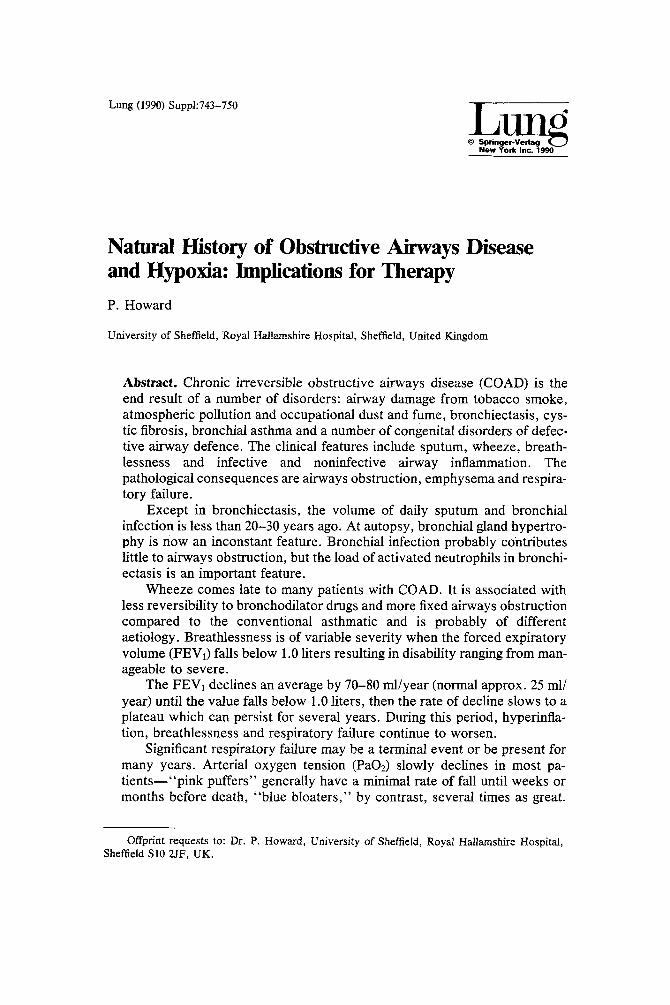
Lung (1990) Suppl:743-750
New York Inc. 1990
Natural History of Obstructive Airways Disease and Hypoxia: Implications for Therapy P. Howard
University of Sheffield, Royal Hallamshire Hospital, Sheffield, United Kingdom
Abstract. Chronic irreversible obstructive airways disease (COAD) is the end result of a number of disorders: airway damage from tobacco smoke, atmospheric pollution and occupational dust and fume, bronchiectasis, cys- tic fibrosis, bronchial asthma and a number of congenital disorders of defec- tive airway defence. The clinical features include sputum, wheeze, breath- lessness and infective and noninfective airway inflammation. The pathological consequences are airways obstruction, emphysema and respira- tory failure.
Except in bronchiectasis, the volume of daily sputum and bronchial infection is less than 20-30 years ago. At autopsy, bronchial gland hypertro- phy is now an inconstant feature. Bronchial infection probably contributes little to airways obstruction, but the load of activated neutrophils in bronchi- ectasis is an important feature.
Wheeze comes late to many patients with COAD. It is associated with less reversibility to bronchodilator drugs and more fixed airways obstruction compared to the conventional asthmatic and is probably of different aetiology. Breathlessness is of variable severity when the forced expiratory volume (FEV1) falls below 1.0 liters resulting in disability ranging from man- ageable to severe.
The FEVI declines an average by 70-80 ml/year (normal approx. 25 ml/ year) until the value falls below 1.0 liters, then the rate of decline slows to a plateau which can persist for several years. During this period, hyperinfla- tion, breathlessness and respiratory failure continue to worsen.
Significant respiratory failure may be a terminal event or be present for many years. Arterial oxygen tension (PaOz) slowly declines in most pa- t ientsm"pink puffers" generally have a minimal rate of fall until weeks or months before death, "blue bloaters," by contrast, several times as great.
Offprint requests to: Dr. P. Howard, University of Sheffield, Royal Hallamshire Hospital, Sheffield S10 2JF, UK.

744 P. H o w ~ d
Oxygen therapy, corticosteroids and other bronchodilator drugs do not influ- ence the rate of deterioration. Pulmonary vascular remodelling is an impor- tant part of the pathology of hypoxic COAD. Oxygen therapy relieves hy- poxia but does not arrest deterioration of airways obstruction. New therapeutic approaches are needed to tackle the steady decline of airway function.
Key words: Chronic obstructive airways disease--Hypoxia--Forced expir- atory volume in one second--Natural history.
Chronic bronchitis and emphysema are the most common cause of irreversible airways obstruction. Clinical features include sputum expectoration, wheeze, breathlessness and clinical manifestations of inflammation, infective or nonin- fective. The most important symptom is breathlessness, its appearance herald- ing disability. At this point, results of tests of airway function, notably the forced expiratory volume in one second (FEV), and the forced vital capacity (FVC), have fallen to less than half of predicted normal values. The decline of the FEVI, probably the most reliable test of the natural history of airways obstruction, is exponential, with an accelerated fall in earlier but asymptomatic years. In the later stages, the FEV~ plateaus for a number of years at very low levels, below values of 1 liter. It is often surprising how long patients can survive with forced expiratory volumes of around half a liter. The decline of FEV1 in earlier years has been admirably described by Fletcher and Peto [1] in their study of London postmen (Fig. 1).
Decline is related to the susceptibility of smoking effects and the amount smoked. The decline returns to more normal levels if smoking ceases before extreme disability is realized. The average normal rate of decline with age is said to be between 20 and 30 ml/year ofFEV1. In the urban smoking individual the rate of decline is between 70 and 100 ml/year of FEV1, but it is likely to be somewhat higher than this in earlier phases of the disease. Most patients die in respiratory failure. Disability from breathlessness is variably expressed in rela- tion to the FEV1 when it falls below 2 liters. For example, a patient with an FEV1 of 1.0 liters may sometimes have serious limitation to effort and walking and yet others have only disability on heavy exercise, hills or stairs (Fig. 2). Equally, the appearance of respiratory failure of either Type 1 or Type 2 formu- lation may be an early feature of the fall of FEV~ below 2 liters or a very late feature appearing only a few weeks before death. Therefore it seems likely that the decline of airway function, disability and respiratory failure are discontinu- ous features. This is substantiated by Biernacki et al. [2] who recently found little association between pulmonary haemodynamics, gas exchange and the severity of emphysema as assessed by quantitative CT scan.
Death rate attributable to bronchitis and emphysema has declined in recent years probably due to reduction of smoking and atmospheric and occupational pollution (Fig. 3) [3]. In contrast, for patients attending hospital clinics, the rate of decline of FEVI below 2 liters has changed little despite these improve-

Natural History of COPD
100-
~, 75- r=
== so-
"6
25-
0 25
Never smoked or not
~ oke
Smoked regularly and / ~ -'~'~...._ ~" susceptible to its effect.s ~ -'~"~. Stopped at 45
. . . . . . . . . . . . . . . . . . . . _ ' _ ' 5 _ - . r~^~,k " ~ ' ~ . . <~ Stopped at 65
. . . . . . . . . ~ . . 2 " - - . . . . .
I I 50 75
Age in years
745
Fig. 1. Graph of the decline in FEVt in smokers and nonsmokers. Cessation of an established smoking habit is associated with a reduced rate of subsequent deterioration. (Reproduced by permission, [1].)
FEV 4 .0 -
2 .0 -
0 0
litres
50 100
VA Breathlessness
Fig. 2. Breathlessness measured by visual analogue scale in relation to FEV~. Breathlessness varies widely in severity once FEV~ falls below 2.0 liters.
ments. Thus, although there are less patients with bronchitis and emphysema compared with 20 years ago, those who acquire the most aggressive forms of the disorder continue to deteriorate at the same rate.
The Problem of Hypoxia
Once the FEV1 falls below 2 liters most patients will have a degree of hypox- aemia--some being severely hypoxaemic and hypercapnic almost simulta- neously and others having arterial oxygen levels only a few mmHg below accepted normal values. The rate of decline of arterial oxygen tension is equally variable. In those with the clinical manifestations of emphysema-- hyperinflation, wasting, inappropriate dyspnoea--the decline of arterial oxy- gen tension tends to be slower giving rise to the synonym "pink puffer." The rather more obese, somnolent patient has a more rapidly deteriorating hypox- aemia and, in many instances, also hypercapnia. He develops oedema early giving rise to the synonym "blue bloater." There is a spectrum of effect with patients whose features are midway between the extremes, but the rate of deterioration of arterial hypoxaemia at the "cor pulmonale end" can be very severe between 10 and 20 mmHg or 1-3 kPa per annum.

746 200] o ° 15° 1
2O t ~
i i i i I i i I 1968 70 72 74 76 78 80 82
P. Howard
Fig. 3. Decline in death rate from bronchitis in British and American patients: England/Wales, males (11) and females (O); USA, males ([]) and females (&). (Reproduced by permission, [3].)
Table 1. Features of hypoxic cor pulmonale associated with COAD
Clinical Physiologic
Overweight Somnolent Less dyspnoea Central cyanosis Recurrent edema
PaOz < 7.3 kPa (55 mmHg) PaCO2 > 6.0 kPa (45 mmHg) Hct > 45% PAP > 22 mmHg (range 18-50 mmHg) FEVt < 1.5 1 FVC < 2.0 1
PaO2 and PaCO2--arterial oxygen and carbon dioxide tensions, Hct haematocrit, PAP mean pulmonary artery pres- sure supine, FEVt and FVC--forced expiratory volume and vital capacity, respectively
Table 1 shows the critical physiological findings when patients commence with exacerbations of oedema. Such patients were studied using oxygen ther- apy in the now famous NOTT [4] and MRC trials [5] which quite clearly demonstrated benefit to survival. It is now possible to follow-up such patients after 10 years of long term domiciliary oxygen therapy [6]. Table 2 shows the characteristics of 72 such patients. They had all of the physiological character- istics described in Table 1. The mean values are given in Table 2, and the mean survival compared with the control group of the MRC males, is shown in Table 3. The survival of the Sheffield patients at 10 years approximates that of the MRC no-oxygen control male group (the only control population available) at five years. There is clearly a sharp difference in survival of both groups com- pared with the derived normal population. The conclusion must be that L T O T

Natural History of COPD
Table 2. Features of 72 patients with hypoxic cor pulmonale and COAD treated with oxygen for up to 10 years
M/F 53/19 Age (years) 60.5 - 7.5 FEVt (liters) 0.78 ± 0.31 FVC (liters) 1.90 ± 0.64 TLCO (kPa .1-Ls -I) 3.48 ± 1.87 PaO2 (kPa) 6.1 --+ 1.0 PaO2 (on 05) (kPa) 9.1 -- 1.4 PaCO2 (kPa) 6.9 ± 1.2 PaCO2 (on O2) (kPa) 7.3 ± 1.4
TLCO, transfer factor for carbon monoxide; mean - SD (courtesy of Thorax, 1987-see [5])
747
Table 3. Cumulative survival proportions at 5 and 10 years from onset of LTOT in 72 Sheffield patients compared with Medical Re- search Council Control subjects and a derived normal population
Group Survival at Survival at 5 years (%) I0 years (%)
MRC control males 20 - - Sheffield patients 65 20 Normal population of same age and sex 90 78
Cumulative survival proportions are rounded up for compar- ison
is worth about five years of extended survival, which is useful but somewhat shorter than originally anticipated. The cause of death was investigated and correlations made with survival characteristics. In the treated group, survival was not related to arterial blood gases, haematocrit or the level of mean pulmo- nary artery pressure but to the FEV~. Oxygen therapy thus relieves the trouble- some effects of hypoxaemia but fails to influence the continuing decline of airway function which eventually determines the clinical outcome.
This point is reinforced in a further study of 37 deaths [7] in patients with hypoxaemic cor pulmonale associated with COAD (Table 4). There were 27 males and 10 females. Table 4 compares physiological parameters at the start of treatment with those immediately prior to death. The mean period of treatment was five years. The FEV1 continued to deteriorate throughout the period of therapy. This is also true of the arterial blood gases breathing air, confirming that oxygen therapy does not arrest the underlying natural history of the disease.

748
Table 4. Study of 37 deaths in patients with hypoxic cor pulmonale and COAD, physiological measurements
Onset of LTOT Prior to death
Age yrs 60.1 --- 8.0 65 -- 7.9 FEVz 0.78 + 0.33 0.57 +-- 0.20 PaO2, air, kPa 6.6 --- 1.0 5.1 --- 1.1 PaCOz, air, kPa 6.7 - 0.9 7.2 -4-_ 1.3
27 males and 10 females, mean --- SD
P. Howard
Table 5. Summary of indications for long-term oxygen therapy--Brit ish Guidelines
Category Clinical features Physiologic features
PaOz* PaCO2** FEVI FVC (kPa) (kPa) (liters) (liters)
1 "Blue bloater" syndrome <7.3 >6.0 <1.5 <2.0 of bronchitis and emphysema
2 Breathlessness; emphysema <7.3 <1.5 <2.0 and respiratory failure
3 Respiratory failure associated <7.3 with any terminal disease; palliative short-term therapy
* 55 mmHg; ** 45 mmHg
Outcome of Current Domiciliary Oxygen Treatment
All of these studies have selected patients with particularly severe hypoxaemia, hypercapnia and oedema, which poses problems for the selection of patients for oxygen therapy in everyday clinical practice. This was recognized in the writing of the British Guidelines to General Practitioners for the selection of patients (Table 5).
Category 1 describes the classic "blue bloater" patient. There seems little doubt that oxygen therapy in this group is beneficial. Category 2 patients are more of a problem. These are likely to be patients with clinical features of emphysemambreathless, hyperinflated and wasted--but who actually have hy- poxaemia. Hypercapnia and oedema will usually appear only in preterminal phases. In effect they compose the "pink puffer" patients who will finally enter the stage of respiratory failure. What are their prospects for benefit from long- term domiciliary oxygen therapy (LTOT). The FEV1 is extremely low, usually less than 1.0 liters, and, as the more important determinant of survival than treated hypoxaemia, will truncate survival prospects. In the United Kingdom,

Natural History of COPD 749
General Practitioners are selecting patients on the grounds of central cyanosis and dyspnoea. The average duration of an oxygen concentrator installation is less than one year. Although more precise studies are needed, it seems that Category 2 patients will have limited benefit from LTOT.
Hypoxaemia without Oedema
It is possible to look at the survival characteristics of hypoxaemia alone from two studies, the IPPB American trial [8] and the Multicenter VIMS European Almitrine study [9]. Both groups of patients had advanced obstructive airways disease. The former study selected patients with no hypoxaemia to determine whether intermittent assisted breathing would help survival and disability. The second study investigated the effects of Almitrine bismesylate, a chemorecep- tor agonist, on hypoxic patients with COAD, very few having reached the oedematous phase. Five hundred patients treated with IPPB of mean age 60.5 yrs had an initial arterial oxygen tension of 9.4 kPa (70 mmHg) and were normocapnic. For the 701 patients in the VIMS study, the mean age at entry was 62 yrs., the mean arterial oxygen tension was 7.6kPa (57 mmHg) and they were, on average, normocapnic, 6.0 kPa (45 mmHg). The major difference clinically was that the VIMS patients were notably more hypoxaemic. Cumula- tive survival proportions of approximately 85% in both studies showed no major differences at two years. Moderate hypoxaemia in association with ad- vancing obstructive airways disease would therefore seem not to be a risk factor for survival.
Which features of severe hypoxaemia cause rapid death of the "blue bloater" patient when in earlier phases it creates little problem? It is now appreciated that, as hypoxaemia progresses, tissue responses to the chronic deficiency may change. Acclimatization can break down. Alterations of func- tion occur in vitally affected organs such as the carotid body, the kidney, the autonomic nervous system, the peripheral nervous system and perhaps the lung. These features should now be studied to elucidate the effects of lack of oxygen to enable more precise targeting of long-term domiciliary oxygen ther- apy (LTOT) to those patients showing metabolic disturbances.
Treatment of Early Hypoxaemia without Oedema
Oxygen therapy has failed to alter the natural history of COAD. It merely relieves tissue hypoxaemia temporarily. In early phases it is unlikely to influ- ence survival. The emphasis must surely be on prevention of progressive hypo- xaemia which is more likely to yield to pharmacologic therapy. Almitrine bis- mesylate should be reexamined in this light, and other compounds should be studied which might prevent the loss of tissue acclimatization to advancing hypoxic disease.

750 P. Howard
References
1. Fletcher C, Peto R (1977) The natural history of chronic airflow obstruction. Brit Med J i: 1645- 1648
2. Biernacki W, Gould GA, Whyte KF, Flerdey DC (1989) Pulmonary haemodynamics, gas ex- change and the severity of emphysema as assessed by quantitative CT scan in chronic bronchi- tis and emphysema. Amer Rev Respir Dis 139:1509-1515
3. Seaton A, Seaton D, Leitch Gordon A (1989) Croften and Douglas Respiratory Diseases, 4th edition. Blackwell Scientific: Oxford
4. Nocturnal Oxygen Therapy Trial Group (1980) Continuous or nocturnal oxygen therapy in hypoxaemic chronic obstructive airways disease--a clinical trial. Ann Intern Med 93:391-398
5. Medical Research Council Working Party (1981) Long term domiciliary oxygen therapy in chronic hypoxic cor pulmonale complicating chronic bronchitis and emphysema. Lancet i:681- 683
6. Cooper CB, Waterhouse JC, Howard P (1987) Twelve year clinical study of patients with hypoxic cor pulmonale given long term domiciliary oxygen therapy. Thorax 42:105-110
7. Personal observations--unpublished 8. Intermittent Positive Pressure Breathing Trial Group (1983) Report of a trial. Ann Intern Med
99:612-620 9. Voisin C, Howard P, Ansquer JC (1987) Almitrine bismesylate--a placebo controlled trial in
COAD--VIMS. Bull Europ Physiopathol Respir 23 (suppl 11):169-182