Embed Size (px)
Citation preview
HAL Id: hal-03154304https://hal.archives-ouvertes.fr/hal-03154304
Submitted on 28 Feb 2021
HAL is a multi-disciplinary open accessarchive for the deposit and dissemination of sci-entific research documents, whether they are pub-lished or not. The documents may come fromteaching and research institutions in France orabroad, or from public or private research centers.
L’archive ouverte pluridisciplinaire HAL, estdestinée au dépôt et à la diffusion de documentsscientifiques de niveau recherche, publiés ou non,émanant des établissements d’enseignement et derecherche français ou étrangers, des laboratoirespublics ou privés.
Nanosized microporous crystals: emerging applicationsSvetlana Mintova, Maguy Jaber, Valentin Valtchev
To cite this version:Svetlana Mintova, Maguy Jaber, Valentin Valtchev. Nanosized microporous crystals: emerging ap-plications. Chemical Society Reviews, Royal Society of Chemistry, 2015, 44 (20), pp.7207-7233.�10.1039/C5CS00210A�. �hal-03154304�
1
Nanosized microporous crystals: emerging applications
Svetlana Mintova,1,
* Maguy Jaber,2,3
Valentin Valtchev1
1Laboratoire Catalyse & Spectrochimie, ENSICAEN - Université de Caen - CNRS
6, boulevard Maréchal Juin, 14050 Caen, France
2UPMC Paris 06, UMR 8220, Laboratoire d’archéologie moléculaire et structurale, LAMS,
F-75005, 4 place Jussieu, BP225, Paris, France
3CNRS, UMR 8220, LAMS, F-75005, Paris, France
Abstract
This review highlights recent developments in the synthesis and unconventional applications
of nanosized microporous crystals including namely framework (zeolites) and layered (clays)
type materials. Owing to their micropore nature nanosized zeolites and clays exhibit novel
properties, different from those of bulk materials. The factors controlling the formation of
nanosized microporous crystals are first revised. The most promising approaches from the
viewpoint of large-scale production of nanosized zeolites and clays are discussed in depth.
The preparation and advanced applications of nanosized zeolites and clays in free (suspension
and powder forms) and fixed (films) forms are summarized. Further the review is accentuated
on the new, non-conventional for porous materials, applications. A comprehensive analysis of
the emerging applications of microporous nanosized crystals in the fields of semiconductor
industry, optical materials, chemical sensors, medicine, cosmetics, and food industry is
presented. Finally, the future needs and perspectives for nanosized microporous materials
(zeolites and clays) are addressed.
1 Introduction
Zeolites and clays are minerals with widespread occurrence in various Earth's crust
environments and with great impact in different areas of the chemical process industries and
nanotechnology.1 Both zeolites and clays crystallize in water-rich and low pressure
environment under wide range of temperature.2,3
Under such conditions low-density water-
containing structures are formed. The major difference between these two groups of minerals
is the type of the structure formed, which in the case of clays is two dimensional (2D) while
2
the zeolites exhibit a three dimensional framework (3D). Both clays and zeolite contain
exchangeable cations and water molecules in the intra-crystalline space. In the case of clay
minerals cation-water complexes are situated between sheets built of SiO4 and other cations.
While, three-dimensional zeolite frameworks contain well define pores and cavities able to
discriminate species with different sizes less than 1 Å. The zeolite frameworks are fairly rigid
and exhibit limited flexibility. Zeolites exhibit shape selectivity of molecules of dimension
similar with their channels. Depending on the pore opening the zeolites are subdivided of
small, medium and large pore channels, as the later are able to process molecules with size
between 7 and 8 Å. Extra large pore (> 8 Å) zeolites were also obtained, however, because of
limited framework stability and difficulties in tuning framework composition they are not
used for practical applications. In contrast, the inter-lamellar space of clays can be expanded
substantially and thus they could adsorb molecules larger than zeolites. The zeolites and clays
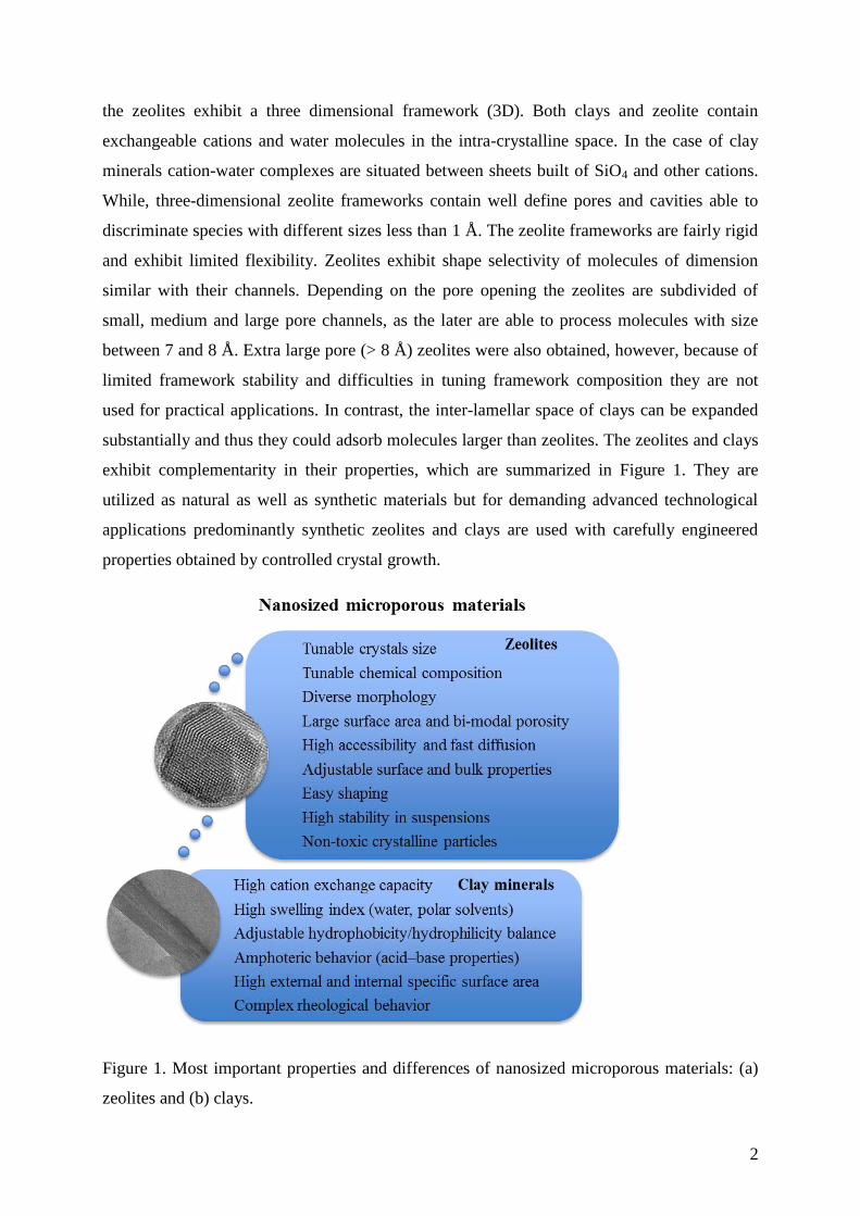
exhibit complementarity in their properties, which are summarized in Figure 1. They are
utilized as natural as well as synthetic materials but for demanding advanced technological
applications predominantly synthetic zeolites and clays are used with carefully engineered
properties obtained by controlled crystal growth.
Figure 1. Most important properties and differences of nanosized microporous materials: (a)
zeolites and (b) clays.
3
Zeolites and clays are widely used in chemical process industry as heterogeneous catalysts,
adsorbents and ion exchangers. Their importance is enormous for such key areas as petroleum
refining, petrochemicals and fine chemicals production, separation of toxic and radioactive
wastes, air pollution abatement, industrial effluent and water purification. The use of clays
and zeolites ranges from heavy chemical processes and environmental protection, to
household needs and lately to advanced applications. The wide use of these materials is due to
their particular structures and related properties, such as accessible intracrystalline volume
and thus much larger specific surface area in respect to dense materials. The presence of
channels with molecular dimension and active sites that can be tuned make these materials
excellent heterogeneous catalysts and molecular sieves. Although porous with high specific
surface area and accessible crystalline volume, the size of zeolites and clays is important for
their performance.
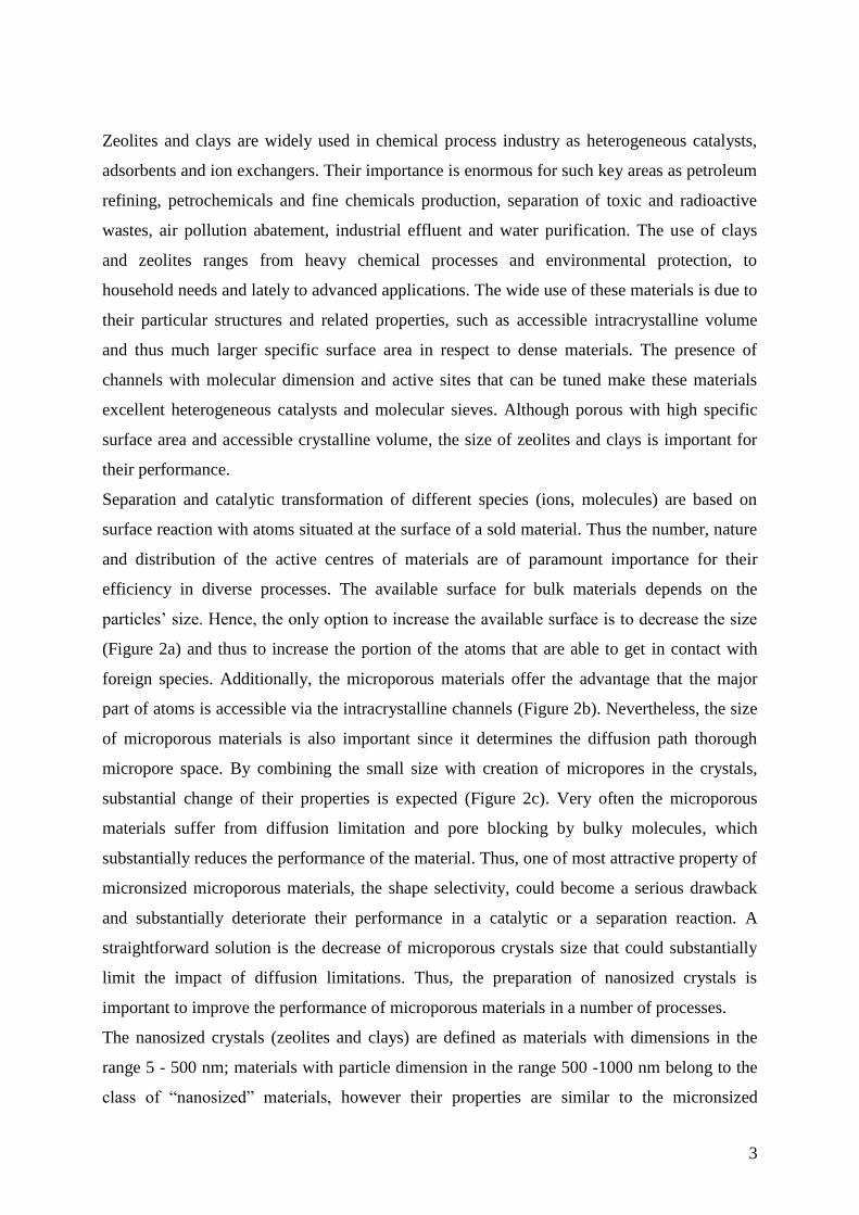
Separation and catalytic transformation of different species (ions, molecules) are based on
surface reaction with atoms situated at the surface of a sold material. Thus the number, nature
and distribution of the active centres of materials are of paramount importance for their
efficiency in diverse processes. The available surface for bulk materials depends on the
particles’ size. Hence, the only option to increase the available surface is to decrease the size
(Figure 2a) and thus to increase the portion of the atoms that are able to get in contact with
foreign species. Additionally, the microporous materials offer the advantage that the major
part of atoms is accessible via the intracrystalline channels (Figure 2b). Nevertheless, the size
of microporous materials is also important since it determines the diffusion path thorough
micropore space. By combining the small size with creation of micropores in the crystals,
substantial change of their properties is expected (Figure 2c). Very often the microporous
materials suffer from diffusion limitation and pore blocking by bulky molecules, which
substantially reduces the performance of the material. Thus, one of most attractive property of
micronsized microporous materials, the shape selectivity, could become a serious drawback
and substantially deteriorate their performance in a catalytic or a separation reaction. A
straightforward solution is the decrease of microporous crystals size that could substantially
limit the impact of diffusion limitations. Thus, the preparation of nanosized crystals is
important to improve the performance of microporous materials in a number of processes.
The nanosized crystals (zeolites and clays) are defined as materials with dimensions in the
range 5 - 500 nm; materials with particle dimension in the range 500 -1000 nm belong to the
class of “nanosized” materials, however their properties are similar to the micronsized
4
counterparts. A substantial increase of the fraction of the atoms on zeolite/clay surface could
also be used to perform reactions with very bulky molecules that cannot defuse in micropore
space. Finally, nanosized zeolite crystals and clays are important for a number of emerging
applications related with the development of advanced materials.
Figure 2. Available surface area of crystals increased by (a) decreasing the size of particles,
(b) introduction of pores, and by combining both (c) decrease of size and introduction of
pores in nanosized crystals.
There are already a number of papers that review the synthesis, modification and applications
of nanosized porous particles.4–6
The aim of present article is to overview latest development
in the field paying special attention to the emerging uses of microporous crystals. We have
limited our study to classical silica-based microporous crystals since the major part of the new
advanced applications are based on such materials.
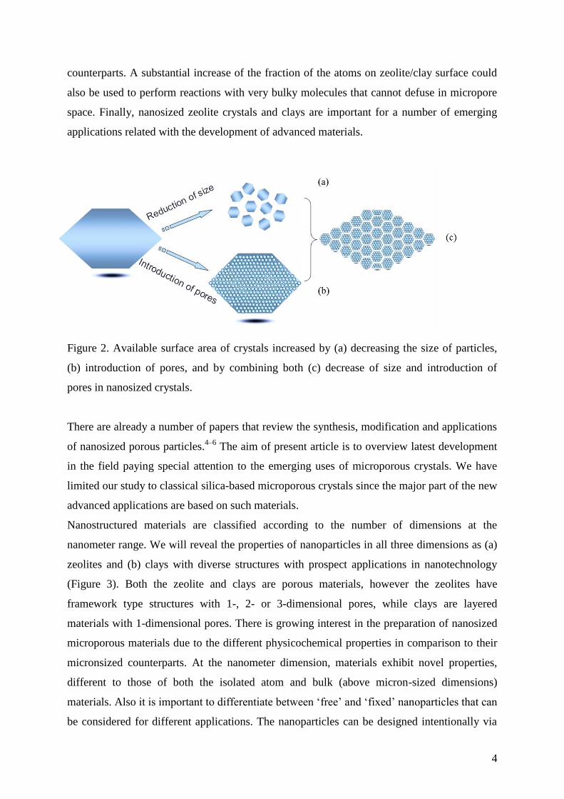
Nanostructured materials are classified according to the number of dimensions at the
nanometer range. We will reveal the properties of nanoparticles in all three dimensions as (a)
zeolites and (b) clays with diverse structures with prospect applications in nanotechnology
(Figure 3). Both the zeolite and clays are porous materials, however the zeolites have
framework type structures with 1-, 2- or 3-dimensional pores, while clays are layered
materials with 1-dimensional pores. There is growing interest in the preparation of nanosized
microporous materials due to the different physicochemical properties in comparison to their
micronsized counterparts. At the nanometer dimension, materials exhibit novel properties,
different to those of both the isolated atom and bulk (above micron-sized dimensions)
materials. Also it is important to differentiate between ‘free’ and ‘fixed’ nanoparticles that can
be considered for different applications. The nanoparticles can be designed intentionally via
5
“synthesis engineering” or the nanoparticles can be prepared by post-synthesis treatments. In
this review, the emphasis will be on in situ prepared nanosized microporous materials.
Figure 3. Key issues of nanosized microporous materials: zeolites and clays.
2 Nanosized zeolites
Zeolite nucleation is a spontaneous process leading to the formation of viable nuclei that grow
into crystals.7,8
The zeolite yielding systems are fairly different in terms of chemistry and
physical appearance of the precursor. They can vary from water clear suspensions to dry
powders containing only traces of water. The common feature of laboratory syntheses is that
the crystallization is performed in close systems and very rarely fresh reactants are added in
the course of zeolite crystallization. Synthesis under such conditions allows control of the
ultimate crystal size via the nucleation process. More precisely, the number of the viable
nuclei in the system determines the ultimate crystal size since after the conversion of the
amorphous precursors into crystals the growth process stops. Hence, the systems with
abundant nucleation yield small crystallites, whereas the ones that generate a few nuclei
provide relatively large ones. It has to be underlined, however, that the size of the crystallites
can be small (5 - 500 nm) but they might form large intergrown agglomerates (above 1 m).
Often the performance of such agglomerates is similar to micron-sized crystals. Therefore, in
order to take advantage of small crystal size, single not agglomerated nanocrystals are highly
desired.
6
In general, the synthesis of zeolites is performed from a hydrogel. These systems exhibit
distinct solid (gel) and liquid (mother liquor) part and they are highly non-uniform. Usually
the products from such systems are zeolite crystals with size of about one micron, the
intergrowth and the agglomeration from one system to another differ and this is highly
dependent of the initial reactants used, the alkali and water contents. Briefly the process of
zeolite nucleation and growth in hydrogel system is highly unpredictable and far from well
controlled.
2.1 Non-conventional synthesis of nanosized zeolites
The difficulties in the control of the crystals size in conventional zeolite-yielding systems
resulted in the development of alternative methods of synthesis. Such an approach that
circumvents the difficulties in the control of zeolite nucleation is the confined space synthesis.
In this case the ultimate crystal size is determined by the available free space, which is limited
by some physical or chemical barrier. The first example of such synthesis was published by
Madsen and Jacobsen.9 They synthesized zeolites in the voids of porous carbon particles and
removed the carbon by combustion to obtain nanosized zeolite crystals. Later on, the hard
carbon templates were replaced by organic matrixes, such as strach10
and polymer
hydrogels.11
Besides hard and soft templates were also used to control the growth of zeolite crystals.
Serrano and co-workers have demonstrated that ZSM-5 growth can be controlled by surface
functionalization of growing crystals.12,13
Different types of silanes containing amino,
mercapto and allyl surface groups were used. In all cases the synthesized crystals were
smaller than those synthesized from the same initial system without using silanes. It should be
mentioned that beside the size, the presence of silanes changed the Si/Al ratio and the surface
charge of the nanosized zeolites.
Jo et al. reported the use of multivalent surfactants for restricting the growth of zeolite crystals
and thus the formation of nanosized particles.14
The effect of surfactant on the zeolite crystals
size is attributed to the multiply binding of surfactant’s functional groups to zeolite surface,
which limits the growth. Zeolites with MOR, FAU, CHA and MFI framework topology where
synthesized. In any case, however, the zeolite nanocrystals were heavily aggregated. Similar
approach was used by Jamil et al., who employed a polyoxyethilene-based surfactant to
control the size and the crystal aspect ratio of TON-type zeolite.15
A decrease of the crystal
length from 300 to 90 nm was observed. Microwave (MW) radiation was used in this
7
synthesis. Again MW heating, but employing a water-in-oil microemulsion was used to
synthesize FAU type zeolite nanocrystals.16
Small water droplets were used as microreactors
to limit the zeolite growth. The size of synthesized FAU type crystals (zeolite X) varied
between 10 and 30 nm. Conventional heating yielded crystals with size of about 150 nm.
MW heating offers the advantage of rapid and uniform heating in the reaction vessel,17
usually the result is more homogeneous and abundant nucleation in the system. Thus,
different types of microporous materials were synthesized in the form of nanocrystrals.5 It
should be mentioned that the method is more efficient in the synthesis of aluminophosphate
than aluminosilicate microporous materials. For instance, MW irradiation was used to initiate
the nucleation of SAPO-34 and then the system was subjected to conventional hydrothermal
treatment at 200 °C for 24 h.18
The authors reported a pronounced effect of the MW power on
the nucleation process. The nucleation initiated at higher MW power (210 W) is more
abundant, but the crystals tend to aggregate, whereas the use of lower (140 W) power yields
less but non-aggregated nuclei.
Similarly to MW heating the sonification provides smaller crystals than the conventional
synthesis.19
Recently nanosized zeolite NaP was synthesized by sonochemical of the initial
gel followed by conventional hydrothermal treatment.20
Small about 50 nm zeolite P crystals
were obtained. However, most of the crystals were agglomerated and form large particles.
The authors underlined the importance of sonication energy for the formation of active
radicals, which promote the rapid zeolite formation.
Dry-gel conversion (DGC) method for preparation of zeolites could also be considered as a
non-conventional synthesis approach for nanocrystals. The method is based on the
preliminary preparation of an initial zeolite yielding gel, which is dried and then subjected to
crystallization in vapour phase. In such ultra-dense gel usually the nucleation process is
abundant. However, closely situated nucleuses usually formed aggregates. Yue et al.
employed the DGC process to prepare shaped ZSM-5 agglomerates.21
A particularity in their
approach is the use of seeds (MFI-type) that were mixed with aluminoslicate precursor and
extruded to shape columned bodies. The synthesis was performed in vapour phase at 175 °C
for different periods of time. Besides water, the vapour phase contained NH4OH, ethylamine
or butylamine. The best results were obtained with ethylamine, which combines high
alkalinity with high-saturated vapour pressure. Depending on the synthesis conditions, zeolite
particles with size between 200 and 800 nm were synthesized. The reference experiment
without addition of the seeds yielded relatively large ZSM-5 crystals. Zeolite bodies exhibited
significant mechanical strength.
8
2.2 Conventional synthesis of nanosized zeolites
The synthesis of a zeolite from a hydrogel precursor in a close reactor upon conventional
heating is considered as “conventional synthesis”. First synthesis of nanosized zeolite was
reported by Meng et al.22
Nanosized zeolite L crystals with uniform particle size of 50-100
nm building larger agglomerates were obtained.
First syntheses of nanosized single zeolite crystals that do not agglomerate were reported in
the early 90s by Schoeman et al.23–25
The syntheses of ZSM-2, FAU-, LTA- and SOD-type
zeolites were described in these studies. After purification zeolite nanocrystals were stabilized
in the form of colloidal suspensions with narrow particle size distribution. The systems
yielding such uniform nanocrystals were called “clear solutions”. In fact, the initial systems
used were optically clear suspensions containing discrete colloidal particles. Although not
completely homogeneous these systems provide the necessary condition for a uniform
nucleation and crystal growth. Consequently the obtained nanocrystals are uniform in size and
the agglomeration of the particles is limited.
The stabilization of a suspension containing discrete zeolite particles requires specific
conditions in each stage of their preparation. First the employed reactants and the conditions
have to be properly selected. Further, the alkali metal content has to be as low as possible in
order to avoid the aggregation between the particles. The absence of alkali cations is
compensated with abundant amounts of tetraalkylammonium hydroxides (TAAOH) that keep
the basicity of the system high and act as structure directing agents (SDAs). The
homogenisation of the reactants is gentle to avoid the aggregation of precursor particles. Thus
a suspension containing uniform in size particle is stabilized. Usually the system used is very
diluted to avoid the aggregation between the growing crystals. Crystallization temperature is
typically lower than the one traditionally used for the synthesis of a particular zeolite, which
favours the nucleation over the growth. Upon heating the amorphous precursor particles
convert into nanosized zeolite. With increase the number and the size of colloidal particles
initially optically clear solution turns to turbid and then to milky white. Besides the specific
preparation procedure, the nanosized zeolites require a specific post-synthesis treatment in
order to avoid severe aggregation between the particles. The excess of organic templates and
unreacted silica/alumina are separated by high-speed centrifugation, and redispersion of
zeolite nanoparticles in distilled water is usually performed. The procedure is repeated several
times and then the pH of resultant suspension is adjusted to about 9 with diluted NH4OH. The
9
goal of latter procedure is to keep the negative charge of colloidal zeolite crystals high. The
repulsive forces of negatively charged particles protect the particles from further aggregation.
Freeze drying is used when dry powder of zeolite nanocrystals is needed. This process limits
the aggregation between the crystallites. The calcination of organic template-containing
zeolite nanocrystals is usually performed under the conditions used for the micron-sized
counterpart. The micropore volume of highly crystalline nanosized zeolites is expected to be
similar to the micronsized crystals. This is in the case of fully crystalline nanosized zeolites
(see Figure 4). As can be anticipated the nanocrystals exhibit substantially higher external
surface area.
The use of clear suspension is very efficient process that provides high quality nanosized
zeolite particles. However, because of specific conditions used, namely highly diluted systems
crystallizing at relatively low temperature, the crystalline yield is much lower than that from a
gel-rich initial system. On the other hand, clear precursor suspensions were found very useful
in studies devoted to the zeolite crystallization mechanism. The uniformity of the initial
system and the simultaneity of the crystallization events make them indispensable for
understanding of the zeolite nucleation and growth processes. For instance, the precursor
suspension with a chemical composition 9TPAOH:25SiO2:480H2O (TPAOH is
tetrapropylammonium hydroxide), first used by Persson et al. to synthesize nanosized
silicalite-1 is one of most largely exploited system.26
With slight variations of the TPAOH and
H2O contents, this system was used in tenth of studies devoted to zeolite nucleation and
substantial advances in understanding of the nature of colloidal precursors yielding zeolite
were achieved.27–33
Another “clear” precursor suspension allowed visualizing the “birth” of LTA-type zeolite.34
The system used was 13.4(TMA)O2:0.3Na2O:1.8Al2O3:11.25SiO2:700H2O, where as organic
template (TMAOH is tetramethylammonium hydroxide) was used. The crystallization was
performed at room temperature for a week. After mixing the initial reactants 40-80 nm
amorphous particles were formed. Each amorphous particle yielded a zeolite crystal. The
formation of zeolite nuclei was observed in the centre of each particle and then the crystalline
network propagated outward direction till the complete transformation of the amorphous
particles into crystalline was reached. It is noteworthy that the size and the morphology of the
crystal were similar with the amorphous precursor particles. According to the X-ray
diffraction (XRD) and transmission electron microscopy (TEM) study these particle were
fully crystalline. Only after heating at elevated temperature larger cubic shape zeolite A
crystals were observed. Similar process of formation was observed in the case of FAU-type
10
zeolite synthesized from TMAOH-rich initial system.35
Again a low Na-content initial
solution was used, but the crystallization was performed at elevated temperature. The
crystallization mechanism followed at elevated temperature shows that it is not specific for
ambient temperature conditions only. The question that arises is whether colloidal precursor
particles can be stabilized in a Na-rich initial system and further to be transformed into
nanosized zeolite by a similar pathway.
Very recently ultra small EMT-type zeolite, which is the hexagonal counterpart of cubic
FAU-type zeolite, was synthesized from an organic template-free Na-rich initial system.36
The
mechanism of formation was similar to the one observed in the synthesis of LTA- and FAU-
type zeolites synthesized from TMAOH-rich colloidal precursors. First amorphous colloidal
particles were formed and upon moderate heating (30-40°C) these particles have gradually
transformed into EMT-type zeolite crystals with no substantial changes of their size. The
dynamic light scattering (DLS) measurements showed almost the same particles size for the
amorphous precursor and crystalline zeolite particles. Ultra small plate-like hexagonal EMT-
type crystals were synthesized. The crystal thickness along a and c directions was 13-15 nm
and 4 nm, respectively. These dimensions correspond approximately of 7 x 2 unit cells
crystallite. Such small single nanocrystals were not synthesized up to this study, including in
organic template-containing initial systems.4,6
The factors playing key role in this spectacular
synthesis were: first the stabilization of colloidal precursor suspension in the presence of large
amount of Na, and second the synthesis perform at low temperature (40 C). It is important to
note that in order to avoid an uncontrolled polymerization of the aluminosilicate precursor
particles, special precaution was taken to the preparation of initial suspensions and the order
of reagents mixing. An aluminate and a silicate solution with similar NaOH content were
prepared and cooled down to 4 °C. The addition of small portion of one solution to another
under vigorous stirring ensured the formation of discrete amorphous particles. The
hydrothermal treatment under a moderate temperature allowed the conversion of the
amorphous precursor into crystalline zeolite without favouring of the Ostwald repining. It is
worth nothing that the synthesis at low temperature was important not only to limit the growth
of the crystals, but also to avoid the formation of competing phases as LTA-, SOD, GIS- or
FAU-type zeolites.37
Thus the ultimate crystals were similar in size with the amorphous
particles. This synthesis is equally remarkable by the fact that EMT-type zeolite was
synthesized solely with sodium as a structure-directing agent. Previously this zeolite was
obtained using a 18-crown-6 ether.38,39
The use of this very expensive SDA was the main
11
obstacle to industrial use of EMT-type zeolite, which exhibit great potential in catalytic and
separation processes.
Recently, organic additives were used to control the formation of EMT type zeolite in a
sodium-rich initial system.40
Triethanolamine, tetramethylammonium and
tetraethylammonium were employed in this study. The triethanolamine had the most
pronounced effect on the crystal growth process of the EMT type zeolite providing larger
crystals (100 nm) with framework composition different from the counterpart obtained in an
organic-free precursor system. The effect of triethanolamine was attributed to the
immobilization of Al in the initial suspension, and thus partial suppression of zeolite
nucleation was observed.
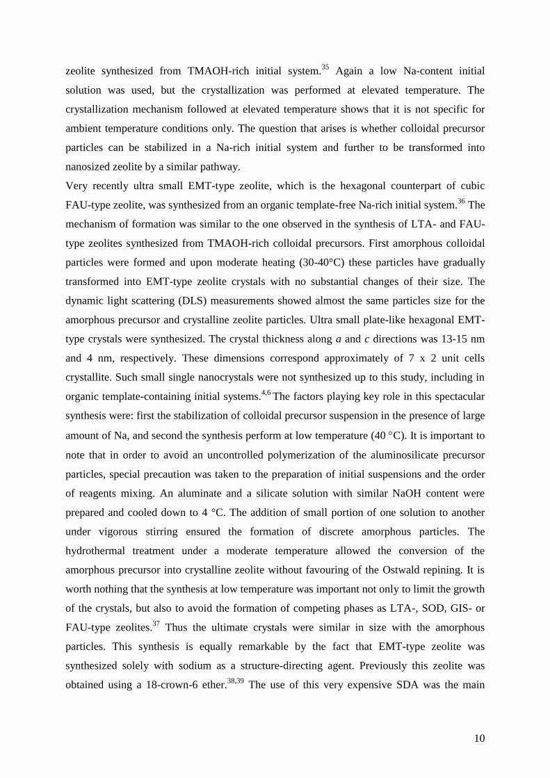
Figure 4. Nitrogen adsorption/desorption isotherms of (a) nanosized (particles of 10 nm) and
(b) micronsized (1 m) FAU type zeolites.
The strategy employed in the preparation of Na-EMT nanocrystals was further applied
in the synthesis of FAU-type zeolite. Again the stabilization of clear suspension of discrete
gel particles was amongst the key factors in the synthesis of organic template-free FAU-type
nanocrystals. The zeolite was synthesized at low temperature and also at 100 °C, which is
common for the synthesis of FAU-type zeolite. Both, zeolite Y and zeolite X were obtained
by varying the initial precursor composition. It is important to note that the zeolite Y particles
with size 10, 70 and 400 nm were synthesized.41
Products with high crystallinity and
0.0 0.2 0.4 0.6 0.8 1.00
200
400
600
800
1000
Vo
lum
e a
dso
rbed
(cm
3 g
-1)
Partial pressure (P/P0)
(a)
(b)
12
micropore volume equal to micron-sized crystals were obtained (Figure 4). It is remarkable
that ultra small zeolite crystals can be synthesized with such high crystallinity and preserved
micropore volume. The nanosized FAU zeolite exhibits a mix of Type I and IV isotherms
with a large H1 type hysteresis (Figure 4a). Such a feature is associated with textural pores
formed by the close packing of monodispersed and well-shaped nanosized crystallites; the
total pore volume of this sample is 1.54 cm3g
-1 while the micron-sized FAU
zeolite has 0.35
cm3g
-1. The results underline the high crystallinity and quality of the nanosized FAU crystals
prepared from organic template-free precursor suspension; this is not observed when
nanosized FAU crystals are synthesized in the presence of organic agents.35
The micropore
volume for both the nanosized and micronsized FAU zeolites is equal to 0.31 cm3g
-1; a value
typical of highly crystalline material. Another important feature of the nanosized FAU zeolite
is the high external surface area, 270 m2g
-1, opening opportunities for processes taking place
specifically on this part of the zeolite in comparison to 30 m2g
-1 for the micronsized FAU
zeolite. The prospects for this material, both in new advanced applications and traditional
zeolite uses, are bright.
2.3 Synthesis of bi-dimensional zeolites
A particular form of nanosized zeolites is so called zeolite nanosheets. In this case the crystals
are relatively large, but the thickness in one direction is one to several unit cells. Although not
nanometric in all three directions, the zeolite nanosheets are included in this review since they
exhibit particular properties that attracted a lot of interest over last 5 years.
The concept was first introduced by Corma and co-workers, who prepared such sheets by a
top-down approach.42
A MWW-type zeolite precursor was subjected to treatment to swallow
hexadecyltrimethylammonium and then delaminated by ultrasonic treatment. The same
approach was used to delaminate FER- and NSI-type zeolite precursors.43,44
An in situ method for generation of zeolite nanosheets was reported by Ryoo and co-
workers.45
A specific surfactant that comprised two quaternary ammonium groups separated
by C6H12 linkage is used to promote the formation of MFI-type zeolite. The two quaternary
ammonium groups are linked with long chain (C18H37) alkyl group that suppress the growth of
zeolite. Thus bi-dimensional zeolite crystals with thickness of one unit cell were synthesized.
The extension of synthesis reaction leads to the formation of ordered multilamelar structures
that are more appropriate for practical applications.46
Tsapatsis and co-workers demonstrated
13
the possibility to synthesize nanosheets by replacing the surfactant with bi-functional structure
directing agent.47
Shaped zeolite nanocrystals with three-dimensionally ordered mesoporous (3DOm) were
synthesized and successfully used for preparation of oriented films. The ordered mesoporous
carbon replicas were considered as attractive materials for carrying out confined synthesis of
zeolite crystals with a wide range of crystal morphologies and size-tuneable crystallites,
ranging between 10 and 40 nm.48–50
After these pioneering studies different structure directing agents and synthesis conditions
were explored to obtain zeolite sheets.51–55
The acidic properties and the potential of zeolite
sheets were tested in a number of catalytic reactions. For instance, the selective formation of
propylene from methanol over high-silica nanosheets was studied by Hu et al.56
The open
highly accessible active sites of zeolite sheets are particularly appropriate for bulky molecules
processing. Therefore, Lee et al. used such a catalyst for upgrading bio-oil derived from
biomass constituents.57
Zeolite nanosheets produced bio-oil of better quality in respect to a
reference Al-SBA-15 material. Tin-modified MFI nanosheets were used in Baeyer Villiger
Oxidation to Cyclic Ketones, where they exhibited drastically higher thermal and
hydrothermal stability in respect to a reference Sn-MCM-41 reference material.58
The list of the studies devoted on the preparation of zeolite sheets is much longer. Recent
reviews covered the main achievements in the preparation of zeolite nanosheets and zeolites
with bi-modal porosity (hierarchical zeolites) are available.59,60
3 Nanoclays
3.1 Properties of nanoclays
Clay nanomaterials have been used long before their structure was determined and
understood, as witnessed by the well-known example of “Mayan blue”, a nanocomposite of
indigo pigments and clay minerals.61–65
Although phyllosilicates like Laponite and Allophane
form nanosized particles, the term ‘nanoclays’ is generally used to denote clays whose
particles have at least one dimension, usually along the c axis, in the nanoscale range (1-100
nm). In this respect nanoclays resemble of zeolite nanosheets reviewed in the previous
section.
The clays used for the preparation of their nanosized analogies belong in most of the reported
cases to smectite group, which are also known as 2:1 phyllosilicates. The basic structural unit
of the latter consists of an octahedral sandwiched between two tetrahedral sheets. Because of
isomorphous substitution of Al3+
for Si4+
in the tetrahedral sheet and Mg2+
for Al3+
in the
14
octahedral sheet, the layers of many clay minerals carry a negative charge, which is balanced
by exchangeable alkali or alkali earth (Na+, Ca
2+) cations occupying the interlayer space. Clay
mineral layers have the tendency to be stacked in face-to-face arrangement leading to particles
with at least 10 stacked dehydrated layers.66,67
The aspect ratio, defined as the average ratio of
the width to the thickness of the particles (length/diameter ratio), is an important characteristic
for clay materials. Besides the ion exchange and adsorption capacity, this characteristic
determines the ability of a clay mineral to exfoliate. The terms “exfoliation” and
“delamination” are used to designate the separation between the planar faces of two adjacent
layers. The layers may eventually become completely independent from one another, with a
loss of crystallographic orientation. In this case each unit is then freely oriented in space.
There is not clear distinction between these two terms but as recommended in the Handbook
of Clay Science,66
in the present article we will use the term exfoliation to denote the stage
when the separated clay units are isotropically dispersed in a solvent.
The term nanoclays is generally confused with nanocomposites based on clays minerals. Clay
minerals have been used for a long time as nanofillers because they are easily available on all
the continents at relatively low cost. Sodium-Montmorillonite appears as the most interesting
smectite for bio-nanocomposite preparations, due to its tendency to swell in the presence of
water. This feature favours the exfoliation of the layer silicates, therefore facilitating the
access of biopolymers, which are hydrophilic compounds, to the interlayer space of the clay.
However, the reinforcing ability of natural clays is poor due to their large particle thickness
and low affinity to organic polymers. Therefore the conversion of natural clays in nanoclays
has been developed in order to make them compatible with polymers.66
Depending on the exfoliation conditions nanoclays have a particle size varying from a few
stacked layers up to much greater numbers. The degree of stacking strongly influences all
basic characteristics of nanoclays.
The cation exchange capacity (CEC) is among the most important properties of clay minerals.
The CEC represents the total amount of cations available for exchange at a given pH. It is
commonly expressed as meq/100 g of calcined clay. Typical CEC values of 2:1
phyllosilicates vary from 20 meq/100 for the group of Sepiolite-Palygorskite to 120 meq/100g
for Montmorillonite. The CEC is directly linked to the charge density of the layers, which
itself corresponds to the density of isomorphous substitutions.68
The ability to swell in water or in any polar solvent is also a defining property of the smectite
group of cationic clay minerals. Swelling is easily observable at the macroscopic level, by the
formation of gel-like phases when water is added to clay minerals, with a large increase in
15
volume as compared to the dry solid. A swelling index may be defined as the gel volume (in
mL) obtained per 10 g of clay mineral, and it may reach values of 25–30 mL/10.69,70
The basal surface atoms of a 2:1 clay mineral are the oxygens of the Si tetrahedron. In the
absence of isomorphous substitution and defect sites, the clay mineral surface is composed of
oxygen atoms involved in Si–O bonds. The latter have considerable covalent character and
the surface is hydrophobic. Hydrophilicity is introduced by isomorphous substitution,
involving the presence of the exchangeable cations, which are hydrophilic.71
The edges of
clay particles should exhibit a different reactivity from that of basal planes similar to that of
amphoteric oxides: surface group such as Si–OH may react both with a proton (thus giving
rise to Si–OH2+ in acidic conditions), or with a hydroxide (thus giving rise to Si–O
_ in basic
conditions). The amphoteric behaviour means that clay minerals have acid–base properties;
aqueous smectite dispersions have pH values from 7.5 for Montmorillonite to 10 for Laponite.
Smectites possess two types of specific surface area, external and internal. The external
surface area is the basal and lateral area constituted by layers stacking. The external surface
varies from 30 to 130 m2g
-1 depending on the size and aspect ratio of the considered clay
mineral. The internal surface corresponds to the basal surface of the layers that are “stuck”
together.72
For porous clay minerals, lenticular pores are often observed by TEM. The pores can be
accessible or not depending on physicochemical characteristics of a nanoclay, but also on the
preparation conditions such as for instance air- or freeze- drying processing.72,73
In addition, each clay material has particularities, which influence their properties and
determine their use. For instance smectites display a complex rheological behaviour when
dispersed in an aqueous medium. They form viscous gel phases at low volume fractions, a
property which is widely exploited in industries.74
3.2 Synthesis of nanoclays
The major problem in the use of natural nanoclays is the presence of a mixture of several
phases. To overcome this inconvenience synthetic methods for production of clay minerals
have been developed. A lot of research was devoted to the synthesis of well-crystalline clay
minerals with controlled composition and properties. The syntheses are performed under
hydrothermal conditions that involves, similarly to zeolites, a nucleation and a crystal growth
stages. The crystallization temperature is usually between 150 and 220 °C under autogenous
pressure. In particular cases the pressure can reach up to 1500 bars. Syntheses performed at
lower temperatures are not efficient and provide products of low quality.75
Similarly to
16
zeolites the initial system composition, the nature of the reactants and temperature/time
conditions are the key parameters governing the synthesis of clays. The major difference
between zeolite and clay syntheses is that the latter can be formed at low pH. A typical
example is the Montmorillonite that can be obtained in acidic medium.76–79
Low pH (2-6)
favours also the formation of 1:1 clay minerals such as kaolinite. In basic medium but at
moderate pH (7-10 for Al3+
, up to 12 for Fe3+
) usually 2:1 clay minerals such as smectites and
micas are formed. High pH (>12) favours the formation of zeolite in the presence of with Al3+
or aegerine in the presence of Fe3+
minerals.
The most often synthesized nanoclays is Laponite. The synthetic Laponite exhibits nanosized
dimensions in all three directions. The synthesis of Laponite involves combining sodium
silicate, magnesium, sodium and lithium salts to produce a hydrogel, which is subjected to
hydrothermal treatment at 200 °C.
67 Synthetic Laponite is a nanomaterial of disc-shaped
platelets, much smaller than naturally occurring counterpart. Compared to the natural product
the synthetic Laponite exfoliates much easier.
Nanosized Imogolite and Halloysite are also obtained in nanosized form, but similarly to
exfoliated clay minerals they are nanosized only along the c axis. A specific feature of
synthetic Imogolite and Halloysite is the formation of nanotubes. The first synthesis of
Imogolite nanotubes was reported by Farmer et al.80
The freeze dried product had a cotton-
like appearance.81
Halloysite nanoparticles with tubular structure have also been
synthesized.82
Halloysite is an aluminosilicate clay mineral with chemical formula of
Al2Si2O5(OH)4 H2O that belongs to the 1:1 phyllosilicates.
3.3 Nanoclays by exfoliation
There are several routes to exfoliate clay-type materials based on chemical and/or physical
processes. A largely used chemical process is based on the intercalation of polymers or
biopolymers. Several examples have been reported in literature, all of them require the
modification of the clay mineral by organic moieties in order to be more compatible with the
polymers.
Intercalation reactions can occur according to several routes depending on the nature of the
compound intercalated. Neutral organic molecules can be adsorbing by complexation of the
interlayer cations, ion–dipole interaction, acid–base reactions, charge transfer, van der Waals
forces or H-bonding. However, the use of charged species such as alkylammonium cations is
the most often used approach for pre-treatment of clays prior to the exfoliation step.
17
Grafting different organic moieties of clay surface is commonly used approach prior to
polymer intercalation.66
The most successful one is the grafting of organosilanes. The
covalent bonds established by condensation of some reactive groups on the organic chain with
the clay silanol surface groups or with the hydroxyl groups of octahedral layer as in the case
of kaolinite allowing the expansion of the interlayer space. Consequently, the surface
becomes hydrophobic and compatible with the polymers required to exfoliate. It differs from
ion exchange procedures by the creation of covalent bonds between the clay mineral surface
and the silsesquioxane. This results in irreversible organophilization, while intercalation by
ion exchange is usually reversible.
“Acid activation” or “acid dissolution” is reported to facilitate the exfoliation. During the acid
treatment, the surface of clay is protonated on the edge groups while introducing H3O+ cations
in the interlayer space. The acid treatment leaches metal ions, usually from the octahedral
layer first. It also increases the external surface area of the clay and introduces permanent
mesoporosity.83
Among the physical methods, ultrasonic radiation has showed excellent results in the
exfoliation of clay minerals compared to other mechanical methods. After sonication,
significant delamination and lateral size reduction in phyllosilicates occurs.84,85
The products
retained their crystalline structure and their morphology. However, when the interactions
between the layers and the interlayer space are strong enough, treatment by sonication is
clearly not sufficient and other physical or chemical methods should be applied.
Some patents reported on the use of supercritical fluids (SCFs) to exfoliate silicate minerals.
In this process, silicate mineral particles are dispersed in SCFs to diffuse fluids into the
interlayer space, followed by rapid depressurization or cooling to gasefy the SCFs. The
expansion of SCFs in the interlayers breaks the interlayer forces resulting in exfoliation of the
silicate minerals.86
A freeze-drying approach has been developed for the exfoliation of clay minerals.87
It consists
in the nucleation and propagation of ice crystals in the interlayer space.88–91
The expansion of
the layers and the cracks remained after the water sublimation change substantially the
properties of the clay mineral.
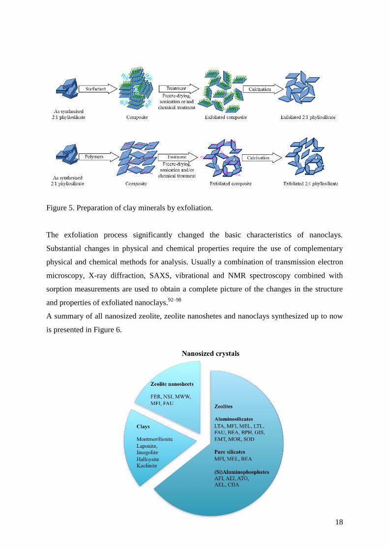
The generation of nanoclay minerals by combining chemical and physical treatments is
schematically presented in Figure 5. First, an organophilisation is performed by the
intercalation of cationic surfactants or polymers. The resulting composites are then freeze-
dried or sonicated in order to reach a high level of exfoliation.
18
Figure 5. Preparation of clay minerals by exfoliation.
The exfoliation process significantly changed the basic characteristics of nanoclays.
Substantial changes in physical and chemical properties require the use of complementary
physical and chemical methods for analysis. Usually a combination of transmission electron
microscopy, X-ray diffraction, SAXS, vibrational and NMR spectroscopy combined with
sorption measurements are used to obtain a complete picture of the changes in the structure
and properties of exfoliated nanoclays.92–98
A summary of all nanosized zeolite, zeolite nanoshetes and nanoclays synthesized up to now
is presented in Figure 6.

19
Figure 6. Synthetic nanosized zeolites (19 types), zeolite nanoshetes (5 types) and nanoclay (5
types).
4 Applications
4.1. Nanosized zeolites
The potential uses of nanosized zeolites and nanoclays are going much beyond traditional
catalysis, sorption and ion exchanges processes. These materials are considered in a number
of advanced applications, where namely bulk materials were used. Recent developments of
synthesis procedures for nanosized zeolites and their arrangements in thin-to-thick films,
membranes and hierarchical forms have pushed them into new and advanced applications that
have not been considered before.99–107
The following sections provide a comprehensive
overview of the use of nanosized zeolites for nanotechnology applications including sensors,
optical layers, medicine, pharmaceutical industry, cosmetics and food.
4.1.1 Sensors based on nanosized zeolites
The global market for gas sensors is expected to reach $ 2,512.4 million by 2020, according to
a market report by Grand View Research Inc., driven largely by new regulatory initiatives.108
The adsorption capacity, high-surface area and porosity, presence of mobile ions, and
catalytic activity make zeolites attractive candidates for chemical sensing/detection. In
addition, nanosized zeolites offer a fast response and facile processing that make them
particular appropriate for sensing devices. Consequently many fabrication methods were used
in the production of zeolite-based chemical sensors. Common techniques for preparation of
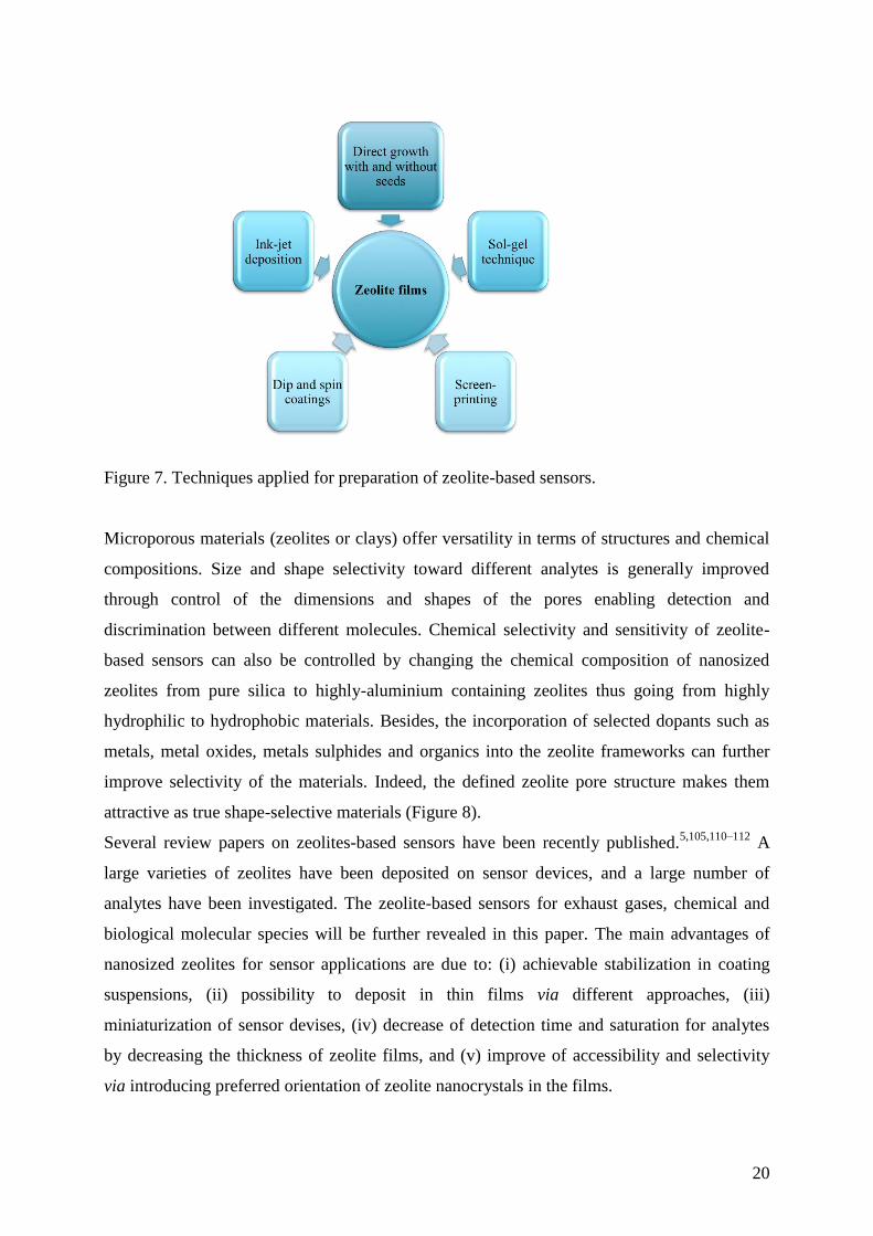
sensors include (1) direct growth with and without pre-seeding of the substrates, (2) sol-gel
techniques, (3) screen-printing, (4) dip and spin coatings, and very recently (5) ink-jet
deposition (Figure 7). A review analysing in depth the techniques for preparation of
microporous thin film is available.109
The primary challenge in utilizing nanosized zeolites
remains the ability to fabricate or pattern thin films with appropriate micro-nano-scale
features. The attractive properties of zeolites combined with the ability to choose and adept an
appropriate patterning technique is expected to enhance their utilization in device fabrication.
In the chemical sensors devices, the zeolite may act as a main functional element, which is the
case for sensor principles relying directly on conductive, adsorptive, or catalytic properties of
one specific type molecular sieve and its interaction with the analytes. The zeolites can also
be included as supplementary or secondary element in the device.
20
Figure 7. Techniques applied for preparation of zeolite-based sensors.
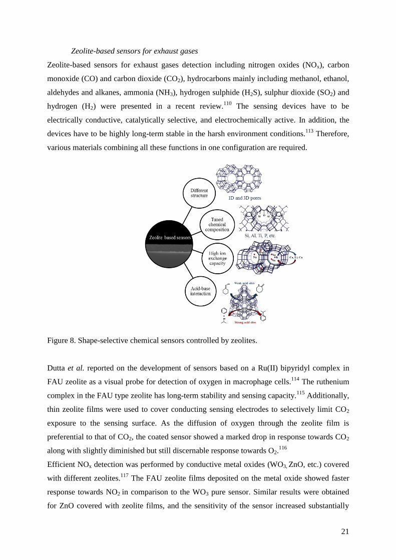
Microporous materials (zeolites or clays) offer versatility in terms of structures and chemical
compositions. Size and shape selectivity toward different analytes is generally improved
through control of the dimensions and shapes of the pores enabling detection and
discrimination between different molecules. Chemical selectivity and sensitivity of zeolite-
based sensors can also be controlled by changing the chemical composition of nanosized
zeolites from pure silica to highly-aluminium containing zeolites thus going from highly
hydrophilic to hydrophobic materials. Besides, the incorporation of selected dopants such as
metals, metal oxides, metals sulphides and organics into the zeolite frameworks can further
improve selectivity of the materials. Indeed, the defined zeolite pore structure makes them
attractive as true shape-selective materials (Figure 8).
Several review papers on zeolites-based sensors have been recently published.5,105,110–112
A
large varieties of zeolites have been deposited on sensor devices, and a large number of
analytes have been investigated. The zeolite-based sensors for exhaust gases, chemical and
biological molecular species will be further revealed in this paper. The main advantages of
nanosized zeolites for sensor applications are due to: (i) achievable stabilization in coating
suspensions, (ii) possibility to deposit in thin films via different approaches, (iii)
miniaturization of sensor devises, (iv) decrease of detection time and saturation for analytes
by decreasing the thickness of zeolite films, and (v) improve of accessibility and selectivity
via introducing preferred orientation of zeolite nanocrystals in the films.
21
Zeolite-based sensors for exhaust gases
Zeolite-based sensors for exhaust gases detection including nitrogen oxides (NOx), carbon
monoxide (CO) and carbon dioxide (CO2), hydrocarbons mainly including methanol, ethanol,
aldehydes and alkanes, ammonia (NH3), hydrogen sulphide (H2S), sulphur dioxide (SO2) and
hydrogen (H2) were presented in a recent review.110
The sensing devices have to be
electrically conductive, catalytically selective, and electrochemically active. In addition, the
devices have to be highly long-term stable in the harsh environment conditions.113
Therefore,
various materials combining all these functions in one configuration are required.
Figure 8. Shape-selective chemical sensors controlled by zeolites.
Dutta et al. reported on the development of sensors based on a Ru(II) bipyridyl complex in
FAU zeolite as a visual probe for detection of oxygen in macrophage cells.114
The ruthenium
complex in the FAU type zeolite has long-term stability and sensing capacity.115
Additionally,
thin zeolite films were used to cover conducting sensing electrodes to selectively limit CO2
exposure to the sensing surface. As the diffusion of oxygen through the zeolite film is
preferential to that of CO2, the coated sensor showed a marked drop in response towards CO2
along with slightly diminished but still discernable response towards O2.116
Efficient NOx detection was performed by conductive metal oxides (WO3, ZnO, etc.) covered
with different zeolites.117
The FAU zeolite films deposited on the metal oxide showed faster
response towards NO2 in comparison to the WO3 pure sensor. Similar results were obtained
for ZnO covered with zeolite films, and the sensitivity of the sensor increased substantially
22
too.118
Further the sensitivity of the sensors toward NOx was improved via using Pt doped
FAU zeolite crystals.119
The detection of CO and CO2 is a major target for sensing, which is based on either metal
oxides encapsulated in zeolites or in polymer/zeolite composites. The ferrierite (FER) layers
showed enhanced sensitivity towards CO at 300 °C, while semiconducting tungsten oxide and
chromium titanium oxide covered with zeolite were found to be sensitive to CO.120
Additionally a polyaniline/zeolite (Ca-LTA) sensor with high selectivity towards CO was
reported.121
The Ca-LTA zeolite exhibits more accessible pores than the K- and Na- forms,
and it has shown to have no great influence on the diffusion of CO through the conductive
polymer. Another report was on the utilization of poly(3,4-ethylenedioxythiophene) doped
with polystyrene sulfonic acid and ZSM-5 zeolite crystals. The sensitivity increased when the
Si/Al ratio of the ZSM-5 zeolite decreased.122
Recently, we also demonstrated that metal-
containing zeolite Beta with high selectivity towards CO in the presence of water can be
prepared.123,124
The high sensitivity of the Pt- and Pd-zeolite films to CO in very low
concentrations (2-100 ppm) in the presence of highly concentrated vapours of methanol or
pentane (400-4000 ppm) was reported.
A composite based on FAU zeolite crystals grown on a Metglas magnetoelastic strip for CO2
sensing has also been developed.125
In this case, a dry atmosphere was required to avoid the
interference with water. A similar device was prepared by the same group using hydrophobic
pure silica MFI type zeolite (silicalite-1). The device was used for sensing of CO2 in air thus
avoiding the use of nitrogen.
Zeolite-based sensors for hydrocarbons
A very extensive research has been devoted to the selective chemical sensing of hydrocarbons
using zeolite films. Many different framework-type microporous materials were used to sense
and capture hydrocarbons and volatile organic compounds (VOCs).126
The detection of VOCs
and hydrocarbon by zeolites layers considering mainly the detection limit and stability under
different conditions were reported.127,128
Tungsten and zinc oxides covered with zeolite film
have also been described as excellent ethanol sensors.118
The oxide sensors covered with LTA
type zeolite showed selectivity toward ethanol in comparison to isopropanol.117
The
conductive polymer (polypyrrole, polyamide-6) combined with K-LTA zeolite crystals were
applied for sensing of acetone, methylether ketone, methanol, and toluene.129
Selective
detection of n-butane in the presence of i-butane using zeolites was reported too.130
A zeolite
sensor showed the lowest detection for o-xylene (6 ppm), however the sensor suffered from a
23
lack of selectivity with cross-sensitivity to p-xylene. ZSM-5 crystals doped with Pt and Au
showed a great selectivity toward propane and propylene.131
The best performance of the
sensors was obtained using zeolite layers with high silica content, as well as using thin layers
with properly oriented crystals.132,133
High silica MFI film was reported for the detection of
isopropanol (16 ppm) and toluene (22 ppb). The fast response toward isopropanol than for
toluene was presented, which was explained with the smaller size of the former molecule.134
Metal containing (Pt, Fe) MFI and FAU type zeolite films showed a strong cross-sensitivity to
several analytes including NO, CO, CO2, ammonia, and hydrocarbons with low concentration
(1-5 ppm).135
The highest sensitivity toward ammonia was reported for Fe containing MFI
zeolite layers, which was explained by the high acidity, pore volume and surface area of the
zeolite. The preparation of reversible NH3 sensors comprised of HClO4-doped poly(3-
thiopheneacetic acid) polymer with FAU type zeolite crystals gave excellent results.136
Very
recently, the potential of Ag loaded FAU zeolites for NH3 detection based on the mobility
change of the silver cations (Ag+) in the presence of NH3 were presented.
137 No cross-
sensitivity for O2, CO, CO2 and propane was measured.
The effect of acidity and pore diameter of zeolites on the detection of base molecules
including water, acetonitrile, ammonia, benzonitrile, pyridine, aniline, and triethylamine was
investigated. It was pointed out that acid–base interaction is the major factor when molecular
diameter is smaller than the zeolite pore diameter (Figure 8). As for larger molecules
(benzonitrile, pyridine, aniline, and triethylamine) of which the molecular diameter is nearly
the pore diameter of zeolites, the response magnitudes were lower than those of the smaller
molecules.138
The sensitivity of the zeolite sensors to various base molecules as a function of
proton affinity, which can be regarded as a measure of base strength of the molecules have
been investigated. The sensitivity generally increased with the increase in the proton affinity.
In the case of large molecules such as pyridine and triethylamine, the sensitivity was strongly
dependent on the type of zeolites (MOR > BEA > MFI) indicating the significant contribution
of diffusion in the detection of larger molecules.139
Additional functionality in the zeolite
sensors has been introduced via using their ion-exchange capacity (Figure 8) and thus the
selectivity towards desired molecules was achieved. It was found that the propane sensor
effect takes place at the zeolite/Au electrode and was controlled by bulk diffusion of propane
into the pores of the zeolite.140
Zeolite-based biosensors
A human breath analysis was performed with twenty sensors connected in series for the
24
diagnosis of asthma since NO in breath increases from 5-10 ppb (normal state) to 100 ppb in
an asthmatic state.141
Zeolite nanocrystals were also applied to enrich and identify low-abundance
peptides/proteins, as well as to immobilize enzymes for bio sensing.142
Magnetically separable
nanosized zeolites were fabricated by combining their surface properties with the super
paramagnetism of Fe3O4 nanoparticles. Attractive perspectives in bio-applications such as
magnetically controllable biosensors and microfluidic biochips were considered.143
The development of oxygen-enhanced membrane assemblies suitable for use in sensors and
biosensors involving oxygen for the detection of the targeted substance was reported.144
Great attempts were focused at the evaluation of the adsorption efficiency of zeolites versus
toxins in different media.145–147
A removal of uremic toxins by adsorption process (e.g. during
the hemodialysis) using zeolites instead of conventional polymer membranes was
reported.146,148
The high toxicity of p-cresol or 1,4-methylphenol responsible for the
intoxication of patients, ultimately causing heart attack due to increased serum concentration,
was studied.149,150
Recently we studied the detection limit for uremic toxins such as o- and p- cresol with EMT,
LTL and MFI type zeolites assembled in thin films. Low concentrations of o- and p-cresol (5-
50 ppm) in the presence of water vapour (100 ppm) with the three-zeolite films were detected.
Between the three zeolites, the EMT shows the fastest response and saturation for o- and p-
cresol at low concentration (5 ppm), while the pure silica MFI film had the highest sorption
capacity due to its hydrophobic nature.
Impedance-based detection of highly toxic organophosphate nerve agent Sarin in the presence
of dimethylmethylphosphonate (DMMP) using FAU type sensors was investigated.151
The
surface of the zeolite had negligible effects on the decomposition of DMMP, while the intra-
zeolite cations motion facilitated by the DMMP reorientation was found to be responsible for
the decrease in the impedance. The cations with larger size in the supercage of FAU zeolite
showed an increase in impedance when exposed to DMMP, probably due to the repulsive
interactions that counteract the ionic motion. The change in the Na-FAU impedance was used
as a sensor for detection of organophosphates at elevated temperature. The selectivity to
DMMP in the presence of other molecules including methane and propane was found to
increased.151
The concept of physically and functionally integrating zeolite films with the long period fiber
grating (LPFG) had offered new opportunities in the development of a variety of chemical
sensors with high sensitivities for detection of chemical and biological molecular species. The
25
advantages of this type of sensors were mentioned to be operation simplicity, and
compactness, which are highly desirable for portable device applications.134
Optical devices to measure the change in optical reflectivity upon adsorption of chemical and
biological molecular species were designed too. In recent years, colour tuneable Bragg stacks
composed of various materials, including hydrogels, inorganic materials such as silicon,
titania, silica etc. were found to be responsive to a wide variety of analytes through optical
thickness variations of the specific nanostructures.152–154
The unique combination of chemical
and optical properties of nanosized zeolite crystals opened up the possibility of using them as
a part of tuneable Bragg stacks. The porous Bragg stacks are considered for chemical sensing
application with optical encoding. The idea behind is that the change of refractive index
and/or thickness of the layers in the Bragg stack will be due to ion-exchange, sorption, and
capillary condensation of molecules adsorbed in the pores which will result in a shift of the
high reflectance band and consequent change of their colour will be observed.155–157
Recently
we have shown that by using materials with high optical contrast such as GeSe2 (nH=2.65) and
nanosized zeolites (nL=1.19), it is possible an omnidirectional reflectance to be obtained.158
However finding porous materials with high refractive index is not a trivial task. To overcome
this problem, the preparation of quasi-omnidirectional (q-ODR) photonic crystals (PhC)
exhibiting high reflectance for all polarization but in narrower range of incident angles as
compared to omnidirectional PhC was considered. Additionally, the preparation of one-
dimensional photonic crystals based on MEL type nanosized zeolite and Nb2O5 was
reported.153
MEL type zeolite nanocrystals were used for both sensing and transducing
elements, while the sufficient optical contrast is ensured by sol-gel derived Nb2O5 film. The
particular choice of MEL type nanosized zeolite was made considering their hydrophobicity
and high sensitivity towards acetone. The sensing properties of the multi-layered structures
toward acetone were studied by measuring transmittance spectra prior and after vapour
exposure. The adsorption/desorption cycles of acetone with photonic crystals based on
different number of Nb2O5 and MEL zeolite layers are presented in Figure 9.
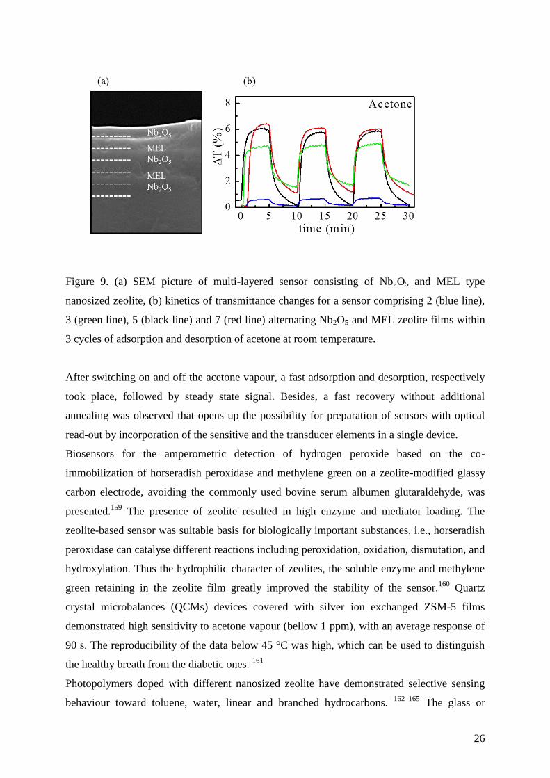
26
Figure 9. (a) SEM picture of multi-layered sensor consisting of Nb2O5 and MEL type
nanosized zeolite, (b) kinetics of transmittance changes for a sensor comprising 2 (blue line),
3 (green line), 5 (black line) and 7 (red line) alternating Nb2O5 and MEL zeolite films within
3 cycles of adsorption and desorption of acetone at room temperature.
After switching on and off the acetone vapour, a fast adsorption and desorption, respectively
took place, followed by steady state signal. Besides, a fast recovery without additional
annealing was observed that opens up the possibility for preparation of sensors with optical
read-out by incorporation of the sensitive and the transducer elements in a single device.
Biosensors for the amperometric detection of hydrogen peroxide based on the co-
immobilization of horseradish peroxidase and methylene green on a zeolite-modified glassy
carbon electrode, avoiding the commonly used bovine serum albumen glutaraldehyde, was
presented.159
The presence of zeolite resulted in high enzyme and mediator loading. The
zeolite-based sensor was suitable basis for biologically important substances, i.e., horseradish
peroxidase can catalyse different reactions including peroxidation, oxidation, dismutation, and
hydroxylation. Thus the hydrophilic character of zeolites, the soluble enzyme and methylene
green retaining in the zeolite film greatly improved the stability of the sensor.160
Quartz
crystal microbalances (QCMs) devices covered with silver ion exchanged ZSM-5 films
demonstrated high sensitivity to acetone vapour (bellow 1 ppm), with an average response of
90 s. The reproducibility of the data below 45 °C was high, which can be used to distinguish
the healthy breath from the diabetic ones. 161
Photopolymers doped with different nanosized zeolite have demonstrated selective sensing
behaviour toward toluene, water, linear and branched hydrocarbons. 162–165
The glass or
27
plastic substrates were covered with nanosized zeolites and exposed directly to the
environmental conditions.162–165
Absorption of gases (hydrocarbons, CO, CO2, NO, NO2) by
reflection gratings recorded in an acrylamide-based films doped with nanosized zeolites
resulted in reversible adsorption, and the resultant change in periodicity of the reflection
grating caused a shift in the reconstructed wavelength. The same principle has been adopted
for the development of liquid-phase alcohols and pH sensors. The sensing ability of
transmission gratings recorded in zeolite-doped acrylamide photopolymer layers have been
studied from the same group. Polymers doped with high silica zeolites (BEA, MFI and MEL)
were used for detection of alcohols, which are one of the most widely used groups of
chemicals in research and industry, as they have important application as solvents and play an
important role in the production of other chemicals. Therefore the development of sensitive
and selective alcohol sensors is of great interest. The developed optical sensors have
advantages over other sensor types such as semiconductor and electrochemical sensors due to
their potential for fast response times and high gas specificity, as well as allowing for label-
free, in-situ, real-time measurements.
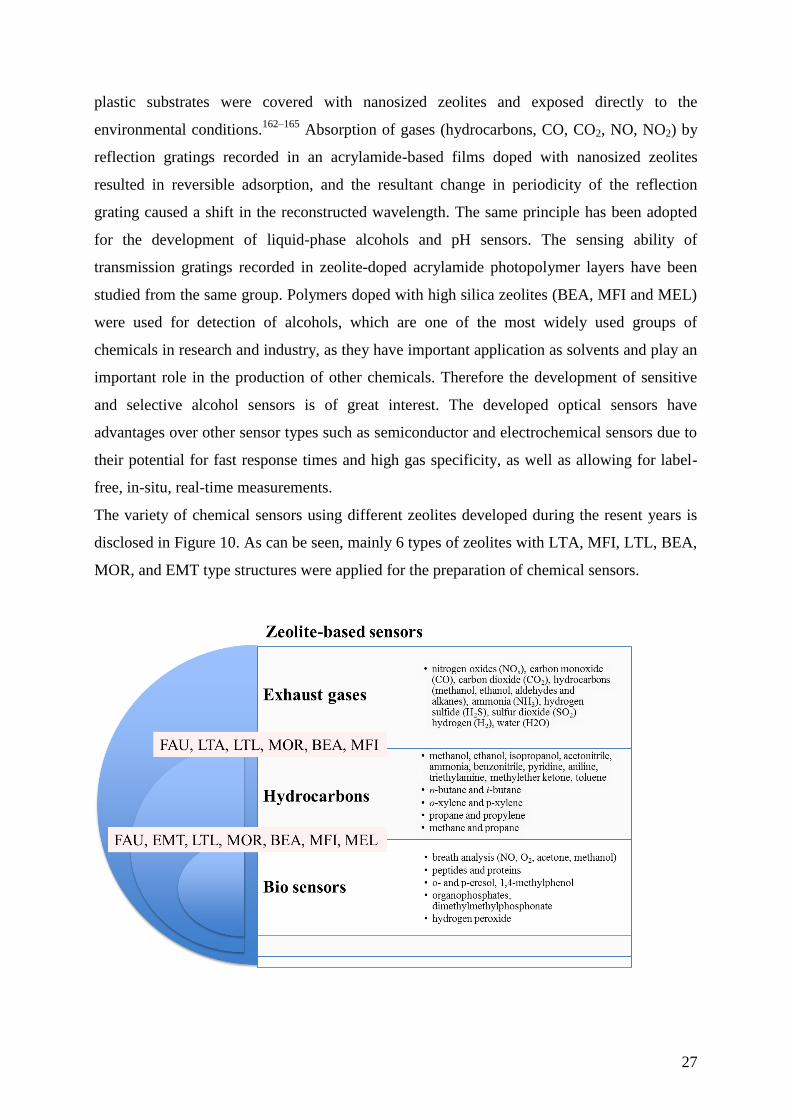
The variety of chemical sensors using different zeolites developed during the resent years is
disclosed in Figure 10. As can be seen, mainly 6 types of zeolites with LTA, MFI, LTL, BEA,
MOR, and EMT type structures were applied for the preparation of chemical sensors.
28
Figure 10. Chemical sensors based on nanosized zeolites.
4.1.2. Other devices based on nanosized zeolites
The fabrication of optical devices using nanosized zeolites attracted considerable attention
due to their rigid structure, high thermal stability and availability in different morphologies.
Zeolite films are used for preparation of optical quality films with a thickness in the range 50-
170 nm through spin-on or sol-gel deposition methods.99,100
The optical properties of the
zeolite films including refractive index, extinction coefficient, thickness, and hardness were
found to depend on the method of preparation and on the use of different binders with organic
vs. inorganic natures. The free pore volume of the zeolite films was found to depend on the
type of the crystalline structure and chemical nature of binders.
The encapsulation of organic dyes in the channels of one-dimensional LTL nanosized zeolite
for light harvesting and energy transfer was presented as an original model of zeolite-based
photovoltaic solar cell.166
Multilevel organization of zeolite crystals of 40 nm in hybrid films
for optoelectronic applications was reported from the same group. The functionalization of the
zeolite surface resulted in different organizations inside the zeolite cavities and on the outer
surface of the polymeric structure. The ordered LTL films allowed arrangement of single dye
loading in the zeolite cavities with a conjugated polymer. The hybrid films contained three
levels of organization: (1) organization of the dye molecules in the one dimensional LTL
zeolite channels, (2) organization of the zeolite crystals inside the polymer cavities, and (3)
micro- or nano-structuration of the polymer.167
The working principal was based on the
absorption of light by the dye-zeolite composite containing semi conductors, which is
responsible for the electron-hole formation. Another concept was based on TiO2 introduced in
the cages of FAU type zeolite and modified by organic acids or nitrogen doping.168
The
photovoltaic activity with the doped FAU zeolites were 20 times less compared to the pure
TiO2 based solar cells. However, considering the low loading of titanium dioxide within the
zeolite (4.8 %) this photovoltaic activity was considered as relatively strong.
New photovoltaic solar cells based on quantum dots (QDs = CdS and PdS) encapsulated in
the voids of FAU zeolite were presented.169
The results showed that the activity of devices
depends on the type of the QDs, and the best performance was obtained for FAU type crystals
doped with more than one type of quantum dots. In addition, Ag nanoparticles selectively
deposited in the channels or on the surface of nanosized zeolites with one- and three-
dimensional pore structures were considered for the same application.
29
Zeolite films were applied as corrosion resistant layers too.170
A review on the advanced
application of zeolites including semiconducting industry and space stations was published.103
The emphasis was given on the development of zeolite films as low-dielectric constant layers
for (i) future generation computer chips, (ii) environmentally benign corrosion-resistant
coatings for aerospace alloys, and (iii) hydrophilic and bacteriological coatings for gravity-
independent water separation in space stations. Numerous examples were presented for
zeolites exhibiting either hydrophobic or hydrophilic properties with low or high ion
exchange capacity, respectively. The zeolite layers show great mechanical stability and
hardness, which make them interesting for reverse osmosis membranes for seawater
desalination and proton exchange membrane fuel cells.
4.1.23 Biological and medical applications of nanosized zeolites
The increasing utilization of engineered nanoparticles for a range of application in fields such
as electronics, photonics and biomedicine demands an assessment of risk associated with
deliberate or accidental exposure. Major health concerns in relation to exposure to
nanoparticles arise from the same property which is of great importance for their potential
industrial applications, that is, the characteristic of high surface to mass ratio and potentially
high surface adhesion and reactivity compared to their larger counterparts. However, for
many applications the toxicology of the nanomaterial has not been addressed, although its
importance is now being realized. The cytotoxicity of inorganic nanoparticles such as zeolites,
metals and metal oxides is gradually recognized as an important issue in “nanosociety”.
Nanoparticles in general may have an adverse effect on human health compared to their
micrometer counterparts and therefore nano-safety regulations may become necessary.
Nanoparticles can enter the human body via: (i) the lungs where a rapid translocation through
the blood stream to vital organs is possible, including crossing the blood brain barrier (BBB),
and absorption, (ii) the intestinal tract, or (iii) the skin.
The interest in the toxicity of zeolite nanocrystals is very high due to their wide use in
industry and commercial products. One of the reasons for zeolite nanoparticles not to be
extensively studied for their biocompatibility is a relatively low occurrence of direct contact
with humans. A recent review on the synthesis and commercial potential of microporous
nano-materials for environmental applications connected with biological toxicity was
published.171
Studies on the biocompatibility of zeolite coated on commercially potent
titanium to pluripotent mouse embryonic stem cells were performed and it was found higher
adhesion and rate of cellular proliferation.172
However, since different cells demonstrate
30
different types and magnitude of responses on exposure to nanoparticles, it is important to
analyse the response of human cells on exposure to various nanosized zeolites.
The nanosized zeolites free of organic templates show no or very low toxicity to the HeLa
cells.173–175
The low cytotoxicity of the nanosized LTL type zeolite was reported.176
At high
concentrations a toxic response was observed, which was explained with the active positively
charged crystalline surface. However, the pure silica zeolites, or low Al containing zeolites
were reported to be non-toxic no matter the amount used. All measurements were carried out
with HeLa cancerous cell line.177
It was concluded that the nanosized zeolites had good
thermal and chemical stability due to their highly crystalline structures, and their toxicity
differences on HeLa cell had to be mainly depend on their sizes, shapes and surface properties
rather than the release of their toxic species.
Kihara et al. investigated the toxicities of nanosized zeolites with LTA, MFI (both ZSM-5 and
silicalite-1) and LTL type structures using human embryonic kidney 293 (HEK-293),
RAW264.7 macrophage and HeLa cells.177,
The toxicities of zeolite nanoparticles were found
to be dependent on their size (30 - 500 nm), Si/Al ratio and shape. Pure siliceous MFI type
zeolite (silicalite-1) is not toxic, but the other three aluminium containing nanosized zeolites
show a dose-dependent toxic manner.
The important factors considered for nanosized zeolites were particle size and crystalline
morphologies. Recent work showed a definitive preference for cellular necrosis rather than
programmed cell death via apoptotic mechanisms for the assayed conditions on particles with
different sizes and morphology.174
The zeolites with and without functionalization showed
different cytoxicity; the zeolites modified with amine were the most toxic.178
Further toxicity study of nanosized zeolites in different biological systems is needed. When
considering potential application of biocompatible materials, their interaction with free
radicals needs to be considered at an early exploratory stage. It is well known that free
radicals play a key role in the human body, and therefore a convenient screening protocol was
developed to identify the anti-oxidant and pro-oxidant properties of highly dispersed
nanosized zeolites. It was found that TS-1 zeolite (Ti- containing MFI type zeolite) exhibits
comparable or even better anti-oxidant activity in comparison to cerium oxide, while pro-
oxidant activity was demonstrated for several other zeolites.178
A clear finding was that the
toxicity of zeolites is dependent on the cell types used in the study. This implies that in vitro
studies have to be carried out carefully using representative methodology.
The properties of crystalline MFI type nanosized zeolites (sizes of 50 nm and 100 nm) and
their toxicological effects on human lung alveolar (A549) cells under in vitro conditions were
31
studied too. The biocompatibility of the nanoparticles by analysing their effect on the
biological properties of cellular proliferation, cytotoxicity and both extracellular and
intracellular free radical generation capacity was investigated.179
Live cell imaging showed
that the nanoparticles precipitated from the colloidal suspension of cell culture media as large
agglomerates, coming in contact with the cell surface through sedimentation. A cellular
proliferative capacity test showed that the zeolite nanoparticles exhibit no cytotoxicity. DNA
fragmentation analysis disclosed that the MFI nanosized zeolites cause genotoxicity in a
concentration dependent manner. The conclusion drawn from this study showed that the pure
silica MFI type nanosized zeolites were relatively non-reactive and cytotoxic to the human
alveolar cells. The observed oxidative stress, heightened mitochondrial activity and genotoxic
damages occur at environmentally non-relevant high dosage levels. Further studies are needed
to provide a better understanding of the role of mitochondrial and other cell in causing
oxidative stress and their possible relation with observed genotoxicity.
Another study reported on in vitro cytotoxicity test of nanosized zeolites with FAU and LTA
type structures.173
For the cytotoxicological experiments human alveolar epithelial cells
(A549), endothelial cells (EA.hy926) and differentiated monocytes (THP-1) were used. The
cytotoxic response of macrophages, epithelial and endothelial cells to nanosized zeolites
(sizes of 25-100 nm) was investigated, and no significant cytotoxic activity was detected for
doses up to 500 μg/mL for 24 h. More importantly, the nanosized zeolites showed lower
toxicity in comparison to amorphous silica, which make them interesting for future
applications in medicine and biochemical field. After exposure to 500 mg/ml nanosized
zeolites, up to 50 % of the administered nanoparticle mass was found to be associated with the
cells. However, only few nanosized zeolites were internalized in macrophage cells. There was
no evidence for uptake in endothelial and epithelial cells. This suggested that the internal
surface of microporous particles did not contribute to their biological activity.
Recently, we studied the toxicity of two types of ultra-small zeolites with LTL and EMT type
structures synthesized from organic template free systems.180
Both the LTL and EMT type
zeolites belong to the same group of molecular sieves; they have large pores (~7.3 Å) and low
silica content (Si/Al = 1.1 – 3.5). The cellular interactions with the two types of zeolite
nanocrystals were evaluated by cell viability, reactive oxygen species and cell life cycle-
assays. It was found that various concentrations of zeolites have negligible effects on the cell
life cycle. Moreover, the zeolites did not cause extensive oxidative stress on the cells.
Although it is seen that the zeolites extensively entered in the cells, there is no sign of toxicity
for all employed concentrations of ultra-small EMT and LTL zeolites. Additionally, no
32
abnormality during the DNA replication by the zeolite exposure was observed. Very
importantly, the EMT nanosized zeolite shows a high affinity for fibrinogen, moderate for
apoA-II and complement factor 3, and trace affinity for albumin, which is the most abundant
protein of human plasma. Thus the EMT nanosized zeolite can be considered as very
promising material for purification of fibrinogen and lipoproteins, while keeping the highest
concentration of albumin, when they are exposed to in vivo-like human plasma
concentration.181
Fibrinogen is a very complex, hydrophilic and bipolar molecule. In contrary
to fibrinogen, apolipoprotein C-III (APOC-III) is a simple, highly hydrophobic and non-polar
molecule. The selective sorption behaviour of human plasma proteins on EMT and FAU
nanosized zeolites with a size of 10 nm was presented.180,181
The protein composition of
corona in EMT and FAU zeolite nanocrystals was strongly dependent on the plasma protein
concentrations, zeolite external surface area and hydrophilicity, but it was independent of the
concentration of zeolites used. In conclusion, the property of the EMT and FAU zeolite
nanocrystals might be adapted to hemophilic patients (hemophilia A (F-VIII deficient) and
hemophilia B (F-IX deficient) with a risk of bleeding.
The stability of natural zeolite (clinoptilolite) in simulated digestion conditions and their
interactions with digestive media and with Caco-2 (human colon adenocarcinoma) cells was
investigated.182
The cytotoxicity test using colon cancer cells showed that the natural
clinoptilolite has cytotoxic effect against Caco-2 cells, and cytotoxicity did not significantly
change with respect to simulated digestion process.182
Non-toxic nanosized zeolites with
interesting physicochemical properties offer considerable advantages in everyday life uses as
stated by Cola et al.183
In summary, most of the toxicity tests were performed on five types nanosized zeolites (LTA,
MOR, LTL, FAU and MFI) (Table 1). The low cytotoxic activity of nanosized zeolites with
different sizes, compositions and shapes are expected to enlarge their future industrial and
medical applications. However, currently there are no standards for toxicological study of
nanosized zeolites. Therefore, the safety analysis of zeolites with different structures,
morphology, size and chemical compositions is of highest priority given the expected
worldwide use and the probability of exposure to human being.
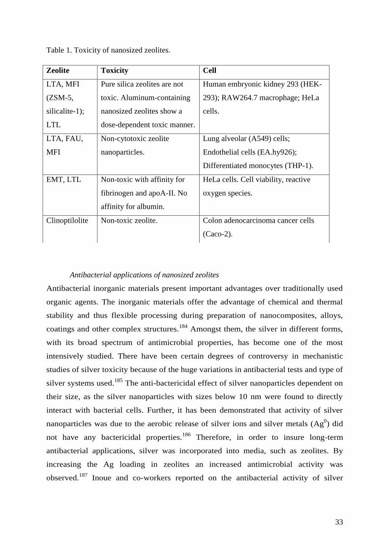
33
Table 1. Toxicity of nanosized zeolites.
Antibacterial applications of nanosized zeolites
Antibacterial inorganic materials present important advantages over traditionally used
organic agents. The inorganic materials offer the advantage of chemical and thermal
stability and thus flexible processing during preparation of nanocomposites, alloys,
coatings and other complex structures.184
Amongst them, the silver in different forms,
with its broad spectrum of antimicrobial properties, has become one of the most
intensively studied. There have been certain degrees of controversy in mechanistic
studies of silver toxicity because of the huge variations in antibacterial tests and type of
silver systems used.185
The anti-bactericidal effect of silver nanoparticles dependent on
their size, as the silver nanoparticles with sizes below 10 nm were found to directly
interact with bacterial cells. Further, it has been demonstrated that activity of silver
nanoparticles was due to the aerobic release of silver ions and silver metals (Ag0) did
not have any bactericidal properties.186
Therefore, in order to insure long-term
antibacterial applications, silver was incorporated into media, such as zeolites. By
increasing the Ag loading in zeolites an increased antimicrobial activity was
observed.187
Inoue and co-workers reported on the antibacterial activity of silver
Zeolite Toxicity Cell
LTA, MFI
(ZSM-5,
silicalite-1);
LTL
Pure silica zeolites are not
toxic. Aluminum-containing
nanosized zeolites show a
dose-dependent toxic manner.
Human embryonic kidney 293 (HEK-
293); RAW264.7 macrophage; HeLa
cells.
LTA, FAU,
MFI
Non-cytotoxic zeolite
nanoparticles.
Lung alveolar (A549) cells;
Endothelial cells (EA.hy926);
Differentiated monocytes (THP-1).
EMT, LTL Non-toxic with affinity for
fibrinogen and apoA-II. No
affinity for albumin.
HeLa cells. Cell viability, reactive
oxygen species.
Clinoptilolite Non-toxic zeolite. Colon adenocarcinoma cancer cells
(Caco-2).
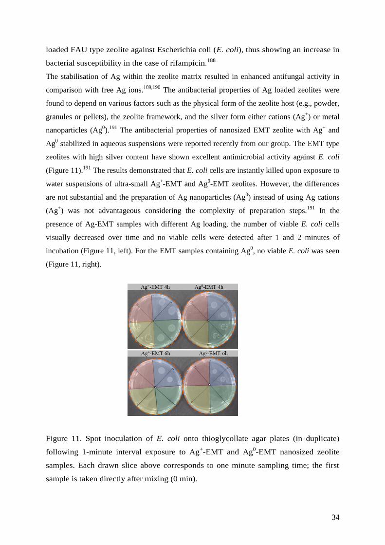
34
loaded FAU type zeolite against Escherichia coli (E. coli), thus showing an increase in
bacterial susceptibility in the case of rifampicin.188
The stabilisation of Ag within the zeolite matrix resulted in enhanced antifungal activity in
comparison with free Ag ions.189,190
The antibacterial properties of Ag loaded zeolites were
found to depend on various factors such as the physical form of the zeolite host (e.g., powder,
granules or pellets), the zeolite framework, and the silver form either cations (Ag+) or metal
nanoparticles (Ag0).
191 The antibacterial properties of nanosized EMT zeolite with Ag
+ and
Ag0 stabilized in aqueous suspensions were reported recently from our group. The EMT type
zeolites with high silver content have shown excellent antimicrobial activity against E. coli
(Figure 11).191
The results demonstrated that E. coli cells are instantly killed upon exposure to
water suspensions of ultra-small Ag+-EMT and Ag
0-EMT zeolites. However, the differences
are not substantial and the preparation of Ag nanoparticles (Ag0) instead of using Ag cations
(Ag+) was not advantageous considering the complexity of preparation steps.
191 In the
presence of Ag-EMT samples with different Ag loading, the number of viable E. coli cells
visually decreased over time and no viable cells were detected after 1 and 2 minutes of
incubation (Figure 11, left). For the EMT samples containing Ag0, no viable E. coli was seen
(Figure 11, right).
Figure 11. Spot inoculation of E. coli onto thioglycollate agar plates (in duplicate)
following 1-minute interval exposure to Ag+-EMT and Ag
0-EMT nanosized zeolite
samples. Each drawn slice above corresponds to one minute sampling time; the first
sample is taken directly after mixing (0 min).
35
The effect of silver-exchanged mordenite (MOR) against the growth of six fungi (Rhizopus
oryzae, Mucor circinelloides, Geotrichum candidum, Saccharomyces cerevisiae,
Debaryomyces hansenii and Zygosaccharomyces rouxii) was studied.192
It was shown that the
Ag released from the Ag-mordenite have effective antifungal action and being more effective
than the free silver ions in solutions. Interestingly, the Geotrichum candidum was the more
resistant, while the yeasts were more sensitive than filamentous fungi.
The antibacterial activity of cutting boards containing silver-based layers was studied
too.193
The humidity was found to have noticeable effect on bacteria killing
(staphylococci, Escherichia coli, Salmonella, Serratia and Listeria monocytogenes). If
no substantial water content was present during the use, then the antibacterial activity
of the silver was negligible. Additionally, patterned-zeolite films consisting of FAU
zeolite crystals were also found to be efficient towards eradication of E. coli.194
Another example is a nitrogen oxide (NO) loaded Zn-containing zeolite, which was
also, used as bi-functional anti-bacterial material.195
In summary, the nanosized zeolites would be very advantageous for antibacterial applications
having in mind that they can be deliver in different stable forms such as (i) suspensions, (ii)
discrete solid particles, (iii) deposited in films and membranes, and (iii) self-supported bodies
(diverse sizes and shapes) with very high external surfaces and great accessibility. Thus the
efficiency of the nanosized zeolites as antibacterial carrier is expected to be higher than their
micronsized equivalents.
Drug delivery application of nanosized zeolites
The immobilization of biomolecules on the surface of porous nanocrystals (zeolites and
mesoporous silica) is the focus of intense activity in biotechnology and biomedicine.
Especially, amine groups in proteins can be bound to the surface of zeolite particles and
stabilized electrostatically. In addition, nanosized zeolites show great promise as substrates to
immobilize proteins due to their small size and high functional groups density for further
grafting. The hydrophilic/hydrophobic nature, high stability and high sorption capacity of
nanosized zeolites make them important for further application in pharmaceutical industry and
medicine.
Drugs or contrasting agents can be confined within the pores or cages of zeolites. There are
cases where the drug of interest is attached to the external surfaces, and therefore the interest
is in the synthesis of zeolites with high external surface area. Examples of using nanosized
zeolites as multifunctional nanocarriers to deliver organic molecules into living cells were
36
presented.196
Both the surface and the internal pore volume of the LTL nanosized zeolite were
used after post synthesis functionalization, and the release of a drug molecule (4', 6-
diamidino-2-phenylindole, DAPI) from the zeolite pores was demonstrated.196
The application of nanosized zeolites for biomedical applications, controlled carry and
delivery of probes are envisioned for theranostic and gene therapy applications too.197–204
New
horizons in clinical medicine have been opened using nanosized zeolites, in particular the
multifunctional therapeutic delivery systems.205
Mostly, LTL type zeolite was applied as drug
carrier, and a great promise for immobilization of proteins due to their small size, sufficient
functional groups, surface hydrophilic/hydrophobic distribution and stable colloidal properties
was shown. The LTL nanosized zeolite were used as a trypsin-immobilizing carrier and
patterned in a poly(Me methacrylate) (PMMA) microfluidic channel for the fabrication of
enzyme microreactor. Due to the high trypsin immobilization capacity and unique surface
characteristics of LTL nanosized zeolite, the resulting trypsin microreactor had the efficient
digestion of proteins at low concentrations for a very short reaction time.206
On the other hand, zeolites have been investigated for encapsulation of drugs in their cavities
not only in the channels like in the case of the LTL nanosized zeolite. The ability of storing
and releasing of a variety of drugs in the FAU type zeolite was reported.207–209
The FAU
zeolite demonstrated a slow release for various anthelmintic drugs.210
FAU type zeolite was
used as a slow-release carrier for some anthelmintic drugs. Besides, doxorubicin and paraquat
molecules have been successfully loaded in the FAU zeolite functionalized with a
trimethylsilyl. The controlled release of sulfonamide antibiotics adsorbed on the FAU type
zeolite was investigated by changing the pore mouth opening, modifying the external surface,
and the nature of the grafted functional groups.201
The controlled release of ibuprofen
encapsulated in de-aluminated FAU zeolite was another example of an efficient drug
delivery.211,212
As indicated before, the nanosized zeolites possess both the micropores and textural
mesopores that are potentially large enough to accommodate a wide-range of different drug
molecules, such as sulfonamide antibiotics, ibuprofen, doxorubicin, mitoxantrone, aspirin and
5-Fluorouracil (5-FU).202,213–216
The ibuprofen immobilized in the FAU zeolite showed better
release behaviour than the observed for mesoporous silica (MCM-41 type material).
Therapy with classical drugs (5-FU) has often-toxic side effects. Thus, the encapsulation of 5-
FU in zeolites may contribute to reduce these side effects and may allow an oral
administration as well. Encapsulation may allow drugs to be released in a controlled way to
the cancer area, thus preventing toxicity. The anticancer drug (5-FU) was encapsulated in

37
FAU and LTL zeolites.217
In vitro drug study revealed the release of 80 - 90 % of 5-FU in the
first 10 min. The drug delivery system (DDS) was also evaluated using two human colorectal
carcinoma cell lines (HCT-15 and RKO). Unloaded zeolites presented no toxicity to both
cancer cells, while all DDS showed an important 5-FU effect on the cell viability. DDS based
on zeolites were able to increase the efficiency of 5-FU, a widely used anticancer drug.217
These systems should be further explored in other cancer models, e.g. in vivo models, to
confirm their efficiency. The same group reported on the preparation of zeolite with
anticancer drug cyano-4-hydroxycinnamic acid (CHC) and demonstrated its efficacy against
colorectal carcinoma cells.204,218
The anticancer drugs were loaded into the pores of zeolite Y
(FAU), zeolite A (LTA) and zeolite Beta (BEA), and they have shown a noticeable cytotoxic
effect when applied to cancer cells.204,219
However, the zeolites alone reveal no toxicity to
cancer cells, while, importantly, anticancer drug-zeolite led to an inhibition of cell viability up
to 585-fold when compared to the non-encapsulated drug.
Larsen and co-workers investigated the loading and release of Aspirin and 5-FU from the
FAU type zeolite with different SiO2/Al2O3 ratios of 5, 30, and 60.215
They found that the
aluminum content in the zeolite plays a critical role in determining the drug loading and
release profiles. Aspirin and 5-fluorouracil loadings were found to increase with increasing
aluminum content in the FAU zeolite crystals. In this release system, the aluminum has an
integral role in governing the interactions between zeolite crystals and drug molecules. The
zeolite with the highest aluminum content may interact with the drug molecule species by
hydrogen bonding of the carbonyl oxygen atoms with the zeolite hydroxyl groups as well
through coordination of the drug molecules to the aluminum atoms.215
Aspirin was bonded to
silanol sites via hydrogen bonding on FAU zeolite with higher SiO2/Al2O3 ratio. The drug
release study from the zeolite matrix at neutral (pH = 7.4) in aqueous media revealed nearly
complete release of the aspirin from the FAU zeolite with high alumina loading, while partial
release of the aspirin for high silica FAU zeolite was observed. The partial release of the drug
was attributed to the increased hydrophobicity of the materials due to an increase of van der
Waals interactions. The nature of intermolecular aspirin-zeolite interactions was studied in
detail by spectroscopic methods.218,220
Zeolites X (FAU type) and A (LTA type) were used for encapsulation and controlled release
of Ketoprofen drugs.213
The ketoprofen was encapsulated in the zeolite matrix by a soaking
procedure, and it was shown that no drug release in acid conditions is observed, thus
suggesting that the material has great potential for controlled delivery of ketoprofen.213
Immobilization of famotidine (sulphonamide derivative) on two zeolite structures (MFI and
38
MOR) revealed that the amount of famotidine adsorbed was generally greater in ion-
exchanged-zeolites, suggesting that these cations act as binding sites for the famotidine
molecules. The equilibrium and kinetic characteristics of the drug on these materials were
studied by varying the incubation time, the famotidine concentration and the pH of the
solution.221
Zeolite crystals modified with polyaniline were prepared and used for controlled
release of an antibiotic (cefuroxime). The zeolite doped with the antibiotic showed longer
time for cefuroxime discharge, and about 42 % remained in the zeolite after 6 h. 222
Natural enzymes frequently suffer limitations in various applications due to denaturation.
They are easily deactivated under extreme temperatures and/or pH. For these reasons, zeolites
were also used as drug carriers and delivery systems for a number of enzymes such as glucose
oxidase and antibodies.223,224
Exploring the possibility of using zeolites in tissue engineering scaffolds is a potential
approach in regenerative medicine because of their biocompatibility and cation exchange
ability.225
A novel method to synthesize formaldehyde cross-linked gelatine/zeolite scaffolds
by lyophilisation technique was reported. Swelling and degradation of scaffolds were tuned
by adjusting the concentrations of zeolites in the composite scaffolds. All these results
suggest that they can be further investigated for their application in tissue engineering.
The preparation of novel zeolite-hydroxyapatite composite coating on titanium alloys and
stainless steel for regenerative medicine was presented recently.226
The zeolite-hydroxyapatite
coating is super-hydrophilic and outperforms in corrosion resistance tests in aggressive
sodium chloride media and phosphate buffer solution. Moreover, the zeolite coating
eliminated the elastic modulus mismatch between the coating and the bone. In addition, it
was observed faster post-surgical recovery.
Medical imaging based on nanosized zeolites
Nanosized zeolites modified with folic acid are recognized for application in targeting
treatment of damaged cells. Manufacturing of such multifunctional systems in a way to carry
bioactive molecules, able to recognize specific targets in living cells, represents an
achievement towards the development of highly selective tools for both diagnosis and
therapeutics. A review summarizing the applications of zeolites nanocrystals for
immobilization of enzymes and their use in magnetic resonance imaging was published.227
The materials possessing unique properties that enable radionuclides and contrast agent
metals to be concentrated and thus to be more efficient in magnetic resonance imaging (MRI)
and nuclear medicine were identified.228
The possible application of zeolites in positron
39
emission tomography (PET) was also studied. The ion-exchange capabilities of FAU type
zeolites make it an ideal vehicle for ionic metal MRI contrast agents including Gd3+
and
positron-emitting radionuclides (68
Ga3+
). Besides, the zeolite crystals were subjected to
surface modification via trialkoxysilane condensation, which add biological selective moieties
to the zeolite surface, and led to the concentration of the crystals at specific locations in the
body.
Zeolites functionalized with molecular targeting vectors (e.g., peptides, aptamers) that enable
specific high-affinity binding to in vivo molecular targets (such as cell surface receptors) were
investigated. In addition, these materials possess unique properties that enable radionuclides
and contrast agent metals to be concentrated for applications in magnetic resonance imaging
(MRI) and positron emission tomography (PET). Some luminescent molecules such as
iridium (Ir) and europium (Eu) complexes and organic dye in LTL nanosized zeolite were
introduced and used for biology imaging.198,229,230
The light responsive components in
combination with the small zeolites (diameter of 30 nm) have been very successfully prepared.
The LTL nanosized zeolites were loaded with an emitter, and the surface was functionalized
with a phthalocyanine derivative and amino groups, thus non-covalent binding of the hybrid
nanomaterial to the bacterial surface was achieved. The results demonstrated that hybrid
nanomaterials efficiently produced singlet oxygen and adhered to bacterial surfaces, leading
to targeting, labelling, and photoinactivation capabilities against antibiotic-resistant bacteria.
Additionally, iron oxide nanoparticles feature in vivo applications including magnetic
resonance, imaging contrast enhancement, tissue repair, drug delivery and treatment of
hypothermia. Pure silica (silicalite-1) nanosized crystals containing magnetite were prepared
and the sorption of hemoglobin (178 mg) at neutral pH 7 was studied.231
The possibility of
combining the properties of the zeolite with the magnetite super paramagnetic nanoparticles
was demonstrated.
Vectorized therapies for tumours based on inorganic nanomaterials have recently been
proposed to be an exciting approach. The nanoparticles with vectorezation properties were
used to deliver different drugs in tumours.232
The advances in the design of nanoscale stimuli-
responsive systems able to control (i) drug bio-distribution under variation of temperature,
and (ii) in the presence of magnetic field, ultrasound, light or electric pulses, or (iii) under
changes of the pH, enzyme concentration or redox gradients, were described.233
Additionally
they were used for synchronized MRI and focused-ultrasound enhanced brain-tumour drug
delivery.
40
Gas storage in nanosized zeolites for medical applications
Another applications for nanosized zeolites include storage and controlled delivery of gases in
biological systems. Reversible adsorption is a good method of storing a desire gas in a
material from which a controlled delivery is required. The application of oxygen
concentrators for the supply of medical oxygen in remote places has important cost saving
and supply implications. The zeolite trapped nitrogen from compressed room air, and thus
concentrates the oxygen in the expressed gas. At delivery flow rates of 2 to 5 l per minute,
oxygen concentrations greater than 80 % was maintained.234
The possibility of using zeolites
for oxygen concentrator was demonstrated for packed LTA zeolite, which produces
humidified O2-enriched air because H2O is adsorbed and recovered together with the
oxygen. Oxygen concentrators for the delivery of supplemental oxygen in remote high-
altitude areas were constructed using LTL zeolite.234
Another paper reports on the use of both
5A (Ca-LTA-type) and partially Ag- and Li-exchanged 13X (FAU- type) zeolites, wherein the
oxygen purity was greater than 90%. The zeolite carriers for oxygen with reduced size are
interesting for personal and medical applications.235
Besides, the efficient oxygen supply is a
continuing challenge for the fabrication of successful tissue engineered constructs. Therefore
an effort to enhance oxygen delivery via using fluorinated zeolite particles embedded in three-
dimensional polyurethane scaffolds was exploited.236
The concentration of oxygen
substantially increased via treatment of the zeolite with 1H,1H,2H,2H-
perfluorodecyltriethoxysilane. The fluorinated-zeolite particles embedded in 3-D tissue
engineered polyurethane scaffolds had high homogeneity. It was shown that polyurethane
scaffolds with zeolites could be used for tissue engineering with enhanced oxygen delivery to
the cells. Artificial mechanical red blood cells (respirocytes) mimic the oxygen and the carbon
dioxide function as transport of red blood cells. They can deliver more than 200 times oxygen
per unit volume than a natural red blood cell.237
The tuneable nature of NO release from ion exchanged LTA zeolites extends their potential
applications to tubing and instruments used in procedures such as bypass surgery and renal
replacement therapy.238
As specific potential, the use for NO-releasing zeolite in the
cardiovascular arena centers on the prevention of thrombosis on artificial surfaces coming in
contact with blood was considered. Additionally the zeolite loaded with NO was studied for
application in catheter implantations, and stents. It was demonstrated that between different
ion exchanged zeolites, the Co-containing LTA zeolite has the highest affinity for NO even at
low pressure. The amount of NO released from the zeolite in water solution was measured
electrochemically and between the numerous samples, the Co-exchanged zeolites released the
41
most NO while the Na-LTA zeolite released least NO. It was shown that the amount of NO
released by the zeolite was dependent not only on the type of transition metals but also on the
amount and state of the metal present. In conclusion, the loading with NO in the zeolite was
simple and the release occurred by a reaction with water. Besides, the NO releasing zeolites
inhibit platelet aggregation and adhesion in plasma, which is of potential importance in
prevention of thrombus formation. The application of NO-release zeolites will extend far
beyond the confines of reduced thrombogenicity of prosthetic grafts, catheters, and
extracorporeal blood conduits. Other medical uses including antimicrobial adjuncts to prevent
infection, for example, in chronically implanted catheters and in wound dressings for
enhanced angiogenesis were considered from the same group.
Typically applied nitric oxide induces T-Lymphocyte infiltration in human skin, but minimal
inflammation.239
Nanoparticles of natural zeolite (clinoptilolite) were applied on textiles as
protective finishes material. The nanosized zeolite crystals with increased active surface area
scatter the light and thus provide a textile with a high level UV protection.
The main biological and medical applications for zeolites are summarised in Table 2.
Table 2. Drugs and gases loaded in zeolites for biological systems and medical applications.
Zeolite Drug Application
LTL 4',6-diamidino-2-
phenylindole
Multifunctional therapeutic delivery systems.
LTL Trypsin Enzyme microreactor.
FAU Anthelmintic compounds Slow release carrier.
FAU Doxorubicin, Paraquat
molecules, Antibiotics,
Sulfonamide, Ibuprofen,
Mitoxantrone, Aspirin,
5-Fluorouracil
Efficient drug loading. Controlled release.
LTA,
FAU
Ketoprofen Controlled and selective delivery (no release in
acidic media).
LTA,
FAU,
BEA
Anticancer drug, Cyano-4-
hydroxycinnamic acid,
5-Fluorouracil
Inhibition of cell viability. High efficiency to
colorectal carcinoma cells.
42
MFI,
MOR
Famotidine (sulphonamide
derivative), Cefuroxime
High loading dependent on pH and zeolite/drug
concentrations.
FAU Natural enzymes, Glucose
oxidase, Antibodies
Stabilization at various pH and temperatures.
LTA,
Co-LTA
O2, CO2, NO
Storage and controlled delivery of gases.
Treatment of cardiovascular centers. Prevention
of thrombosis on artificial surfaces.
Antimicrobial adjuncts for prevention of
infections.
4.1.4 Nanosized zeolites in cosmetics and food
The nanosized zeolites are very attractive and can be safely applied in processes to (1) absorb
unpleasant odours, (2) for skin cleaning, (3) protect from harmful emissions, microorganisms
and rheumatic problems, (4) speed the healing of skin wounds and (5) regulate the alkalinity.
Zeolite crystals containing di- and poly- valent metal cations were used for delivery of
important biological compounds to skin and hair care agents. Additionally, delivery of certain
metals, including trace metals, and certain zirconium aluminum amino acids that provide
antiperspirant benefits were introduced in the cosmetic compounds.240
Another application is
based on ion exchange zeolites mainly with Cu and Zn as fillers in cosmetics. This cosmetic
has curative-and-prophylactic anti-inflammatory effect on facial skin. Besides, the cosmetics
with zeolite fillers were used to remove toxins and to decrease greasiness and to deliver
necessary micro- and macro-elements and to regenerate the skin.241
Another trends are in the
preparation of self-heating cosmetic compositions containing zeolites.242,243
The zeolites were
loaded with hydrocarbon oil, caprylic triglyceride, cyclomethicone, and ethanol. During the
application of the zeolite, the skin maintains warm and increases softness and reduces
dryness. In most cases, non-aqua exothermic cosmetics containing LTA type zeolites was
used, which effectively eliminates impurities on skin and promotes blood circulation during
the exothermal process of treatment. This cosmetic was applied to skin and hair, where the
temperature was increased of 16 °C for 1 min under mixing with water. The cosmetics were
loaded with 25 wt.% zeolites and dimethylsiloxane-methyl(polyoxyethylene-
polyoxypropylene)siloxane copolymer. These exothermic cosmetics contained ethoxylate
alcohols (C5-25) and hydrophilic non-aqua solvents including glycerine, propylene glycol and
1,3-butylene glycol. The exothermic cosmetic based on zeolites was prepared in the form of
creams, lotions, gels, packs, pastes and deodorant sticks. Besides, it was found that the self-

43
heated cosmetic was very efficient for massage and face cleansing. Modified LTA zeolite
with polyethylene glycol, 1,3-butylene glycol, glycerine, Ca(OH)2, H3PO4, and surfactants
was used. The LTA type zeolite was modified with magnesium or calcium halide as well. The
halides were used to adjust the pH of the cosmetics at a value suitable for the skin. Cosmetic
compounds containing zeolites were applied as cleansing creams, hand cleaner, hand lotions,
cream shampoo and toothpaste compounds too.244
Metal-exchanged zeolites (copper, silver) exhibit good antibacterial activity for both
gram-negative and gram-positive bacteria, and this effect was developed in a short
period of time. A new antifungal material, Ag-zeolite, was combined with a
commercial tissue conditioner as a successful agent against Candida albicans growth
and acid production. The inhibitory effects of these materials on fungal growth were
decreased by the presence of a saliva coat, particularly with zeolite in a tissue
conditioner. A silver zeolite mouth rinse was prepared by suspending zeolite powder
into phosphate-buffered saline at a concentration of 3 wt. %.
In addition to the use of zeolites in cosmetics and household items, they were applied in the
beer industry as energy supply.245
The concept for heat supplying is based on water adsorption
heat within zeolites.
The zeolites were also used as ingredients in packing materials for food.246
The metal (silver,
zinc, copper) containing zeolites were incorporated into food contact polymers to enhance
their mechanical strength and to prevent the degradation of plastics. Besides, the silver
containing zeolites worked as effective antimicrobial compounds, decrease ethylene oxidation
play the role of oxygen scavenger, thus increase the lifetime of food.
Zeolite nanocrystals were used as selective adsorbents for palm oil purification.247,248
The
zeolites with electrostatic charged framework tend to allow polar compounds such as moisture
contaminant and carbonyl oxidized products to be adsorbed and trapped in their pores, while
do not interact with the non-polar oil molecules. Recently, the use of zeolite nanocrystals as
eco-friendly anti-oxidant additives was reported.249
Zeolite nanocrystals with different
framework structures (LTL, EMT and FAU) were found to possess distinctive anti-oxidation
behaviour in palm oil during heat treatment (180 C) for a long period of time (40 days). It
was shown that the delay of palm oil oxidation is the result of free radicals inactivation, C=C
bonds stabilization and also adsorption of polar primary (hydroperoxides) and secondary
(carboxylic acids) oxidation products by zeolites. The FAU nanosized zeolite prepared from
template-free precursor suspensions in different ion exchange forms (Li, Na, K, and Ca) were
used as additives in palm oil (Figure 12). It was clearly shown that only 1 % zeolite
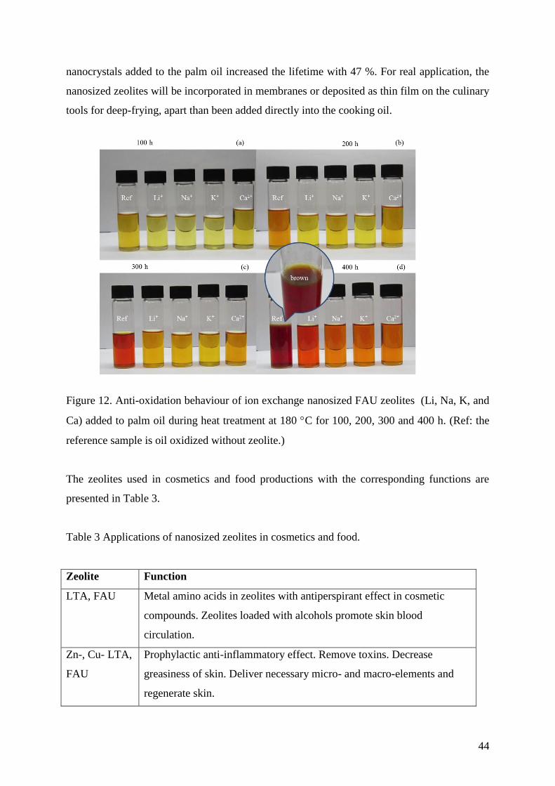
44
nanocrystals added to the palm oil increased the lifetime with 47 %. For real application, the
nanosized zeolites will be incorporated in membranes or deposited as thin film on the culinary
tools for deep-frying, apart than been added directly into the cooking oil.
Figure 12. Anti-oxidation behaviour of ion exchange nanosized FAU zeolites (Li, Na, K, and
Ca) added to palm oil during heat treatment at 180 C for 100, 200, 300 and 400 h. (Ref: the
reference sample is oil oxidized without zeolite.)
The zeolites used in cosmetics and food productions with the corresponding functions are
presented in Table 3.
Table 3 Applications of nanosized zeolites in cosmetics and food.
Zeolite Function
LTA, FAU Metal amino acids in zeolites with antiperspirant effect in cosmetic
compounds. Zeolites loaded with alcohols promote skin blood
circulation.
Zn-, Cu- LTA,
FAU
Prophylactic anti-inflammatory effect. Remove toxins. Decrease
greasiness of skin. Deliver necessary micro- and macro-elements and
regenerate skin.
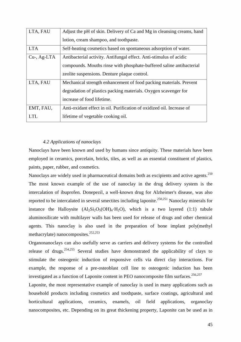
45
LTA, FAU Adjust the pH of skin. Delivery of Ca and Mg in cleansing creams, hand
lotion, cream shampoo, and toothpaste.
LTA Self-heating cosmetics based on spontaneous adsorption of water.
Cu-, Ag-LTA Antibacterial activity. Antifungal effect. Anti-stimulus of acidic
compounds. Mouths rinse with phosphate-buffered saline antibacterial
zeolite suspensions. Denture plaque control.
LTA, FAU Mechanical strength enhancement of food packing materials. Prevent
degradation of plastics packing materials. Oxygen scavenger for
increase of food lifetime.
EMT, FAU,
LTL
Anti-oxidant effect in oil. Purification of oxidized oil. Increase of
lifetime of vegetable cooking oil.
4.2 Applications of nanoclays
Nanoclays have been known and used by humans since antiquity. These materials have been
employed in ceramics, porcelain, bricks, tiles, as well as an essential constituent of plastics,
paints, paper, rubber, and cosmetics.
Nanoclays are widely used in pharmaceutical domains both as excipients and active agents.250
The most known example of the use of nanoclay in the drug delivery system is the
intercalation of ibuprofen. Donepezil, a well-known drug for Alzheimer's disease, was also
reported to be intercalated in several smectites including laponite.250,251
Nanoclay minerals for
instance the Halloysite (Al2Si2O5(OH)4·H2O), which is a two layered (1:1) tubule
aluminosilicate with multilayer walls has been used for release of drugs and other chemical
agents. This nanoclay is also used in the preparation of bone implant poly(methyl
methacrylate) nanocomposites.252,253
Organonanoclays can also usefully serve as carriers and delivery systems for the controlled
release of drugs.254,255
Several studies have demonstrated the applicability of clays to
stimulate the osteogenic induction of responsive cells via direct clay interactions. For
example, the response of a pre-osteoblast cell line to osteogenic induction has been
investigated as a function of Laponite content in PEO nanocomposite film surfaces.256,257
Laponite, the most representative example of nanoclay is used in many applications such as
household products including cosmetics and toothpaste, surface coatings, agricultural and
horticultural applications, ceramics, enamels, oil field applications, organoclay
nanocomposites, etc. Depending on its great thickening property, Laponite can be used as in
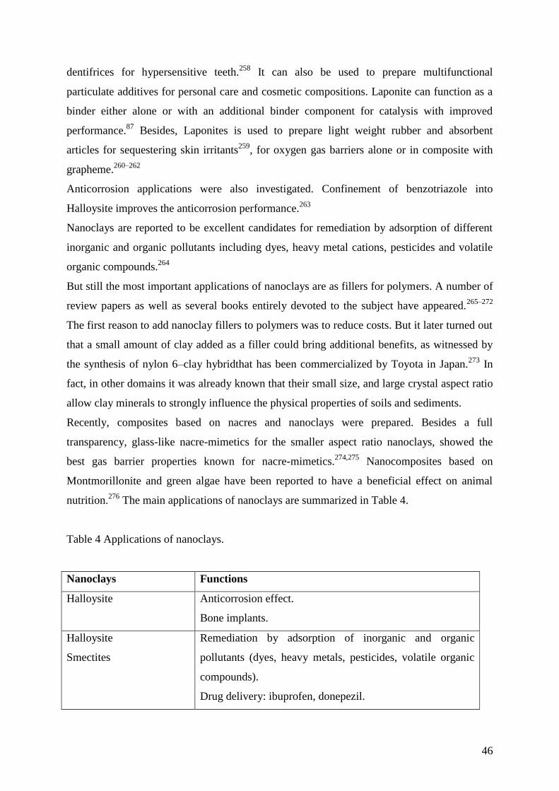
46
dentifrices for hypersensitive teeth.258
It can also be used to prepare multifunctional
particulate additives for personal care and cosmetic compositions. Laponite can function as a
binder either alone or with an additional binder component for catalysis with improved
performance.87
Besides, Laponites is used to prepare light weight rubber and absorbent
articles for sequestering skin irritants259
, for oxygen gas barriers alone or in composite with
grapheme.260–262
Anticorrosion applications were also investigated. Confinement of benzotriazole into
Halloysite improves the anticorrosion performance.263
Nanoclays are reported to be excellent candidates for remediation by adsorption of different
inorganic and organic pollutants including dyes, heavy metal cations, pesticides and volatile
organic compounds.264
But still the most important applications of nanoclays are as fillers for polymers. A number of
review papers as well as several books entirely devoted to the subject have appeared.265–272
The first reason to add nanoclay fillers to polymers was to reduce costs. But it later turned out
that a small amount of clay added as a filler could bring additional benefits, as witnessed by
the synthesis of nylon 6–clay hybridthat has been commercialized by Toyota in Japan.273
In
fact, in other domains it was already known that their small size, and large crystal aspect ratio
allow clay minerals to strongly influence the physical properties of soils and sediments.
Recently, composites based on nacres and nanoclays were prepared. Besides a full
transparency, glass-like nacre-mimetics for the smaller aspect ratio nanoclays, showed the
best gas barrier properties known for nacre-mimetics.274,275
Nanocomposites based on
Montmorillonite and green algae have been reported to have a beneficial effect on animal
nutrition.276
The main applications of nanoclays are summarized in Table 4.
Table 4 Applications of nanoclays.
Nanoclays Functions
Halloysite
Anticorrosion effect.
Bone implants.
Halloysite
Smectites
Remediation by adsorption of inorganic and organic
pollutants (dyes, heavy metals, pesticides, volatile organic
compounds).
Drug delivery: ibuprofen, donepezil.
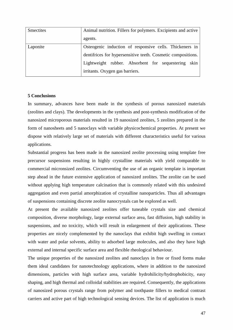
47
Smectites Animal nutrition. Fillers for polymers. Excipients and active
agents.
Laponite Osteogenic induction of responsive cells. Thickeners in
dentifrices for hypersensitive teeth. Cosmetic compositions.
Lightweight rubber. Absorbent for sequestering skin
irritants. Oxygen gas barriers.
5 Conclusions
In summary, advances have been made in the synthesis of porous nanosized materials
(zeolites and clays). The developments in the synthesis and post-synthesis modification of the
nanosized microporous materials resulted in 19 nanosized zeolites, 5 zeolites prepared in the
form of nanosheets and 5 nanoclays with variable physicochemical properties. At present we
dispose with relatively large set of materials with different characteristics useful for various
applications.
Substantial progress has been made in the nanosized zeolite processing using template free
precursor suspensions resulting in highly crystalline materials with yield comparable to
commercial micronsized zeolites. Circumventing the use of an organic template is important
step ahead in the future extensive application of nanosized zeolites. The zeolite can be used
without applying high temperature calcination that is commonly related with this undesired
aggregation and even partial amorphization of crystalline nanoparticles. Thus all advantages
of suspensions containing discrete zeolite nanocrystals can be explored as well.
At present the available nanosized zeolites offer tuneable crystals size and chemical
composition, diverse morphology, large external surface area, fast diffusion, high stability in
suspensions, and no toxicity, which will result in enlargement of their applications. These
properties are nicely complemented by the nanoclays that exhibit high swelling in contact
with water and polar solvents, ability to adsorbed large molecules, and also they have high
external and internal specific surface area and flexible rheological behaviour.
The unique properties of the nanosized zeolites and nanoclays in free or fixed forms make
them ideal candidates for nanotechnology applications, where in addition to the nanosized
dimensions, particles with high surface area, variable hydrohilicity/hydrophobicity, easy
shaping, and high thermal and colloidal stabilities are required. Consequently, the applications
of nanosized porous crystals range from polymer and toothpaste fillers to medical contrast
carriers and active part of high technological sensing devices. The list of application is much
48
longer as can be seen in the previous sections and continues growing. Most of these
applications require customized nanosized materials. Hence, we believe that future efforts will
be oriented to better control of the properties and more efficient production of nanosized
zeolites and nanoclays. Their processing to ultimate product will also be further optimized.
The nanosized zeolites and nanoclays will be more and more exposed to human via
inhalation, skin or swallowing. Therefore information on the toxicity of nanosized zeolites
and nanoclays prior further application is of great interests. The low cytotoxic activity of
nanosized zeolites with different sizes, compositions and shapes were reported and this is
expected to enlarge their future industrial and medical applications. Zeolite materials have
been considered for medical use due to their stability in biological environments. Based on the
advantages such as small size and controlled release, zeolites and clays modified with
targeting molecules are recognized for application in controlled drug treatments. Additionally
nanosized zeolites are also investigated as magnetic resonance imaging agents, and enzyme-
immobilizing carriers. The multifunctional capabilities of zeolites extend applications to
fluorescent imaging and heat therapy. Nanosized zeolites have been studied as dietary
supplements, antimicrobial agents, or adjuvants in anticancer therapy. They are expected to
play an ever-increasing role in in new products in medical, food and cosmetic industries.
Similarly to zeolites the nanoclays are used for adsorption of inorganic and organic pollutants
(dyes, heavy metals, pesticides, volatile organic compounds), for drug deliver and bone
implants, as fillers for polymers and lightweight devices, anticorrosion layers and oxygen gas
barrier.
Although the nanosized zeolites and nanoclays are used in similar fields, they are
complementary in their used due to the specificity of their structures and related
physicochemical properties. Therefore even larger “cooperation” between these two families
of nanosized porous materials can be expected in the future.
Acknowledgements
The financial support from ZEOXY Emergence and the Lower-Normandy Region (France) is
acknowledged. VV acknowledges the financial support from the MicroGreen project (ANR-
12-IS08-0001-01).
References
1 R. M. Barrer, Zeolites and clay minerals as sorbents and molecular sieves, London,
1978.
49
2 R. W. Tschernich, Zeolites of the world, Phoenix, 1992.
3 R. M. Barrer, Hydrothermal chemistry of zeolites, Academic Press, London, 1982.
4 L. Tosheva and V. P. Valtchev, Chem. Mater., 2005, 17, 2494–2513.
5 S. Mintova, J.-P. Gilson and V. Valtchev, Nanoscale, 2013, 5, 6693–703.
6 V. Valtchev and L. Tosheva, Chem. Rev., 2013, 113, 6734–6760.
7 C. S. Cundy and P. A. Cox, Microporous Mesoporous Mater., 2005, 82, 1–78.
8 D. P. Serrano and R. van Grieken, J. Mater. Chem., 2001, 11, 2391–2407.
9 C. Madsen and C. J. H. Jacobsen, Chem. Commun., 1999, 673–674.
10 B. Wang, H. Z. Ma and Q. Z. Shi, Chinese Chem. Lett., 2002, 13, 385–391.
11 H. Wang, B. a. Holmberg and Y. Yan, J. Am. Chem. Soc., 2003, 125, 9928–9929.
12 D. P. Serrano, J. Aguado, J. M. Escola, J. M. Rodríguez and Á. Peral, Chem. Mater.,
2006, 18, 2462–2464.
13 J. Aguado, D. P. Serrano and J. M. Rodríguez, Microporous Mesoporous Mater., 2008,
115, 504–513.
14 C. Jo, J. Jung, H. S. Shin, J. Kim and R. Ryoo, Angew. Chemie - Int. Ed., 2013, 52,
10014–10017.
15 A. K. Jamil, O. Muraza, M. Sanhoob, T. Tago, H. Konno, Y. Nakasaka and T. Masuda,
J. Anal. Appl. Pyrolysis, 2014, 110, 338–345.
16 Z. Cheng, S. Han, W. Sun and Q. Qin, Mater. Lett., 2013, 95, 193–196.
17 G. A. Thompset, W. C. Conner and K. S. Yngvesson, ChemPhysChem, 2013, 7, 295–
304.
18 F. M. Shalmani, R. Halladj and S. Askari, Powder Technol., 2012, 221, 395–402.
19 S. Askari, S. Miar Alipour, R. Halladj and M. H. Davood Abadi Farahani, J. Porous
Mater., 2012, 20, 285–302.
20 P. Pal, J. K. Das, N. Das and S. Bandyopadhyay, Ultrason. Sonochem., 2013, 20, 314–
321.
21 M. B. Yue, N. Yang, W. Q. Jiao, Y. M. Wang and M. Y. He, Solid State Sci., 2013, 20,
1–7.
22 X. Meng, Y. Zhang, M. Meng and W. Pang, in Proceedings 9th Int. Zeolite
Conference, eds. R. von Ballmoos, J. B. Higgins and M. M. J. Treacy, Butterworth-
Heinemann, Stoneham, MA, 1993, pp. 297–304.
23 B. . Schoeman, J. Sterte and J.-E. Otterstedt, Zeolites, 1994, 14, 110–116.
24 B. J. Schoeman, J. Sterte and J.-E. Otterstedt, Zeolites, 1994, 14, 208–216.
50
25 B. J. Schoeman, J. Sterte and J.-E. Otterstedt, J. Colloid Interface Sci., 1995, 170, 449–
456.
26 A. E. Persson, B. J. Schoeman, J. Sterte and J.-E. Otterstedt, Zeolites, 1994, 14, 557–
567.
27 B. J. Schoeman and O. Regev, Zeolites, 1996, 17, 447–456.
28 J. N. Watson, A. S. Brown, L. E. Iton and J. W. White, J. Chem. Soc. Faraday Trans.,
1998, 94, 2181–2186.
29 N. D. Hould and R. F. Lobo, Chem. Mater., 2008, 20, 5807–5815.
30 T. M. Davis, T. O. Drews, H. Ramanan, C. He, J. Dong, H. Schnablegger, M. a
Katsoulakis, E. Kokkoli, A. V McCormick, R. L. Penn and M. Tsapatsis, Nat. Mater.,
2006, 5, 400–408.
31 S. Kumar, Z. Wang, R. Lee Perm and M. Tsapatsis, J. Am. Chem. Soc., 2008, 130,
17284–17286.
32 C. E. A. Kirschhock, V. Buschmann, S. Kremer, R. Ravishankar, C. J. Y. Houssin, B.
L. Mojet, R. A. Van Santen, P. J. Grobet, P. A. Jacobs and J. A. Martens, Angew.
Chemie - Int. Ed., 2001, 40, 2637–2640.
33 C. E. A. Kirschhock, S. P. B. Kremer, J. Vermant, G. Van Tendeloo, P. A. Jacobs and
J. A. Martens, Chem. - A Eur. J., 2005, 11, 4306–4313.
34 S. Mintova, N. H. Olson, V. Valtchev and T. Bein, Science (80-. )., 1999, 283, 958–
960.
35 S. Mintova, N. Olson and T. Bein, Angew. Chem. Int. Ed. Engl., 1999, 38, 3201–3204.
36 E.-P. Ng, D. Chateigner, T. Bein, V. Valtchev and S. Mintova, Science (80-. )., 2012,
335, 70–73.
37 E.-P. Ng, J.-M. Goupil, A. Vicente, C. Fernandez, R. Retoux, V. Valtchev and S.
Mintova, Chem. Mater., 2012, 24, 4758–4765.
38 Eur. Pat., 364352, 1990.
39 G. Lischke, E. Schreier, B. Parlitz, I. Pitsch, U. Lohse and M. Woettke, Appl. Catal. A
Gen., 1995, 129, 57–67.
40 V. Georgieva, A. Vicente, C. Fernandez, R. Retoux, A. Palcic, V. Valtchev and S.
Mintova, Cryst. Growth Des., 2015, 150225102729005.
41 H. Awala, J.-P. Gilson, R. Retoux, P. Boullay, J.-M. Goupil, V. Valtchev and S.
Mintova, Nat. Mater., 2015.
42 A. Corma, V. Fornes, S. B. Pergher, T. L. M. Maesen and J. G. Buglass, 1998, 396,
353–356.
51
43 A. Corma, U. Diaz, M. Domine and V. Fornés, Angew. Chem. Int. Ed. Engl., 2000, 39,
1499–1501.
44 A. Corma, V. Fornés and U. Díaz, Chem. Commun., 2001, 2642–2643.
45 M. Choi, K. Na, J. Kim, Y. Sakamoto, O. Terasaki and R. Ryoo, Nature, 2009, 461,
246–249.
46 K. Na, W. Park, Y. Seo and R. Ryoo, Chem. Mater., 2011, 23, 1273–1279.
47 X. Zhang, D. Liu, D. Xu, S. Asahina, K. a. Cychosz, K. V. Agrawal, Y. Al Wahedi, a.
Bhan, S. Al Hashimi, O. Terasaki, M. Thommes and M. Tsapatsis, Science (80-. ).,
2012, 336, 1684–1687.
48 W. Fan, M. a Snyder, S. Kumar, P.-S. Lee, W. C. Yoo, A. V McCormick, R. Lee Penn,
A. Stein and M. Tsapatsis, Nat. Mater., 2008, 7, 984–991.
49 W. C. Yoo, S. Kumar, Z. Wang, N. S. Ergang, W. Fan, G. N. Karanikolos, A. V.
McCormick, R. L. Penn, M. Tsapatsis and A. Stein, Angew. Chemie - Int. Ed., 2008,
47, 9096–9099.
50 Z. Wang, P. Dornath, C. C. Chang, H. Chen and W. Fan, Microporous Mesoporous
Mater., 2013, 181, 8–16.
51 R. Kore, R. Srivastava and B. Satpati, Chem. - A Eur. J., 2014, 20, 11511–11521.
52 Q. Zhang, H. Yang and W. Yan, RSC Adv., 2014, 4, 56938–56944.
53 S. Lin, L. Shi and T. Yu, J. Phys. Chem. C, 2015, 119, 1008–1015.
54 D. Xu, Z. Jing, F. Cao, H. Sun and S. Che, Chem. Mater., 2014, 26, 4612–4619.
55 Y. Seo, K. Cho, Y. Jung and R. Ryoo, ACS Catal., 2013, 3, 713–720.
56 S. Hu, J. Shan, Q. Zhang, Y. Wang, Y. Liu, Y. Gong, Z. Wu and T. Dou, Appl. Catal.
A Gen., 2012, 445-446, 215–220.
57 H. W. Lee, S. H. Park, J.-K. Jeon, R. Ryoo, W. Kim, D. J. Suh and Y.-K. Park, Catal.
Today, 2014, 232, 119–126.
58 H. Y. Luo, L. Bui, W. R. Gunther, E. Min and Y. Román-Leshkov, ACS Catal., 2012,
2, 2695–2699.
59 K. Na, M. Choi and R. Ryoo, Microporous Mesoporous Mater., 2013, 166, 3–19.
60 K. Möller and T. Bein, Chem. Soc. Rev., 2013, 42, 3689–3707.
61 F. Bergaya and G. Lagaly, In Clay-Based Polymer, Nanocomposites, Chantilly, VA,
1997.
62 A. Doménech, M. T. Doménech-Carbó, M. Sánchez del Río, M. L. Vázquez de
Agredos Pascual and E. Lima, New J. Chem., 2009, 33, 2371–2379.
52
63 B. Hubbard, W. Kuang, A. Moser, G. A. Facey and C. Detellier, Clays Clay Miner.,
2003, 51, 318–326.
64 M. Jose-Yacaman, L. Rendon, J. Arenas and M. C. Serra Puche, Science (80-. )., 1996,
273, 223–225.
65 H. Van Olphen, Science, 1966, 154, 645–646.
66 J. F. L. F. Bergaya, M. Jaber, in Rubber-Clay Nanocomposites: Science, Technology,
and Applications, ed. Maurizio Galimberti, John Wiley & Sons, Inc, 2011, pp. 3–44.
67 M. Jaber, S. Komarneni and C.-H. Zhou, Handbook of Clay Science, Elsevier, 2013,
vol. 5.
68 L. Ammann, F. Bergaya and G. Lagaly, Clay Miner., 2005, 40, 441–453.
69 G. E. Christidis, A. E. Blum and D. D. Eberl, Appl. Clay Sci., 2006, 34, 125–138.
70 K. Norrish, Discuss. Faraday Soc., 1954, 18, 120–134.
71 R. W. Mooney, A. G. Keenan and L. A. Wood, J. Am. Chem. Soc., 1952, 74, 1371–
1374.
72 L. G. and J. J. F. F. Annabi-Bergaya, M. I. Cruz, Mineral. Soc., 1979, 249–258.
73 L. J. Michot and F. Villiéras, Handbook of Clay Science, Elsevier, 2006, vol. 1.
74 L. Bailey, H. N. W. Lekkerkerker and G. C. Maitland, Soft Matter, 2015, 11, 222–236.
75 J. H. Rayner, in Colloques Internationaux du CNRS, Genèse et Synthèse des Argiles,
1962, pp. 124–127.
76 A. Decarreau, Bull. Minéralogie, 1980, 103, 579–590.
77 A. Decarreau, S. Petit, P. Vieillard and N. Dabert, Eur. J. Mineral., 2004, 16, 85–90.
78 F. J. Huertas, J. Cuadros, F. Huertas and J. Linares, Am. J. Sci., 2000, 300, 504–527.
79 J. T. Kloprogge, S. Komarneni and J. E. Amonette, Clays Clay Miner., 1999, 47, 529–
554.
80 V. C. Farmer, A. R. Fraser and J. M. Tait, J. Chem. Soc. Chem. Commun., 1977, 462–
463.
81 S. Mukherjee, V. M. Bartlow and S. Nair, Chem. Mater., 2005, 17, 4900–4909.
82 US pat., 4098676 A, 1978.
83 F. Kooli, Y. Z. Khimyak, S. F. Alshahateet and F. Chen, Langmuir, 2005, 21, 8717–
8723.
84 C. Lam, K. Lau, H. Cheung and H. Ling, Mater. Lett., 2005, 59, 1369–1372.
85 L. A. Pérez-Maqueda, F. Franco and J. L. Pérez-Rodríguez, J. Eur. Ceram. Soc., 2005,
25, 1463–1470.
86 M. Daneshvar and E. Gulari, J. Supercrit. Fluids, 1992, 5, 143–150.
53
87 US pat., 7055566 B2, 2006.
88 E. C. Bate-Smith, Nature, 1965, 206, 538–539.
89 C. B. Agbugba, B. A. Hendriksen, B. Z. Chowdhry and M. J. Snowden, Colloids
Surfaces A Physicochem. Eng. Asp., 1998, 137, 155–164.
90 M. Birgersson, O. Karnland and U. Nilsson, Phys. Chem. Earth, Parts A/B/C, 2008, 33,
S527–S530.
91 D. M. Anderson and A. R. Tice, Soil Sci. Soc. Am. J., 1971, 35, 47–54.
92 D. F. Eckel, M. P. Balogh, P. D. Fasulo and W. R. Rodgers, J. Appl. Polym. Sci., 2004,
93, 1110–1117.
93 A. Bafna, G. Beaucage, F. Mirabella and S. Mehta, Polymer (Guildf)., 2003, 44, 1103–
1115.
94 S. I. Marras, I. Zuburtikudis and C. Panayiotou, Eur. Polym. J., 2007, 43, 2191–2206.
95 H.-M. Park, X. Li, C.-Z. Jin, C.-Y. Park, W.-J. Cho and C.-S. Ha, Macromol. Mater.
Eng., 2002, 287, 553–558.
96 E. Balnois, S. Durand-Vidal and P. Levitz, Langmuir, 2003, 19, 6633–6637.
97 R. S. Alvim and C. R. Miranda, Phys. Chem. Chem. Phys., 2015, 17, 4952–4960.
98 S. Aoyama, Y. T. Park, C. W. Macosko, T. Ougizawa and G. Haugstad, Langmuir,
2014, 30, 12950–12959.
99 Z. Wang, H. Wang, A. Mitra, L. Huang and Y. Yan, Adv. Mater., 2001, 13, 746–749.
100 Z. B. Wang, A. Mitra, H. T. Wang, L. M. Huang and Y. Yan, Adv. Mater., 2001, 13,
1463–1466.
101 A. Mitra, L. Huang and S. Li, Ind. Eng. Chem. Res., 2004, 43, 2946–2949.
102 C.-H. Chen, S.-Y. Li, A. S. T. Chiang, A. T. Wu and Y. S. Sun, Sol. Energy Mater. Sol.
Cells, 2011, 95, 1694–1700.
103 C. M. Lew, R. Cai and Y. Yan, Acc. Chem. Res., 2010, 43, 210–219.
104 C.-S. Wu, J.-Y. Liao, S.-Y. Fang and A. S. T. Chiang, Adsorption, 2009, 16, 69–74.
105 X. Xu, J. Wang and Y. Long, Sensors, 2006, 6, 1751–1764.
106 K. Sahner, G. Hagen, D. Schönauer, S. Reiß and R. Moos, Solid State Ionics, 2008,
179, 2416–2423.
107 Y. Zheng, X. Li and P. K. Dutta, Sensors, 2012, 12, 5170–5194.
108 http://www.grandviewresearch.com/industry-analysis/gas-sensors-market, 2014.
109 S. Mandal, H. L. Williams and H. K. Hunt, Microporous Mesoporous Mater., 2015,
203, 245–258.
110 D. Banerjee, Z. Hu and J. Li, Dalton Trans., 2014, 43, 10668–10685.
54
111 T. Bein and S. Mintova, Zeolites and Ordered Mesoporous Materials: Progress and
Prospects, Elsevier, 2005, vol. 157.
112 M. Zaarour, B. Dong, I. Naydenova, R. Retoux and S. Mintova, Microporous
Mesoporous Mater., 2014, 189, 11–21.
113 K. Sahner and R. Moos, Sens. Lett., 2008, 6, 808–811.
114 P. Payra and P. K. Dutta, Microporous Mesoporous Mater., 2003, 64, 109–118.
115 T. A. Ruda-Eberenz, A. Nagy, W. J. Waldman and P. K. Dutta, Langmuir, 2008, 24,
9140–9147.
116 J.-C. Yang, J. V. Spirig, D. Karweik, J. L. Routbort, D. Singh and P. K. Dutta, Sensors
Actuators B Chem., 2008, 131, 448–454.
117 R. Binions, A. Afonja, S. Dungey, D. W. Lewis, I. P. Parkin and D. E. Williams, IEEE
Sens. J., 2011, 11, 1145–1151.
118 D. C. Pugh, E. J. Newton, a. J. T. Naik, S. M. V. Hailes and I. P. Parkin, J. Mater.
Chem. A, 2014, 2, 4758–4764.
119 J.-C. Yang and P. K. Dutta, J. Phys. Chem. C, 2007, 111, 8307–8313.
120 R. Binions, H. Davies, A. Afonja, S. Dungey, D. Lewis, D. Williams and I. Parkin, J.
Electrochem. Soc., 2009, 156, J46 – J51.
121 N. Densakulprasert, L. Wannatong, D. Chotpattananont, P. Hiamtup, A. Sirivat and J.
Schwank, Mater. Sci. Eng. B, 2005, 117, 276–282.
122 P. Chanthaanont and A. Sirivat, e-Polymers, 2012, 12, 106–116.
123 S. Thomas, P. Bazin, L. Lakiss, V. De Waele and S. Mintova, Langmuir, 2011, 27,
14689–14695.
124 L. Lakiss, S. Thomas, P. Bazin, V. de Waele and S. Mintova, Microporous
Mesoporous Mater., 2014, 200, 326–333.
125 I. G. Giannakopoulos, D. Kouzoudis, C. A. Grimes and V. Nikolakis, Adv. Funct.
Mater., 2005, 15, 1165–1170.
126 F. L. Dickert, P. Achatz and K. Halikias, Fresenius. J. Anal. Chem., 2001, 371, 11–15.
127 M. ilaseca, . oronas, A. irera, A. ornet, . . Morante and . antamar a, Catal.
Today, 2003, 82, 179–185.
128 D. Jadsadapattarakul, C. Thanachayanont, J. Nukeaw and T. Sooknoi, Sensors
Actuators B Chem., 2010, 144, 73–80.
129 L. Wannatong and A. Sirivat, React. Funct. Polym., 2008, 68, 1646–1651.
130 T. Baimpos, L. Gora, V. Nikolakis and D. Kouzoudis, Sensors Actuators A Phys.,
2012, 186, 21–31.
55
131 S. Reiß, G. Hagen and R. Moos, Sensors, 2008, 8, 7904–7916.
132 A. Zampieri, A. Dubbe, W. Schwieger, A. Avhale and R. Moos, Microporous
Mesoporous Mater., 2008, 111, 530–535.
133 G. Hagen and R. Moos, Sens. Lett., 2011, 9, 110–113.
134 J. Zhang, X. Tang, J. Dong, T. Wei and H. Xiao, Sensors Actuators B Chem., 2009,
135, 420–425.
135 M. E. Franke, U. Simon, R. Moos, A. Knezevic, R. Mueller and C. Plog, Phys. Chem.
Chem. Phys., 2003, 5, 5195–5198.
136 S. Konkayan, P. Chanthaanont, W. Prissanaroon, P. Hormnirun and A. Sirivat, Mater.
Sci. Technol., 2013, 28, 332–338.
137 Y. Zheng, M. R. Mullen, J. Wang and P. K. Dutta, Sensors Actuators B Chem., 2014,
193, 542–551.
138 A. Satsuma, D. Yang and K. Shimizu, Microporous Mesoporous Mater., 2011, 141,
20–25.
139 U. Simon and M. . Franke, Microporous Mesoporous Mater., 2000, 41, 1–36.
140 A. Dubbe, Solid State Ionics, 2008, 179, 1645–1647.
141 S. P. Mondal, P. K. Dutta, G. W. Hunter, B. J. Ward, D. Laskowski and R. A. Dweik,
Sensors Actuators B Chem., 2011, 158, 292–298.
142 T. Yu, Y. Zhang, C. You, J. Zhuang, B. Wang, B. Liu, Y. Kang and Y. Tang, Chem. -
A Eur. J., 2006, 12, 1137–1143.
143 W. Shan, T. Yu, B. Wang, J. Hu, Y. Zhang, X. Wang and Y. Tang, Chem. Mater.,
2006, 18, 3169–3172.
144 L. Lin and J. T. Guthrie, J. Memb. Sci., 2006, 278, 173–180.
145 D. Bergé-Lefranc, C. Vagner, R. Calaf, H. Pizzala, R. Denoyel, P. Brunet, H.
Ghobarkar and O. Schäf, Microporous Mesoporous Mater., 2012, 153, 288–293.
146 L. Narasimhan, P. Boulet, B. Kuchta, C. Vagner, O. Schäf and R. Denoyel, Appl. Surf.
Sci., 2010, 256, 5470–5474.
147 V. Wernert, O. Schäf, V. Faure, P. Brunet, L. Dou, Y. Berland, P. Boulet, B. Kuchta
and R. Denoyel, J. Biotechnol., 2006, 123, 164–173.
148 V. Wernert, O. Schäf, H. Ghobarkar and R. Denoyel, Microporous Mesoporous Mater.,
2005, 83, 101–113.
149 G. Lesaffer, Nephrol. Dial. Transplant., 2000, 15, 50–57.
150 P. Brunet, L. Dou, C. Cerini and Y. Berland, Adv. Ren. Replace. Ther., 2003, 10, 310–
320.
56
151 X. Li and P. K. Dutta, J. Phys. Chem. C, 2010, 114, 7986–7994.
152 B. V. Lotsch, C. B. Knobbe and G. a. Ozin, Small, 2009, 5, 1498–1503.
153 K. Lazarova, H. Awala, S. Thomas, M. Vasileva, S. Mintova and T. Babeva, Sensors,
2014, 14, 12207–12218.
154 T. Babeva, H. Awala, M. Vasileva, J. El Fallah, K. Lazarova, S. Thomas and S.
Mintova, Dalton Trans., 2014, 43, 8868–8876.
155 L. D. Bonifacio, B. V. Lotsch, D. P. Puzzo, F. Scotognella and G. A. Ozin, Adv.
Mater., 2009, 21, 1641–1646.
156 J. Kobler, B. V. Lotsch, G. a. Ozin and T. Bein, ACS Nano, 2009, 3, 1669–1676.
157 B. V Lotsch, F. Scotognella, K. Moeller, T. Bein and G. a Ozin, Proc. SPIE - Int. Soc.
Opt. Eng., 2010, 7713, 77130V–77130V–10.
158 T. Babeva, R. Todorov, B. Gospodinov, N. Malinowski, J. El Fallah and S. Mintova, J.
Mater. Chem., 2012, 22, 18136–18138.
159 B. Liu, F. Yan, J. Kong and J. Deng, Anal. Chim. Acta, 1999, 386, 31–39.
160 A. P. Turner, Science, 2000, 290, 1315–1317.
161 H. Huang, J. Zhou, S. Chen, L. Zeng and Y. Huang, Sensors Actuators B Chem., 2004,
101, 316–321.
162 D. ody, E. Mihaylova, L. O’Neill, T. Babeva, H. Awala, . etoux, . Mintova and I.
Naydenova, Opt. Mater. (Amst)., 2014, 37, 181–187.
163 M. Moothanchery, I. Naydenova, S. Mintova and V. Toal, Opt. Express, 2011, 19,
25786–25791.
164 E. Leite, I. Naydenova, S. Mintova, L. Leclercq and V. Toal, Appl. Opt., 2010, 49,
3652–3660.
165 E. Leite, T. Babeva, E. P. Ng, V. Toal, S. Mintova and I. Naydenova, J. Phys. Chem. C,
2010, 114, 16767–16775.
166 G. Calzaferri, S. Huber, H. Maas and C. Minkowski, Angew. Chemie - Int. Ed., 2003,
42, 3732–3758.
167 V. Vohra, A. Bolognesi, G. Calzaferri and C. Botta, Langmuir, 2009, 25, 12019–
12023.
168 P. Atienzar, S. Valencia, A. Corma and H. García, ChemPhysChem, 2007, 8, 1115–
1119.
169 H. S. Kim, N. C. Jeong and K. B. Yoon, Langmuir, 2011, 27, 14678–14688.
170 US pat., 20100119736 A1, 2010.
171 S. E. Lehman and S. C. Larsen, Environ. Sci. Nano, 2014, 1, 200–213.
57
172 R. S. Bedi, D. E. Beving, L. P. Zanello and Y. Yan, Acta Biomater., 2009, 5, 3265–
3271.
173 L. C. J. Thomassen, D. Napierska, D. Dinsdale, N. Lievens, J. Jammaer, D. Lison, C.
E. A. Kirschhock, P. H. Hoet and J. A. Martens, Nanotoxicology, 2012, 6, 472–485.
174 A. Petushkov, J. Intra, J. B. Graham, S. C. Larsen and A. K. Salem, Chem. Res.
Toxicol., 2009, 22, 1359–1368.
175 Z. Li, J. Hüve, C. Krampe, G. Luppi, M. Tsotsalas, J. Klingauf, L. De Cola and K.
Riehemann, Small, 2013, 9, 1809–1820.
176 H. Zhang, D. R. Dunphy, X. Jiang, H. Meng, B. Sun, D. Tarn, M. Xue, X. Wang, S.
Lin, Z. Ji, R. Li, F. L. Garcia, J. Yang, M. L. Kirk, T. Xia, J. I. Zink, A. Nel and C. J.
Brinker, J. Am. Chem. Soc., 2012, 134, 15790–15804.
177 T. Kihara, Y. Zhang, Y. Hu, Q. Mao, Y. Tang and J. Miyake, J. Biosci. Bioeng., 2011,
111, 725–730.
178 R. Mellaerts, J. Delvaux, P. Levêque, B. Wuyts, G. Van den Mooter, P. Augustijns, B.
Gallez, I. Hermans and J. Martens, RSC Adv., 2013, 3, 900–909.
179 K. Bhattacharya, P. C. Naha, I. Naydenova, S. Mintova and H. J. Byrne, Toxicol. Lett.,
2012, 215, 151–160.
180 S. Laurent, E.-P. Ng, C. Thirifays, L. Lakiss, G.-M. Goupil, S. Mintova, C. Burtea, E.
Oveisi, C. Hébert, M. de Vries, M. M. Motazacker, F. Rezaee and M. Mahmoudi,
Toxicol. Res. (Camb)., 2013, 2, 270–279.
181 N. Tirtaatmadja, G. Mortimer, E.-P. Ng, H. A. Ahmad, S. Mintova, V. Serpooshan, R.
F. Minchin and M. Mahmoudi, J. Iran. Chem. Soc., 2014, 12, 317–323.
182 D. D. Kavak and S. Ülkü, J. Porous Mater., 2012, 20, 331–338.
183 C. A. Strassert, M. Otter, R. Q. Albuquerque, A. Hone, Y. Vida, B. Maier and L. De
Cola, Angew. Chemie - Int. Ed., 2009, 48, 7928–7931.
184 D. Campoccia, L. Montanaro and C. R. Arciola, Biomaterials, 2013, 34, 8533–8554.
185 L. Rizzello and P. P. Pompa, Chem. Soc. Rev., 2014, 43, 1501–1518.
186 Z. M. Xiu, Q. B. Zhang, H. L. Puppala, V. L. Colvin and P. J. J. Alvarez, Nano Lett.,
2012, 12, 4271–4275.
187 L. Tosheva, A. Brockbank, B. Mihailova, J. Sutula, J. Ludwig, H. Potgieter and J.
Verran, J. Mater. Chem., 2012, 22, 16897–16905.
188 Y. Inoue, J. Biomater. Nanobiotechnol., 2012, 03, 114–117.
189 P. Saint-Cricq, Y. Kamimura, K. Itabashi, A. Sugawara-Narutaki, A. Shimojima and T.
Okubo, Eur. J. Inorg. Chem., 2012, 2012, 3398–3402.
58
190 L. Ferreira, A. M. Fonseca, G. Botelho, C. A.- Aguiar and I. C. Neves, Microporous
Mesoporous Mater., 2012, 160, 126–132.
191 B. Dong, S. Belkhair, M. Zaarour, L. Fisher, J. Verran, L. Tosheva, R. Retoux, J.-P.
Gilson and S. Mintova, Nanoscale, 2014, 6, 10859–10864.
192 C. Chiericatti, J. C. Basílico, M. L. Z. Basílico and J. M. Zamaro, Microporous
Mesoporous Mater., 2014, 188, 118–125.
193 T. Møretrø, G. S. Høiby-Pettersen, C. K. Halvorsen and S. Langsrud, Food Control,
2012, 28, 118–121.
194 S. Sabbani, D. Gallego-Perez, A. Nagy, W. James Waldman, D. Hansford and P. K.
Dutta, Microporous Mesoporous Mater., 2010, 135, 131–136.
195 S. Fox, T. S. Wilkinson, P. S. Wheatley, B. Xiao, R. E. Morris, A. Sutherland, a. J.
Simpson, P. G. Barlow, A. R. Butler, I. L. Megson and A. G. Rossi, Acta Biomater.,
2010, 6, 1515–1521.
196 A. Bertucci, H. Lülf, D. Septiadi, A. Manicardi and R. Corradini, Adv. Healthc. Mater.,
2014, 3, 1812–1817.
197 É. Csajbók, I. Bányai, L. Vander Elst, R. N. Muller, W. Zhou and J. a. Peters, Chem. -
A Eur. J., 2005, 11, 4799–4807.
198 M. M. Tsotsalas, K. Kopka, G. Luppi, S. Wagner, M. P. Law, M. Schäfers and L. De
Cola, ACS Nano, 2010, 4, 342–348.
199 N. Ndiege, R. Raidoo, M. K. Schultz and S. Larsen, Langmuir, 2011, 27, 2904–2909.
200 H. Zhang, Y. Kim and P. K. Dutta, Microporous Mesoporous Mater., 2006, 88, 312–
318.
201 I. Braschi, G. Gatti, G. Paul, C. E. Gessa, M. Cossi and L. Marchese, Langmuir, 2010,
26, 9524–9532.
202 I. Braschi, S. Blasioli, L. Gigli, C. E. Gessa, A. Alberti and A. Martucci, J. Hazard.
Mater., 2010, 178, 218–225.
203 A. Martucci, L. Pasti, N. Marchetti, A. Cavazzini, F. Dondi and A. Alberti,
Microporous Mesoporous Mater., 2012, 148, 174–183.
204 N. Vilaça, R. Amorim, O. Martinho, R. M. Reis, F. Baltazar, A. M. Fonseca and I. C.
Neves, J. Mater. Sci., 2011, 46, 7511–7516.
205 Z. Popović, M. Otter, G. alzaferri and L. De ola, Angew. Chemie - Int. Ed., 2007,
46, 6188–6191.
206 Y. Zhang, Y. Liu, J. Kong, P. Yang, Y. Tang and B. Liu, Small, 2006, 2, 1170–1173.
207 M. E. Davis, Nature, 2002, 417, 813–821.
59
208 T. Farías, A. R. Ruiz-Salvador and A. Rivera, Microporous Mesoporous Mater., 2003,
61, 117–125.
209 A. Kulak, S. R. Hall and S. Mann, Chem. Commun. (Camb)., 2004, 576–577.
210 T. Ukmar, T. Čendak, M. Mazaj, . Kaučič and G. Mali, J. Phys. Chem. C, 2012, 116,
2662–2671.
211 P. Horcajada, A. Rámila, G. Férey and M. Vallet-Regí, Solid State Sci., 2006, 8, 1243–
1249.
212 P. Horcajada, C. Márquez-Alvarez, A. Rámila, J. Pérez-Pariente and M. Vallet-Regí,
Solid State Sci., 2006, 8, 1459–1465.
213 M. G. Rimoli, M. R. Rabaioli, D. Melisi, A. Curcio, S. Mondello, R. Mirabelli and E.
Abignente, J. Biomed. Mater. Res. - Part A, 2008, 87, 156–164.
214 S. Grund, T. Doussineau, D. Fischer and G. J. Mohr, J. Colloid Interface Sci., 2012,
365, 33–40.
215 A. Datt, D. Fields and S. C. Larsen, J. Phys. Chem. C, 2012, 116, 21382–21390.
216 A. Datt, E. A. Burns, N. A. Dhuna and S. C. Larsen, Microporous Mesoporous Mater.,
2013, 167, 182–187.
217 N. Vilaça, R. Amorim, A. F. Machado, P. Parpot, M. F. R. Pereira, M. Sardo, J. Rocha,
A. M. Fonseca, I. C. Neves and F. Baltazar, Colloids Surfaces B Biointerfaces, 2013,
112, 237–244.
218 R. Amorim, N. Vilaça, O. Martinho, R. M. Reis, M. Sardo, J. Rocha, A. M. Fonseca, F.
Baltazar and I. C. Neves, J. Phys. Chem. C, 2012, 116, 25642–25650.
219 S. Ferdov, E. Shikova, Z. Ivanova, L. T. Dimowa, R. P. Nikolova, Z. Lin and B. L.
Shivachev, RSC Adv., 2013, 3, 8843–8848.
220 Q. Tang, Y. Xu, D. Wu and Y. Sun, J. Solid State Chem., 2006, 179, 1513–1520.
221 A. Tavolaro, I. I. Riccio and P. Tavolaro, Microporous Mesoporous Mater., 2013, 167,
62–70.
222 A. C. Nechifor, A. Ivan, S. I. Voicu, V. Danciulescu and R. Trusca, in Key Engineering
Materials, 2014, vol. 583, pp. 91–94.
223 M. Vallet-Regí, F. Balas and D. Arcos, Angew. Chemie - Int. Ed., 2007, 46, 7548–
7558.
224 I. I. Slowing, J. L. Vivero-Escoto, C.-W. Wu and V. S.-Y. Lin, Adv. Drug Deliv. Rev.,
2008, 60, 1278–1288.
225 N. Ninan, Y. Grohens, A. Elain, N. Kalarikkal and S. Thomas, Eur. Polym. J., 2013,
49, 2433–2445.
60
226 R. S. Bedi, G. Chow, J. Wang, L. Zanello and Y. S. Yan, Adv. Eng. Mater., 2012, 14,
200–206.
227 M. Danilczuk, K. Długopolska, T. uman and D. Pogocki, Mini Rev. Med. Chem.,
2008, 8, 1407–1417.
228 S. a Osseni, S. Lechevallier, M. Verelst, P. Perriat, J. Dexpert-Ghys, D. Neumeyer, R.
Garcia, F. Mayer, K. Djanashvili, J. a Peters, E. Magdeleine, H. Gros-Dagnac, P. Celsis
and R. Mauricot, Nanoscale, 2014, 6, 555–564.
229 J. A. Peters and K. Djanashvili, Eur. J. Inorg. Chem., 2012, 2012, 1961–1974.
230 F. Lerouge, O. Melnyk, J.-O. Durand, L. Raehm, P. Berthault, G. Huber, H. Desvaux,
A. Constantinesco, P. Choquet, J. Detour and M. Smaïhi, J. Mater. Chem., 2009, 19,
379–386.
231 H.-L. Hsu, R. Selvin, J.-W. Cao, L. S. Roselin and M. Bououdina, Sci. Adv. Mater.,
2011, 3, 939–943.
232 C. H. Fan, C. Y. Ting, H. J. Lin, C. H. Wang, H. L. Liu, T. C. Yen and C. K. Yeh,
Biomaterials, 2013, 34, 3706–3715.
233 S. Mura, J. Nicolas and P. Couvreur, Nat. Mater., 2013, 12, 991–1003.
234 J. A. Litch and R. A. Bishop, Wilderness Environ. Med., 2000, 11, 189–191.
235 V. Rama Rao, S. Farooq and W. B. Krantz, AIChE J., 2010, 56, 354–370.
236 D. G. Seifu, T. T. Isimjan and K. Mequanint, Acta Biomater., 2011, 7, 3670–3678.
237 K. Bogunia-Kubik and M. Sugisaka, Biosystems, 2002, 65, 123–138.
238 P. S. Wheatley, A. R. Butler, M. S. Crane, S. Fox, B. Xiao, A. G. Rossi, I. L. Megson
and R. E. Morris, J. Am. Chem. Soc., 2006, 128, 502–509.
239 M. Mowbray, X. Tan, P. S. Wheatley, A. G. Rossi, R. E. Morris and R. B. Weller, J.
Invest. Dermatol., 2008, 128, 352–360.
240 US pat., 20090130154 A1, 2009.
241 Russ. Pat., 2151591 C1, 2004.
242 Int. pat., 8605389, 1986.
243 Eur. pat., 1172088 A3, 2003.
244 Int. pat., 9317661 (A1), 1993.
245 A. Diaconu, W. Russ, B. Klitzing and I. Ionel, J. Environ. Prot. Ecol., 2012, 13, 2331–
2337.
246 A. Llorens, E. Lloret, P. A. Picouet, R. Trbojevich and A. Fernandez, Trends Food Sci.
Technol., 2012, 24, 19–29.
247 E. P. Ng, L. Delmotte and S. Mintova, ChemSusChem, 2009, 2, 255–260.
61
248 E.-P. Ng and S. Mintova, in Topics in Chemistry and Material Science: Advanced
Micro- and Mesoporous Materials, ed. S. M. and G. V. K. Hadjiivanov, V. Valtchev,
Coronet Books Incorporated, 2008, pp. 322–329.
249 K.-H. Tan, E.-P. Ng, H. Awala, R. R. Mukti, K.-L. Wong, S.-K. Yeong and S.
Mintova, in Topics in Chemistry and Material Science: Advanced Micro- and
Mesoporous Materials, eds. K. Hadjiivanov, V. Valtchev, S. Mintova and G.
Vayssilov, 2014, pp. 102–108.
250 C. Viseras, P. Cerezo, R. Sanchez, I. Salcedo and C. Aguzzi, Appl. Clay Sci., 2010, 48,
291–295.
251 C. Aguzzi, P. Cerezo, C. Viseras and C. Caramella, Appl. Clay Sci., 2007, 36, 22–36.
252 Y. M. Lvov, D. G. Shchukin, H. Möhwald and R. R. Price, ACS Nano, 2008, 2, 814–
820.
253 N. G. Veerabadran, R. R. Price and Y. M. Lvov, Nano, 2007, 02, 115–120.
254 H. A. Patel, R. S. Somani, H. C. Bajaj and R. V. Jasra, Bull. Mater. Sci., 2006, 29,
133–145.
255 M. T. Droy-Lefaix and F. Tateo, Handbook of Clay Science, Elsevier, 2006, vol. 1.
256 J. H.-Y. Chung, A. Simmons and L. A. Poole-Warren, Expert Opin. Drug Deliv., 2011,
8, 765–778.
257 J. I. Dawson and R. O. C. Oreffo, Adv. Mater., 2013, 25, 4069–4086.
258 Int. pat., 2002062310 A1, 2002.
259 US pat., 6485733 B1, 2002.
260 S. M. Alhassan, S. Qutubuddin and D. a. Schiraldi, Langmuir, 2012, 28, 4009–4015.
261 J. Yoo, S. B. Lee, C. K. Lee, S. W. Hwang, C. Kim, T. Fujigaya, N. Nakashima and J.
K. Shim, Nanoscale, 2014, 10824–10830.
262 D. C. Marcano, D. V. Kosynkin, J. M. Berlin, A. Sinitskii, Z. Sun, A. Slesarev, L. B.
Alemany, W. Lu and J. M. Tour, ACS Nano, 2010, 4, 4806–4814.
263 D. G. Shchukin, S. V. Lamaka, K. A. Yasakau, M. L. Zheludkevich, M. G. S. Ferreira
and H. Mohwald, J. Phys. Chem. C, 2008, 112, 958–964.
264 D. Rawtani and Y. K. Agrawal, Rev. Adv. Mater. Sci., 2012, 30, 282–295.
265 A. Okada and A. Usuki, Mater. Sci. Eng. C, 1995, 3, 109–115.
266 E. P. Giannelis, Adv. Mater., 1996, 8, 29–35.
267 P. LeBaron, Appl. Clay Sci., 1999, 15, 11–29.
268 M. Zanetti, S. Lomakin and G. Camino, Macromol. Mater. Eng., 2000, 279, 1–9.
269 R. A. Vaia, H. Ishii and E. P. Giannelis, Chem. Mater., 1993, 5, 1694–1696.
62
270 I. A. Ramphal and M. E. Hagerman, Langmuir, 2015, 31, 1505–1515.
271 O. Eksik, J. Gao, S. A. Shojaee, A. Thomas, P. Chow, S. F. Bartolucci, D. a. Lucca and
N. Koratkar, ACS Nano, 2014, 8, 5282–5289.
272 E. Kharlampieva, V. Kozlovskaya, B. Wallet, V. V. Shevchenko, R. R. Naik, R. Vaia,
D. L. Kaplan and V. V. Tsukruk, ACS Nano, 2010, 4, 7053–7063.
273 T.-M. Wu and J.-Y. Wu, J. Macromol. Sci. Part B, 2007, 41, 17–31.
274 P. Das, J.-M. Malho, K. Rahimi, F. H. Schacher, B. Wang, D. E. Demco and A.
Walther, Nat. Commun., 2015, 6, 5967.
275 Q. Wang, J. Zhang and A. Wang, Appl. Clay Sci., 2014, 87, 7–13.
276 Fr. pat., 2882997 (A1), 2006.